TOP2A and EZH2 Provide Early Detection of an Aggressive ...
26
Thomas Jefferson University Thomas Jefferson University Jefferson Digital Commons Jefferson Digital Commons Department of Radiation Oncology Faculty Papers Department of Radiation Oncology 11-15-2017 TOP2A and EZH2 Provide Early Detection of an Aggressive TOP2A and EZH2 Provide Early Detection of an Aggressive Prostate Cancer Subgroup. Prostate Cancer Subgroup. David P. Labbé Harvard Medical School; Dana-Farber Cancer Institute; McGill University and Research Institute Christopher J. Sweeney Harvard Medical School Myles Brown Harvard Medical School; Dana-Farber Cancer Institute Phillip Galbo Roswell Park Cancer Institute Spencer Rosario Roswell Park Cancer Institute See next page for additional authors Follow this and additional works at: https://jdc.jefferson.edu/radoncfp Part of the Oncology Commons, and the Radiology Commons Let us know how access to this document benefits you Recommended Citation Recommended Citation Labbé, David P.; Sweeney, Christopher J.; Brown, Myles; Galbo, Phillip; Rosario, Spencer; Wadosky, Kristine M.; Ku, Sheng-Yu; Sjöström, Martin; Alshalalfa, Mohammed; Erho, Nicholas; Davicioni, Elai; Karnes, R. Jeffrey; Schaeffer, Edward M.; Jenkins, Robert B.; Den, Robert B.; Ross, Ashley E.; Bowden, Michaela; Huang, Ying; Gray, Kathryn P.; Feng, Felix Y.; Spratt, Daniel E.; Goodrich, David W.; Eng, Kevin H.; and Ellis, Leigh, "TOP2A and EZH2 Provide Early Detection of an Aggressive Prostate Cancer Subgroup." (2017). Department of Radiation Oncology Faculty Papers. Paper 110. https://jdc.jefferson.edu/radoncfp/110 This Article is brought to you for free and open access by the Jefferson Digital Commons. The Jefferson Digital Commons is a service of Thomas Jefferson University's Center for Teaching and Learning (CTL). The Commons is a showcase for Jefferson books and journals, peer-reviewed scholarly publications, unique historical collections from the University archives, and teaching tools. The Jefferson Digital Commons allows researchers and interested readers anywhere in the world to learn about and keep up to date with Jefferson scholarship. This article has been accepted for inclusion in Department of Radiation Oncology Faculty Papers by an authorized administrator of the Jefferson Digital Commons. For more information, please contact: [email protected].
Transcript of TOP2A and EZH2 Provide Early Detection of an Aggressive ...
Thomas Jefferson University Thomas Jefferson University
Jefferson Digital Commons Jefferson Digital Commons
Department of Radiation Oncology Faculty Papers Department of Radiation Oncology
11-15-2017
TOP2A and EZH2 Provide Early Detection of an Aggressive TOP2A and EZH2 Provide Early Detection of an Aggressive
Prostate Cancer Subgroup. Prostate Cancer Subgroup.
David P. Labbé Harvard Medical School; Dana-Farber Cancer Institute; McGill University and Research Institute
Christopher J. Sweeney Harvard Medical School
Myles Brown Harvard Medical School; Dana-Farber Cancer Institute
Phillip Galbo Roswell Park Cancer Institute
Spencer Rosario Roswell Park Cancer Institute
See next page for additional authors
Follow this and additional works at: https://jdc.jefferson.edu/radoncfp
Part of the Oncology Commons, and the Radiology Commons
Let us know how access to this document benefits you
Recommended Citation Recommended Citation
Labbé, David P.; Sweeney, Christopher J.; Brown, Myles; Galbo, Phillip; Rosario, Spencer;
Wadosky, Kristine M.; Ku, Sheng-Yu; Sjöström, Martin; Alshalalfa, Mohammed; Erho, Nicholas;
Davicioni, Elai; Karnes, R. Jeffrey; Schaeffer, Edward M.; Jenkins, Robert B.; Den, Robert B.; Ross,
Ashley E.; Bowden, Michaela; Huang, Ying; Gray, Kathryn P.; Feng, Felix Y.; Spratt, Daniel E.;
Goodrich, David W.; Eng, Kevin H.; and Ellis, Leigh, "TOP2A and EZH2 Provide Early Detection of
an Aggressive Prostate Cancer Subgroup." (2017). Department of Radiation Oncology Faculty
Papers. Paper 110.
https://jdc.jefferson.edu/radoncfp/110
This Article is brought to you for free and open access by the Jefferson Digital Commons. The Jefferson Digital Commons is a service of Thomas Jefferson University's Center for Teaching and Learning (CTL). The Commons is a showcase for Jefferson books and journals, peer-reviewed scholarly publications, unique historical collections from the University archives, and teaching tools. The Jefferson Digital Commons allows researchers and interested readers anywhere in the world to learn about and keep up to date with Jefferson scholarship. This article has been accepted for inclusion in Department of Radiation Oncology Faculty Papers by an authorized administrator of the Jefferson Digital Commons. For more information, please contact: [email protected].

Authors Authors David P. Labbé, Christopher J. Sweeney, Myles Brown, Phillip Galbo, Spencer Rosario, Kristine M. Wadosky, Sheng-Yu Ku, Martin Sjöström, Mohammed Alshalalfa, Nicholas Erho, Elai Davicioni, R. Jeffrey Karnes, Edward M. Schaeffer, Robert B. Jenkins, Robert B. Den, Ashley E. Ross, Michaela Bowden, Ying Huang, Kathryn P. Gray, Felix Y. Feng, Daniel E. Spratt, David W. Goodrich, Kevin H. Eng, and Leigh Ellis
This article is available at Jefferson Digital Commons: https://jdc.jefferson.edu/radoncfp/110

TOP2A and EZH2 provide early detection of an aggressive prostate cancer subgroup
David P. Labbé1,2,15, Christopher J. Sweeney1, Myles Brown1,2, Phillip Galbo3, Spencer Rosario3, Kristine M. Wadosky3, Sheng-Yu Ku3, Martin Sjöström4, Mohammed Alshalalfa5, Nicholas Erho5, Elai Davicioni5, R. Jeffrey Karnes6, Edward M. Schaeffer7, Robert B. Jenkins8, Robert B. Den9, Ashley E. Ross5, Michaela Bowden1, Ying Huang1, Kathryn P. Gray10, Felix Y. Feng11, Daniel E. Spratt12, David W. Goodrich3, Kevin H. Eng13, and Leigh Ellis14,§
1Department of Medical Oncology, Dana-Farber Cancer Institute, Harvard Medical School, Boston, MA, USA
2Center for Functional Cancer Epigenetics, Dana-Farber Cancer Institute, Boston, MA, USA
3Department of Pharmacology and Therapeutics, Roswell Park Cancer Institute, Buffalo, NY, USA
4Department of Clinical Sciences Lund, Oncology and Pathology, Lund University and Skåne University Hospital, Lund, Sweden
5GenomeDx Biosciences, Vancouver, British Columbia, Canada
6Department of Urology, Mayo Clinic, Rochester, MN, USA
7Department of Urology, Northwestern University, IL, USA
8Department of Pathology and Laboratory Medicine, Mayo Clinic, Rochested, MN, USA
9Department of Radiation Oncology, Sidney Kimmel Medical College, Thomas Jefferson University, Philadelphia, PA, USA
10Deparment of Biostatistics and Computational Biology, Dana-Farber Cancer Institute, Harvard T.H. Chan School of Public Health, Boston, MA, 02215
11Departement of Radiation Oncology, University of California at San Francisco, San Francisco, CA, USA
12Department of Radiation Oncology, Michigan Center for Translational Pathology, Comprehensive Cancer Center, University of Michigan, Ann Arbor, MI, USA
§Correspondence: Dr. Leigh Ellis, Assistant Professor, Department of Oncologic Pathology, Dana-Farber Cancer Institute, Brigham and Women’s Hospital, Harvard Medical School, 450 Brookline Avenue, Boston, MA 02215, [email protected] address: Division of Urology, Department of Surgery, McGill University and Research Institute of the McGill University Health Centre, Montréal, Québec, Canada
Author’s ContributionsD.P.L. contributed to experimental design, data analysis and manuscript writing. C.J.S. edited the manuscript and provided vital reagents. M.Brown contributed to manuscript editing. S.R. performed experiments, analyzed data, and edited the manuscript. P.G. & K.M.W. performed experiments and analyzed data. S-Y.K. & D.W.G. provided vital reagents and contributed to manuscript editing. M.S., M.A., N.E, E.D., R.J.K., E.M.S, R.B.J., R.B.D., A.E.R., F.Y.F & D.E.S., analyzed and/or provided clinical data. M.Bowden, Y.H. & K.P.G. performed experiments and/or analyzed data. K.H.E. analyzed data. L.E. designed experiments, performed research, analyzed data, and wrote the manuscript.
HHS Public AccessAuthor manuscriptClin Cancer Res. Author manuscript; available in PMC 2018 November 15.
Published in final edited form as:Clin Cancer Res. 2017 November 15; 23(22): 7072–7083. doi:10.1158/1078-0432.CCR-17-0413.
Author M
anuscriptA
uthor Manuscript
Author M
anuscriptA
uthor Manuscript
13Department of Biostatistics and Bioinformatics, Roswell Park Cancer Institute, Buffalo, NY, USA
14Department of Oncologic Pathology, Dana-Farber Cancer Institute and Brigham and Women’s Hospital, Harvard Medical School, MA, USA
Abstract
Purpose—Current clinical parameters do not stratify indolent from aggressive prostate cancer
(PCa). Aggressive PCa, defined by the progression from localized disease to metastasis, is
responsible for the majority of PCa-associated mortality. Recent gene expression profiling has
proven successful in predicting the outcome of PCa patients, however they have yet to provide
targeted therapy approaches that could inhibit a patient’s progression to metastatic disease.
Experimental Design—We have interrogated a total of seven primary PCa cohorts (N = 1,900),
two metastatic castration resistant PCa datasets (N = 293) and one prospective cohort (N = 1,385)
to assess the impact of TOP2A and EZH2 expression on PCa cellular program and patient
outcomes. We also performed immunohistochemical staining for TOP2A and EZH2 in a cohort of
primary PCa patients (N = 89) with known outcome. Finally, we explored the therapeutic potential
of a combination therapy targeting both TOP2A and EZH2 using novel PCa-derived murine cell
lines.
Results—We demonstrate by genome-wide analysis of independent primary and metastatic PCa
datasets that concurrent TOP2A and EZH2 mRNA and protein up-regulation selected for a
subgroup of primary and metastatic patients with more aggressive disease and notable overlap of
genes involved in mitotic regulation. Importantly, TOP2A and EZH2 in PCa cells act as key
driving oncogenes, a fact highlighted by sensitivity to combination-targeted therapy.
Conclusions—Overall, our data supports further assessment of TOP2A and EZH2 as
biomarkers for early identification of patients with increased metastatic potential that may benefit
from adjuvant or neo-adjuvant targeted therapy approaches.
Introduction
Aggressive prostate cancer (PCa), as defined by the progression to metastatic disease after
therapy for localized disease accounts for almost two thirds of prostate cancer deaths (1).
Therefore, the need to more accurately identify lethal PCa, in an effort to personalize
medicine for those in need, has led to a large-scale push for biomarker development in the
field (2–4). With this, it is important to recognize, at the earliest time point, which patients
are at a higher risk for developing aggressive disease and intercept their disease progression
with appropriate therapies. We have previously demonstrated that combination therapy using
an EZH2 inhibitor with the TOP2 poison etoposide, resulted in significantly increased
therapeutic efficacy in models of lethal PCa associated with enhanced DNA damage (5).
While each of these genes were independently associated with metastatic PCa (6–9), they
have never been studied together in the context of early detection of patients with PCa that
will relapse after localized therapy with curative intent.
The systematic study of TOP2A and EZH2 mRNA expression in primary PCa samples from
two publicly available datasets (10,11) allowed us to confirm that overexpression of these
Labbé et al. Page 2
Clin Cancer Res. Author manuscript; available in PMC 2018 November 15.
Author M
anuscriptA
uthor Manuscript
Author M
anuscriptA
uthor Manuscript

two genes can select a subgroup of high-risk patients with a decreased time to biochemical
recurrence and enriched for a mitotic gene signatures. Interrogation of additional datasets
also revealed that PCa patients with high expression of both TOP2A and EZH2 mRNA
and/or protein were more likely to progress to a metastatic disease and even die of their
disease. In support, PCa metastatic datasets (12,13) also demonstrated that patients with
increased TOP2A and EZH2 mRNA expression maintained enrichment of mitotic related
gene signatures, suggesting that indeed TOP2A and EZH2 could provide early detection of
primary tumors with metastatic potential. Altogether, our study supports the usefulness of
TOP2A and EZH2 as valuable prognostic biomarkers to identify patients that are more likely
to develop an aggressive, metastatic disease. Finally, using a novel cell line derived from a
highly aggressive genetically engineered mouse models (GEMM) of PCa, we provide the
rational that a targeted combination therapy against TOP2A and EZH2 in a subset of patients
may have the potential to intercept progression to metastatic disease in the context of neo-
adjuvant or adjuvant clinical trials.
Materials and Methods
Differentially Expressed Genes Analysis
Gene expression data were analyzed using Bioconductor 3.1 (http://bioconductor.org),
running on R 3.1.3. To identify significant differences in gene expression in TOP2A+/
EZH2+ and Other patients, moderated Student’s t-tests were performed using empirical
Bayes statistics in the “Limma” package, and the resulting P-values were adjusted for
multiple testing using the false discovery rate (FDR) Benjamini and Hochberg method;
probe sets with adjusted P value FDR q < 0.05, and logFC > 1.5, were called differentially
expressed (DEG).
Primary Prostate Cancer Analysis
Data from The Cancer Genome Atlas (10) was collected from multiple institutes from
primary prostatectomies of patients with PCa. Samples then underwent RNA-sequencing on
the Illumina HiSeq 2500 platform. Patient data (497 samples) was acquired as RSEM
“counts” gene expression data from Firehose, a Broad Institute Software, with 52 matched
normal samples. Data from The Memorial Sloan Kettering Cancer Center (11) set was
collected from a single institute from primary prostatectomies of patients with PCa. Samples
then underwent microarray analysis on the Affymetrix Human Exon 1.0 ST Array platform.
Patient data (131 samples) was acquired as log2 transformed gene expression data from the
Gene Set Omnibus (GEO) with 29 matched normal samples. Expression data for TOP2A and EZH2 was isolated for each patient separately, then split into high or low expression
based on z-scores over normal expression, with the designations High as z > 1.5 and Other z
< 1.5. Distributions for TOP2A and EZH2 were then overlapped, with patients in the high
for both TOP2A and EZH2 being designated as TOP2A+/EZH2+ and all other samples
being relegated to the “Other” groups. Samples were then pre-processed, by removing all
genes consisting of read outs of 0 for more than 80% of samples. The data underwent scale
normalization using the “Limma” package in R statistical software, and were then Voom
Transformed. DEGs were isolated as described above. Those genes were then utilized to
generate a scaled heatmap, constructed using the “Gplots” and “Heatmap.2” packages in R.
Labbé et al. Page 3
Clin Cancer Res. Author manuscript; available in PMC 2018 November 15.
Author M
anuscriptA
uthor Manuscript
Author M
anuscriptA
uthor Manuscript

Euclidian distances and hierarchal clustering was applied using “h.clust”, and further
Principal Component Analysis was later conducted utilizing the “PrComp” package in R, to
determine how similar samples were to one another.
Localized Prostate Cancer from Decipher GRID analysis
We used a total of 3,565 genome-wide expression profiles from PCa RP tissues from the
Decipher Genomic Ressource Information Database (GRID) (Supplement Table 1).
Expression profiling, specimen selection, RNA extraction and microarray hybridization were
done in a Clinical Laboratory Improvement Amendments (CLIA)-certified laboratory
facility (GenomeDx Biosciences, San Diego, CA, USA) as previously described (14).
Briefly, total RNA was extracted and purified using the RNeasy FFPE kit (Qiagen, Valencia,
CA). RNA was amplified and labeled using the Ovation WTA FFPE system (NuGen, San
Carlos, CA) and hybridized to Human Exon 1.0 ST GeneChips (Affymetrix, Santa Clara,
CA). Quality control was performed using Affymetrix Power Tools and normalization was
performed using the Single Channel Array Normalization (SCAN) algorithm (15). For this
analyses, patients with expression of TOP2A and/or EZH2 above the 75th percentile in the
cohort were categorized as TOP2A+ and/or EZH2+. To test associations between TOP2A expression, EZH2 expression and outcomes, we grouped patients into TOP2A+/EZH2+
(expression of both genes is greater than the 75th percentile), TOP2A−/EZH2− (expression
of both genes is less than the 75th percentile) or patients with the expression of either gene
greater than the 75th percentile (TOP2A+ or EZH2+). Then we conducted survival analysis
using the Kaplan Meier method, with case-cohort reweighting when appropriate, across 4
case-cohort/cohort studies (Mayo Clinic Validation, N = 232; Thomas Jefferson University,
N = 133; Johns Hopkins University post-RP, N = 262, Johns Hopkins University post-BCR,
N = 213) to assess the prognostic impact of the four groups to predict metastasis outcome
after RP or prostate cancer specific mortality (PCSM) if the data was available. P-values
were calculated with the log-rank test. We also conducted univariate and multivariate
analyses to associate subtypes with clinical outcome after adjusting for other
clinicopathological variables including Gleason score, lymph node invasion, surgical
margins, extracapsular extension, seminal vesicle invasion and pre-operative prostate
specific antigen (PSA) levels in the Mayo Clinic Discovery (N = 545) case control cohort
and a “Meta Cohort” from which we utilized genome-wide expression profiles of 751
patients with metastatic outcome follow-up from the Decipher GRID. These patients were
pooled from four studies of either case-cohort or cohort design. Patients for these studies
came from four institutes: John Hopkins University post-RP (N = 260)(16), Mayo II (N =
235)(17), Thomas Jefferson University (N = 139)(18) and Durham VA (N = 117)(19). A
total of 120 non-randomly selected patients from case-cohort studies were removed before
pooling the studies to avoid bias in estimating the hazard ratio. 631 patients were thus
eligible for analysis, 70 of which developed metastasis. Median follow-up time for censored
patients was 8 years and the median age at radical prostatectomy was 61 years. Finally, we
used 2,293 prospective samples to associate the expression of TOP2A and EZH2 with
metastatic outcome. But since we don’t have metastasis outcome, we used the Decipher test
risk stratification as a surrogate for metastasis given the well validated evidence that
Decipher is the strongest predictor of metastasis currently available (20).
Labbé et al. Page 4
Clin Cancer Res. Author manuscript; available in PMC 2018 November 15.
Author M
anuscriptA
uthor Manuscript
Author M
anuscriptA
uthor Manuscript
Metastatic Prostate Cancer Analysis
Data from the Robinson et al. metastatic castration resistant prostate cancer (mCRPC)
dataset was collected from 6 different institutes from metastatic lesions in patients diagnosed
with prostate cancer (13). Samples then underwent RNA-sequencing on the Illumina HiSeq
2500 platform and patient data (N = 229) was acquired as transcripts per million (TPM).
Data from Kumar et al. mCRPC dataset was collected from a single institute from metastatic
lesions in patients diagnosed with prostate cancer (12). Samples then underwent microarray
analysis on the Agilent-016162 PEDB Whole Human Genome Microarray 4×44K platform.
Patient data (154 samples from 64 patients) was acquired from the Gene Set Omnibus
(GEO) using “GEO2R” as log2 transformed values. Expression data for TOP2A and EZH2 was isolated for each patient separately, then split into quartiles with the designations high,
intermediate high, intermediate low, and low. Quartile distributions for TOP2A and EZH2 were then overlapped, with patients in the highest quartile for both TOP2A and EZH2 being
designated as TOP2A+/EZH2+ and all other samples being relegated to the “Other” group.
Samples were then pre-processed, by removing all genes consisting of read outs of 0 for
more than 80% of samples. The data underwent scale normalization using the “Limma”
package in R statistical software, and were then Voom Transformed. DEGs were isolated as
described above. Those genes were then utilized to generate a scaled heatmap, constructed
using the “Gplots” and “Heatmap.2” packages in R. Euclidian distances and hierarchal
clustering was applied using “h.clust”, and further Principal Component Analysis was later
conducted utilizing the “PrComp” package in R, to determine how similar samples were to
one another.
In silico Gene Set Enrichment Analysis
A rank list for each dataset was constructed by taking the gene name and the log2 fold
change value for each of the DEGs in each set. The Gene Set Enrichment Analysis (GSEA)
tool (http://www.broadinstitute.org/-gsea) was then used to analyze relationship of existing
gene expression profiles (Hallmarks and Oncogenic Signatures) in the Molecular Signature
Database (MSigDB) with the rank list generated. The GseaPreranked tool, with 1000
permutations, and a failure to collapse dataset to gene symbols, was used for GSEA analysis.
All statistically significant data (P < 0.05 and FDR < 0.15) is provided (Supplement Tables 2
and 3). We acknowledge our use of the gene set enrichment analysis, GSEA software, and
Molecular Signature Database (MSigDB; http://www.broad.mit.edu/gsea/) (21).
Tissue Microarray analysis of TOP2A and EZH2 (DFCI cohort)
Patients and Samples—A retrospective cohort of prostate cancer patients from a
treatment facility of Dana Farber Cancer Institute (DFCI) Prostate Clinical Research
Information Systems (CRIS) database was identified to include patients who had tissues
available from radical prostatectomy (RP) or transurethral resections of the prostate (TURP)
with 3 pre-defined tumor micro-array (TMAs). The IHC staining was performed and a
multiplexed tyramide signal amplification method was performed on 4-μm sections of the
TMA for detection of TOP2A and EZH2 proteins. TSA-plus fluorescence
immunohistochemistry combined with spectral imaging was used to measure TOP2A and
EZH2 expressions/cell positivity. The analysis cohort included 89 patient samples after
Labbé et al. Page 5
Clin Cancer Res. Author manuscript; available in PMC 2018 November 15.
Author M
anuscriptA
uthor Manuscript
Author M
anuscriptA
uthor Manuscript

quality control to ensure for proper assessment of marker values (Supplement Table 4). The
analysis endpoints included time to biochemical recurrence (BCR) defined as time from RP
to biochemical recurrence and time to lethal disease defined as time from RP to the
development of metastases. The distribution of time to BCR or metastases according to
TOP2A and EZH2 marker status (low vs high) in the combination 4 groups used Kaplan-
Meier method. Cox proportional hazards model was used to assess the associations of time
to events and marker status with estimate hazard ratio (HR), 95% CI. The multivariate model
adjusted for clinical-pathologic factors of age, Gleason score, and pathological grade was
also used. Additional details are provided in the Supplement Material and Methods.
Definition of TOP2A, EZH2 marker values—The case level positivity (= positive T
cell #/total T cell #*100 %) for both markers, used a cutoff value on cell intensity readout
level to define TOP2A and EZH2 positive cell. According to the cell data distribution,
combined with representative images review, the top 90% cell intensity as chosen to be
cutoff value for TOP2A and EZH2, respectively. The marker values were then dichotomized
into low vs high categories using the pre-specified lower quartile level as cut-off, i.e.,
TOP2A- if <=lower quartile TOP2A cell positivity (%) or TOP2A+ otherwise; EZH2- if
<=lower quartile EZH2 cell positivity (%) or EZH2+ otherwise. Subsequently 4 groups from
the both markers were defined as: 1) TOP2A+/EZH2+ (reference group); 2) TOP2A-/
EZH2-; 3) EZH2+; 4) TOP2A+.
Analysis of gene and protein expression in sKO and dKO murine prostatic tissues
Total RNA extraction was performed using prostate dorsal and lateral lobes of of Pten−/−
(sKO) or Pten−/− ; Rb1−/− (dKO) PCa mouse models (22). Reverse transcription and
quantitative real-time PCR (qRT-PCR) were done as previously described (5) and data was
normalized according to Rpl32 levels. The oligonucleotides used for the analysis of gene
expression were Top2a (Forward: AGGATTCCGCAGTTACGTGG; Reverse:
CATGTCTGCCGCCCTTAGAA), Ezh2 (Forward: GCCAGACTGGGAAGAAATCTG;
Reverse: TGTGCTGGAAAATCCAAGTCA) and Rpl32 (Forward:
TTCCTGGTCCACAACGTCAAG; Reverse: TGTGAGCGATCTCGGCAC). Detection of
TOP2A and EZH2 by fluorescence immunochemistry was done on formalin-fixed, paraffin
embedded prostate tissues from a sKO and a dKO mouse and performed as described for the
tissue microarray (Supplement Material and Methods).
sKO and dKO cell line establishment
A conditional reprogramming method was used to establish cell lines from sKO or dKO PCa
mouse models (24). Briefly, tumor samples from the prostate of a 51-week-old sKO mouse
or a 38-week-old dKO mouse was chopped into 2-mm fragments followed by digestion in a
mixture of Collagenase/Hyaluronidase (Stem Cell Technologies, #7912) at 37 ⁰C for three
hours. Cell clumps were then incubated with 0.25% trypsin for 1 hour on ice followed by
Dispase (Stem Cell Technologies, #7913) and DNaseI (Sigma, #DN25) for 1 min. Cell
suspensions were filtered through a 40-μm cell strainer, and then co-cultured with irradiated
Swiss-3T3 feeder layer in F medium containing 10 μM Y-27632 (Enzo Life Sciences,
#ALX-270-333) for two months. Epithelial colonies surrounded by irradiated fibroblast
were harvested and trypsinized into single cells, plated in a 96-well plate. sKO and dKO cell
Labbé et al. Page 6
Clin Cancer Res. Author manuscript; available in PMC 2018 November 15.
Author M
anuscriptA
uthor Manuscript
Author M
anuscriptA
uthor Manuscript

lines were established from the homogeneous population in one single well and then
continuously maintained in F medium (described previously (24)) with the supplement of 10
μM Y-27632 and 1nM R1881. Both cell lines were finally cultured using DMEM containing
10% fetal bovine serum.
Analysis of protein expression
Analysis of protein expression in sKO and dKO cell lines was performed as described
previously described (5). Briefly cells were harvested and whole cell lysates was extracted
using lysis buffer (PIPES 20 mM, NaCl 150 mM, EGTA 1 mM, MgCl2 1.5 mM, Triton
X-100, pH 7.4) supplemented with both protease (Pierce, #88265) and phosphatase
inhibitors (Pierce, #88667). Equal amount of denatured protein were resolved on a 4–15%
SDS-PAGE Mini-PROTEAN® TGX™ gel (BioRad, #456-1083) and transferred to
polyvinylidene difluoride membrane. Membranes were blocked (5% BSA), washed and then
probed with the following rabbit monoclonal antibodies according to the manufacturer’s
instructions: anti-EZH2 (Cell Signaling, #5246, 1:1000), anti-TOP2A (Abcam, #52934,
1:500), or anti-GAPDH (Cell Signaling, #2118, 1:10000).
Adherent Clonogenics
sKO and dKO were plated in a 6 well plate (5×10⁴ cells/well). Following treatment as
indicated with Etoposide (Sigma-Aldrich), EPZ6438, GSK126 (Xcess Biosciences Inc.) or
DMSO (vehicle) for 24 or 72 hours, cells were trypsinized and replated either 500 or 1000
cells per well. Colony formation was assessed 10 days post plating by crystal violet staining.
Cell cycle analysis
sKO and dKO cells (5×10⁴ cells/well) were seeded into 6-well plates (BD Bioscience), left
to adhere, and treated as indicated. Following treatments, adherent and non-adherent cells
were collected and washed in 1x PBS, and fixed in 50% ethanol at 4°C overnight. Cells were
stained with propidium iodide solution containing RNase A (Sigma) for 15 minutes at 37°C.
DNA content analysis was performed by using a FACS caliber cytometer.
Statistical analysis
Data are displayed as mean ±SEM. Differences were determined using two-tailed unpaired
t-tests, using GraphPad Prism software. P values < 0.05 were considered statistically
significant.
Results
We performed a cross-platform analysis to stratify high-risk prostate cancer patients
(aggressive disease) from low-risk patients (indolent disease) within the primary disease
setting. In The Cancer Genome Atlas (TCGA) (10) and Taylor, et al. (11) primary PCa
datasets, samples were divided into patients with increased levels of both TOP2A+/EZH2+
and other. Unsupervised hierarchical clustering and principal component analysis (PCA)
demonstrated that TOP2A+/EZH2+ primary PCa patients displayed a unique set of
differentially expressed genes (DEG) (21) compared to other patients (Figure 1A,B;
Supplement Figure 1A, B). In the TCGA dataset, high levels of TOP2A and EZH2 were
Labbé et al. Page 7
Clin Cancer Res. Author manuscript; available in PMC 2018 November 15.
Author M
anuscriptA
uthor Manuscript
Author M
anuscriptA
uthor Manuscript

associated with a shorter time to biochemical recurrence (Figure 1C), a feature that was also
observed in the Taylor et al. dataset (Supplement Figure 1C). Comparing those patients’
tumors with increased levels of TOP2A and EZH2 to patients without increased levels of
both genes in the TCGA and Taylor et al. datasets generated a DEG list, consisting of 269
and 362 genes respectively, with 86 of those genes shared between both primary tumor
datasets (Figure 1D; Supplement Tables 5, 6). Gene Sets Enrichment Analyses (GSEA)
using the Hallmark and Oncogenic Signatures gene sets revealed that DEG in patients with
concurrent TOP2A+/EZH2+ expression in both datasets enriches for similar cell programs
geared toward cellular proliferation (Figure 1E; Supplement Figure 2; Supplement Tables 2,
3). Additional interrogation revealed that TCGA TOP2A+/EZH2+ patients had statistically
significant down regulation of CDKN1A, FGFR1 and PMP22, genes that were previously
shown to be down regulated in lethal PCa (23) (Supplement Figure 3).
Given that high levels of both TOPA2 and EZH2 expression in patients are associated to a
poorly differentiated primary disease and a shorter time to biochemical recurrence, we
hypothesized that those patients might be more prone to progress to an advance, metastatic
disease. To test this hypothesis, we used 5 retrospective radical prostatectomy (RP) cohorts
with outcome data (total N = 1,272) and one prospective cohort (N = 2,293) (Supplement
Table 1) from the Decipher Genomic Ressource Information Database (GRID). In the Mayo
Clinic Validation study (N = 232), a case-cohort consisting of high risk PCa patients (17)
with a median follow-up of 7 years, we found that TOP2A+/EZH2+ patients had a faster
progression to metastatic disease (Figure 2A). This finding was also confirmed in the
Thomas Jefferson University dataset (N = 133), a cohort of PCa patients treated with
radiation therapy (18) (Figure 2B) and in two Johns Hopkins University case-cohorts
consisting of post-RP patients who received no therapy until the onset of metastasis (N =
262) (16) (Figure 2C) or of patients who had developed biochemical recurrence (post-BCR)
and again received no therapy until the onset of metastasis (N = 213) (24) (Figure 2D).
However, concurrent TOP2A and EZH2 expression in those datasets was not associated to a
shorter time to biochemical recurrence (Supplement Figure S4). Importantly, TOP2A+/
EZH2+ patients in both Johns Hopkins University cohorts were also more likely to die of
their disease compared to patients with low levels of TOP2A and EZH2 expression (Both
Low) as shown by a shorter time to PCa specific mortality (PCSM; Figure 2E, F). Strikingly,
univariate and multivariate analyses using the Mayo Clinic Discovery dataset (N = 545) (25)
demonstrate that concurrent TOP2A and EZH2 expression had a similar prognostic power
than a high Gleason grade (> 8) in predicting the development of an aggressive, metastatic
disease and outperformed all other clinicopathological parameters analyzed (Figure 2G, H).
Additional univariate and multivariate analyses using a “Meta Cohort” (see the Materials
and Methods for details) confirmed the prognostic power of TOP2 and EZH2 expression in
predicting PCa progression (Supplement Tables 7 and 8). Finally, using 2,293 prospective
RP patients whose PCa agressivness was stratified by the Decipher test, a validated surrogate
of metastasis (20), we found that almost all patients with high expression of TOP2A and
EZH2 were categorized as high risk of progressing to a metastatic disease (Figure 2I).
Moreover, multivariate survival analysis of Decipher adjusting for TOP2A+/EZH2+ and
other clinicopathologic risk factors revealed that both Decipher and TOP2A+/EZH2+ are
significantly associated with metastasis and contain unique prognostic information
Labbé et al. Page 8
Clin Cancer Res. Author manuscript; available in PMC 2018 November 15.
Author M
anuscriptA
uthor Manuscript
Author M
anuscriptA
uthor Manuscript

(Supplement Table 9). Altogether, we found that high levels of TOP2A and EZH2 expression is consistently associated to the progression to a metastatic and lethal disease.
In order to evaluate if TOP2A and EZH2 protein levels could also discriminate patients
bearing an indolent from those bearing an aggressive tumor, we performed
immunohistochemical (IHC) staining utilizing a tissue microarray (TMA) of primary PCa
patients who had a prostatectomy and either did or did not relapse with metastatic disease
(Dana-Farber Cancer Institute cohort; Supplement Table 4). TOP2A and EZH2 protein
expression was quantified and patients were then classified in one of four categories
(TOP2A+/EZH2+, EZH2+, TOP2A+ or TOP2A-/EZH2-; Figure 3A). Kaplan Meier analysis
showed that patients with high levels of both proteins had a higher rate and significantly
shorter time to BCR and metastatic disease progression (Figure 3B, C). Moving forward,
this initial finding requires external validation with a larger sample size, but our data shows
that in addition to TOP2A and EZH2 gene expression levels, protein expression levels
predict for patients who will develop metastatic/lethal prostate cancer with high accuracy in
small tissue samples (Figure 3D).
With evidence that expression of TOP2A and EZH2 predict metastatic free survival from
primary PCa samples, it was hypothesized that metastatic PCa samples harboring concurrent
up-regulation of TOP2A and EZH2 would enrich for similar gene expression sets as
identified in our primary PCa analysis. Using the Robinson et al. (13) and Kumar et al. (12)
metastatic castration resistant prostate cancer (mCRPC) datasets, we observed that TOP2A+/
EZH2+ patients expressed a unique gene signature as assessed by hierarchical clustering and
PCA (Figure 4A, B). A generated DEG list, showed a total of 315 and 144 genes driven by
high TOP2A/EZH2 expression in the Robinson et al. and the Kumar et al. datasets
respectively, with a 107 gene overlap (Figure 4C; Supplement Tables 5, 6). Interestingly, as
observed with primary PCa, GSEA using DEG characteristic of TOP2A+/EZH2+ patients
revealed a consistent enrichment for gene sets related to mitosis in metastatic tumors (Figure
4D; Supplement Tables 2, 3), a hallmark that has been previously associated with aggressive
disease (26,27).
Next we compared primary and metastatic TOP2A+/EZH2+ patient gene signatures from
RNA-sequencing and microarray platforms. The DEG lists provided a high level of overlap
between the metastatic and primary PCa samples (Figure 4E, F; Supplement Table 6) with a
core of 45 DEGs conserved in all four dataset (Supplement Table 6), as well as a high level
of overlap in mitotic spindle deregulation and G2M/mitotic hallmarks (Figures 1F and 4D).
This study and others demonstrate that biomarkers of aggressive PCa are often associated
with mitotic deregulation (27–29). Mitotic deregulation and mitotic gene conversion is
associated with therapy resistance and increased genome instability (30,31). This knowledge
may give insight into underlying mechanisms as to why our identified patients with
increased enrichment for mitotic related DEG progress more rapidly with BCR, metastatic
disease and ultimately PCa specific mortality.
To test our biomarkers as targetable actions for therapeutic intervention we took advantage
of newly generated preclinical model of highly aggressive GEMM of PCa (22). While
prostate-specific deletion of Pten (sKO) is sufficient to initiate PCa development, the co-
Labbé et al. Page 9
Clin Cancer Res. Author manuscript; available in PMC 2018 November 15.
Author M
anuscriptA
uthor Manuscript
Author M
anuscriptA
uthor Manuscript

deletion of Pten and Retinoblastoma-1 (Rb1) (dKO) results in a robust PCa progression with
spontaneous metastasis, a feature not observed in the sKO mice (22). Interestingly, dKO
primary tumors show greater levels of TOP2A/EZH2 mRNA and protein compared to sKO
GEMMs (Figure 5A, B), a feature that prompted us to generate cell line models (32) from
primary tumors resected from those GEMM of PCa (Figure 5C). Importantly, the dKO cell
line retained high levels of TOP2A and EZH2 expression compared to the sKO cell line
(Figure 5D). Our previous work identified a significant increase in DNA damage and
therapeutic efficacy by combining etoposide with EZH2 inhibitors (EZH2i) in pre-clinical
models of lethal PCa (5). With this, we proposed that dKO cells would be more sensitive to
combination treatment with etoposide and clinically relevant EZH2i, namely GSK126 and
EPZ6438 (33,34). Both sKO and dKO cells were treated with either the vehicle control
(DMSO) or combination therapy consisting of 1μM etoposide and 1μM or 5μM of EZH2i.
Although intrinsically more proliferative than sKO cells, dKO cell lines lose their
clonogenic ability upon combined inhibition of TOP2A and EZH2, a feature not observed in
sKO cells (Figure 5E, Supplement Figure 5A). This hypersensitivity to TOP2A and EZH2
inhibition is likely related to the significant increase in accumulation of a SubG1 population
(Figures 5F, Supplement Figure 5B), indicative of apoptosis associated with DNA damage as
we have previously demonstrated (5).
Discussion
While an improved understanding of the molecular basis of PCa tumorigenesis has
generated increased prognostic and predictive measures, the early identification of
aggressive primary PCa is still not resolved. Genetic pathways and/or gene expression
panels to predict PCa outcome and response to therapeutic interventions (35) are promising.
Three recent gene expression panels have reported success in predicting PCa patient
outcome, in terms of BCR, mortality, and metastatic progression (36–40). While these
panels have indicated patient outcomes, they have yet to provide and/or validate targeted
therapy approaches that could intercept a patient’s progression to metastatic disease.
Provoked by this and our previous work (5,41), this current study presents data regarding
two genes associated with metastatic PCa, which has never been considered as potential
cooperating partners in the identification and treatment of aggressive primary PCa. Results
within, convincingly demonstrate the ability to predict metastatic potential of primary PCa
by expression analysis of TOP2A and EZH2. Further, in vitro PCa models demonstrate the
ability to predict response to a combination therapy approach based on TOP2A and EZH2
gene or protein expression.
In addition, we found that high TOP2A and EZH2 gene expression in TCGA kidney cancer
datasets (42–44) also selected for a majority of patients with worse progression-free survival
involving progression to metastatic disease (Supplement Figure 6A, B). However,
stratification of primary TCGA bladder cancer patients (45) by our methods did not predict
metastatic potential or progression-free survival (data not shown). Overall, these data
indicate that the potential of TOP2A and EZH2 expression to identify patients with
metastatic potential can be extended to other genitourinary cancers and should be further
explored in additional models.
Labbé et al. Page 10
Clin Cancer Res. Author manuscript; available in PMC 2018 November 15.
Author M
anuscriptA
uthor Manuscript
Author M
anuscriptA
uthor Manuscript
While this study focused on the use of etoposide combination with EZH2 inhibitors, other
targeted therapies maybe considered. Recently, exciting clinical results showed the dramatic
impact of PARP inhibitors for treatment of patients with metastatic PCa with defects in
DNA repair genes (46). Because our data highlight a subgroup of patients with increased
mitotic DEGs and potential of increased genomic instability, PARP inhibitors may offer an
alternative therapeutic opportunity. Overall these data could provide a novel direction for
PCa risk stratification and the clinical management of primary disease (Figure 6).
Supplementary Material
Refer to Web version on PubMed Central for supplementary material.
Acknowledgments
The authors thank Clyde Bango for technical assistance and Noriko Uetani for figure design and drawing.
Grant Support
D.P.L. is supported by a Prostate Cancer Foundation Young Investigator Award, a Cancer Research Society Next Generation of Scientist Scholarship, and a Canadian Institute of Health Research Fellowship. L.E. is supported by a Roswell Park Cancer Institute (RPCI) and a Dana-Farber Cancer Institute faculty start-up funds, and a Prostate Cancer Foundation Young Investigator Award. This study used shared resources supported by the RPCI Cancer Center Support Grant from the NCI (P30CA016056) and was supported by grants from the NIH (R21CA179907 to D.W.G. and R01CA207757 to D.W.G. & L.E.).
Financial support:
D.P.L. & L.E.: Prostate Cancer Foundation Young Investigator Award
D.W.G., K.H.E. & L.E.: NCI P30 (CA016056)
D.W.G. & L.E.: NIH R01 (CA207757)
References
1. Siegel RL, Miller KD, Jemal A. Cancer statistics, 2015. CA Cancer J Clin. 2015; 65(1):5–29. DOI: 10.3322/caac.21254 [PubMed: 25559415]
2. Madu CO, Lu Y. Novel diagnostic biomarkers for prostate cancer. J Cancer. 2010; 1:150–77. [PubMed: 20975847]
3. Prensner JR, Rubin MA, Wei JT, Chinnaiyan AM. Beyond PSA: the next generation of prostate cancer biomarkers. Sci Transl Med. 2012; 4(127):127rv3.doi: 10.1126/scitranslmed.3003180
4. Rizzardi AE, Rosener NK, Koopmeiners JS, Isaksson Vogel R, Metzger GJ, Forster CL, et al. Evaluation of protein biomarkers of prostate cancer aggressiveness. BMC Cancer. 2014; 14:244.doi: 10.1186/1471-2407-14-244 [PubMed: 24708576]
5. Kirk JS, Schaarschuch K, Dalimov Z, Lasorsa E, Ku S, Ramakrishnan S, et al. Top2a identifies and provides epigenetic rationale for novel combination therapeutic strategies for aggressive prostate cancer. Oncotarget. 2015; 6(5):3136–46. DOI: 10.18632/oncotarget.3077 [PubMed: 25605014]
6. Cheville JC, Karnes RJ, Therneau TM, Kosari F, Munz JM, Tillmans L, et al. Gene panel model predictive of outcome in men at high-risk of systemic progression and death from prostate cancer after radical retropubic prostatectomy. J Clin Oncol. 2008; 26(24):3930–6. DOI: 10.1200/JCO.2007.15.6752 [PubMed: 18711181]
7. de Resende MF, Vieira S, Chinen LT, Chiappelli F, da Fonseca FP, Guimaraes GC, et al. Prognostication of prostate cancer based on TOP2A protein and gene assessment: TOP2A in prostate cancer. J Transl Med. 2013; 11:36.doi: 10.1186/1479-5876-11-36 [PubMed: 23398928]
Labbé et al. Page 11
Clin Cancer Res. Author manuscript; available in PMC 2018 November 15.
Author M
anuscriptA
uthor Manuscript
Author M
anuscriptA
uthor Manuscript

8. Karnes RJ, Cheville JC, Ida CM, Sebo TJ, Nair AA, Tang H, et al. The ability of biomarkers to predict systemic progression in men with high-risk prostate cancer treated surgically is dependent on ERG status. Cancer Res. 2010; 70(22):8994–9002. DOI: 10.1158/0008-5472.CAN-10-1358 [PubMed: 21062978]
9. Melling N, Thomsen E, Tsourlakis MC, Kluth M, Hube-Magg C, Minner S, et al. Overexpression of enhancer of zeste homolog 2 (EZH2) characterizes an aggressive subset of prostate cancers and predicts patient prognosis independently from pre- and postoperatively assessed clinicopathological parameters. Carcinogenesis. 2015; 36(11):1333–40. DOI: 10.1093/carcin/bgv137 [PubMed: 26392259]
10. Cancer Genome Atlas Research N. The Molecular Taxonomy of Primary Prostate Cancer. Cell. 2015; 163(4):1011–25. DOI: 10.1016/j.cell.2015.10.025 [PubMed: 26544944]
11. Taylor BS, Schultz N, Hieronymus H, Gopalan A, Xiao Y, Carver BS, et al. Integrative genomic profiling of human prostate cancer. Cancer Cell. 2010; 18(1):11–22. DOI: 10.1016/j.ccr.2010.05.026 [PubMed: 20579941]
12. Kumar A, Coleman I, Morrissey C, Zhang X, True LD, Gulati R, et al. Substantial interindividual and limited intraindividual genomic diversity among tumors from men with metastatic prostate cancer. Nat Med. 2016; 22(4):369–78. DOI: 10.1038/nm.4053 [PubMed: 26928463]
13. Robinson D, Van Allen EM, Wu YM, Schultz N, Lonigro RJ, Mosquera JM, et al. Integrative clinical genomics of advanced prostate cancer. Cell. 2015; 161(5):1215–28. DOI: 10.1016/j.cell.2015.05.001 [PubMed: 26000489]
14. Glass AG, Leo MC, Haddad Z, Yousefi K, du Plessis M, Chen C, et al. Validation of a Genomic Classifier for Predicting Post-Prostatectomy Recurrence in a Community Based Health Care Setting. J Urol. 2016; 195(6):1748–53. DOI: 10.1016/j.juro.2015.11.044 [PubMed: 26626216]
15. Piccolo SR, Sun Y, Campbell JD, Lenburg ME, Bild AH, Johnson WE. A single-sample microarray normalization method to facilitate personalized-medicine workflows. Genomics. 2012; 100(6):337–44. DOI: 10.1016/j.ygeno.2012.08.003 [PubMed: 22959562]
16. Ross AE, Johnson MH, Yousefi K, Davicioni E, Netto GJ, Marchionni L, et al. Tissue-based Genomics Augments Post-prostatectomy Risk Stratification in a Natural History Cohort of Intermediate- and High-Risk Men. Eur Urol. 2016; 69(1):157–65. DOI: 10.1016/j.eururo.2015.05.042 [PubMed: 26058959]
17. Karnes RJ, Bergstralh EJ, Davicioni E, Ghadessi M, Buerki C, Mitra AP, et al. Validation of a genomic classifier that predicts metastasis following radical prostatectomy in an at risk patient population. J Urol. 2013; 190(6):2047–53. DOI: 10.1016/j.juro.2013.06.017 [PubMed: 23770138]
18. Den RB, Feng FY, Showalter TN, Mishra MV, Trabulsi EJ, Lallas CD, et al. Genomic prostate cancer classifier predicts biochemical failure and metastases in patients after postoperative radiation therapy. Int J Radiat Oncol Biol Phys. 2014; 89(5):1038–46. DOI: 10.1016/j.ijrobp.2014.04.052 [PubMed: 25035207]
19. Freedland SJ, Choeurng V, Howard L, De Hoedt A, du Plessis M, Yousefi K, et al. Utilization of a Genomic Classifier for Prediction of Metastasis Following Salvage Radiation Therapy after Radical Prostatectomy. Eur Urol. 2016; 70(4):588–96. DOI: 10.1016/j.eururo.2016.01.008 [PubMed: 26806658]
20. Spratt DE, Yousefi K, Deheshi S, Ross AE, Den RB, Schaeffer EM, et al. Individual Patient-Level Meta-Analysis of the Performance of the Decipher Genomic Classifier in High-Risk Men After Prostatectomy to Predict Development of Metastatic Disease. J Clin Oncol. 2017; 35(18):1991–8. DOI: 10.1200/JCO.2016.70.2811 [PubMed: 28358655]
21. Subramanian A, Tamayo P, Mootha VK, Mukherjee S, Ebert BL, Gillette MA, et al. Gene set enrichment analysis: a knowledge-based approach for interpreting genome-wide expression profiles. Proc Natl Acad Sci U S A. 2005; 102(43):15545–50. DOI: 10.1073/pnas.0506580102 [PubMed: 16199517]
22. Ku SY, Rosario S, Wang Y, Mu P, Seshadri M, Goodrich ZW, et al. Rb1 and Trp53 cooperate to suppress prostate cancer lineage plasticity, metastasis, and antiandrogen resistance. Science. 2017; 355(6320):78–83. DOI: 10.1126/science.aah4199 [PubMed: 28059767]
23. Irshad S, Bansal M, Castillo-Martin M, Zheng T, Aytes A, Wenske S, et al. A molecular signature predictive of indolent prostate cancer. Sci Transl Med. 2013; 5(202):202ra122.doi: 10.1126/scitranslmed.3006408
Labbé et al. Page 12
Clin Cancer Res. Author manuscript; available in PMC 2018 November 15.
Author M
anuscriptA
uthor Manuscript
Author M
anuscriptA
uthor Manuscript

24. Benzon B, Zhao SG, Haffner MC, Takhar M, Erho N, Yousefi K, et al. Correlation of B7-H3 with androgen receptor, immune pathways and poor outcome in prostate cancer: an expression-based analysis. Prostate Cancer Prostatic Dis. 2017; 20(1):28–35. DOI: 10.1038/pcan.2016.49 [PubMed: 27801901]
25. Erho N, Crisan A, Vergara IA, Mitra AP, Ghadessi M, Buerki C, et al. Discovery and validation of a prostate cancer genomic classifier that predicts early metastasis following radical prostatectomy. PLoS One. 2013; 8(6):e66855.doi: 10.1371/journal.pone.0066855 [PubMed: 23826159]
26. Clermont PL, Lin D, Crea F, Wu R, Xue H, Wang Y, et al. Polycomb-mediated silencing in neuroendocrine prostate cancer. Clin Epigenetics. 2015; 7:40.doi: 10.1186/s13148-015-0074-4 [PubMed: 25859291]
27. Smith BA, Sokolov A, Uzunangelov V, Baertsch R, Newton Y, Graim K, et al. A basal stem cell signature identifies aggressive prostate cancer phenotypes. Proc Natl Acad Sci U S A. 2015; 112(47):E6544–52. DOI: 10.1073/pnas.1518007112 [PubMed: 26460041]
28. Ellis L, Loda M. Advanced neuroendocrine prostate tumors regress to stemness. Proc Natl Acad Sci U S A. 2015; 112(47):14406–7. DOI: 10.1073/pnas.1519151112 [PubMed: 26578812]
29. Tzelepi V, Zhang J, Lu JF, Kleb B, Wu G, Wan X, et al. Modeling a lethal prostate cancer variant with small-cell carcinoma features. Clin Cancer Res. 2012; 18(3):666–77. DOI: 10.1158/1078-0432.CCR-11-1867 [PubMed: 22156612]
30. Hicks WM, Kim M, Haber JE. Increased mutagenesis and unique mutation signature associated with mitotic gene conversion. Science. 2010; 329(5987):82–5. DOI: 10.1126/science.1191125 [PubMed: 20595613]
31. Zhang W, Mao JH, Zhu W, Jain AK, Liu K, Brown JB, et al. Centromere and kinetochore gene misexpression predicts cancer patient survival and response to radiotherapy and chemotherapy. Nat Commun. 2016; 7:12619.doi: 10.1038/ncomms12619 [PubMed: 27577169]
32. Liu X, Ory V, Chapman S, Yuan H, Albanese C, Kallakury B, et al. ROCK inhibitor and feeder cells induce the conditional reprogramming of epithelial cells. Am J Pathol. 2012; 180(2):599–607. DOI: 10.1016/j.ajpath.2011.10.036 [PubMed: 22189618]
33. McCabe MT, Ott HM, Ganji G, Korenchuk S, Thompson C, Van Aller GS, et al. EZH2 inhibition as a therapeutic strategy for lymphoma with EZH2-activating mutations. Nature. 2012; 492(7427):108–12. DOI: 10.1038/nature11606 [PubMed: 23051747]
34. Knutson SK, Kawano S, Minoshima Y, Warholic NM, Huang KC, Xiao Y, et al. Selective inhibition of EZH2 by EPZ-6438 leads to potent antitumor activity in EZH2-mutant non-Hodgkin lymphoma. Mol Cancer Ther. 2014; 13(4):842–54. DOI: 10.1158/1535-7163.MCT-13-0773 [PubMed: 24563539]
35. Bostrom PJ, Bjartell AS, Catto JW, Eggener SE, Lilja H, Loeb S, et al. Genomic Predictors of Outcome in Prostate Cancer. Eur Urol. 2015; 68(6):1033–44. DOI: 10.1016/j.eururo.2015.04.008 [PubMed: 25913390]
36. Cooperberg MR, Davicioni E, Crisan A, Jenkins RB, Ghadessi M, Karnes RJ. Combined value of validated clinical and genomic risk stratification tools for predicting prostate cancer mortality in a high-risk prostatectomy cohort. Eur Urol. 2015; 67(2):326–33. DOI: 10.1016/j.eururo.2014.05.039 [PubMed: 24998118]
37. Cooperberg MR, Simko JP, Cowan JE, Reid JE, Djalilvand A, Bhatnagar S, et al. Validation of a cell-cycle progression gene panel to improve risk stratification in a contemporary prostatectomy cohort. J Clin Oncol. 2013; 31(11):1428–34. DOI: 10.1200/JCO.2012.46.4396 [PubMed: 23460710]
38. Cullen J, Rosner IL, Brand TC, Zhang N, Tsiatis AC, Moncur J, et al. A Biopsy-based 17-gene Genomic Prostate Score Predicts Recurrence After Radical Prostatectomy and Adverse Surgical Pathology in a Racially Diverse Population of Men with Clinically Low- and Intermediate-risk Prostate Cancer. Eur Urol. 2015; 68(1):123–31. DOI: 10.1016/j.eururo.2014.11.030 [PubMed: 25465337]
39. Cuzick J, Swanson GP, Fisher G, Brothman AR, Berney DM, Reid JE, et al. Prognostic value of an RNA expression signature derived from cell cycle proliferation genes in patients with prostate cancer: a retrospective study. The Lancet Oncology. 2011; 12(3):245–55. DOI: 10.1016/S1470-2045(10)70295-3 [PubMed: 21310658]
Labbé et al. Page 13
Clin Cancer Res. Author manuscript; available in PMC 2018 November 15.
Author M
anuscriptA
uthor Manuscript
Author M
anuscriptA
uthor Manuscript

40. You S, Knudsen BS, Erho N, Alshalalfa M, Takhar M, Al-Deen Ashab H, et al. Integrated Classification of Prostate Cancer Reveals a Novel Luminal Subtype with Poor Outcome. Cancer Res. 2016; 76(17):4948–58. DOI: 10.1158/0008-5472.CAN-16-0902 [PubMed: 27302169]
41. Ellis L. Determination of synthetic lethal interactions to provide therapeutic direction to end aggressive prostate cancer. Future Oncol. 2015; 11(10):1451–4. DOI: 10.2217/fon.15.61 [PubMed: 25963421]
42. Cancer Genome Atlas Research N. Comprehensive molecular characterization of clear cell renal cell carcinoma. Nature. 2013; 499(7456):43–9. DOI: 10.1038/nature12222 [PubMed: 23792563]
43. Cancer Genome Atlas Research N. Linehan WM, Spellman PT, Ricketts CJ, Creighton CJ, Fei SS, et al. Comprehensive Molecular Characterization of Papillary Renal-Cell Carcinoma. N Engl J Med. 2016; 374(2):135–45. DOI: 10.1056/NEJMoa1505917 [PubMed: 26536169]
44. Davis CF, Ricketts CJ, Wang M, Yang L, Cherniack AD, Shen H, et al. The somatic genomic landscape of chromophobe renal cell carcinoma. Cancer Cell. 2014; 26(3):319–30. DOI: 10.1016/j.ccr.2014.07.014 [PubMed: 25155756]
45. Cancer Genome Atlas Research N. Comprehensive molecular characterization of urothelial bladder carcinoma. Nature. 2014; 507(7492):315–22. DOI: 10.1038/nature12965 [PubMed: 24476821]
46. Mateo J, Carreira S, Sandhu S, Miranda S, Mossop H, Perez-Lopez R, et al. DNA-Repair Defects and Olaparib in Metastatic Prostate Cancer. N Engl J Med. 2015; 373(18):1697–708. DOI: 10.1056/NEJMoa1506859 [PubMed: 26510020]
Labbé et al. Page 14
Clin Cancer Res. Author manuscript; available in PMC 2018 November 15.
Author M
anuscriptA
uthor Manuscript
Author M
anuscriptA
uthor Manuscript
STATEMENT OF TRANSLATIONAL RELEVANCE
Metastatic PCa accounts for the majority of PCa specific mortality. However, the ability
to distinguish primary prostate cancer with metastatic potential has not been achieved.
Here, we show that primary human PCa with concurrent increased expression of TOP2A
and EZH2 have a faster time to biochemical recurrence, are more susceptible to progress
to a metastatic disease and thereby resulting in increased PCa specific death. Importantly,
we demonstrate, using preclinical prostate cancer mouse cell lines models that concurrent
increase of TOP2A and EZH2 result in a hypersensitivity to combination treatment with
etoposide, a TOP2 poison, with inhibitors of EZH2. Together, our work provides
substantial evidence for the utility of TOP2A and EZH2 as prognostic biomarkers and
therapeutic targets to identify and intercept aggressive PCa progressing to a metastatic
disease.
Labbé et al. Page 15
Clin Cancer Res. Author manuscript; available in PMC 2018 November 15.
Author M
anuscriptA
uthor Manuscript
Author M
anuscriptA
uthor Manuscript

Figure 1. Concurrent TOP2A and EZH2 expression is associated with biochemical recurrence and a mitotic gene signature(A) Unsupervised clustering analysis of TCGA primary prostate cancer data demonstrates
that patients with concurrent high TOP2A and EZH2 expression (TOP2A+/EZH2+; green)
tightly cluster apart from other patients (purple) based on their differentially expressed genes
(DEGs). (B) Unique DEGs between TOP2A+/EZH2+ and other patients was validated by
principle component analysis (PCA). (C) Kaplan-Meier curves reveals that TOP2A+/EZH2+
patients have a faster progression to biochemical recurrence. (D) Comparison of DEGs in
TOP2A+/EZH2+ patients versus other patients in TCGA (2015) and Taylor et al. (2010)
independent primary datasets. (E) Gene Set Enrichment Analysis (GSEA) revealed
statistically significant overlapping gene signatures involved in mitosis and E2F signaling (P < 0.05 and FDR < 0.15; Supplement Table 2).
Labbé et al. Page 16
Clin Cancer Res. Author manuscript; available in PMC 2018 November 15.
Author M
anuscriptA
uthor Manuscript
Author M
anuscriptA
uthor Manuscript

Figure 2. Concurrent TOP2A and EZH2 expression leads to faster progression to metastasis and prostate cancer specific mortality(A, B) Patients with concurrent high TOP2A and EZH2 expression in primary prostatic
tumors (TOP2A+/EZH2+) have a shorter time to metastatic progression. (C, D) Progression
to metastasis is also significantly faster for TOP2A+/EZH2+ patients who received no
therapy after radical prostatectomy (RP) until the onset of metastasis (C) or for patients who
develop biochemical recurrence (BR) and received no therapy until the onset of metastasis
(D). (E, F) TOP2A+/EZH2+ patients have an increase prostate cancer specific mortality
(PCSM) compared to TOP2A−/EZH2− patients. (G, H) Univariate and multivariate analyses
Labbé et al. Page 17
Clin Cancer Res. Author manuscript; available in PMC 2018 November 15.
Author M
anuscriptA
uthor Manuscript
Author M
anuscriptA
uthor Manuscript

revealed that high TOP2A and EZH2 expression is a strong prognostic factor for progression
to metastasis compared to standard clinical parameters. (I) Patients with high TOP2A and
EZH2 expressiong are mostly classified as high risk patients according to the Decipher test,
a surrogate of PCa metastasis (Low N = 815; Intermediate N = 512; High N = 966; R =
Pearson’s correlation). (Abbreviations: lymph node invasion, LNI; surgical margins, SM;
extracapsular extension, ECE; seminal vesicle invasion, SVI; prostate specific antigen, PSA)
Labbé et al. Page 18
Clin Cancer Res. Author manuscript; available in PMC 2018 November 15.
Author M
anuscriptA
uthor Manuscript
Author M
anuscriptA
uthor Manuscript

Figure 3. Patients with concurrent TOP2A and EZH2 expression develop an aggressive, metastatic disease(A) Example of fluorescence immunohistochemistry for alpha-methylacyl-CoA racemase
(AMACR; tumor epithelial cells) with 4′,6-diamidino-2-phenylindole (DAPI; cell nucleus),
TOP2A and EZH2 on a representative TOP2A+/EZH2+ prostate tumor microarray core. (B,
C) High expression of TOP2A and EZH2 proteins (TOP2A+/EZH2+) is associated to
decreased time to BCR (B) and to metastatic event (C). (D) Multivariate CoxPH model of
assessing the associations of marker groups (Others vs. TOP2A+/EZH2+) adjusting for
clinical covariates.
Labbé et al. Page 19
Clin Cancer Res. Author manuscript; available in PMC 2018 November 15.
Author M
anuscriptA
uthor Manuscript
Author M
anuscriptA
uthor Manuscript

Figure 4. Metastatic tumors with concurrent TOP2A and EZH2 expression maintain a set of differentially expressed genes that overlap with TOP2A+/EZH2+ primary PCa(A) Unsupervised clustering analysis of metastatic PCa data demonstrates that patients with
concurrent high TOP2A and EZH2 expression (TOP2A+/EZH2+; green) tightly cluster apart
from other patients (purple) based on their differentially expressed genes (DEGs). (B)
Unique DEGs between TOP2A+/EZH2+ and other patients was validated by PCA. (C) An
important proportion of DEGs in TOP2A+/EZH2+ metastatic prostate cancer patients are
shared between Robinson et al. (2015) and Kumar et al. (2016) datasets. (D) GSEA revealed
statistically significant enrichment of gene signatures involved mitosis and E2F signaling, P
Labbé et al. Page 20
Clin Cancer Res. Author manuscript; available in PMC 2018 November 15.
Author M
anuscriptA
uthor Manuscript
Author M
anuscriptA
uthor Manuscript
< 0.05 and FDR < 0.15. (E, F) Comparison of gene expression platforms including (E)
RNA-seq and (F) microarray revealed that shared gene expression was maintained between
TOP2A+/EZH2+ primary and metastatic PCa patients.
Labbé et al. Page 21
Clin Cancer Res. Author manuscript; available in PMC 2018 November 15.
Author M
anuscriptA
uthor Manuscript
Author M
anuscriptA
uthor Manuscript
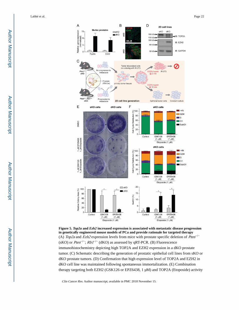
Figure 5. Top2a and Ezh2 increased expression is associated with metastatic disease progression in genetically engineered mouse models of PCa and provide rationale for targeted therapy(A) Top2a and Ezh2 expression levels from mice with prostate specific deletion of Pten−/−
(sKO) or Pten−/−; Rb1−/− (dKO) as assessed by qRT-PCR. (B) Fluorescence
immunohistochemistry depicting high TOP2A and EZH2 expression in a dKO prostate
tumor. (C) Schematic describing the generation of prostatic epithelial cell lines from sKO or
dKO prostate tumors. (D) Confirmation that high expression level of TOP2A and EZH2 in
dKO cell line was maintained following spontaneous immortalization. (E) Combination
therapy targeting both EZH2 (GSK126 or EPZ6438, 1 μM) and TOP2A (Etoposide) activity
Labbé et al. Page 22
Clin Cancer Res. Author manuscript; available in PMC 2018 November 15.
Author M
anuscriptA
uthor Manuscript
Author M
anuscriptA
uthor Manuscript

induces loss of clonogenicity specific to dKO cells. (top: representative experiment;
unpaired t test; * P < 0.05, triplicate, mean ±SEM). (F) Cell cycle analysis indicates that
combination therapy induces apoptosis specifically in dKO cells as indicated by increased
SubG1 accumulation (unpaired t test; * P < 0.05, triplicate, mean ±SEM).
Labbé et al. Page 23
Clin Cancer Res. Author manuscript; available in PMC 2018 November 15.
Author M
anuscriptA
uthor Manuscript
Author M
anuscriptA
uthor Manuscript

Figure 6. Schematic figure indicating the potential use of TOP2A and EZH2 for the early
identification and treatment direction of aggressive primary PCa.
Labbé et al. Page 24
Clin Cancer Res. Author manuscript; available in PMC 2018 November 15.
Author M
anuscriptA
uthor Manuscript
Author M
anuscriptA
uthor Manuscript