Top 10 Health Technology Hazards for 2017 · Top 10 Health Technology Hazards for ... and using...
52
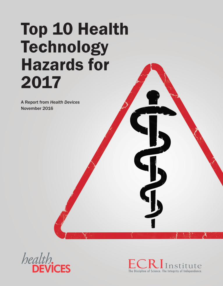
Top 10 Health Technology Hazards for 2017 A Report from Health Devices November 2016
-
Upload
nguyendien -
Category
Documents
-
view
219 -
download
0
Transcript of Top 10 Health Technology Hazards for 2017 · Top 10 Health Technology Hazards for ... and using...
Top 10 Health Technology Hazards for 2017 A Report from Health Devices November 2016
Health Devices Editorial and Scientific PolicyThrough its comparative equipment Evaluations, product ratings, patient safety alerts, and guidance articles, Health Devices provides inde-pendent, objective judgment for selecting, purchasing, managing, and using medical devices, equipment, and systems.
Charter and General Policy
Who we are. ECRI Institute is an independent nonprofit that researches the best approaches to improving patient care. Since 1971, we have been producing Health Devices material to help fulfill our mission of improving the effectiveness, safety, and economy of health services.
Peer review. The material on the Health Devices website is produced by ECRI Institute staff. Our content routinely receives intensive review by engineering and clinical professionals, both within and outside the organization, before publication.
Impartiality. ECRI Institute respects and is impartial toward all ethical medical device companies and practices. Neither ECRI Institute nor any of its staff members has a direct or indirect financial interest in promoting the sale of any medical device. Our employees do not under-take private consulting work for the medical device industry or own stock in medical device companies. We accept no royalties, gifts, finder’s fees, or commissions from the medical device industry, nor does Health Devices accept advertising. ECRI Institute prohibits manufacturers from using or referring to our product ratings or reports, in whole or in part, in advertising or promotional materials.
Comparative Evaluations
Scope. Product Evaluations, unless otherwise noted, cover only the specific models discussed. We caution readers against applying the ratings to models that we did not evaluate. If we do not include a currently marketed device in an Evaluation, this does not necessarily mean that it is no longer available or imply anything about its value, safety, or performance.
Accountability. Neither ECRI Institute nor the Health Devices program implies any warranty, including a warranty of merchantability or fitness for a particular purpose, or assumes liability for the safety, performance, or effectiveness of the evaluated products. We invite manufacturers to discuss their products and to review test data before publication. However, ECRI Institute assumes responsibility for the final Evaluation of a device.
Restrictions on the use of Health Devices content. As an impartial evaluator of biomedical technology, ECRI Institute does not endorse any specific brand or model of device. Reproducing excerpts from our product Evaluations in promotional materials implies endorsement, contra-venes ECRI Institute policy, and may violate copyright law. Please report any instances of improper use of our published materials directly to: Legal Department, ECRI Institute.
ALL MATERIAL COPYRIGHT ©2016 ECRI INSTITUTE
All rights reserved. All rights are reserved under international and Pan-American copyright conventions. All material in ECRI Institute publica-tions is protected by copyright.
Reproduction. Except where otherwise noted, ECRI Institute prohibits reproduction of Health Devices material.
This report reprints material from the Health Devices website as of November 4, 2016. It does not reflect modifications that may have been made after that date.
jim
Highlight
jim
Highlight
Top 10 Health Technology Hazards for 2017 A Report from Health Devices November 2016
1www.ecri.org/2017hazards ©2016 ECRI Institute. Health Devices 2016 November
ECRI Institute prohibits the direct dissemination, posting, or republishing of this work without prior permission. Subscribing institutions may reproduce this page for internal distribution only.
Top 10 Health Technology Hazards for 2017The safe use of health technology—from basic infusion pumps to large, complex imaging systems—requires identifying possible sources of danger or difficulty with those technologies and taking steps to minimize the likelihood that adverse events will occur. This report, which is the 10th edition of our Top 10 list, will help healthcare facilities do that.
Produced each year by ECRI Institute’s Health Devices Group, the Top 10 Health Technology Hazards list (1) identifies the potential sources of danger that we believe warrant the greatest attention for the coming year and (2) offers practical recommendations for reducing the risks.
The List for 2017Following are the 10 technology hazards that make up our list for 2017. Details about these topics can be found on the pages that follow.
1. Infusion Errors Can Be Deadly If Simple Safety Steps Are Overlooked
2. Inadequate Cleaning of Complex Reusable Instruments Can Lead to Infections
3. Missed Ventilator Alarms Can Lead to Patient Harm
4. Undetected Opioid-Induced Respiratory Depression
5. Infection Risks with Heater-Cooler Devices Used in Cardiothoracic Surgery
6. Software Management Gaps Put Patients, and Patient Data, at Risk
7. Occupational Radiation Hazards in Hybrid ORs
8. Automated Dispensing Cabinet Setup and Use Errors May Cause Medication Mishaps
9. Surgical Stapler Misuse and Malfunctions
10. Device Failures Caused by Cleaning Products and Practices
The Purpose of the ListECRI Institute’s Health Devices Group produces this list to highlight the technology safety topics that we believe warrant particular attention for the coming year. The list does not enumerate the most frequently reported problems or the ones associated with the most severe consequences—although we do consider such information in our analysis. Rather, the list reflects our judgment about which risks should receive priority now.
All the items on our list represent problems that can be avoided or risks that can be minimized through the careful manage-ment of technologies. For each topic, we describe the problem and provide practical recommendations for action. We also point to sources of additional information or guidance. In this way, the list serves as a tool that healthcare facilities can use to prioritize their patient safety efforts. While not all hazards on the list will apply at all healthcare facilities, the list can pro-vide a starting point for patient safety discussions.
jim
Highlight
2
www.ecri.org/2017hazards ©2016 ECRI Institute. Health Devices 2016 November ECRI Institute prohibits the direct dissemination, posting, or republishing of this work without prior permission. Subscribing institutions may reproduce this page for internal distribution only.
How Topics Are SelectedThis list focuses on what we call generic hazards—problems that result from the risks inherent to the use of certain types or combinations of medical technologies. It does not discuss risks or problems that pertain to specific models or suppliers.
ECRI Institute engineers, scientists, clinicians, and other patient safety analysts nominate topics for consideration based on their own expertise and insight gained through:
• Investigating incidents
• Testing medical devices
• Observing operations and assessing hospital practices
• Reviewing the literature
• Speaking with clinicians, clinical engineers, technology managers, purchasing staff, health systems administrators, and device suppliers
Staff also consider the thousands of health-technology-related problem reports that we receive through our Problem Reporting Network and through data that participating facilities share with our patient safety organization, ECRI Institute PSO.
After the topic nomination phase, professionals from ECRI Institute’s many program areas, as well as members of some of our external advisory committees, review these topics and select their top 10. We use this feedback to produce the final list, weighing factors such as the following:
• Severity. What is the likelihood that the hazard could cause serious injury or death?
• Frequency. How likely is the hazard? Does it occur often?
• Breadth. If the hazard occurs, are the consequences likely to spread to affect a great number of people, either within one facility or across many facilities?
• Insidiousness. Is the problem difficult to recognize? Could the problem lead to a cascade of downstream errors before it is identified or corrected?
• Profile. Is the hazard likely to receive significant publicity? Has it been reported in the media, and is an affected hospital likely to receive negative attention? Has the hazard become a focus of regulatory bodies or accrediting agencies?
• Preventability. Can actions be taken now to prevent the problem or at least minimize the risks? Would raising aware-ness of the hazard help reduce future occurrences?
All the topics we select for the list must, to some degree, be preventable. But any one of the other criteria can, on its own, warrant including a topic on the list. We encourage readers to examine these same factors when judging the criticality of these and other hazards at their own facilities.
Note that the exclusion of a topic that was included on a previous year’s list should not be interpreted to mean that the topic no longer deserves attention. Most of these hazards persist, and hospitals should continue working toward minimizing them. Rather, our experts determined that other topics should receive greater attention in 2017.

3www.ecri.org/2017hazards ©2016 ECRI Institute. Health Devices 2016 November
ECRI Institute prohibits the direct dissemination, posting, or republishing of this work without prior permission. Subscribing institutions may reproduce this page for internal distribution only.
Infusion Errors Can Be Deadly If Simple Safety Steps Are Overlooked
Most large-volume infusion pumps incorporate safety mechanisms for reducing the risks of potentially deadly intravenous (IV) infusion errors. These mechanisms have greatly improved infusion safety, but can’t eliminate all potential errors. And the mechanisms themselves have been known to fail.
ECRI Institute continues to learn about and investigate incidents of infusion errors involving pump or adminis-tration set failures, staff unknowingly defeating a safety mechanism, or incorrect infusion programming. Such errors—particularly those that result in the uncontrolled flow of medication to the patient, known as “IV free flow”—can lead to patient harm and even death.
In many of these incidents, harm could have been averted if staff had:
• Noticed signs of physical damage to infusion pump components
• Made appropriate use of the roller clamp on the IV tubing
• Checked the drip chamber beneath the medication reservoir for unexpected flow
Once commonplace, these simple practices are now often overlooked—perhaps because staff implicitly trust the pump’s advanced safety features.
1

4
www.ecri.org/2017hazards ©2016 ECRI Institute. Health Devices 2016 November ECRI Institute prohibits the direct dissemination, posting, or republishing of this work without prior permission. Subscribing institutions may reproduce this page for internal distribution only.
Problem1. Most large-volume infusion pumps incorporate effective mechanisms for reducing the risks of infusion
errors. However:
a) These safety features are not able to eliminate all potential errors.b) The mechanisms themselves have been known to fail or be unknowingly defeated.
2. ECRI Institute continues to receive reports and investigate incidents of infusion errors that involve:
a) Pump or administration set failures or staff unknowingly defeating a safety mechanism, resulting in uncontrolled flow (i.e., free flow) of medication to the patient
b) Incorrect infusion programming, resulting in infusion errors
3. In many of these incidents, patient harm could have been averted if staff had:
a) Noticed signs of physical damage to pump componentsb) Made use of the roller clamp (and slide clamp, when available) on the intravenous (IV) tubing at
appropriate times, as defined below in the ECRI Institute Recommendations section.c) Checked the drip chamber located below the medication reservoir for unexpected flow
4. These simple practices, which used to be commonplace, are now frequently overlooked—perhaps because staff implicitly trust the pump’s safety features.
5. While the presence of free-flow protection mechanisms on infusion pumps and administration sets has significantly reduced the risk of IV free flow, occasional incidents of uncontrolled flow are still reported. Causes include:
a) Broken or damaged components on the pump or administration setb) Incorrect loading of the administration set c) Failure of the free-flow mechanism to engage
6. Similarly, even “smart” pumps that incorporate dosing safeguards can be misprogrammed in a way that leads to gross flow-rate errors. Mistakes that can lead to entry of an incorrect dose rate, flow rate, or concentration include:
a) Field-swap errors—for example, a dose rate is entered into the flow rate field, or vice versab) Zero-entry errors—for example, entering “20” as “200”c) Entry or selection of an incorrect drug or concentrationd) Overriding of dose limit/concentration alerts
7. Infusion errors can lead to patient harm and even death, especially when potent high-alert medications are being delivered.
ECRI Institute RecommendationsClinical Staff
1. Do not use any infusion pump that shows signs of physical damage, such as:
a) A component that appears to be bent or brokenb) A pump door that won’t close completely or that requires unexpected force to close

5www.ecri.org/2017hazards ©2016 ECRI Institute. Health Devices 2016 November
ECRI Institute prohibits the direct dissemination, posting, or republishing of this work without prior permission. Subscribing institutions may reproduce this page for internal distribution only.
2. During use of the pump, close the roller clamp before opening the pump door anytime you will be changing the administration set or removing the set from the pump.
3. Keep the roller clamp closed:
a) After priming the administration set b) During programming, if the infusion is not already ongoingc) If the infusion is paused or on standby
4. Open the roller clamp when the infusion is initiated.
5. Check the drip chamber anytime an infusion is initiated or the flow rate or dose has been changed. Before leaving the bedside:
a) Verify that medication is flowing from the correct medication bag. b) Assess the drip rate for any gross inconsistency between the observed drip rate and the intended
flow rate or dose. (1) Note that for weight-based infusions, the flow rate is calculated by the pump. (2) In such circumstances, check the resulting flow rate based on the dose entered to get an idea
of the approximate drip rate frequency that should be observed in the drip chamber.
6. Also check the drip chamber anytime the infusion has been stopped to verify that there is no medication flow in the drip chamber. This includes:
a) Whenever the roller clamp is closedb) Anytime the administration set is outside the pump
Charge Nurses and Nurse Educators
1. Establish practices whereby nurses:
a) Examine the infusion pump for obvious signs of physical damage (e.g., bent or cracked com-ponents, a door that won’t close) when initiating an infusion or changing or removing the administration set. A damaged component could, for example, prevent an anti-free-flow clamp from engaging.
b) Close the roller clamp at appropriate times as defined above. This step can minimize the risk of uncontrolled medication flow to the patient.
c) Observe the flow in the drip chamber before leaving the patient room. This quick check can help nurses identify gross inconsistencies in the infusion flow rate.
d) Have proven competency with the devices prior to use.
2. Educate staff about the importance of these practices.
a) Discuss the hazardous conditions that these steps can help prevent. b) Share with staff the ECRI Institute Hazard Reports (see References and Resources, below) that
describe the kinds of incidents that have occurred.
3. Assist staff in implementing these practices.
a) Teach staff how to broadly assess the drip rate for common flow rates used on the pumps in your facility. For example, review the differences that can be observed between:(1) Low flow rates (e.g., 10 mL/hr)
6
www.ecri.org/2017hazards ©2016 ECRI Institute. Health Devices 2016 November ECRI Institute prohibits the direct dissemination, posting, or republishing of this work without prior permission. Subscribing institutions may reproduce this page for internal distribution only.
(2) Average flow rates (e.g., 100 mL/hr)(3) High flow rates (e.g., 999 mL/hr)(4) Unrestricted gravity flow (i.e., with the set removed from pump and all clamps open)
b) Monitor nursing staff compliance—for example, verify during nursing rounds that the roller clamp is consistently being closed and the drip chamber is being checked as specified above in the recom-mendations for clinical staff.
4. Review with nurses the procedures to follow in the event that a problem is observed.
a) If an infusion error is suspected, consult the attending physician or charge nurse to determine the safest course of action.
b) If a pump is damaged or suspected to be faulty or if patient harm is suspected: (1) Remove the pump from clinical use and save the infusion tubing.(2) Tag the pump as faulty; include a short description of the suspected fault or the issue (e.g.,
“Uncontrolled flow when pump turned off”).(3) Report the problem to clinical engineering.
Clinical Engineers
1. Recognize that broken or bent components can affect the performance of the pump or its safety mechanisms.
2. Replace or repair such components, as appropriate, before returning the pump to service.
Background1. While most infusion pumps incorporate effective safety features, users must recognize the limitations
of such features. For example:
a) Infusion pumps cannot detect uncontrolled flow.(1) Although some infusion pumps can alarm for a potential free-flow condition, this alarm does
not result from a determination of uncontrolled flow; rather, it indicates only that an anti-free-flow mechanism is not appropriately engaged.
(2) Infusion pump manufacturers specify that the roller clamp is the primary method of ensuring that no flow is going to the patient. A pump’s anti-free-flow mechanism should be considered a secondary protective measure.
b) Damage to or failure of a crucial component of the pump can affect the functioning of a pump’s safety features—for example, a damaged component could prevent an anti-free-flow clamp from engaging.
2. ECRI Institute has published several articles related to pump and administration set failures wherein identification of damage to a pump component, use of the roller clamp as specified above, or checking of the drip chamber could have minimized the risk of patient harm occurring.
a) Note that incidents have occurred with pumps from all major suppliers.b) Refer to the list of articles in under Member Resources, on the following page.

7www.ecri.org/2017hazards ©2016 ECRI Institute. Health Devices 2016 November
ECRI Institute prohibits the direct dissemination, posting, or republishing of this work without prior permission. Subscribing institutions may reproduce this page for internal distribution only.
References and ResourcesMember Resources
1. Baxter—SIGMA Spectrum infusion pumps: incorrect technique for changing administration set may lead to unintentional free flow. Health Devices Alerts 2013 Sep 26 (Accession No. H0215).
2. CareFusion—Alaris pump modules: damaged door components may fail to engage anti free flow mecha-nism, potentially leading to gravity flow [ECRI Exclusive Hazard Report]. Health Devices Alerts 2016 Aug 25 (Accession No. H0337).
3. Hospira—Plum Series infusion pumps: close all upstream clamps to prevent primary and secondary fluid mixing when door is open [ECRI Exclusive Hazard Report]. Health Devices Alerts 2015 Nov 5 (Accession No. H0212 01).
4. Incorrect installation of free-flow clamp in B. Braun Infusomat Space infusion pumps may result in gravity flow. Health Devices Alerts 2011 Jan 13 (Accession No. H0134).
Additional Resources 1. Gorski L, Hadaway L, Hagle ME, et al. Infusion therapy
standards of practice. J Infus Nurs 2016;39(Suppl 1): S1-S159.
2. CareFusion: a) Alaris™ Pump Module FAQs. 2016 Jul 15.b) Improper Set Loading Guide for the Alaris® Pump
Module. 2013.c) Set Loading and Unloading Guide for the Alaris®
Pump Module. 2015.d) User Manual: Alaris™ System with Guardrails™
Suite MX (with Alaris™ PC unit, Model 8015 Soft-ware Version 9.19). 2015 Dec.
3. Hospira. Hospira MedNet Plum 360 System Operating Manual.
4. Baxter: a) Loading and Unloading Baxter IV Administration
Sets with Sigma Spectrum Infusion System.b) Operator’s Manual: Sigma Spectrum Infusion
Pump with Master Drug Library: 35700BAX2. 5. B. Braun:
a) Infusomat® Space and Accessories Manual.b) Infusomat® Space Infusion Pump System 2nd
Generation Software Quick Reference Guide.

8
www.ecri.org/2017hazards ©2016 ECRI Institute. Health Devices 2016 November ECRI Institute prohibits the direct dissemination, posting, or republishing of this work without prior permission. Subscribing institutions may reproduce this page for internal distribution only.
2
The use of contaminated medical instruments can lead to disabling or deadly patient infections or instrument malfunctions.
Outbreaks associated with the use of contaminated duodenoscopes—such as those that caused headlines in recent years—illustrate the severity of this issue. But duodenoscopes are not the only devices that warrant atten-tion. ECRI Institute has received reports involving a variety of contaminated medical instruments that have been used, or almost used, on patients.
Complex, reusable instruments—such as endoscopes, cannulated drills, and arthroscopic shavers—are of particular concern. They can be difficult to clean and then disinfect or sterilize (i.e., reprocess) between uses, and the presence of any lingering contamination on, or in, the instrument can be difficult to detect.
Often, we find that inattention to the cleaning steps within the reprocessing protocol is a contributing factor. Healthcare facilities should verify that comprehensive reprocessing instructions are available to staff and that all steps are consistently followed, including precleaning of the device at the point of use.
Inadequate Cleaning of Complex Reusable Instruments Can Lead to Infections
jim
Highlight
jim
Highlight

9www.ecri.org/2017hazards ©2016 ECRI Institute. Health Devices 2016 November
ECRI Institute prohibits the direct dissemination, posting, or republishing of this work without prior permission. Subscribing institutions may reproduce this page for internal distribution only.
Problem1. ECRI Institute periodically receives reports of contaminated medical instruments that have been
presented for use on a patient.
a) Of particular concern are complex, reusable instruments whose design makes the instruments difficult to clean and then disinfect or sterilize (i.e., reprocess) between uses.
b) Examples include: endoscopes, cannulated drills, perforator drills, and arthroscopic shavers.c) In addition to hindering reprocessing efforts, the complex design of such instruments can make it
harder for reprocessing staff or users to detect whether any contamination remains on, or in, the instrument.
d) In fact, in numerous cases, lingering contamination had not been identified until after the device had been used on one or more patients.
e) Highly publicized infection outbreaks associated with the use of contaminated duodenoscopes illustrate the severity of this issue. (We discussed this example in detail in last year’s list; see Hazard #1—Top 10 Health Technology Hazards for 2016.)
2. When contaminated instruments are presented for use, regardless of whether it is in the OR or an endoscopy suite, the cause can often be traced to an operational failure involving the instrument cleaning steps (including precleaning at the point of use).
3. Such contamination can lead to disabling or deadly patient infections or to instruments malfunctioning during use.
4. Incidents can also expose healthcare facilities to liability that can jeopardize their reputation.
a) If a healthcare facility discovers that instruments used on a patient were contaminated, it is obli-gated to notify any affected patients that they have been exposed and could develop an infection.
b) The anxiety patients experience when learning that they might have been treated with contaminated instruments is also a liability concern for healthcare facilities.
ECRI Institute Recommendations1. Verify that both (a) staff responsible for precleaning at the point of use and (b) reprocessing staff have
ready access to reprocessing instructions for the instruments they will encounter.
2. For complex instruments in particular—that is, for instruments featuring difficult-to-clean components such as lumens, hinges, cannulated blades, stopcocks, or O-rings—confirm that the reprocessing instructions are comprehensive. This documentation should include:
a) Instructions for precleaning the instrument immediately after use.b) Information about any special accessories required for cleaning. (Recognize that if the required
accessories are not available, reprocessing staff might resort to using unapproved tools, as discussed in ECRI Institute’s Hazard Report Use of Unapproved Brushes for Cleaning Endoscope Channels Is Not Recommended.)
c) Information about compatible cleaning agents.d) Instructions for disassembly and reassembly, if applicable (including photos or diagrams).e) Information about the expected time required for each cleaning step.
jim
Highlight
jim
Highlight
jim
Highlight
jim
Highlight
jim
Highlight
10
www.ecri.org/2017hazards ©2016 ECRI Institute. Health Devices 2016 November ECRI Institute prohibits the direct dissemination, posting, or republishing of this work without prior permission. Subscribing institutions may reproduce this page for internal distribution only.
3. When reprocessing instructions lack this information, contact the manufacturer and request the information needed. If the manufacturer is unable to provide the information, consider purchasing alternative instruments when replacement is required.
4. Verify that staff have been trained to perform the reprocessing procedures correctly.
a) Confirm (e.g., through periodic competency testing) that effective reprocessing can be achieved by the staff at your facility, including the steps to be performed by clinical staff at the point of use (e.g., precleaning immediately after use) and those to be performed by reprocessing staff.
b) Consider having point-of-use and reprocessing staff observe how their counterparts prepare instru-ments for reprocessing (i.e., complete their precleaning or cleaning tasks). This cross-observation could help the two groups appreciate the importance of completing their tasks properly, and it may help build an effective team.
5. Verify that the current inventory of instruments affords sufficient time for each cleaning step to be performed properly.
6. Before purchasing instruments that are new to the facility:
a) Confirm that appropriate reprocessing instructions are available.b) Review the cleaning requirements with reprocessing and clinical staff.c) Confirm that staff will be able to meet the requirements.
To help with this process, consider using a checklist like the Patient Safety Impact Assessment Tool published by the Pennsylvania Patient Safety Authority. That tool provides a reminder to address repro-cessing concerns before new types of instruments are introduced into the workflow.
7. Consider purchasing only instruments for which the manufacturer has validated its cleaning instructions. When applicable, request written information explaining the validation process.
8. Remind relevant clinical staff that precleaning immediately after use is critical.
a) Without precleaning, instrument reprocessing can be compromised, sometimes irreversibly, by dried debris and biofilm formation.
b) In many instances, clinical staff should be responsible for precleaning because they have the most timely access to instruments immediately after use.
Background 1. Precleaning and cleaning remain largely manual processes; consequently, they are perhaps the
reprocessing steps most prone to inconsistent completion.
2. Historically, the problem was exacerbated by manufacturers’ reprocessing instructions that were of inconsistent quality or that lacked adequate detail. In particular, the cleaning steps within these instructions sometimes lacked necessary details.
3. To help ensure consistent end results, point-of-use and reprocessing staff should have ready access to validated cleaning instructions that include adequate detail.
jim
Highlight
jim
Highlight
jim
Highlight

11www.ecri.org/2017hazards ©2016 ECRI Institute. Health Devices 2016 November
ECRI Institute prohibits the direct dissemination, posting, or republishing of this work without prior permission. Subscribing institutions may reproduce this page for internal distribution only.
4. While process validation does not guarantee that a process will never fail, it does demonstrate that the provided cleaning procedures can be effective when completed properly. This additional level of assurance is important and was evidently lacking with the duodenoscopes associated with the recent carbapenem-resistant Enterobacteriaceae (CRE) infections.
5. FDA has become increasingly aware of the inconsistent quality of cleaning and reprocessing instruc-tions. The agency now recommends that manufacturers of reusable instruments validate cleaning instructions, as discussed in the March 2015 Reprocessing Medical Devices in Health Care Settings: Validation Methods and Labeling.
6. Staff education should emphasize the importance of instrument cleaning, both at the point of use and during reprocessing, and how vital cleaning is to keeping patients safe.
References and ResourcesMember Resources
1. ECRI Institute’s guidance related to the reprocessing function:a) Use of unapproved brushes for cleaning endo-
scope channels is not recommended [ECRI Exclusive Hazard Report]. Health Devices Alerts 2016 Feb 11 (Accession No. H0306).
b) Inadequate cleaning of flexible endoscopes before disinfection can spread deadly pathogens. Hazard #1—top 10 health technology hazards for 2016. Health Devices 2015 Nov 7.
c) Duodenoscope reprocessing challenges lead to CRE exposures: update on a top 10 hazard. Health Devices 2015 Mar 11.
d) Inadequate reprocessing of endoscopes and surgical instruments. Hazard #4—top 10 health technology hazards for 2015. Health Devices 2014 Nov 24.
e) Clear channels: ensuring effective endoscope reprocessing. Health Devices 2010 Oct 1.
2. ECRI Institute’s series of alerts pertaining specifically to the CRE infection issue:a) Endoscopic retrograde cholangiopancreatogra-
phy (ERCP) duodenoscopes: design may impede effective cleaning. Health Devices Alerts 2015 Feb 20 (Accession No. H0245).
b) ECRI Institute recommends culturing duodeno-scopes as a key step to reducing CRE infections. Health Devices Alerts 2015 May 7 (Accession No. H0245 02).
c) ECRI Institute provides perspectives on FDA’s recent supplemental measures to enhance duo-denoscope reprocessing. Health Devices Alerts 2015 Aug 7 (Accession No. H0245 03).
3. ECRI Institute’s September 30, 2015, web confer-ence: Tracking scopes: best practices for identifying endoscopes during cleaning and patient use.
Additional Resources1. Centers for Disease Control and Prevention (CDC),
U.S. Immediate need for healthcare facilities to review procedures for cleaning, disinfecting, and sterilizing reusable medical devices. CDC Health Advisory 2015 Sep 11.
2. Food and Drug Administration (FDA), U.S. Reprocess-ing medical devices in health care settings: validation methods and labeling guidance for industry and Food and Drug Administration staff. 2015 Mar 17.
3. Pennsylvania Patient Safety Authority:a) Davis J. Equipment, environment, and ergonomics:
an enigma of infection risk. Pa Patient Saf Advis 2015 Mar;12(1):37-40. Available from: http://patientsafetyauthority.org/ADVISORIES/AdvisoryLi-brary/2015/mar;12(1)/Pages/37.aspx.
b) Patient safety impact assessment tool: con-siderations when integrating equipment into the workplace. 2015. Available from: http://patientsafetyauthority.org/EducationalTools/PatientSafetyTools/ergonomics_inf/Pages/assessment.aspx.
4. Whitman E. Hidden danger: dirty medical tools. Mod Healthc 2016 Sep 3.
jim
Highlight
12
www.ecri.org/2017hazards ©2016 ECRI Institute. Health Devices 2016 November ECRI Institute prohibits the direct dissemination, posting, or republishing of this work without prior permission. Subscribing institutions may reproduce this page for internal distribution only.
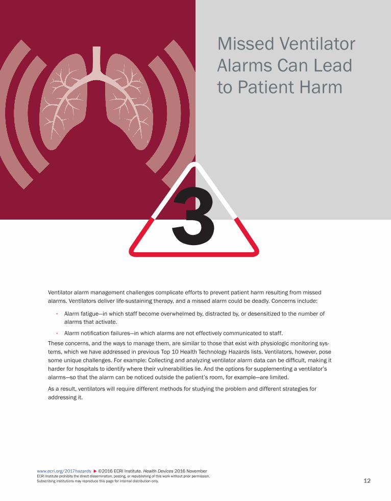
Missed Ventilator Alarms Can Lead to Patient Harm
3Ventilator alarm management challenges complicate efforts to prevent patient harm resulting from missed alarms. Ventilators deliver life-sustaining therapy, and a missed alarm could be deadly. Concerns include:
• Alarm fatigue—in which staff become overwhelmed by, distracted by, or desensitized to the number of alarms that activate.
• Alarm notification failures—in which alarms are not effectively communicated to staff.
These concerns, and the ways to manage them, are similar to those that exist with physiologic monitoring sys-tems, which we have addressed in previous Top 10 Health Technology Hazards lists. Ventilators, however, pose some unique challenges. For example: Collecting and analyzing ventilator alarm data can be difficult, making it harder for hospitals to identify where their vulnerabilities lie. And the options for supplementing a ventilator’s alarms—so that the alarm can be noticed outside the patient’s room, for example—are limited.
As a result, ventilators will require different methods for studying the problem and different strategies for addressing it.

13www.ecri.org/2017hazards ©2016 ECRI Institute. Health Devices 2016 November
ECRI Institute prohibits the direct dissemination, posting, or republishing of this work without prior permission. Subscribing institutions may reproduce this page for internal distribution only.
Problem1. Challenges related to ventilator alarm management may prevent hospitals from effectively addressing
the risks of missed ventilator alarms.
2. Causes of missed ventilator alarms (or alarm conditions) include:
a) Alarm fatigue, in which staff become overwhelmed by, distracted by, or desensitized to the number of alarms that activate. Ventilators are the most common sources of alarms after physiologic moni-toring systems.
b) Alarm notification failures, in which alarms are not effectively communicated to staff. Limited ancil-lary notification options make ventilator alarms particularly challenging to manage.
3. Ventilators deliver life-sustaining therapy, and a missed alarm could lead to severe patient harm or death.
ECRI Institute RecommendationsIn broad terms, the process for improving the management of ventilator alarms will be similar to that used for physiologic monitoring alarms. (We detail this process in our Alarm Safety Handbook, which is avail-able to ECRI Institute members from our Alarm Management Resources web page; see the list of Member Resources, below.) However, ventilators pose some unique challenges that will require different methods for studying the problem and different strategies for addressing it. The recommendations that follow are intended to help healthcare facilities begin the process of overcoming these challenges.
1. Initiate a comprehensive, multidisciplinary effort to address the potential hazards associated with ven-tilator alarm management, paying particular attention to the specific challenges with this technology.
a) The effort should be spearheaded by the existing alarm management committee.b) Appropriate stakeholders should be represented on the committee. These include respiratory
therapists and other clinical personnel (e.g., nurses, pulmonologists, intensivists) who routinely address and support ventilator alarms.
2. Understand how ventilator alarms are used at your facility—focusing on the alarm load in each care area and the effectiveness of the established notification pathways. A successful alarm management program will require identifying where your vulnerabilities lie and developing appropriate strategies to limit the hazards. To gain this understanding:
a) Observe how the many different alarms are handled in each care area. Much can be learned by walking around, observing what happens on the care unit, and engaging frontline staff about their concerns.
b) Review your reports of adverse events and near misses.c) Consider collecting and analyzing alarm data to obtain a quantitative measure of the number and
types of alarms that activate per device within a care area. (1) Ventilators are not typically networked in a hospital, and are therefore not connected to a
vendor-supplied server that would allow access to alarm data from a central location, the way most physiologic monitoring systems are. This makes quantifying the number and types of ventilator alarms a particular challenge.
14
www.ecri.org/2017hazards ©2016 ECRI Institute. Health Devices 2016 November ECRI Institute prohibits the direct dissemination, posting, or republishing of this work without prior permission. Subscribing institutions may reproduce this page for internal distribution only.
(2) Options that can be considered include accessing and analyzing alarm log data from individual ventilators or using third-party alarm analytics software.
d) Assess whether alarms are adequately heard by clinicians in each care area. Examine whether any environmental factors—such as the architectural layout of the care area, the presence of closed doors, or the distance of rooms from the nurses’ station—or other circumstances could be hinder-ing staff recognition of or response to ventilator alarms.
3. Identify and implement strategies for reducing the alarm load.
a) Using the information collected during the steps listed above, analyze the most frequently occurring alarms for each care area and categorize them as follows:(1) Alarms that are clinically actionable—for example, low-pressure alarms (which could signify a
disconnection or leak) or low-volume alarms(2) Alarms that are not clinically actionable—for example, transient high-pressure alarms caused
by a patient coughingb) Work with frontline staff to identify and implement appropriate strategies for reducing the number
of nonactionable alarms in each care area. For example: The appropriate use of ventilator modes to promote better synchrony between the patient and the ventilator can be an effective strategy for reducing unnecessary alarms.
4. Identify and implement strategies to improve staff awareness of and response to ventilator alarms.
a) Investigate whether staffing levels or staff deployment can be adjusted to improve responsiveness to the needs of ventilator patients. For example, in our experience, assigning respiratory therapists to a specific care area, rather than having them float between multiple care areas, leads to the best alarm and patient response.
b) Consider enhancing notification of ventilator alarms with secondary notification pathways. Several alternatives are available, but each has limitations (as detailed in the Background section below).(1) Alternatives include:
(a) Using the nurse call system as a means of alerting users to the presence of certain (but not necessarily all) ventilator alarms. (The ventilator alarms that can be communicated in this manner will depend on the capabilities of the ventilator model and the nurse call system.)
(b) Integrating ventilators with patient monitoring systems to allow notification of ventilator alarms via the associated central stations and ancillary displays.
(c) Using ancillary alarm notification/alarm integration systems to send specific alarms to end-user communication devices.
These systems can also be used to configure delays so that self-correcting conditions do not add to the alarm load. For example, configuring a delay for high-pressure alarms could reduce the number of alarms staff receive for transitory conditions like a patient cough.
(2) With any of these approaches, it is important to test the systems before implementation to:(a) Examine whether and how each alarm is communicated to the clinician(b) Understand the type of information (e.g., alarm type, priority level, patient) that is and is
not communicated

15www.ecri.org/2017hazards ©2016 ECRI Institute. Health Devices 2016 November
ECRI Institute prohibits the direct dissemination, posting, or republishing of this work without prior permission. Subscribing institutions may reproduce this page for internal distribution only.
Background 1. We have addressed the need to improve the safety of clinical alarm systems in every edition of our Top
10 Health Technology Hazards list since its inception in 2007. The importance of this effort is high-lighted by the Joint Commission’s National Patient Safety Goal on clinical alarm safety, which went into full effect in January 2016.
2. While many healthcare facilities’ alarm improvement efforts have, to date, focused on the alarms generated by physiologic monitoring systems, healthcare facilities must not ignore the risks associated with other devices, such as ventilators.
3. Ventilators are life-sustaining devices that generate a large number of alarms—and managing these alarms poses some unique challenges.
4. Ultimately, efforts to improve the safety of ventilator alarm systems must balance the same two oppos-ing needs as with physiologic monitoring systems:
a) The need to reliably detect and notify appropriate staff about all conditions—with either the patient or the medical device—that require their attention
b) The need to reduce the overall number of alarms to which caregivers are exposed, to combat alarm fatigue
5. With ventilators, however, limited options are available for addressing these needs.
6. With respect to improving the reliability of alarm notification:
a) Most ventilator alarms sound only at the patient’s bedside. Ventilators do not include a central station that could display information from all the ventilators in the care area in one location, nor do they typically provide a means to allow the display of alarms outside the patient’s room.
b) While some alternative notification options are available to help improve staff awareness of activating ventilator alarms, these options all have limitations. For example:(1) While nurse call systems can be used to provide an indication outside the patient’s room of a
ventilator alarm, they do not provide any indication of the cause or priority of the alarm. Also, users may not be able to distinguish this notification from other nurse call alarms, such as patient assist.
(2) Physiologic monitoring systems can be used as a pathway for ventilator alarms, but based on our experience, the systems may not provide adequate indication of the cause or priority of the alarm and may sometimes misrepresent alarms. For example, our testing showed that in some cases critical ventilator alarms were communicated as medium-priority alarms by the monitor.
(3) Alarm middleware systems offer extensive alarm management capabilities, but they can be an expensive option. In the absence of a server, middleware systems require proprietary hardware that must be connected to each ventilator to collect and distribute alarm data.
16
www.ecri.org/2017hazards ©2016 ECRI Institute. Health Devices 2016 November ECRI Institute prohibits the direct dissemination, posting, or republishing of this work without prior permission. Subscribing institutions may reproduce this page for internal distribution only.
References and ResourcesMember Resources
1. Alarm hazards: inadequate alarm configuration policies and practices. Hazard #1—top 10 health tech-nology hazards for 2015. Health Devices 2014 Nov 24.
2. Alarm Management Resources—This online resource page provides access to ECRI Institute’s Alarm Safety Handbook and the accompanying Alarm Safety Workbook.
3. Evaluation background: ancillary alarm notification systems. Health Devices 2016 Sep 28. This resource provides an overview of, and links to, our evaluations of four ancillary alarm notification systems.
4. Interfacing monitoring systems with ventilators: how well do they communicate alarms? Health Devices 2012 May 1.
5. Missed alarms can have fatal consequences. Haz-ard #2—top 10 health technology hazards for 2016. Health Devices 2015 Nov 7.
6. Ventilator disconnections not caught because of mis-set or missed alarms. Hazard #5—top 10 health technology hazards for 2015. Health Devices 2014 Nov 24.
Additional Resources1. American Association for Respiratory Care (AARC) and
University HealthSystem Consortium’s (UHC) Respira-tory Care Network. Safe initiation and management of mechanical ventilation [white paper]. 2016.
2. Joint Commission. The Joint Commission announces 2014 National Patient Safety Goal. Jt Comm Perspect 2013 Jul;33(7):1-3.

17www.ecri.org/2017hazards ©2016 ECRI Institute. Health Devices 2016 November
ECRI Institute prohibits the direct dissemination, posting, or republishing of this work without prior permission. Subscribing institutions may reproduce this page for internal distribution only.
Undetected Opioid-Induced Respiratory Depression
4
Patients receiving opioids—such as morphine, hydromorphone, or fentanyl—are at risk for drug-induced respiratory depression. If not detected, this condition can quickly lead to anoxic brain injury or death. Thus, spot checks every few hours of a patient’s oxygenation and ventilation are inadequate.
Drug-induced respiratory depression is of particular concern for patients receiving parenteral and neuraxial opioids in medical-surgical and general care areas. However, it is also of concern for hospital or ambulatory surgery/endos-copy facility patients receiving opioids during procedural sedation and while in the postanesthesia care unit (PACU).
Even if they are otherwise healthy, such patients can be at risk if, for example:
• They are receiving another drug that also has a sedating effect
• They have diagnosed or undiagnosed sleep apnea or other conditions that predispose them to respiratory compromise
• They receive more medication than intended—for example, because of a medication errorECRI Institute recommends that healthcare facilities implement measures to continuously monitor the adequacy of ventilation of these patients and has recently tested and rated monitoring devices for this application.
18
www.ecri.org/2017hazards ©2016 ECRI Institute. Health Devices 2016 November ECRI Institute prohibits the direct dissemination, posting, or republishing of this work without prior permission. Subscribing institutions may reproduce this page for internal distribution only.
Problem1. Patients receiving opioids—such as morphine, hydromorphone, or fentanyl—are at risk for drug-induced
respiratory depression, which can lead to anoxic brain injury or death.
a) This is of particular concern for patients receiving parenteral and neuraxial opioids in medical-surgical and general care areas.
b) However, it is also of concern for hospital or ambulatory surgery/endoscopy facility patients receiving opioids:(1) During procedural sedation(2) While in the postanesthesia care unit (PACU)
2. Even if they are otherwise healthy, such patients can be at risk if, for example:
a) They are receiving another drug that also has a sedating effect.b) They have diagnosed or undiagnosed comorbidities (e.g., morbid obesity, sleep apnea) that predis-
pose them to respiratory compromise.c) They receive more medication than intended—for example, because of a medication error, such as:
(1) An incorrect dose or concentration being programmed into a patient-controlled analgesic (PCA) pump—Factor-of-10 errors have resulted in delivery rates 10 times higher than ordered.
(2) A prefilled syringe of hydromorphone being selected when morphine had been prescribed—The mix-up would produce an opioid effect approximately seven times greater than intended.
3. Some patients can deteriorate from normal to insufficient respiration in a few minutes, so spot-check-ing every few hours may not be adequate for reliably detecting opioid-induced respiratory depression.
ECRI Institute Recommendations1. Work with medical leadership to create and implement policies and procedures for continuous
monitoring of:
a) Patients receiving parenteral and neuraxial opioids in medical-surgical and general care areasb) Patients receiving opioids in hospitals and ambulatory surgery/endoscopy facilities during proce-
dural sedation and while in PACUs
2. Monitor the adequacy of ventilation of these patients either with capnography—that is, the measurement of end-tidal carbon dioxide (EtCO2)—or by assessing minute ventilation.
a) ECRI Institute does not recommend purchasing pulse oximeters (which monitor adequacy of oxy-genation) for this application.
b) For additional discussion, refer to ECRI Institute’s Evaluation Background on monitors for detecting respiratory depression.
3. Verify that monitor alarms can be recognized throughout the care area.
a) Consider remote alarm annunciation using the existing nurse call system.b) Alternatively, a facility may wish to connect through a third-party integration solution to alarm
management middleware.

19www.ecri.org/2017hazards ©2016 ECRI Institute. Health Devices 2016 November
ECRI Institute prohibits the direct dissemination, posting, or republishing of this work without prior permission. Subscribing institutions may reproduce this page for internal distribution only.
4. Review and consider other relevant measures recommended by the Joint Commission in its Sentinel Event Alert on the safe use of opioids in hospitals. These measures include:
a) Serial assessment of the quality and adequacy of the patient’s respiration and depth of sedationb) The review of pain management plans by a pharmacist or pain management specialist c) Using drug delivery methods that can provide dosing feedback (e.g., smart pumps, conversion
support systems to verify orders and delivery routes)d) Educating clinicians about:
(1) The effect of opioid therapy on sedation and respiratory drive(2) Assessing patients for adverse drug events
Background1. In last year’s Top 10 Hazards list, we addressed the issue of opioid-induced respiratory depression
specifically with hospitalized, postoperative patients (see Hazard #3—Top 10 Health Technology Hazards for 2016). This year’s topic reflects an expansion in scope.
a) Based on our research and consultations with representatives of the Institute for Safe Medication Practices (ISMP) and the Joint Commission, we’re now including patients in other care areas and types of facilities.
b) For a few examples of published guidance and incident reports supporting the expanded scope, see: ASA/ASRA 2016, ISMP 2013 Mar 21, and Wong 2016.
2. For statistics illustrating the likelihood of a patient experiencing opioid-induced respiratory depression, refer to the Background section of last year’s report.
3. Multiple coexisting conditions and risk factors are associated with opioid-induced respiratory depres-sion (Weinger and Lee 2011). These include:
a) Obesityb) Sleep apnea (more than 75% of individuals with moderate to severe sleep apnea are undiagnosed)c) Age—elderly patients are at greater risk (the risk is 2.8 times higher for patients aged 61 to 70 and
5.4 times higher for patients aged 71 to 80) d) Organ system dysfunction or disease e) Concurrent use of a central nervous system depressant (e.g., benzodiazepines, sedatives)f) Preoperative chronic opioid tolerance
4. However, it is not possible to reliably predict opioid responsiveness.
a) Patient sensitivity to opioids may vary 20- to 40-fold between patients (Dahan et al. 2013).b) See, for example, these accounts of the deaths of three teenage patients: Gapinski 2016 Mar 29,
ISMP 2013 Mar 21, and Promise to Amanda (foundation website).
5. Thus, applying electronic monitoring only selectively—based upon the presumed risk—is likely to miss incidents of respiratory depression in patients with unrecognized risk factors.

20
www.ecri.org/2017hazards ©2016 ECRI Institute. Health Devices 2016 November ECRI Institute prohibits the direct dissemination, posting, or republishing of this work without prior permission. Subscribing institutions may reproduce this page for internal distribution only.
6. While we recommend the continuous monitoring of hospitalized patients receiving opioids in general care areas, we acknowledge the challenges associated with such implementations. These include:
a) Financial constraints—It is difficult to find funding to acquire monitors and establish an operating budget. ECRI Institute estimates an annual per-monitor cost of $7,000 to $14,000.
b) Confusion about what monitoring technology to select and how to implement it.c) Concerns about alarm fatigue resulting from the activation of nonactionable alarms.d) The lack of peer-reviewed studies showing that continuous monitoring of low-acuity patients is
safer than intermittent spot checks.
7. We discussed several of these challenges in our Evaluation Background on monitors for detecting respiratory depression. More detailed guidance on how to implement monitoring will be presented in an upcoming Health Devices article.
8. The Association for the Advancement of Medical Instrumentation’s (AAMI) National Coalition to Promote Continuous Monitoring of Patients on Opioids is also exploring these challenges (e.g., by developing an ROI model).
References and ResourcesMember Resources
1. Evaluation background: monitors for detecting respi-ratory depression—recommended for patients on opioids. Health Devices 2016 Oct 5.
2. Evaluation: Masimo Root capnographic monitoring system—findings for detection of respiratory depres-sion. Health Devices 2016 Oct 5.
3. Evaluation: Medtronic Capnostream 20p moni-tor—findings for detection of respiratory depression. Health Devices 2016 Oct 5.
4. Evaluation: Respiratory Motion ExSpiron 1Xi moni-tor—findings for detection of respiratory depression. Health Devices 2016 Oct 5.
5. Failure to effectively monitor postoperative patients for opioid-induced respiratory depression can lead to brain injury or death. Hazard #3—top 10 health tech-nology hazards for 2016. Health Devices 2015 Nov 7.
Additional Resources1. American Hospital Association. Addressing the Opioid
Epidemic (website).2. American Society of Anesthesiologists (ASA) and the
American Society of Regional Anesthesia and Pain Medicine (ASRA). Practice guidelines for the preven-tion, detection, and management of respiratory depression associated with neuraxial opioid adminis-tration. Anesthesiology 2016 Mar;124(3):535-52.
3. Anesthesia Patient Safety Foundation (APSF):a) Geralemou S, Probst S, Gan TJ. The role of capnog-
raphy to prevent postoperative respiratory adverse events. APSF Newsletter 2016 Oct;31(2):42-3.
b) Stoelting RK, Overdyk FJ. Essential monitor-ing strategies to detect clinically significant drug-induced respiratory depression in the postop-erative period—conclusions and recommendations from June 08, 2011, Conference on Electronic Monitoring Strategies.
c) Weinger MB, Lee LA. “No patient shall be harmed by opioid-induced respiratory depression” [Proceed-ings of “Essential Monitoring Strategies to Detect Clinically Significant Drug-Induced Respiratory Depression in the Postoperative Period” confer-ence]. APSF Newsletter 2011 Fall;26(2):21, 26-8.
4. Dahan A, Niesters M, Olofsen E, et al. Opioids. In: Barash P, Cullen BF, Stoelting RK, et al., eds. Clinical anesthesia. 7th ed. Philadelphia (PA): Lippincott Wil-liams & Wilkins; 2013. p. 501-22.

21www.ecri.org/2017hazards ©2016 ECRI Institute. Health Devices 2016 November
ECRI Institute prohibits the direct dissemination, posting, or republishing of this work without prior permission. Subscribing institutions may reproduce this page for internal distribution only.
5. Davies EC, Green CF, Taylor S, et al. Adverse drug reactions in hospital in-patients: a prospective analy-sis of 3695 patient-episodes. PLoS ONE 2009 Feb 11;4(2):e4439.
6. ECRI Institute PSO:a) Pain relief: how to keep opioid administration safe.
PSO Navigator 2013 May (subscription required).b) Patient risk factors for opioid-induced respiratory
depression. PSO Research Response 2013 Jul (subscription required).
7. Gapinski K. The 6 nursing lessons I learned from the sleep apnea death of my teenage son. Outpatient Surg 2016 Mar 29.
8. Institute for Safe Medication Practices (ISMP):a) Drawn curtains, muted alarms, and diverted atten-
tion lead to tragedy in the postanesthesia care unit. ISMP Med Saf Alert 2013 Mar 21.
b) Fatal PCA adverse events continue to happen . . . better patient monitoring is essential to prevent harm. ISMP Med Saf Alert 2013 May 30.
c) Risk control strategies for reducing patient harm with hydromorphone. 2016.
9. Joint Commission. Safe use of opioids in the hospital. Sentinel Event Alert 2012 Aug 8;(49):1-5.
10. Lee LA, Caplan RA, Stephens LS, et al. Postoperative opioid-induced respiratory depression: a closed claims analysis. Anesthesiology 2015 Mar;122:659-65.
11. Lynn LA, Curry JP. Patterns of unexpected in-hospital deaths: a root cause analysis. Patient Saf Surg 2011 Feb 11;5(1):3.
12. Michigan Opioid Safety Score (MOSS). Described in: Soto R, Yaldou B. The Michigan Opioid Safety Score (MOSS): a patient safety and nurse empowerment tool. J Perianesth Nurs 2015 Jun;30(3):196-200.
13. National Coalition to Promote Continuous Monitor-ing of Patients on Opioids. Organized by the AAMI Foundation and more than a dozen co-convening organizations, including ECRI Institute, this coalition was developed to produce data-driven financial results and share strategies to overcome barriers to continu-ous monitoring.
14. Pennsylvania Patient Safety Authority. Opioid Knowl-edge Self-Assessment. An assessment tool that accompanies: Grissinger M. Results of the opioid knowledge assessment from the PA Hospital Engage-ment Network adverse drug event collaboration. Pa Patient Saf Advis 2013 Mar;10(1):27-33.
15. Promise to Amanda—A Foundation Focused on Moni-toring CO2 (website).
16. San Diego Patient Safety Council. Tool kit: patient controlled analgesia (PCA) guidelines of care for the opioid naïve patient. 2009 Dec.
17. Troxel DB. REMS: opioid-related patient safety and liability. Doctor’s Advocate 2012 fourth quarter.
18. Wong M. Medical malpractice: 5 lessons from the Joan Rivers lawsuit. Outpatient Surg 2016 Jul.
19. Wright A, Feblowitz J, Phansalkar S, et al. Prevent-ability of adverse drug events involving multiple drugs using publicly available clinical decision support tools. Am J Health Syst Pharm 2012 Feb 1;69(3):221-7.

22
www.ecri.org/2017hazards ©2016 ECRI Institute. Health Devices 2016 November ECRI Institute prohibits the direct dissemination, posting, or republishing of this work without prior permission. Subscribing institutions may reproduce this page for internal distribution only.
Infection Risks with Heater-Cooler Devices Used in Cardiothoracic Surgery
5Heater-cooler systems have been identified as a potential source of nontuberculous mycobacteria (NTM) infec-tions in heart surgery. The likelihood of infection during surgery is not fully understood. However, these infections can be life-threatening and have resulted in patient deaths.
Heater-cooler systems are used in cardiothoracic surgeries to warm or cool the patient by extracorporeal heat exchange with the patient’s blood during heart-lung bypass procedures. These devices circulate warm or cold water through a closed circuit. Water in the circuit is not intended to come into direct contact with the patient or the patient’s circulating blood. However, aerosolized water carried by air from the exhaust vents of contaminated heater-coolers has been suggested as a cause of NTM infections.
Initial reports focused on one specific model of heater-cooler, but models from other suppliers could likewise become contaminated under certain circumstances and if appropriate precautions are not taken.
The U.S. Food and Drug Administration has issued recommendations for all heater-cooler devices; they are intended to help prevent and manage device contamination risks and to minimize patient exposure to heater-cooler exhaust air, which may contain aerosolized contaminated water.
23www.ecri.org/2017hazards ©2016 ECRI Institute. Health Devices 2016 November
ECRI Institute prohibits the direct dissemination, posting, or republishing of this work without prior permission. Subscribing institutions may reproduce this page for internal distribution only.
Problem 1. Heater-cooler systems have been identified as a potential source of nontuberculous mycobacteria
(NTM) infections in heart surgery.
a) The likelihood of infection during surgery is not fully understood.b) The infections can be life-threatening and have resulted in patient deaths.
2. NTM contamination occurs in the water bath and/or circuit of the heater-cooler system.
a) Some devices may have been contaminated during manufacture.b) Devices can become contaminated through use error (e.g., filling or topping off with unfiltered tap
water, connecting to other contaminated circuit components).
3. Aerosolized water entrained into air emitting from the exhaust vents of contaminated heater-coolers has been suggested as a cause of NTM infections acquired during cardiothoracic surgery.
4. While initial reports focused on one specific model of heater-cooler, models from other suppliers could likewise become contaminated under certain circumstances and if appropriate precautions are not taken.
5. FDA has issued recommendations for all heater-cooler devices; they are intended to do the following:
a) Help prevent and manage device contamination risksb) Minimize patient exposure to heater-cooler exhaust air, which may contain aerosolized
contaminated water
ECRI Institute RecommendationsUsers of heater-cooler devices should take the following steps:
1. Remove from service heater-cooler devices that have tested positive for NTM, that have been associ-ated with a patient infection, or that visibly appear to be contaminated unless clinical circumstances dictate continued use. For example, if no alternative is available and a patient’s life depends on use of the device, the benefit of using the device likely outweighs the risk.
2. Precisely follow model-specific cleaning and disinfection instructions.
3. Use sterile or filtered (0.2-micron filter) water in heater-coolers (and ice machines). This includes when filling, rinsing, topping off, and adding ice to the heater-cooler unit.
4. Direct the exhaust vent of the heater-cooler away from the sterile surgical area, preferably toward the operating room (OR) exhaust vent.
For additional information regarding these recommendations, refer to Health Devices Alerts Hazard Reports H0284 and H0343.
24
www.ecri.org/2017hazards ©2016 ECRI Institute. Health Devices 2016 November ECRI Institute prohibits the direct dissemination, posting, or republishing of this work without prior permission. Subscribing institutions may reproduce this page for internal distribution only.
Background 1. NTM is a family of bacteria commonly found in tap water.
2. NTM exposure rarely causes illness in healthy individuals. However, NTM infections have emerged in recent years as a potentially lethal risk of cardiothoracic surgeries in which a contaminated heater-cooler system is used during the open-chest procedure.
3. Heater-cooler systems are used in cardiothoracic surgeries to warm or cool the patient by extracor-poreal heat exchange with the patient’s blood during heart-lung bypass procedures. These devices circulate warm or cold water through a closed circuit to an external heat exchanger or warming/cooling blanket. Water in the circuit is not intended to come into direct contact with the patient or the patient’s circulating blood.
4. The infections may have been introduced via aerosolized water entrained into air emitted from the exhaust vents of contaminated heater-cooler systems.
5. The likelihood of NTM infection in surgery is believed to be low but has not been firmly established: Symptoms may not appear for more than a year, making it difficult to associate an infection with the surgery.
6. Once a heater-cooler becomes contaminated with NTM, and especially if a biofilm forms, the device may be difficult or impossible to disinfect with the chemical disinfection procedure alone.
7. Attention has recently been refocused on LivaNova/Sorin 3T units manufactured before September 2014 because of the possibility of contamination of units at the site of manufacture.
8. ECRI Institute is concerned that facilities may overlook the inherent infection risks associated with all heater-cooler devices used for heart surgery.
9. Heater-cooler devices are important in patient care, and in appropriately selected patients, the benefits of temperature control during open-chest cardiothoracic procedures generally outweigh the risk of infection transmission associated with the devices.
References and ResourcesMember ResourcesFor actions covered in Health Devices Alerts related to this issue, see the following:
1. Sorin/LivaNova—3T heater-cooler systems: manu-facturer announces plan to follow CDC and FDA recommendations. Health Devices Alerts 2016 Oct 20 (Accession No. A27411).
2. The S0287 series of Special Reports that culminated in S0287 02:a) Sorin/LivaNova—3T heater-cooler systems: FDA
issues new recommendations related to Mycobac-terium chimaera infection risks [update]. Health Devices Alerts 2016 Oct 14 (Accession No. S0287 02).
b) Sorin—3T heater-cooler systems: FDA warns of Mycobacterium chimaera infections associated with use of systems. Health Devices Alerts 2016 Oct 13 (Accession No. S0287 01).
c) Sorin—3T heater-cooler systems: FDA plans to restrict importation into U.S. Health Devices Alerts 2016 Jan 15 (Accession No. S0287).
3. Maquet—heater-cooler units: may become contami-nated with bacteria. Health Devices Alerts 2016 Oct 5 (Accession No. A27329).
4. Bard—Medivance ARCTICSUN temperature manage-ment systems: manufacturer updates warnings to exclude use of in operating room. Health Devices Alerts 2016 Oct 24 (Accession No. A27250).
25www.ecri.org/2017hazards ©2016 ECRI Institute. Health Devices 2016 November
ECRI Institute prohibits the direct dissemination, posting, or republishing of this work without prior permission. Subscribing institutions may reproduce this page for internal distribution only.
5. The A24508 series of Alerts that culminated in A24508 03:a) Sorin—heater cooler 1T and 3T devices: non-
compliance with maintenance and disinfection instructions may yield contaminated water [update]. Health Devices Alerts 2016 Jun 21 (Accession No. A24508 03).
b) Sorin—heater cooler 1T and 3T devices: non-compliance with maintenance and disinfection instructions may yield contaminated water [update]. Health Devices Alerts 2016 Jun 14 (Accession No. A24508 02).
c) Sorin—heater cooler 1T and 3T devices: non-compliance with maintenance and disinfection instructions may yield contaminated water [update]. Health Devices Alerts 2016 Aug 19 (Accession No. A24508 01).
d) Sorin—heater cooler 1T and 3T devices: non-compliance with maintenance and disinfection instructions may yield contaminated water. Health Devices Alerts 2016 Aug 17 (Accession No. A24508).
6. Cincinnati Sub-Zero—Hemotherm dual-reservoir cooler-heaters and ECMO heaters: manufacturer reminds users of proper cleaning procedures to reduce risk of Nontuberculous Mycobacterium infec-tions. Health Devices Alerts 2016 May 20 (Accession No. A25355).
7. FDA warns of nontuberculous mycobacterium infec-tions associated with heater-cooler devices. Health Devices Alerts 2015 Oct 22 (Accession No. H0284).
8. Heater-cooler devices used in cardiothoracic surgeries—careful adherence to manufacturer’s model-specific disinfection procedures is essential to prevent life-threatening patient infections [ECRI Exclu-sive Hazard Report]. Health Devices Alerts 2016 Oct 26 (Accession No. H0343).
9. Various heater-coolers used with cardiopulmonary bypass machines: may become contaminated with Mycobacterium, potentially leading to patient infec-tion. Health Devices Alerts 2015 Jun 11 (Accession No. A24543).
10. Sorin—heater cooler devices: may become contami-nated. Health Devices Alerts 2014 Aug 6 (Accession No. A22770).
Additional Resources1. Centers for Disease Control and Prevention, U.S.
Notes from the field: Mycobacterium chimaera con-tamination of heater-cooler devices used in cardiac surgery — United States. 2016 Oct 14.
2. Food and Drug Administration, U.S.:a) Nontuberculous mycobacterium infections asso-
ciated with heater-cooler devices: FDA safety communication. 2015 Oct 15.
b) MedWatch. Heater-cooler devices: FDA safety communication—use of devices associated with nontuberculous mycobacteria infections. 2016 Mar 28.
c) Mycobacterium chimaera infections associated with Sorin Group Deutschland GmbH Stöckert 3T heater-cooler system: FDA safety communication. 2016 Jun 1.
d) FDA’s ongoing investigation of nontuberculous mycobacteria infections associated with heater-cooler devices. 2016 Jun 8.
e) Heater-cooler devices. 2016 Aug 30.f) UPDATE: Mycobacterium chimaera infections asso-
ciated with LivaNova PLC (formerly Sorin Group Deutschland GmbH) Stöckert 3T heater-cooler system: FDA safety communication. 2016 Oct 13.
3. Schwartz S. FDA issues warning on heater-cooler use during surgery, citing risk for serious bacterial infec-tion. MedScape. 2016 Oct 17.

26
www.ecri.org/2017hazards ©2016 ECRI Institute. Health Devices 2016 November ECRI Institute prohibits the direct dissemination, posting, or republishing of this work without prior permission. Subscribing institutions may reproduce this page for internal distribution only.
Software Management Gaps Put Patients, and Patient Data, at Risk
6
Inadequate medical device software management can delay a facility’s responses to safety alerts, allow cybersecurity vulnerabilities to be exploited, and impact patient safety.
Maintaining a central repository of up-to-date and easily retrievable information about the software versions used in a healthcare facility’s medical devices is challenging. But failure to do so leaves the facility ill-prepared to effec-tively manage software updates and alerts.
Mismanagement of software updates and alerts can adversely affect patient care or impact patient/staff safety—for example, by:
• Causing downtime or otherwise affecting the performance of medical devices or interconnected systems• Delaying identification and implementation of key software updates, including those that address safety
concerns• Allowing cybersecurity vulnerabilities to persist, possibly leading to lost, stolen, or inaccessible data
To address the hazard, a healthcare facility should verify that its computerized maintenance management system (CMMS) provides the capabilities needed to effectively track software versions for its medical devices and sys-tems. In addition, the facility should establish practices for keeping the software version information in the CMMS current and complete.
27www.ecri.org/2017hazards ©2016 ECRI Institute. Health Devices 2016 November
ECRI Institute prohibits the direct dissemination, posting, or republishing of this work without prior permission. Subscribing institutions may reproduce this page for internal distribution only.
Problem 1. Maintaining a central repository of up-to-date and easily retrievable information about the software
used in a healthcare facility’s medical devices and equipment is a challenging process.
2. Some facilities do not adequately track software information and may be ill-prepared to effectively manage software updates and field corrective actions.
3. Mismanagement of software updates and alerts can adversely affect patient care or impact patient/staff safety—for example, by:
a) Causing downtime or otherwise affecting the performance of medical devices and interconnected systems
b) Delaying identification and implementation of key software updates, including those that address safety concerns
c) Allowing cybersecurity vulnerabilities to persist, possibly leading to lost, stolen, or inaccessible data
4. ECRI Institute, in collecting information on events and hazards, has noted several incidents in which the above concerns led to patient harm or the potential for harm.
ECRI Institute Recommendations1. Assess whether your computerized maintenance management system (CMMS) provides the
capabilities you need to effectively track software versions for the medical devices and systems in your inventory. CMMS software should:
a) Facilitate software version tracking by including fields for software versionb) Provide a means for recording additional relevant information, such as MAC address or operating
systemc) Allow facilities to modify or create fields to suit their needsd) Enable customization of workflows and processes to support future growth and procedurese) Support dependency mapping for determining which connected devices may be affected by a
software or operating system update
2. If your facility currently uses a CMMS that does not meet your software tracking needs, consider replacing it with a CMMS that is better suited to your requirements.
3. Record in the inventory database the software version information for clinical equipment and for medi-cal IT systems that directly interface with medical devices. Coordinate clinical engineering and IT efforts when necessary, such as for interconnected systems; dependency mapping can streamline this process.
4. For newly purchased devices and systems, record the software version before the device or system is implemented, such as at the time of acceptance inspection.
5. For currently owned equipment:
a) Develop a plan, including specific goals and due dates, for:(1) Recording the software version for devices and systems that are currently in your CMMS.
Consider prioritizing devices such as:(a) Networked devices

28
www.ecri.org/2017hazards ©2016 ECRI Institute. Health Devices 2016 November ECRI Institute prohibits the direct dissemination, posting, or republishing of this work without prior permission. Subscribing institutions may reproduce this page for internal distribution only.
(b) Life-support devices(c) Other high-risk devices (devices for which a malfunction would create a high risk of
patient harm)(2) Verifying on an ongoing basis that the correct software version is recorded in inventory. For
instance, this could be done at the time of inspection and preventive maintenance for each device.b) Consider investigating the availability and desirability of software updates for each device model on an
annual basis or as notified by the device manufacturer (e.g., for field corrections). This would involve:(1) Determining whether software or operating system updates are available and what those
updates are intended to accomplish(2) Determining, on an individual basis, whether each update should be applied to the devices in
your inventory(3) Developing a plan for applying appropriate updates to all applicable devices(4) Recording within the CMMS any changes that are made
Background1. Between January 1, 2016, and September 26, 2016, ECRI Institute posted over 200 supplier and
regulatory alerts targeting specific software versions or involving software updates or software upgrades.
2. Software updates for medical devices and medical IT equipment are frequently intended to:
a) Improve device functionality, such as through user-interface or workflow enhancements b) Enable new features to improve device performance or safetyc) Patch known device bugs, including resolving patient safety risks and cybersecurity vulnerabilities
3. Tracking software versions allows facilities to:
a) Track implementation of software updatesb) Identify devices affected by an alert or recallc) Assess the burden of managing software updatesd) Monitor compatibility of connected systemse) Discuss with device manufacturers how software updates might affect connected devices and
whether manufacturers recommend user training updates
4. Many facilities currently perform some level of software version tracking, but they lack a formal proce-dure or appropriate software for doing so on a more extensive basis.
5. Failure to maintain up-to-date software inventories and to manage updates, upgrades, and vulnerabilities can result in device malfunction and the potential for patient harm. See, for example, the following reports from ECRI Institute’s Health Devices Alerts:
a) Failure to act on gamma camera manufacturer notices that require equipment servicing or modification can lead to serious patient injury or death. Health Devices Alerts 2015 Jun 11 (Accession No. H0259).
b) Varian—ARIA Radiation Therapy Management Prescribe Treatment software: organ at risk dose- volume constraint values may be displayed incorrectly if prescription was created in previous version. Health Devices Alerts 2016 Sep 15 (Accession No. A27200).
29www.ecri.org/2017hazards ©2016 ECRI Institute. Health Devices 2016 November
ECRI Institute prohibits the direct dissemination, posting, or republishing of this work without prior permission. Subscribing institutions may reproduce this page for internal distribution only.
c) Hospira—MedNet Medication Management Suite used with SapphirePlus infusion pumps: incorrect bolus amounts may be calculated for specific drugs. Health Devices Alerts 2016 Sep 29 (Accession No. A27131).
6. Failure to coordinate software tracking and software updates between the clinical engineering and IT departments can result in device malfunction and the potential for patient harm. See, for example, the following Health Devices Alerts reports:
a) Biomed-IT collaboration critical to ensuring proper functioning of medical devices residing on hospital IT infrastructure. Health Devices Alerts 2013 Jun 6 (Accession No. S0241).
b) Windows 10 upgrade may result in medical device malfunction. Health Devices Alerts 2015 Dec 10 (Accession No. H0296).
7. In some cases, failure to apply appropriate updates can enable cybersecurity vulnerabilities that can then be exploited by hackers or ransomware, such as in these incidents:
a) Hospital Network’s Failure Led to Massive Hack (CNN Money)b) Hackers Broke into Hospitals Despite Software Flaw Warnings (AP: The Big Story)
8. In Hazard #7 in the list of the Top 10 Health Technology Hazards for 2014, ECRI Institute discussed the need to implement an effective change management approach for updating or changing net-worked devices and systems. Maintaining a complete and accurate inventory of devices and software versions is a fundamental step in this process. Key recommendations in that report included:
a) Increasing coordination between clinical engineering and IT staff. Medical devices and systems are increasingly networked; thus, updates cannot be effectively addressed by IT or clinical engineering alone.
b) Maintaining an inventory of interfaced devices/systems.c) Testing and validating changes in a controlled environment.
9. Integrating the Healthcare Enterprise (IHE) is currently developing a Medical Equipment Management Device Management Communication (MEMDMC) profile.
a) This profile is intended to facilitate the management of and improve overall operation of medical devices.
b) If the profile is accepted, compliant devices will regularly communicate unsolicited information about their status to compatible CMMSs through a network.
c) Information to be communicated would include:(1) Software version(2) Battery charge status(3) Network connection status(4) Alarms, alerts, and advisories

30
www.ecri.org/2017hazards ©2016 ECRI Institute. Health Devices 2016 November ECRI Institute prohibits the direct dissemination, posting, or republishing of this work without prior permission. Subscribing institutions may reproduce this page for internal distribution only.
References and ResourcesMember Resources
1. Biomed-IT collaboration critical to ensuring proper functioning of medical devices residing on hospital IT infrastructure. Health Devices Alerts 2013 Jun 6 (Accession No. S0241).
2. Failure to act on gamma camera manufacturer notices that require equipment servicing or modifica-tion can lead to serious patient injury or death. Health Devices Alerts 2015 Jun 11 (Accession No. H0259).
3. Hospira—MedNet Medication Management Suite used with SapphirePlus infusion pumps: incorrect bolus amounts may be calculated for specific drugs. Health Devices Alerts 2016 Sep 29 (Accession No. A27131).
4. Neglecting change management for networked devices and systems. In: Top 10 health technology hazards for 2014: key safety threats to manage in the coming year (Hazard #7). Health Devices 2013 Nov 1.
5. Varian—ARIA Radiation Therapy Management Pre-scribe Treatment software: organ at risk dose-volume constraint values may be displayed incorrectly if prescription was created in previous version. Health Devices Alerts 2016 Sep 15 (Accession No. A27200).
6. Windows 10 upgrade may result in medical device malfunction. Health Devices Alerts 2015 Dec 10 (Accession No. H0296).
Additional ResourceIntegrating the Healthcare Enterprise (IHE) Patient Care Device Technical Committee. IHE Patient Care Device Technical Framework supplement: Medical Equipment Management Device Management Communication (MEM-DMC)—trial implementation. 2015 Oct 14.

31www.ecri.org/2017hazards ©2016 ECRI Institute. Health Devices 2016 November
ECRI Institute prohibits the direct dissemination, posting, or republishing of this work without prior permission. Subscribing institutions may reproduce this page for internal distribution only.
Occupational Radiation Hazards in Hybrid ORs
7Clinicians working in hybrid ORs—operating suites that include built-in x-ray imaging systems—are at risk of unnecessary occupational exposures to ionizing radiation if appropriate precautions are not consistently followed.
Particular concern exists in this environment because hybrid OR staff may be less knowledgeable than radiology and interventional radiology staff about the risks of radiation exposure, and they may be less experienced at taking appropriate precautions.
In addition, with the increasing reliance on x-ray imaging systems during complex OR procedures, an increasing number of specialists and staff members who previously would have had little exposure to ionizing radiation during surgeries are now participating in these procedures.
Because long-term exposure to radiation increases the risk of cancer, it is imperative that hybrid OR staff obtain OR-specific radiation protection training, that they put this training into action, and that available tools and methods be used to minimize radiation exposures.

32
www.ecri.org/2017hazards ©2016 ECRI Institute. Health Devices 2016 November ECRI Institute prohibits the direct dissemination, posting, or republishing of this work without prior permission. Subscribing institutions may reproduce this page for internal distribution only.
Problem 1. Clinicians working in hybrid ORs—operating suites that include fixed x-ray imaging systems—are at risk
of unnecessary occupational exposures to ionizing radiation if appropriate precautions are not consistently followed.
2. Particular concern exists in this environment because hybrid OR staff may be:
a) Less knowledgeable than radiology and interventional radiology staff about the risks of radiation exposure
b) Less experienced at practicing appropriate precautions
3. In addition, with the increasing reliance on x-ray imaging systems during complex OR procedures, an increasing number of specialists and staff members who previously would have had little exposure to ionizing radiation during surgeries are now participating in these procedures. Long-term exposure to radiation increases the risk of cancer.
ECRI Institute Recommendations1. Verify that all hybrid OR staff (including surgeons) obtain OR-specific radiation protection training and
that they put this training into action. Consult with a medical or health physicist when developing your radiation protection and safety program. Training should address:
a) The use of personal protective equipment (PPE)b) Optimal fluoroscopic parameter settings c) Means of effective shielding
2. Assess the existing radiation protection infrastructure and, if needed, implement additional personal radiation safety equipment, such as specialized radiation shield garments.
a) Lead aprons should be available in a range of sizes and in sufficient quantities to meet the needs of all the staff who may come into the room during the procedure.
b) Laminated posters or other instructional materials describing what safety equipment is needed and how it is to be worn (e.g., where the radiation badge is to be placed) should be present in the staff area.
3. Use the tools and methods that are available to reduce the radiation dose to the patient. (The lower the exposure to the patient, the lower the risk to staff as well.) Examples include:
a) Collimating the beam during fluoroscopy to the minimum required visual fieldb) Using the lowest pulse rate possible during fluoroscopyc) Importing images from previous scans (e.g., CT images)d) Using lower-dose alternatives whenever possible—for example: advanced fluoroscopy roadmapping
tools can be used instead of the higher-dose standard digital subtraction angiography (DSA) tools for some applications
4. Make sure that staff are warned when radiation will be activated and that they know how to respond. For example:
a) Place auxiliary displays in various locations around the OR so that staff can see when fluoroscopy is active.

33www.ecri.org/2017hazards ©2016 ECRI Institute. Health Devices 2016 November
ECRI Institute prohibits the direct dissemination, posting, or republishing of this work without prior permission. Subscribing institutions may reproduce this page for internal distribution only.
b) During high-dose (e.g., cine, 3-D) activations, instruct all staff to move to a protected area (e.g., behind a lead shield) unless their presence near the patient is absolutely necessary for the patient’s safety. Sterility procedures should continue to be observed at all times.
c) Remind staff that the greater their distance from the patient, the lower their risk of exposure.
5. Verify that staff radiation exposure monitoring is appropriate for the expected level of exposure.
a) Determine which staff members will require a radiation monitoring badge (based on their annual expected exposure level).
b) Verify that badges are properly worn and maintained and that the data collected from them is appropriately reviewed.
c) Periodically assess whether changes to an individual’s expected exposure level warrant changing the monitoring status for that staff member.
6. Additionally, consider implementing real-time dose monitoring devices to help clinicians learn and adopt safe behaviors.
7. Encourage staff to take an active role in each other’s safety, and institute a process for reporting concerns about unsafe practices.
Background1. Hybrid ORs are operating suites with built-in x-ray systems. These systems bring advanced imaging
capabilities into the surgical environment to guide complex minimally invasive procedures that may need to quickly transition to open procedures.
2. The growth of hybrid ORs has increased dramatically in recent years, raising concerns about radiation exposure risks for the increasing number of staff members who now may find themselves working in an environment with unfamiliar risks.
a) Personnel in radiology departments and catheterization labs, where imaging devices have a long history, are generally well versed in the occupational risks associated with ionizing radiation and well educated in the safety precautions that must be taken.
b) Outside those more controlled environments, however, knowledge of the risks and experience in executing precautions may be lacking—a situation that could lead to unnecessary radiation exposures to clinicians working in a hybrid OR on a daily basis.
3. Healthcare facilities that have implemented a hybrid OR must have in place a radiation protection pro-gram that provides staff who will be working in these ORs with the knowledge and tools they need to minimize radiation exposures in this unique environment.
4. Three key components of any radiation protection program are training, shielding, and monitoring.
a) Training:(1) An appropriate training program will address the specific needs of staff who may not have
extensive experience with imaging technologies.(2) The program should educate them about the risks of ionizing radiation and the protective
measures that should be taken—some of which may not be intuitive. For example, the angulation of the imaging system can affect the radiation dose received by the staff.
34
www.ecri.org/2017hazards ©2016 ECRI Institute. Health Devices 2016 November ECRI Institute prohibits the direct dissemination, posting, or republishing of this work without prior permission. Subscribing institutions may reproduce this page for internal distribution only.
b) Shielding:(1) Lead aprons are the first line of defense for all staff working in the vicinity of the equipment.
To be effective, an apron must fit well and, of course, it must be worn. (Aprons can be uncom-fortable and cumbersome; and they can contribute to musculoskeletal problems. But they are a necessary means of protecting staff from ionizing radiation.)
(2) Shielding can also be provided by additional lead barriers, such as those suspended from the ceiling. These barriers likewise will be effective only if they are actually used.
c) Monitoring:(1) Radiation monitoring badges are worn by certain staff members so that facilities can track
the individual’s cumulative radiation exposure.(a) Facilities use these badges to create the required permanent radiation record for staff
members who are expected to receive exposures that exceed a certain level. (b) Badge data is periodically reviewed to verify that regulatory dose limits are not exceeded.(c) This form of monitoring is not needed for all staff, however. Thus, employers need a pro-
cess for determining which staff members will require monitoring in this fashion and for assessing when changes to an individual’s monitoring status are warranted.
(2) To augment the use of traditional badges (which would still be needed to create the legal radiation record for some individual staff members), facilities may also choose to institute the use of electronic badges that provide real-time readings of the dose rate. (a) Whereas traditional badges are unique to a specific clinician and provide a cumulative
radiation dose reading only when the badge is later analyzed, electronic badges can be used by any staff member at any time and allow dose rate readings to be instantly dis-played and warnings to be provided in real time.
(b) With knowledge of radiation risks as they occur, staff can immediately adjust their behavior—for example, repositioning themselves to reduce their exposure.
(c) These badges are useful as a training tool because the efficacy of the behavioral adjust-ment is immediately evident, thus providing positive reinforcement for safe practices.
(d) A study by Sandblom et al. (2013) showed that when staff were able to see their real-time exposure levels, they were able to take measures to reduce the dose they were receiving by 40% to 60%.
(e) Real-time dose monitoring devices can be used with new or existing systems.
References and ResourcesMember ResourceOccupational radiation hazards in hybrid ORs (Hazard No. 5). In: Top 10 health technology hazards for 2014: key safety threats to manage in the coming year. Health Devices 2013 Nov 1.
Reference Sandblom V, Mai T, Almen A, et al. Evaluation of the impact of a system for real-time visualisation of occupational radi-ation dose rate during fluoroscopically guided procedures. J Radiol Prot 2013 Jul 4;33(3):693-702.

35www.ecri.org/2017hazards ©2016 ECRI Institute. Health Devices 2016 November
ECRI Institute prohibits the direct dissemination, posting, or republishing of this work without prior permission. Subscribing institutions may reproduce this page for internal distribution only.
Additional ResourcesECRI Institute has not completed a full literature review on this topic. However, we list below a number of studies that are likely to be of interest:
1. Bartal G, Roguin A, Paulo G. Call for implementing a radiation protection culture in fluoroscopically guided interventional procedures. Am J Roentgenol 2016 May;206(5):1110-1. [For abstract, see: https://www.ncbi.nlm.nih.gov/pubmed/27002777.]
2. ECRI Institute. Hybrid operating rooms with a focus on endovascular hybrid ORs: Planning guidelines, pricing, and procurement trends powered by ECRI Institute’s SELECTplus Market Analytics [white paper]. 2015.
3. Gilligan P, Lynch J, Eder H, et al. Assessment of clinical occupational dose reduction effect of a new interven-tional cardiology shield for radial access combined with a scatter reducing drape. Catheter Cardiovasc Interv 2015 Nov;86(5):935-40. [For abstract, see: https://www.ncbi.nlm.nih.gov/pubmed/26154301.]
4. Goodman BS, Carnel CT, Mallempati S, et al. Reduc-tion in average fluoroscopic exposure times for interventional spinal procedures through the use of pulsed and low-dose image settings. Am J Phys Med Rehabil 2011 Nov;90(11):908-12. [For abstract, see: https://www.ncbi.nlm.nih.gov/pubmed/21952213.]
5. Jurado-Román A, Sánchez-Pérez I, Lozano Ruíz-Poveda F, et al. Effectiveness of the implementation of a simple radiation reduction protocol in the cath-eterization laboratory. Cardiovasc Revasc Med 2016 Jul-Aug;17(5):328-32. [For abstract, see: https://www.ncbi.nlm.nih.gov/pubmed/27085219.]
6. Kendrick DE, Miller CP, Moorehead PA, et al. Com-parative occupational radiation exposure between fixed and mobile imaging systems. J Vasc Surg 2016 Jan;63(1):190-7. [For abstract, see: https://www.ncbi.nlm.nih.gov/pubmed/26454685.]
7. Kirkwood ML, Arbique GM, Guild JB, et al. Surgeon education decreases radiation dose in complex endo-vascular procedures and improves patient safety. J Vasc Surg 2013 Sep;58(3):715-21. [For abstract, see: https://www.ncbi.nlm.nih.gov/pubmed/23688628.]
8. Lim H, Linet MS, Van Dyke ME, et al. Changing pat-terns in the performance of fluoroscopically guided interventional procedures and adherence to radiation safety practices in a U.S. cohort of radiologic technolo-gists. AJR Am J Roentgenol 2016 Aug 30:1-10. [Epub ahead of print]. [For abstract, see: https://www.ncbi.nlm.nih.gov/pubmed/27575031.]
9. Mangiarotti M, D’Ercole L, Quaretti P, et al. Evalu-ation of an active personal dosimetry system in
interventional radiology and neuroradiology: prelimi-nary results. Radiat Prot Dosimetry 2015 Dec 8. pii: ncv502. [Epub ahead of print]. [For abstract, see: https://www.ncbi.nlm.nih.gov/pubmed/26656079.]
10. McLean D, Hadaya D, Tse J. Eye dose to staff involved in interventional and procedural fluo-roscopy. J Phys Conf Ser 2016;694:012054. Available from: http://iopscience.iop.org/article/10.1088/1742-6596/694/1/012054/pdf.
11. Meisinger QC, Stahl CM, Andre MP, et al. Radiation protection for the fluoroscopy operator and staff. AJR Am J Roentgenol 2016 Jul 19:1-10. [Epub ahead of print]. [For abstract, see: https://www.ncbi.nlm.nih.gov/pubmed/27440524.]
12. Mohapatra A, Greenberg RK, Mastracci TM, et al. Radiation exposure to operating room personnel and patients during endovascular procedures. J Vasc Surg 2013 Sep;58(3):702-9. [For abstract, see: https://www.ncbi.nlm.nih.gov/pubmed/23810300.]
13. Plastaras C, Appasamy M, Sayeed Y, et al. Fluoros-copy procedure and equipment changes to reduce staff radiation exposure in the interventional spine suite. Pain Physician 2013 Nov-Dec;16(6):E731-8. [For abstract, see: https://www.ncbi.nlm.nih.gov/pubmed/24284854.]
14. Poudel S, Weir L, Dowling D, et al. Changes in occu-pational radiation exposures after incorporation of a real-time dosimetry system in the interventional radiology suite. Health Phys 2016 Aug;111(2 Suppl 2):S166-71. [For abstract, see: https://www.ncbi.nlm.nih.gov/pubmed/27356166.]
15. Racadio J, Nachabe R, Carelsen B, et al. Effect of real-time radiation dose feedback on pediatric interventional radiology staff radiation exposure. J Vasc Interv Radiol 2014 Jan;25(1):119-26. [For abstract, see: https://www.ncbi.nlm.nih.gov/pubmed/24094673.]
16. Rajaraman P, Doody MM, Yu CL, et al. Cancer risks in U.S. radiologic technologists working with fluoroscopi-cally guided interventional procedures, 1994-2008. AJR Am J Roentgenol 2016 May;206(5):1101-8. [For abstract, see: https://www.ncbi.nlm.nih.gov/pubmed/26998721.]
17. Yamashita K, Higashino K, Wada K, et al. Radiation exposure to the surgeon and patient during a fluo-roscopic procedure: how high is the exposure dose? A cadaveric study. Spine (Phila Pa 1976) 2016 Aug 1;41(15):1254-60. [For abstract, see: https://www.ncbi.nlm.nih.gov/pubmed/26953671.]
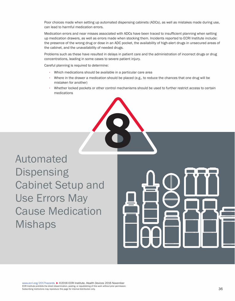
36
www.ecri.org/2017hazards ©2016 ECRI Institute. Health Devices 2016 November ECRI Institute prohibits the direct dissemination, posting, or republishing of this work without prior permission. Subscribing institutions may reproduce this page for internal distribution only.
8
Poor choices made when setting up automated dispensing cabinets (ADCs), as well as mistakes made during use, can lead to harmful medication errors.
Medication errors and near misses associated with ADCs have been traced to insufficient planning when setting up medication drawers, as well as errors made when stocking them. Incidents reported to ECRI Institute include: the presence of the wrong drug or dose in an ADC pocket, the availability of high-alert drugs in unsecured areas of the cabinet, and the unavailability of needed drugs.
Problems such as these have resulted in delays in patient care and the administration of incorrect drugs or drug concentrations, leading in some cases to severe patient injury.
Careful planning is required to determine:
• Which medications should be available in a particular care area• Where in the drawer a medication should be placed (e.g., to reduce the chances that one drug will be
mistaken for another)• Whether locked pockets or other control mechanisms should be used to further restrict access to certain
medications
Automated Dispensing Cabinet Setup and Use Errors May Cause Medication Mishaps
37www.ecri.org/2017hazards ©2016 ECRI Institute. Health Devices 2016 November
ECRI Institute prohibits the direct dissemination, posting, or republishing of this work without prior permission. Subscribing institutions may reproduce this page for internal distribution only.
Problem 1. Medication errors and near misses associated with the use of automated dispensing cabinets (ADCs)
have been traced to:
a) Insufficient planning when configuring the drawers, including decisions related to:(1) Which medications would be available in a particular care area(2) Where in the drawer a medication would be placed(3) Whether additional control mechanisms (e.g., lidded and locked pockets within the drawer)
would be used to further restrict access for certain medicationsb) Stocking errors
2. ECRI Institute has received reports involving:
a) ADC pockets that contained:(1) The wrong medication(2) The wrong concentration (dose) of the correct medication(3) Two different drugs(4) Two different concentrations of the same drug (5) Expired drugs
b) ADC drawers that contained:(1) Drugs with similar-sounding names or similar-looking packaging(2) High-alert drugs in an unsecured area (e.g., a drawer or pocket without a lock or other control
mechanism)(3) Drugs that are inappropriate for the care area
3. We have also received reports of instances in which drugs that were needed in the care area were not available in the ADC.
4. Problems such as these have resulted in:
a) Delays in patient careb) The administration of incorrect drugs or concentrations of drugs, leading to:
(1) The need for additional patient monitoring(2) Severe patient injury
ECRI Institute RecommendationsWhen implementing an ADC in any care area:
1. Work closely with the device supplier to understand and implement the ADC features that are avail-able to minimize the risk of medication errors. Such features include:
a) Secured, lidded pocketsb) Bar-code scanning

38
www.ecri.org/2017hazards ©2016 ECRI Institute. Health Devices 2016 November ECRI Institute prohibits the direct dissemination, posting, or republishing of this work without prior permission. Subscribing institutions may reproduce this page for internal distribution only.
c) Software for tracking the cabinet contents (e.g., the medications that have been added, drug expiration dates)
2. Review the inventory of medications (i.e., the formulary) within the ADC to verify the appropriateness of each drug for that care area.
a) Only stock medications necessary for the specified care area.b) Be sure to stock a sufficient supply of medications that might be needed for urgent situations.
3. Carefully plan where medications will be located within each drawer and how those medications can be accessed.
a) Consider the use of secured, lidded pockets. High-alert drugs, in particular, should be stored only in secured drawers or pockets.
b) Store similar-looking drugs and drugs with similar-sounding names in physically distinct areas of the cabinet—for example, in different drawers or within different lidded pockets.
c) Avoid arranging drugs in a drawer alphabetically, and differentiate drugs by dosage form in adjoin-ing pockets to increase visual contrast.
d) Consider the use of bar-code scanning, which can help prevent mix-ups when stocking the cabinet and when dispensing medications.
4. Have an independent reviewer assess the planned configuration to verify that it adequately reduces the risks of, for example, similar-sounding or similar-looking medications being confused for one another.
5. Establish a training and competency-assessment program on the ADC systems that emphasizes:
a) The importance of verifying that the medication (drug and concentration) that has been retrieved from the cabinet is, in fact, what the doctor or nurse intended.
b) The importance of returning unused medications to the ADC’s return bin—not to the original drawer or pocket. Returned medications should be processed by the pharmacy.
c) The value of reporting any errors or near misses that occur, so that prevention steps can be considered and implemented.
During use of an ADC:
1. Perform an independent double-check of the cabinet’s contents each time it is restocked. Implementing software that tracks the medications added to the cabinet can facilitate this process.
2. Additionally, for ADCs used in the OR:
a) Dispense medications for surgical procedures only when the patient is in the OR. b) Limit the practice of pre-dispensing medications for surgical cases that will be performed later in
the day.
Background1. ADCs are used:
a) To provide access to medications in patient care areas (e.g., nursing units, the emergency department, the OR)
b) To control drug distribution; interfaces with other systems support tracking, stocking, and billing

39www.ecri.org/2017hazards ©2016 ECRI Institute. Health Devices 2016 November
ECRI Institute prohibits the direct dissemination, posting, or republishing of this work without prior permission. Subscribing institutions may reproduce this page for internal distribution only.
2. The cabinets are highly configurable. They can be equipped with a variety of drawer designs and access-limiting controls. Options can include:
a) Locking or nonlocking drawersb) Bar-code access, requiring an electronic scan of a bar code to allow stocking and dispensing of
medicationsc) Drawers that allow open access to some pockets, but locked access to othersd) A return bin that clinicians can use to deposit—but not remove—unused medications
3. ADCs are often stocked in the care area by a pharmacy technician, who has a supply of medications for that care area that has been checked by the pharmacist.
4. During the period from January through September 2016, ECRI Institute PSO received reports of 227 patient safety events associated with ADCs.* These events occurred in multiple care areas and included errors and near misses involving the following drugs or concentrations:
a) Ephedrine instead of epinephrineb) Phenobarbital instead of dronabinol c) Methylphenidate (Ritalin) mixed with hydromorphone d) Lyrica instead of hydromorphone e) Oxycodone XL instead of oxycodone IRf) Albuterol 2.5 mg instead of 0.5 mgg) Toradol 60 mg/2 mL mixed with 15 mg/mLh) Atropine (for vagal response) not stocked i) Unsecured fentanyl, hydromorphone, and remifentanilj) Insulin NPH instead of insulin aspart k) Expired drugs
* ECRI Institute PSO is a federally listed Patient Safety Organization and a component of ECRI Institute. With the goal of helping healthcare providers learn from near misses and adverse events to improve patient care, ECRI Institute PSO collects and analyzes incident reports and distributes its analyses and recommendations in a variety of formats.
40
www.ecri.org/2017hazards ©2016 ECRI Institute. Health Devices 2016 November ECRI Institute prohibits the direct dissemination, posting, or republishing of this work without prior permission. Subscribing institutions may reproduce this page for internal distribution only.
References and ResourcesMember ResourceMedication management systems, decentralized. Healthcare Product Comparison System 2014 Nov. (Available to members of Health Devices Gold and SELECTplus.)
Additional Resources 1. American Society of Hospital Pharmacists. ASHP tech-
nical assistance bulletin on hospital drug distribution and control. Am J Hosp Pharm 1980;37:1097-103.
2. Australian Commission on Safety and Quality in Health Care. Automated dispensing systems. Evidence Brief-ings on Interventions to Improve Medication Safety 2013 Jun;1(2).
3. Ferencz N. Safety of automated dispensing systems. US Pharm 2014 Aug;39(8 Pharm&Tech suppl):8-12.
4. Grissinger M. Safeguards for using and design-ing automated dispensing cabinets. P T 2012 Sep;37(9):490-1, 530.
5. Institute for Safe Medication Practices (ISMP):a) Follow ISMP guidelines to safeguard the design
and use of automated dispensing cabinets (ADCs). ISMP Med Saf Alert 2009 Feb 12.
b) Guidance on the interdisciplinary safe use of auto-mated dispensing cabinets. 2008.
6. Pennsylvania Patient Safety Authority. Problems asso-ciated with automated dispensing cabinets. PA PSRS Patient Saf Advis 2005 Sep;2(3):21-3.

41www.ecri.org/2017hazards ©2016 ECRI Institute. Health Devices 2016 November
ECRI Institute prohibits the direct dissemination, posting, or republishing of this work without prior permission. Subscribing institutions may reproduce this page for internal distribution only.
9Problems associated with the use and functioning of surgical staplers can lead to intraoperative hemorrhaging, tissue damage, unexpected postoperative bleeding, failed anastomoses, and other forms of patient harm.
Surgical staplers require meticulous technique to operate, and problems during use are not uncommon. The U.S. Food and Drug Administration receives thousands of adverse event reports related to surgical staplers each year, and ECRI Institute likewise consistently receives reports of surgical stapler problems. Although severe injuries are infrequent, they do occur: We have investigated fatalities and other cases of serious patient harm.
Commonly reported problems include: misfiring or difficulty in firing, misapplied staples, unusual sounds during firing (which can indicate a damaged or malfunctioning mechanism), and tissue becoming “jammed” in the mechanism.
To prevent patient harm, users must be familiar with device operation, they must carefully select the appropriate staple size for the patient and tissue type, and they must be alert to the signs that the stapler may not be func-tioning as intended.
Surgical Stapler Misuse and Malfunctions
42
www.ecri.org/2017hazards ©2016 ECRI Institute. Health Devices 2016 November ECRI Institute prohibits the direct dissemination, posting, or republishing of this work without prior permission. Subscribing institutions may reproduce this page for internal distribution only.
Problem 1. Problems associated with the use and functioning of surgical staplers can lead to intraoperative
hemorrhaging, tissue damage, unexpected postoperative bleeding, failed anastomoses, and other forms of patient harm.
a) ECRI Institute consistently receives reports of surgical stapler problems and has investigated many incidents in which patient care was adversely affected.
b) Although severe injuries are uncommon, they do occur: We have investigated fatalities and other cases involving significant patient harm.
2. Commonly reported problems include:
a) Misfiring or difficulty in firing—this includes instances in which:(1) Squeezing the stapler handle requires more force than usual(2) Staples are not fully deployed after the device is fired(3) The stapler does not articulate or rotate as intended
b) Misapplied staples—as can occur when:(1) Staples of the wrong size are used(2) Staples are applied to the wrong tissue(3) The staple is applied at an angle such that it does not fully reach across the structure(4) The stapler is applied across a harder material (e.g., another previously deployed staple or a
surgical instrument) that results in device damage and inappropriate staple formation(5) Tissue in the stapler’s jaws is distributed unevenly
c) Unusual sounds during firing. This can indicate a damaged or malfunctioning internal mechanism.d) Failure to release tissue after firing—that is, tissue becoming “jammed” in the mechanism.
3. Such problems can prolong surgery or cause complications that require subsequent medical interventions.
4. Stapler problems have been known to result in:
a) Severe intraoperative hemorrhageb) Tissue damagec) Postoperative complications, such as unexpected bleeding or gastrointestinal (GI) leaking from
incompletely sealed structuresd) Infection
ECRI Institute RecommendationsClinicians—Before Use
1. Read and follow the stapler manufacturer’s instructions for use (IFU) before using the device for the first time or for the first time in a while.
a) Recognize that staplers from different manufacturers operate differently. b) Be aware of the intended use and potential contraindications of using these devices.
43www.ecri.org/2017hazards ©2016 ECRI Institute. Health Devices 2016 November
ECRI Institute prohibits the direct dissemination, posting, or republishing of this work without prior permission. Subscribing institutions may reproduce this page for internal distribution only.
2. Have an appropriate range of stapler cartridge sizes available for use.
a) Understand the array of possible staple sizes and their associated typical applications for a given stapler model. Then adjust the staple selection as the situation requires.
b) Keep in mind that tissue varies not only across patients but within the same patient, so there may be no single staple size that is suitable for all applications for a specific patient or patient tissue.
3. Have additional staplers and other means of closure available for immediate use.
a) Be prepared to switch staplers if a problem is identified with the original unit.b) Alternative means of closure, such as manual sutures, should also be available.
4. Inspect the stapler for damage before using it. (Preferably, this should be done both by the scrub technician/nurse who opens the instrument pouch and by the surgeon who will use the instrument.)
a) Inspect the anvil and the cartridge to ensure proper alignment.b) Verify that the shaft of the device is uniform and without any defects.c) Verify that all the components of reusable staplers are present and that, when the device is
assembled, the components are positioned properly and tightly fastened together. d) In the case of battery-powered staplers, make sure the device passes its designated self-tests for
handle, adapter, and reload.e) If the surgical stapler does not meet these criteria, use a different stapler.
Clinicians—During and After Use
1. Do not use a stapler if you are unable to visually assess (either directly or with an endoscope) secure closure and hemostasis.
2. Before firing the stapler, pause to ensure that the staples are appropriately sized for the intended tissue. If you have difficulty squeezing the handle of the stapler, you may need to resize.
3. If the stapler makes unusual sounds or is difficult to fire during use:
a) Visually inspect the stapler either directly (during open procedures) or using an endoscope.b) Check for excessive tissue, foreign material, or interference from another instrument and proceed
with caution.c) If sounds persist, continue the procedure with another stapler.
4. Adhere to the practices recommended by Fuller et al. (2014); specifically:
a) Avoid applying clips near an intended staple line site or stapling over an existing staple line. Contact with objects in the stapler’s path could damage the cartridge knife.
b) If a malfunction is noted when the stapler is closed on the tissue, “clamp or ligate the vessel before releasing the stapler.”
c) If tissue is “jammed” in the stapler, avoid forcefully freeing the tissue; consider dissecting proximal to the stapler closure.
5. Once tissue has been stapled, check the application site for secure closure and hemostasis.
6. If you identify a faulty stapler:
a) In the absence of injury, return the stapler (with its device packaging if possible) to the supplier for analysis and credit.
44
www.ecri.org/2017hazards ©2016 ECRI Institute. Health Devices 2016 November ECRI Institute prohibits the direct dissemination, posting, or republishing of this work without prior permission. Subscribing institutions may reproduce this page for internal distribution only.
b) If an injury has occurred, retain the device for possible third-party investigation.c) Contact ECRI Institute to report the problem.
Administrators and Other PersonnelFuller et al. (2014) additionally recommend the following:
1. For materials management: Communicate with clinicians during the device procurement process to ensure that surgeons can work with their preferred devices.
2. For the chief of surgery/OR director:
a) Facilitate focused and systematic training on surgical devices for all members of the surgical team to increase comfort with the device and its approved uses, as well as to raise awareness about known types of misuse and malfunction.
b) Make provisions to allow clinicians to have effective hands-on practice time with surgical devices.
Background 1. Surgical staplers are single-use or reusable devices intended for anastomosis and to seal and transect
tissues.
a) This report covers linear, circular, curved, cutter, and battery-powered surgical staplers.b) Surgical applications include: GI tract, gynecological, pediatric, urologic, and thoracic surgeries.
2. Staplers are complex devices requiring meticulous technique to operate, and problems during use are not uncommon.
a) On its Surgical Stapler Information web page, updated in 2015, FDA estimated that 8,000 to 9,000 adverse event reports related to surgical staplers were filed per year over the preceding five years.
b) In a retrospective study of 349 colorectal resections using a circular stapler, 19% of the proce-dures included a technical/use error that was associated with a higher risk of GI bleeding, the need for transfusions, and unplanned proximal diversions (Offodile et al. 2010). That is, nearly 1 in 5 of these cases featured a technical error associated with stapler use.
3. Among the stapler-related events of which ECRI Institute is aware, use errors such as the following were identified as contributing factors:
a) Selecting an incorrect staple size (see the figure on the next page).(1) Using an undersized staple on tissue that is too thick can prevent staples from securely con-
tacting the anvil and being bent into their fully closed shape; these staples can work their way free, allowing the staple line to open.
(2) Using an oversized staple on tissue that is too thin allows the staples to contact the anvil and be bent into the correct shape, but the staples will not completely compress the target tissue. Though the staples will remain in place, the resulting gaps in the staple line may allow leak-age from between the tissue halves.
b) Interposing other instruments or excessive tissue between the jaws.c) Failure to follow the device IFU, including adhering to contraindications.

45www.ecri.org/2017hazards ©2016 ECRI Institute. Health Devices 2016 November
ECRI Institute prohibits the direct dissemination, posting, or republishing of this work without prior permission. Subscribing institutions may reproduce this page for internal distribution only.
4. Raising awareness about surgical stapler hazards and educating staff about proper use can have a positive impact:
a) In a multiple-choice test created to evaluate surgical residents’ knowledge on stapler use a 24% increase in average scores (from 53% to 77%) was obtained after a 40-minute instructional ses-sion (McColl et al. 2009).
b) Though the study was small (n = 26, general surgery), it may indicate an existing knowledge gap in the material and the value of directed training. Hands-on training may even provide a more marked improvement.
5. The bottom line? Stapler operators need to be knowledgeable about staple size selection; the mecha-nisms of closure, deployment, and removal; and the ways in which a specific device might be misused (Deng et al. 2002).
References and ResourcesMember Resources
1. Before a surgical stapler fails [guidance article]. Health Devices 2001 Oct.
2. Surgical staplers: recommendations to reduce the risk of commonly reported problems. Health Devices Alerts 2016 Mar 17 (Accession No. H0312).
3. Surgical stapler hazards. In: Top 10 technology haz-ards: high-priority risks and what to do about them (Hazard #8). Health Devices 2009 Nov.
4. Using the wrong size surgical stapler cartridge can injure patients [hazard report]. Health Devices 2009 Apr.
References1. Deng DY, Meng MV, Nguyen HT, et al. Laparo-
scopic linear cutting stapler failure. Urology 2002 Sep;60(3):415-9.
2. Food and Drug Administration, U.S. (FDA). Surgical stapler information [updated 2015 May 8].
3. Fuller HJ, Paul DE, Williams LC. Surgical stapler adverse events in the Veterans Health Administra-tion: root causes and lessons learned. Proceedings of the International Symposium on Human Factors and Ergonomics in Health Care: Advancing the Cause. 2014 Jun;3(1):153-6.
4. McColl RJ, Karmali S, Reso A, et al. The effect of a focused instructional session on knowledge of surgi-cal staplers in general surgery residents. J Surg Educ 2009 Sep-Oct;66(5):288-91.
5. Offodile AC 2nd, Feingold DL, Nasar A, et al. High incidence of technical errors involving the EEA circular stapler: a single institution experience. J Am Coll Surg 2010 Mar;210(3):331-5.
Figure. Matching stapler cartridge size to tissue thickness (as shown in the third image) is important for secure closure and hemostasis.
Undersized staple(staple line won’t hold)
Oversized staple(tissue can move, resulting in gaps)
Staple size matchedto tissue thickness

46
www.ecri.org/2017hazards ©2016 ECRI Institute. Health Devices 2016 November ECRI Institute prohibits the direct dissemination, posting, or republishing of this work without prior permission. Subscribing institutions may reproduce this page for internal distribution only.
Device Failures Caused by Cleaning Products and Practices
10
The use of cleaning agents or cleaning practices that are incompatible with the materials used in a medical device’s construction, or that are otherwise inappropriate for the device’s design, can cause the device to malfunction or to fail prematurely, possibly affecting patient care. Specifically:
• Repeated use of incompatible cleaning agents can damage equipment surfaces and degrade plastics, often resulting in device breakage—possibly with no visible warning signs.
• The use of improper cleaning practices can damage seals, degrade lubricants, and cause fluid intrusion. This can result in damage to electronics, power supplies, and motors.
Because there is no single cleaner or cleaning process that will work with all devices, hospitals must stock and use multiple cleaning products and familiarize staff with device-specific cleaning methods—tasks that pose a sig-nificant burden. Nevertheless, failure to do so can lead to ineffective cleaning (a potentially deadly circumstance), as well as excessive component breakage and premature equipment failures (which can affect patient care and be a significant financial burden).

47www.ecri.org/2017hazards ©2016 ECRI Institute. Health Devices 2016 November
ECRI Institute prohibits the direct dissemination, posting, or republishing of this work without prior permission. Subscribing institutions may reproduce this page for internal distribution only.
Problem 1. The use of cleaning agents or cleaning practices that are incompatible with the materials used in a
medical device’s construction, or that are otherwise inappropriate for the device’s design, can cause the device to malfunction or to fail prematurely, possibly affecting patient care. Specifically:
a) Repeated use of incompatible cleaning agents can damage equipment surfaces and degrade plastics, often resulting in device breakage—possibly with no visible warning signs.
b) The use of improper cleaning practices can damage seals, degrade lubricants, and cause fluid intrusion. This can result in damage to electronics, power supplies, and motors.
2. Such damage can lead to excessive alarms or other device errors or equipment failures that can compromise patient safety.
3. In addition to the potential impact on patient care, excessive component breakage and premature equipment failures can be a significant financial burden. ECRI Institute is aware of several instances of such failures, each of which had cost a healthcare facility thousands of dollars before the cause of the failure was identified.
ECRI Institute Recommendations1. Recognize that there is no single cleaner or cleaning process that will work with all devices.
2. Review manufacturers’ information to identify recommended procedures and compatible cleaning solutions and wipes.
a) If this information is not provided, contact the manufacturer and request the information needed. b) If the manufacturer is unable to provide this information, consider purchasing alternative instru-
ments when replacement is needed.
3. Make recommended cleaning instructions and suitable cleaners easily accessible by all clinical and equipment maintenance staff.
4. Instruct staff to:
a) Use only cleaning solutions and wipes that are recommended for use with the equipment to be cleaned.
b) Allow sufficient wet and dry times per cleaning agent and device instructions.
5. Regularly inspect all plastic components and moving parts for deterioration and wear.
6. Consider stocking replacement parts and evaluate the need for backup devices in case of failure.
Background1. The need to stock and use multiple cleaning products, along with the requirement to familiarize staff
with device-specific cleaning methods, is a significant burden for hospitals.
48
www.ecri.org/2017hazards ©2016 ECRI Institute. Health Devices 2016 November ECRI Institute prohibits the direct dissemination, posting, or republishing of this work without prior permission. Subscribing institutions may reproduce this page for internal distribution only.
2. Nevertheless, a failure to do so can lead to ineffective cleaning (a potentially deadly circumstance), as well as premature device failure.
a) Repeated use of incompatible cleaning agents can cause environmental stress cracking in plastics, which in turn can cause parts to become prematurely brittle, often with no visible indication of impending failure.
b) Incorrect cleaning of battery modules or electrical connections may result in breakage of compo-nents that can cause excessive alarms, device errors, or equipment failures.
c) Using excessive amounts of cleaning solutions can lead to fluid ingress, which can cause corrosion or short circuits that result in device failures.
d) Not allowing sufficient wet (dwell) time can lead to inadequate disinfection.e) Not inspecting battery compartments for fluid intrusion can result in premature battery failure.
3. Even when manufacturers publish equipment-specific cleaning instructions, this information is not always readily available to clinical staff.
4. To read more about some of the incidents that ECRI Institute has investigated or learned about, refer to the Member Resources list below.
References and ResourcesMember Resources
1. Baxter—SIGMA Spectrum infusion pumps: incorrect cleaning of pump battery connections may lead to battery alarms and errors. [ECRI Exclusive Hazard Report]. Health Devices Alerts 2015 Sep 10 (Acces-sion No. H0274).
2. Berchtold—OPERON surgical table hand controls: unin-tended table section movement may occur [Update]. Health Devices Alerts 2014 Jul 31 (Accession No. A19816 01).
3. Covidien/Medtronic—Genius 2 ear tympanic ther-mometers: may crack from use of incompatible disinfectants. [ECRI Exclusive Hazard Report]. Health Devices Alerts 2015 Sep 10 (Accession No. H0275).
4. ECRI Institute User Experience Network—lower door hinge may crack on Carefusion Alaris 8100 Pump Modules. Health Devices Alerts 2014 May 15 (Acces-sion No. S0251 01).
5. Fresenius—2008 series hemodialysis machines: manufacturer reminds users of cleaning guidelines. Health Devices Alerts 2015 Aug 12 (Accession No. A24922).
6. Haemonetics—various cell processor and collection system devices: improper cleaning may damage pump rollers, potentially leading to device malfunction [Update]. Health Devices Alerts 2016 Feb 23 (Acces-sion No. A25476 01).
7. Infusion pumps—failure to follow manufacturers’ recommended cleaning instructions may cause premature device failures. [ECRI Exclusive Hazard Report]. Health Devices Alerts 2016 Jun 16 (Acces-sion No. H0328).
8. Intuitive—da Vinci Surgical Systems: certain disin-fectant wipes may damage instrument arm plastic components. Health Devices Alerts 2016 Jan 14 (Accession No. A25621).
9. PARI—PARI SOLE N and PARI SOLE N Tracheo inhala-tion devices: liquid may damage temperature gauge. Health Devices Alerts 2013 Nov 12 (Accession No. A21201).
10. Philips M4841a telemetry transmitters may experience short circuit and overheat if battery com-partment tab breaks. [Hazard Report]. Health Devices Alerts 2013 Feb 28 (Accession No. H0192).
11. Siemens—cleaning solutions used with ADVIA Centaur Systems: hypochlorite may degrade at a higher-than-expected rate. Health Devices Alerts 2014 Nov 26 (A23371).
ZZ UNITED STATES 5200 Butler Pike Plymouth Meeting, PA 19462-1298, USA Telephone +1 (610) 825-6000 Fax +1 (610) 834-1275
ZZ EUROPE Suite 104, 29 Broadwater Road Welwyn Garden City Hertfordshire, AL7 3BQ, UK Telephone +44 (1707) 871 511 Fax +44 (1707) 393 138
ZZ ASIA PACIFIC 11-3-10, Jalan 3/109F Danau Business Centre Taman Danau Desa 58100 Kuala Lumpur, Malaysia Telephone +60 3 7988 1919 Fax +60 3 7988 1170
ZZ MIDDLE EAST/INTEGRA Office 708-6, Fairmont Hotel Sheikh Zayed Road P.O. Box 128740 Dubai, United Arab Emirates Telephone +971 4 3042460 Fax +971 4 3042461
Objectives of the Health Devices SystemTo improve the effectiveness, safety, and economy of health services by:
Providing independent, objective judgment for selecting, purchasing, managing, and using medical devices, equipment, and systems.
Functioning as an information clearinghouse for hazards and deficiencies in medical devices.
Encouraging the improvement of medical devices through an informed marketplace.