Tools for Improving the Quality of Aged, Degraded, Damaged, or
30
The author(s) shown below used Federal funds provided by the U.S. Department of Justice and prepared the following final report: Document Title: Tools for Improving the Quality of Aged, Degraded, Damaged, or Otherwise Compromised DNA Evidence Author: John R. Battista Document No.: 237840 Date Received: February 2012 Award Number: 2007-DN-BX-K146 This report has not been published by the U.S. Department of Justice. To provide better customer service, NCJRS has made this Federally- funded grant final report available electronically in addition to traditional paper copies. Opinions or points of view expressed are those of the author(s) and do not necessarily reflect the official position or policies of the U.S. Department of Justice.
Transcript of Tools for Improving the Quality of Aged, Degraded, Damaged, or

The author(s) shown below used Federal funds provided by the U.S. Department of Justice and prepared the following final report:
Document Title: Tools for Improving the Quality of Aged, Degraded, Damaged, or Otherwise Compromised DNA Evidence
Author: John R. Battista Document No.: 237840
Date Received: February 2012 Award Number: 2007-DN-BX-K146
This report has not been published by the U.S. Department of Justice. To provide better customer service, NCJRS has made this Federally-funded grant final report available electronically in addition to traditional paper copies.
Opinions or points of view expressed are those of the author(s) and do not necessarily reflect
the official position or policies of the U.S. Department of Justice.

1
Final Technical Report
Title: Tools for Improving the Quality of Aged, Degraded, Damaged, or
Otherwise Compromised DNA Evidence
Award: 2007-DN-BX-K146
Author: John R. Battista
Abstract
Current forensic DNA genotyping technology requires 0.2-2.0 nanograms duplex DNA
that is at least 100-500 base pair in length. However, many evidence samples fail to meet
these minimal requirements, because the target DNA has been exposed to environments
capable of extensively damaging DNA. This damage reduces the size of the target DNA,
making it too small to be amplified and can block PCR amplification. Since DNA
samples found at crime scenes exhibit varying degrees of damage, the effectiveness of
DNA typing technology is limited by sample condition, and at present there are no
methods of circumventing this problem. Our principle objective was to develop tools to
facilitate STR DNA genotyping through a) improving the quality of the DNA found in
degraded forensic samples, and b) enhancing the ability to retrieve amplifiable DNA
from forensic samples. Our efforts were focused on two areas. First, we attempted
repairing damaged DNA in vitro using cell extracts isolated from repair proficient
microorganisms. Extracts were emphasized because there is no way of knowing a priori
what type of DNA damage is present in biological evidence, and intact cells express an
extensive array of DNA repair proteins capable of dealing with most DNA damage.
These efforts met with minimal success; it appears we were able to repair DNA in vitro,
but the technique was difficult to reproduce. The cell extracts inhibited the reaction used
to monitor repair and prevent an accurate assessment of the extent of repair. Second, we
have developed a method for retrieving selected DNA fragments from complex mixtures.
DNA damage occurs randomly and so the double strand breaks resulting from that
damage are also random. A sample – even a highly degraded sample – is expected to
have a distribution of fragment sizes, and our efforts are aimed at isolating fragments that
include the intact STR sequences from such mixtures. To that end we have developed an
in vitro method using three purified proteins that permits selective capture of specific
DNA sequences. This method relies on the use of a targeting DNA fragment, a sequence
that is complementary to the sequence you wish to retrieve. Combining this fragment,
the three proteins, and appropriate cofactors allows recovery of homologous DNA from
solution with high efficiency.
This document is a research report submitted to the U.S. Department of Justice. This report has not been published by the Department. Opinions or points of view expressed are those of the author(s) and do not necessarily reflect the official position or policies of the U.S. Department of Justice.

2
This document is a research report submitted to the U.S. Department of Justice. This report has not been published by the Department. Opinions or points of view expressed are those of the author(s) and do not necessarily reflect the official position or policies of the U.S. Department of Justice.

3
Executive Summary
Since its inception in the 1980s, DNA typing of biological evidence has taken on a
central role in forensic science. This technology, in conjunction with the CODIS network
and the implementation of national standards for convicted offender DNA databasing,
provides law enforcement with a highly effective method for solving violent crimes
nationwide. In addition, DNA evidence has led to the exoneration of a number of
wrongly convicted persons. Current forensic DNA technology requires 0.2-2.0
nanograms duplex DNA that is at least 100-500 base pair in length. However, many
evidence samples fail to meet these minimal requirements, because the target DNA has
been exposed to environments capable of extensively damaging DNA. This damage can
reduce the size of the target DNA, making it too small to be amplified by the polymerase
chain reaction (PCR) during the typing protocol, and some forms of damage result in
modification of nitrogenous bases that block PCR amplification. Since DNA samples
typically found at crime scenes exhibit varying degrees of damage, the effectiveness of
DNA typing technology is limited by sample condition, and at present there are no
methods of circumventing this problem. If treatments can be found that improve the
condition of degraded samples without affecting the genetic profile of DNA evidence, the
use of DNA typing can be expanded to the benefit of law enforcement and society.
The long-term objective of this project is to develop a protocol for restoring heavily
degraded DNA evidence to a condition suitable for STR DNA genotyping – a task that
requires the capacity to increase the amount of available undamaged DNA fragments in
the necessary 100-500bp range prior to amplification via the PCR process. With
appropriate technical training, it is relatively straightforward to generate a genotype from
an abundant pristine sample, such as that obtained from a cheek scraping. However,
forensic scientists do not always have the luxury of abundant high quality DNA evidence
to work with. Samples are affected by their history. DNA evidence is often exposed to
the environment for varying lengths of time; in some situations, it could be decades
between the time a crime is committed and the time the evidence is collected and
analyzed. Sample DNA can be damaged in a myriad of natural and unnatural processes,
and this degradation in sample quality makes obtaining a genotype difficult, if not
impossible.
Two general types of degradation are particularly important when considering forensic
genotyping. First, DNA damage can modify DNA bases, and these modifications can
physically block movement of the DNA polymerase used in PCR amplification, reducing
or eliminating the signals (derived from the STRs) used to create the genotype. Second,
DNA damage causes DNA strand breaks, and as the number of strand breaks increases
the probability of separating the STR from one of the primer sites needed for
amplification increases. The number of strand breaks increases with time, creating
smaller and smaller fragments. Eventually, the accumulation of strand breaks causes
individual STR signals to drop out of the genotype, the larger STRs disappearing first.
The approach taken during this project toward improving DNA genotyping focused on 1)
improving damaged sample DNA prior to attempts at PCR amplification, and 2)
This document is a research report submitted to the U.S. Department of Justice. This report has not been published by the Department. Opinions or points of view expressed are those of the author(s) and do not necessarily reflect the official position or policies of the U.S. Department of Justice.

4
retrieving those specific sequences needed for PCR amplification, thereby improving the
chances for obtaining a genotype. The protocols described are not intended to supplant
existing methodology. The effort was intended to provide a “pretreatment” that would
help guarantee successful genotyping, augmenting existing protocols approved for
forensic applications. These efforts were guided by the knowledge that DNA repair
systems are present in every living cell – systems that have evolved to repair damaged
DNA efficiently without introducing sequence changes. If these systems could be
harnessed for use in vitro with degraded DNA samples, it may be possible to increase the
efficiency with which forensic samples are processed.
During the funding period, two lines of investigation were pursued. First, extracts from
Deinococcus radiodurans R1 were examined for their ability to repair DNA damage in
vitro. D. radiodurans is one of the most DNA damage tolerant species known and that
capability is the consequence of an unusually efficient DNA repair. Second, an in vitro
system for repairing DNA double strand breaks was developed and used as a mechanism
for capturing and concentrating any DNA sequence.
Repairing DNA in vitro using extracts of D. radiodurans R1
The experimental design for this study was straightforward. Samples of damaged DNA
would be treated with extracts obtained from cultures of D. radiodurans exposed to
3000Gy dose of ionizing radiation. Earlier work had established that aged forensic
samples exhibit a pattern of DNA damage similar to that generated when DNA is
exposed to ionizing radiation. To follow DNA repair we used a bioassay in which
viability was the end point. The bacterial strain Acinteobacter baylyi ADP6 carries a
defect that prevents this strain from growing in the presence of 4-hydroxy-benzoate
(POB). Transferring the pcaEFDBCHG operon into ADP6 can restore POB prototrophy.
Since A. baylyi strains are easily and efficiently transformed with naked DNA, we
designed a system in which we attempted to repair damaged plasmids carrying the
pcaEFDBCHG operon with the extracts from D. radiodurans. Restoration of POB
prototrophy in ADP6 transformed with a damaged plasmid would be taken as evidence of
in vitro repair.
Initial trials suggested that extracts were repairing damaged DNA, but the controls
indicated that undamaged DNA was adversely affected by the extract, lowering
transformation efficiency. This effect of the extract made it impossible to accurately
assess the extent of DNA repair in vitro. Attempts to determine the mechanism
responsible for the inhibition by the extract were unsuccessful, and indicated that there
could be more than one inhibitory factor present. After obtaining this result, no further
studies of the use cell extracts for in vitro DNA repair were pursued.
In vitro DNA double strand break repair and selective capture
A procedure for the joining together two DNA molecules that share an 18- 55 nucleotide
region of homology was developed during the funding period. The process, referred to as
This document is a research report submitted to the U.S. Department of Justice. This report has not been published by the Department. Opinions or points of view expressed are those of the author(s) and do not necessarily reflect the official position or policies of the U.S. Department of Justice.

5
in vitro double-strand break repair reaction, requires only three proteins: RecA protein,
single strand binding (SSB) protein and a DNA polymerase. To date, all three proteins
are obtained from Escherichia coli. RecA and SSB are needed for identifying the region
of homology and promoting invasion by the 3’ end of single strand DNA molecule
recombining with duplex DNA. The DNA polymerase extends the 3’ ends, sealing the
spliced fragments formed by the action of RecA and SSB.
The purpose of this reaction is twofold. First, it can be used to repair double strand
breaks found between the STR and the primer sites for STR amplification. If the single
strand fragment used to initiate in vitro double strand break repair (referred to as a
targeting fragment) bridges the strand break the resulting union eliminates that break.
Second, if the targeting fragment includes at its 5’ end a feature that allows efficient
retrieval such as biotin that can be harvested with streptavidin-coated magnetic beads,
recombined fragments can be isolated and concentrated for further manipulation (e.g.
PCR amplification).
To establish that in vitro double strand break repair was possible, an assay was developed
that utilized the plasmid pUC19 cleaved by a pair of restriction enzymes: AatII and PstI.
The plasmid pUC19 DNA digested with AatII shares homology with the ends of the PstI
digested pUC19 DNA. The ends overlap by 504 nucleotides and 2182 nucleotides.
Strand pairing, strand exchange, and extension of the 3’ end of the exchanged strand can
theoretically produce two products: one of 3190 nucleotides and another of 4868
nucleotides. When attempted, the reaction generated all of the expected products with
unprecedented efficiency. It is estimated that as much as 40% of the starting material
recombined. Additional investigation established that the following components were
necessary for the reaction: RecA (in the presence of ATP), SSB, DNA Pol I, and all four
dNTPs; omit any one of these components and the products are not formed
To test the feasibility of using synthetic oligonucleotides in the double strand break
reaction, biotinylated capture fragments of 50, 100, 150 and 200 nucleotides in length
were examined for the ability to initiate the reaction. Each fragment was complementary
to DNA sequence upstream of the gltS gene of Escherichia coli, which simulated a target
sequence. An attempt was made to recover this sequence from purified E. coli genomic
DNA. The double strand break reaction was conducted as described above. The 100,
150, and 200 base oligonucleotides initiate the double strand break reaction and allow a
discrete set of fragments that are larger than 1500 base pairs to be retrieved.
It appears that as long as complementary DNA sequence is available, targeting DNA
fragments can be designed that will promote in vitro DNA double strand break repair. In
theory, this protocol can be used to selectively capture any DNA sequence. In addition to
autosomal STRs, Y chromosome STRs, and mitochondrial DNA sequences could be
retrieved by this procedure. This procedure has three properties that make it potentially
useful for forensic applications: i) it does not require knowledge of DNA sequence on
both ends of a DNA segment to be captured, as in PCR, ii) it does not introduce any
enzymes that could damage or alter a target DNA sample, and iii) it should allow the
retrieval of desired DNA segments even from samples that are highly contaminated with
This document is a research report submitted to the U.S. Department of Justice. This report has not been published by the Department. Opinions or points of view expressed are those of the author(s) and do not necessarily reflect the official position or policies of the U.S. Department of Justice.

6
DNA from other species.
The capacity to selectively target DNA sequences within a complex mixture is one of the
most useful features of the method, allowing one to retrieve the proverbial “needle from
the haystack” because the method relies on a RecA-facilitated search for
complementarity between a targeting DNA fragment and the genomic DNA sequence
with a sequence of interest. The mechanism of a RecA-mediated search for homology is
not well understood, but in vitro evidence (derived from the literature) indicates that
RecA can identify its target within an excess of 200,000-fold heterologous substrate
within 15 minutes – far faster than if the same DNA fragment were added to this mixture
in the absence of RecA.
It is expected that selective capture through in vitro double strand break repair will
enhance the ability to recover the DNA fragments from low abundance samples,
degraded samples, and samples that are contaminated with mixtures of DNA from other
species. The incredible specificity of RecA-mediated reactions, all but excludes cross-
reaction. The ability to selective capture a sequence of interest and recover it with
magnetic beads simplifies subsequent steps in genotyping and will remove ambiguity
from those analyses generated from samples obtained under less than ideal
circumstances.
Final Technical Report
I. Introduction
A. Statement of the problem
This project sought to answer a single question. Can DNA evidence that has been deemed
quantitatively and qualitatively inadequate for PCR-based genotyping be restored to a
usable condition if pre-treated with mixtures of proteins that either repair existing damage
or allow efficient recovery of DNA sequence suitable for amplification? During the
funding period, we have worked toward developing methods for repairing damaged DNA
in vitro and for retrieving DNA fragments of interest to forensic scientists from evidence
samples.
The long-term objective of this project is development of protocols for facilitating STR
DNA genotyping from heavily degraded forensic DNA evidence; a task that requires the
capacity to increase the amount of available undamaged DNA fragments in the necessary
100-500bp range prior to amplification via the PCR process. Two approaches have been
taken: 1) We have attempted to add extracts from highly repair proficient bacteria to
extensively-degraded biological evidence in the belief that we can improve the quality of
a large enough fraction of the target DNA in that sample to support STR genotyping, and
2) we have developed a method that allows us to capture and concentrate the targets of
STR typing.
This document is a research report submitted to the U.S. Department of Justice. This report has not been published by the Department. Opinions or points of view expressed are those of the author(s) and do not necessarily reflect the official position or policies of the U.S. Department of Justice.

7
B. Literature citations and review
1. STR typing
DNA genotyping based on the PCR amplification and electrophoretic analysis of Short
Tandem Repeats (STRs) plays a prominent role in forensic science (Butler, 2006). A
STR is a polymorphism found in mammalian DNA, a sequence of nucleotides (ranging
between 2-10 bases) that is tandemly repeated at a locus. By examining several STR loci
one can establish the unique genetic profile of an individual, linking biological evidence
from a crime to the perpetrator or to other crimes by the same person.
Tetranucleotide repeats are the mainstay of forensic DNA typing and criminal offender
databasing (Butler, 2006). There are only 33 possible tetranucleotide motifs (Jin et al.,
1997), and the consensus motif sequences, mostly AGAT and GATA, are ubiquitous in
the human genome. The number of repeat units at these loci varies from as few as four to
as many as 50. In 1997 the forensic community in the United States chose thirteen STR
loci to form the essential core of its Combined DNA Index System (CODIS) casework
and offender databases. These loci are: D3S1358, D5S818, D7S820, D8S1179, D13S317,
D16S539, D18S51, D21S11, CSF1PO, FGA, Th01, TPOX, and vWA. There are enough
different alleles at these STR loci in any major population or subpopulation to ensure that
individuals will be heterozygous at most loci, enabling unambiguous identification
(Butler, 2006; Jin et al., 1997).
2. DNA Degradation and STR Typing
Under conditions where biological samples are well preserved, genotyping with STRs is
a robust technology that can be applied with confidence. However, forensic scientists are
often confronted with biological evidence in which the DNA is present in a degraded
form that interferes with PCR amplification, limiting the effectiveness of this technology
(Hochmeister, 1998; Hoff-Olsen et al., 2001; Pfeiffer et al., 1999). In these samples the
DNA is highly fragmented and contains a large number of modified nucleotides. STR
analysis generates PCR fragments of between 100 – 500 base pairs; if the fragments of
target DNA are on average smaller than this, effective PCR amplification will not be
obtained. Although many other types of DNA damage can interfere with PCR
amplification, double strand breaks are the major impediment to successful DNA
genotyping in degraded DNA samples. Any double strand break in the region between
the primers used to amplify the DNA at a particular locus will prevent amplification of
that segment.
3. DNA Repair
Given the fundamental role of DNA in the storage and transmission of genetic
information, it is somewhat surprising that this molecule is relatively unstable in vivo and
in vitro (Lindahl, 1993). DNA is susceptible to spontaneous decomposition and it can be
damaged by a myriad of physical and chemical agents derived from endogenous and
exogenous sources (Friedberg et al., 2005). The effect that DNA damage has on a cell is
This document is a research report submitted to the U.S. Department of Justice. This report has not been published by the Department. Opinions or points of view expressed are those of the author(s) and do not necessarily reflect the official position or policies of the U.S. Department of Justice.

8
in large part determined by the interaction between the lesion formed and the replicative
DNA polymerase (Wellinger and Thoma, 1996). Lesions frequently inhibit this
polymerase, blocking DNA replication, and unless the blockage is removed the cell will
die. As a consequence, all characterized species express proteins that either detect and
repair DNA damage or allow the offending lesion to be bypassed. The primary function
of most DNA repair proteins in vivo is to correct DNA damage before the polymerase
reaches the lesion, thus avoiding the problem. An impressive arsenal of proteins is
dedicated to this function and repair is accomplished by direct reversal of base damage,
or by excising damage and using the undamaged complementary strand to restore the
original sequence (Friedberg et al., 2005). Mechanisms for reversing a lesion are not as
common as that involving excision repair. Direct reversion is of necessity lesion specific
and known mechanisms affect only pyrimidine dimers and certain methylated bases. In
contrast many lesions are targeted by excision repair systems. There are two types of
excision repair: nucleotide excision repair (NER) and base excision repair (BER).
During NER, the lesion is recognized and the repair complex nicks the DNA backbone 5’
and 3’ of the lesion. When the lesion is removed a 12 or 13 base gap is generated in the
strand. A DNA polymerase restoring the original DNA sequence fills this gap. The
proteins that catalyze NER recognize unnatural bends that are introduced by the damaged
base into DNA; they do not identify specific lesions. As a consequence NER can remove
many types of DNA damage and, in general, a single species encodes only one NER
complex. Base excision repair utilizes a similar strategy, but there are many types of BER
proteins, as they tend to be specific in their substrates, each protein recognizing a limited
number of lesions. The damaged base is removed during BER, leaving an
apurinic/apyrimidinic (AP) site in the DNA. The sugar remaining in the AP site is then
removed by the action of an AP endonuclease and deoxyribophosphodiesterase, resulting
in a one base gap in the DNA that is filled in by a DNA polymerase. If the lesion cannot
be removed by one of these processes and the polymerase is stopped, there are back up
systems that allow the cell either to repair the stalled replication fork and correct the
damage found there, or to bypass the damage with a special class of DNA polymerases
capable of translesion DNA synthesis (tls) (Battista and Earl, 2004; Friedberg et al.,
2005). The repair of stalled replication forks employs genetic recombination, utilizing the
redundant genetic information found in sister duplexes to replace the damaged DNA and
permit accurate replication restart (Cox et al., 2000). The tls DNA polymerases are not
affected by blocking lesions, and insert a nucleotide opposite the lesion to extend the
growing DNA chain and effect bypass (Lopes et al., 2006; Rattray and Strathern, 2003;
Woodgate, 1999). Thus, DNA repair processes limit the lethal and mutagenic effects of
DNA damage as it arises in vivo, and so maintain viability and preserve the fidelity of
DNA replication. For the purposes of this proposal, it is instructive to recognize that the
problems DNA damage causes in vivo are not unlike those that occur during attempts to
amplify degraded DNA evidence during STR genotyping. In both situations damaged
DNA bases block the movement of a DNA polymerase. By removing the offending base
prior to the polymerase reaching the site of damage in vivo, the cell avoids a potentially
lethal circumstance. If damage to DNA evidence can be repaired in vitro prior to PCR, it
should be possible to successfully genotype that sample. It was our contention that
proteins utilized in vivo by all species to repair DNA damage have potential use as
This document is a research report submitted to the U.S. Department of Justice. This report has not been published by the Department. Opinions or points of view expressed are those of the author(s) and do not necessarily reflect the official position or policies of the U.S. Department of Justice.
9
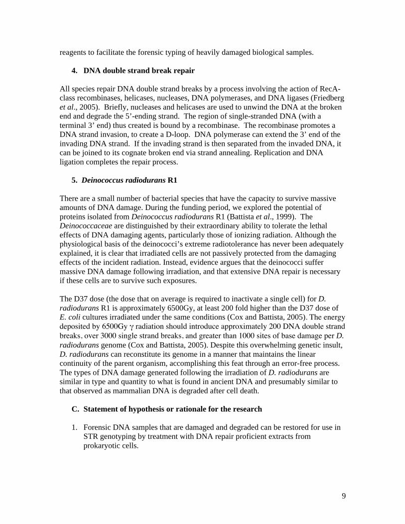
reagents to facilitate the forensic typing of heavily damaged biological samples.
4. DNA double strand break repair
All species repair DNA double strand breaks by a process involving the action of RecA-
class recombinases, helicases, nucleases, DNA polymerases, and DNA ligases (Friedberg
et al., 2005). Briefly, nucleases and helicases are used to unwind the DNA at the broken
end and degrade the 5’-ending strand. The region of single-stranded DNA (with a
terminal 3’ end) thus created is bound by a recombinase. The recombinase promotes a
DNA strand invasion, to create a D-loop. DNA polymerase can extend the 3’ end of the
invading DNA strand. If the invading strand is then separated from the invaded DNA, it
can be joined to its cognate broken end via strand annealing. Replication and DNA
ligation completes the repair process.
5. Deinococcus radiodurans R1
There are a small number of bacterial species that have the capacity to survive massive
amounts of DNA damage. During the funding period, we explored the potential of
proteins isolated from Deinococcus radiodurans R1 (Battista et al., 1999). The
Deinococcaceae are distinguished by their extraordinary ability to tolerate the lethal
effects of DNA damaging agents, particularly those of ionizing radiation. Although the
physiological basis of the deinococci’s extreme radiotolerance has never been adequately
explained, it is clear that irradiated cells are not passively protected from the damaging
effects of the incident radiation. Instead, evidence argues that the deinococci suffer
massive DNA damage following irradiation, and that extensive DNA repair is necessary
if these cells are to survive such exposures.
The D37 dose (the dose that on average is required to inactivate a single cell) for D.
radiodurans R1 is approximately 6500Gy, at least 200 fold higher than the D37 dose of
E. coli cultures irradiated under the same conditions (Cox and Battista, 2005). The energy deposited by 6500Gy radiation should introduce approximately 200 DNA double strand breaks, over 3000 single strand breaks, and greater than 1000 sites of base damage per D.
radiodurans genome (Cox and Battista, 2005). Despite this overwhelming genetic insult,
D. radiodurans can reconstitute its genome in a manner that maintains the linear
continuity of the parent organism, accomplishing this feat through an error-free process.
The types of DNA damage generated following the irradiation of D. radiodurans are
similar in type and quantity to what is found in ancient DNA and presumably similar to
that observed as mammalian DNA is degraded after cell death.
C. Statement of hypothesis or rationale for the research
1. Forensic DNA samples that are damaged and degraded can be restored for use in
STR genotyping by treatment with DNA repair proficient extracts from
prokaryotic cells.
This document is a research report submitted to the U.S. Department of Justice. This report has not been published by the Department. Opinions or points of view expressed are those of the author(s) and do not necessarily reflect the official position or policies of the U.S. Department of Justice.

10
2. Intact short tandem repeats suitable for forensic analysis can be recovered with
high efficiency from extensively degraded DNA samples.
II. Methods
During the three years of this projects existence, we have pursued two lines of
investigation. For the first one and one half years, we attempted to use cell extracts
derived from D. radiodurans R1 as a means of “repairing” extensively damaged sample
DNA in vitro. This approach resulted in the creation a convenient assay for in vitro
repair, followed by iterative exploration of the best conditions for isolating and adding
extracts to the samples of interest. The last one and one half years of the project focused
on the development of a technique we refer to as in vitro DNA double strand break
repair. This method allows us to selectively capture DNA containing STR sequences,
concentrating the sequences of interest for genotyping, and making those sequences
available for further repair, if necessary. Each sub-project will be dealt with in separate
sections.
A. The use of cell extracts to repair damaged DNA
1. Background
At the outset it was critical to establish a set of assays to facilitate the screening of cell
extracts and sets of conditions for repair activities relevant to STR typing. We believed
that the initial assays should be simple, rapid, cost-effective, and sensitive enough to
detect weak signals that can later be enhanced by optimizing reagents and conditions.
Initially, assays involving non-human DNA were used to provide a proof of concept and
help identify the most successful strategies. Assays employing human DNA samples
(derived from cultured cells) and the more expensive National DNA Index System
(NDIS)-approved STR typing kits were planned for later stages in the investigations
provided the assays worked in the simpler system.
The assay designed relied on properties of the bacterium Acinetobacter baylyi ADP1
(Vaneechoutte et al., 2006). ADP1 will efficiently take up exogenous DNA when added
to the growth medium, and if that DNA shares homology with sequences in the A. baylyi
genome, the exogenous DNA is incorporated into the A. baylyi chromosome with
observed transformation frequencies ranging up to 10%. Wild type strains of ADP1 can
grow on media in which 4-hydroxy benzoate (POB) is the sole carbon source (Young et
al., 2005). Growth on POB requires a number of catabolic enzymes found in a large
operonic cluster (pcaEFDBCHG) in the ADP1 genome. The pathway (known as the
beta-ketoadipate pathway) converts POB or benzoic acid to succinyl-CoA and acetyl-
CoA is diagrammed in Figure 1.
There are derivatives of ADP1 (for example ADP6) that cannot grow on POB due to
alterations in the pcaEFDBCHG operon (Young et al., 2005). The plasmid pZR1
expresses the cloned operon and can complement the ADP6 genotype; growth on POB is,
therefore, dependent on successful transformation with the appropriate plasmid. Since
This document is a research report submitted to the U.S. Department of Justice. This report has not been published by the Department. Opinions or points of view expressed are those of the author(s) and do not necessarily reflect the official position or policies of the U.S. Department of Justice.
11
introducing DNA damage to this plasmid dramatically reduces the efficiency of
transformation to POB prototrophy with pZR1, the plan was to assess the function of
extracts or individual DNA repair proteins on in vitro repair of pZR1. Since we can
control the type and extent of damage introduced into the transforming DNA, we felt this
system could be used to quickly screen extracts for sub-fractions (or repair proteins)
capable of restoring DNA in vitro. If the efficiency of transformation in incubations
containing a repair protein or extract is better than that of the damaged plasmid alone, we
could infer that the repair protein or extract is repairing the damaged DNA. A biological
assay of this type has the potential to detect very small numbers of successful DNA repair
events, and was expected to be especially useful in the early stages of our studies. After
incubation with extract, each damaged pZR1 DNA sample added to an ADP6 culture,
spotted onto plates containing only POB as a carbon source, and then scored the next day
for colonies containing repaired DNA. Controls to determine the number of colonies
produced with the same DNA, incubated without extract, were included in each study.
Increases in colony number were assumed to signal successful repair.
2. Inactivation of pZR1 by gamma irradiation
The plasmid pZR1 was created by cloning the pcaEFDBCHG operon of Acinetobacter
baylyi ADP1 (Doten et al., 1987) into the plasmid pUC18, allowing easy propagation of
the plasmid in the E. coli strain JM109. JM109/pZR1 was grown in LB broth containing
100μg/ml ampicillin. Large-scale isolation of the plasmid pZR1 was accomplished using
the Qiagen Maxiprep Protocol (QIAGEN Inc., Valencia, CA). The purified plasmid was
placed in sterile eppendorf tubes in a volume equivalent to 5μg of plasmid DNA. The
plasmid samples were then dried in desiccators maintained at 5% relative humidity.
Drying and rehydrating did not affect the plasmids ability to transform ADP6 and
provided a convenient method of storing the plasmid without need for refrigeration.
We artificially degraded pZR1 DNA by exposing the hydrated plasmid to gamma
radiation. Gamma radiation “ages” the DNA, introducing the same pattern of damage
found in forensics samples that have been exposed to the environment for prolonged
periods of time (Paabo, 1989). Five micrograms of pZR1 was rehydrated in 15μl of
water and exposed in Model 484R 60
Co irradiator (J. L. Shepherd & Associates, San
Fernando, CA) at a dose rate of 16Gy/min. Plasmids were removed after exposure to
10,000, 20,000, 30,000, 40,000, and 50,000 Gray (Gy). (One Gray is an absorbed dose of
100 rad.)
3. Transformation of Acinetobacter baylyi ADP6
Transformation of ADP6 was carried out by adding one microgram of linearized plasmid
DNA to 0.1 ml of an exponential phase culture of ADP6 (between 1.5 x 10⁶ and 1x
107cfu/ml) and incubating for 30 min at 37
oC before plating on M9 minimal media plates
containing 5mM POB. An EcoR1 digest was used to linearize pZR1. This step is
necessary to ensure that the product of recombination is stable, arising through a double
crossover event. After incubation with transforming DNA, cells were diluted to 1ml with
0.9% saline. 0.1 ml aliquots were plated, and plates stored at 37oC for 24 hours to permit
This document is a research report submitted to the U.S. Department of Justice. This report has not been published by the Department. Opinions or points of view expressed are those of the author(s) and do not necessarily reflect the official position or policies of the U.S. Department of Justice.

12
colony formation. Transformation efficiency was determined by dividing the number of
cells transformed to POB prototrophy by the number of cells to which transforming DNA
was added. Titers of the number of cells to which transforming DNA was added were
determined by serial dilution on plates of M9 minimal medium containing 10mM
succinate. Three independent trials were conducted for each transformation with three
replicates plated for each trial.
4. Generating extracts from D. radiodurans R1
Previous experiments indicated that D. radiodurans cells were able to fully recover
growth within three hours after -irradiation at 3,000Gy under optimal growth conditions
in TGY medium (Battista, 1997), suggesting that cells efficiently repaired massive DNA
damages following irradiation. Subsequent studies of gene expression indicated that the
proteins most necessary for conferring radioresistance of this species were synthesized
one hour post-irradiation (Tanaka et al., 2004). Cells extracts used in all studies were
obtained from cultures of D. radiodurans one hour post-irradiation after exposure to
3000Gy -radiation.
TGY broth (200 ml) was inoculated with a 2 ml culture (2 x 10⁸cfu/ml) of D.
radiodurans, and allowed to grow to approximately 1x 10⁷cfu/ml at 30
oC. Cultures were
irradiated and harvested by centrifugation at 4oC at 6,000 x g for 15 min. Pellets were re-
suspended in 20 ml 95% ethanol and held at room temperature for 10 minutes to remove
D. radiodurans' outer membrane. The ethanol-stripped cells were collected by
centrifugation at 4oC at 6000-x g for 15 min and the resulting pellet gently re-suspended
in 1 ml of 2 mg/ml lysozyme (Sigma Chemical, St. Louis, MO) in TE buffer (10mM
Tris-HCl, 0.1mM EDTA, pH 8.0). This mixture was incubated at 37oC for 30 min and
centrifuged at 4oC at 6000 x g for 15 min to remove debris and unbroken cells. This
extract was frozen and stored at -80oC in 0.1ml aliquots. To examine the in vitro DNA
repair capability of the extracts, the frozen aliquots were thawed on ice and varying
amounts added to damaged and undamaged plasmid pZR1 prior to attempts to transform
that plasmid into ADP6.
B. In vitro DNA double strand break repair
1. Background
We have developed a procedure for the joining together two DNA molecules that share a
significant region of homology, using a simple but robust in vitro double-strand break
repair reaction. The process requires only three proteins: RecA protein, single strand
binding (SSB) protein and a DNA polymerase. To date, all three proteins are obtained
from Escherichia coli.
Figure 2 illustrates the mechanism of the method developed. Once a sequence to be
retrieved is decided upon, a single stranded DNA targeting fragment (which is
complementary to a region adjacent to the sequence of interest) is designed. The
targeting fragment typically shares at least 18- 55 nucleotides of homology with this
This document is a research report submitted to the U.S. Department of Justice. This report has not been published by the Department. Opinions or points of view expressed are those of the author(s) and do not necessarily reflect the official position or policies of the U.S. Department of Justice.
13
DNA sequence, and must be single-stranded to initiate the reaction.
The single-stranded DNA targeting fragment is then bound by RecA protein or a RecA
homologue and SSB, and used to promote a strand invasion reaction into the duplex
genomic DNA sequence.
A DNA polymerase, displacing one strand of the invaded duplex DNA in the process,
then extends the 3’ end of the invading targeting strand. DNA polymerase also extends
the 3’ end of the invaded duplex. The result is a lengthened DNA that combines
sequences from both DNA fragments.
To exploit the specificity of this reaction, the targeting DNA fragment includes at its 5’
end a feature that allows efficient retrieval such as biotin that can be harvested with
streptavidin-coated magnetic beads. Retrieving the targeting fragment and the target in
this manner allows concentration of the target sequence and further manipulation of the
target (e.g. PCR amplification) without the potential interference of biological and
chemical contaminants found in the mixture from which the target was obtained.
2. Protocol: in vitro DNA double strand break repair
a. Proteins, DNA, and Reagents
Escherichia coli proteins RecA and SSB were purified natively as described previously
(Lohman et al., 1986; Shan et al., 1996). DNA substrates were derived from pUC19
DNA that was purified using CsCl2 banding. DNA was digested with either AatII or PstI
(NEB (Ipswich, MA)). The reaction was then subjected to phenol/chloroform/isoamyl
alcohol (25:25:1) extraction. Finally, the DNA was ethanol precipitated and re-
suspended in water. DNA polymerase I was purchased from NEB (Ipswich, MA). The
dNTPs were purchased from Promega (Madison, WI). ATP, creatine phosphokinase,
phosphocreatine, formamide, phenymethanesulfonyl fluoride and DTT were purchased
from Sigma (St. Louis, MO). SYBR Green I dye was purchased from Invitrogen
(Carlsbad, CA).
b. Double-strand break repair
The AatII digested pUC19 DNA was heated in water to 100oC for 10 min in a
thermocycler, then quick-chilled in an ice-water bath for 10 min. To this DNA (1.6 M)
was added a reaction mix containing the following (concentrations reported as final):
RecA buffer (25mM Tris-Acetate, 5% glycerol, 3mM potassium glutamate, 10mM
magnesium acetate, pH 7.5), ATP (2mM), dithiotheritol (DTT) (1mM), additional
glycerol (7.5%), and an ATP-regeneration system of creatine phosphokinase (10U) and
phosphocreatine oC for 3
oC for 3 minutes. The PstI digested pUC19 DNA was then added and
allowed to incubate at 37oC for 10 minutes. Finally, DNA polymerase I (1 unit) was
This document is a research report submitted to the U.S. Department of Justice. This report has not been published by the Department. Opinions or points of view expressed are those of the author(s) and do not necessarily reflect the official position or policies of the U.S. Department of Justice.
14
added with all four dNTPs (2mM), and allowed to incubate at 37oC for at least 30
minutes. Proteins were digested with 1μL of proteinase K undiluted from manufacturer
[NEB (Ipswich, MA), 20mg/ml], and incubated at 37oC for 30 minutes. Loading buffer
(20mM EDTA, 8.3% glycerol, 0.07% bromophenol blue, in water) was added to the
reaction and the entire aliquot was loaded onto a 1% agarose for electrophoresis.
Following electrophoresis, the gel was stained with SYBR Green I dye (Invitrogen
(Carlsbad, CA)), and the DNA was visualized using an Amersham Typhoon Imager
model 9410.
c. The use of synthetic oligonucleotides in selective capture
A specific single-strand DNA capture fragment can be chemically synthesized using
available genome sequence data to guide that synthesis. The process is inexpensive and
synthetic oligonucleotides as large as 200 nucleotides may be purchased commercially.
The protocol for selective capture is simplified if biotinylated oligonucleotides specific
for the target sequences of interest can be prepared on demand.
To test the feasibility of using synthetic oligonucleotides in the double strand break
reaction, we designed biotinylated capture fragments of 50, 100, 150 and 200 nucleotides
in length. Each fragment was complementary to DNA sequence upstream of the gltS
gene of Escherichia coli, which simulated a target sequence. A double strand break
reaction was conducted as described above.
One nanomole of the biotinylated oligonucleotide is added to the reaction mixture
composed of 1mM DTT, 2mM ATP, 7.5% glycerol, 12mM phosphocreatine, and 10U
creatine phosphokinase. RecA from E. coli is then added to a final concentration of
2μM, and incubated for three minutes at 37oC. Single-stranded binding protein from E.
coli is added to the reaction mixture to a final concentration of 0.8μM, and incubated an
additional three minutes at 37oC. 250ng of target DNA is added to the reaction mixture
and incubated for 30 minutes at 37oC before the addition of one unit of DNA polymerase
I from E. coli and deoxynucleotide triphosphates (2mM final concentration). This final
reaction mixture is incubated for 30 minutes at 37oC. The reaction is stopped by adding
Proteinase K (1mg/ml final concentration) for one half hour at 37°C, followed by the
addition of phenymethanesulfonyl fluoride (5 M final concentration).
Phenymethanesulfonyl fluoride was left in the reaction mixture for one hour.
The streptavidin-coated magnetic beads (Dynal M-280) used to recover the capture
fragment and bound target sequence from each reaction is washed twice in phosphate
buffered saline prior to use. Fifty microliters the washed bead suspension is combined
with the reaction mixture and incubated for three hours at room temperature with
agitation. The beads are separated from reaction mixture with a magnet, and washed two
times with phosphate buffered saline (37 mM NaCl, 2.7 mM KCl, 10 mM sodium
phosphate dibasic, 2 mM potassium phosphate monobasic, pH 7.4) and once with
deionized water. The magnet is removed; 95% formamide in 10mM EDTA is added to
the beads and the mixture heated for 5 minutes at 65°C. A magnet is used to separate the
free beads; the supernatant is collected to retrieve the captured target DNA.
This document is a research report submitted to the U.S. Department of Justice. This report has not been published by the Department. Opinions or points of view expressed are those of the author(s) and do not necessarily reflect the official position or policies of the U.S. Department of Justice.

15
III. Results
A. Statement of Results
1. The use of cell extracts to repair damaged DNA
a. Application of ionizing radiation reduces the transformation of ADP6
to POB prototrophy. Table 1 illustrates that increasing doses of ionizing
radiation result in a progressive decrease in transformation efficiency. A
dose of 30kGy was chosen for attempting in vitro repair as it was felt that
this dose would provide the clearest indication of an incremental increase
in transformation efficiency.
b. Addition of cell extract appeared to increase the transformation
efficiency of irradiated plasmid DNA. The assay we are using is
straightforward; extracts of D. radiodurans R1 are added to the damaged
plasmid (pZR1*) and if repair is taking place, the efficiency of plasmid
transformation increases. If increases in transformation efficiency take
place, it is direct evidence of in vitro DNA repair. Table 2 indicates the
effect of D. radiodurans cell extracts on transformation efficiency in the
three trials described. The extract increases the transformation efficiency
of the irradiated plasmid 8.8 fold. However, the extract is also inhibiting
transformation with the undamaged plasmid, suggesting that extract
contains components that severely limit our ability to judge the
effectiveness of in vitro DNA repair and so our ability to use the extract to
improve the condition of evidence samples. Since this assay depends on
the ability to a) reproducibly generate an active extract and b) specifically
repair the plasmid, our efforts on this project shifted toward trying to
determine what in the extract was limiting transformation efficiency of
undamaged pZR1. This effort has been unsuccessful to date. We are
convinced that there is at least one nuclease activity present that destroys
the plasmid, but we have not been able to identify it.
2. In vitro DNA double strand break repair
a. In vitro double strand break repair occurs in vitro. To establish that in vitro
double strand break repair was possible an assay was developed the utilized the
plasmid pUC19 cleaved by a pair of restriction enzymes: AatII and PstI (Fig. 2).
The plasmid pUC19 DNA digested with AatII shares homology with the ends of
the PstI digested pUC19 DNA. The ends overlap by 504 nucleotides and 2182
nucleotides. Strand pairing, strand exchange, and extension of the 3’ end of the
exchanged strand can theoretically produce two products: one of 3190 nucleotides
and another of 4868 nucleotides. So for example, invasion of the PstI digested
pUC19 DNA with a single strand of AatII digested pUC19 DNA produces a joint
molecule with two 3’ ends that can be extended by DNA polymerase I (Pol I) to
This document is a research report submitted to the U.S. Department of Justice. This report has not been published by the Department. Opinions or points of view expressed are those of the author(s) and do not necessarily reflect the official position or policies of the U.S. Department of Justice.
16
produce a fully double-stranded molecule of the above mentioned lengths. The
results we obtained show the appearance of two products of almost equal
intensity, of approximately 3.2kb and 4.8kb (Fig 3). The appearance of these two
higher molecular weight bands is dependent on the presence of the two DNAs
(AatII digested pUC19 that has been rendered single-stranded, and the PstI
digested pUC19, which is double-stranded), RecA (in the presence of ATP), SSB,
DNA PolI, and all four dNTPs. Omit any one of these components and the
products are not formed (Fig. 4). Thus, we have shown that these components (a
ssDNA and a dsDNA sharing a region of homology, RecA (in the presence of
ATP), SSB, DNA PolI, and all four dNTPs) are both necessary and sufficient for
product formation.
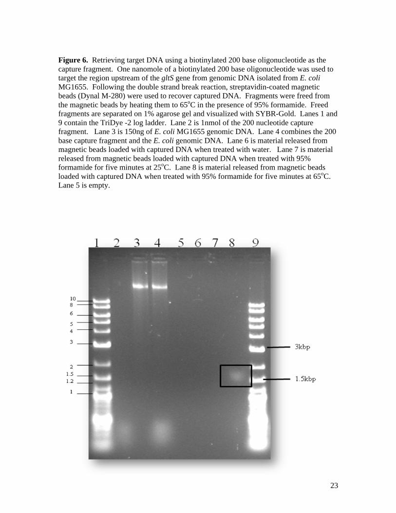
b. A biotinylated oligonucleotide can be used in an in vitro double strand break
repair reaction to retrieve a specific DNA sequence from purified DNA in
solution. As illustrated in Figure 6, a 200 base oligonucleotide initiates the
double strand break reaction and allows us to retrieve a discrete set of fragments
that are larger than 1500 base pairs (Lane 8, boxed in the figure). The protocol
for retrieval and release was successful, but only when 95% formamide and heat
treatment (65oC for five minutes) was used to separate streptavidin and biotin.
Double strand break repair was accomplished when using oligonucleotides of 150
and 100 bases in length were used as well, but the double strand break reaction
was not initiated with a 50 base oligonucleotide.
This document is a research report submitted to the U.S. Department of Justice. This report has not been published by the Department. Opinions or points of view expressed are those of the author(s) and do not necessarily reflect the official position or policies of the U.S. Department of Justice.

17
3. Tables
Table 1. The inactivation of plasmid pZR1 by gamma radiation. Increasing radiation
dose results in a reduction in the efficiency of pZR1 to transform ADP6 to POB
prototrophy, presumably by damaging DNA in a manner that prevents recombination of
the wild type pcaEFDBCHG sequence with the ADP6 genome. Efficiencies are the
mean of values obtained from three independent experiments with three replicates per
experiment.
Dose (kGy) Transformation Efficiency (%)
0 4.6 ± 0.03
5 4.2 ± 0.05
10 0.4 ± 0.01
20 0.04 ± 0.01
30 0.002 ± 0.0004
40 --a
50 -- a Transformation not detected
Table 2. Attempt to increase the efficiency of transformation to heavily irradiated pZR1.
50μl of undiluted extract from cultures of D. radiodurans R1 irradiated exposed to 3kGy
-radiation was added to 5 μg of pZR1 that had been exposed to 30kGy -radiation.
Irradiated pZR1 is denoted as pZR1*. Efficiencies are the mean of values obtained from
three independent experiments with three replicates per experiment.
Transformation Efficiency
Trial pZR1 pZR1* Extract + pZR1 Extract +
pZR1*
1 8.6± 0.03 0.0008± 0.0005 0.4 ± 0.01 0.0074± 0.003
2 11± 0.01 0.002± 0.001 0.7 ± 0.09 0.016± 0.008
3 9.2± 0.05 0.002± 0.0009 1.4 ± 0.13 0.017± 0.006
This document is a research report submitted to the U.S. Department of Justice. This report has not been published by the Department. Opinions or points of view expressed are those of the author(s) and do not necessarily reflect the official position or policies of the U.S. Department of Justice.

18
4. Figures
Figure 1. The -ketoadipate pathway of Acinetobacter baylyi. This pathway comprises
two parallel metabolic branches. One branch, mediated by six enzymes encoded by the
cat genes, converts catechol to succinate and acetyl coenzyme A (acetyl-CoA); the other
branch, catalyzed by products of the pca genes, converts protocatechuate to succinate and
acetyl-CoA by six metabolic reactions analogous or identical to those of the catechol
sequence.
This document is a research report submitted to the U.S. Department of Justice. This report has not been published by the Department. Opinions or points of view expressed are those of the author(s) and do not necessarily reflect the official position or policies of the U.S. Department of Justice.

19
Figure 2. A double-strand break repair reaction in vitro. The reaction shown is the
splicing together of two DNA segments with an overlapping region of sequence, shown
in red. One of the segments is rendered single-stranded, preferably by heat denaturation,
nuclease/helicase action, or asymmetric PCR (PCR amplification using only one PCR
primer). The 3’-ending strand is bound by RecA and SSB, and is used to invade the other
duplex DNA. DNA polymerase I extends both available 3’ ends and displaces one strand
of DNA segment 2. The result is a new segment in which the sequences of segments 1
and 2 are combined.
This document is a research report submitted to the U.S. Department of Justice. This report has not been published by the Department. Opinions or points of view expressed are those of the author(s) and do not necessarily reflect the official position or policies of the U.S. Department of Justice.

20
Figure 3. In the test reaction a single plasmid DNA is cut in two different ways to
generate two different full-length linear segments. These two segments have all the same
nucleotide sequences, but the sequences at the ends are different, and the two segments
can overlap in two different ways. This situation leads to two different double-strand
break repair reactions and two possible products.
cut with ycut with X
Product 1 Product 2
x
5’
x
5’
5’
5’
y
x
This document is a research report submitted to the U.S. Department of Justice. This report has not been published by the Department. Opinions or points of view expressed are those of the author(s) and do not necessarily reflect the official position or policies of the U.S. Department of Justice.

21
Figure 4. The plasmid used in this experiment is pUC19 (2686 bp), cleaved with either
AatII or PstI. The first lane after the marker ladder shows heat-denatured AatII cleaved
pUC19 (single-stranded DNA or ssDNA). Lane 2 shows PstI cleaved pUC19 (linear
duplex DNA or dsDNA). Lane 3 shows both of these DNA substrates together. Lane 4
shows the ssDNA after 3 min of incubation with RecA protein (some has reannealed,
some is in the well). Lane 5 shows the DNA after addition of RecA and SSB. Lane 6
shows the DNA after the addition of the dsDNA, just prior to the addition of DNA
polymerase I. The final three lanes show the generation of products at 10, 20, and 30 min
after the addition of DNA polymerase I. S: substrate DNA; P: expected products; I:
reaction intermediates.
This document is a research report submitted to the U.S. Department of Justice. This report has not been published by the Department. Opinions or points of view expressed are those of the author(s) and do not necessarily reflect the official position or policies of the U.S. Department of Justice.

22
Figure 5. Requirements for double-strand break repair in vitro. The same marker ladder
is used in lane 1, and lanes 2-4 are again simply the denatured and intact DNA segments,
as in panel B. The next three lanes (5-7) show the effects of incubating the indicated
components with denatured DNA (ssDNA) at 37 ºC for 46 min. Lanes 8-10 contain the
same components incubated in the same way with intact dsDNA. Lanes 11-16 illustrate
reactions containing both single stand and double strand DNA substrates. In lanes 11-15,
one or more components were omitted. Lane 16 shows the complete reaction. Wherever
DNA polymerase I was present, dNTPs were also added. S: substrate DNA; P: expected
products; I: reaction intermediates.
This document is a research report submitted to the U.S. Department of Justice. This report has not been published by the Department. Opinions or points of view expressed are those of the author(s) and do not necessarily reflect the official position or policies of the U.S. Department of Justice.
23
Figure 6. Retrieving target DNA using a biotinylated 200 base oligonucleotide as the
capture fragment. One nanomole of a biotinylated 200 base oligonucleotide was used to
target the region upstream of the gltS gene from genomic DNA isolated from E. coli
MG1655. Following the double strand break reaction, streptavidin-coated magnetic
beads (Dynal M-280) were used to recover captured DNA. Fragments were freed from
the magnetic beads by heating them to 65oC in the presence of 95% formamide. Freed
fragments are separated on 1% agarose gel and visualized with SYBR-Gold. Lanes 1 and
9 contain the TriDye -2 log ladder. Lane 2 is 1nmol of the 200 nucleotide capture
fragment. Lane 3 is 150ng of E. coli MG1655 genomic DNA. Lane 4 combines the 200
base capture fragment and the E. coli genomic DNA. Lane 6 is material released from
magnetic beads loaded with captured DNA when treated with water. Lane 7 is material
released from magnetic beads loaded with captured DNA when treated with 95%
formamide for five minutes at 25oC. Lane 8 is material released from magnetic beads
loaded with captured DNA when treated with 95% formamide for five minutes at 65oC.
Lane 5 is empty.
This document is a research report submitted to the U.S. Department of Justice. This report has not been published by the Department. Opinions or points of view expressed are those of the author(s) and do not necessarily reflect the official position or policies of the U.S. Department of Justice.

24
IV. Conclusions
1. Discussion of findings
a. Our efforts to develop a method for repairing damaged DNA using extracts from
DNA repair proficient bacteria were unsuccessful. We expected that the assay
system (conversion of ADP6 to POB prototrophy following transformation with
plasmid DNA) would be sensitive enough to detect small increases in repair and
that ultimately we could use these results to isolate the proteins responsible for
those repairs. While we were able to see a nine fold increase in transformation
efficiency in trials described in Table 2, we did not expect the extracts to reduce
the transformation efficiency observed when undamaged DNA was used in the
assay. Clearly this inhibitory activity limits the utility of the assay, preventing us
from assessing the true extent of repair. Our attempts to isolate the inhibitor by
fractionating the extract were not successful; several different protein fractions
exhibited inhibitory activity. The simplest explanation assumes that there is more
than one inhibitory agent. Rather than be bogged down in investigating further,
we changed strategy and focused on developing a method of in vitro double
strand break repair.
b. We have developed a procedure for the joining together of two DNA molecules
that share a significant region of homology, using a simple but robust in vitro
double-strand break repair reaction. The process requires only three proteins:
RecA protein, SSB and a DNA polymerase. All three proteins are obtained from
bacterial sources. As long as complementary DNA sequence is available, in vitro
DNA double strand break repair can be used to selectively capture any DNA
sequence. In addition to autosomal STRs, Y chromosome STRs, and
mitochondrial DNA sequences could be retrieved by this procedure. To exploit
the specificity of this reaction, the targeting DNA fragment may include at its 5’
end a feature that allows efficient retrieval of that fragment and any DNA with
which it has recombined. Non-limiting examples of this feature include a ligand
such as biotin that can be harvested with streptavidin-coated magnetic beads, or a
DNA sequence that binds tightly to a specific protein affinity column. Retrieving
the targeting fragment and the target in this manner allows concentration of the
target sequence and further manipulation of the target (e.g. PCR amplification)
without the potential interference of biological and chemical contaminants found
in the mixture from which the target was obtained.
c. This procedure has three properties that make it potentially useful for forensic
applications: i) it does not require knowledge of DNA sequence on both ends of a
DNA segment to be captured, as in PCR, ii) it does not introduce any enzymes
that could damage or alter a target DNA sample, and iii) it should allow the
retrieval of desired DNA segments even from samples that are highly
contaminated with DNA from other species.
This document is a research report submitted to the U.S. Department of Justice. This report has not been published by the Department. Opinions or points of view expressed are those of the author(s) and do not necessarily reflect the official position or policies of the U.S. Department of Justice.