TOE Standard Views Expanded 21 Jan 2014 Small
12
1 IMAGE ACQUISITION VIEW & STRUCTURES IMAGED SIMULATOR TOE IMAGE MIDOESOPHAGEAL MO 4C MO (30 40cm) 020° +/ slight retroflexion LV: basal, mid and apical anteroseptal and inferolateral walls LA RV RA MV: A3, P1 TV: (Ant, Septal) & IAS MO LV 5C MO 020° Withdraw probe slightly LV: anteroseptal , inferolateral LVOT MO LV 2C MO 80100° LV: entire anterior and inferior walls
-
Upload
chris-nickson -
Category
Documents
-
view
3 -
download
0
description
TOE Standard Views proforma from The Alfred ICU.
Transcript of TOE Standard Views Expanded 21 Jan 2014 Small
1
IMAGE ACQUISITION
VIEW & STRUCTURES IMAGED SIMULATOR TOE IMAGE
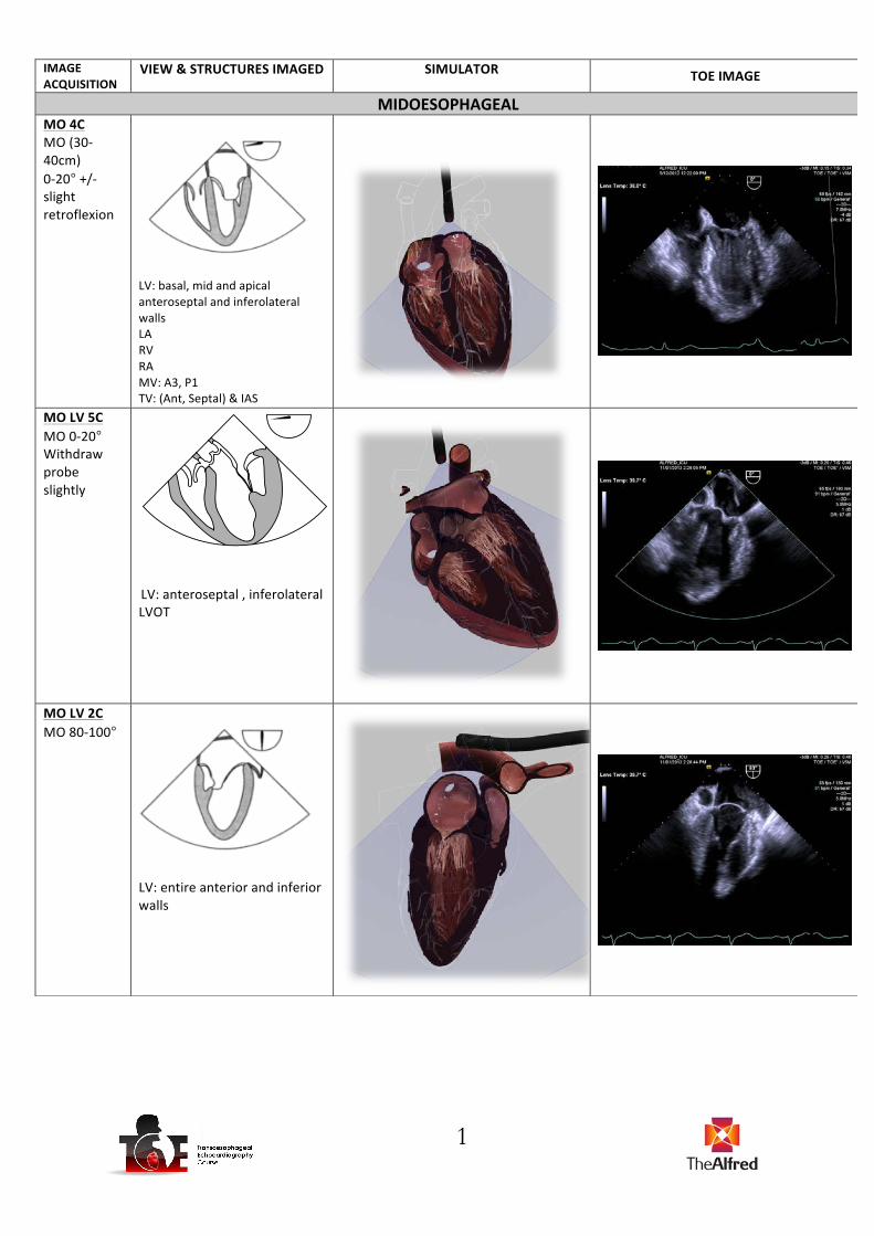
MIDOESOPHAGEAL MO 4C MO (30-‐40cm) 0-‐20° +/-‐ slight retroflexion
LV: basal, mid and apical anteroseptal and inferolateral walls LA RV RA MV: A3, P1 TV: (Ant, Septal) & IAS
MO LV 5C MO 0-‐20° Withdraw probe slightly
LV: anteroseptal , inferolateral LVOT
MO LV 2C MO 80-‐100°
LV: entire anterior and inferior walls
2
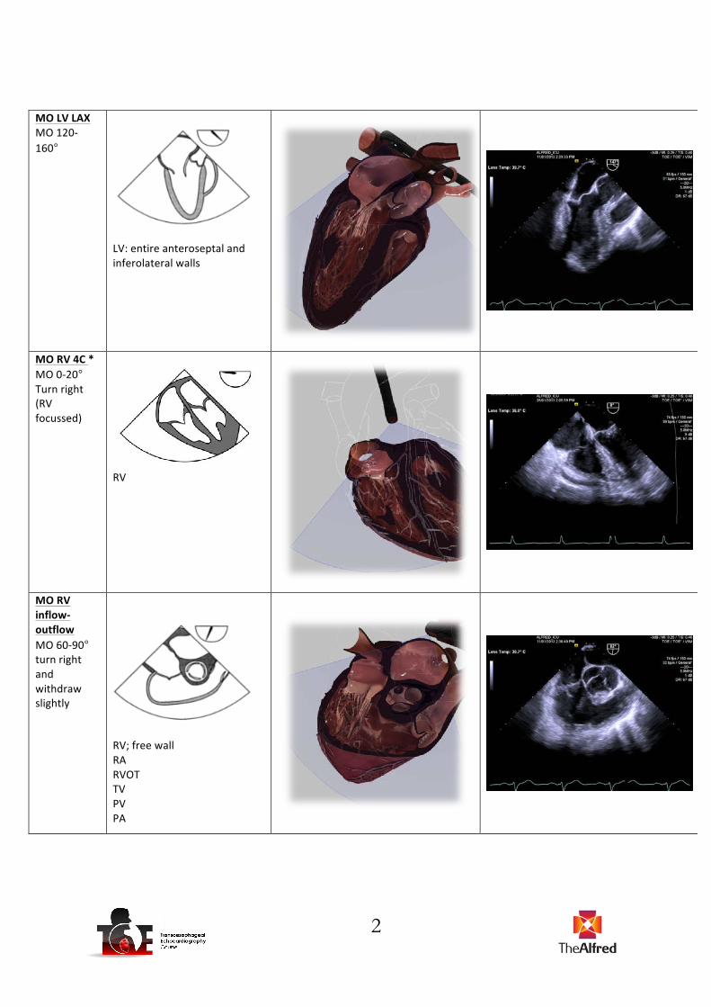
MO LV LAX MO 120-‐160°
LV: entire anteroseptal and inferolateral walls
MO RV 4C * MO 0-‐20° Turn right (RV focussed)
RV
MO RV inflow-‐outflow MO 60-‐90° turn right and withdraw slightly
RV; free wall RA RVOT TV PV PA
3
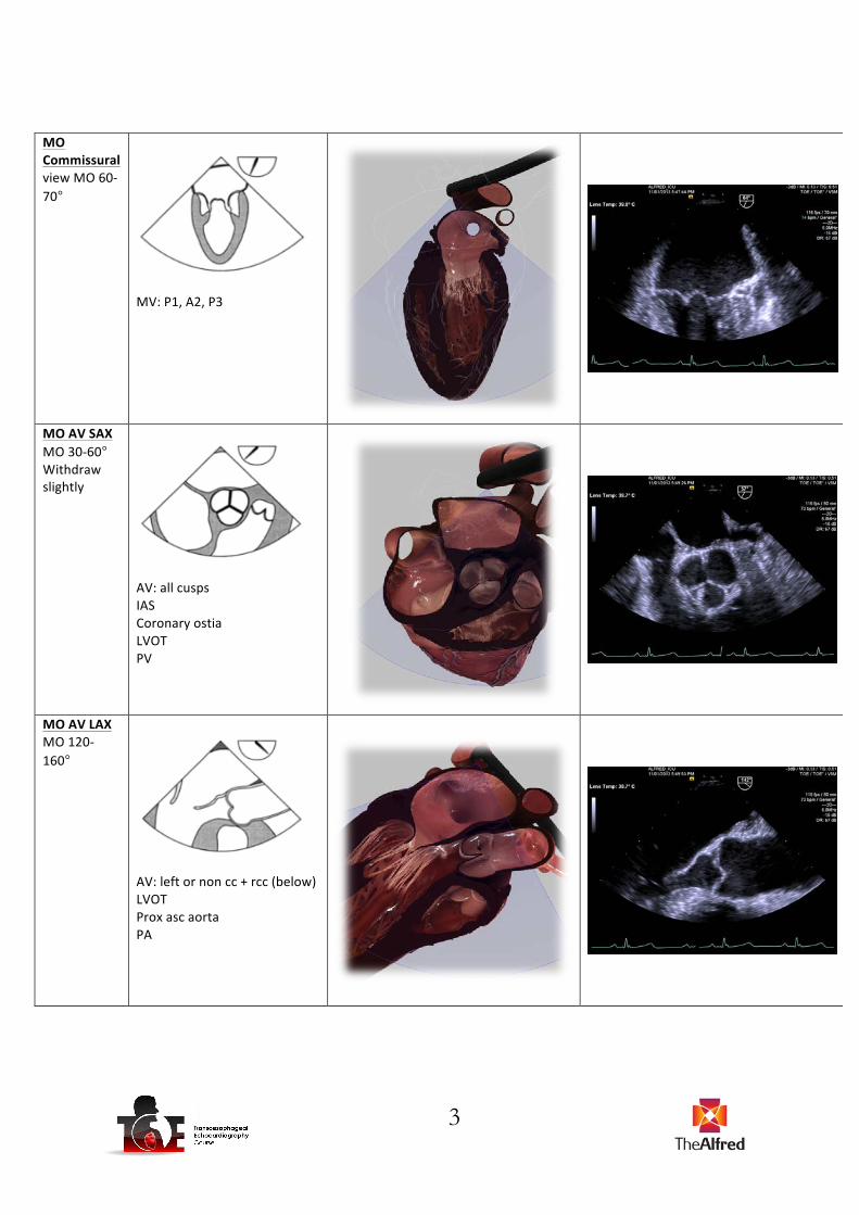
MO Commissural view MO 60-‐70°
MV: P1, A2, P3
MO AV SAX MO 30-‐60° Withdraw slightly
AV: all cusps IAS Coronary ostia LVOT PV
MO AV LAX MO 120-‐160°
AV: left or non cc + rcc (below) LVOT Prox asc aorta PA

4
MO bicaval MO 80-‐110° + turn right slightly
SVC LA RA IAS IVC
MO modified bicaval MO 110-‐130º after centred on IAS. May also be obtained at 50-‐70o as noted in Hahn
SVC LA RA IAS TV
MO LAA * MO 0-‐90°, centre and rotate through lobes
LAA Ligament of Marshall
5
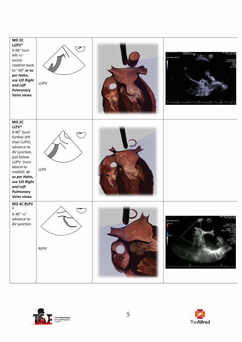
MO 2C LUPV* 0-‐90° turn left +/-‐ sector rotation back to ~60° or as per Hahn, use UO Right and Left Pulmonary Veins views
LUPV
MO 2C LLPV* 0-‐90° (turn further left than LUPV) advance to AV junction, just below LUPV (runs lateral to medial) or as per Hahn, use UO Right and Left Pulmonary Veins views
LLPV
MO 4C RLPV * 0-‐30° +/-‐ advance to AV junction
RLPV

6
MO 4C RUPV 0-‐30° turn right +/-‐ retroflexion +/-‐ withdraw
RUPV Mid asc aorta SVC
MO Bicaval RUPV * MO 80-‐110° + turn left (consider sector back to 90° for Doppler line-‐up)
RUPV
MO asc aorta SAX MO 0-‐60°
Asc aorta SVC PA R) PA

7
MO asc aorta LAX MO 100-‐150°
Asc Aorta R) PA
MO coronary sinus *
Coronary Sinus
Desc aortic SAX 0-‐10° Diaphragm to Left subclavian. Consider increasing frequency and adjust TGC
Desc aorta Left pleura
8
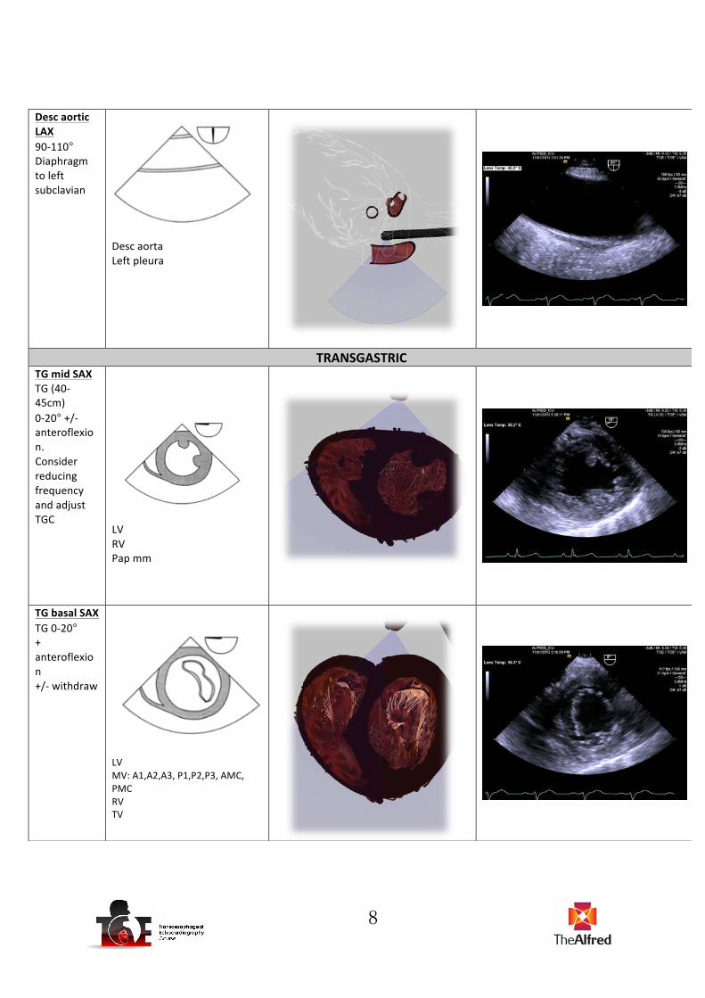
Desc aortic LAX 90-‐110° Diaphragm to left subclavian
Desc aorta Left pleura
TRANSGASTRIC TG mid SAX TG (40-‐45cm) 0-‐20° +/-‐ anteroflexion. Consider reducing frequency and adjust TGC
LV RV Pap mm
TG basal SAX TG 0-‐20° + anteroflexion +/-‐ withdraw
LV MV: A1,A2,A3, P1,P2,P3, AMC, PMC RV TV
9
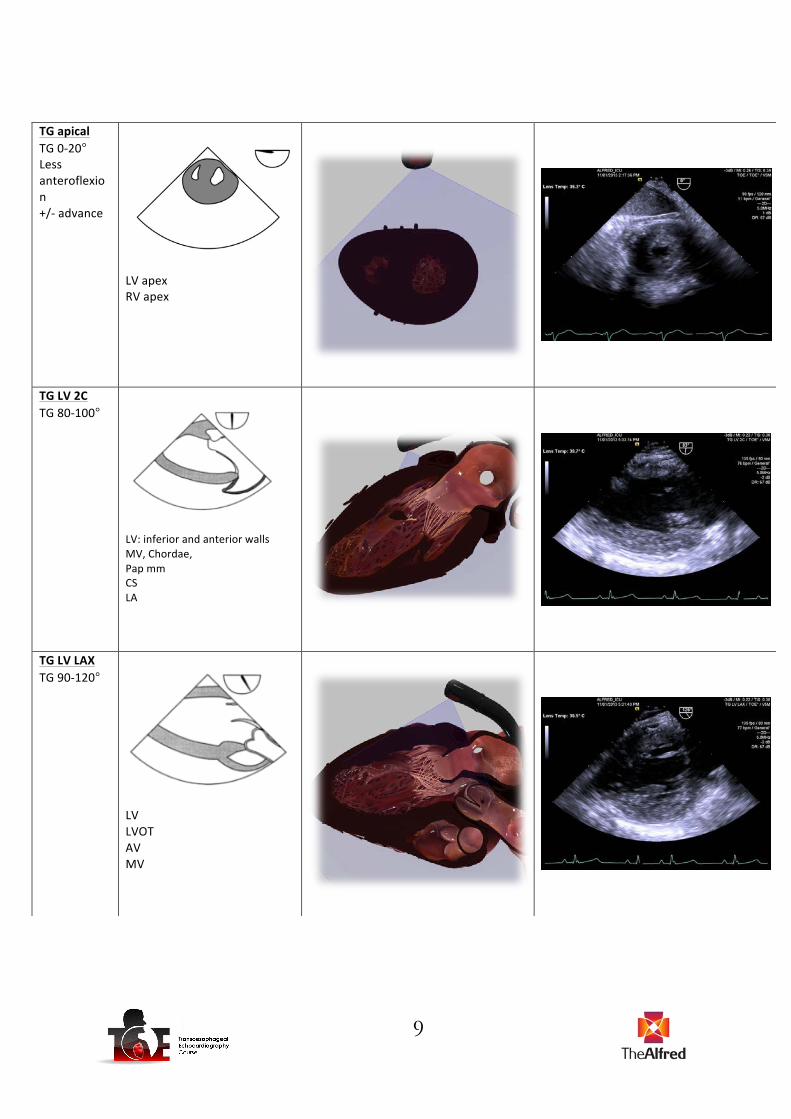
TG apical TG 0-‐20° Less anteroflexion +/-‐ advance
LV apex RV apex
TG LV 2C TG 80-‐100°
LV: inferior and anterior walls MV, Chordae, Pap mm CS LA
TG LV LAX TG 90-‐120°
LV LVOT AV MV

10
TG RV SAX* TG 0-‐10° Turn right
RV: free and septal walls
TG RV inflow TG 100-‐120°
RV TV RA TV chordae Pap mm
DEEP TRANSGASTRIC Deep TG LAX Deep TG 45-‐50cm 0-‐20° Advance past apex + anteflexion
LVOT AV Asc aorta Aortic arch

11
TG/Deep TG RV basal Deep TG 0-‐20º+anteflexion, withdraw + turn right
RV RVOT PV PA
UPPER OESOPHAGEAL Upper oesophageal (UO) aortic arch SAX UO (20-‐25cm) 90°
Aortic arch Pulm artery PV
UO aortic arch LAX UO 0-‐10°
Aortic Arch
12
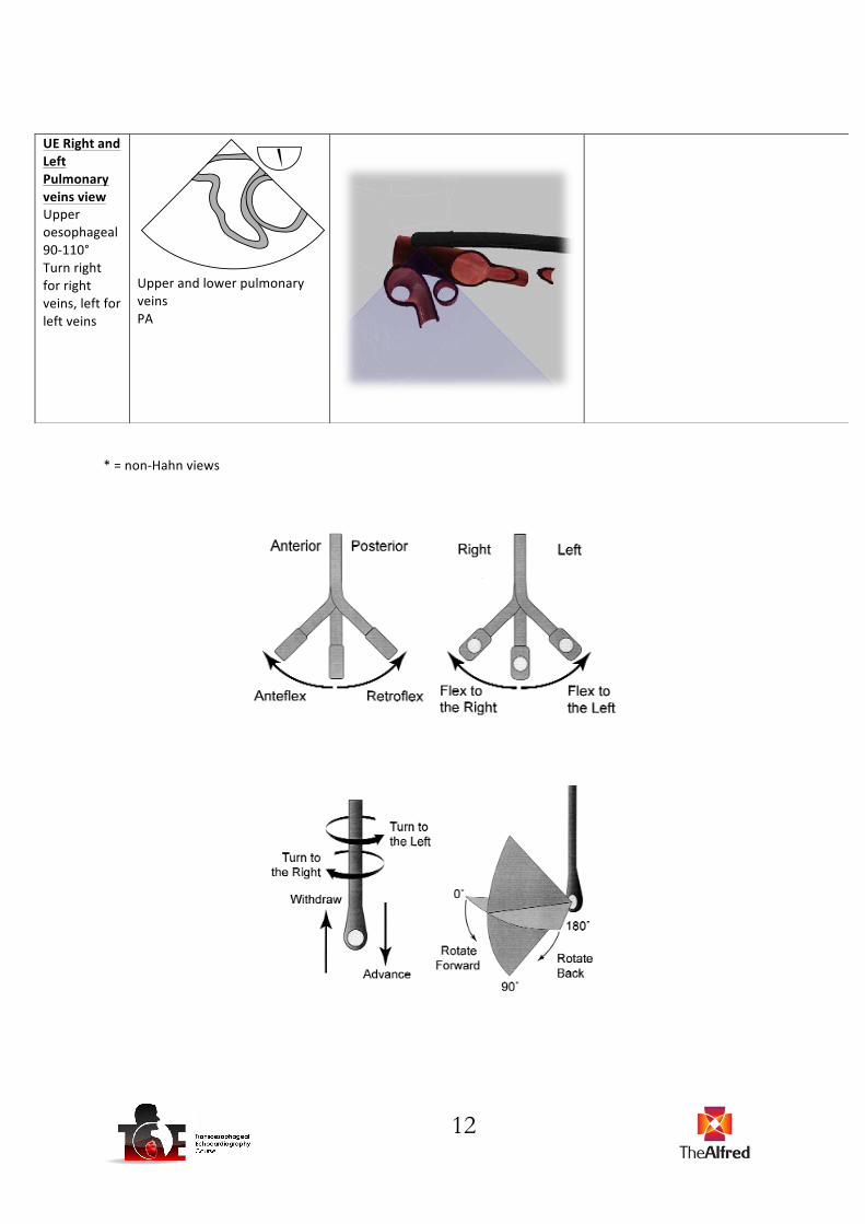
* = non-‐Hahn views
UE Right and Left Pulmonary veins view Upper oesophageal 90-‐110° Turn right for right veins, left for left veins
Upper and lower pulmonary veins PA