ToadHeartUtilizesExclusivelySlowSkeletalMuscle … found in toad heart, whereas cardiac muscles of...
13
Toad Heart Utilizes Exclusively Slow Skeletal Muscle Troponin T AN EVOLUTIONARY ADAPTATION WITH POTENTIAL FUNCTIONAL BENEFITS * □ S Received for publication, April 17, 2012, and in revised form, July 2, 1012 Published, JBC Papers in Press, July 9, 2012, DOI 10.1074/jbc.M112.373191 Han-Zhong Feng, Xuequn Chen, M. Moazzem Hossain, and Jian-Ping Jin 1 From the Department of Physiology, Wayne State University School of Medicine, Detroit, Michigan 48201 Background: The heart of dry land toads has adapted to sustain circulation in a wide range of body fluid changes. Results: The toad cardiac muscle expresses exclusively slow skeletal troponin T with cardiac forms of other myofilament proteins and exhibits functional benefit. Conclusion: This finding reflects a novel adaptation of toad heart. Significance: The results indicate a molecular mechanism to improve systolic function. The three isoforms of vertebrate troponin T (TnT) are nor- mally expressed in a muscle type-specific manner. Here we report an exception that the cardiac muscle of toad (Bufo) expresses exclusively slow skeletal muscle TnT (ssTnT) together with cardiac forms of troponin I and myosin as determined using immunoblotting, cDNA cloning, and/or LC-MS/MS. Using RT-PCR and 3- and 5-rapid amplification of cDNA ends on toad cardiac mRNA, we cloned full-length cDNAs encoding two alternatively spliced variants of ssTnT. Expression of the cloned cDNAs in Escherichia coli confirmed that the toad car- diac muscle expresses solely ssTnT, predominantly the low molecular weight variant with the exon 5-encoded NH 2 -termi- nal segment spliced out. Functional studies were performed in ex vivo working toad hearts and compared with the frog (Rana) hearts. The results showed that toad hearts had higher contract- ile and relaxation velocities and were able to work against a sig- nificantly higher afterload than that of frog hearts. Therefore, the unique evolutionary adaptation of utilizing exclusively ssTnT in toad cardiac muscle corresponded to a fitness value from improving systolic function of the heart. The data demon- strated a physiological importance of the functional diversity of TnT isoforms. The structure-function relationship of TnT may be explored for the development of new treatment of heart failure. Toad and frog are two closely related amphibian species. However, toads are more adapted to living on land than the wet habitats in which frogs reside. In land habitats, the volumes of toad body fluid and blood change with large ranges between rain and dry seasons (1) or with different heat exposures (2). The heart of toad apparently adapts well to working with a big range of blood volume changes (2). The function of the vertebrate circulatory system is critically dependent on and closely regulated by the volume of circulating blood (3). Decreases of blood volume such as that in humans during hemorrhagic shock would result in decreased cardiac output due to both decreased ventricular filling and increased peripheral arterial resistance from the compensatory increase in sympathetic tone (4). Therefore, to understand the mecha- nism(s) by which the toad heart is able to cope with the drastic decrease in blood volume such as that occurs during dry season (1) or under experimental conditions (2) and sustain cardiac output is of physiological and medical importance. A foundation of heart function is the myofilament proteins that form the contractile machinery in cardiac muscle (5). Comparative studies of the myofilament contractile and regu- latory proteins in the hearts of toad and frog are valuable in revealing the molecular basis of the functional feature of toad cardiac muscle. In the present study, we examined the isoform contents of representative myofilament proteins in the toad cardiac mus- cle. Solely slow skeletal muscle isoform of troponin T (ssTnT) 2 was found in toad heart, whereas cardiac muscles of frog and other vertebrate species examined all expressed exclusively car- diac TnT. The unique presence of slow TnT in toad heart was confirmed by cDNA cloning. Normal cardiac troponin I (TnI) and cardiac myosin heavy chain (MHC) were present in toad heart. Functional studies demonstrated that toad hearts had higher contractility and were able to work against higher after- load than that of frog hearts, suggesting a physiological value of the unique evolutionary adaptation of utilizing ssTnT in toad cardiac muscle. MATERIALS AND METHODS Animals—American toads (Bufo americanus) and bullfrogs (Rana catesbeiana) with comparable body weights were obtained from commercial sources in non-hibernating season. Toads were kept in moist moss and frogs in a water tank at room temperature for a few days before use. Tadpoles of both species were purchased from a commercial source and eutha- * This work was supported, in whole or in part, by National Institutes of Health Grants AR048816 and HL098945 (to J. P. J.). □ S This article contains supplemental Fig. S1 and Table S1. The nucleotide sequence(s) reported in this paper has been submitted to the Gen- Bank TM /EBI Data Bank with accession number(s) AY773671, AY773672, and AY773673. 1 To whom correspondence may be addressed: 540 East Canfield St., Scott Hall 5374, Detroit, MI 48201. Tel.: 313-577-1520; Fax: 313-577-5494; E-mail: [email protected]. 2 The abbreviations used are: ssTnT, slow skeletal muscle troponin T; bpm, beat per minute; MHC, myosin heavy chain; TnI, troponin I; TnT, troponin T. THE JOURNAL OF BIOLOGICAL CHEMISTRY VOL. 287, NO. 35, pp. 29753–29764, August 24, 2012 © 2012 by The American Society for Biochemistry and Molecular Biology, Inc. Published in the U.S.A. AUGUST 24, 2012 • VOLUME 287 • NUMBER 35 JOURNAL OF BIOLOGICAL CHEMISTRY 29753 by guest on July 16, 2018 http://www.jbc.org/ Downloaded from
-
Upload
trankhuong -
Category
Documents
-
view
214 -
download
0
Transcript of ToadHeartUtilizesExclusivelySlowSkeletalMuscle … found in toad heart, whereas cardiac muscles of...
Toad Heart Utilizes Exclusively Slow Skeletal MuscleTroponin TAN EVOLUTIONARY ADAPTATION WITH POTENTIAL FUNCTIONAL BENEFITS*□S
Received for publication, April 17, 2012, and in revised form, July 2, 1012 Published, JBC Papers in Press, July 9, 2012, DOI 10.1074/jbc.M112.373191
Han-Zhong Feng, Xuequn Chen, M. Moazzem Hossain, and Jian-Ping Jin1
From the Department of Physiology, Wayne State University School of Medicine, Detroit, Michigan 48201
Background: The heart of dry land toads has adapted to sustain circulation in a wide range of body fluid changes.Results: The toad cardiac muscle expresses exclusively slow skeletal troponin T with cardiac forms of other myofilamentproteins and exhibits functional benefit.Conclusion: This finding reflects a novel adaptation of toad heart.Significance: The results indicate a molecular mechanism to improve systolic function.
The three isoforms of vertebrate troponin T (TnT) are nor-mally expressed in a muscle type-specific manner. Here wereport an exception that the cardiac muscle of toad (Bufo)expresses exclusively slow skeletalmuscleTnT (ssTnT) togetherwith cardiac forms of troponin I and myosin as determinedusing immunoblotting, cDNA cloning, and/or LC-MS/MS.Using RT-PCR and 3�- and 5�-rapid amplification of cDNA endson toad cardiac mRNA, we cloned full-length cDNAs encodingtwo alternatively spliced variants of ssTnT. Expression of thecloned cDNAs in Escherichia coli confirmed that the toad car-diac muscle expresses solely ssTnT, predominantly the lowmolecular weight variant with the exon 5-encoded NH2-termi-nal segment spliced out. Functional studies were performed inex vivo working toad hearts and compared with the frog (Rana)hearts. The results showed that toad hearts had higher contract-ile and relaxation velocities and were able to work against a sig-nificantly higher afterload than that of frog hearts. Therefore,the unique evolutionary adaptation of utilizing exclusivelyssTnT in toad cardiac muscle corresponded to a fitness valuefrom improving systolic function of the heart. The data demon-strated a physiological importance of the functional diversity ofTnT isoforms. The structure-function relationship of TnT maybe explored for the development of new treatment of heartfailure.
Toad and frog are two closely related amphibian species.However, toads aremore adapted to living on land than the wethabitats in which frogs reside. In land habitats, the volumes oftoad body fluid and blood change with large ranges betweenrain and dry seasons (1) or with different heat exposures (2).The heart of toad apparently adapts well to working with a bigrange of blood volume changes (2).
The function of the vertebrate circulatory system is criticallydependent on and closely regulated by the volumeof circulatingblood (3). Decreases of blood volume such as that in humansduring hemorrhagic shock would result in decreased cardiacoutput due to both decreased ventricular filling and increasedperipheral arterial resistance from the compensatory increasein sympathetic tone (4). Therefore, to understand the mecha-nism(s) by which the toad heart is able to cope with the drasticdecrease in blood volume such as that occurs during dry season(1) or under experimental conditions (2) and sustain cardiacoutput is of physiological and medical importance.A foundation of heart function is the myofilament proteins
that form the contractile machinery in cardiac muscle (5).Comparative studies of the myofilament contractile and regu-latory proteins in the hearts of toad and frog are valuable inrevealing the molecular basis of the functional feature of toadcardiac muscle.In the present study, we examined the isoform contents of
representative myofilament proteins in the toad cardiac mus-cle. Solely slow skeletal muscle isoform of troponin T (ssTnT)2was found in toad heart, whereas cardiac muscles of frog andother vertebrate species examined all expressed exclusively car-diac TnT. The unique presence of slow TnT in toad heart wasconfirmed by cDNA cloning. Normal cardiac troponin I (TnI)and cardiac myosin heavy chain (MHC) were present in toadheart. Functional studies demonstrated that toad hearts hadhigher contractility and were able to work against higher after-load than that of frog hearts, suggesting a physiological value ofthe unique evolutionary adaptation of utilizing ssTnT in toadcardiac muscle.
MATERIALS AND METHODS
Animals—American toads (Bufo americanus) and bullfrogs(Rana catesbeiana) with comparable body weights wereobtained from commercial sources in non-hibernating season.Toads were kept in moist moss and frogs in a water tank atroom temperature for a few days before use. Tadpoles of bothspecies were purchased from a commercial source and eutha-
* This work was supported, in whole or in part, by National Institutes of HealthGrants AR048816 and HL098945 (to J. P. J.).
□S This article contains supplemental Fig. S1 and Table S1.The nucleotide sequence(s) reported in this paper has been submitted to the Gen-
BankTM/EBI Data Bank with accession number(s) AY773671, AY773672, andAY773673.
1 To whom correspondence may be addressed: 540 East Canfield St., ScottHall 5374, Detroit, MI 48201. Tel.: 313-577-1520; Fax: 313-577-5494; E-mail:[email protected].
2 The abbreviations used are: ssTnT, slow skeletal muscle troponin T; bpm,beat per minute; MHC, myosin heavy chain; TnI, troponin I; TnT, troponin T.
THE JOURNAL OF BIOLOGICAL CHEMISTRY VOL. 287, NO. 35, pp. 29753–29764, August 24, 2012© 2012 by The American Society for Biochemistry and Molecular Biology, Inc. Published in the U.S.A.
AUGUST 24, 2012 • VOLUME 287 • NUMBER 35 JOURNAL OF BIOLOGICAL CHEMISTRY 29753
by guest on July 16, 2018http://w
ww
.jbc.org/D
ownloaded from
nized upon arrival to collect tissue samples. The experimentalprotocols were approved by the Institutional Animal Care andUse Committee.Muscle Samples—Immediately after euthanasia, atrial and
ventricular muscles as well as various skeletal muscles of toadand frogwere dissected according to illustrations of frogmuscleanatomy.Wholemuscle tissuewas homogenized in 20 volumes(w/v) of SDS gel sample buffer containing 2%SDS, 10%glycerol,50 mM Tris base, 2% 2-mercaptoethanol, pH 8.8, using a highspeed mechanical homogenizer (PRO Scientific Inc.). Thehomogenized muscle samples were immediately heated at80 °C for 5 min, clarified by high speed centrifugation, andstored at �80 °C for SDS-PAGE and Western blot analysis.Development of Cardiac TnT-specificMonoclonal Antibodies
(mAbs)—Using human cardiac TnT expressed from clonedcDNA in Escherichia coli as immunigen, hybridoma cell lineswere generated to produce specific mAbs. The expression andpurification of human cardiac TnT, immunization of Balb/cmice, hybridoma fusion, screening, and subcloning, mAb pro-duction, and immunoglobulin isotyping were carried out asdescribed previously using standardmethods (6). Isoform spec-ificity of the anti-TnT mAbs obtained was examined withWestern blots on cardiac and skeletal muscle samples frommultiple vertebrate species.SDS-PAGE and Western Blotting—Protein samples were
resolved on 14% Laemmli gel with an acrylamide:bisacrylamideratio of 180:1. The protein bands were visualized by stainingthe gel with Coomassie Brilliant Blue R-250. Duplicate gelswere electrically transferred to blot nitrocellulose membranes.Tris-buffered saline (TBS) containing 1% bovine serum albu-min (BSA) was used to block the nitrocellulose membranes atroom temperature for 30min. Themembranes were then incu-bated with anti-TnT mAb 2C8 recognizing all three muscletype-specific isoforms across vertebrate species (7), mAb CT3recognizing cardiac TnT and slow TnT (8), mAb 4B8 recogniz-ing only cardiac TnT (characterized in the present study), anti-TnImAbTnI-1 (6), and anti-cardiacMHCmAbFA2 (9) dilutedin TBS containing 0.1% BSA at 4 °C overnight. After high strin-gency washes with TBS plus 0.5% Triton X-100 and 0.05% SDS,the membranes were further incubated with alkaline phospha-tase-conjugated goat anti-mouse IgG second antibody (SantaCruz Biotechnology, Santa Cruz, CA), washed again as above,and developed in 5-bromo-4-chloro-3-indolylphosphate/nitroblue tetrazolium substrate solution. In some experiments, themembrane, after the blot image was documented by scanning,was re-blotted with another mAb at doubled concentration byrepeating the above procedure.Myosin heavy chain isoforms were examined using glycerol/
SDS-PAGE as described previously (10). The muscle proteinsamples were resolved on 8% SDS-polyacrylamide gel contain-ing 30% glycerol run in an icebox at constant voltage of 100 Vfor 24 h.Cloning of cDNAs Encoding Toad ssTnT—Total RNA was
extracted from toad hearts using the TRIzol reagent asinstructed in the product manual (Invitrogen). Integrity of theRNA isolated was verified using agarose gel electrophoresis.Poly(A)� mRNA was reverse transcribed using an anchoredoligo(dT) primer (TV20) at 42 °C for 2 h. Using degenerated
primers derived from amino acid sequences in conservedregions of vertebrate ssTnT, partial cDNA encoding toadssTnT was cloned with polymerase chain reaction (PCR). Full-length toad ssTnT cDNAs were then obtained using 3�- and5�-rapid amplification of cDNA ends and sequenced.Cloning of cDNA Encoding Toad Cardiac TnI—From total
cardiac cDNA synthesized as above, cDNA encoding toad car-diac TnI was cloned with reverse transcription-coupled PCRusing degenerated primers derived from the sequence of Xeno-pus cardiac TnI (GenBankTM accession number L25721.1) andsequenced.Expression of Cloned Toad ssTnT cDNA in E. coli—The cod-
ing region of toad ssTnT cDNA was inserted into expressionplasmid vector pET24. The plasmid DNA was used to trans-form BL21(ED3)pLysS E. coli for the expression of toad ssTnT.The expressed protein was examined using SDS-PAGE andWestern blot as described above.Preparation of ex Vivo Working Hearts and Functional
Measurements—Toad and frog hearts were isolated and cannu-lated on a perfusion apparatus modified from a working heartsystem previously used for mouse and rat heart studies (11).Experiments were performed at 16 °C controlled with circu-
lating water from a refrigerated bath. The left side aorta of toador frog heart was cannulated and retrograde perfused through ablunted 16-gauge needle. The right side aorta was sutured witha 3-0 silk thread. Preload was established by connecting theanterior vena cava to the preload reservoir through a 16-gaugecannula. All other vena cavae connected to sinus venosus weresutured to prevent leaking. The perfusion solution contains (inmM) 115 NaCl, 2.5 KCl, 1.0 CaCl2, 2.15 Na2HPO4, 0.85NaH2PO4, and 5.6 D-glucose. pH was adjusted to 7.20 by theaddition of Na2HPO4 and equilibrated with air (12).
The heart rate was controlled at 32 beats per minutes (bpm)through pacing with a pair of electrodes attached to the rightatrium. Pulse stimulation of 4 ms duration with a constant cur-rent 30% above the threshold was given using an isolated stim-ulator. A pressure transducer (MLT844, Capto, Horten, Nor-way) was connected to the aortic cannula through a three-wayconnector at the ventricle level tomeasure changes in the aorticpressure. A 1.2 French pressure-volume catheter was insertedinto the ventricular chamber through the apex tomeasure ven-tricular pressure and volume changes. Data were collected con-tinuously using Chart 5 software through an A/D interface(Power Lab).In functional studies, afterload pressure of 25 cm H2O and
preload pressure of 4 cm H2O were set as the baseline condi-tions with reference to the levels of aortic trunk and left atrium,respectively. Preload and afterload were experimentally alteredby moving the reservoirs up or down to test preload and after-load responses.LC-MS/MS Analysis of MHCs—MHC bands separated in
glycerol/SDS-PAGE were stained with GelCode Blue reagent(Thermo Scientific) and manually excised from the gel. For ingel protein digestion, the gel slices were washed with 50 mM
ammonium bicarbonate, reduced in 10mMDTT at 37 °C for 45min, and then alkylated with 55 mM iodoacetamide at roomtemperature for 30 min. Subsequently, four proteases, Lys-C,trypsin, Glu-C, and chymotrypsin, were added to four replicate
Slow Skeletal Muscle Troponin T in Toad Heart
29754 JOURNAL OF BIOLOGICAL CHEMISTRY VOLUME 287 • NUMBER 35 • AUGUST 24, 2012
by guest on July 16, 2018http://w
ww
.jbc.org/D
ownloaded from
reaction tubes each at 10 ng/�l and the digestionwas incubatedat 37 °C overnight except for chymotrypsin that was incubatedat room temperature.The resulting peptides were analyzed by nanoLC-MS/MS.
The peptides were first separated on a reversed-phase C18 col-umn with a 90-min gradient using a Dionex Ultimate HPLCsystem. MS and MS/MS spectra were then acquired on anApplied Biosystems QSTAR XL mass analyzer using informa-tion dependent acquisitionmode.MS scanwas performed fromm/z 400–1,500 for 1 s followed by product ion scans on the twomost intense multiply charged ions. Peak lists were submittedto Mascot server to search against the NCBInr data base for allentries with carbamidomethyl (C) used as a fixed modificationand oxidation (M), N-acetylation (protein N terminus) as vari-able modifications. To confirm the peptide mapping results, analiquot of the tryptic peptides was also analyzed on a LTQ XLmass spectrometer.MHC Peptide Mapping and BLAST Analysis—Because the
sequence of toad cardiac MHC was unavailable in any publicprotein sequence databases, our strategy was to obtain a partialsequence of this large protein by mapping the MS/MS data toMHC orthologs from related species. Therefore, we selected allentries in the NCBInr database for the Mascot searches. Tomaximize the sequence coverage, toad MHC bands weredigested with the four different proteases described above.To determine that theMHC in toad heart was cardiac like or
skeletal muscle like, we used the compiled partial proteinsequence of toad MHC to perform a BLAST analysis in theNCBInr database.DataAnalysis—DNAand protein sequence analysis was per-
formed using the DNAStar software (Lasergene). Two-dimen-
sional densitometry was done to quantify SDS gel andWesternblots on images scanned at 600 dpi. Statistical significance forquantitative datawere determined using Student’s t test or two-way analysis of variance test.
RESULTS
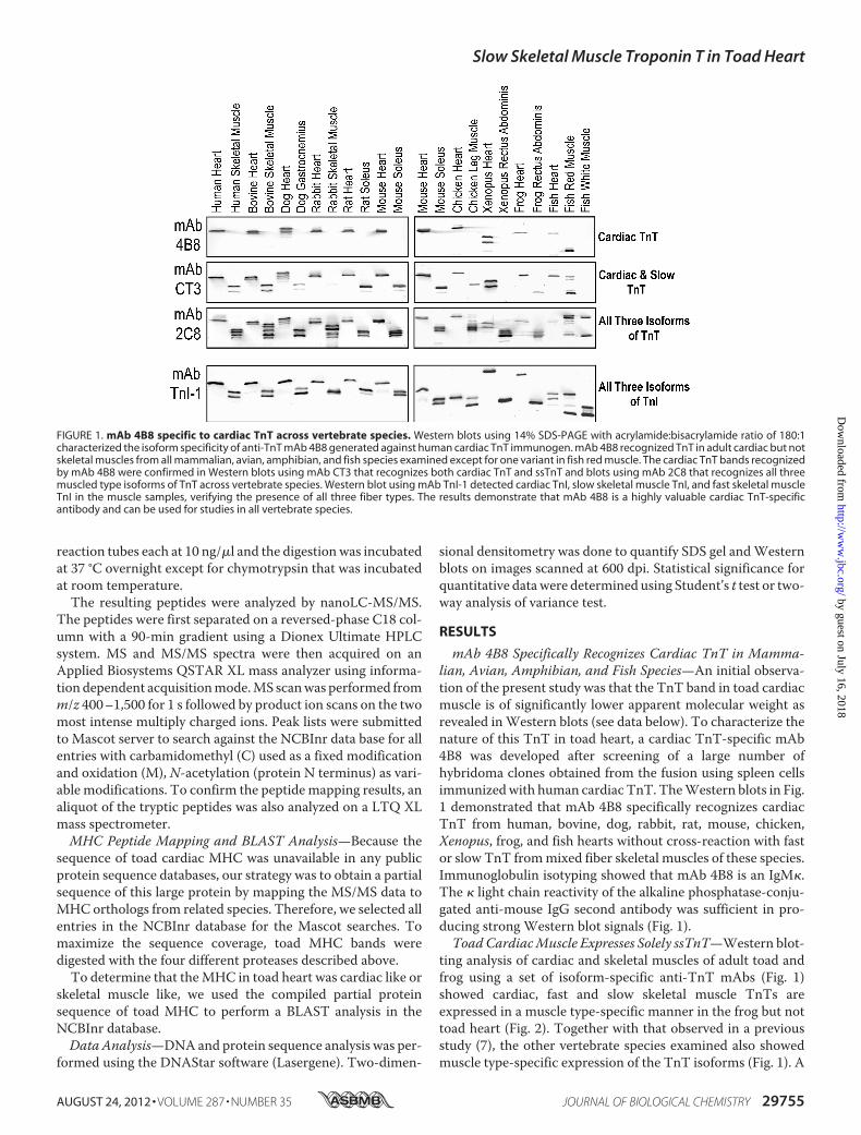
mAb 4B8 Specifically Recognizes Cardiac TnT in Mamma-lian, Avian, Amphibian, and Fish Species—An initial observa-tion of the present study was that the TnT band in toad cardiacmuscle is of significantly lower apparent molecular weight asrevealed inWestern blots (see data below). To characterize thenature of this TnT in toad heart, a cardiac TnT-specific mAb4B8 was developed after screening of a large number ofhybridoma clones obtained from the fusion using spleen cellsimmunizedwith human cardiac TnT. TheWestern blots in Fig.1 demonstrated that mAb 4B8 specifically recognizes cardiacTnT from human, bovine, dog, rabbit, rat, mouse, chicken,Xenopus, frog, and fish hearts without cross-reaction with fastor slow TnT frommixed fiber skeletal muscles of these species.Immunoglobulin isotyping showed that mAb 4B8 is an IgM�.The � light chain reactivity of the alkaline phosphatase-conju-gated anti-mouse IgG second antibody was sufficient in pro-ducing strong Western blot signals (Fig. 1).ToadCardiacMuscle Expresses Solely ssTnT—Western blot-
ting analysis of cardiac and skeletal muscles of adult toad andfrog using a set of isoform-specific anti-TnT mAbs (Fig. 1)showed cardiac, fast and slow skeletal muscle TnTs areexpressed in a muscle type-specific manner in the frog but nottoad heart (Fig. 2). Together with that observed in a previousstudy (7), the other vertebrate species examined also showedmuscle type-specific expression of the TnT isoforms (Fig. 1). A
FIGURE 1. mAb 4B8 specific to cardiac TnT across vertebrate species. Western blots using 14% SDS-PAGE with acrylamide:bisacrylamide ratio of 180:1characterized the isoform specificity of anti-TnT mAb 4B8 generated against human cardiac TnT immunogen. mAb 4B8 recognized TnT in adult cardiac but notskeletal muscles from all mammalian, avian, amphibian, and fish species examined except for one variant in fish red muscle. The cardiac TnT bands recognizedby mAb 4B8 were confirmed in Western blots using mAb CT3 that recognizes both cardiac TnT and ssTnT and blots using mAb 2C8 that recognizes all threemuscled type isoforms of TnT across vertebrate species. Western blot using mAb TnI-1 detected cardiac TnI, slow skeletal muscle TnI, and fast skeletal muscleTnI in the muscle samples, verifying the presence of all three fiber types. The results demonstrate that mAb 4B8 is a highly valuable cardiac TnT-specificantibody and can be used for studies in all vertebrate species.
Slow Skeletal Muscle Troponin T in Toad Heart
AUGUST 24, 2012 • VOLUME 287 • NUMBER 35 JOURNAL OF BIOLOGICAL CHEMISTRY 29755
by guest on July 16, 2018http://w
ww
.jbc.org/D
ownloaded from

single TnT bandwas detected in the toad heart by the pan-TnTmAb 2C8 with a size in the range of skeletal muscle TnTs (Fig.2). Western blot using anti-cardiac and ssTnT mAb CT3 iden-tified this toad heart TnT with identical size to ssTnT in toadrectus abdominis muscle (Fig. 2). No band was detected in the
Western blot of toad cardiac muscle by the cardiac TnT-spe-cific mAb 4B8 that clearly recognizes cardiac TnT in the froghearts (Fig. 2). Re-blotting of the 4B8-probed membrane usingmAb CT3 confirmed the presence of ssTnT in the toad car-diac muscle blot (Fig. 2). Altogether, theWestern blots usingTnT isoform-specific mAbs indicated that the toad cardiacmuscle expresses a single TnT that is ssTnT and no cardiacTnT was detectable. SDS-PAGE gels and Western blots fur-ther showed that toad tadpole heart also expresses exclu-sively ssTnT (Fig. 3).Confirmation of ssTnT in Toad Heart by cDNA Cloning—
From toad heart poly(A)� RNA, RT-PCR using two degener-ated ssTnT primers followed by 3�- and 5�-rapid amplificationof cDNAends successfully cloned two variants of cDNAencod-ing toad ssTnT (Fig. 4A). DNA sequencing revealed that thefull-length toad ssTnT cDNA cloned included 71-bp of 5�-un-translated region and 251-bp of 3�-untranslated region. Thesequences also revealed that the two species of toad ssTnTmRNA in the adult toad heart were generated from alternativesplicing with the inclusion or exclusion of the exon 5-encodedsegment corresponding to an acidic segment of 8 amino acids inthe NH2-terminal variable region (DYGEHIEE) (Fig. 4A). Thetwo splicing variants of toad ssTnT contain 264 and 256 aminoacids, with predicted isoelectric points (pI) of 5.45 and 5.81,respectively. The cDNA and amino acid sequences of toadssTnT have been deposited to GenBank with accession num-bers AY773671 and AY773672.Consistently, two toad ssTnT bands were detected in toad
skeletal and cardiac muscles by Western blot using mAb CT3with the low molecular weight variant being predominant (Fig.4B). The ssTnT proteins expressed in E. coli from the clonedtoad ssTnT cDNA showed sizes identical to the ssTnT proteinsin toad heart and skeletal muscle (Fig. 4B). These data furtherconfirm the presence of solely ssTnT in toad cardiac muscle.A phylogenetic tree of vertebrate ssTnT was generated from
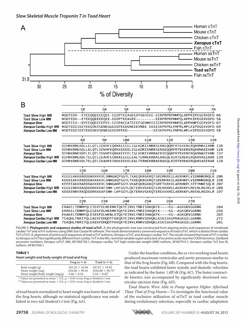
aligning amino acid sequences of cardiac and skeletal muscleTnT isoforms of representative species. The results showedthat toad ssTnT is closely related to avian and mammalianssTnTs (73–80% residue identities) although significantlydiverged from cardiac TnT (Fig. 5A).Alignment of amino acid sequences of the toad ssTnT iso-
forms with the ssTnT and cardiac TnT of Xenopus laevis pro-vided a comparison for their primary structures. In addition todifferences in the NH2-terminal hypervariable region, amphib-ian ssTnT differs from amphibian cardiac TnT by a lack of sixamino acids near the COOH terminus (Fig. 5B).Lower Specific Stroke Volume but Faster Kinetics of ToadVer-
sus Frog Hearts—At similar body weights, toad hearts werefound to be larger than frog hearts, resulting in a significantlyhigher heart weight to body weight ratio (Table 1). This featuremay be an evolutionary adaptation with functional significanceand further investigation is needed.Ex vivoworking heart experiments revealed interesting func-
tional differences between toad and frog hearts. At the baselinecondition of 4 cmH2Opreload, 25 cmH2O afterload, and heartrate paced at 32 bpm, the toad hearts produced similar absolutestroke volumes as frog hearts (Fig. 6A). However, due to thehigher heart weight of toad hearts (Table 1), the stroke volume
FIGURE 2. Toad cardiac muscle expresses solely ssTnT in the absence ofcardiac TnT. A single TnT band was detected in toad heart with a significantlylower molecular weight than that of frog cardiac TnT as shown in the Westernblot using mAb 2C8 recognizing all three muscle-type isoforms of TnT (Fig. 1).This TnT band in toad heart was recognized in the Western blot using mAbCT3 that reacts to both cardiac TnT and ssTnT with a molecular weightsimilar to that of ssTnT in toad and frog skeletal muscles. In contrast, it wasnot recognized in Western blots using the cardiac TnT-specific mAb 4B8.Reblotting of the 4B8 blot with mAb CT3 confirmed its presence on themembrane. The results demonstrated that the sole TnT expressed in toadheart is ssTnT.
Slow Skeletal Muscle Troponin T in Toad Heart
29756 JOURNAL OF BIOLOGICAL CHEMISTRY VOLUME 287 • NUMBER 35 • AUGUST 24, 2012
by guest on July 16, 2018http://w
ww
.jbc.org/D
ownloaded from
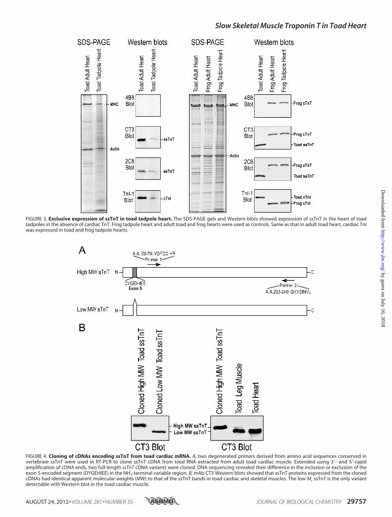
FIGURE 3. Exclusive expression of ssTnT in toad tadpole heart. The SDS-PAGE gels and Western blots showed expression of ssTnT in the heart of toadtadpoles in the absence of cardiac TnT. Frog tadpole heart and adult toad and frog hearts were used as controls. Same as that in adult toad heart, cardiac TnIwas expressed in toad and frog tadpole hearts.
FIGURE 4. Cloning of cDNAs encoding ssTnT from toad cardiac mRNA. A, two degenerated primers derived from amino acid sequences conserved invertebrate ssTnT were used in RT-PCR to clone ssTnT cDNA from total RNA extracted from adult toad cardiac muscle. Extended using 3�- and 5�-rapidamplification of cDNA ends, two full-length ssTnT cDNA variants were cloned. DNA sequencing revealed their difference in the inclusion or exclusion of theexon 5-encoded segment (DYGEHIEE) in the NH2-terminal variable region. B, mAb CT3 Western blots showed that ssTnT proteins expressed from the clonedcDNAs had identical apparent molecular weights (MW) to that of the ssTnT bands in toad cardiac and skeletal muscles. The low Mr ssTnT is the only variantdetectable with Western blot in the toad cardiac muscle.
Slow Skeletal Muscle Troponin T in Toad Heart
AUGUST 24, 2012 • VOLUME 287 • NUMBER 35 JOURNAL OF BIOLOGICAL CHEMISTRY 29757
by guest on July 16, 2018http://w
ww
.jbc.org/D
ownloaded from
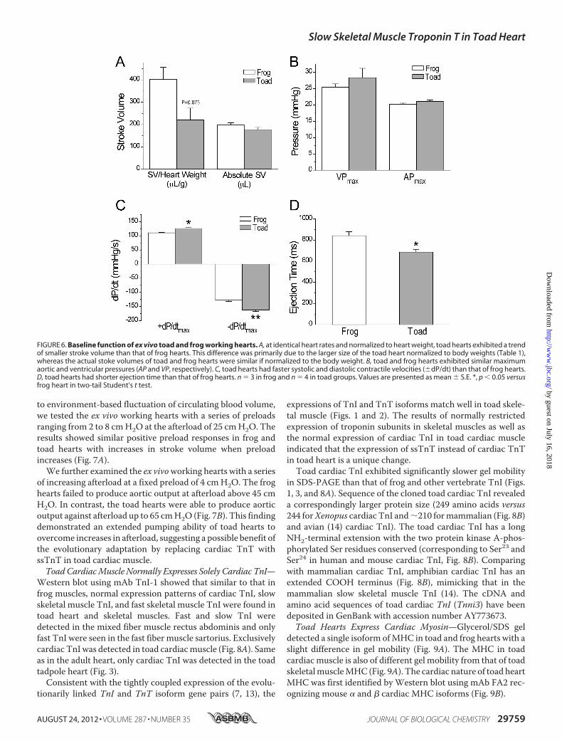
of toad hearts normalized to heartweightwas lower than that ofthe frog hearts, although no statistical significance was estab-lished in two-tail Student’s t test (Fig. 6A).
Under the baseline condition, the ex vivoworking toad heartsproduced maximum ventricular and aortic pressures similar tothat of the frog hearts (Fig. 6B). Compared with the frog hearts,the toad hearts exhibited faster systolic and diastolic velocitiesas indicated by the faster�dP/dt (Fig. 6C). The faster contract-ile kinetics was accompanied by significantly shortened ven-tricular ejection time (Fig. 6D).Toad Hearts Were Able to Pump against Higher Afterload
Than That of Frog Hearts—To investigate the functional valueof the exclusive utilization of ssTnT in toad cardiac muscleduring evolutionary selection, especially in cardiac adaptation
FIGURE 5. Phylogenetic and sequence studies of toad ssTnT. A, the phylogenetic tree was constructed from aligning amino acid sequences of vertebratecardiac TnT and ssTnT isoforms using DNA Star Clustal W software. The result demonstrated a conserved sequence of toad ssTnT, which is distinct from cardiacTnT (cTnT). B, alignment of amino acid sequences of toad ssTnT isoforms, Xenopus ssTnT, and Xenopus cardiac TnT. The results showed that toad ssTnT is similarto Xenopus ssTnT but significantly different from cardiac TnT in the NH2-terminal variable region and a lack of six amino acids near the COOH terminus. GenBankaccession numbers: Xenopus ssTnT, NM_001092738.1; Xenopus cardiac TnT high molecular weight (MW) isoform, AF467919.1; Xenopus cardiac TnT low Mrisoform, AF467920.1.
TABLE 1Heart weight and body weight of toad and frog
Frog (n � 4) Toad (n � 6)
Body weight (g) 181.25 � 16.94 187.67 � 19.92Heart weight (mg) 634.48 � 99.45 1016.08 � 90.75aHeart weight/body weight (mg/g) 3.46 � 0.24 5.54 � 0.42b
a Values are presented as mean � S.E., p � 0.05 versus frog in Student’s t test.b Values are presented as mean � S.E., p � 0.01 versus frog in Student’s t test.
Slow Skeletal Muscle Troponin T in Toad Heart
29758 JOURNAL OF BIOLOGICAL CHEMISTRY VOLUME 287 • NUMBER 35 • AUGUST 24, 2012
by guest on July 16, 2018http://w
ww
.jbc.org/D
ownloaded from
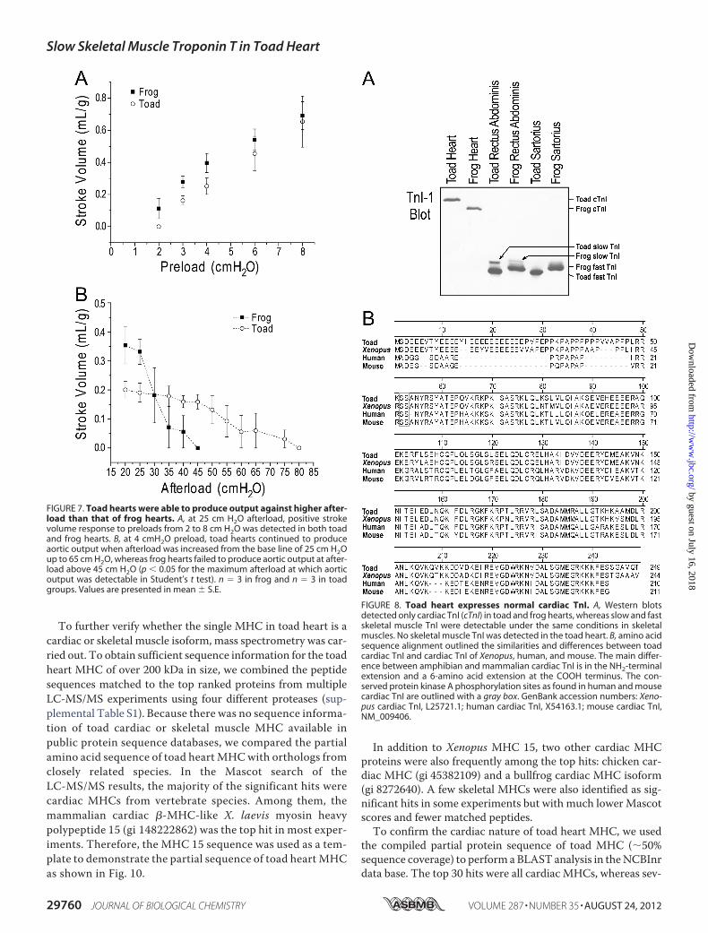
to environment-based fluctuation of circulating blood volume,we tested the ex vivo working hearts with a series of preloadsranging from 2 to 8 cmH2O at the afterload of 25 cmH2O. Theresults showed similar positive preload responses in frog andtoad hearts with increases in stroke volume when preloadincreases (Fig. 7A).We further examined the ex vivoworking hearts with a series
of increasing afterload at a fixed preload of 4 cmH2O. The froghearts failed to produce aortic output at afterload above 45 cmH2O. In contrast, the toad hearts were able to produce aorticoutput against afterload up to 65 cmH2O (Fig. 7B). This findingdemonstrated an extended pumping ability of toad hearts toovercome increases in afterload, suggesting a possible benefit ofthe evolutionary adaptation by replacing cardiac TnT withssTnT in toad cardiac muscle.ToadCardiacMuscle Normally Expresses Solely Cardiac TnI—
Western blot using mAb TnI-1 showed that similar to that infrog muscles, normal expression patterns of cardiac TnI, slowskeletal muscle TnI, and fast skeletal muscle TnI were found intoad heart and skeletal muscles. Fast and slow TnI weredetected in the mixed fiber muscle rectus abdominis and onlyfast TnI were seen in the fast fiber muscle sartorius. Exclusivelycardiac TnIwas detected in toad cardiacmuscle (Fig. 8A). Sameas in the adult heart, only cardiac TnI was detected in the toadtadpole heart (Fig. 3).Consistent with the tightly coupled expression of the evolu-
tionarily linked TnI and TnT isoform gene pairs (7, 13), the
expressions of TnI and TnT isoforms match well in toad skele-tal muscle (Figs. 1 and 2). The results of normally restrictedexpression of troponin subunits in skeletal muscles as well asthe normal expression of cardiac TnI in toad cardiac muscleindicated that the expression of ssTnT instead of cardiac TnTin toad heart is a unique change.Toad cardiac TnI exhibited significantly slower gel mobility
in SDS-PAGE than that of frog and other vertebrate TnI (Figs.1, 3, and 8A). Sequence of the cloned toad cardiac TnI revealeda correspondingly larger protein size (249 amino acids versus244 forXenopus cardiac TnI and�210 formammalian (Fig. 8B)and avian (14) cardiac TnI). The toad cardiac TnI has a longNH2-terminal extension with the two protein kinase A-phos-phorylated Ser residues conserved (corresponding to Ser23 andSer24 in human and mouse cardiac TnI, Fig. 8B). Comparingwith mammalian cardiac TnI, amphibian cardiac TnI has anextended COOH terminus (Fig. 8B), mimicking that in themammalian slow skeletal muscle TnI (14). The cDNA andamino acid sequences of toad cardiac TnI (Tnni3) have beendeposited in GenBank with accession number AY773673.Toad Hearts Express Cardiac Myosin—Glycerol/SDS gel
detected a single isoform ofMHC in toad and frog hearts with aslight difference in gel mobility (Fig. 9A). The MHC in toadcardiac muscle is also of different gel mobility from that of toadskeletalmuscleMHC (Fig. 9A). The cardiac nature of toad heartMHC was first identified by Western blot using mAb FA2 rec-ognizing mouse � and � cardiac MHC isoforms (Fig. 9B).
FIGURE 6. Baseline function of ex vivo toad and frog working hearts. A, at identical heart rates and normalized to heart weight, toad hearts exhibited a trendof smaller stroke volume than that of frog hearts. This difference was primarily due to the larger size of the toad heart normalized to body weights (Table 1),whereas the actual stoke volumes of toad and frog hearts were similar if normalized to the body weight. B, toad and frog hearts exhibited similar maximumaortic and ventricular pressures (AP and VP, respectively). C, toad hearts had faster systolic and diastolic contractile velocities (�dP/dt) than that of frog hearts.D, toad hearts had shorter ejection time than that of frog hearts. n � 3 in frog and n � 4 in toad groups. Values are presented as mean � S.E. *, p � 0.05 versusfrog heart in two-tail Student’s t test.
Slow Skeletal Muscle Troponin T in Toad Heart
AUGUST 24, 2012 • VOLUME 287 • NUMBER 35 JOURNAL OF BIOLOGICAL CHEMISTRY 29759
by guest on July 16, 2018http://w
ww
.jbc.org/D
ownloaded from
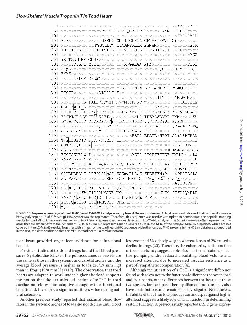
To further verify whether the single MHC in toad heart is acardiac or skeletal muscle isoform, mass spectrometry was car-ried out. To obtain sufficient sequence information for the toadheart MHC of over 200 kDa in size, we combined the peptidesequences matched to the top ranked proteins from multipleLC-MS/MS experiments using four different proteases (sup-plemental Table S1). Because there was no sequence informa-tion of toad cardiac or skeletal muscle MHC available inpublic protein sequence databases, we compared the partialamino acid sequence of toad heart MHCwith orthologs fromclosely related species. In the Mascot search of theLC-MS/MS results, the majority of the significant hits werecardiac MHCs from vertebrate species. Among them, themammalian cardiac �-MHC-like X. laevis myosin heavypolypeptide 15 (gi 148222862) was the top hit in most exper-iments. Therefore, the MHC 15 sequence was used as a tem-plate to demonstrate the partial sequence of toad heart MHCas shown in Fig. 10.
In addition to Xenopus MHC 15, two other cardiac MHCproteins were also frequently among the top hits: chicken car-diac MHC (gi 45382109) and a bullfrog cardiac MHC isoform(gi 8272640). A few skeletal MHCs were also identified as sig-nificant hits in some experiments but with much lowerMascotscores and fewer matched peptides.To confirm the cardiac nature of toad heart MHC, we used
the compiled partial protein sequence of toad MHC (�50%sequence coverage) to perform aBLAST analysis in theNCBInrdata base. The top 30 hits were all cardiac MHCs, whereas sev-
FIGURE 7. Toad hearts were able to produce output against higher after-load than that of frog hearts. A, at 25 cm H2O afterload, positive strokevolume response to preloads from 2 to 8 cm H2O was detected in both toadand frog hearts. B, at 4 cmH2O preload, toad hearts continued to produceaortic output when afterload was increased from the base line of 25 cm H2Oup to 65 cm H2O, whereas frog hearts failed to produce aortic output at after-load above 45 cm H2O (p � 0.05 for the maximum afterload at which aorticoutput was detectable in Student’s t test). n � 3 in frog and n � 3 in toadgroups. Values are presented in mean � S.E.
FIGURE 8. Toad heart expresses normal cardiac TnI. A, Western blotsdetected only cardiac TnI (cTnI) in toad and frog hearts, whereas slow and fastskeletal muscle TnI were detectable under the same conditions in skeletalmuscles. No skeletal muscle TnI was detected in the toad heart. B, amino acidsequence alignment outlined the similarities and differences between toadcardiac TnI and cardiac TnI of Xenopus, human, and mouse. The main differ-ence between amphibian and mammalian cardiac TnI is in the NH2-terminalextension and a 6-amino acid extension at the COOH terminus. The con-served protein kinase A phosphorylation sites as found in human and mousecardiac TnI are outlined with a gray box. GenBank accession numbers: Xeno-pus cardiac TnI, L25721.1; human cardiac TnI, X54163.1; mouse cardiac TnI,NM_009406.
Slow Skeletal Muscle Troponin T in Toad Heart
29760 JOURNAL OF BIOLOGICAL CHEMISTRY VOLUME 287 • NUMBER 35 • AUGUST 24, 2012
by guest on July 16, 2018http://w
ww
.jbc.org/D
ownloaded from

eral skeletal MHCs were matched with a lower percentage ofidentity. To validate our conclusion based on BLAST analysisusing a partial protein sequence, we also did a BLAST searchusing peptide mapping from a mouse cardiac MHC (�57%sequence coverage) obtained in a separate LC-MS/MS study. Asimilar overall pattern of the BLAST result was obtained (sup-plemental Fig. S1), which is consistentwith the toadheartMHCbeing a cardiac MHC.These thorough proteomic analysis concluded that the toad
heart MHC is a cardiac MHC. Therefore, the expression solelyof ssTnT in toad cardiac muscle is accompanied by normalexpressions of other thin and thick filament proteins. Accord-ingly, the novel functional differences between toad and froghearts is likely due to the unique replacement of cardiac TnTwith ssTnT.
DISCUSSION
More than four decades of extensive studies have establishedthe central position of troponin in the regulation of striatedmuscle contraction (15–17). TnT is the thin filament anchoringsubunit of the troponin complex and plays a central role in theregulation of striated muscle contraction (15). Three homolo-gous genes have evolved encoding the cardiac, slow skeletal andfast skeletal muscle isoforms of TnT in vertebrate cardiac andskeletal muscles (14, 18). Muscle type-specific isoforms of TnThave been identified in vertebrates inclusively from fish tohuman (7), but their physiological significances are not fullyunderstood.Multiple studies have documented that genes encodingmus-
cle type-specific TnT andTnI isoforms are expressed in specificfiber types and during development under stringent regulations
(14, 18). Cardiac muscle has evolved to have its specific set ofmyofilament protein isoforms adapted to accomplish the spe-cific function of the heart. With no other myofilament proteinisoforms changed, the expression of solely ssTnT to completelyreplace cardiac TnT in toad cardiac muscle is a unique case.Two lines of evidence indicated that the exclusive expression
of ssTnT to replace cardiac TnT in the toad heart is an evolu-tionary selection of certain beneficial value to cardiac function.The first is that all other vertebrate species from fish to humanincluding closely related amphibian species such as frog andXenopus, the immediate ancestor of toads and frogs, all expresssolely cardiac TnT in the cardiac muscle (7) (Fig. 1). Therefore,the expression of ssTnT in toad hearts is not a residually ances-tral trait but an acquired adaptation. The other evidence is thattoad heart expresses cardiac forms of other thin and thick fila-ment proteins, such as cardiac TnI (Fig. 8) and cardiac MHC(Fig. 9). Therefore, the exclusive expression of ssTnT in toadcardiac muscle cells is not due to a differentiation of the entirecellular environment but a selective activation of the ssTnTgene and inactivation of the cardiac TnT (Tnnt2) gene in nor-mally differentiated adult cardiomyocytes.We detected no cardiac TnT expression in either heart or
skeletal muscle of the toad. Therefore, the toad cardiac TnTgene is either lost or inactivated during evolution. Because thevertebrate cardiac TnT gene is closely linked to the slow TnI(Tnni1) gene (7) and a chromosomal deletion event to removethe cardiac TnT gene would likely impair the upstream slowTnI gene. However, the slow TnI expression is apparently nor-mal in toad skeletal muscle (Fig. 8A). Therefore, the cardiacTnT gene probably remains in the toad genome but isinactivated.The molecular mechanisms of this novel gene switch
remains to be investigated. Considering the early evolutionaryemerging of the three muscle type isoforms of TnT in fish andpossibly in the vertebrate ancestor hagfish (7), it would be inter-esting to further investigatewhether the adaptive switch ofTnTgenes also occurred in the heart of any other amphibians orprimitive reptiles.This intriguing finding provides a novel lead and experimen-
tal system to investigate the adaptive values of TnT isoforms inmuscle contractility and cardiac function. The ex vivo workingheart system used in our study allows analyses of cardiac func-tion excluding neurohumoral and vascular influences. Thisexperimental approach also permits precisely controlling theheart rate and altering preload and/or afterload to explore thefunctional differences between toad and frog hearts. To copewith the fact that toad heart and frog heart are both typicalthree-chamber amphibian hearts with two atria and one ventri-cle, we have successfully modified a system built for mouse andrat heart studies (10, 11) for the study of amphibian hearts.Two major differences found in the comparison of ex vivo
toad and frog working hearts support the proposed functionalbenefit of utilizing ssTnT in toad cardiac muscle. One is thefaster contractile and relaxation velocity of the toad ventricularmuscle reflecting higher contractility (Fig. 6C) and the other isthe higher afterload that the toad heart could work against toproduce aortic output (Fig. 7B). The link between the replace-ment of cardiac TnTwith ssTnT and the enhanced pumping of
FIGURE 9. Toad heart expresses cardiac MHC. A, MHC isoforms in the heartsand skeletal muscles of toad, frog, and Xenopus were resolved in glycerol/SDSgel. Mouse diaphragm muscle was analyzed in parallel as control. A singleMHC band was detected in toad heart with a slightly different gel mobilityfrom that of frog cardiac MHC or the MHC bands in toad rectum abdominisand sartorius muscles. B, mAb FA2 specific to cardiac �- and �-MHC (cardiac�-MHC is the same as MHC I in slow skeletal muscle) recognized the MHCband in both toad and frog hearts, supporting their nature as cardiac MHC.
Slow Skeletal Muscle Troponin T in Toad Heart
AUGUST 24, 2012 • VOLUME 287 • NUMBER 35 JOURNAL OF BIOLOGICAL CHEMISTRY 29761
by guest on July 16, 2018http://w
ww
.jbc.org/D
ownloaded from
toad heart provided organ level evidence for a functionalbenefit.Previous studies of toads and frogs found that blood pres-
sures (systolic/diastolic) in the pulmocutaneous vessels arethe same as those in the systemic and carotid arches, and theaverage blood pressure is higher in toads (26/19 mm Hg)than in frogs (15/8 mm Hg) (19). The observation that toadhearts are adapted to work under higher afterload supportsthe notion that the exclusive utilization of ssTnT in toadcardiac muscle was an adaptive change with a functionalbenefit and, therefore, a significant fitness value during nat-ural selection.Another previous study reported that maximal blood flow
rates in the systemic arches of toads did not decline until blood
loss exceeded 5% of body weight, whereas losses of 2% caused adecline in frogs (20). Therefore, the enhanced systolic functionof toad hearts may suggest a role of ssTnT inmaintaining effec-tive pumping under reduced circulating blood volume andincreased afterload due to increased vascular resistance as apart of sympathetic compensation (4).Although the utilization of ssTnT is a significant difference
foundwith relevance to the functional differences between toadand frog hearts, other differences between the hearts of thesetwo species, for example, other myofilament proteins, may alsohave contributions and remain to be investigated. Nonetheless,the ability of toadhearts to produce aortic output against higherafterload suggests a likely role of TnT function in determiningsystolic function. A previous study reported ssTnT gene expres-
FIGURE 10. Sequence coverage of toad MHC from LC-MS/MS analyses using four different proteases. A database search showed that cardiac-like myosinheavy polypeptide 15 of X. laevis (gi 148222862) was the top match. Therefore, this sequence was used as a template to demonstrate the peptide mappingresults for toad MHC. Amino acids marked with black letters represent sequences detected in LC-MS/MS analyses. The boxed lowercase letters represent aminoacids that are different from the Xenopus MHC 15 sequence. X represents amino acid residues in the MHC of the Xenopus MHC 15 sequence, which are notcovered in the LC-MS/MS results. Together with a match of the toad heart MHC sequence with other cardiac MHC proteins in the NCBInr database as describedin the text, the data confirmed that the MHC in toad heart is a cardiac isoform.
Slow Skeletal Muscle Troponin T in Toad Heart
29762 JOURNAL OF BIOLOGICAL CHEMISTRY VOLUME 287 • NUMBER 35 • AUGUST 24, 2012
by guest on July 16, 2018http://w
ww
.jbc.org/D
ownloaded from
sion in hypertensive rats, which is an established pressure over-load model (21). The exclusive expression of ssTnT in toadheart provides a natural animal model to study the functionalsignificance of TnT modifications in cardiac muscle.The mechanism for ssTnT to enhance the systolic function
of heart remains to be investigated. Knocking down ssTnTinduced a slow-to-fast type muscle switch in mice withimpaired fatigue tolerance of the diaphragm muscle (22).Although ssTnTmay have a unique function in skeletal musclecontractility, its value in cardiac muscle function needs morestudy. On the other hand, transgenic mice overexpressing fastskeletal muscle TnT in the heart decreased cardiac function(23). Therefore, the apparently benign compatibility of ssTnTin toad cardiac myofilaments merits further investigation.The present study also raises an interesting question of how the
muscle type-specific TnT isoform genes are regulated. Similar tothe frog ssTnT gene, the toad ssTnT gene was normally expressedin rectus abdominis muscles containing slow fibers but not in thepure fast fiber sartorius muscle (Fig. 2). Therefore, the specificexpression of toad ssTnT gene in slow muscle fibers is preservedwhile it also became activated in cardiac muscle.In vertebrate striated muscles, the expression of TnT isoform
genes is coupled with TnI isoforms and MHC isoforms (24, 25).Slow skeletal muscle TnI is the sole TnI expressed in embryoniccardiacmuscle but replacedby cardiacTnI duringperinatal devel-opment (26, 27). However, toad heart expresses normal cardiacforms of othermyofilament proteins. No skeletalmuscle isoformsof othermyofilament proteins was seen in the toad heart, indicat-ing no overall change in cellular gene regulations. The selectivelyactivation of the ssTnT gene and inactivation of the cardiac TnTgene in adult toad heart, therefore, suggested a change in specificregulatory factors other than the altered overall cellular environ-ment or a loosened ssTnT gene regulation. To further investigatethe transcriptional regulation of the toad ssTnT genemay lead to abetter understanding of the regulation of cardiac-specific genes ingeneral. More investigation is also needed to understand why thecardiac TnT gene ceased expression in the toad heart.
In summary, the significanceofour findings is 2-fold.The first isthat cardiacmuscle cells have the potential, at least in lower verte-brates, to switchTnT isoform gene expression in functional adap-tations. The other is that by changing the function of a single thinfilament regulatory protein, TnT, it is possible to increase the sys-tolic function of the heart. The finding that the function of TnTmay improve systolic function of the heart suggests a potentialtarget for the treatment of heart failure, in which pharmacologi-cally altering the function of normal cardiac TnT, rather thanswitching to ssTnT, may provide similar therapeutic benefits.
Acknowledgments—We thank HuiWang for technical assistance. Dr.Jimin Gao participated in the cloning of cDNAs encoding toad ssTnTand cardiac TnI when this research group was at the ENH ResearchInstitute and Northwestern University Feinberg School of Medicine.
REFERENCES1. Boral, M. C., and Deb, C. (1970) Seasonal changes in body fluids and
hematology in toad (Bufo melanostictus)—a poikilothermic cold torpor.Proc. Indian Natl. Sci. Acad. 36, 369–373
2. Deb, C., Chatterjee, S., and Boral, M. C. (1974) Body fluid and hematolog-
ical changes in toads following heat exposure. Am. J. Physiol. 226,408–410
3. Schadt, J. C., and Ludbrook, J. (1991) Hemodynamic and neurohumoralresponses to acute hypovolemia in conscious mammals. Am. J. Physiol.260, H305-H318
4. Peitzman, A. B., Billiar, T. R., Harbrecht, B. G., Kelly, E., Udekwu, A. O.,and Simmons, R. L. (1995) Hemorrhagic shock. Curr. Probl. Surg. 32,925–1002
5. Gordon, A. M., Homsher, E., and Regnier, M. (2000) Regulation of con-traction in striated muscle. Physiol. Rev. 80, 853–924
6. Jin, J. P., Yang, F. W., Yu, Z. B., Ruse, C. I., Bond, M., and Chen, A. (2001)The highly conserved COOH terminus of troponin I forms a Ca2�-mod-ulated allosteric domain in the troponin complex. Biochemistry 40,2623–2631
7. Chong, S. M., and Jin, J. P. (2009) To investigate protein evolution bydetecting suppressed epitope structures. J. Mol. Evol. 68, 448–460
8. Jin, J. P., Chen, A., Ogut, O., and Huang, Q. Q. (2000) Conformationalmodulation of slow skeletalmuscle troponinT by anNH2-terminalmetal-binding extension. Am. J. Physiol. Cell Physiol. 279, C1067-C1077
9. Jin, J. P., Malik, M. L., and Lin, J. J. (1990) Monoclonal antibodies againstcardiac myosin heavy chain. Hybridoma 9, 597–608
10. Feng, H. Z., Chen, M.,Weinstein, L. S., and Jin, J. P. (2008) Removal of theN-terminal extension of cardiac troponin I as a functional compensationfor impaired myocardial �-adrenergic signaling. J. Biol. Chem. 283,33384–33393
11. Feng, H. Z., Biesiadecki, B. J., Yu, Z. B., Hossain, M.M., and Jin, J. P. (2008)Restricted N-terminal truncation of cardiac troponin T. A novel mecha-nism for functional adaptation to energetic crisis. J. Physiol 586,3537–3550
12. Mazza, R., Gattuso, A., Mannarino, C., Brar, B. K., Barbieri, S. F., Tota, B.,and Mahata, S. K. (2008) Catestatin (chromogranin A344–364) is a novelcardiosuppressive agent. Inhibition of isoproterenol and endothelin sig-naling in the frog heart. Am. J. Physiol. Heart Circ. Physiol 295,H113-H122
13. Brotto, M. A., Biesiadecki, B. J., Brotto, L. S., Nosek, T. M., and Jin, J. P.(2006) Coupled expression of troponin T and troponin I isoforms in singleskeletal muscle fibers correlates with contractility. Am. J. Physiol. CellPhysiol. 290, C567-C576
14. Jin, J. P., Zhang, Z., and Bautista, J. A. (2008) Isoform diversity, regulation,and functional adaptation of troponin and calponin. Crit. Rev. Eukaryot.Gene Expr. 18, 93–124
15. Perry, S. V. (1998) Troponin T. Genetics, properties and function. J. Mus-cle Res. Cell Motil. 19, 575–602
16. Perry, S. V. (1999) Troponin I. Inhibitor or facilitator.Mol. Cell. Biochem.190, 9–32
17. Perry, S. V. (2008) Background to the discovery of troponin and SetsuroEbashi’s contribution to our knowledge of the mechanism of relaxation instriated muscle. Biochem. Biophys. Res. Commun. 369, 43–48
18. Wei, B., and Jin, J. P. (2011) Troponin T isoforms and posttranscriptionalmodifications. Evolution, regulation, and function. Arch. Biochem. Bio-phys. 505, 144–154
19. Simons, J. R. (1957) J. Physiol. 137, 12–2120. Hillman, S. S., andWithers, P. C. (1988) The hemodynamic consequences
of hemorrhage and hypernatremia in two amphibians. J. Comp. Physiol. B157, 807–812
21. Barton, P. J., Felkin, L. E., Koban, M. U., Cullen, M. E., Brand, N. J., andDhoot, G. K. (2004) The slow skeletal muscle troponin T gene is ex-pressed in developing and diseased human heart. Mol. Cell Biochem.263, 91–97
22. Feng, H. Z., Wei, B., and Jin, J. P. (2009) Deletion of a genomic segmentcontaining the cardiac troponin I gene knocks down expression of theslow troponin T gene and impairs fatigue tolerance of diaphragmmuscle.J. Biol. Chem. 284, 31798–31806
23. Huang, Q. Q., Feng, H. Z., Liu, J., Du, J., Stull, L. B., Moravec, C. S., Huang,X., and Jin, J. P. (2008) Co-expression of skeletal and cardiac troponin Tdecreases mouse cardiac function. Am. J. Physiol. Cell Physiol 294,C213-C222
24. Feng, H. Z., Chen, M., Weinstein, L. S., and Jin, J. P. (2011) Improved
Slow Skeletal Muscle Troponin T in Toad Heart
AUGUST 24, 2012 • VOLUME 287 • NUMBER 35 JOURNAL OF BIOLOGICAL CHEMISTRY 29763
by guest on July 16, 2018http://w
ww
.jbc.org/D
ownloaded from

fatigue resistance in G�s-deficient and aging mouse skeletal muscles dueto adaptive increases in slow fibers. J. Appl. Physiol. 111, 834–843
25. Yu, Z. B., Gao, F., Feng, H. Z., and Jin, J. P. (2007) Differential regulationof myofilament protein isoforms underlying the contractility changesin skeletal muscle unloading. Am. J. Physiol. Cell Physiol. 292,C1192–C1203
26. Jin, J. P. (1996) Alternative RNA splicing-generated cardiac troponin Tisoform switching. A non-heart-restricted genetic programming synchro-nized in developing cardiac and skeletal muscles. Biochem. Biophys. Res.Commun. 225, 883–889
27. Saggin, L., Gorza, L., Ausoni, S., and Schiaffino, S. (1989) Troponin Iswitching in the developing heart. J. Biol. Chem. 264, 16299–16302
Slow Skeletal Muscle Troponin T in Toad Heart
29764 JOURNAL OF BIOLOGICAL CHEMISTRY VOLUME 287 • NUMBER 35 • AUGUST 24, 2012
by guest on July 16, 2018http://w
ww
.jbc.org/D
ownloaded from
Han-Zhong Feng, Xuequn Chen, M. Moazzem Hossain and Jian-Ping JinBENEFITS
EVOLUTIONARY ADAPTATION WITH POTENTIAL FUNCTIONAL Toad Heart Utilizes Exclusively Slow Skeletal Muscle Troponin T: AN
doi: 10.1074/jbc.M112.373191 originally published online July 9, 20122012, 287:29753-29764.J. Biol. Chem.
10.1074/jbc.M112.373191Access the most updated version of this article at doi:
Alerts:
When a correction for this article is posted•
When this article is cited•
to choose from all of JBC's e-mail alertsClick here
Supplemental material:
http://www.jbc.org/content/suppl/2012/07/09/M112.373191.DC1
http://www.jbc.org/content/287/35/29753.full.html#ref-list-1
This article cites 27 references, 3 of which can be accessed free at
by guest on July 16, 2018http://w
ww
.jbc.org/D
ownloaded from