To access additional material or resources available with ...
43
Transcript of To access additional material or resources available with ...

To access additional material or resources available with this e-book,please visit http://www.thieme.com/bonuscontent. After completing ashort form to verify your e-book purchase, you will be provided with theinstructions and access codes necessary to retrieve any bonus content.
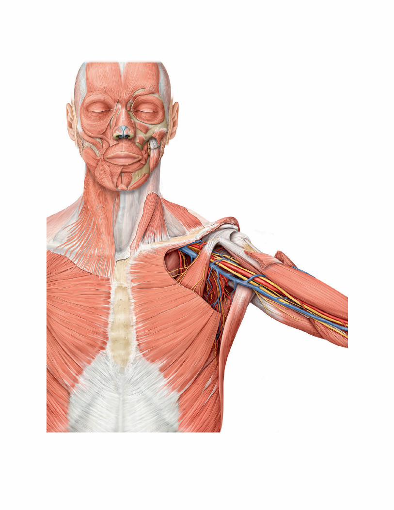
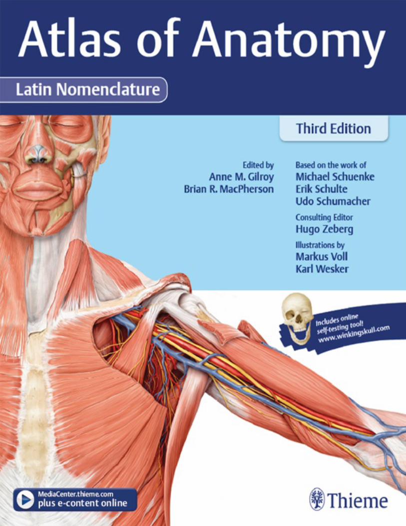
Atlas of Anatomy
Third Edition
Latin Nomenclature
Edited byAnne M. Gilroy, MAAssociate ProfessorDepartment of RadiologyUniversity of Massachusetts Medical SchoolWorcester, Massachusetts
Brian R. MacPherson, PhDProfessor and Vice-ChairDepartment of Anatomy and NeurobiologyUniversity of Kentucky College of MedicineLexington, Kentucky
Consulting EditorHugo Zeberg, MD, PHDLecturerDepartment of NeuroscienceKarolinska InstitutetStockholm, Sweden
Based on the work ofMichael Schuenke, MD, PhDInstitute of AnatomyChristian Albrecht University KielKiel, Germany
Erik Schulte, MDDepartment of Functional and Clinical AnatomyUniversity MedicineJohannes Gutenberg UniversityMainz, Germany
Udo Schumacher, MD, FRCPath, CBiol, FSB, DSc

Institute of Anatomy and Experimental MorphologyCenter for Experimental MedicineUniversity Cancer CenterUniversity Medical Center Hamburg-EppendorfHamburg, Germany
Illustrations byMarkus VollKarl Wesker
1935 illustrations
ThiemeNew York • Stuttgart • Delhi • Rio de Janeiro

Editorial Director, Educational Products: Anne M. SydorDevelopmental Editor: Julie O'MearaManaging Editor: Judith TomatEditorial Assistant: Tony PaeseDirector, Editorial Services: Mary Jo CaseyInternational Production Director: Andreas SchabertInternational Marketing Director: Fiona HendersonInternational Sales Director: Louisa TurrellDirector of Sales, North America: Mike RosemanSenior Vice President and Chief Operating Officer: Sarah VanderbiltPresident: Brian D. ScanlanIllustrations: Markus Voll and Karl WeskerProduction Editor: Barbara ChernowCompositors: Carol Pierson, Chernow Editorial Services, Inc. Toppan Best-set Premedia Limited
Library of Congress Cataloging-in-Publication Data is available from the publisher upon request.
Copyright ©2017 by Thieme Medical Publishers, Inc.Thieme Publishers New York333 Seventh Avenue, New York, NY 10001 USA+1 800 782 3488, [email protected]
Thieme Publishers StuttgartRüdigerstrasse 14, 70469 Stuttgart, Germany+49 [0]711 8931 421, [email protected]
Thieme Publishers DelhiA-12, Second Floor, Sector-2, Noida-201301Uttar Pradesh, India+91 120 45 566 00, [email protected]
Thieme Publishers Rio de Janeiro, Thieme Publicações Ltda.Edifício Rodolpho de Paoli, 25o andarAv. Nilo Peçanha, 50 – Sala 2508Rio de Janeiro 20020-906 Brasil+55 21 3172 2297
Printed in China by Everbest Printing Ltd5 4 3 2 1
ISBN 978-1-62623-522-9
Also available as an e-book:eISBN 978-1-62623-523-6
Important note: Medicine is an ever-changing science undergoing continual development. Researchand clinical experience are continually expanding our knowledge, in particular our knowledge of propertreatment and drug therapy. Insofar as this book mentions any dosage or application, readers may restassured that the authors, editors, and publishers have made every effort to ensure that such references
are in accordance with the state of knowledge at the time of production of the book.Nevertheless, this does not involve, imply, or express any guarantee or responsibility on the part of
the publishers in respect to any dosage instructions and forms of applications stated in the book. Everyuser is requested to examine carefully the manufacturers’ leaflets accompanying each drug and tocheck, if necessary in consultation with a physician or specialist, whether the dosage schedulesmentioned therein or the contraindications stated by the manufacturers differ from the statements madein the present book. Such examination is particularly important with drugs that are either rarely used orhave been newly released on the market. Every dosage schedule or every form of application used isentirely at the user's own risk and responsibility. The authors and publishers request every user to reportto the publishers any discrepancies or inaccuracies noticed. If errors in this work are found afterpublication, errata will be posted at www.thieme.com on the product description page.
Some of the product names, patents, and registered designs referred to in this book are in factregistered trademarks or proprietary names even though specific reference to this fact is not alwaysmade in the text. Therefore, the appearance of a name without designation as proprietary is not to beconstrued as a representation by the publisher that it is in the public domain.
This book, including all parts thereof, is legally protected by copyright. Any use, exploitation, orcommercialization outside the narrow limits set by copyright legislation without the publisher's consentis illegal and liable to prosecution. This applies in particular to photostat reproduction, copying,mimeographing or duplication of any kind, translating, preparation of microfilms, and electronic dataprocessing and storage.
Dedication
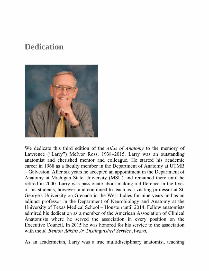
We dedicate this third edition of the Atlas of Anatomy to the memory ofLawrence (“Larry”) McIvor Ross, 1938–2015. Larry was an outstandinganatomist and cherished mentor and colleague. He started his academiccareer in 1968 as a faculty member in the Department of Anatomy at UTMB– Galveston. After six years he accepted an appointment in the Department ofAnatomy at Michigan State University (MSU) and remained there until heretired in 2000. Larry was passionate about making a difference in the livesof his students, however, and continued to teach as a visiting professor at St.George's University on Grenada in the West Indies for nine years and as anadjunct professor in the Department of Neurobiology and Anatomy at theUniversity of Texas Medical School – Houston until 2014. Fellow anatomistsadmired his dedication as a member of the American Association of ClinicalAnatomists where he served the association in every position on theExecutive Council. In 2015 he was honored for his service to the associationwith the R. Benton Adkins Jr. Distinguished Service Award.
As an academician, Larry was a true multidisciplinary anatomist, teaching

histology, neuroanatomy, gross anatomy, and embryology to thousands ofmedical and graduate students as well as numerous non-medical groups. Asan author, he will be remembered best for his work with Thieme Publishers.From 2005 to 2007 he co-edited the English translation of all three volumesof Prometheus: Atlas of Anatomy. Following the critical success of the three-volume atlas in English- speaking countries, he was instrumental in helpingThieme create the concept for the single-volume Atlas of Anatomy. This atlas,now in its third edition, is highly acclaimed in its own right and is distributedworldwide and translated in over 14 languages. As his co-authors, we aremost grateful to Larry for his mentorship. He was responsible for bringing usonto the project and into the world of medical publications. We feelpersonally indebted for all he did for us, and will fondly remember him as agreat mentor, friend, and colleague.
Anne and Brian
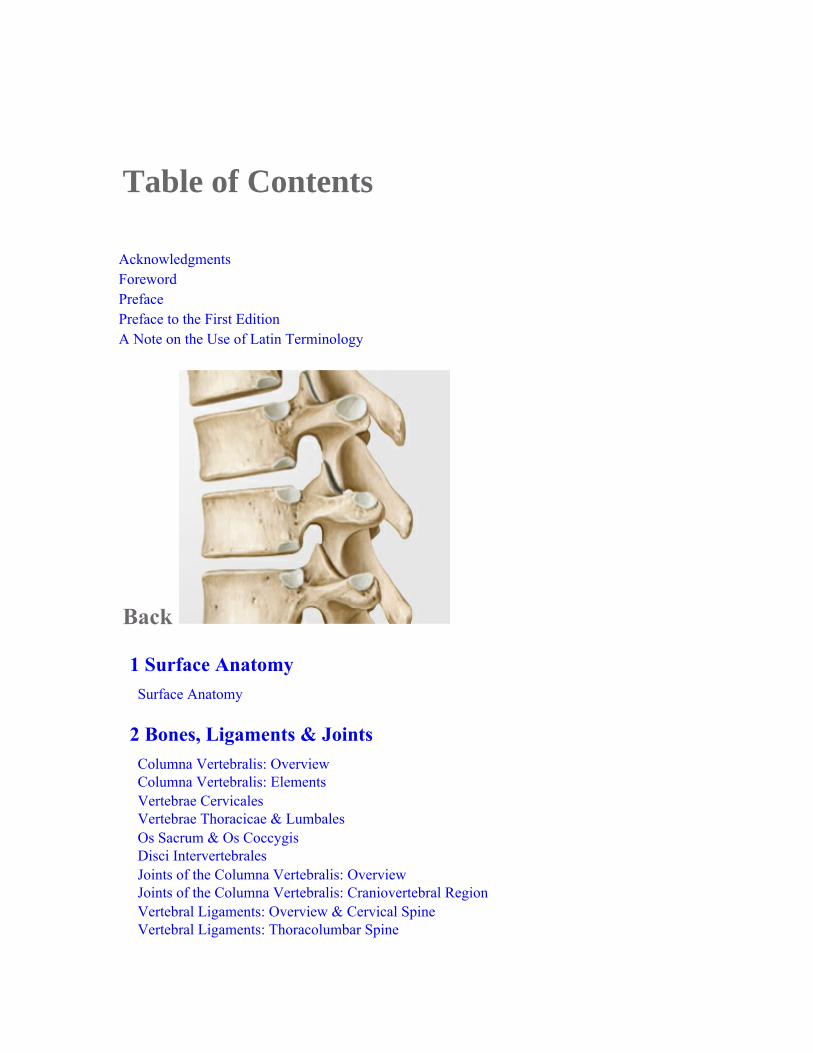
Table of Contents
AcknowledgmentsForewordPrefacePreface to the First EditionA Note on the Use of Latin Terminology
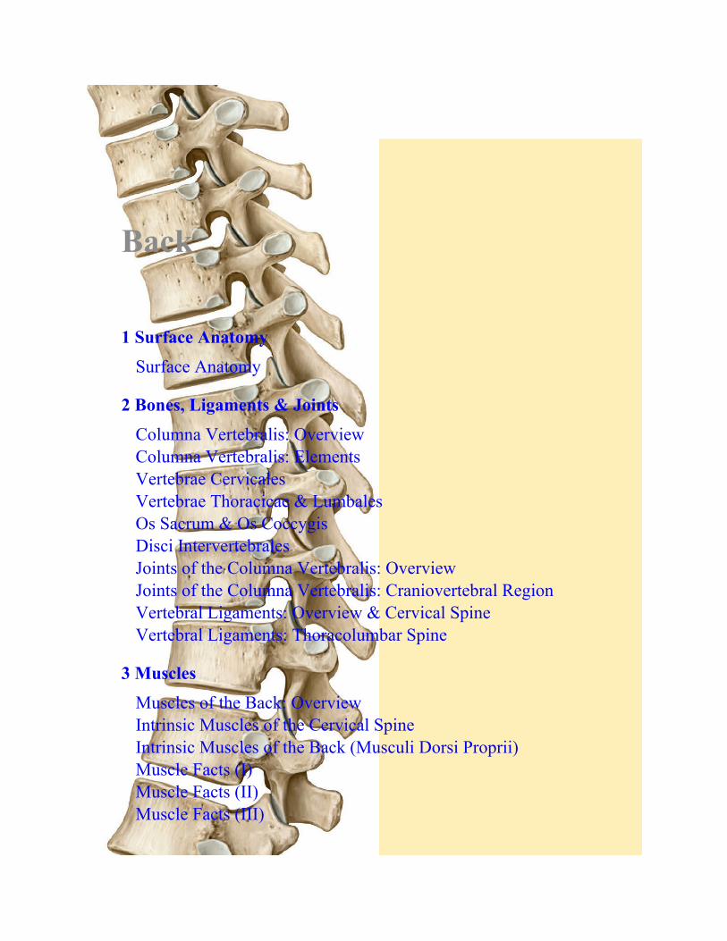
Back
1 Surface AnatomySurface Anatomy
2 Bones, Ligaments & JointsColumna Vertebralis: OverviewColumna Vertebralis: ElementsVertebrae CervicalesVertebrae Thoracicae & LumbalesOs Sacrum & Os CoccygisDisci IntervertebralesJoints of the Columna Vertebralis: OverviewJoints of the Columna Vertebralis: Craniovertebral RegionVertebral Ligaments: Overview & Cervical SpineVertebral Ligaments: Thoracolumbar Spine
3 MusclesMuscles of the Back: OverviewIntrinsic Muscles of the Cervical SpineIntrinsic Muscles of the Back (Musculi Dorsi Proprii)Muscle Facts (I)Muscle Facts (II)Muscle Facts (III)
4 NeurovasculatureArteries & Veins of the BackNerves of the BackMedulla Spinalis (Spinal Cord)Segments of the Medulla Spinalis & Nervi SpinalesArteries & Veins of the Medulla SpinalisNeurovascular Topography of the Back
5 Sectional & Radiographic AnatomyRadiographic Anatomy of the Back (I)Radiographic Anatomy of the Back (II)
Thorax
6 Surface AnatomySurface Anatomy
7 Thoracic WallThoracic SkeletonSternum & CostaeJoints of the Thoracic Cage
Thoracic Wall Muscle FactsDiaphragmaNeurovasculature of the DiaphragmaArteries & Veins of the Thoracic WallNerves of the Thoracic WallNeurovascular Topography of the Thoracic WallMamma (Breast)Lymphatics of the Mamma
8 Thoracic CavityDivisions of the Thoracic CavityArteries of the Thoracic CavityVeins of the Thoracic CavityLymphatics of the Thoracic CavityNerves of the Thoracic Cavity
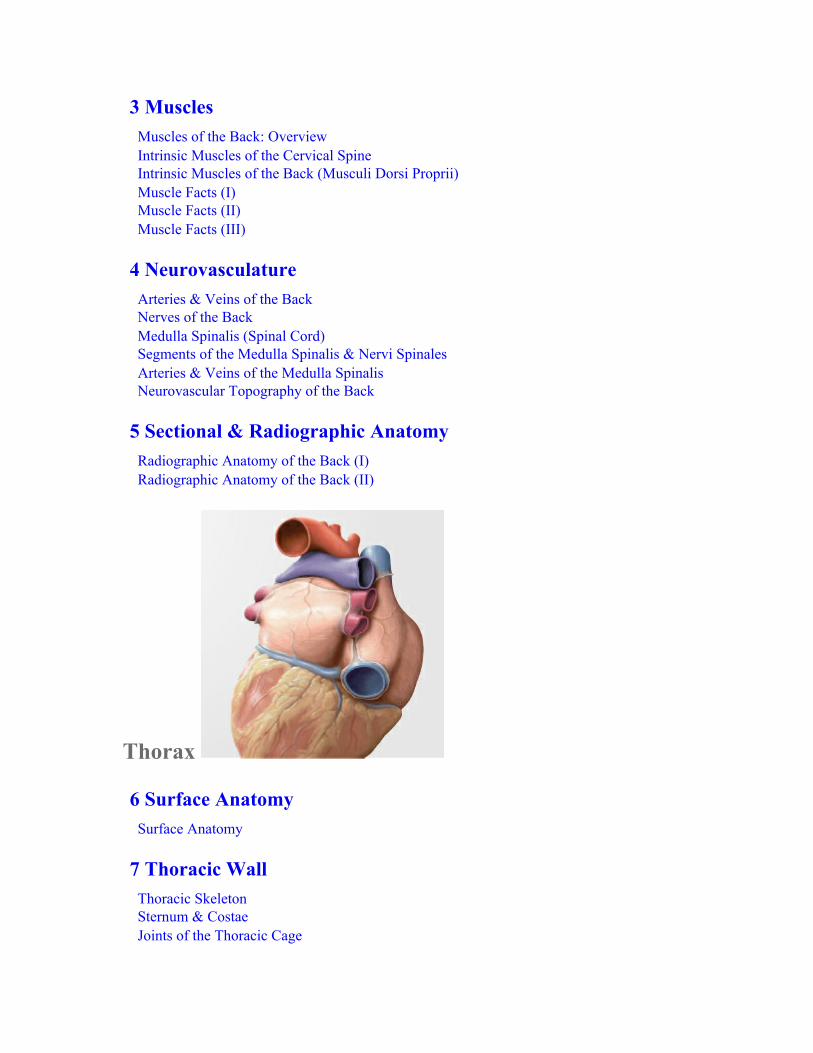
9 MediastinumMediastinum: OverviewMediastinum: StructuresHeart (Cor): Functions and RelationsPericardiumHeart: Surfaces & ChambersHeart: ValvesArteries & Veins of the HeartConduction & Innervation of the HeartPre- & Postnatal CirculationOesophagusNeurovasculature of the OesophagusLymphatics of the Mediastinum
10 Pulmonary CavitiesPulmonary CavitiesPleura: Subdivisions, Recesses & InnervationLungs (Pulmones)Bronchopulmonary SegmentsTrachea & Bronchial Tree (Arbor Bronchialis)Respiratory MechanicsPulmonary Arteries & VeinsNeurovasculature of the Tracheobronchial TreeLymphatics of the Pleural Cavity
11 Sectional & Radiographic AnatomySectional Anatomy of the Thorax

Radiographic Anatomy of the Thorax (I)Radiographic Anatomy of the Thorax (II)Radiographic Anatomy of the Thorax (III)
Abdomen
12 Surface AnatomySurface Anatomy
13 Abdominal WallBony Framework for the Abdominal WallMuscles of the Anterolateral Abdominal WallMuscles of the Posterior Abdominal Wall & DiaphragmaAbdominal Wall Muscle FactsRegio Inguinalis & Canalis InguinalisFuniculus Spermaticus, Scrotum & TestisAnterior Abdominal Wall & Inguinal Hernias
14 Cavitas Abdominis & SpacesDivisions of the Cavitas AbdominopelvicaCavitas Peritonealis & Greater SacBursa Omentalis, or Lesser SacMesenteria & Posterior Wall
15 Internal OrgansGasterDuodenumJejunum & IleumCaecum, Appendix Vermiformis & ColonHepar: Overview
Hepar: Lobi & SegmentaVesica Biliaris & Bile DuctsPancreas & SplenRenes & Glandulae Suprarenales (I)Renes & Glandulae Suprarenales (II)
16 NeurovasculatureArteries of the AbdomenPars Abdominalis Aortae & Arteriae RenalesTruncus CoeliacusArteria Mesenterica Superior & Arteria Mesenterica InferiorVeins of the AbdomenVena Cava Inferior & Venae RenalesVena Portae HepatisVena Mesenterica Superior & Vena Mesenterica InferiorLymphatics of the Abdominal OrgansNodi Lymphoidei of the Posterior Abdominal WallNodi Lymphoidei of the Supracolic OrgansNodi Lymphoidei of the Infracolic OrgansNerves of the Abdominal WallAutonomic Innervation: OverviewAutonomic PlexusesInnervation of the Abdominal OrgansInnervation of the Intestines
17 Sectional & Radiographic AnatomySectional Anatomy of the AbdomenRadiographic Anatomy of the Abdomen (I)Radiographic Anatomy of the Abdomen (II)
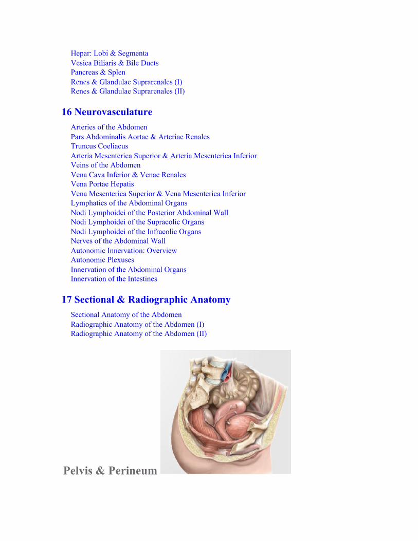
Pelvis & Perineum
18 Surface AnatomySurface Anatomy
19 Bones, Ligaments & MusclesCingulum PelvicumFemale & Male PelvisFemale & Male Pelvic MeasurementsPelvic LigamentsMuscles of the Diaphragma Pelvis & PerineumDiaphragma Pelvis & Perineal Muscle Facts
20 SpacesContents of the PelvisPeritoneal RelationshipsPelvis & Perineum
21 Internal OrgansRectum & Canalis AnalisUreteresVesica Urinaria & UrethraOverview of the Genital OrgansUterus & OvariaLigaments & Fasciae of the Deep PelvisVaginaFemale External GenitaliaPenis, Testis & EpididymisMale Accessory Sex Glands
22 NeurovasculatureArteries & Veins of the PelvisArteries & Veins of the Rectum & GenitaliaNodi Lymphoidei of the Abdomen & PelvisNodi Lymphoidei of the GenitaliaAutonomic Plexuses of the PelvisAutonomic Innervation: Urinary & Genital Organs & RectumNeurovasculature of the Female Perineum & GenitaliaNeurovasculature of the Male Perineum & Genitalia
23 Sectional & Radiographic AnatomySectional Anatomy of the Pelvis & PerineumRadiographic Anatomy of the Female PelvisRadiographic Anatomy of the Male Pelvis

Upper Limb
24 Surface AnatomySurface Anatomy
25 Shoulder & ArmBones of the Upper LimbClavicula & ScapulaHumerusJoints of the ShoulderJoints of the Shoulder: Articulatio HumeriSubacromial Space & BursaeAnterior Muscles of the Shoulder & Arm (I)Anterior Muscles of the Shoulder & Arm (II)Posterior Muscles of the Shoulder & Arm (I)Posterior Muscles of the Shoulder & Arm (II)Muscle Facts (I)Muscle Facts (II)Muscle Facts (III)Muscle Facts (IV)
26 Elbow & ForearmRadius & UlnaArticulatio CubitiLigaments of the Articulatio CubitiArticulationes RadioulnaresMuscles of the Forearm: Compartimentum AnteriusMuscles of the Forearm: Compartimentum PosteriusMuscle Facts (I)Muscle Facts (II)Muscle Facts (III)

27 Wrist & HandBones of the Wrist (Carpus) & Hand (Manus)The Carpal BonesJoints of the Wrist & HandLigaments of the HandLigaments and Compartments of the WristLigaments of the FingersMuscles of the Hand: Superficial & Middle LayersMuscles of the Hand: Middle & Deep LayersDorsum of the HandMuscle Facts (I)Muscle Facts (II)
28 NeurovasculatureArteries of the Upper LimbVeins & Lymphatics of the Upper LimbNerves of the Upper Limb: Plexus BrachialisPars Supraclavicularis & Fasciculus PosteriorFasciculus Posterior: Nervus Axillaris & Nervus RadialisFasciculus Medialis & Fasciculus LateralisNervus Medianus & Nervus UlnarisSuperficial Veins & Nerves of the Upper LimbPosterior Shoulder & ArmAnterior ShoulderAxillaAnterior Arm & Regio CubitalisRegio Antebrachii Anterior & Regio Antebrachii PosteriorRegio CarpalisPalm of the HandDorsum of the Hand
29 Sectional & Radiographic AnatomySectional Anatomy of the Upper LimbRadiographic Anatomy of the Upper Limb (I)Radiographic Anatomy of the Upper Limb (II)Radiographic Anatomy of the Upper Limb (III)
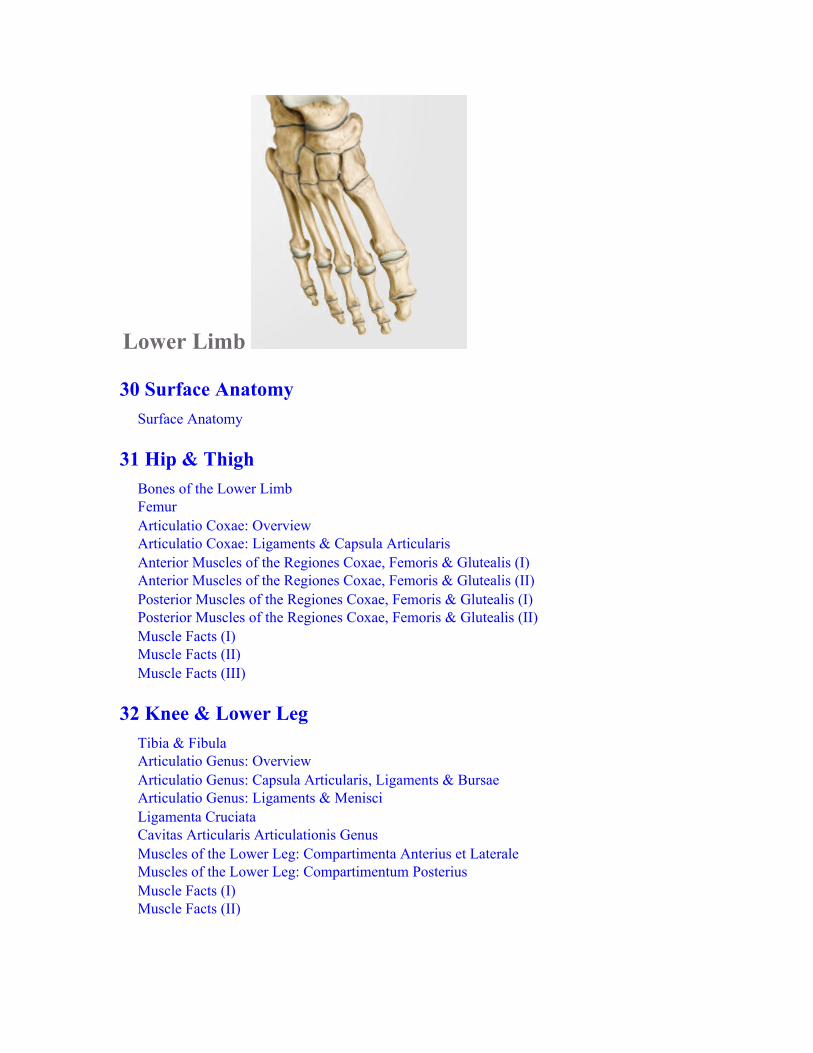
Lower Limb
30 Surface AnatomySurface Anatomy
31 Hip & ThighBones of the Lower LimbFemurArticulatio Coxae: OverviewArticulatio Coxae: Ligaments & Capsula ArticularisAnterior Muscles of the Regiones Coxae, Femoris & Glutealis (I)Anterior Muscles of the Regiones Coxae, Femoris & Glutealis (II)Posterior Muscles of the Regiones Coxae, Femoris & Glutealis (I)Posterior Muscles of the Regiones Coxae, Femoris & Glutealis (II)Muscle Facts (I)Muscle Facts (II)Muscle Facts (III)
32 Knee & Lower LegTibia & FibulaArticulatio Genus: OverviewArticulatio Genus: Capsula Articularis, Ligaments & BursaeArticulatio Genus: Ligaments & MenisciLigamenta CruciataCavitas Articularis Articulationis GenusMuscles of the Lower Leg: Compartimenta Anterius et LateraleMuscles of the Lower Leg: Compartimentum PosteriusMuscle Facts (I)Muscle Facts (II)

33 Tarsus & FootBones of the FootArticulationes Pedis (I)Articulationes Pedis (II)Articulationes Pedis (III)Ligaments of the Tarsus & FootPlantar Vault & Arches of the FootMuscles of the Planta PedisMuscles & Vaginae Tendinum of the FootMuscle Facts (I)Muscle Facts (II)
34 NeurovasculatureArteries of the Lower LimbVeins & Lymphatics of the Lower LimbPlexus LumbosacralisNerves of the Plexus LumbalisNerves of the Plexus Lumbalis: Nervus Obturatorius & Nervus FemoralisNerves of the Plexus SacralisNerves of the Plexus Sacralis: Nervus IschiadicusSuperficial Nerves & Vessels of the Lower LimbTopography of the Inguinal Region (Inguen)Topography of the Regio GlutealisTopography of the Regiones Femoris Anterior et PosteriorTopography of the Compartimentum Cruris Posterius & FootTopography of the Compartimenta Cruris Laterale et Anterius and Dorsum PedisTopography of the Planta Pedis
35 Sectional & Radiographic AnatomySectional Anatomy of the Lower LimbRadiographic Anatomy of the Lower Limb (I)Radiographic Anatomy of the Lower Limb (II)Radiographic Anatomy of the Lower Limb (III)
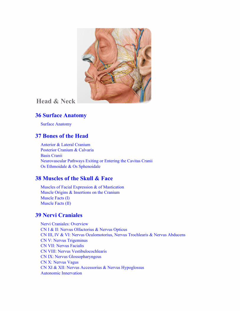
Head & Neck
36 Surface AnatomySurface Anatomy
37 Bones of the HeadAnterior & Lateral CraniumPosterior Cranium & CalvariaBasis CraniiNeurovascular Pathways Exiting or Entering the Cavitas CraniiOs Ethmoidale & Os Sphenoidale
38 Muscles of the Skull & FaceMuscles of Facial Expression & of MasticationMuscle Origins & Insertions on the CraniumMuscle Facts (I)Muscle Facts (II)
39 Nervi CranialesNervi Craniales: OverviewCN I & II: Nervus Olfactorius & Nervus OpticusCN III, IV & VI: Nervus Oculomotorius, Nervus Trochlearis & Nervus AbducensCN V: Nervus TrigeminusCN VII: Nervus FacialisCN VIII: Nervus VestibulocochlearisCN IX: Nervus GlossopharyngeusCN X: Nervus VagusCN XI & XII: Nervus Accessorius & Nervus HypoglossusAutonomic Innervation

40 Neurovasculature of the Cranium & FaceInnervation of the FaceArteries of the Head & NeckArteria Carotis Externa: Anterior, Medial & Posterior BranchesArteria Carotis Externa: Terminal BranchesVeins of the Head & NeckMeningesSinus Durae MatrisTopography of the Superficial FaceTopography of the Regio Parotideomasseterica & Fossa TemporalisTopography of the Fossa InfratemporalisTopography of the Fossa Pterygopalatina
41 Orbita & EyeBones of the OrbitaMusculi OrbitaeNeurovasculature of the OrbitaTopography of the OrbitaOrbita & PalpebraBulbus OculiCornea, Iris & Lens
42 Cavitas Nasi & NasusBones of the Cavitas NasiSinus ParanasalesNeurovasculature of the Cavitas Nasi
43 Os Temporale & EarOs TemporaleAuris Externa & Meatus Acusticus ExternusAuris Media: Cavitas TympaniAuris Media: Ossicula Auditus & Membrana TympanicaArteries of the Auris MediaAuris Interna
44 Cavitas Oris & PharynxBones of the Cavitas OrisArticulatio TemporomandibularisTeethCavitas Oris Muscle FactsInnervation of the Cavitas OrisTongue

Topography of the Cavitas Oris & Glandulae SalivariaeTonsillae & PharynxMusculi PharyngisNeurovasculature of the Pharynx
45 NeckMuscle Facts (I)Muscle Facts (II)Muscle Facts (III)Arteries & Veins of the NeckLymphatics of the NeckInnervation of the NeckLarynx: Cartilage & StructureLarynx: Muscles & LevelsNeurovasculature of the Larynx, Glandulae Thyroidea & ParathyroideaTopography of the Neck: Regiones & FasciaTopography of the Regio Cervicalis AnteriorTopography of the Regio Cervicalis Anterior & Regio Cervicalis LateralisTopography of the Regio Cervicalis LateralisTopography of the Regio Cervicalis Posterior
46 Sectional & Radiographic AnatomySectional Anatomy of the Head & Neck (I)Sectional Anatomy of the Head & Neck (II)Sectional Anatomy of the Head & Neck (III)Sectional Anatomy of the Head & Neck (IV)Sectional Anatomy of the Head & Neck (V)Radiographic Anatomy of the Head & Neck (I)Radiographic Anatomy of the Head & Neck (II)Radiographic Anatomy of the Head & Neck (III)
Brain & Nervous System
47 BrainNervous System: OverviewEncephalon, Macroscopic OrganizationDiencephalonTruncus Encephali & CerebellumVentricles & CSF Spaces
48 Blood Vessels of the BrainVeins and Venous Sinuses of the BrainArteriae Encephali
49 Functional SystemsAnatomy & Organization of the Medulla SpinalisSensory & Motor Pathways
50 Autonomic Nervous SystemAutonomic Nervous System (I): OverviewAutonomic Nervous System (II)
51 Sectional & Radiographic AnatomySectional Anatomy of the Nervous SystemRadiographic Anatomy of the Nervous System
Index
Acknowledgments
We would like to thank the authors of the original award-winning ThiemeAtlas of Anatomy three-volume series, Michael Schuenke, Erik Schulte, andUdo Schumacher, and the illustrators, Karl Wesker and Marcus Voll, for theirwork over the course of many years.We thank the many instructors and students who have pointed out to us whatwe have done well and brought to our attention errors, ambiguities, and newinformation, or have suggested how we could present a topic moreeffectively. This input, combined with our experience teaching with theAtlas, have guided our work on this edition.
We again cordially thank the members of the first edition Advisory Board fortheir contributions:• Bruce M. Carlson, MD, PhD
University of MichiganAnn Arbor, Michigan
• Derek Bryant (Class of 2011)University of Toronto Medical SchoolBurlington, Ontario
• Peter Cole, MDGlamorum Healing CentreOrangeville, Ontario
• Michael Droller, MDThe Mount Sinai Medical CenterNew York, New York
• Anthony Firth, PhDImperial College London London
• Mark H. Hankin, PhD

University of Virginia, School of MedicineCharlottesville, Virginia
• Katharine Hudson (Class of 2010)McGill Medical SchoolMontreal, Quebec
• Christopher Lee (Class of 2010)Harvard Medical SchoolCambridge, Massachusetts
• Francis Liuzzi, PhDLake Erie College of Osteopathic MedicineBradenton, Florida
• Graham Louw, PhDUniversity of Cape Town Medical SchoolUniversity of Cape Town
• Estomih Mtui, MDWeill Cornell Medical CollegeNew York, New York
• Srinivas Murthy, MDHarvard Medical SchoolBoston, Massachusetts
• Jeff Rihn, MDThe Rothman InstitutePhiladelphia, Pennsylvania
• Lawrence Rizzolo, PhDYale UniversityNew Haven, Connecticut
• Mikel Snow, PhDUniversity of Southern CaliforniaLos Angeles, California
• Kelly Wright (Class of 2010)Wayne State University School of MedicineDetroit, Michigan
It has been a great honor to act as a consulting editor, with responsibility forthe Latin nomenclature, for Atlas of Anatomy, Third Edition. There wereseveral people from whom I received a great deal of assistance and guidance,and must express my gratitude towards. Regarding the discussion ofnomenclature, I would wish to thank my mentor Prof. Peter Århem, Ph.D.,my father Lennart Zeberg, M.D., and Prof. Jonas Broman, Ph.D. In addition,I would also like to express my gratitude to Prof. Björn Meister, M.D., Ph.D.,and Prof. Kaj Fried, Ph.D.Moreover, I am deeply grateful to the staff at Thieme Medical Publishers thatI have been in close contact with, in particular, the editorial director AnneSydor, Ph.D., managing editor Judith Tomat, editorial assistant Tony Paese,and marketing agent David Towle.I would also like to acknowledge the Federative International Programme forAnatomical Terminology (FIPAT) for their work towards a standardnomenclature in the field of anatomy.Hugo Zeberg

Foreword
This Atlas of Anatomy, in my opinion, is the finest single-volume atlas ofhuman anatomy that has ever been created. Two factors make it so: theimages and the way they have been organized.
The artists, Markus Voll and Karl Wesker, have created a new standard ofexcellence in anatomical art. Their graceful use of transparency and theirsensitive representation of light and shadow give the reader an accurate three-dimensional understanding of every structure.
The authors have organized the images so that they give just the flow ofinformation a student needs to build up a clear mental image of the humanbody. Each two-page spread is a self-contained lesson that unobtrusivelyshows the hand of an experienced and thoughtful teacher. I wish I could haveheld this book in my hands when I was a student; I envy any student whodoes so now.
Robert D. AclandLouisville, Kentucky

Preface
It is with a mix of pride and humility that we offer our 3rd edition of the Atlasof Anatomy. As with the previous editions, we have tried to respond to therequests, comments, and criticisms of our readers. Although this edition wasprepared without the contributions of our friend and co-author, LawrenceRoss, who passed away in 2015, we have tried to maintain the same qualityof excellence and attention to detail that he helped bring to the previouseditions.
In this latest edition we focused our attention on three major tasks. The firstreflects an understanding that anatomy is a changing science. Our readersunderstand that it is a dynamic part of clinical medicine, itself a scienceundergoing constant evolution. Concepts and terminology changeaccordingly and we feel a responsibility to pass on to our readers the mostaccurate and current information available.
Our second task was to add additional examples of sectional and radiographicimages to help students apply their knowledge of anatomic structure andrelationships to comparable clinical representations. While radiology as aclinical discipline is a specialty that requires expertise in diagnoses andtreatment (and as such is not addressed here), the topographic interpretationof radiographic images is a natural companion to the study of anatomy. Tothis end, we have moved some images that were previously integrated intoearlier chapters and added many new images to create a new Sectional andRadiographic Anatomy chapter in each unit.
Finally, we have expanded areas that deserved greater attention. A newlytitled Brain and Nervous System unit replaces the former Neuro- anatomyunit. Here, the reader will find a greater focus on the gross anatomy of thebrain and peripheral nervous system. We've also added new spreads on theautonomic nervous system, a topic that needed to be expanded. In the Pelvisand Perineum unit, some images were removed and others revised to

illustrate current anatomic theory. In addition, new art that betterdemonstrates the complex pelvic anatomy has been added throughout theunit.
As always, we thank reviewers, colleagues, and students who commented onprevious editions and have suggested appropriate corrections.
We recognize that our efforts, though important, are just one part of theprocess that brings this textbook to your desk. Support from the entireThieme Publishers team has been essential in creating the third edition. Weare especially grateful to Julie O'Meara, Developmental Editor; Tony Paese,Editorial Assistant; Anne M. Sydor, PhD, Editorial Director, EducationalProducts; Barbara Chernow, PhD, Production Manager; and Carol Pierson,compositor, for excellence in their individual areas of expertise and theirunwavering confidence in our ability to produce a quality manuscript.
Anne M. GilroyWorcester, Massachusetts
Brian R. MacPhersonLexington, Kentucky

Preface to the First Edition
Each of the authors was amazed and impressed with the extraordinary detail,accuracy, and beauty of the illustrations that were created for the ThiemeAtlas of Anatomy. We feel these images are one of the most significantadditions to anatomical education in the past 50 years. It was our intent to usethese exceptional illustrations as the cornerstone of our effort in creating aconcise single volume Atlas of Anatomy for the curious and eager healthscience student.
Our challenge was first to select from this extensive collection those imagesthat are most instructive and illustrative of current dissection approaches.Along the way, however, we realized that creating a single-volume atlas wasmuch more than choosing images: each image has to convey a significantamount of detail while the appeal and labeling need to be clean and soothingto the eye. Therefore, hundreds of illustrations were drawn new or modifiedto fit the approach of this new atlas. In addition, key schematic diagrams andsimplified summary-form tables were added wherever needed. Dozens ofapplicable radiographic images and important clinical correlates have beenadded where appropriate. Additionally, surface anatomy illustrations areaccompanied by questions designed to direct the student's attention toanatomic detail that is most relevant in conducting the physical exam.Elements from each of these features are arranged in a regional format tofacilitate common dissection approaches. Within each region, the variouscomponents are examined systemically, followed by topographical images totie the systems together within the region. In all of this, a clinical perspectiveon the anatomical structures is taken. The unique two facing pages “spread”format focuses the user to the area/topic being explored.
We hope these efforts—the results of close to 100 combined years experienceteaching the discipline of anatomy to bright, enthusiastic students—hasresulted in a comprehensive, easy-to-use resource and reference.

We would like to thank our colleagues at Thieme Publishers who soprofessionally facilitated this effort. We cannot thank enough Cathrin E.Schulz, MD, Editorial Director, Educational Products, who so graciouslyreminded us of deadlines, while always being available to “trouble shoot”problems. More importantly, she encouraged, helped, and complimented ourefforts.
We also wish to extend very special thanks and appreciation to BridgetQueenan, Developmental Editor, who edited and developed the manuscriptwith an outstanding talent for visualization and intuitive flow of information.We are very grateful to her for catching many details along the way whilealways patiently responding to requests for artwork and labeling changes.
Cordial thanks to Elsie Starbecker, Senior Production Editor, who with greatcare and speed produced this atlas with its over 2,200 illustrations. Finally,thanks to Rebecca McTavish, Developmental Editor, for joining the team inthe correction phase. So very much of their hard work has made the Atlas ofAnatomy a reality.
Anne M. GilroyWorcester, Massachusetts
Brian R. MacPhersonLexington, Kentucky
Lawrence M. RossHouston, Texas

A Note on the Use of Latin Terminology
To introduce the Latin nomenclature into an English textbook is a delicatetask, particularly because the many Latin loanwords have passed into generaluse. Some loanwords are so common that fluency of the text would bedisturbed if they were to be translated back into Latin. The Latin loanwordshave typically undergone several adaptations before becoming part of theEnglish language. A term such as sympathetic trunk (lat. truncus sympaticus)has undergone morphological adaptation (through the loss of masculinesuffix -us), orthographical adaptation (through the substitution of a‘Germanic’ k for a Latin c), and phonological adaptation (th and e instead of tand i). In addition, the word order has been reversed. The Latin termsympaticus is in fact borrowed from late Greek sympathetikos (fromsympathes ‘having a fellow feeling, affected by like feelings’), therebyillustrating that terms move between languages when cultures meet. Otheranatomical terms are so colloquial (e.g., hand), that a Latin term (e.g., manus)would be inappropriate to use at all occasions. Clearly, the text would easilybecome unreadable if a strict translation of all English terms into Latin wereimposed. As a result, Latin has been used as long as it does not disrupt theflow of the text and whenever possible in figures and tables. In some cases,dual terminology has been used, with either the English or Latin word inparentheses. As much as possible, the terminology of TerminologiaAnatomica (1998) has been followed.
Hugo ZebergStockholm, Sweden

Back
1 Surface AnatomySurface Anatomy
2 Bones, Ligaments & JointsColumna Vertebralis: OverviewColumna Vertebralis: ElementsVertebrae CervicalesVertebrae Thoracicae & LumbalesOs Sacrum & Os CoccygisDisci IntervertebralesJoints of the Columna Vertebralis: OverviewJoints of the Columna Vertebralis: Craniovertebral RegionVertebral Ligaments: Overview & Cervical SpineVertebral Ligaments: Thoracolumbar Spine
3 MusclesMuscles of the Back: OverviewIntrinsic Muscles of the Cervical SpineIntrinsic Muscles of the Back (Musculi Dorsi Proprii)Muscle Facts (I)Muscle Facts (II)Muscle Facts (III)

4 NeurovasculatureArteries & Veins of the BackNerves of the BackMedulla Spinalis (Spinal Cord)Segments of the Medulla Spinalis & Nervi SpinalesArteries & Veins of the Medulla SpinalisNeurovascular Topography of the Back
5 Sectional & Radiographic AnatomyRadiographic Anatomy of the Back (I)Radiographic Anatomy of the Back (II)

1 Surface Anatomy
Surface Anatomy
Fig. 1.1 Palpable structures of the back Posterior view.
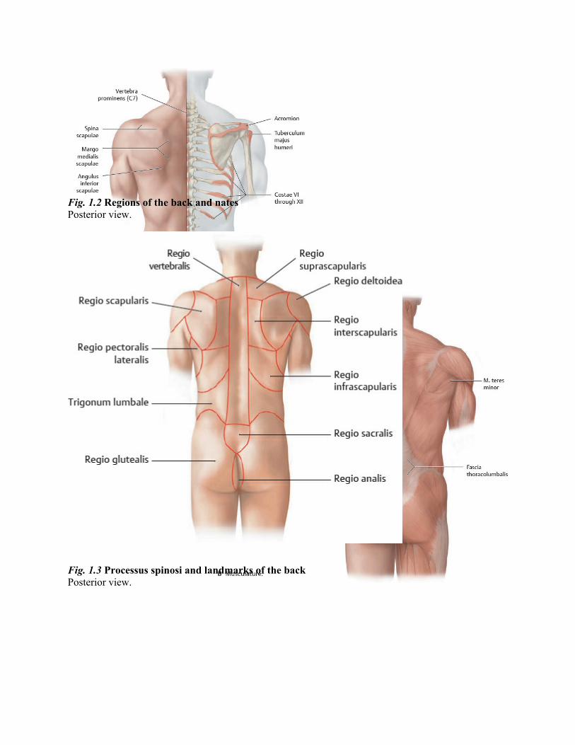
Fig. 1.2 Regions of the back and nates Posterior view.
Fig. 1.3 Processus spinosi and landmarks of the back Posterior view.
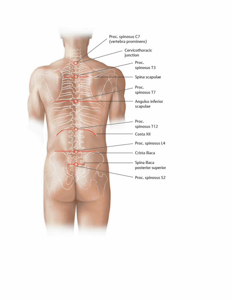

Table 1.1 Reference lines of the back
Linea mediana posterior Posterior trunk midline at the level of the procc.spinosi
Linea paravertebralis Line at the level of the procc. transversi
Linea scapularis Line through the angulus inferior scapulae
Table 1.2 Processus spinosi that provide useful posterior landmarks
Vertebral processus spinosus Posterior landmark
C7 Vertebra prominens (the projecting proc. spinosus of C7 isclearly visible and palpable)
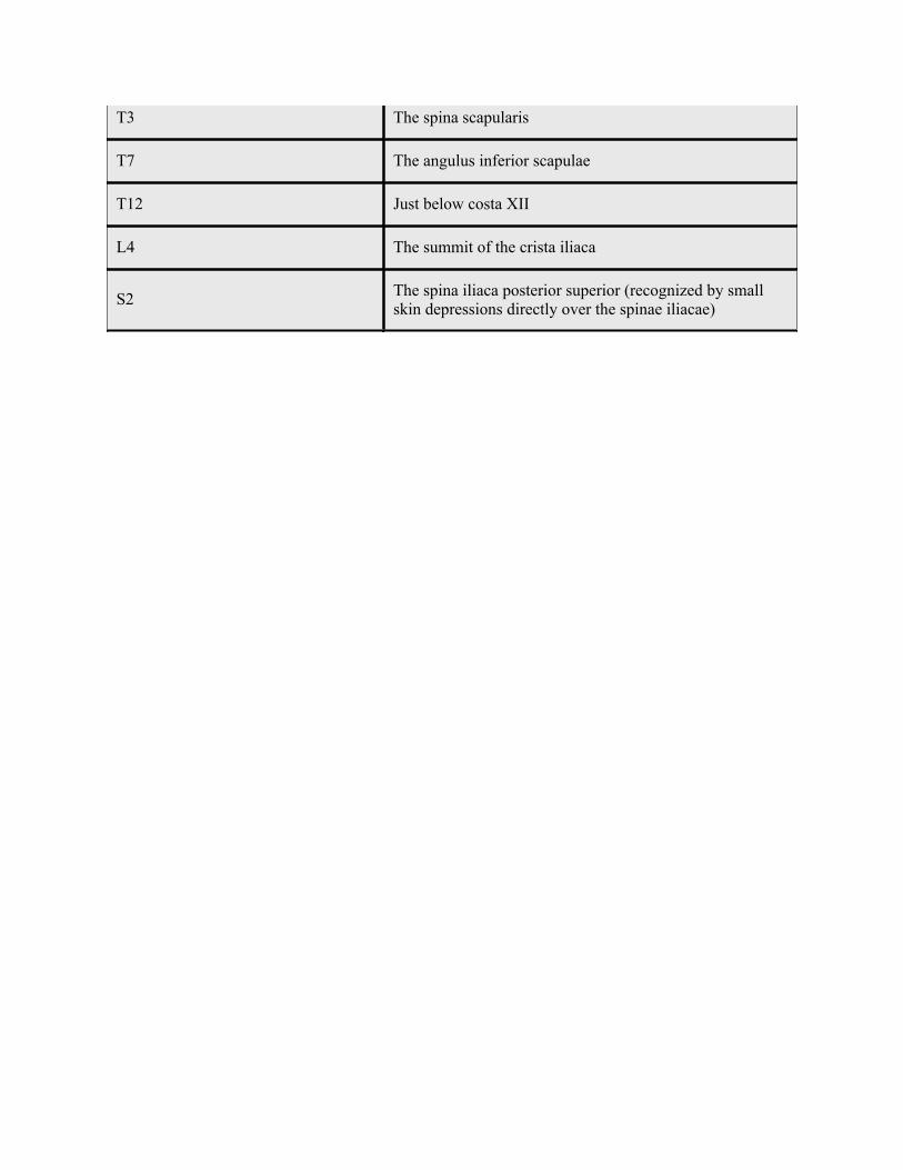
T3 The spina scapularis
T7 The angulus inferior scapulae
T12 Just below costa XII
L4 The summit of the crista iliaca
S2 The spina iliaca posterior superior (recognized by smallskin depressions directly over the spinae iliacae)
2 Bones, Ligaments & Joints
Columna Vertebralis: Overview
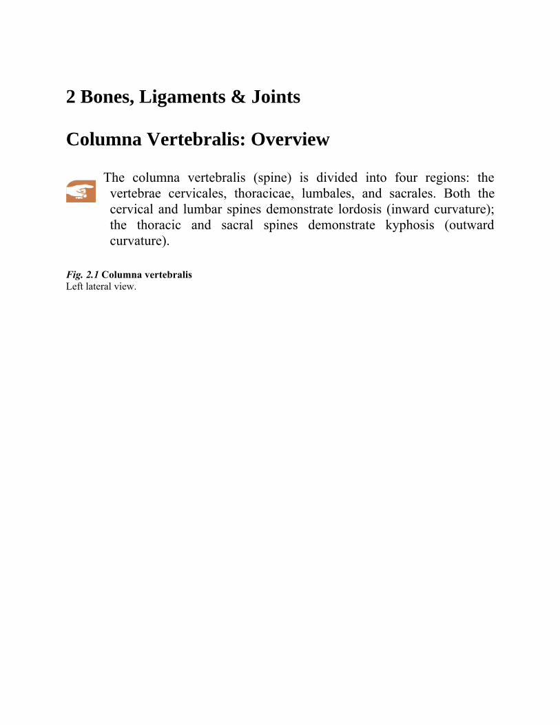
The columna vertebralis (spine) is divided into four regions: thevertebrae cervicales, thoracicae, lumbales, and sacrales. Both thecervical and lumbar spines demonstrate lordosis (inward curvature);the thoracic and sacral spines demonstrate kyphosis (outwardcurvature).
Fig. 2.1 Columna vertebralis Left lateral view.
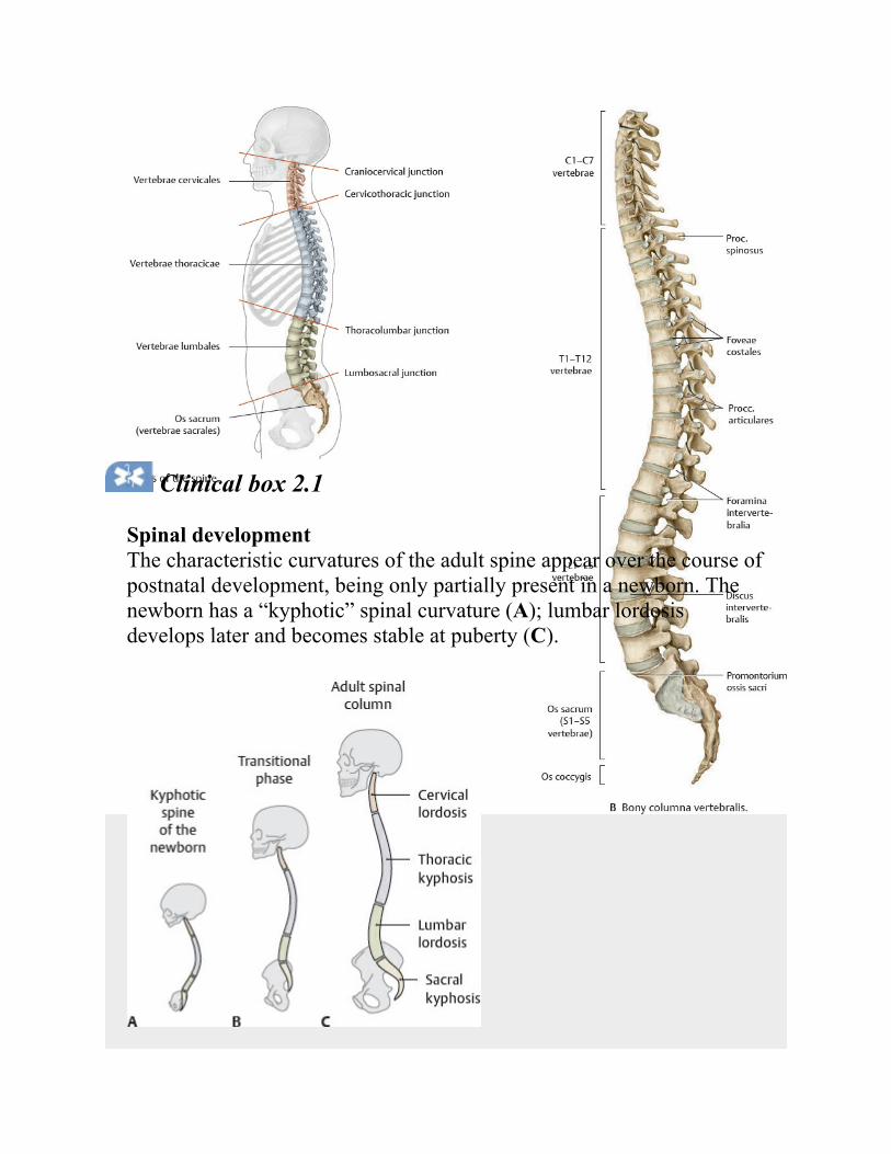
Clinical box 2.1
Spinal developmentThe characteristic curvatures of the adult spine appear over the course ofpostnatal development, being only partially present in a newborn. Thenewborn has a “kyphotic” spinal curvature (A); lumbar lordosisdevelops later and becomes stable at puberty (C).
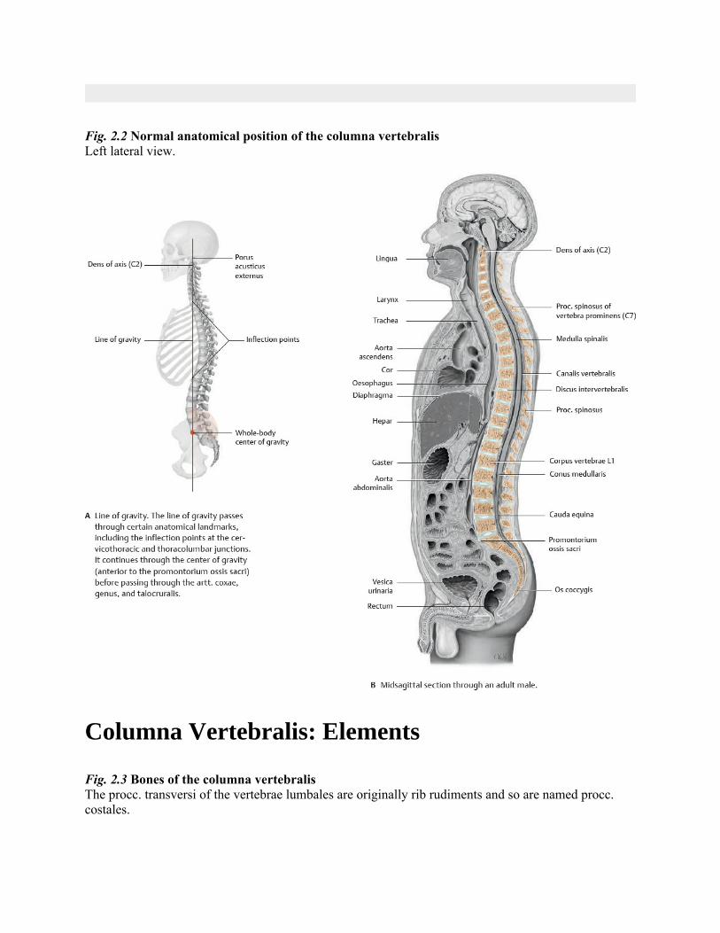
Fig. 2.2 Normal anatomical position of the columna vertebralisLeft lateral view.
Columna Vertebralis: Elements
Fig. 2.3 Bones of the columna vertebralisThe procc. transversi of the vertebrae lumbales are originally rib rudiments and so are named procc.costales.