TLR5 Signaling Enhances the Proliferation of Human ... · TLR5 Signaling Enhances the Proliferation...
9
TLR5 Signaling Enhances the Proliferation of Human Allogeneic CD40-Activated B Cell Induced CD4 hi CD25 + Regulatory T Cells Ping-Lung Chan, Jian Zheng, Yinping Liu, Kwok-Tai Lam, Zheng Xiang, Huawei Mao, Yuan Liu, Gang Qin, Yu-Lung Lau, Wenwei Tu* Department of Paediatrics and Adolescent Medicine, Li Ka Shing Faculty of Medicine, University of Hong Kong. Hong Kong SAR, China Abstract Although diverse functions of different toll-like receptors (TLR) on human natural regulatory T cells have been demonstrated recently, the role of TLR-related signals on human induced regulatory T cells remain elusive. Previously our group developed an ex vivo high-efficient system in generating human alloantigen-specific CD4 hi CD25 + regulatory T cells from naı ¨ve CD4 + CD25 2 T cells using allogeneic CD40-activated B cells as stimulators. In this study, we investigated the role of TLR5- related signals on the generation and function of these novel CD4 hi CD25 + regulatory T cells. It was found that induced CD4 hi CD25 + regulatory T cells expressed an up-regulated level of TLR5 compared to their precursors. The blockade of TLR5 using anti-TLR5 antibodies during the co-culture decreased CD4 hi CD25 + regulatory T cells proliferation by induction of S phase arrest. The S phase arrest was associated with reduced ERK1/2 phosphorylation. However, TLR5 blockade did not decrease the CTLA-4, GITR and FOXP3 expressions, and the suppressive function of CD4 hi CD25 + regulatory T cells. In conclusion, we discovered a novel function of TLR5-related signaling in enhancing the proliferation of CD4 hi CD25 + regulatory T cells by promoting S phase progress but not involved in the suppressive function of human CD40-activated B cell-induced CD4 hi CD25 + regulatory T cells, suggesting a novel role of TLR5-related signals in the generation of induced regulatory T cells. Citation: Chan P-L, Zheng J, Liu Y, Lam K-T, Xiang Z, et al. (2013) TLR5 Signaling Enhances the Proliferation of Human Allogeneic CD40-Activated B Cell Induced CD4 hi CD25 + Regulatory T Cells. PLoS ONE 8(7): e67969. doi:10.1371/journal.pone.0067969 Editor: Kin Mang Lau, The Chinese University of Hong Kong, Hong Kong Received April 15, 2013; Accepted May 23, 2013; Published July 3, 2013 Copyright: ß 2013 Chan et al. This is an open-access article distributed under the terms of the Creative Commons Attribution License, which permits unrestricted use, distribution, and reproduction in any medium, provided the original author and source are credited. Funding: This work was supported in part by NSFC/RGC Joint Research Scheme (N_HKU 747/11), General Research Fund, Research Grants Council of Hong Kong (HKU 781211M); Area of Excellence program (AoE/M-12/06), University Grants Committee of Hong Kong Special Administrative Region. The funders had no role in study design, data collection and analysis, decision to publish, or preparation of the manuscript. Competing Interests: The authors have declared that no competing interests exist. * E-mail: [email protected] Introduction Natural regulatory T cells (nTregs) and induced regulatory T cells (iTregs) are important to the self-tolerance of the human body and the tolerance to transplanted organs or tissues [1,2]. Impairments in the development or functions of these cells can cause autoimmune diseases such as immunodysregulation poly- endocrinopathy enteropathy X-linked syndrome [3], and systemic lupus erythematosus [4], which is either fatal or severely reduces the quality of life of patients, and graft rejection in transplantation. Although many efficient strategies have been developed to treat autoimmune diseases and graft rejection, their severe side effects lead to an urgent need for novel therapeutic strategies, such as adoptive transfer of antigen-specific regulatory T cells [5]. As a result, investigation in the biology of regulatory T cells is crucial for understanding these diseases and the development of novel therapeutic strategies for treating and managing autoimmune diseases and graft rejections. It is known that activation and function of regulatory T cells require signals from both T cell receptor (TCR) [6] and CD28 [7,8]. However, as increasing number of co-stimulatory molecules, such as OX-40 and PD-1, were discovered to be implicated in the activation and function of regulatory T cells [9,10], it is speculated that co-stimulatory molecules may also play diverse and crucial roles in the activation and function of these cells [11]. Reports about the non-absolute requirement of TCR signal in T cell function further support this speculation [12,13]. As a result, investigation in the role of co-stimulatory molecules in regulatory T cells is warranted. Although toll-like receptors (TLR) are thought to mainly participate in the antigen recognition and activation of innate immune cells [14], they are also crucial co- stimulatory molecules involved in the function of T cells. In vitro data suggested that TLR2, 4, 5, 7, and 8 could promote the proliferation of CD4 + T cells [15,16], and compelling evidence from the experiment of Marsland et al. demonstrated that CpG DNA stimulation could activate CD4 + T cells from PKC-h 2/2 mice and causing EAE, indicating that TLR stimulation could support the activation and differentiation of CD4 + T cells in the absence of TCR signaling [17]. TLRs are also involved in the activation and function of nTregs. Direct stimulation of mice CD4 + nTregs with TLR2 ligand Pam3Cys increased the proliferation and concomitantly abrogated the function of the cells [18,19], while stimulation of human nTregs with TLR4 ligand LPS and IL-2 up-regulated FOXP3 expression and the suppressive function [20]. In vivo result from TLR9 2/2 mice also PLOS ONE | www.plosone.org 1 July 2013 | Volume 8 | Issue 7 | e67969
-
Upload
hoangtuyen -
Category
Documents
-
view
217 -
download
0
Transcript of TLR5 Signaling Enhances the Proliferation of Human ... · TLR5 Signaling Enhances the Proliferation...

TLR5 Signaling Enhances the Proliferation of HumanAllogeneic CD40-Activated B Cell Induced CD4hiCD25+
Regulatory T CellsPing-Lung Chan, Jian Zheng, Yinping Liu, Kwok-Tai Lam, Zheng Xiang, Huawei Mao, Yuan Liu, Gang Qin,
Yu-Lung Lau, Wenwei Tu*
Department of Paediatrics and Adolescent Medicine, Li Ka Shing Faculty of Medicine, University of Hong Kong. Hong Kong SAR, China
Abstract
Although diverse functions of different toll-like receptors (TLR) on human natural regulatory T cells have been demonstratedrecently, the role of TLR-related signals on human induced regulatory T cells remain elusive. Previously our group developedan ex vivo high-efficient system in generating human alloantigen-specific CD4hiCD25+ regulatory T cells from naı̈veCD4+CD252 T cells using allogeneic CD40-activated B cells as stimulators. In this study, we investigated the role of TLR5-related signals on the generation and function of these novel CD4hiCD25+ regulatory T cells. It was found that inducedCD4hiCD25+ regulatory T cells expressed an up-regulated level of TLR5 compared to their precursors. The blockade of TLR5using anti-TLR5 antibodies during the co-culture decreased CD4hiCD25+ regulatory T cells proliferation by induction of Sphase arrest. The S phase arrest was associated with reduced ERK1/2 phosphorylation. However, TLR5 blockade did notdecrease the CTLA-4, GITR and FOXP3 expressions, and the suppressive function of CD4hiCD25+ regulatory T cells. Inconclusion, we discovered a novel function of TLR5-related signaling in enhancing the proliferation of CD4hiCD25+
regulatory T cells by promoting S phase progress but not involved in the suppressive function of human CD40-activated Bcell-induced CD4hiCD25+ regulatory T cells, suggesting a novel role of TLR5-related signals in the generation of inducedregulatory T cells.
Citation: Chan P-L, Zheng J, Liu Y, Lam K-T, Xiang Z, et al. (2013) TLR5 Signaling Enhances the Proliferation of Human Allogeneic CD40-Activated B Cell InducedCD4hiCD25+ Regulatory T Cells. PLoS ONE 8(7): e67969. doi:10.1371/journal.pone.0067969
Editor: Kin Mang Lau, The Chinese University of Hong Kong, Hong Kong
Received April 15, 2013; Accepted May 23, 2013; Published July 3, 2013
Copyright: � 2013 Chan et al. This is an open-access article distributed under the terms of the Creative Commons Attribution License, which permitsunrestricted use, distribution, and reproduction in any medium, provided the original author and source are credited.
Funding: This work was supported in part by NSFC/RGC Joint Research Scheme (N_HKU 747/11), General Research Fund, Research Grants Council of Hong Kong(HKU 781211M); Area of Excellence program (AoE/M-12/06), University Grants Committee of Hong Kong Special Administrative Region. The funders had no role instudy design, data collection and analysis, decision to publish, or preparation of the manuscript.
Competing Interests: The authors have declared that no competing interests exist.
* E-mail: [email protected]
Introduction
Natural regulatory T cells (nTregs) and induced regulatory T
cells (iTregs) are important to the self-tolerance of the human body
and the tolerance to transplanted organs or tissues [1,2].
Impairments in the development or functions of these cells can
cause autoimmune diseases such as immunodysregulation poly-
endocrinopathy enteropathy X-linked syndrome [3], and systemic
lupus erythematosus [4], which is either fatal or severely reduces
the quality of life of patients, and graft rejection in transplantation.
Although many efficient strategies have been developed to treat
autoimmune diseases and graft rejection, their severe side effects
lead to an urgent need for novel therapeutic strategies, such as
adoptive transfer of antigen-specific regulatory T cells [5]. As a
result, investigation in the biology of regulatory T cells is crucial
for understanding these diseases and the development of novel
therapeutic strategies for treating and managing autoimmune
diseases and graft rejections.
It is known that activation and function of regulatory T cells
require signals from both T cell receptor (TCR) [6] and CD28
[7,8]. However, as increasing number of co-stimulatory molecules,
such as OX-40 and PD-1, were discovered to be implicated in the
activation and function of regulatory T cells [9,10], it is speculated
that co-stimulatory molecules may also play diverse and crucial
roles in the activation and function of these cells [11]. Reports
about the non-absolute requirement of TCR signal in T cell
function further support this speculation [12,13]. As a result,
investigation in the role of co-stimulatory molecules in regulatory
T cells is warranted. Although toll-like receptors (TLR) are
thought to mainly participate in the antigen recognition and
activation of innate immune cells [14], they are also crucial co-
stimulatory molecules involved in the function of T cells. In vitro
data suggested that TLR2, 4, 5, 7, and 8 could promote the
proliferation of CD4+ T cells [15,16], and compelling evidence
from the experiment of Marsland et al. demonstrated that CpG
DNA stimulation could activate CD4+ T cells from PKC-h2/2
mice and causing EAE, indicating that TLR stimulation could
support the activation and differentiation of CD4+ T cells in the
absence of TCR signaling [17]. TLRs are also involved in the
activation and function of nTregs. Direct stimulation of mice
CD4+ nTregs with TLR2 ligand Pam3Cys increased the
proliferation and concomitantly abrogated the function of the
cells [18,19], while stimulation of human nTregs with TLR4
ligand LPS and IL-2 up-regulated FOXP3 expression and the
suppressive function [20]. In vivo result from TLR92/2 mice also
PLOS ONE | www.plosone.org 1 July 2013 | Volume 8 | Issue 7 | e67969

suggested that TLR9 signaling enhanced nTregs function through
induction of IDO [21].
TLR5 is expressed in both CD4+ T cells and nTregs [22,23].
Since the TLR5 ligand, flagellin, is commonly expressed in
different bacteria species [24,25], TLR5 may be particularly
important to the induction of tolerance to intestinal commensal
bacteria and of oral tolerance [26]. Currently, there is only a single
report investigated on the direct effect of TLR5-related signals on
human nTregs. Crellin et al. reported that stimulation of human
nTregs with anti-CD3/CD28 and flagellin up-regulated FOXP3
expression and the suppressive function [27]. Since the direct
effect of TLR5-related signals on iTregs remains unexamined, the
function of TLR5 in human iTregs is investigated in this study.
Previously our laboratory has developed a simple and cost
effective novel protocol of large-scale in vitro induction and
expansion of human alloantigen specific CD4hiCD25+ regulatory
T cells with therapeutic potential from naı̈ve
CD4+CD252CD45RO2 precursors using human allogeneic
CD40-activated B cells as stimulators without the use of exogenous
cytokine. Co-culture of human naı̈ve CD4+CD252 T cells with
allogeneic CD40-activated B cells at T cell to B cell ratio of 10:1
induced a population of CD4hiCD25+ regulatory T cells [28]. The
CD4hiCD25+ T cells were alloantigen specific
CD45RO+CCR72CD62L+ memory T cells and expressed
FOXP3, IFN-c, CTLA-4, and GITR [28,29]. Suppressive MLR
experiment demonstrated that these cells could suppress T cell
proliferation in a cell-cell contact dependent manner which was
partially dependent on the surface CTLA-4, indicating that these
cells are iTregs [28]. In this experiment, we investigated the role of
TLR5-related signals in the generation and function of human
CD4hiCD25+ regulatory T cells induced by allogeneic CD40-
activated B cells and have unveiled a novel function of TLR5-
related signaling in iTregs. Our results indicated that TLR5-
related signaling enhances the proliferation but not the suppressive
function of human CD4hiCD25+ regulatory T cells induced by
allogeneic CD40-activated B cells.
Materials and Methods
Ethics StatementWritten consent for the use of buffy coat for research purposes
was obtained from the donors by the Hong Kong Red Cross Blood
Transfusion Services at the time of blood donation. The use of
buffy coat for this experiment was approved by the Institutional
Review Board of the University of Hong Kong/Hospital Authority
Hong Kong West Cluster (IRB Reference Number: UW 07-390).
Generation of CD40-activated B CellsPBMC were isolated from buffy coat of healthy adult donors
from the Hong Kong Red Cross Blood Transfusion Services.
CD40-acitvated B cells were generated from PBMC via CD40
stimulation using lethally irradiated (96Gy) NIH3T3 cells trans-
fected with the human CD40 ligand (t-CD40-L cells) as stimulator
in B cell medium as we described previously [30,31]. Briefly,
isolated PBMC were co-cultured with the lethally irradiated
(96Gy) t-CD40-L cells in IMDM (GIBCO-BRL, Life Technolo-
gies, CA) supplement with the 2ng/ml of IL-4 (R&D systems,
MN), 5.561027 M of cyclosporine A (Sigma-Aldrich, MO),
50 mg/ml of transferrin (Sigma-Aldrich, MO), 5 mg/ml of insulin
(Sigma-Aldrich, MO), 15 mg/ml of gentamycin (GIBCO-BRL,
Life Technologies, CA), and 10% of heat-inactivated human AB
serum (Innovative Research, MI) at 37uC in 5% CO2. Cells were
sub-cultured to new 6-well plates of t-CD40-L cells every 3 to 4
days. After 14 days of co-culture, more than 95% of the viable
suspended cells are routinely CD19 positive. These B cells were
cryopreserved in 10% DMSO medium for future use.
Isolation of Naı̈ve CD4+CD252CD45RO2 T Cells andInduction of CD4hiCD25+ Regulatory T CellsThe CD4hiCD25+ regulatory T cells were induced by the co-
culture of the CD4+CD252CD45RO2 T cells with the allogeneic
CD40-activated B cells at a T-cell: B-cell ratio of 10:1 for 6 days as
described previously [28] unless otherwise specified. Human naı̈ve
CD4+CD252CD45RO2 T cells were isolated from healthy
donors PBMC by CD4+ T cell enrichment using the human
CD4 T Cells Enrichment Cocktails (StemCell Technologies,
Canada), followed by negative selection using a human naı̈ve
CD4+ T Cell Isolation Kit and LD Column (Miltenyi Biotec,
Germany) according to manufacturer’s instructions.
TLR5 Blockade and Chemical Inhibition of ERK1/2Phosphorylation10 mg/ml of anti-TLR5 mAb, and its relevant isotype control
(Invivogen, CA) were used for the blockade of TLR5. 20 mM of
PD98059 and its solvent control DMSO (Merck, Germany) were
used for chemical inhibition of ERK1/2 phosphorylation.
Antibodies and PD98059 were added to CD4+CD252CD45RO2
T cells one hour before co-culturing with allogeneic CD40-
activated B cells and were replenished when cell culture medium
was changed.
Flow Cytometric AssaysAll fluorescence-conjugated antibodies were from BD-Biosci-
ences unless otherwise specified: CD4-pacific blue (Biolegend,
CA), CD25-APC-Cy7, CTLA-4-PE, GITR-PE, TLR5-PE (Im-
genex, CA), human Foxp3 staining kit (clone: PCH101) (eBios-
ciences, CA), p-p44/42 (Thr202/Tyr204)-AlexaFluor-488 (Cell
Signaling, MA). Annexin V/propidium iodide (Gibco-BRL, Life
Technologies, CA) was used for measuring apoptosis. Propidium
iodide (Gibco-BRL, Life Technologies, CA) was used for cell cycle
analysis. For measuring cell proliferation, naı̈ve
CD4+CD252CD45RO2 T cells were stained with CFSE before
co-culturing with allogeneic CD40-activated B cells. Cells were
analyzed using FACS LSRII (BD Biosciences, CA) and results
were analyzed using FlowJo v8.8.2 (Tree Star, OR). Cell cycle
analysis results were analyzed using ModFit (Verity Software
House, ME).
Mixed Lymphocyte Reaction (MLR) AssaysCD4hiCD25+ regulatory T cells generated with or without the
blockade of TLR5 were sorted using FACSAria after 9 days of co-
culture. The sorted CD4hiCD25+ regulatory T cells were titrated
and co-cultured with 56104 responder CD4+CD252 T cells from
the same donor of the CD4hiCD25+ regulatory T cells and 56104
c-irradiated target PBMC from the donor of the CD40-activated B
cells as stimulator for 3 days. 3H-thymidine was added to the co-
culture in the last 18 hours and the proliferation was analyzed by3H-thymidine incorporation assay as we described previously [28].
Statistical AnalysisGraphs and statistical analysis were performed using Prism 5.0
for Windows software (GraphPad Software, CA). One-way
ANOVA with Tukey’s pairwise comparisons was used for
comparing the percentage of regulatory T cells, apoptotic T cells,
and percentage of CD4hiCD25+ regulatory T cells in S phase. p
value of ,0.05 was considered to be significant.
TLR5 Enhances Induced Treg Proliferation
PLOS ONE | www.plosone.org 2 July 2013 | Volume 8 | Issue 7 | e67969

Results
TLR5-related Signals Enhance the Generation ofCD4hiCD25+ Regulatory T Cells Independent of CellApoptosisWe first investigated the TLR5 expression in the CD4hiCD25+
regulatory T cells. A population of CD4hiCD25+ regulatory T cells
and a population of CD4+CD252 T cells without any regulatory
function could be identified in the co-culture of naı̈ve
CD4+CD252CD45RO2 T cells with allogeneic CD40-activated
B cells for 6 days (Figure 1A). As shown in Figure 1 B-E,
CD4hiCD25+ regulatory T cells exhibited an up-regulated surface
(Figure 1B and C) and total TLR5 protein expression (Figure 1D
and E). Interestingly, in CD4+CD252 T cells, surface TLR5
expression level was lower than that of naı̈ve
CD4+CD252CD45RO2 T cells while total TLR5 expression
was the same (Figure 1 B-E).
The up-regulated TLR5 expression in CD4hiCD25+ regulatory
T cells prompted us to investigate the effect of TLR5-related
signals on the generation and function of CD4hiCD25+ regulatory
T cells. It was found that the blockade of TLR5 using anti-TLR5
blocking antibodies decreased CD4hiCD25+ regulatory T cells
generation (Figure 1F and G). Frequency of CD4hiCD25+
regulatory T cells decreased from 61% of total CD4+ T cells to
about 36% after 6 days of co-culture (p,0.001) (Figure 1G),
indicating that TLR5 signaling was involved in CD4hiCD25+
regulatory T cells generation. Since TLR5 was reported to be anti-
apoptotic [32], and could promote the survival of cells and mice
subjected to lethal irradiation [33,34], we further studied whether
the reduced CD4hiCD25+ regulatory T cells generation was due to
increased apoptosis of CD4+ T cells. Surprisingly, cell death
analysis using annexin V/podium iodide staining indicated that
the blockade of TLR5 did not increase the apoptosis of either
CD4hiCD25+ regulatory T cells or CD4+CD252 T cells.
Approximate 5% of CD4+CD252 T cells and 2% of CD4hiCD25+
regulatory T cells were in either early or late apoptotic phase and
TLR5 blockade did not alter the percentage (Figure 1H). These
results indicated that the reduction of CD4hiCD25+ regulatory T
cell generation by blocking TLR5-related signals is not dependent
on cell apoptosis.
TLR5-related Signals Endorse the Proliferation ofCD4hiCD25+ Regulatory T Cells by Promoting the Processof S PhaseUnaltered apoptosis of CD4+ T cells after the blockade of TLR5
suggested that the reduced CD4hiCD25+ regulatory T cells
Figure 1. LR5 blockade reduced the generation of CD4hiCD25+ regulatory T cells and was independent of apoptosis. (A) Flowcytometric analysis of the percentage of CD4hiCD25+ regulatory T cells generated on Day 6 (right panel) from naı̈ve CD4+CD252CD45RO2 T cells (leftpanel). (B) Flow cytometric analysis of the expression of surface TLR5 in freshly isolated naı̈ve CD4+CD252CD45RO2 T cells (dotted line), andCD4+CD252 (dashed line) and CD4hiCD25+ regulatory T cells (solid line) after 6 days of co-culture of naı̈ve CD4+CD252CD45RO2 T cells withallogeneic CD40-activated B cells. Filled histogram indicates the staining obtained from isotype-matched mAb controls. (C) Mean fluorescenceintensity (MFI) of the expression of surface TLR5. Data show Mean+SEM, n = 6. (D) Flow cytometric analysis of total TLR5 in freshly isolated naı̈veCD4+CD252CD45RO2 T cells (dotted line), CD4+CD252 (dashed line), and CD4hiCD25+ regulatory T cells (solid line) after 6 days of co-culture of naı̈veCD4+CD252CD45RO2 T cells with allogeneic CD40-activated B cells. Filled histogram indicates the staining obtained from isotype-matched mAbcontrol. (E) Mean fluorescence intensity (MFI) of the expression of total TLR5. Data show Mean+SEM, n = 6. (F) Flow cytometric analysis of thegeneration of CD4hiCD25+ regulatory T cells with no treatment (left panel), with isotype-matched mAb (middle panel), and with anti-TLR5 blockingmAb (right panel) during the co-culture. (G) Mean percentage of CD4hiCD25+ regulatory T cells generated with no treatment, with isotype-matchedmAb, and with anti-TLR5 blocking mAb. Data shown Mean+SEM, n = 6. (H) Flow cytometric analysis of the percentage of apoptotic CD4hiCD25+
regulatory T cells (upper panel) or CD4+CD252 T cells (lower panel) after 6 days of co-culture of naı̈ve CD4+CD252CD45RO2 T cells with allogeneicCD40-activated B cells. All results shown are representative of three independent experiments. *p,0.05, **p,0.01, ***p,0.001, one way ANOVA withTukey’s pairwise comparisons.doi:10.1371/journal.pone.0067969.g001
TLR5 Enhances Induced Treg Proliferation
PLOS ONE | www.plosone.org 3 July 2013 | Volume 8 | Issue 7 | e67969
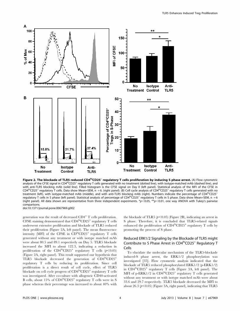
generation was the result of decreased CD4+ T cells proliferation.
CFSE staining demonstrated that CD4hiCD25+ regulatory T cells
underwent extensive proliferation and blockade of TLR5 reduced
their proliferation (Figure 2A, left panel). The mean fluorescence
intensity (MFI) of the CFSE in CDhiCD25+ regulatory T cells
generated without any treatment or with isotype matched mAb
were about 80.5 and 89.1 respectively on Day 5. TLR5 blockade
increased the MFI to about 122.3, indicating a reduction in
proliferation of the CD4hiCD25+ regulatory T cells (p,0.05)
(Figure 2A, right panel). This result supported our hypothesis that
TLR5 blockade decreased the generation of CD4hiCD25+
regulatory T cells by reducing its proliferation. Since cell
proliferation is a direct result of cell cycle, effect of TLR5
blockade on cell cycle progress of CD4hiCD25+ regulatory T cells
was investigated. After co-culture with allogeneic CD40-activated
B cells, about 15% of CD4hiCD25+ regulatory T cells were in S
phase whereas their percentage was increased to about 40% with
the blockade of TLR5 (p,0.05) (Figure 2B), indicating an arrest in
S phase. Therefore, it is concluded that TLR5-related signals
enhanced the proliferation of CD4hiCD25+ regulatory T cells by
promoting the process of S phase.
Reduced ERK1/2 Signaling by the Blockade of TLR5 mightContribute to S Phase Arrest in CD4hiCD25+ Regulatory TCellsTo elucidate the molecular mechanism of the TLR5-blockade
induced-S phase arrest, the ERK1/2 phosphorylation was
investigated [35]. Flow cytometric analysis indicated that the
blockade of TLR5 reduced phosphorylated ERK1/2 (p-ERK1/2)
in CD4hiCD25+ regulatory T cells (Figure 3A, left panel). The
MFI of p-ERK1/2 in CD4hiCD25+ regulatory T cells generated
without any treatment or with isotype matched mAb were about
33.6 and 29.7 respectively. TLR5 blockade decreased the MFI to
about 26.3 (p,0.05) (Figure 3A, right panel), indicating that TLR5
Figure 2. The blockade of TLR5 reduced CD4hiCD25+ regulatory T cells proliferation by inducing S phase arrest. (A) Flow cytometricanalysis of the CFSE signal in CD4hiCD25+ regulatory T cells generated with no treatment (dotted line), with isotype-matched mAb (dashed line), andwith anti-TLR5 blocking mAb (solid line). Filled histogram is the CFSE signal on Day 0 (left panel). Statistical analysis of the MFI of the CFSE inCD4hiCD25+ regulatory T cells. Data show Mean+SEM, n = 6. (right panel). (B) Cell cycle analysis of CD4hiCD25+ regulatory T cells generated with notreatment (left), with isotype-matched mAb (middle), and with anti-TLR5 blocking mAb (right). Numbers indicate the percentage of CD4hiCD25+
regulatory T cells in S phase (left panel). Statistical analysis of percentage of CD4hiCD25+ regulatory T cells in S phase. Data show Mean+SEM, n = 6(right panel). All data shown are representative from three independent experiments. *p,0.05, **p,0.01, one way ANOVA with Tukey’s pairwisecomparisons.doi:10.1371/journal.pone.0067969.g002
TLR5 Enhances Induced Treg Proliferation
PLOS ONE | www.plosone.org 4 July 2013 | Volume 8 | Issue 7 | e67969
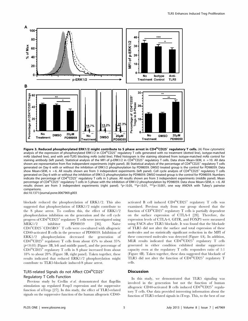
blockade reduced the phosphorylation of ERK1/2. This also
suggested that phosphorylation of ERK1/2 might contribute to
the S phase arrest. To confirm this, the effect of ERK1/2
phosphorylation inhibition on the generation and the cell cycle
progress of CD4hiCD25+ regulatory T cells were investigated using
MEK1/2 inhibitor PD98059 [36]. Naı̈ve
CD4+CD252CD45RO2 T cells were co-cultured with allogeneic
CD40-activated B cells in the presence of PD98059. Inhibition of
ERK1/2 phosphorylation decreased the generation of
CD4hiCD25+ regulatory T cells from about 45% to about 35%
(p,0.05) (Figure 3B, left and middle panel), and the percentage of
CD4hiCD25+ regulatory T cells in S phase increased from about
18% to about 28% (Figure 3B, right panel). Taken together, these
results indicated that reduced ERK1/2 phosphorylation might
contribute to TLR5-blockade induced-S phase arrest.
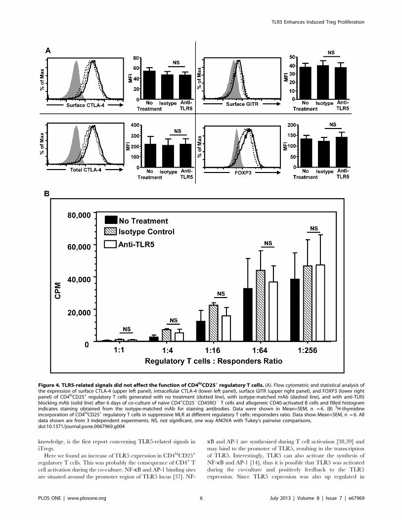
TLR5-related Signals do not Affect CD4hiCD25+
Regulatory T Cells FunctionPrevious study by Crellin et al. demonstrated that flagellin
stimulation up regulated Foxp3 expression and the suppressive
function of nTregs [27]. In this study, the effect of TLR5-related
signals on the suppressive function of the human allogeneic CD40-
activated B cell induced CD4hiCD25+ regulatory T cells was
examined. Previous study from our group showed that the
function of CDhiCD25+ regulatory T cells is partially dependent
on the surface expression of CTLA-4 [28]. Therefore, the
expression levels of CTLA-4, GITR, and FOXP3 were measured
using FACS after TLR5 blockade. It was found that the blockade
of TLR5 did not alter the surface and total expression of these
molecules and no statistically significant reduction in the MFI of
these concerned molecules was detected (Figure 4A). In addition,
MLR results indicated that CD4hiCD25+ regulatory T cells
generated in either condition exhibited similar suppressive
capacity even at the regulatory T cells: responders ratio of 1:1
(Figure 4B). Taken together, these data suggested that blockade of
TLR5 did not alter the function of CD4hiCD25+ regulatory T
cells.
Discussion
In this study, we demonstrated that TLR5 signaling was
involved in the generation but not the function of human
allogeneic CD40-activated B cells induced CD4hiCD25+ regula-
tory T cells. Our data provided interesting information about the
function of TLR5-related signals in iTregs. This, to the best of our
Figure 3. Reduced phosphorylated ERK1/2 might contribute to S phase arrest in CD4hiCD25+ regulatory T cells. (A) Flow cytometricanalysis of the expression of phosphorylated ERK1/2 in CD4hiCD25+ regulatory T cells generated with no treatment (dotted line), isotype-matchedmAb (dashed line), and with anti-TLR5 blocking mAb (solid line). Filled histogram is the staining obtained from isotype-matched mAb control forstaining antibody (left panel). Statistical analysis of the MFI of p-ERK1/2 in CD4hiCD25+ regulatory T cells. Data show Mean+SEM, n = 10. All datashown are representative from five independent experiments (right panel). (B) Statistical analysis of the percentage of CD4hiCD25+ regulatory T cellsgenerated on Day 6 with or without the inhibition of ERK1/2 phosphorylation by PD98059. DMSO treated group is the control for PD98059. Datashow Mean+SEM, n = 6. All results shown are from 3 independent experiments (left panel). Cell cycle analysis of CD4hiCD25+ regulatory T cellsgenerated on Day 6 with or without the inhibition of ERK1/2 phosphorylation by PD98059. DMSO treated group is the control for PD98059. Numbersindicate the percentage of CD4hiCD25+ regulatory T cells in S phase. All results shown are from 3 independent experiments (middle panel). Meanpercentage of CD4hiCD25+ regulatory T cells in S phase with the inhibition of ERK1/2 phosphorylation by PD98059. Data show Mean+SEM, n = 6. Allresults shown are from 3 independent experiments (right panel). *p,0.05, **p,0.01, ***p,0.001, one way ANOVA with Tukey’s pairwisecomparisons.doi:10.1371/journal.pone.0067969.g003
TLR5 Enhances Induced Treg Proliferation
PLOS ONE | www.plosone.org 5 July 2013 | Volume 8 | Issue 7 | e67969
knowledge, is the first report concerning TLR5-related signals in
iTregs.
Here we found an increase of TLR5 expression in CD4hiCD25+
regulatory T cells. This was probably the consequence of CD4+ T
cell activation during the co-culture. NF-kB and AP-1 binding sites
are situated around the promoter region of TLR5 locus [37]. NF-
kB and AP-1 are synthesized during T cell activation [38,39] and
may bind to the promoter of TLR5, resulting in the transcription
of TLR5. Interestingly, TLR5 can also activate the synthesis of
NF-kB and AP-1 [14], thus it is possible that TLR5 was activated
during the co-culture and positively feedback to the TLR5
expression. Since TLR5 expression was also up regulated in
Figure 4. TLR5-related signals did not affect the function of CD4hiCD25+ regulatory T cells. (A). Flow cytometric and statistical analysis ofthe expression of surface CTLA-4 (upper left panel), intracellular CTLA-4 (lower left panel), surface GITR (upper right panel), and FOXP3 (lower rightpanel) of CD4hiCD25+ regulatory T cells generated with no treatment (dotted line), with isotype-matched mAb (dashed line), and with anti-TLR5blocking mAb (solid line) after 6 days of co-culture of naı̈ve CD4+CD252CD45RO2 T cells and allogeneic CD40-activated B cells and filled histogramindicates staining obtained from the isotype-matched mAb for staining antibodies. Data were shown in Mean+SEM, n = 6. (B) 3H-thymidineincorporation of CD4hiCD25+ regulatory T cells in suppressive MLR at different regulatory T cells: responsders ratio. Data show Mean+SEM, n = 6. Alldata shown are from 3 independent experiments. NS, not significant, one way ANOVA with Tukey’s pairwise comparisons.doi:10.1371/journal.pone.0067969.g004
TLR5 Enhances Induced Treg Proliferation
PLOS ONE | www.plosone.org 6 July 2013 | Volume 8 | Issue 7 | e67969

resting nTregs [22], it is possible that Foxp3 also up regulate the
TLR5 expression but the precise mechanism remains to be
investigated.
In this study, we further found that blockade of TLR5 using
anti-TLR5 blocking antibody reduced the proliferation of
CD4hiCD25+ regulatory T cells through S phase arrest but did
not increase the apoptosis of CD4hiCD25+ regulatory T cells or
CD4+CD252 T cells. Since TLR5 was reported to be anti-
apoptotic [40], it was surprising that blockade of TLR5 did not
increase the apoptosis of the cells. This may be explained by the
observation from our previous investigation that large amount of
IL-2 was produced by the CD40-activated B cells [28], thus it is
possible that these IL-2 molecules rescued the CD4+ T cells from
apoptosis.
The S phase arrest of the CD4hiCD25+ regulatory T cells may
be explained by the associated reduction of the ERK1/2
phosphorylation after TLR5 blockade. It is known that S phase
exit or G2/M phase entry is controlled by cdk2 and cyclin A [41],
the cdk2 is in turn activated by cdc25a [42], which could be
activated and phosphorylated by p-ERK1/2 [43]. Therefore, it is
speculated that the reduced ERK1/2 phosphorylation in the
CD4hiCD25+ regulatory T cells decreased the expression and
activation of cdc25a, thus in turn, the cdk2 activation, causing S
phase arrest. However, the precise molecular mechanism between
the reduced ERK1/2 phosphorylation and the S phase arrest
remains to be elucidated. In addition, the reduced proliferation of
the CD4hiCD25+ regulatory T cells may also be the result of
reduced production of different cytokines. It was reported that
stimulation of TLR5 using flagellin resulted in IL-8 production in
epithelial cells and gastric cancer cells, increasing the proliferation
of these cells [44,45], and the production of IFN-c [46].
Therefore, it is possible that TLR5-related signals may enhance
the production of IFN-c, which in turn increases the proliferation
of CD4hiCD25+ regulatory T cells. However, the relative
importance between cell cycle control and cytokine production
in regulating the proliferation of the CD4hiCD25+ regulatory T
cells remains to be elucidated.
Our results demonstrated that TLR5 is not involved in the
suppressive function of CD4hiCD25+ regulatory T cells. This is in
contrast with previous studies by Crellin et al. that flagellin
stimulation of natural regulatory T cells enhanced the FOXP3
expression and function [27]. Maximal Foxp3 expression in
peripheral thymic derived regulatory T cells requires signals from
TCR [47], CD28 [48], and IL-2 [49]. IL-2 promotes Foxp3
expression by activating STAT5, which binds to the promoter of
Foxp3 locus. In Crellin et al. experiment, IL-2 was not used to
activate nTregs [27] and the nTregs might express a low level of
Foxp3, which could be up regulated by flagellin stimulation.
Flagellin stimulation promoted AP-1 activation and the binding of
AP-1 to the promoter of Foxp3 locus, thus in turn, the
transcription of Foxp3 [50] while IL-2 secreted from the CD40-
activated B cells [31] might compensate the effect of TLR5
blockade on the function of CD4hiCD25+ regulatory T cells in our
induction system. The lack of IL-2 in Crellin et al. experiment may
also explain the contrasting results in CD4hiCD25+ regulatory T
cells proliferation. nTregs is hyporesponsive for proliferation and
its proliferation requires the co-existence of CD3, CD28, and IL-2
signaling. It is possible that TLR5 signaling was not potent enough
to break this hyporesponsiveness. In contrast, the CD4hiCD25+
regulatory T cells are induced from naı̈ve
CD4+CD252CD45RO2 T cells, which are not hyporesponsive
for proliferation, and the strength of TLR5 signaling may be
sufficient in promoting the proliferation of CD4hiCD25+ regula-
tory T cells.
In conclusion, our results demonstrated that TLR5-related
signals were involved in the proliferation of CD4hiCD25+
regulatory T cells by promoting the progress of S phase arrest
and ERK1/2 signaling may be involved. However, TLR5-related
signals did not affect the function of CD4hiCD25+ regulatory T
cells. The role of TLR5-related signaling in CD4hiCD25+
regulatory T cells is more resemble to CD4+ effector T cells than
CD4+ nTregs as reflected by the contrasting responses in
proliferation and suppressive function after the blockade of
TLR5. However, whether the same phenomenon can be observed
in other types of iTregs such as Tr1 and Th3 remains to be
elucidated. Our result also indicated that, unlike TLR2, TLR5-
related signaling promoted the proliferation of CD4hiCD25+
regulatory T cells without diminishing the suppressive function
[19]. This suggests that flagellin may be a potential ligand for
increasing the number of iTregs and suppressing inflammation in
organs such as intestines where induced regulatory T cells are
abundant.
Author Contributions
Conceived and designed the experiments: PLC WT. Performed the
experiments: PLC JZ Yuan Liu KTL ZX HM GQ. Analyzed the data:
PLC JZ Yinping Liu WT. Contributed reagents/materials/analysis tools:
YLL. Wrote the paper: PLC WT.
References
1. Lourenco EV, La Cava A (2011) Natural regulatory T cells in autoimmunity.
Autoimmunity 44: 33–42.2. Bilate AM, Lafaille JJ (2012) Induced CD4+Foxp3+ regulatory T cells in
immune tolerance. Annu Rev Immunol 30: 733–758.
3. Le Bras S, Geha RS (2006) IPEX and the role of Foxp3 in the development andfunction of human Tregs. J Clin Invest 116: 1473–1475.
4. Kuhn A, Beissert S, Krammer PH (2008) CD4(+)CD25 (+) regulatory T cells inhuman lupus erythematosus. Arch Dermatol Res 5: 5.
5. Alpdogan O, van den Brink MR (2012) Immune tolerance and transplantation.
Semin Oncol 39: 629–642.6. Corthay A (2009) How do regulatory T cells work? Scand J Immunol 70: 326–
336.7. Sansom DM, Walker LS (2006) The role of CD28 and cytotoxic T-lymphocyte
antigen-4 (CTLA-4) in regulatory T-cell biology. Immunol Rev 212: 131–148.8. Gogishvili T, Luhder F, Goebbels S, Beer-Hammer S, Pfeffer K, et al. (2013)
Cell-intrinsic and -extrinsic control of Treg-cell homeostasis and function
revealed by induced CD28 deletion. Eur J Immunol 43: 188–193.9. Redmond WL, Ruby CE, Weinberg AD (2009) The role of OX40-mediated co-
stimulation in T-cell activation and survival. Crit Rev Immunol 29: 187–201.10. Wang L, Pino-Lagos K, de Vries VC, Guleria I, Sayegh MH, et al. (2008)
Programmed death 1 ligand signaling regulates the generation of adaptive
Foxp3+CD4+ regulatory T cells. Proc Natl Acad Sci U S A 105: 9331–9336.
11. Wakamatsu E, Mathis D, Benoist C (2013) Convergent and divergent effects of
costimulatory molecules in conventional and regulatory CD4+ T cells. Proc Natl
Acad Sci U S A 110: 1023–1028.
12. Szymczak-Workman AL, Workman CJ, Vignali DA (2009) Cutting edge:
regulatory T cells do not require stimulation through their TCR to suppress.
J Immunol 182: 5188–5192.
13. Schmidt-Weber CB, Alexander SI, Henault LE, James L, Lichtman AH (1999)
IL-4 enhances IL-10 gene expression in murine Th2 cells in the absence of TCR
engagement. J Immunol 162: 238–244.
14. Akira S, Uematsu S, Takeuchi O (2006) Pathogen recognition and innate
immunity. Cell 124: 783–801.
15. Gelman AE, Zhang J, Choi Y, Turka LA (2004) Toll-like receptor ligands
directly promote activated CD4+ T cell survival. J Immunol 172: 6065–6073.
16. Caron G, Duluc D, Fremaux I, Jeannin P, David C, et al. (2005) Direct
stimulation of human T cells via TLR5 and TLR7/8: flagellin and R-848 up-
regulate proliferation and IFN-gamma production by memory CD4+ T cells.
J Immunol 175: 1551–1557.
17. Marsland BJ, Nembrini C, Grun K, Reissmann R, Kurrer M, et al. (2007) TLR
ligands act directly upon T cells to restore proliferation in the absence of protein
kinase C-theta signaling and promote autoimmune myocarditis. J Immunol 178:
3466–3473.
TLR5 Enhances Induced Treg Proliferation
PLOS ONE | www.plosone.org 7 July 2013 | Volume 8 | Issue 7 | e67969

18. Sutmuller RP, den Brok MH, Kramer M, Bennink EJ, Toonen LW, et al. (2006)
Toll-like receptor 2 controls expansion and function of regulatory T cells. J ClinInvest 116: 485–494.
19. Oberg HH, Ly TT, Ussat S, Meyer T, Kabelitz D, et al. (2010) Differential but
direct abolishment of human regulatory T cell suppressive capacity by variousTLR2 ligands. J Immunol 184: 4733–4740.
20. Milkova L, Voelcker V, Forstreuter I, Sack U, Anderegg U, et al. (2009) TheNF-kappaB signalling pathway is involved in the LPS/IL-2-induced upregula-
tion of FoxP3 expression in human CD4CD25 regulatory T cells. Exp Dermatol
21: 21.21. Fallarino F, Volpi C, Zelante T, Vacca C, Calvitti M, et al. (2009) IDO mediates
TLR9-driven protection from experimental autoimmune diabetes. J Immunol183: 6303–6312.
22. Kabelitz D (2007) Expression and function of Toll-like receptors in Tlymphocytes. Curr Opin Immunol 19: 39–45.
23. Mansson A, Adner M, Cardell LO (2006) Toll-like receptors in cellular subsets of
human tonsil T cells: altered expression during recurrent tonsillitis. Respir Res 7:36.
24. Fournier B, Williams IR, Gewirtz AT, Neish AS (2009) Toll-like receptor 5-dependent regulation of inflammation in systemic Salmonella enterica Serovar
typhimurium infection. Infect Immun 77: 4121–4129.
25. Kinnebrew MA, Ubeda C, Zenewicz LA, Smith N, Flavell RA, et al. (2010)Bacterial flagellin stimulates Toll-like receptor 5-dependent defense against
vancomycin-resistant Enterococcus infection. J Infect Dis 201: 534–543.26. Sun J, Fegan PE, Desai AS, Madara JL, Hobert ME (2007) Flagellin-induced
tolerance of the Toll-like receptor 5 signaling pathway in polarized intestinalepithelial cells. Am J Physiol Gastrointest Liver Physiol 292: G767–778.
27. Crellin NK, Garcia RV, Hadisfar O, Allan SE, Steiner TS, et al. (2005) Human
CD4+ T cells express TLR5 and its ligand flagellin enhances the suppressivecapacity and expression of FOXP3 in CD4+CD25+ T regulatory cells.
J Immunol 175: 8051–8059.28. Tu W, Lau YL, Zheng J, Liu Y, Chan PL, et al. (2008) Efficient generation of
human alloantigen-specific CD4+ regulatory T cells from naive precursors by
CD40-activated B cells. Blood 112: 2554–2562.29. Zheng J, Liu Y, Qin G, Lam KT, Guan J, et al. (2011) Generation of human
Th1-like regulatory CD4(+) T cells by an intrinsic IFN-gamma- and T-bet-dependent pathway. Eur J Immunol 41: 128–139.
30. Zheng J, Liu Y, Liu M, Xiang Z, Lam KT, et al. (2013) Human CD8+regulatory T cells inhibit GVHD and preserve general immunity in humanized
mice. Science translational medicine 5: 168ra169.
31. Zheng J, Liu Y, Lau YL, Tu W (2010) CD40-activated B cells are more potentthan immature dendritic cells to induce and expand CD4(+) regulatory T cells.
Cellular & molecular immunology 7: 44–50.32. Salamone GV, Petracca Y, Fuxman Bass JI, Rumbo M, Nahmod KA, et al.
(2010) Flagellin delays spontaneous human neutrophil apoptosis. Lab Invest 90:
1049–1059.
33. Neish AS (2007) TLRS in the gut. II. Flagellin-induced inflammation and
antiapoptosis. Am J Physiol Gastrointest Liver Physiol 292: G462–466.34. Burdelya LG, Krivokrysenko VI, Tallant TC, Strom E, Gleiberman AS, et al.
(2008) An agonist of toll-like receptor 5 has radioprotective activity in mouse and
primate models. Science 320: 226–230.35. Huang F, Xiong X, Wang H, You S, Zeng H (2010) Leptin-induced vascular
smooth muscle cell proliferation via regulating cell cycle, activating ERK1/2 andNF-kappaB. Acta biochimica et biophysica Sinica 42: 325–331.
36. Nishimoto S, Nishida E (2006) MAPK signalling: ERK5 versus ERK1/2.
EMBO reports 7: 782–786.37. (2000) Champion ChiP Transcription Factor Search Portal. 2012–05–12 ed.
CA: Qiagen.38. Lupino E, Ramondetti C, Piccinini M (2012) IkappaB kinase beta is required for
activation of NF-kappaB and AP-1 in CD3/CD28-stimulated primary CD4(+) Tcells. Journal of immunology 188: 2545–2555.
39. Schmidt A, Oberle N, Weiss EM, Vobis D, Frischbutter S, et al. (2011) Human
regulatory T cells rapidly suppress T cell receptor-induced Ca(2+), NF-kappaB,and NFAT signaling in conventional T cells. Sci Signal 4: ra90.
40. Vijay-Kumar M, Wu H, Jones R, Grant G, Babbin B, et al. (2006) Flagellinsuppresses epithelial apoptosis and limits disease during enteric infection.
Am J Pathol 169: 1686–1700.
41. Schafer KA (1998) The cell cycle: a review. Vet Pathol 35: 461–478.42. Collins K, Jacks T, Pavletich NP (1997) The cell cycle and cancer. Proc Natl
Acad Sci U S A 94: 2776–2778.43. Chen S, Gardner DG (2004) Suppression of WEE1 and stimulation of CDC25A
correlates with endothelin-dependent proliferation of rat aortic smooth musclecells. J Biol Chem 279: 13755–13763.
44. Steiner TS, Ivison SM, Yao Y, Kifayet A (2010) Protein kinase D1 and D2 are
involved in chemokine release induced by toll-like receptors 2, 4, and 5. CellImmunol 264: 135–142.
45. Song EJ, Kang MJ, Kim YS, Kim SM, Lee SE, et al. (2011) Flagellin promotesthe proliferation of gastric cancer cells via the Toll-like receptor 5. Int J Mol Med
28: 115–119.
46. Simone R, Floriani A, Saverino D (2009) Stimulation of Human CD4 TLymphocytes via TLR3, TLR5 and TLR7/8 Up-Regulates Expression of
Costimulatory and Modulates Proliferation. Open Microbiol J 3: 1–8.47. Josefowicz SZ, Wilson CB, Rudensky AY (2009) Cutting edge: TCR stimulation
is sufficient for induction of Foxp3 expression in the absence of DNAmethyltransferase 1. J Immunol 182: 6648–6652.
48. Soligo M, Camperio C, Caristi S, Scotta C, Del Porto P, et al. (2011) CD28
costimulation regulates FOXP3 in a RelA/NF-kappaB-dependent mechanism.Eur J Immunol 41: 503–513.
49. Haiqi H, Yong Z, Yi L (2011) Transcriptional regulation of Foxp3 in regulatoryT cells. Immunobiology 216: 678–685.
50. von Boehmer H, Nolting J (2008) What turns on Foxp3? Nat Immunol 9: 121–
122.
TLR5 Enhances Induced Treg Proliferation
PLOS ONE | www.plosone.org 8 July 2013 | Volume 8 | Issue 7 | e67969

Reproduced with permission of the copyright owner. Further reproduction prohibited withoutpermission.