TLR4 Ligand/H O Enhances TGF-b1 Signaling to … · suggesting that TGF-b1 might not be able to...
10
TLR4 Ligand/H 2 O 2 Enhances TGF-b1 Signaling to Induce Metastatic Potential of Non-Invasive Breast Cancer Cells by Activating Non-Smad Pathways Yuan-Hong Zhou, Sheng-Jun Liao, Dong Li, Jing Luo, Jing-Jing Wei, Bin Yan, Rui Sun, Yu Shu, Qi Wang, Gui-Mei Zhang*, Zuo-Hua Feng* Department of Biochemistry & Molecular Biology, Tongji Medical College, Huazhong University of Science & Technology, Wuhan, The People’s Republic of China Abstract TGF-b1 has the potential to activate multiple signaling pathways required for inducing metastatic potential of tumor cells. However, TGF-b1 was inefficient in inducing metastatic potential of many non-invasive human tumor cells. Here we report that the enhancement of TGF-b1 signaling is required for inducing metastatic potential of non-invasive breast cancer cells. TGF-b1 alone could not efficiently induce the sustained activation of Smad and non-Smad pathways in non-invasive breast cancer cells. TLR4 ligand (LPS) and H 2 O 2 cooperated with TGF-b1 to enhance the sustained activation of non-Smad pathways, including p38MAPK, ERK, JNK, PI3K, and NF-kB. The activation of MAPK and PI3K pathways resulted in a positive feed-back effect on TGF-b1 signaling by down-regulating Nm23-H1 expression and up-regulating the expression of TbRI and TbRII, favoring further activation of multiple signaling pathways. Moreover, the enhanced TGF-b1 signaling induced higher expression of SNAI2, which also promoted TbRII expression. Therefore, the sustained activation levels of both Smad and non-Smad pathways were gradually increased after prolonged stimulation with TGF-b1/H 2 O 2 /LPS. Consistent with the activation pattern of signaling pathways, the invasive capacity and anoikis-resistance of non-invasive breast cancer cells were gradually increased after prolonged stimulation with TGF-b1/H 2 O 2 /LPS. The metastatic potential induced by TGF-b1/ H 2 O 2 /LPS was sufficient for tumor cells to extravasate and form metastatic foci in an experimental metastasis model in nude mice. The findings in this study suggested that the enhanced signaling is required for inducing higher metastatic capacity of tumor cells, and that targeting one of stimuli or signaling pathways might be potential approach in comprehensive strategy for tumor therapy. Citation: Zhou Y-H, Liao S-J, Li D, Luo J, Wei J-J, et al. (2013) TLR4 Ligand/H 2 O 2 Enhances TGF-b1 Signaling to Induce Metastatic Potential of Non-Invasive Breast Cancer Cells by Activating Non-Smad Pathways. PLoS ONE 8(5): e65906. doi:10.1371/journal.pone.0065906 Editor: Todd W. Miller, Dartmouth, United States of America Received February 15, 2013; Accepted April 29, 2013; Published May 29, 2013 Copyright: ß 2013 Zhou et al. This is an open-access article distributed under the terms of the Creative Commons Attribution License, which permits unrestricted use, distribution, and reproduction in any medium, provided the original author and source are credited. Funding: This work was supported by National Science Foundation of China (No. 30830095 to ZHF, No. 81272314 to GMZ) (http://isisn.nsfc.gov.cn/egrantweb/ main), and National Development Program (973) for Key Basic Research of China (No. 2009CB521806 to ZHF) (http://www.973.gov.cn/areamana.aspx). The funders have no role in study design, data collection and analysis, decision to publish, or preparation of the manuscript. Competing Interests: The authors have declared that no competing interests exist. * E-mail: [email protected] (ZHF); [email protected] (GMZ) Introduction The efficient activation of multiple signaling pathways is the important driving force for tumor cell metastasis [1–3]. Compared with high-invasive human cancer cells, non-invasive human cancer cells have constitutively lower activation of signaling pathways [4,5]. Given that tumor microenvironment can influence the metastatic capacity of tumor cells [6], the metastatic potential of non-invasive tumor cells might be induced by modulatory factor(s) in tumor milieu. Since the activation of single signaling pathway is not sufficient for inducing the metastasis of non- invasive tumor cells [4,5], the factor(s) which could activate multiple signaling pathways might be responsible for inducing metastasis of non-invasive tumor cells. Transforming growth factor b1 (TGF-b1), the most potent factor to induce epithelial to mesenchymal transition (EMT) [7,8], has the potential to activate multiple signaling pathways, including Smad pathway and non-Smad pathways such as p38MAPK, ERK, JNK, PI3K, and NF-kB [8,9]. The increased production of TGF-b1 has been observed in many types of carcinomas [7,10]. The carcinomas with excess TGF-b1 production are more motile and invasive, and exhibit increased tumor cell metastasis in athymic mice [7]. All of these findings implicate the important roles of TGF-b1 in tumor metastasis. However, many non-invasive tumor cells could not undergo TGF-b1-induced EMT in vitro [7], suggesting that TGF-b1 might not be able to efficiently activate multiple signaling pathways in non-invasive tumor cells. The effect of TGF-b1 in tumor milieu might be enhanced by other factor(s) which could cooperate with TGF-b1 to induce sufficient activation of multiple signaling pathways, and promote the metastatic capacity, including invasion and extravasation, of tumor cells. It has been found that TLR4 ligand and H 2 O 2 also have the potential to activate non-Smad pathways which could be activated by TGF-b1 [11–16], suggesting the possibility that TLR4 ligand and/or H 2 O 2 might cooperate with TGF-b1 to induce sufficient activation of multiple signaling pathways, favoring metastatic potential of non-invasive tumor cells. TLR4 ligands could be existent in vivo due to surgery, damage of tumor cells, or the existence of bacteria in tumor [17–20]. H 2 O 2 , one of the molecules involved in inflammation, is abundantly existent in tumor milieu [21]. Therefore, in this study we investigated PLOS ONE | www.plosone.org 1 May 2013 | Volume 8 | Issue 5 | e65906
Transcript of TLR4 Ligand/H O Enhances TGF-b1 Signaling to … · suggesting that TGF-b1 might not be able to...
TLR4 Ligand/H2O2 Enhances TGF-b1 Signaling to InduceMetastatic Potential of Non-Invasive Breast Cancer Cellsby Activating Non-Smad PathwaysYuan-Hong Zhou, Sheng-Jun Liao, Dong Li, Jing Luo, Jing-Jing Wei, Bin Yan, Rui Sun, Yu Shu, Qi Wang,
Gui-Mei Zhang*, Zuo-Hua Feng*
Department of Biochemistry & Molecular Biology, Tongji Medical College, Huazhong University of Science & Technology, Wuhan, The People’s Republic of China
Abstract
TGF-b1 has the potential to activate multiple signaling pathways required for inducing metastatic potential of tumor cells.However, TGF-b1 was inefficient in inducing metastatic potential of many non-invasive human tumor cells. Here we reportthat the enhancement of TGF-b1 signaling is required for inducing metastatic potential of non-invasive breast cancer cells.TGF-b1 alone could not efficiently induce the sustained activation of Smad and non-Smad pathways in non-invasive breastcancer cells. TLR4 ligand (LPS) and H2O2 cooperated with TGF-b1 to enhance the sustained activation of non-Smadpathways, including p38MAPK, ERK, JNK, PI3K, and NF-kB. The activation of MAPK and PI3K pathways resulted in a positivefeed-back effect on TGF-b1 signaling by down-regulating Nm23-H1 expression and up-regulating the expression of TbRIand TbRII, favoring further activation of multiple signaling pathways. Moreover, the enhanced TGF-b1 signaling inducedhigher expression of SNAI2, which also promoted TbRII expression. Therefore, the sustained activation levels of both Smadand non-Smad pathways were gradually increased after prolonged stimulation with TGF-b1/H2O2/LPS. Consistent with theactivation pattern of signaling pathways, the invasive capacity and anoikis-resistance of non-invasive breast cancer cellswere gradually increased after prolonged stimulation with TGF-b1/H2O2/LPS. The metastatic potential induced by TGF-b1/H2O2/LPS was sufficient for tumor cells to extravasate and form metastatic foci in an experimental metastasis model in nudemice. The findings in this study suggested that the enhanced signaling is required for inducing higher metastatic capacity oftumor cells, and that targeting one of stimuli or signaling pathways might be potential approach in comprehensive strategyfor tumor therapy.
Citation: Zhou Y-H, Liao S-J, Li D, Luo J, Wei J-J, et al. (2013) TLR4 Ligand/H2O2 Enhances TGF-b1 Signaling to Induce Metastatic Potential of Non-Invasive BreastCancer Cells by Activating Non-Smad Pathways. PLoS ONE 8(5): e65906. doi:10.1371/journal.pone.0065906
Editor: Todd W. Miller, Dartmouth, United States of America
Received February 15, 2013; Accepted April 29, 2013; Published May 29, 2013
Copyright: � 2013 Zhou et al. This is an open-access article distributed under the terms of the Creative Commons Attribution License, which permitsunrestricted use, distribution, and reproduction in any medium, provided the original author and source are credited.
Funding: This work was supported by National Science Foundation of China (No. 30830095 to ZHF, No. 81272314 to GMZ) (http://isisn.nsfc.gov.cn/egrantweb/main), and National Development Program (973) for Key Basic Research of China (No. 2009CB521806 to ZHF) (http://www.973.gov.cn/areamana.aspx). Thefunders have no role in study design, data collection and analysis, decision to publish, or preparation of the manuscript.
Competing Interests: The authors have declared that no competing interests exist.
* E-mail: [email protected] (ZHF); [email protected] (GMZ)
Introduction
The efficient activation of multiple signaling pathways is the
important driving force for tumor cell metastasis [1–3]. Compared
with high-invasive human cancer cells, non-invasive human
cancer cells have constitutively lower activation of signaling
pathways [4,5]. Given that tumor microenvironment can influence
the metastatic capacity of tumor cells [6], the metastatic potential
of non-invasive tumor cells might be induced by modulatory
factor(s) in tumor milieu. Since the activation of single signaling
pathway is not sufficient for inducing the metastasis of non-
invasive tumor cells [4,5], the factor(s) which could activate
multiple signaling pathways might be responsible for inducing
metastasis of non-invasive tumor cells.
Transforming growth factor b1 (TGF-b1), the most potent
factor to induce epithelial to mesenchymal transition (EMT) [7,8],
has the potential to activate multiple signaling pathways, including
Smad pathway and non-Smad pathways such as p38MAPK,
ERK, JNK, PI3K, and NF-kB [8,9]. The increased production of
TGF-b1 has been observed in many types of carcinomas [7,10].
The carcinomas with excess TGF-b1 production are more motile
and invasive, and exhibit increased tumor cell metastasis in
athymic mice [7]. All of these findings implicate the important
roles of TGF-b1 in tumor metastasis. However, many non-invasive
tumor cells could not undergo TGF-b1-induced EMT in vitro [7],
suggesting that TGF-b1 might not be able to efficiently activate
multiple signaling pathways in non-invasive tumor cells. The effect
of TGF-b1 in tumor milieu might be enhanced by other factor(s)
which could cooperate with TGF-b1 to induce sufficient activation
of multiple signaling pathways, and promote the metastatic
capacity, including invasion and extravasation, of tumor cells.
It has been found that TLR4 ligand and H2O2 also have the
potential to activate non-Smad pathways which could be activated
by TGF-b1 [11–16], suggesting the possibility that TLR4 ligand
and/or H2O2 might cooperate with TGF-b1 to induce sufficient
activation of multiple signaling pathways, favoring metastatic
potential of non-invasive tumor cells. TLR4 ligands could be
existent in vivo due to surgery, damage of tumor cells, or the
existence of bacteria in tumor [17–20]. H2O2, one of the
molecules involved in inflammation, is abundantly existent in
tumor milieu [21]. Therefore, in this study we investigated
PLOS ONE | www.plosone.org 1 May 2013 | Volume 8 | Issue 5 | e65906

whether TGF-b1 could induce the metastatic potential of non-
invasive tumor cells in presence of TLR4 ligand and/or H2O2,
and whether TLR4 ligand and/or H2O2 could enhance TGF-b1
signaling in non-invasive tumor cells. Our results showed that
TLR4 ligand and H2O2 could cooperate with TGF-b1 to induce
sustained activation of multiple signaling pathways in non-invasive
human breast cancer cells, promoting the metastatic potential
sufficient for invasion and extravasation of tumor cells.
Results
TGF-b1/H2O2/LPS promotes the invasive migration ofnon-invasive breast cancer cells
To investigate whether TGF-b1, H2O2, and TLR4 ligand
might cooperate to promote invasive migration of non-invasive
breast cancer cells, we first cultured non-invasive MCF-7 and T-
47D cells in presence of TGF-b1, H2O2, and LPS (a well known
TLR4 ligand). The capacity of invasive migration of tumor cells
was gradually increased after prolonged stimulation (Figure 1A).
We then further analyzed the effects of TGF-b1, H2O2, and LPS.
The result showed that H2O2 and LPS, alone or in combination,
could not influence the invasive migration of these cells (Figure 1B).
TGF-b1, alone or in combination with either H2O2 or LPS,
slightly promoted the invasive migration of MCF-7 and T-47D
cells. However, the invasive migration of these cells was much
more efficient after treatment with TGF-b1/H2O2/LPS
(Figure 1B). Consistently, the polymerization of actin in tumor
cells in response to ECM molecules (matrigel), which is important
for migratory and invasive properties of tumor cells [22], was also
increased by TGF-b1/H2O2/LPS (Figure S1). Moreover, TGF-
b1/H2O2/LPS significantly up-regulated the expression of avb3
(Figure 1C), which is the key integrin mediating tumor cell arrest
during flow and the invasive migration of tumor cells [4]. The
production of active MMP-9 in response to ECM was also
increased by pretreatment with TGF-b1/H2O2/LPS (Figure 1D).
Taken together, these results indicated that TGF-b1, H2O2, and
LPS could cooperate to promote the invasive migration of non-
invasive breast cancer cells.
Sustained activation of non-Smad pathways is enhancedby co-stimulation with TGF-b1/H2O2/LPS
The requirement for prolonged stimulation implied that the
sustained activation of signaling pathways was important for TGF-
b1/H2O2/LPS to promote invasive migration of non-invasive
breast cancer cells. We then analyzed the effect of TGF-b1/
H2O2/LPS on the sustained activation of non-Smad pathways. To
do this, we first detected the sustained activation of these pathways
by stimulating MCF-7 cells for 7 days with TGF-b1, H2O2, and
LPS. The result showed that the co-stimulation with three stimuli
was most efficient in activating non-Smad pathways (Figure 2A),
suggesting that the cooperation of all three stimuli was required for
the most efficient activation of these pathways. Intriguingly, the
prolonged stimulation resulted in the enhanced activation of these
pathways (Figure 2B), suggesting that the signaling was amplified
after prolonged stimulation.
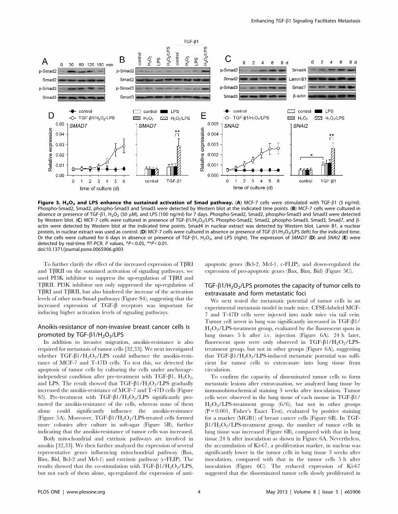
Sustained activation of Smad pathway is enhanced byco-stimulation with TGF-b1/H2O2/LPS
The above data showed that TGF-b1 alone was inefficient in
inducing the sustained activation of non-Smad pathways. We then
asked whether the sustained activation of Smad pathway in non-
invasive breast cancer cells could be induced by TGF-b1. TGF-b1
alone induced transient activation of Smad2/3 in MCF-7 cells
(Figure 3A), but could not induce the sustained activation of
Smad2/3, evaluated by the levels of p-Smad2 and p-Smad3 after
7-d stimulation (Figure 3B). Intriguingly, TGF-b1 could induce the
sustained activation of Smad2/3 in presence of both H2O2 and
LPS (Figure 3B). The sustained activation of Smad2/3 in MCF-7
cells was gradually enhanced in presence of TGF-b1/H2O2/LPS
(Figure 3C). In accordance with the gradually enhanced activation
of Smad2/3, the nuclear translocation of Smad4 was gradually
increased by TGF-b1/H2O2/LPS (Figure 3C). The expression of
Smad7, which is induced by TGF-b1-Smad pathway [23], was
gradually up-regulated by TGF-b1/H2O2/LPS (Figure 3C, 3D).
In addition to Smad7, TGF-b1/H2O2/LPS also increased the
expression of SNAI2 (Figure 3E), a member of Snail family
mediating the effect of TGF-b1, which could be up-regulated by
Smad and ERK pathways [8,24]. These data suggested that H2O2
and LPS enhanced TGF-b1-induced sustained activation of not
only non-Smad pathways but also Smad pathway. However,
although Smad pathway could modulate the expression of p21 and
MYC [25], the expression of p21 and MYC was not significantly
influenced by TGF-b1/H2O2/LPS (Figure S2), possibly due to the
opposite effects of Smad pathway and non-Smad pathways on the
expression of p21 and MYC [25–28].
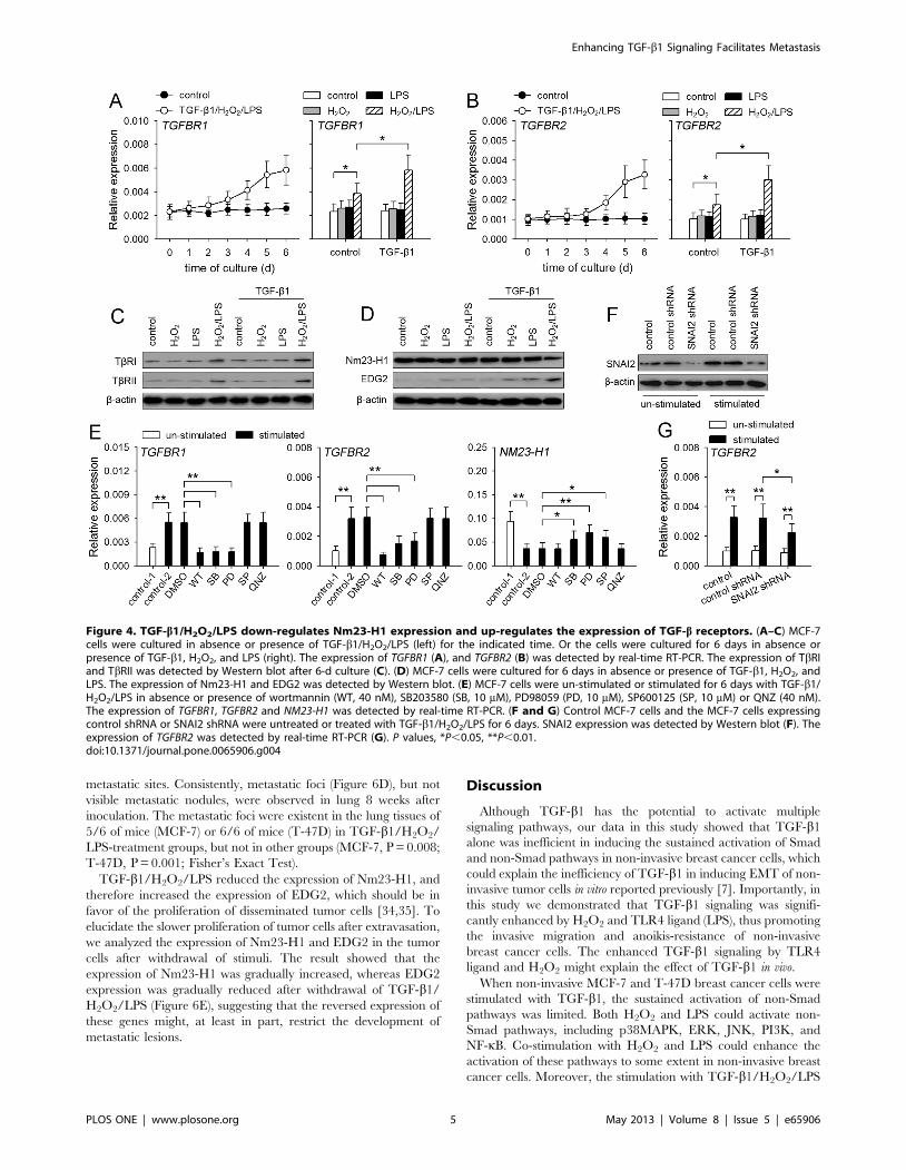
TGF-b1 signaling is enhanced by up-regulating TGF-breceptors and down-regulating Nm23-H1
To further investigate the mechanisms underlying the enhanced
activation of Smad and non-Smad pathways, we analyzed whether
the expression of TLR4 and TGF-b receptors was influenced by
TGF-b1/H2O2/LPS. The expression of TLR4 was not signifi-
cantly changed after stimulation with three stimuli (data not
shown). In presence of TGF-b1/H2O2/LPS, the expression of
TbRI and TbRII was gradually increased after prolonged
stimulation (Figure 4A, 4B). TGF-b1, H2O2 or LPS alone could
not influence TbRI and TbRII expression. H2O2 and LPS could
cooperate to promote the expression of these receptors, and
cooperate with TGF-b1 to further up-regulate the expression of
these receptors (Figure 4A, 4B, 4C).
Since the expression of Smad7, the feed-back inhibitor of
Smad2/3 activation, was not suppressed (Figure 3D), we next
investigated whether TGF-b1/H2O2/LPS might modulate the
expression of Nm23-H1 which has been found to negatively
regulate TGF-b signaling [29]. The result showed that the
expression of Nm23-H1 in the cells was significantly reduced by
TGF-b1/H2O2/LPS, but not one or two of them (Figure S3, 4D).
The down-regulation of Nm23-H1 was further proved by the
increased expression of EDG2 (Figure S3, 4D), since Nm23-H1
has been known to suppress EDG2 expression [30].
We then analyzed whether non-Smad pathways were involved
in modulating the expression of Nm23-H1 and TGF-b receptors,
since TLR4 ligand and H2O2 were required for the modulation.
To do this, we detected the mRNA levels of TbRI, TbRII, and
Nm23-H1 after stimulation with TGF-b1/H2O2/LPS in presence
of wortmannin (PI3K inhibitor), SB203580 (p38MAPK inhibitor),
PD98059 (inhibitor of ERK pathway), SP600125 (JNK inhibitor),
QNZ (NF-kB inhibitor). The results showed that PI3K,
p38MAPK and ERK pathways were required for up-regulating
the expression of TbRI and TbRII, and that p38MAPK, ERK
and JNK pathways were involved in down-regulating Nm23-H1
expression (Figure 4E). As shown in Figure 3E, TGF-b1/H2O2/
LPS could up-regulate the expression of SNAI2, which also
promotes the expression of TbRII [31]. We then used SNAI2
shRNA to inhibit TGF-b1/H2O2/LPS-induced expression of
SNAI2 (Figure 4F), which partially, but significantly, hindered the
up-regulation of TbRII by TGF-b1/H2O2/LPS (Figure 4G).
Enhancing TGF-b1 Signaling Facilitates Metastasis
PLOS ONE | www.plosone.org 2 May 2013 | Volume 8 | Issue 5 | e65906
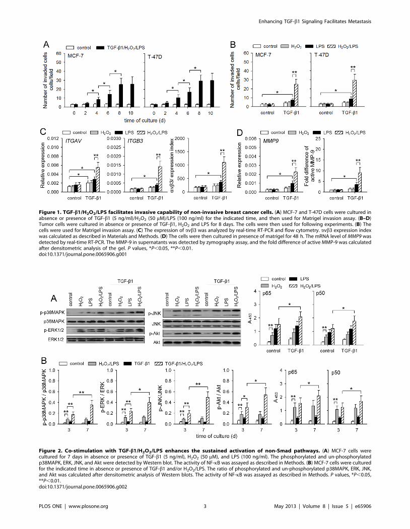
Figure 1. TGF-b1/H2O2/LPS facilitates invasive capability of non-invasive breast cancer cells. (A) MCF-7 and T-47D cells were cultured inabsence or presence of TGF-b1 (5 ng/ml)/H2O2 (50 mM)/LPS (100 ng/ml) for the indicated time, and then used for Matrigel invasion assay. (B–D)Tumor cells were cultured in absence or presence of TGF-b1, H2O2 and LPS for 8 days. The cells were then used for following experiments. (B) Thecells were used for Matrigel invasion assay. (C) The expression of avb3 was analyzed by real-time RT-PCR and flow cytometry. avb3 expression indexwas calculated as described in Materials and Methods. (D) The cells were then cultured in presence of matrigel for 48 h. The mRNA level of MMP9 wasdetected by real-time RT-PCR. The MMP-9 in supernatants was detected by zymography assay, and the fold difference of active MMP-9 was calculatedafter densitometric analysis of the gel. P values, *P,0.05, **P,0.01.doi:10.1371/journal.pone.0065906.g001
Figure 2. Co-stimulation with TGF-b1/H2O2/LPS enhances the sustained activation of non-Smad pathways. (A) MCF-7 cells werecultured for 7 days in absence or presence of TGF-b1 (5 ng/ml), H2O2 (50 mM), and LPS (100 ng/ml). The phosphorylated and un-phosphorylatedp38MAPK, ERK, JNK, and Akt were detected by Western blot. The activity of NF-kB was assayed as described in Methods. (B) MCF-7 cells were culturedfor the indicated time in absence or presence of TGF-b1 and/or H2O2/LPS. The ratio of phosphorylated and un-phosphorylated p38MAPK, ERK, JNK,and Akt was calculated after densitometric analysis of Western blots. The activity of NF-kB was assayed as described in Methods. P values, *P,0.05,**P,0.01.doi:10.1371/journal.pone.0065906.g002
Enhancing TGF-b1 Signaling Facilitates Metastasis
PLOS ONE | www.plosone.org 3 May 2013 | Volume 8 | Issue 5 | e65906
To further clarify the effect of the increased expression of TbRI
and TbRII on the sustained activation of signaling pathways, we
used PI3K inhibitor to suppress the up-regulation of TbRI and
TbRII. PI3K inhibitor not only suppressed the up-regulation of
TbRI and TbRII, but also hindered the increase of the activation
levels of other non-Smad pathways (Figure S4), suggesting that the
increased expression of TGF-b receptors was important for
inducing higher activation levels of signaling pathways.
Anoikis-resistance of non-invasive breast cancer cells ispromoted by TGF-b1/H2O2/LPS
In addition to invasive migration, anoikis-resistance is also
required for metastasis of tumor cells [32,33]. We next investigated
whether TGF-b1/H2O2/LPS could influence the anoikis-resis-
tance of MCF-7 and T-47D cells. To test this, we detected the
apoptosis of tumor cells by culturing the cells under anchorage-
independent condition after pre-treatment with TGF-b1, H2O2,
and LPS. The result showed that TGF-b1/H2O2/LPS gradually
increased the anoikis-resistance of MCF-7 and T-47D cells (Figure
S5). Pre-treatment with TGF-b1/H2O2/LPS significantly pro-
moted the anoikis-resistance of the cells, whereas none of them
alone could significantly influence the anoikis-resistance
(Figure 5A). Moreover, TGF-b1/H2O2/LPS-treated cells formed
more colonies after culture in soft-agar (Figure 5B), further
indicating that the anoikis-resistance of tumor cells was increased.
Both mitochondrial and extrinsic pathways are involved in
anoikis [32,33]. We then further analyzed the expression of several
representative genes influencing mitochondrial pathway (Bax,
Bim, Bid, Bcl-2 and Mcl-1) and extrinsic pathway (c-FLIP). The
results showed that the co-stimulation with TGF-b1/H2O2/LPS,
but not each of them alone, up-regulated the expression of anti-
apoptotic genes (Bcl-2, Mcl-1, c-FLIP), and down-regulated the
expression of pro-apoptotic genes (Bax, Bim, Bid) (Figure 5C).
TGF-b1/H2O2/LPS promotes the capacity of tumor cells toextravasate and form metastatic foci
We next tested the metastatic potential of tumor cells in an
experimental metastasis model in nude mice. CFSE-labeled MCF-
7 and T-47D cells were injected into nude mice via tail vein.
Tumor cell arrest in lung was significantly increased in TGF-b1/
H2O2/LPS-treatment group, evaluated by the fluorescent spots in
lung tissues 5 h after i.v. injection (Figure 6A). 24 h later,
fluorescent spots were only observed in TGF-b1/H2O2/LPS-
treatment group, but not in other groups (Figure 6A), suggesting
that TGF-b1/H2O2/LPS-induced metastatic potential was suffi-
cient for tumor cells to extravasate into lung tissue from
circulation.
To confirm the capacity of disseminated tumor cells to form
metastatic lesions after extravasation, we analyzed lung tissue by
immunohistochemical staining 3 weeks after inoculation. Tumor
cells were observed in the lung tissue of each mouse in TGF-b1/
H2O2/LPS-treatment group (6/6), but not in other groups
(P = 0.001, Fisher’s Exact Test), evaluated by positive staining
for a marker (MGB1) of breast cancer cells (Figure 6B). In TGF-
b1/H2O2/LPS-treatment group, the number of tumor cells in
lung tissue was increased (Figure 6B), compared with that in lung
tissue 24 h after inoculation as shown in Figure 6A. Nevertheless,
the accumulation of Ki-67, a proliferation marker, in nucleus was
significantly lower in the tumor cells in lung tissue 3 weeks after
inoculation, compared with that in the tumor cells 5 h after
inoculation (Figure 6C). The reduced expression of Ki-67
suggested that the disseminated tumor cells slowly proliferated in
Figure 3. H2O2 and LPS enhance the sustained activation of Smad pathway. (A) MCF-7 cells were stimulated with TGF-b1 (5 ng/ml).Phospho-Smad2, Smad2, phospho-Smad3 and Smad3 were detected by Western blot at the indicated time points. (B) MCF-7 cells were cultured inabsence or presence of TGF-b1, H2O2 (50 mM), and LPS (100 ng/ml) for 7 days. Phospho-Smad2, Smad2, phospho-Smad3 and Smad3 were detectedby Western blot. (C) MCF-7 cells were cultured in presence of TGF-b1/H2O2/LPS. Phospho-Smad2, Smad2, phospho-Smad3, Smad3, Smad7, and b-actin were detected by Western blot at the indicated time points. Smad4 in nuclear extract was detected by Western blot. Lamin B1, a nuclearprotein, in nuclear extract was used as control. (D) MCF-7 cells were cultured in absence or presence of TGF-b1/H2O2/LPS (left) for the indicated time.Or the cells were cultured for 6 days in absence or presence of TGF-b1, H2O2, and LPS (right). The expression of SMAD7 (D) and SNAI2 (E) weredetected by real-time RT-PCR. P values, *P,0.05, **P,0.01.doi:10.1371/journal.pone.0065906.g003
Enhancing TGF-b1 Signaling Facilitates Metastasis
PLOS ONE | www.plosone.org 4 May 2013 | Volume 8 | Issue 5 | e65906
metastatic sites. Consistently, metastatic foci (Figure 6D), but not
visible metastatic nodules, were observed in lung 8 weeks after
inoculation. The metastatic foci were existent in the lung tissues of
5/6 of mice (MCF-7) or 6/6 of mice (T-47D) in TGF-b1/H2O2/
LPS-treatment groups, but not in other groups (MCF-7, P = 0.008;
T-47D, P = 0.001; Fisher’s Exact Test).
TGF-b1/H2O2/LPS reduced the expression of Nm23-H1, and
therefore increased the expression of EDG2, which should be in
favor of the proliferation of disseminated tumor cells [34,35]. To
elucidate the slower proliferation of tumor cells after extravasation,
we analyzed the expression of Nm23-H1 and EDG2 in the tumor
cells after withdrawal of stimuli. The result showed that the
expression of Nm23-H1 was gradually increased, whereas EDG2
expression was gradually reduced after withdrawal of TGF-b1/
H2O2/LPS (Figure 6E), suggesting that the reversed expression of
these genes might, at least in part, restrict the development of
metastatic lesions.
Discussion
Although TGF-b1 has the potential to activate multiple
signaling pathways, our data in this study showed that TGF-b1
alone was inefficient in inducing the sustained activation of Smad
and non-Smad pathways in non-invasive breast cancer cells, which
could explain the inefficiency of TGF-b1 in inducing EMT of non-
invasive tumor cells in vitro reported previously [7]. Importantly, in
this study we demonstrated that TGF-b1 signaling was signifi-
cantly enhanced by H2O2 and TLR4 ligand (LPS), thus promoting
the invasive migration and anoikis-resistance of non-invasive
breast cancer cells. The enhanced TGF-b1 signaling by TLR4
ligand and H2O2 might explain the effect of TGF-b1 in vivo.
When non-invasive MCF-7 and T-47D breast cancer cells were
stimulated with TGF-b1, the sustained activation of non-Smad
pathways was limited. Both H2O2 and LPS could activate non-
Smad pathways, including p38MAPK, ERK, JNK, PI3K, and
NF-kB. Co-stimulation with H2O2 and LPS could enhance the
activation of these pathways to some extent in non-invasive breast
cancer cells. Moreover, the stimulation with TGF-b1/H2O2/LPS
Figure 4. TGF-b1/H2O2/LPS down-regulates Nm23-H1 expression and up-regulates the expression of TGF-b receptors. (A–C) MCF-7cells were cultured in absence or presence of TGF-b1/H2O2/LPS (left) for the indicated time. Or the cells were cultured for 6 days in absence orpresence of TGF-b1, H2O2, and LPS (right). The expression of TGFBR1 (A), and TGFBR2 (B) was detected by real-time RT-PCR. The expression of TbRIand TbRII was detected by Western blot after 6-d culture (C). (D) MCF-7 cells were cultured for 6 days in absence or presence of TGF-b1, H2O2, andLPS. The expression of Nm23-H1 and EDG2 was detected by Western blot. (E) MCF-7 cells were un-stimulated or stimulated for 6 days with TGF-b1/H2O2/LPS in absence or presence of wortmannin (WT, 40 nM), SB203580 (SB, 10 mM), PD98059 (PD, 10 mM), SP600125 (SP, 10 mM) or QNZ (40 nM).The expression of TGFBR1, TGFBR2 and NM23-H1 was detected by real-time RT-PCR. (F and G) Control MCF-7 cells and the MCF-7 cells expressingcontrol shRNA or SNAI2 shRNA were untreated or treated with TGF-b1/H2O2/LPS for 6 days. SNAI2 expression was detected by Western blot (F). Theexpression of TGFBR2 was detected by real-time RT-PCR (G). P values, *P,0.05, **P,0.01.doi:10.1371/journal.pone.0065906.g004
Enhancing TGF-b1 Signaling Facilitates Metastasis
PLOS ONE | www.plosone.org 5 May 2013 | Volume 8 | Issue 5 | e65906

could further promote the sustained activation of these pathways,
indicating that the co-stimulation was more efficient in activating
non-Smad pathways in non-invasive breast cancer cells.
In addition to co-stimulating, the feed-back regulation was also
involved in inducing the sustained and higher activation levels of
non-Smad pathways. The enhanced activation of PI3K and
MAPK pathways by co-stimulation resulted in a positive feed-back
effect on the sustained activation of non-Smad pathways. By
enhancing the activation of PI3K and MAPK pathways, H2O2/
LPS could directly promote TbRI and TbRII expression, and
cooperate with TGF-b1 to further up-regulate the expression of
these receptors. The increased receptors could amplify the
activating effect of TGF-b1 on non-Smad pathways. Therefore,
TGF-b1/H2O2/LPS-induced activation of non-Smad pathways
could be gradually enhanced after prolonged stimulation. The
increased expression of TGF-b receptors was important for the
gradual increase of the activation levels of non-Smad pathways,
since inhibiting the up-regulation of TGF-b receptors with PI3K
inhibitor could hinder the further activation of all other non-Smad
pathways.
H2O2/LPS could not directly activate Smad pathway. Howev-
er, the sustained activation of PI3K and MAPK pathways could
enhance TGF-b1-induced activation of Smad pathway by up-
regulating TbRI expression and down-regulating Nm23-H1
expression. Smad7 is induced by TGF-b-Smad pathway, and in
turn, inhibits Smad2/3 activation by competitively binding to
TbRI and inducing the degradation of TGF-b receptors [7,9,23].
H2O2 and LPS could cooperate to promote TbRI expression, and
cooperate with TGF-b1 to further up-regulate the expression of
TbRI by activating PI3K and MAPK pathways. The up-
regulation of TbRI might, at least in part, counteract the
inhibitory effect of Smad7. On the other hand, Nm23-H1
negatively regulates the activation of Smad2/3 by promoting the
recruitment of Smad7 to TbRI and stabilizing the binding of
Smad7 with TbRI [29]. The enhanced activation of non-Smad
pathways resulted in the down-regulation of Nm23-H1 expression.
Since Nm23-H1 negatively regulates TGF-b1 signaling in a dose-
dependent manner [29], the down-regulation of Nm23-H1 by
TGF-b1/H2O2/LPS might result in the attenuation of the
interaction between Smad7 and TbRI, favoring the activation of
Smad2/3. Therefore, as shown by our data, TGF-b1 could induce
the sustained activation of Smad2/3 in non-invasive breast cancer
cells in presence of H2O2 and LPS.
The activation of Smad and ERK pathways is required for up-
regulating the expression of SNAI2 [8,24]. The increased
expression of SNAI2 was a part of positive feed-back loop which
enhanced TGF-b1 signaling, since SNAI2 promoted the expres-
sion of TbRII as shown by our data and others [31]. Therefore,
both Smad and non-Smad pathways were involved in the feed-
back regulation of TGF-b1 signaling. Nevertheless, the feed-back
effect was only induced by TGF-b1 in presence of H2O2 and LPS,
but not by TGF-b1 alone, suggesting that TGF-b1 alone could not
switch on the positive feed-back loop in non-invasive tumor cells.
The enhanced activation of non-Smad pathways by co-stimulation
with TGF-b1/H2O2/LPS was sufficient to induce positive feed-
back effect, thus gradually enhancing the activation of Smad and
Figure 5. TGF-b1/H2O2/LPS promotes anoikis-resistance of non-invasive breast cancer cells. MCF-7 and T-47D cells were cultured for 8days in absence or presence of TGF-b1 (5 ng/ml), H2O2 (50 mM), and LPS (100 ng/ml). The cells were then used in following experiments. (A) The cellswere transferred to poly-HEMA-coated plate and cultured for 24 h. The apoptosis of the cells was analyzed by flow cytometry. (B) The cells were thencultured in soft agar for 3 weeks. The colonies were counted. (C) MCF-7 cells were used for the assay of the expression of Bax, Bim, Bid, Bcl-2, Mcl-1and c-FLIP. The expression of these genes was detected by real-time RT-PCR and Western blot. P values, *P,0.05, **P,0.01.doi:10.1371/journal.pone.0065906.g005
Enhancing TGF-b1 Signaling Facilitates Metastasis
PLOS ONE | www.plosone.org 6 May 2013 | Volume 8 | Issue 5 | e65906
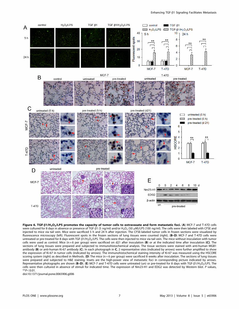
Figure 6. TGF-b1/H2O2/LPS promotes the capacity of tumor cells to extravasate and form metastatic foci. (A) MCF-7 and T-47D cellswere cultured for 8 days in absence or presence of TGF-b1 (5 ng/ml) and/or H2O2 (50 mM)/LPS (100 ng/ml). The cells were then labeled with CFSE andinjected to mice via tail vein. Mice were sacrificed 5 h and 24 h after injection. The CFSE-labeled tumor cells in frozen sections were visualized byfluorescence microscopy (left). Fluorescent spots in the frozen sections of lung tissues were counted (right). (B–D) MCF-7 and T-47D cells wereuntreated or pre-treated for 8 days with TGF-b1/H2O2/LPS. The cells were then injected to mice via tail vein. The mice without inoculation with tumorcells were used as control. Mice (n = 6 per group) were sacrificed on d21 after inoculation (B) or at the indicated time after inoculation (C). Thesections of lung tissues were prepared and subjected to immunohistochemical analysis. The tissue sections were stained with anti-human MGB1antibody (B) or anti-human Ki-67 antibody (C). In each photograph in C, 2 representative sites (indicated by arrows) were further amplified to showthe expression of Ki-67 in tumor cells (indicated by arrows). The immunohistochemical staining intensity of Ki-67 was measured using the HSCOREscoring system (right) as described in Methods. (D) The mice (n = 6 per group) were sacrificed 8 weeks after inoculation. The sections of lung tissueswere prepared and subjected to H&E staining. Insets are the high-power view of metastatic foci in corresponding picture indicated by arrows.Representative photographs are shown (B–D). (E) MCF-7 and T-47D cells were untreated (un) or pre-treated for 8 days with TGF-b1/H2O2/LPS. Thecells were then cultured in absence of stimuli for indicated time. The expression of Nm23-H1 and EDG2 was detected by Western blot. P values,**P,0.01.doi:10.1371/journal.pone.0065906.g006
Enhancing TGF-b1 Signaling Facilitates Metastasis
PLOS ONE | www.plosone.org 7 May 2013 | Volume 8 | Issue 5 | e65906

non-Smad pathways. Therefore, the activation of Smad and non-
Smad pathways induced by TGF-b1 in presence of H2O2 and LPS
was sufficient to modulate the metastatic potential of non-invasive
breast cancer cells.
TGF-b1 could induce apoptosis in a Smad-dependent manner
in some types of tumor cells [36]. However, the sustained
activation of MAPK pathways could promote apoptosis-resistance
of tumor cells, and abolish TGF-b1-indced apoptosis [36]. In
addition, the activation of NF-kB leads to the transcriptional
activation of genes that suppress apoptosis [37]. H2O2/LPS-
enhanced TGF-b1 signaling not only induced the sustained
activation of Smad pathway, but also induced the sustained and
higher activation levels of non-Smad pathways. Therefore, the
enhanced TGF-b1 signaling did not promote apoptosis in non-
invasive breast cancer cells. In contrast, TGF-b1/H2O2/LPS
promoted not only the invasive migration but also the anoikis-
resistance of breast cancer cells.
TGF-b1/H2O2/LPS-induced metastatic potential was sufficient
for tumor cell extravasation as shown by our data in animal test.
The disseminated tumor cells slowly developed into metastatic foci
due to lower proliferation at metastatic sites, since the tumor cells
at metastatic sites showed lower expression of proliferation marker
Ki-67. It has been known that new microenvironment could
restrict the proliferation of disseminated tumor cells [38]. Different
from high-invasive tumor cells, the metastatic tumor cells derived
from non-invasive tumor cells might be more sensitive to the
restriction of new microenvironment. On the other hand, it has
been found that Nm23-H1 has the potential to hinder the growth
of disseminated tumor cells at the metastatic site [34], and that
EDG2 is required for the proliferation of disseminated tumor cells
in new microenvironment [35]. TGF-b1/H2O2/LPS-modulated
expression of Nm23-H1 and EDG2 was reversed when tumor cells
were away from the stimuli, which might partially explain the
slower development of metastatic lesions.
In summary, in this study we demonstrated that H2O2/LPS
could enhance TGF-b1 signaling to induce the sustained
activation of both Smad and non-Smad pathways in non-invasive
breast cancer cells, and thus promoting the metastatic capability of
non-invasive breast cancer cells. Metastases are responsible for
most cancer deaths. Given that the enhanced signaling is required
for inducing higher metastatic capacity of tumor cells, targeting
one of these stimuli or signaling pathways might be potential
approach in comprehensive strategy for tumor therapy.
Materials and Methods
Ethics statementAll animal works were conducted according to relevant national
and international guidelines. They were approved by the
Committee on the Ethics of Animal Experiments of Tongji
Medical College (Permit Number: 2010-S260) and monitored by
the Department of Experimental Animals of Tongji Medical
College.
Cells and reagentsMCF-7 and T-47D cell lines were purchased from the Type
Culture Collection of the Chinese Academy of Sciences (Shanghai,
China), and cultured according to their guidelines. TGF-b1 was
purchased from PeproTech (Rocky Hill, NJ). H2O2 (hydrogen
peroxide) and LPS (lipopolysaccharide) were purchased from
Sigma-Aldrich (St. Louis, MO). Matrigel was purchased from BD
Biosciences (Bedford). SB203580, wortmannin, PD98059,
SP600125, 6-amino-4-(4-phenoxyphenylethylamino) quinazoline
(QNZ) were purchased from Merck4 Biosciences (Calbiochem).
Matrigel invasion assayMatrigel invasion assay was performed using Boyden chambers
(Transwell, Corning, Inc., Corning, NY). The transwell filter
inserts were coated with matrigel. The lower chambers were filled
with DMEM medium containing 10% FBS. 16105 tumor cells
were placed in the upper compartment. After 24-h incubation at
37uC in a humidified incubator with 5% CO2, the non-invading
cells were removed. The invasive cells attached to the lower
surface of membrane insert were fixed, stained, and counted under
a microscope from 7 randomly chosen fields in each membrane.
The average number of the cells per field was calculated.
Analysis for actin polymerizationTumor cells were incubated in matrigel-coated plate for 5 h.
The cells were then fixed in 4% paraformaldehyde, permeabilized
with 0.1% Triton X-100, and then stained with rhodamine-
phalloidin (Invitrogen) according to the manufacturer’s protocol to
visualize the cells with highly polymerized actin.
Assay of gene expression by real-time RT-PCRTotal RNA was extracted from cells with TRIzol reagent
(Invitrogen). The relative quantity of mRNA was determined by
real-time RT-PCR according to MIQE guidelines [39]. Quanti-
fication of the expression of genes was performed using the
comparative CT method. GAPDH, HPRT1 and YWHAZ were
selected as reference genes according to the rules described in
MIQE guidelines. The relative expression of the genes of interest
was expressed as the mRNA level of the gene relative to the
geometric mean of the mRNA levels of three reference genes,
which was calculated using GeNorm software. The primer
sequences were as follows: NM23-H1, sense 59-TCATTGCGAT-
CAAACCAGAT- 39, antisense 59-CAACGTAGT GTTC
CTTGAGA-39; EDG2, sense 59-GCTATGTTCGCCAGAG-
GACT-39, antisense 59-ATC CAGGAGTCCAGCAGATG-39;
ITGAV, sense 59-CTCGGGACTCCTGCTACCTC-39, anti-
sense 59- AAGAAACATCCGGGAAGACG-39; ITGB3, sense
59-CATCCTGGTGGTCCT GCTCT-39, antisense 59-
GCCTCTTTACACAGTGGGTTGTT-39; MMP9, sense 59-
CAGTCCA CCCTTGTGCTCTTCC-39, antisense 59-
CTGCCACCCGAGTGTAACCAT-39; BAX, sense 59-
TTTTGCTTCAGGGTTTCATC-39, antisense 59-GA-
CACTCGCTCAGCTTCTTG-39; BIM, sense 59-CA GAGCCA-
CAAGACAGGA-39, antisense 59-CCATACAAATCTAAGC-
CAGT-39; BID, sense 59-GCCGTCCTTGCTCCGTGAT-39,
antisense 59-ATGCCAGGGCTCCGTCTA-39; BCL2, sense 59-
GGTCATGTGTGTGGAGAGC-39, antisense 59-GATC-
CAGGTGTGCAGGT G-39; MCL1, sense 59-
TTGACTTCTGTTTGTCTTA CGCT-39, antisense 59-
TGGTCCTAACC CTTCCTGG-39; c-FLIP, sense 59-AGAGT-
GAGGCGATTTGACCTG-39, antisense 59-AAG
GTGAGGGTTCCTGAGCA-39; TGFBR1, sense 59-TGAACA-
GAAGTTAAGGCCAAATAT C-39, antisense 59-CAGG-
CAAAGCTGTAGAATTACATTT-39; TGFBR2, sense 59-
CGGTTAA TAACGACATGATAGTCAC-39, anti-sense 59-
TCATGGCAAACTGTCTCTAGTGTTA-39; SNAI2, sense 59-
AGGAATCTGGCTGCTGTG-39, antisense 59-GGA-
GAAAATGCCTTTGGA C-39; SMAD7, sense 59-
TCCTCCTGAGTGCTTGCTT-39, antisense 59-
TCTGCTTCCCCTCT TCCTA-39; p21, sense 59-GGACAG-
CAGAGGAAGACCATGT-39, antisense 59-TGGAGTG GTA-
GAAATCTGTCATGC-39; MYC, sense 59-GCCACGTCTCCA-
CACATCAG-39, antisense 59-
TGGTGCATTTTCGGTTGTTG-39; GAPDH, sense 59-
TCATTGACCTCAACT ACATGGTTT-39, antisense 59-GAA-
Enhancing TGF-b1 Signaling Facilitates Metastasis
PLOS ONE | www.plosone.org 8 May 2013 | Volume 8 | Issue 5 | e65906
GATGGTGATGGGATTTC-39; HPRT1, sense 59-GCTG AG-
GATTTGGAA AGGGTG-39, antisense 59-CAGAGGGCTA-
CAATGTGATGG-39; YWHAZ, sense 59-
GATCTTTCTGGCTCCACTCA-39, antisense 59-CCATT-
CAGGATAGGTAGGGT-39.
Western blot assayWestern blot assay was done as described previously [4]. Abs
were purchased from Santa Cruz Biotechnology and Cell
Signaling Technology.
Flow cytometric analysisTumor cells were stained with FITC-conjugated mouse-anti-
human avb3 (Chemicon) or isotype control IgG1 for flow
cytometric analysis. Parameters were acquired on a FACS Calibur
440E flow cytometer (BD Biosciences) and analyzed with
CellQuest software (BD Biosciences). Percent staining was defined
as the percentage of cells in the gate (M1) which was set to exclude
,99% of isotype control cells. avb3 expression index was
calculated by using the formula: mean fluorescence 6percentage
of avb3+ cells [4].
MMP assay by gelatin zymographyTumor cells were cultured for 48 h in DMEM medium contain-
ing 1% FBS in presence of pre-coated matrigel. The assay of MMP-9
in supernatants was performed as described previously [4].
Assay of activity of NF-kBThe nuclear extract was prepared with Nuclear Extraction Kit
(Millipore, Billerica, MA). The activity of NF-kB in nuclear extract
was determined by NF-kB Assay kit (Millipore) according to the
manufacturer’s protocol.
Cell transfectionFor down-regulation of SNAI2, MCF-7 cells were transduced
with SNAI2 shRNA lentiviral particles, or control shRNA
lentiviral particles (Santa Cruz Biotech, Inc.) according to the
manufacturer’s protocol. After selection with puromycin, the cells
were used for further experiments.
Apoptosis AssayTumor cells were cultured for 24 h in 6-well plates pre-coated
with poly-HEMA (10 mg/ml, Sigma). The cells were then stained
with Annexin V-FITC/Propidium Iodide (PI) apoptosis detection
kit (BD Biosciences, San Diego, CA), and analyzed by flow
cytometry.
Soft agar assayTumor cells were pretreated with the indicated factors for 8
days. The cells were then suspended in 0.3% agar in DMEM (20%
FBS) and plated (16104 cells/well in 6-well plates) on a layer of
0.6% agar in DMEM (20% FBS) in triplicate. After 21-day culture
in absence of stimuli, the cells were stained with 0.005% crystal
violet. The colonies of tumor cells were counted under a
microscope.
Assay of tumor cell arrest in lungFemale athymic nude (nu/nu) mice (4 to 6 weeks old) were
purchased from Beijing HFK Bio-Technology Co, LTD. (Beijing,
China) for studies approved by the Committee on the Ethics of
Animal Experiments of Tongji Medical College. The mice were
maintained in the accredited animal facility of Tongji Medical
College. The mice were randomly divided to several groups
(6 mice/group). For assay of tumor cell arrest in lung, tumor cells
were labeled with CFSE, and injected into mice by i.v. injection
which was performed after inhalation of 2% isoflurane. 16106
cells in 0.1 ml of PBS were slowly injected after sterilization of tail
with alcohol. 5 h and 24 h after the injection, the mice were
sacrificed by CO2 inhalation. Lung tissues were harvested. Frozen
sections were prepared and analyzed by fluorescence microscopy.
Fluorescent spots were counted from 20 randomly chosen fields in
sections of each mouse.
HistologyTumor cells (16106 per mouse) were injected into mice via tail
vein. The lung tissues were harvested at the indicated time, and
embedded in paraffin according to standard histological proce-
dures. Tissue sections were prepared and subjected to immuno-
histochemical analysis or H&E staining. Immunohistochemical
staining for breast cancer cell marker MGB1 (Mammaglobin A)
and for proliferation marker Ki-67 was performed using anti-
human MGB1 antibody and anti-human Ki-67 antibody (Abcam
Biotechnology) as primary antibody. HRP-conjugated anti-rabbit
IgG were used as secondary antibody. Images were obtained using
OLYMPUS-BX51 microscope at 10610, 20610 or 40610
magnification. Staining intensity of cells was evaluated under a
microscope and graded (1, weak; 2, moderate; 3, strong) in a
blinded fashion by two examiners. Staining intensity of tissue
sections was assessed using a semi-quantitative immunohistochem-
ical scoring system, HSCORE. The HSCORE was calculated
using the following equation: HSCORE =gPi(i+1), where i is the
staining intensity of cells and Pi is the percentage of the cells at
each level of intensity [40].
Statistical analysisData are pooled from three independent experiments with a
total of six samples in each group. Results were expressed as mean
value 6 SD and interpreted by one-way ANOVA, except for the
data of experimental lung metastases which were interpreted by
Fisher’s Exact Test. Differences were considered to be statistically
significant when P,0.05.
Supporting Information
Figure S1 TGF-b1/H2O2/LPS augments polymerizationof actin of tumor cells. Tumor cells were cultured in absence
or presence of TGF-b1, H2O2 and LPS for 8 days. The cells were
then incubated in presence of matrigel for 5 h. The cells with
highly polymerized actin were visualized by staining with
rhodamine-phalloidin after incubation (left). Their percentage in
total cells was calculated (right). P values, *P,0.05, **P,0.01.
(TIF)
Figure S2 TGF-b1/H2O2/LPS does not significantlyinfluence the expression of p21 and MYC. MCF-7 cells
were cultured in absence or presence of TGF-b1, H2O2 and LPS
for 7 days. The expression of p21 and MYC were detected by real-
time RT-PCR and Western blot.
(TIF)
Figure S3 TGF-b1/H2O2/LPS modulates the expressionof Nm23-H1 and EDG2. MCF-7 cells were cultured in absence
or presence of TGF-b1/H2O2/LPS (left) for the indicated time.
Or the cells were cultured for 6 days in absence or presence of
TGF-b1, H2O2, and LPS (right). The expression of NM23-H1 (A),and EDG2 (B) was detected by real-time RT-PCR. P values,
**P,0.01.
(TIF)
Enhancing TGF-b1 Signaling Facilitates Metastasis
PLOS ONE | www.plosone.org 9 May 2013 | Volume 8 | Issue 5 | e65906

Figure S4 Inhibiting PI3K suppresses TGF-b receptorexpression and activation of other non-Smad pathways.MCF-7 cells were un-treated or treated with TGF-b1/H2O2/LPS
for the indicated time in absence of presence of wortmannin (WT,
40 nM). (A) The expression of TbRI and TbRII was detected by
Western blot. The relative expression of TbRI and TbRII to b-
actin was calculated after densitometric analysis of Western blot.
(B) The phosphorylated and un-phosphorylated p38MAPK,
ERK, and JNK were detected by Western blot. The ratio of
phosphorylated and un-phosphorylated p38MAPK, ERK, and
JNK was calculated after densitometric analysis of Western blots.
The activity of NF-kB was assayed as described in Methods. P
values, *P,0.05, **P,0.01.
(TIF)
Figure S5 TGF-b1/H2O2/LPS promotes anoikis-resis-tance of non-invasive breast cancer cells. MCF-7 and T-
47D cells were cultured in absence or presence of TGF-b1/H2O2/
LPS for the indicated time. The cells were then transferred to poly-
HEMA-coated plate and cultured for 24 h. The apoptosis of the
cells was analyzed by flow cytometry. P values, *P,0.05,
**P,0.01.
(TIF)
Acknowledgments
The authors would like to thank Professor Sheng-Hong Liu for reviewing
histology data.
Author Contributions
Conceived and designed the experiments: ZHF GMZ. Performed the
experiments: YHZ SJL DL JL JJW BY RS YS QW. Analyzed the data:
YHZ GMZ ZHF. Contributed reagents/materials/analysis tools: GMZ
ZHF. Wrote the paper: ZHF GMZ YHZ.
References
1. Gallego MI, Bierie B, Hennighausen L. (2003) Targeted expression of HGF/SF
in mouse mammary epithelium leads to metastatic adenosquamous carcinomasthrough the activation of multiple signal transduction pathways. Oncogene 22:
8498–8508.2. Li Y, Wang JP, Santen RJ, Kim TH, Park H, et al. (2010) Estrogen stimulation
of cell migration involves multiple signaling pathway interactions. Endocrinology
151: 5146–5156.3. Wu WS, Wu JR, Hu CT. (2008) Signal cross talks for sustained MAPK
activation and cell migration: the potential role of reactive oxygen species.Cancer Metastasis Rev 27: 303–314.
4. Liao SJ, Zhou YH, Yuan Y, Li D, Wu FH, et al. (2012) Triggering of Toll-like
receptor 4 on metastatic breast cancer cells promotes alphavbeta3-mediatedadhesion and invasive migration. Breast Cancer Res Treat 133: 853–863.
5. Krueger JS, Keshamouni VG, Atanaskova N, Reddy KB. (2001) Temporal andquantitative regulation of mitogen-activated protein kinase (MAPK) modulates
cell motility and invasion. Oncogene 20: 4209–4218.
6. Keller ET, Li LY. (2011) The first Tianjin, China forum on tumormicroenvironment. Cancer Res 71: 310–313.
7. Brown KA, Aakre ME, Gorska AE, Price JO, Eltom SE, et al. (2004) Inductionby transforming growth factor-beta1 of epithelial to mesenchymal transition is a
rare event in vitro. Breast Cancer Res 6: R215–231.8. Xu J, Lamouille S, Derynck R. (2009) TGF-beta-induced epithelial to
mesenchymal transition. Cell Res 19:156–172.
9. Parvani JG, Taylor MA, Schiemann WP. (2011) Noncanonical TGF-betasignaling during mammary tumorigenesis. J Mammary Gland Biol Neoplasia 16:
127–146.10. Christeli E, Zoumpourlis V, Kiaris H, Ergazaki M, Vassilaros S, et al. (1996)
TGF-beta 1 overexpression in breast cancer. Oncol Rep 3: 1115–1118.
11. Liu SL, Lin X, Shi DY, Cheng J, Wu CQ, et al. (2002) Reactive oxygen speciesstimulated human hepatoma cell proliferation via cross-talk between PI3-K/
PKB and JNK signaling pathways. Arch Biochem Biophys 406: 173–182.12. Okoh V, Deoraj A, Roy D. (2011) Estrogen-induced reactive oxygen species-
mediated signalings contribute to breast cancer. Biochim Biophys Acta 1815:115–133.
13. Yoon SO, Park SJ, Yoon SY, Yun CH, Chung AS. (2002) Sustained production
of H2O2 activates pro-matrix metalloproteinase-2 through receptor tyrosinekinases/phosphatidylinositol 3-kinase/NF-kappa B pathway. J Biol Chem 277:
30271–30282.14. Diya Z, Lili C, Shenglai L, Zhiyuan G, Jie Y. (2008) Lipopolysaccharide (LPS) of
Porphyromonas gingivalis induces IL-1beta, TNF-alpha and IL-6 production by
THP-1 cells in a way different from that of Escherichia coli LPS. Innate Immun14: 99–107.
15. Fortin CF, Cloutier A, Ear T, Sylvain-Prevost S, Mayer TZ, et al. (2011) A classIA PI3K controls inflammatory cytokine production in human neutrophils.
Eur J Immunol 41: 1709–1719.16. Zhang Y, Jiang J, Ji S, Shan Y, Zhu P, et al. (2001) The regulatory effect of
ERK1/2 signal pathway on production of TNFalpha induced by LPS in mice
Kupffer cells. Chin J Traumatol 4: 139–142.17. Sato Y, Goto Y, Narita N, Hoon DS. (2009) Cancer Cells Expressing Toll-like
Receptors and the Tumor Microenvironment. Cancer Microenvironment 2Suppl 1:205–214.
18. Pidgeon GP, Harmey JH, Kay E, Costa MD, Redmond HP, et al. (1999) The
role of endotoxin/lipopolysaccharide in surgically induced tumor growth in amurine model of metastatic disease. Br J Cancer 81:1311–1317.
19. Tsan MF, Gao B. (2004) Endogenous ligands of Toll-like receptors. J LeukocBiol 76:514–519.
20. Liu L, Yang M, Kang R, Wang Z, Zhao Y, et al. (2011) DAMP-mediated
autophagy contributes to drug resistance. Autophagy 7: 112–114.
21. Szatrowski TP, Nathan CF. (1991) Production of large amounts of hydrogen
peroxide by human tumor cells. Cancer Res 51: 794–798.22. Yilmaz M, Christofori G. (2009) EMT, the cytoskeleton, and cancer cell
invasion. Cancer Metastasis Rev 28: 15–33.23. Nakao A, Afrakhte M, Moren A, Nakayama T, Christian JL, et al. (1997)
Identification of Smad7, a TGFbeta-inducible antagonist of TGF-beta
signalling. Nature 389: 631–635.24. Joseph MJ, Dangi-Garimella S, Shields MA, Diamond ME, Sun L, et al. (2009)
Slug is a downstream mediator of transforming growth factor-beta1-inducedmatrix metalloproteinase-9 expression and invasion of oral cancer cells. J Cell
Biochem 108: 726–736.
25. Massague J. (2004) G1 cell-cycle control and cancer. Nature 432: 298–306.26. Taniguchi F, Harada T, Sakamoto Y, Yamauchi N, Yoshida S, et al. (2003)
Activation of mitogen-activated protein kinase pathway by keratinocyte growthfactor or fibroblast growth factor-10 promotes cell proliferation in human
endometrial carcinoma cells. J Clin Endocrinol Metab 88: 773–780.
27. Acosta JJ, Munoz RM, Gonzalez L, Subtil-Rodriguez A, Dominguez-CaceresMA, et al. (2003) Src mediates prolactin-dependent proliferation of T47D and
MCF7 cells via the activation of focal adhesion kinase/Erk1/2 andphosphatidylinositol 3-kinase pathways. Mol Endocrinol 17: 2268–2282.
28. Mawson A, Lai A, Carroll JS, Sergio CM., Mitchell CJ, et al. (2005) Estrogenand insulin/IGF-1 cooperatively stimulate cell cycle progression in MCF-7
breast cancer cells through differential regulation of c-Myc and cyclin D1. Mol
Cell Endocrinol 229:161–173.29. Seong HA, Jung H, Ha H (2007) NM23-H1 tumor suppressor physically
interacts with serine-threonine kinase receptor-associated protein, a transform-ing growth factor-beta (TGF-beta) receptor-interacting protein, and negatively
regulates TGF-beta signaling. J Biol Chem 282: 12075–12096.
30. Horak CE, Lee JH, Elkahloun AG, Boissan M, Dumont S, et al. (2007) Nm23-H1 suppresses tumor cell motility by down- regulating the lysophosphatidic acid
receptor EDG2. Cancer Res 67: 7238–7246.31. Dhasarathy A, Phadke D, Mav D, Shah RR, Wade PA. (2011) The transcription
factors snail and slug activate the transforming growth factor-Beta signalingpathway in breast cancer. PLoS One 6: e26514.
32. Woods NT, Yamaguchi H, Lee FY, Bhalla KN, Wang HG. (2007) Anoikis,
initiated by Mcl-1 degradation and Bim induction, is deregulated duringoncogenesis. Cancer Res 67: 10744–10752.
33. Marconi A, Atzei P, Panza C, Fila C, Tiberio R, et al. (2004) FLICE/caspase-8activation triggers anoikis induced by beta1-integrin blockade in human
keratinocytes. J Cell Sci 117(Pt24): 5815–5823.
34. Horak CE, Lee JH, Marshall JC, Shreeve SM, Steeg PS. (2008) The role ofmetastasis suppressor genes in metastatic dormancy. APMIS 116: 586–601.
35. Marshall JC, Collins JW, Nakayama J, Horak CE, Liewehr DJ, et al. (2012)Effect of inhibition of the lysophosphatidic Acid receptor 1 on metastasis and
metastatic dormancy in breast cancer. J Natl Cancer Inst 104: 1306–1319.36. Ozaki I, Hamajima H, Matsuhashi S, Mizuta T. (2011) Regulation of TGF-b1-
induced pro-apoptotic signaling by growth factor receptors and extracellular
matrix receptor integrins in the liver. Front Physiol 2: 78.37. Baldwin AS. (2001) Control of oncogenesis and cancer therapy resistance by the
transcription factor NF-kappaB. J Clin Invest 107: 241–246.38. Wikman H, Vessella R, Pantel K. (2008) Cancer micrometastasis and tumour
dormancy. APMIS 116: 754–770.
39. Bustin SA, Benes V, Garson JA, Hellemans J, Huggett J, et al. (2009) The MIQEguidelines: minimum information for publication of quantitative real-time PCR
experiments. Clin Chem 55: 611–622.40. Xiong T, Zhao Y, Hu D, Meng J, Wang R, et al. (2012) Administration of
calcitonin promotes blastocyst implantation in mice by up-regulating integrin
beta3 expression in endometrial epithelial cells. Hum Reprod 27:3540–3551.
Enhancing TGF-b1 Signaling Facilitates Metastasis
PLOS ONE | www.plosone.org 10 May 2013 | Volume 8 | Issue 5 | e65906