TITLE: The Impact of Executive Function on Reading Fluency ...
18
TITLE: The Impact of Executive Function on Reading Fluency in Young Adults with Mild Traumatic Brain Injury PRINCIPAL INVESTIGATOR: Dr. Natalie S. Dailey Co-INVESTIGATOR: Dr. William D.S. Killgore REPORT DATE: March 30, 2020 TYPE OF REPORT: Final Report PREPARED FOR: Woodcock Institute Grant Research Program Texas Woman’s University
Transcript of TITLE: The Impact of Executive Function on Reading Fluency ...

TITLE: The Impact of Executive Function on Reading Fluency in Young Adults with Mild Traumatic Brain Injury
PRINCIPAL INVESTIGATOR: Dr. Natalie S. Dailey Co-INVESTIGATOR: Dr. William D.S. Killgore
REPORT DATE: March 30, 2020
TYPE OF REPORT: Final Report
PREPARED FOR: Woodcock Institute Grant Research Program Texas Woman’s University

1. RESEARCH OVERVIEW
Mild traumatic brain injury (mTBI) accounts for nearly 80% of all reported brain injuries in the United States and a growing number of young adults with mTBI are enrolling in postsecondary education. A mTBI can subtly and microscopically alter the structure of the brain and result in impaired cognitive functions, including slowed processing speed and attention deficits. Furthermore, many young adults who experience mTBI-related cognitive deficits encounter major challenges in the academic setting, yet the impact of mTBI on reading is not well understood. The current study aimed to address this critical gap by identifying the neurocognitive impact of executive function on reading fluency in young adults with mTBI. 2. PROJECT SUMMARY The primary aims of the funded study were to identify the association between executive function and reading fluency in young adults with mTBI and determine the neural correlates of executive function and reading fluency. The study involved one visit to the Social, Cognitive, and Affective Neuroscience (SCAN) Lab at the University of Arizona, which lasted two and a half hours. During the study visit, individuals provided written informed consent, followed by the completion of questionnaires, cognitive tasks, and a 30-minute magnetic resonance imaging (MRI) session. The study progressed on schedule according to the Statement of Work outlined in the funded proposal. Initial IRB approval from the University of Arizona was obtained 11/27/2018 and an amendment was approved on 3/22/2019, to include the Rivermead Post Concussion Symptoms Questionnaire (RPQ) and an additional measure of executive function, a Go/No Go task. All study materials were acquired early Spring 2019. Study Recruitment: Recruitment began in March 2019 and ended November 2019. Our team screened 208 interest forms, assessed 173 individuals for study eligibility, and enrolled 44 participants (see Figure 1). Of the individuals assessed for eligibility, 104 did not meet inclusion/exclusion criteria (see Table 1) and 25 declined to participate. Eligible individuals met inclusion criteria including between 18 and 35 years of age, native English speaker, and normal or corrected-to-normal vision. Individuals with corrected-to-normal vision were required to wear non-tined contacts, as opposed to glasses, during the MRI portion of the study. Those unable to wear contacts were
Figure 1. Consort diagram of screened, enrolled, and completed individuals for this study.
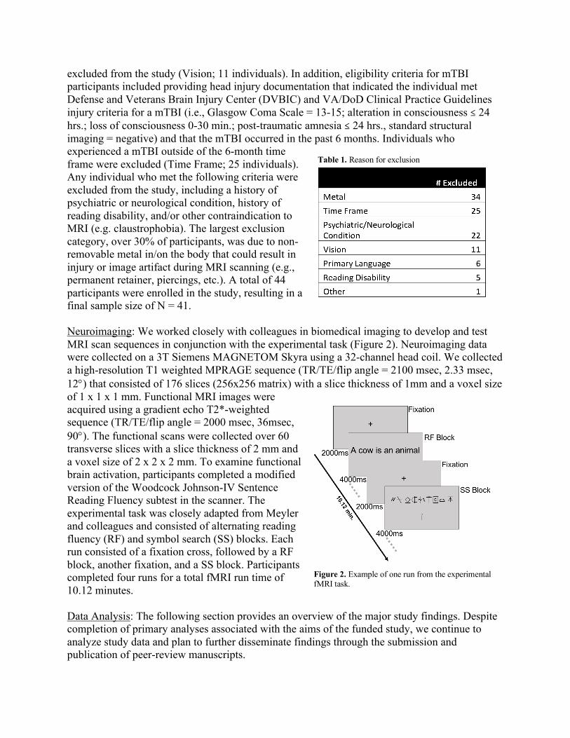
excluded from the study (Vision; 11 individuals). In addition, eligibility criteria for mTBI participants included providing head injury documentation that indicated the individual met Defense and Veterans Brain Injury Center (DVBIC) and VA/DoD Clinical Practice Guidelines injury criteria for a mTBI (i.e., Glasgow Coma Scale = 13-15; alteration in consciousness ≤ 24 hrs.; loss of consciousness 0-30 min.; post-traumatic amnesia ≤ 24 hrs., standard structural imaging = negative) and that the mTBI occurred in the past 6 months. Individuals who experienced a mTBI outside of the 6-month time frame were excluded (Time Frame; 25 individuals). Any individual who met the following criteria were excluded from the study, including a history of psychiatric or neurological condition, history of reading disability, and/or other contraindication to MRI (e.g. claustrophobia). The largest exclusion category, over 30% of participants, was due to non-removable metal in/on the body that could result in injury or image artifact during MRI scanning (e.g., permanent retainer, piercings, etc.). A total of 44 participants were enrolled in the study, resulting in a final sample size of N = 41.
Neuroimaging: We worked closely with colleagues in biomedical imaging to develop and test MRI scan sequences in conjunction with the experimental task (Figure 2). Neuroimaging data were collected on a 3T Siemens MAGNETOM Skyra using a 32-channel head coil. We collected a high-resolution T1 weighted MPRAGE sequence (TR/TE/flip angle = 2100 msec, 2.33 msec, 12°) that consisted of 176 slices (256x256 matrix) with a slice thickness of 1mm and a voxel size of 1 x 1 x 1 mm. Functional MRI images were acquired using a gradient echo T2*-weighted sequence (TR/TE/flip angle = 2000 msec, 36msec, 90°). The functional scans were collected over 60 transverse slices with a slice thickness of 2 mm and a voxel size of 2 x 2 x 2 mm. To examine functional brain activation, participants completed a modified version of the Woodcock Johnson-IV Sentence Reading Fluency subtest in the scanner. The experimental task was closely adapted from Meyler and colleagues and consisted of alternating reading fluency (RF) and symbol search (SS) blocks. Each run consisted of a fixation cross, followed by a RF block, another fixation, and a SS block. Participants completed four runs for a total fMRI run time of 10.12 minutes.
Data Analysis: The following section provides an overview of the major study findings. Despite completion of primary analyses associated with the aims of the funded study, we continue to analyze study data and plan to further disseminate findings through the submission and publication of peer-review manuscripts.
Figure 2. Example of one run from the experimentalfMRI task.
Table 1. Reason for exclusion

Participants. We completed the study with a final sample size of N = 41, including 21 healthy controls (HC) participants and 20 participants with a mTBI. The ratio of males to females in the two groups was not significantly different (c2 = 0.03, p = .87), with 10 males, 11 females in the HC group and 9 males, 11 females in the mTBI group. As shown in Table 2, the groups did not differ significantly on age (t (39) = -0.72, p = .48) or IQ (t (39) = 0.78, p = .44). Demographic characteristics for the groups are reported in Table 2.
With regard to participants in the mTBI group, we obtained documentation on time since injury and collected data on symptom severity and mechanism of injury. The majority of participants suffered a mTBI while engaged in sports-related activities (55%), followed by motor vehicle accidents (20%), being struck by or against an object (15%), and falls (10%). We focused on individuals who were within 6 months of their injury. On average, participants in the mTBI group were roughly 105 days post-injury. The Rivermead Post Concussion Symptoms Questionnaire (RPQ) was administered to evaluate the frequency and severity of 16 post-concussion symptoms. Headaches were the most common RPQ-3 symptom, with 60% of mTBI participants reporting mild to moderate problems after the injury. Sleep disturbance was the most common RPQ-13 symptom, with 35% of mTBI participants reporting mild to severe problems. Injury characteristics for the mTBI group are reported in Table 3.
Reading Ability. Initial analyses were conducted to determine whether groups differed in reading ability. All participants completed the Vocabulary subtest of the Wechsler Abbreviated Scale of Intelligence (WASI-II) to measure vocabulary knowledge, the Letter-Word Identification subtest of Woodcock-Johnson IV Tests of Achievement (WJ-IV) to measure decoding, and the Passage Comprehension subtest of the WJ-IV to measure reading comprehension. One participant in the HC group scored 3 SD beyond the mean and was classified an outlier. Therefore, these analyses included n = 20 HCs and n = 20 mTBI. We conducted a multivariate analysis, with group as the dependent variable and raw scores from the WASI-II and WJ-IV subtests as independent variables, controlling for age, gender, and years of education. Groups differed on vocabulary knowledge, with HC reporting significantly higher
Table 2. Demographic characteristics
Table 3. Mild traumatic brain injury characteristics

vocabulary scores compared to the mTBI group (F(1,35) = 4.64, p = .04, h2 = .12). No other significant differences were found between the groups. Descriptive statistics on reading measures are shown in Table 4.
Executive Function. To address one of the primary aims of the funded study, we
identified the role of executive function in reading following a mTBI. Due to missing data on outcome variables of interest, the final sample for these analyses included 38 participants, 19 HCs and 19 in the mTBI group. In addition to WJ-IV Passage Comprehension and Letter-Word Identification subtests, participants completed a 10-minute computerized Psychomotor Vigilance Test (PVT) and a computerized Berg Card Sorting Test (BCST) to measure attention and executive function, respectively. For all participants, there was a significant relationship between reading comprehension and attention, where faster reaction times were associated with significantly higher passage comprehension scores (r = -0.37, p = .02). When groups were assessed separately, attention and passage comprehension were significantly correlated in the mTBI group (r = -0.47, p = .04), but not in HCs (r = -0.43, p = .07). Linear regressions were conducted to determine the extent to which linguistic and cognitive factors predict reading comprehension. In the sample as a whole, IQ (β = 0.62, p < .0001) and reaction time (β = -0.27, p = .03) predicted comprehension (r2 = 0.48, F = 18.38, p < 0.0001). When we analyzed the groups separately, IQ significantly predicted comprehension (β = 0.80, p < .0001) in HCs, while decoding significantly predicted comprehension (β = 0.62, p = .004) in those with mTBI. These findings suggest that during reading, those who have sustained a mTBI may rely more heavily on attentional networks relative to healthy individuals. Findings from the present study indicate a unique, and arguably important role of sustained attention in broad cognitive tasks, such as reading, specifically in adults who have experienced brain injury. These findings were submitted for presentation at the 2020 Military Health Systems Research Symposium (Thompson, Killgore, & Dailey). A subset of participants (n = 12 HCs; n = 12 mTBI) completed a military-oriented Go/No Go task known as the Military Context Dependent Shoot/No Shoot Task (SNS). This task requires the individual to make a shoot/no-shoot decision based on the context (urban vs. rural) and the color of the uniform of the target (green vs. tan). Moreover, to assess cognitive flexibility, the SNS includes a reversal of contingencies halfway through the task, which requires the individual to use feedback to learn and maintain stimulus-response rules and update these rules after a reversal of contingencies. In this sample, we found that those with mTBI showed profound impairment in cognitive flexibility during the SNS task, particularly post-reversal, compared to HCs. As shown in Figure 3, individuals with a documented mTBI exhibited significantly more errors (left panels) and patterns of perseverative responding (right panels) compared to non-
Table 4. Reading measures

injured controls.
With regard to errors, mTBI subjects showed significantly greater false alarm errors pre- (F(1,22)=7.08, p = 0.01, η2=.24) and post-reversal (F(1,22)=7.08, p = 0.04, η2=.18), compared to HCs. High false alarm rates reflect a failure of inhibitory control, a finding that is consistent with damage to frontal and temporal lobes following brain injury. Response bias was compared between the groups and we found no significant difference pre-reversal (F(1,22)=1.90, p = 0.18, η2=.08). However, post-reversal, mTBI subjects exhibited a significant bias response, perseverating on ‘shoot’ (F(1,22)=8.64, p = 0.008, η2=.28), as compared to HCs. This suggests a subtle deficit in the ability to cope with changing task contingencies that led to an increase in an ineffective and potentially dangerous tendency to perseverate on shoot responses in those with mTBI. These finding were included as pilot data in a grant submission for FY2020 Department of Defense, Combat Readiness-Medical Research Program (PI: WDS Killgore; Co-I: NS Dailey).
Neural Correlates. We analyzed fMRI data to identify regions of activation during a reading fluency task. As described above, participants completed a modified version of the WJ-IV Sentence Reading Fluency subtest. Reading blocks included 8 sentences presented one at a time, for a total of 48 sentences. Symbol search blocks included 8 symbol strings presented independently for a total of 48 strings (see Figure 2 above). The symbol blocks served as the control condition, which accounted for visual scanning and motor aspects of the experimental task. During the task, participants responded “yes” or “no” to each sentence and symbol string using a button box. Independent samples t-test were calculated to compare mTBI and HCs on reading accuracy and speed. High-resolution T1-weighted anatomical images were collected and used to co-register fMRI data. Processing and analysis of neuroimaging data was conducted in SPM12. At the individual level, a general linear model (GLM) was specified to contrast neural activation during the sentence > symbol condition. These contrast images were entered into a second-level independent samples t-test with group (HC vs mTBI) as the independent variable. Primary neuroimaging results from a whole-brain analysis are reported with a threshold p< 0.001 (uncorrected), k (extent)>10 contiguous voxels.
Accuracy and speed on the modified sentence reading fluency task were similar between the groups. We did, however, find differences in cortical activation between the groups. The HC
Figure 3. Cognitive performance on Shoot/No Shoot task. Left panel: False alarm errors by group. Right panel: Response bias by group.

group showed significantly greater cortical activation in the right inferior parietal lobule (k = 31; t = 4.95; p = .026; x = 34; y = -30; z = 24), the right cerebellum (k = 31; t = 4.75; p = .026; x = 14; y = -50; z = -46), and the right superior parietal lobule (k = 27; t = 4.14; p = .036; x = 32; y = -32; z = 22), compared those with a mTBI (see Figure 4). In contrast, the mTBI group exhibited greater activation in the right middle frontal gyrus, a finding that did not reach statistical significance (k = 21; t = 4.55; p = .06; x = 30; y = 18; z = 40) (see Figure 5).
Despite maintaining similar levels of accuracy and speed on the reading fluency task, our findings indicate young adults with mTBI exhibit less cortical activation in superior and inferior parietal regions, but greater cortical activation in the prefrontal cortex during reading. These neuroimaging findings indicate that those with mTBI may require increased allocation of cortical resources associated with executive function to achieve the same level of performance as their healthy peers. These findings raise the possibility that individuals recovering from mTBI might have greater difficulty maintaining other executive functions when reading, due to insufficient prefrontal resources. Because distractions were minimal and controlled in the present study, future research should investigate the relationship of reading fluency and cortical activation when distractors are present to more adequately represent the daily environments people encounter. These findings were submitted for presentation at the 2020 Military Health Systems Research Symposium (Cozine, Killgore, & Dailey).
Figure 4. Regions of cortical activation during reading fluency task for the contrast health control > mild traumatic brain injury.
Figure 5. Region of cortical activation, shown in axial, sagittal, and coronal images, for contrast mild traumatic brain injury > healthy control. Note: neurological orientation.

In addition to functional MRI, we collected diffusion tensor imaging (DTI) data on a subset of participants enrolled in the study. Cognitive deficits observed in those with mTBI can be further linked to structural differences in axonal tracts by detecting white matter abnormalities via DTI. The cingulum bundle is associated with executive control, where significant abnormalities are prevalent among clinical populations including those with mild cognitive impairment. In this exploratory analysis, we aimed to identify white matter pathways where individuals with mTBI may have reduced fractional anisotropy (FA; a metric of axonal integrity) compared to healthy controls and determine the extent to which symptom severity and brain structure were associated with cognitive ability. DTI and cognitive data were collected from 20 HCs (mean age = 21.00 ±1.01 years; 10 females) and 18 participants post-mTBI (mean age = 22.61 ± 1.08 years; 9 females).
Standard processing pipelines in FSL (topup and dtifit) were used to process DTI images and FA was quantified using TBSS. Whole-brain comparisons of FA between the groups are shown in Figure 6. The cingulum bundle was masked for the left and right hemisphere (using the MNI atlas) and mean FA values were extracted for all participants. The cingulum was targeted a-priori, thereby eliminating superfluous comparisons encountered in whole-brain analyses. To determine cingulum integrity following mTBI, a multivariate analysis of covariance was calculated, with gender and IQ entered as covariates (p-value one-tailed). A linear regression was calculated with RPQ-3, cingulum FA values, gender, and IQ to predict PVT reaction time.
Significantly lower FA was found for the right cingulum following mTBI compared to HCs (F = 3.75, p = .03, h2 = .10). However, FA of the left cingulum was similar across the groups (F =
Figure 6. Whole brain comparison of white matter integrity between groups. Top panel: voxels where fractional anisotropy is higher in health controls compared to mild traumatic brain injury. Bottom panel: voxels where fractional anisotropy is higher in mild traumatic brain injury group compared to healthy controls. Note: radiological orientation.

0.56, p = .23, h2 = .02). Symptom severity related to cognitive processes was significantly higher in participants who experienced a mTBI, compared to HCs (t = 4.92, p = 0.03). With regard to cognitive function, higher levels of FA in the right cingulum significantly predicted faster PVT reaction time (β = -0.37, p = .026) in the entire sample. FA is associated with an objective measure of cognitive function. More so, FA in the right cingulum significantly predicted cognitive ability, in that reduced FA indicated slower reaction time on the PVT. Our findings provide support for the utility of DTI metrics as biomarkers for cognitive function following mTBI. These findings were submitted for presentation at the 2020 Military Health Systems Research Symposium (Le, Killgore, & Dailey).
3. BUDGETARY EXPENDITUES
We completed the funded study on time and within the contracted budget. Table 5 provides a detailed use of study funds by category. In August 2019, we established a relationship with the Psychology department and recruited individuals through the PSY 101 undergraduate student research pool. All individuals who participated in the study were compensated for their time, either monetarily or with PSY101 research credits. Of the 44 participants enrolled in the study, 14 received research credits, which reduced the amount of funds necessary to compensate participants. Recruitment through the PSY 101 research pool, as well as ‘word of mouth’, reduced study costs estimated for recruitment. Funds to cover neuroimaging were more than estimated due to two unexpected issues. First, we experienced an acquisition error during data collection for one participant (#016), which increased our time on the scanner by 15 minutes. Second, funds were used to run one additional participant to account for a participant who was excluded from the study after the MRI (#024). Study materials were slightly more than estimated due to additional tax and licensing fees. Despite these unforeseen costs, we completed the study under the budgeted amount.
4. CONCLUSIONS
Results from the study support the role of executive function in reading and, importantly, identify an area of potential deficit in young adults with a history of mTBI. Functional and structural imaging data provide insight into the neural correlates of reading and executive function, as well as highlight potential changes to the brain following a mTBI. Dissemination of these research findings was accomplished through abstract submissions to the upcoming 2020 Military Health Systems Research Symposium. Abstracts referenced in this final report are
Table 5. Use of study funds

attached (as submitted to MHSRS) in Appendix A. Study findings related to executive function and mTBI were used as pilot data for an upcoming Department of Defense grant submission. Furthermore, we are in the beginning stages of manuscript preparation for submission to a peer-reviewed journal in the area of Speech-Language-Hearing Research. This study was made possible through the generous support from the Woodcock Institute for the Advancement of Neurocognitive Research and Applied Practice.

The Effect of Mild Traumatic Brain Injury on Reading Ability
Kyra P. Thompson, William D.S. Killgore, & Natalie S. Dailey
Introduction
Mild traumatic brain injury (mTBI), is one of the most common injuries among military
Servicemembers returning from combat deployments to Iraq and Afghanistan. After
experiencing a mild traumatic brain injury (mTBI), executive function deficits are common,
including slowed processing speed (Gardner et al., 2018; Dautricourt et al., 2017; Dymowski et
al., 2015), as well as impaired working memory, decision making, and attention (Ozga et al.,
2018). Furthermore, research has shown that working memory (Karbach et al., 2015; Söderkvist
et al., 2015), cognitive flexibility (Spencer et al., 2019), and metacognitive behaviors (Cirino et
al., 2018) play a critical role in reading fluency. The ability to read fluently and comprehend
what is read is a crucial skill for warfighters in future multidomain operations. However, the
influence of a recent mTBI on reading ability is poorly understood. Therefore, the current study
aimed to identify the role of executive function in reading ability following a mTBI. We
hypothesized a positive correlation between reading comprehension and attention. We also
hypothesized that attention, decoding, and mental flexibility would significantly predict reading
comprehension.
Methods
A total of 41 individuals completed the study (mean age 21.67 0.51; 19 male),
including 21 healthy controls (HCs) and 20 participants who experienced a mTBI within the last
six months. Due to missing data on outcome variables of interest, the final sample included 38
participants, 19 HCs and 19 in the mTBI group. On average, participants in the mTBI group self-
reported 3 head injuries in their lifetime (mean number of injuries = 2.85 2.18). During the
study, participants completed multiple paper-pencil and computerized tests to measure
intellectual ability, reading, and executive function. The Wechsler Abbreviated Scale of
Intelligence – 2nd Edition (WASI-II) was administered as a measure of intellectual quotient (IQ).
Reading was measured using Passage Comprehension (PC) and Letter-Word Identification (LW)
subtests of the Woodcock-Johnson IV Test of Achievement. The PC subtest, a 52-item measure
of syntactic comprehension, required participants to read a short passage and provide a missing
word to complete the passage. The 78-item LW subtest measure is a measure of decoding and
required participants to read aloud individual words. The 10-minute computerized Psychomotor
Vigilance Test (PVT) was used to measure alertness and sustained attention. During the PVT,
participants viewed a black computer screen and pressed the spacebar each time a red ‘X’
appeared on the screen. Reaction time was the primary outcome measure. Additionally,
participants completed the Berg Card Sorting Test (BCST), a computerized test that requires
participants to sort cards based on color, shape, or number. Participants must incorporate
feedback to determine the sorting rule, which changes throughout the test. Primary outcome
measures on the BCST included categories completed and perseverative errors. A Pearson’s
correlation was calculated to assess the relationship between PC and reaction time on the PVT.
To determine linguistic and cognitive factors that predict reading, linear regressions were
calculated for the entire sample and the groups independently, with comprehension as the
dependent variable and attention, mental flexibility, IQ, and decoding as independent variables.
Results
Appendix A: Submitted Abstracts
There was a significant relationship between PC and PVT reaction time (r = -0.37, p =
.02), in that faster reaction time was associated with higher PC scores for the entire sample.
However, when groups were assessed separately, a significant association between PC and PVT
reaction time was only observed in participants who sustained a mTBI (r = -0.47, p = .04), while
this correlation did not reach significance in HCs (r = -0.43, p = .07). Furthermore, in the sample
as a whole, IQ (β = 0.62, p < .0001) and PVT reaction time (β = -0.27, p = .03) together each
uniquely predicted PC (r2 = 0.48, F = 18.38, p < 0.0001). IQ significantly predicted PC (β = 0.80,
p < .0001) in HCs, while LW decoding significantly predicted PC (β = 0.62, p = .004) in those
with mTBI.
Conclusion
We found a significant negative correlation between sustained attention and passage
comprehension in mTBI participants but not HC. These findings suggest that during reading,
those who have sustained a mTBI may rely more heavily on attentional networks relative to
healthy individuals. Sustained attention has been related to reading comprehension in prior
studies, but the role of sustained attention in reading following mTBI has not been fully
explored. Findings from the present study indicate a unique, and arguably important role of
sustained attention in broad cognitive tasks, such as reading, specifically in adults who have
experienced brain injury. Although not directly tested in the current study, structural damage to
attentional networks resulting from mTBI may be the underlying explanation for the observed
associations, and should be explored further in future studies. Mild traumatic brain injuries
among the most common injuries in military personnel and can impact sustained attention,
decoding, and comprehension, skills that are crucial to the ability to maintain situational
awareness and operate effectively in a chaotic and rapidly changing multidomain environment.
As such, deficits in these domains should be an active area of rehabilitative focus for Service
members who have sustained an mTBI. Future work will need to determine whether similar
outcomes are present across the spectrum of injury types, such as blast injuries versus blunt
impact injuries.
Funding was provided by the Woodcock Institute for the Advancement of Neurocognitive
Research and Applied Practice, awarded to N.S. Dailey. The opinions or assertions
contained herein are the private views of the author, and are not to be construed as
official, or as reflecting true views of the Department of the Army or the Department
of Defense.
Learner Objectives:
Describe cognitive and linguistic factors involved in reading.
Analyze the effect of mTBI on reading.
Discuss the significance of sustained attention, decoding, and comprehension within the mTBI
population
Sources
Cirino, P.T., Miciak, J., Ahmed, Y. et al.(2019). Executive function: association with multiple reading skills.
Read Writ 32, 1819–1846. doi:10.1007/s11145-018-9923-9
Cutting, L.E., Richmond, M.C., Spencer, M. (2019). Considering the Role of Executive Function in Reading
Comprehension: A Structural Equation Modeling Approach. Scientific Studies of Reading.
DOI:10.1080/10888438.2019.1643868
Dautricourt, S., Daws, R., Gorgoraptis, N., Jolly, A., Lorenz, R., Mallas, E.J., Ross, E., Sharp, D. (2017).
Reduced Information Processing Speed and Event-Related EEG Synchronization in Traumatic
Brain Injury. Neurology.149.
Dymowski, A.R., Owens, J.A., Ponsford, J.L., Willmott, C.(2015). Speed of Processing and Strategic Control
of Attention After Traumatic Brain Injury. Journal of Clinical and Experimental
Neuropsychology. 37:10, 1024-1035, DOI: 10.1080/13803395.2015.1074663.
Gardner, R. C. (2018). Early Cognitive Decline Within One Year After Traumatic Brain Injury: A Track-TBI
Study. Alzheimer’s & Dementia: The Journal of the Alzheimer’s Association. Volume 14, Issue
7, P1634
Karbach, J., Schubert, T., Strobach, T.(2015). Adaptive Working-Memory Training Benefits Reading, but Not
Mathematics in Middle Childhood. Child Neuropsychology.21:3. 285-301. DOI:
10.1080/09297049.2014.899336
Ozga, J. E., Povroznik, J. M., Engler-Chiurazzi, E. B., & Vonder Haar, C. (2018). Executive (dys)function after
traumatic brain injury: special considerations for behavioral pharmacology. Behavioural
pharmacology, 29(7), 617–637. doi:10.1097/FBP.0000000000000430
Söderqvist, S., Bergman, N.S. (2015).Working Memory Training is Associated with Long Term Attainments in
Math and Reading. Front. Psychol. 6:1711. doi: 10.3389/fpsyg.2015.01711

A Neuroimaging Study of Cortical Activation during Reading Fluency In Young Adults
With mTBI
Samantha A. Cozine, William D.S. Killgore, & Natalie S. Dailey
Background: Mild traumatic brain injury (mTBI) is the most prevalent traumatic brain injury
affecting military personnel, and the least understood. While most people who have sustained a
mTBI have symptom resolution within a few weeks, the long-term effects on the brain and
cognition are not fully understood, specifically when it comes to reading fluency. The ability to
quickly and accurately decode and comprehend spoken and written language is critical to
mission success throughout multi-domain operations. Here, we evaluate differences in brain
activation during a reading fluency task between young adults who have sustained a mTBI and
healthy controls (HC). We hypothesized that individuals who sustained a mTBI would exhibit
significantly less functional activation within the prefrontal cortex during the reading fluency
task than controls.
Methods: A total of 38 adults participated in the present study. The mTBI group included 19
participants (age: 22.37 ± 4.52y; 11 females) who sustained a mTBI within the last 6 months
(days post-injury: 100.74 ± 62.71 days), while the HC group included 19 participants matched on
age and gender (age: 21.53 ± 4.89y; 10 females). All individuals completed in-lab assessments
and one neuroimaging session. Data reported herein represent a subset of the larger data set, to
be reported elsewhere. During the neuroimaging session, functional magnetic resonance imaging
(fMRI) data was collected while participants completed an experimental task of alternating
reading and symbol blocks. Each of the six reading blocks included 8 sentences from the
Sentence Reading Fluency subtest of the Woodcock-Johnson IV Tests of Achievement, for a total
of 48 sentences. Each of the six symbol search blocks included 8 symbol strings, for a total of 48
strings. This control condition served to control for visual scanning and motor aspects of the
task. Participants responded “yes” or “no” to each sentence and symbol string using a button
box. Independent samples t-test were calculated to compare mTBI and HCs on reading accuracy
and speed.
High-resolution T1-weighted anatomical images were collected and used to co-register fMRI
data. Processing and analysis of neuroimaging data was conducted in SPM12. At the individual
level, a general linear model (GLM) was specified to contrast neural activation during the
sentence > symbol condition. These contrast images were entered into a second-level
independent samples t-test with group (HC vs mTBI) as the independent variable. Primary
neuroimaging results from a whole-brain analysis are reported with a threshold p< 0.001
(uncorrected), k (extent)>10 contiguous voxels.
Results: Reading accuracy (t=-0.610, p=0.546) and speed (t=-0.437, p=0.665) were similar
between mTBI and HC groups. During reading, the HC group showed significantly greater
cortical activation in the right inferior parietal lobule (k = 31; t = 4.95; p = .026; x = 34; y = -30;
z = 24), the right cerebellum (k = 31; t = 4.75; p = .026; x = 14; y = -50; z = -46), and the right
superior parietal lobule (k = 27; t = 4.14; p = .036; x = 32; y = -32; z = 22), compared those with
a mTBI. In contrast, participants with a mTBI showed trend toward greater activation in the right
middle frontal gyrus (k = 21; t = 4.55; p = .06; x = 30; y = 18; z = 40).

Conclusion: Despite maintaining the same level of reading fluency between groups, our findings
indicate young adults with a recent history of mTBI exhibit less cortical activation in superior
and inferior parietal regions, but greater cortical activation in the prefrontal cortex during
reading. These neuroimaging findings indicate that those with an mTBI may require increased
allocation of cortical resources associated with executive function to achieve the same level of
performance as their healthy peers. These findings raise the possibility that individuals
recovering from mTBI might have greater difficulty maintaining other executive functions when
reading, due to insufficient prefrontal resources. Because distractions were minimal and
controlled in the present study, future research should investigate the relationship of reading
fluency and cortical activation when distractors are present to more adequately represent the
daily environments people encounter, particularly during complex multi-domain military
operations. These findings also raise the speculative possibility that targeted rehabilitation
approaches that address executive function deficits might prove useful for facilitating recovery
of reading ability following mTBI.
Funding was provided by the Woodcock Institute for the Advancement of Neurocognitive Research and Applied Practice, awarded to N.S. Dailey.
Learning Objectives:
Describe the overlapping role of the prefrontal cortex in language processing and executive
function.
Discuss the impact of mTBI on brain function and subsequent cognitive performance.
Analyze how these results further illuminate ways in which Service Members may be impacted
by mTBI and potentials areas for targeted rehabilitation.

A Diffusion Tensor Imaging Study on the Relation Between Symptom Severity, Fractional
Anisotropy and Cognitive Ability Among the mTBI Population
Andrew J. Le, William D.S. Killgore, & Natalie S. Dailey
Background
Mild traumatic brain injury (mTBI) is the most prevalent injury sustained by Service members,
accounting for over 80% of reported brain injuries experienced by military personnel (DIVBIC,
2018) [1]. More so, symptoms that arise from mTBI can range from behavioral and emotional, to
deficits and abnormalities in sustained attention and brain structure respectively [2][3][5].
Individuals who sustain a mTBI may experience a combination of symptoms, yet little research
has focused on the direct link between symptom presentation and cognitive deficits due, in part,
to the extreme variability mTBI. Cognitive deficits observed in those with mTBI can be further
linked to structural differences in axonal tracts by detecting white matter abnormalities via
diffusion tensor imaging (DTI) [5]. The cingulum bundle is associated with executive control,
where significant abnormalities are prevalent among clinical populations including those with
Mild Cognitive Impairment [7]. However, the link between symptom severity, cognitive function
and white matter structure following mTBI remains poorly characterized. The aim of the present
study was to identify white matter pathways where individuals with mTBI may have reduced
fractional anisotropy (FA; a metric of fiber pathway directionality and integrity) compared to
healthy controls and determine the extent to which symptom severity and brain structure are
associated with cognitive ability. We hypothesized that the cingulum bundle would show
reduced FA following mTBI and that symptom severity in conjunction with FA would
significantly predict cognitive ability.
Methods
Participants. Neuroanatomical and cognitive data were collected from 20 healthy controls (HCs)
(mean age = 21.00 1.01 years; 10 females) and 18 participants post-mTBI (mean age = 22.61
1.08 years; 9 females). Participants with a mTBI experienced at least one brain injury or blow to
the head within the previous 6 months that resulted in an altered mental state, loss of
consciousness (less than 30 minutes), or post traumatic amnesia (no longer than 24 hours). The
HCs reported no history of brain injury/concussion and all participants indicated no history of
psychiatric or neurological disorder.
Procedures. All participants completed self-report questionnaires, computerized assessments,
and underwent magnetic resonance imaging (MRI). The Rivermead Post Concussion Symptoms
Questionnaire (RPCSQ) used to measure symptom severity. Participants used a four-point scale
(0 = not a problem; 4 = a severe problem) to indicate symptom severity on 16-items. Data from
three items related to cognitive processing, poor concentration, restlessness, and taking longer to
think, were used for the present analysis (3RPCSQ). A 10-minute computerized Psychomotor
Vigilance Task (PVT) was used to measure sustained attention. The PVT required participants to
attend to a computer screen and respond as quickly as possible when a red ‘X’ appeared on the
screen. Reaction time was the measure, in milliseconds, from stimulus onset to button response.
Neuroimaging data were collected from all participants using a 3T Siemens Skyra equipped with

a 32-channel head coil. Standard processing pipelines in FSL (topup and dtifit) were used to
process DTI images and FA, a measure of white matter integrity, was quantified using TBSS.
Data Analysis. The cingulum bundle was masked for the left and right hemisphere (using the
MNI atlas) and mean FA values were extracted for all participants. The cingulum was targeted
a-priori, thereby eliminating superfluous comparisons encountered in whole-brain analyses. To
determine cingulum integrity following mTBI, a multivariate analysis of covariance was
calculated, with gender and IQ entered as covariates (p-value one-tailed). A Linear regression
was calculated with 3RPCSQ, cingulum FA values, gender, and IQ to predict PVT reaction time.
Results
Significantly lower FA was found for the right cingulum following mTBI compared to HCs (F =
3.75, p = .03, 2 = .10). However, FA of the left cingulum was similar across the groups (F =
0.56, p = .23, 2 = .02). Symptom severity related to cognitive processes was significantly higher
in participants who experienced a mTBI, compared to HCs (t = 4.92, p = 0.03). With regard to
cognitive function, higher levels of FA in the right cingulum significantly predicted faster PVT
reaction time (β = -0.37, p = .026) in the entire sample.
Conclusions
FA is associated with objective measures of cognitive function. More so, FA in the right
cingulum significantly predicted cognitive ability, in that reduced FA indicated slower reaction
time on the PVT. Only the right cingulum was associated with sustained attention, a finding that
is consistent with prior research indicating spatial attention is generally lateralized to the right
hemisphere compared to the left [6]. Contrary to our hypothesis, symptom severity was not
predictive of cognitive ability. These results bring to light the variable and often unreliable
nature of self-report assessments to adequately predict cognitive ability. Our findings provide
support for the utility of DTI metrics as biomarkers for cognitive function following mTBI. Next
steps will involve developing models that link DTI metrics to return-to-duty parameters relevant
to specific military occupational specialties.
Funding was provided by the Woodcock Institute for the Advancement of Neurocognitive Research and Applied Practice, awarded to N.S. Dailey.
Learning Objectives:
Describe diffusion tensor imaging and the fractional anisotropy as a metric of fiber pathway
directionality and integrity.
Analyze the predictive relationship between symptom severity, fiber pathway integrity, and cognitive
function following mTBI.
Discuss future research that incorporates these findings associated with utility of self-report
assessments and neuroimaging metrics in return-to-duty decisions after mTBI.

References
1. National Center for Injury Prevention and Control. Report to Congress on Mild Traumatic
Brain Injury in the United States: Steps to Prevent a Serious Public Health Problem. 2003,
Atlanta, GA.
2. Kushner D. Mild Traumatic Brain Injury: Toward Understanding Manifestations and
Treatment. Arch Intern Med. 1998;158(15):1617–1624.
3. Dawn M. Schiehser , Dean C. Delis , J. Vincent Filoteo , Lisa Delano-Wood , S. Duke Han ,
Amy J. Jak , Angela I. Drake & Mark W. Bondi (2011) Are self-reported symptoms of executive
dysfunction associated with objective executive function performance following mild to
moderate traumatic brain injury?, Journal of Clinical and Experimental Neuropsychology, 33:6,
704-714, DOI: 10.1080/13803395.2011.553587
4. Dinges, D.F. and J.W. Powell, Microcomputer analyses of performance on a portable, simple
visual RT task during sustained operations. Behavior Research Methods, Instruments, &
Computers, 1985. 17(6): p. 652-655.
5. Rutgers DR, Toulgoat F, Cazejust J, et al. White matter abnormalities in mild traumatic brain
injury: a diffusion tensor imaging study. AJNR Am J Neuroradiol 2008; 29:514–19
doi:10.3174/ajnr. A0856 pmid:18039754
6. Shulman, G. L., Pope, D. L., Astafiev, S. V., McAvoy, M. P., Snyder, A. Z., & Corbetta, M.
(2010). Right hemisphere dominance during spatial selective attention and target detection
occurs outside the dorsal frontoparietal network. The Journal of neuroscience: the official journal
of the Society for Neuroscience, 30(10), 3640–3651. https://doi.org/10.1523/JNEUROSCI.4085-09.2010
7. Bubb, E. J., Metzler-Baddeley, C., & Aggleton, J. P. (2018). The cingulum bundle: Anatomy,
function, and dysfunction. Neuroscience and biobehavioral reviews, 92, 104–127.
https://doi.org/10.1016/j.neubiorev.2018.05.008