Title: RNA Aptamer-based Functional Ligands of the
53
MOL #78220 1 Title Page Title: RNA Aptamer-based Functional Ligands of the Neurotrophin Receptor, TrkB Authors Yang Zhong Huang, Frank J. Hernandez, Bin Gu, Katie R. Stockdale, Kishore Nanapaneni, Todd E. Scheetz, Mark A. Behlke, Andrew S. Peek, Thomas Bair, Paloma H. Giangrande, James O. McNamara II Department of Neurobiology, Duke University Medical Center, Duke University, Durham, NC, USA. (Y.Z.H.) Department of Internal Medicine, Roy J. and Lucille A. Carver College of Medicine, University of Iowa, Iowa City, IA, USA. (F.J.H., K.R.S., P.H.G., J.O.M.) Department of Pharmacology and Cancer Biology, Duke University Medical Center, Duke University, Durham, NC, USA. (B.G.) Center for Bioinformatics and Computational Biology, Roy J. and Lucille A. Carver College of Medicine, University of Iowa, Iowa City, IA, USA. (K.N., T.E.S.) Department of Biomedical Engineering, Roy J. and Lucille A. Carver College of Medicine, University of Iowa, Iowa City, IA, USA. (K.N., T.E.S.) Department of Ophthalmology and Visual Sciences, Roy J. and Lucille A. Carver College of Medicine, University of Iowa, Iowa City, IA, USA. (T.E.S.) Integrated DNA Technologies (IDT), Coralville, IA, USA. (M.A.B., A.S.P.) DNA Facility, University of Iowa, Iowa City, IA, USA. (T.B.) Molecular Pharmacology Fast Forward. Published on June 29, 2012 as doi:10.1124/mol.112.078220 Copyright 2012 by the American Society for Pharmacology and Experimental Therapeutics. This article has not been copyedited and formatted. The final version may differ from this version. Molecular Pharmacology Fast Forward. Published on June 29, 2012 as DOI: 10.1124/mol.112.078220 at ASPET Journals on April 11, 2019 molpharm.aspetjournals.org Downloaded from
Transcript of Title: RNA Aptamer-based Functional Ligands of the
MOL #78220
1
Title Page
Title: RNA Aptamer-based Functional Ligands of the Neurotrophin Receptor, TrkB
Authors Yang Zhong Huang, Frank J. Hernandez, Bin Gu, Katie R.
Stockdale, Kishore Nanapaneni, Todd E. Scheetz, Mark A. Behlke, Andrew S.
Peek, Thomas Bair, Paloma H. Giangrande, James O. McNamara II
Department of Neurobiology, Duke University Medical Center, Duke University,
Durham, NC, USA. (Y.Z.H.)
Department of Internal Medicine, Roy J. and Lucille A. Carver College of
Medicine, University of Iowa, Iowa City, IA, USA. (F.J.H., K.R.S., P.H.G., J.O.M.)
Department of Pharmacology and Cancer Biology, Duke University Medical
Center, Duke University, Durham, NC, USA. (B.G.)
Center for Bioinformatics and Computational Biology, Roy J. and Lucille A.
Carver College of Medicine, University of Iowa, Iowa City, IA, USA. (K.N., T.E.S.)
Department of Biomedical Engineering, Roy J. and Lucille A. Carver College of
Medicine, University of Iowa, Iowa City, IA, USA. (K.N., T.E.S.)
Department of Ophthalmology and Visual Sciences, Roy J. and Lucille A. Carver
College of Medicine, University of Iowa, Iowa City, IA, USA. (T.E.S.)
Integrated DNA Technologies (IDT), Coralville, IA, USA. (M.A.B., A.S.P.)
DNA Facility, University of Iowa, Iowa City, IA, USA. (T.B.)
Molecular Pharmacology Fast Forward. Published on June 29, 2012 as doi:10.1124/mol.112.078220
Copyright 2012 by the American Society for Pharmacology and Experimental Therapeutics.
This article has not been copyedited and formatted. The final version may differ from this version.Molecular Pharmacology Fast Forward. Published on June 29, 2012 as DOI: 10.1124/mol.112.078220
at ASPE
T Journals on A
pril 11, 2019m
olpharm.aspetjournals.org
Dow
nloaded from

MOL #78220
2
Running Title Page
Running Title: Functional RNA Ligands for TrkB
Corresponding author James O. McNamara II, Ph.D.
Department of Internal Medicine
University of Iowa
375 Newton Road, Room 5204 MERF
Iowa City, IA 52242
Phone: 319-335-8491
E-mail: [email protected]
Figures 9
Tables 1
Number of text pages 26
References 35
Words Abstract 199
Introduction 526
Discussion 1584
Abbreviations: AD, Alzheimer’s Disease; ANOVA, analysis of variance; BDNF, brain-derived neurotrophic factor; BSA, bovine serum albumin; CCD, charge-coupled device; CNS, central nervous system; DMEM, Dulbecco’s modified eagle medium; DNA, deoxyribonucleic acid; DPBS, Dulbecco’s phosphate-buffered saline; ECD, extracellular domain; ECL, enhanced chemiluminescence; EDTA, Ethylenediaminetetraacetic acid; EEG, electroencephalogram; FAM, fluorescein amidite; FITC, fluorescein isothiocyanate; HEK, human embryonic kidney cells; HD, Huntington’s Disease; HEPES, N-(2-Hydroxyethyl)piperazine-N'-(2-ethanesulfonic acid); KA, kainic acid; kDa, kilodaltons; kg, kilograms; LDH, lactate dehydrogenase; μl, microliters; μM, micromolar; ng, nanograms; nM, nanomolar; nm, nanometer; nmol, nanomole; NT4, neurotrophin-4; ml, milliliters; PBS, phosphate-buffered saline; PCR, polymerase chain reaction; PLCγ, phospholipase C-gamma; RNA, ribonucleic acid; RT, reverse transcription; RTK, receptor tyrosine kinase; SDS-PAGE, sodium dodecyl sulfate polyacrylamide gel electrophoresis; SELEX, Systematic Evolution of Ligands by EXponential enrichment; SEM, standard error of the mean; shRNA, short hairpin RNA; tRNA, transfer RNA;
This article has not been copyedited and formatted. The final version may differ from this version.Molecular Pharmacology Fast Forward. Published on June 29, 2012 as DOI: 10.1124/mol.112.078220
at ASPE
T Journals on A
pril 11, 2019m
olpharm.aspetjournals.org
Dow
nloaded from
MOL #78220
3
Abstract
Many cell-surface signaling receptors, such as the neurotrophin receptor, TrkB,
have emerged as potential therapeutic targets for diverse diseases. Reduced activation
of TrkB in particular is thought to contribute to neurodegenerative diseases.
Unfortunately, development of therapeutic reagents that selectively activate particular
cell-surface receptors like TrkB has proven challenging. Like many cell-surface
signaling receptors, TrkB, is internalized upon activation; in this proof of concept study,
we exploited this fact to isolate a pool of nuclease-stabilized RNA aptamers enriched for
TrkB agonists. One of the selected aptamers, C4-3, was characterized with recombinant
protein binding assays, cell-based signaling and functional assays, and in vivo, in a
seizure model in mice. C4-3 binds the extracellular domain of TrkB with high affinity
(KD~2nM), exhibits potent TrkB partial agonistic activity and neuroprotective effects in
cultured cortical neurons. In mice, C4-3 activates TrkB upon infusion into the
hippocampus; systemic administration of C4-3 potentiates kainic acid induced seizure
development. We conclude that C4-3 is a potentially useful therapeutic agent for
neurodegenerative diseases in which reduced TrkB activation has been implicated. We
anticipate that the cell-based aptamer selection approach used here will be broadly
applicable to the identification of aptamer-based agonists for a variety of cell-surface
signaling receptors.
This article has not been copyedited and formatted. The final version may differ from this version.Molecular Pharmacology Fast Forward. Published on June 29, 2012 as DOI: 10.1124/mol.112.078220
at ASPE
T Journals on A
pril 11, 2019m
olpharm.aspetjournals.org
Dow
nloaded from

MOL #78220
4
Introduction
Signaling via the neurotrophin receptor, TrkB, is critical to diverse biological
processes in the CNS, including neuronal survival and differentiation as well as synaptic
structure, function, and plasticity. TrkB is widely expressed in the developing and
mature mammalian central nervous system (CNS). The prototypic neurotrophin ligands
of TrkB, brain-derived neurotrophic factor (BDNF) and neurotrophin-4 (NT4), are 14 kDa
proteins that are packed and released from dense core vesicles of nerve terminals. The
binding of BDNF to the TrkB extracellular domain (ECD) induces receptor dimerization
and subsequent phosphorylation of tyrosine residues within the intracellular domain,
thereby initiating downstream signaling, including MAPK and PLCγ pathways (Huang
and Reichardt, 2001).
Dysregulation of TrkB signaling has been implicated in the pathogenesis of
several CNS disorders, including neurodegenerative diseases. The expression of BDNF
and activation of TrkB in cortical neurons is reduced in striatal neurons in Huntington’s
disease (Zuccato et al, 2001). Reduced BDNF protein levels were also found in the
entorhinal cortex and hippocampus in humans with Alzheimer’s disease (Zuccato and
Cattaneo, 2009). Importantly, supplementing BDNF levels in the brain by viral
expression reverses neural atrophy and ameliorates cognitive impairment in rodent and
primate models of Alzheimer’s disease (Nagahara et al, 2009). Thus enhancing
activation of TrkB may limit neuronal degeneration and progression of some
neurodegenerative diseases. However, excessive activation of TrkB is also sufficient to
induce both epilepsy and neuropathic pain (Croll et al., 1999; Lahteinen et al, 2003;
Coull et al, 2005; Hu and Russek, 2008; He et al, 2010). Ligands used to enhance TrkB
activation for therapeutic purposes therefore must not excessively activate this receptor.
Partial agonists are one type of receptor ligand that is particularly well-suited to
address this problem. A partial agonist is a receptor ligand that elicits a sub-maximal
This article has not been copyedited and formatted. The final version may differ from this version.Molecular Pharmacology Fast Forward. Published on June 29, 2012 as DOI: 10.1124/mol.112.078220
at ASPE
T Journals on A
pril 11, 2019m
olpharm.aspetjournals.org
Dow
nloaded from

MOL #78220
5
level of receptor activation in comparison to that elicited by a full agonist. Thus, the
features of a partial agonist may tilt the balance of TrkB signaling to a level with a
beneficial effect yet prevent excessive activation of TrkB. Indeed, several partial
agonists of neuronal receptors are clinically effective drugs including buspirone
(serotonin 5-HT1A receptor anxiety) (Robinson et al, 1990) and varenicline (α4β2 nicotinic
cholinergic receptor for smoking cessation) (Jorenby et al, 2006). Partial agonists for
TrkB have not been described.
RNA aptamers (synthetic, structural RNAs), which typically bind a target protein
with high affinity and specificity, are emerging as a novel class of therapeutic reagents
with a variety of uses including anticoagulation and targeted therapeutic delivery (Keefe
et al., 2010). One of the most intriguing features of RNA aptamer technology is that the
aptamer identification process known as SELEX (Systematic Evolution of Ligands by
EXponential enrichment) provides a versatile ligand discovery platform that can be
tailored to enrich for receptor ligands with diverse functional properties. Thus we
hypothesized that we could identify RNA aptamers that serve as partial agonists of TrkB.
Here we describe a novel, functional cell-based SELEX approach for the
identification of TrkB agonists in mammalian cells. We identified a TrkB ECD-binding
RNA aptamer that exhibits partial agonistic activity in cultured neurons and upon infusion
into the mouse hippocampus in vivo, yet was also capable of limiting BDNF-mediated
activation of TrkB.
This article has not been copyedited and formatted. The final version may differ from this version.Molecular Pharmacology Fast Forward. Published on June 29, 2012 as DOI: 10.1124/mol.112.078220
at ASPE
T Journals on A
pril 11, 2019m
olpharm.aspetjournals.org
Dow
nloaded from

MOL #78220
6
Materials and Methods
Reagents
BDNF was purchased from Chemicon. Other reagents were obtained from Sigma
unless specified otherwise. Full-length TrkB mammalian expression plasmid pcDNA3-
FLAG-TrkB was described previously (Huang and McNamara, 2010).
Cell culture and transfection
Cortical neuron/glia mixed cultures were prepared from embryonic 18 (E18) pups
of pregnant Spraque-Dawley rat (Charles River) as described previously (Huang et al,
2008). The neurons were cultured in Neurobasal with B27 supplement and Glutamax
(Invitrogen) for 12-24 days in vitro (DIV). HEK293 cells were maintained in DMEM
medium supplemented with 10% fetal bovine serum. For plasmid DNA transfection,
HEK293 cells were plated 12 h before transfection at a density of 5x10⁵ cells/well of 6-
well culture plate and were transfected using Lipofectamine method (GIBCO BRL). 24 h
after transfection, G418 (1mg/ml) was added to medium for 14 days. A single cell clone
was selected and expanded. Animals were handled according to National Institutes of
Health Guide for the Care and Use of the Laboratory Animals and approved by Duke
University Animal Welfare Committee.
Cell Internalization SELEX
An RNA library consisting of 2’-fluoro pyrimidine modified RNAs, 51 nucleotides
long with a central, 20 nucleotide variable region was prepared as described (McNamara
et al, 2008). For library preparation, a template DNA oligo (5'
TCGGGCGAGTCGTCTGNNNNNN NNN NNNNNNNNNNNCCGCATCGTCCTCCC-3’)
and a 5’-oligo (5'-TAATACGACTCACT ATAGGGAGGACGATGCGG) (both synthesized
by Integrated DNA Technologies (IDT), Coralville, IA) were annealed and extended with
This article has not been copyedited and formatted. The final version may differ from this version.Molecular Pharmacology Fast Forward. Published on June 29, 2012 as DOI: 10.1124/mol.112.078220
at ASPE
T Journals on A
pril 11, 2019m
olpharm.aspetjournals.org
Dow
nloaded from

MOL #78220
7
Taq Polymerase (Denville Scientific, Inc.). The resulting duplex was used as template
for in vitro transcription with a mutant (Y639F) T7 RNA Polymerase and the following
nucleotide triphosphates: 1 mM ATP (Roche), 1 mM GTP (Roche), 3 mM 2’-fluoro-CTP
(Trilink) and 3 mM 2’-fluoro-UTP (Trilink). Full-length RNA was gel-purified on a
denaturing urea/acrylamide gel (RNA was visualized with UV-shadowing). 10% of the
RNA recovered from cells after each cell-internalization procedure (outlined below) was
reverse transcribed with a 3’-oligo (5'-TCGGGCGAGTCGTCTG-3’) (IDT) using AMV
Reverse Transcriptase (Roche); the RT reaction was then used as template in a 1 ml,
20-cycle PCR with 5’-oligo and 3’-oligo to generate the transcription template for the
subsequent round of selection.
750 picomoles of library RNA were used for the first round; 500 picomoles of
RNA were used in each subsequent round. The procedure for each round was as
follows: A 150mm dish of HEK cells (~90% confluent) was first blocked by washing the
cells twice with Dulbecco’s Phosphate-Buffered Saline (DBPS) containing calcium and
magnesium (Gibco) followed by incubation at 37ºC for 15 minutes in 15 milliliters DPBS
supplemented with 100μg/ml yeast tRNA (Invitrogen).
To “pre-clear” the library, the DPBS/tRNA was replaced with 15 milliliters of
DPBS containing the library and 100μg/ml yeast tRNA. After a 15 minute incubation at
37ºC, the supernatant was transferred to a centrifuge tube and spun at 2,500rpms in a
table-top centrifuge in order to pellet cellular debris. Following centrifugation, the
supernatant was transferred to a 150mm dish of TrkB-expressing HEK cells (~90%
confluent) that had been blocked with tRNA as described for the HEK cells above. After
a 20 minute incubation at 37ºC, with periodic gentle mixing, the supernatant was
discarded. The cells were washed once with ice cold DPBS, once with ice cold 0.5 M
NaCl and then incubated at 4ºC for 5 minutes in 20 milliliters of ice cold 0.5 M NaCl.
After discarding the 0.5 M NaCl, cells were washed once with DPBS and then total RNA
This article has not been copyedited and formatted. The final version may differ from this version.Molecular Pharmacology Fast Forward. Published on June 29, 2012 as DOI: 10.1124/mol.112.078220
at ASPE
T Journals on A
pril 11, 2019m
olpharm.aspetjournals.org
Dow
nloaded from

MOL #78220
8
was purified from the cells with Trizol (Invitrogen) extraction. Trizol extraction proceeded
as outlined by manufacturer except that 2 μl of 5 mg/ml linear acrylamide (Ambion) was
added to each aqueous phase (after chloroform addition) to facilitate RNA precipitation.
The RNA pellet obtained from the Trizol procedure was suspended in 600 μl DPBS. The
endogenous RNA was then digested at 37ºC for 25 minutes after addition of 6μl of
RNAse A (Fermentas). RNA was then purified with phenol/chloroform/isoamyl alcohol
and chloroform extractions, followed by ethanol precipitation. Each dried RNA pellet
was dissolved in 50μl water and stored at -20ºC. A similar “pre-clearing” strategy was
implemented for each subsequent round of selection.
454 Sequencing of the Round 4 PCR Product
The round 4 PCR product (1.1μM DNA duplex concentration) was diluted 1:100
with water and 1ul of this was used as template for a 20-cycle, 100ul PCR with the
following primers (synthesized by IDT): Primer A.8 (5’-GCCTCCCTCGCGCCATC AGG
TCAGTCAATTAATACGACTCACTATAG) Primer B (5’-GCCTTGCCAGCCCGCTCAGT
CGGGCGAGTCGTCTG). The 3’ portions of each primer provided annealing
(amplification) sites for the round 4 DNA duplex, while the 5’ portions provided annealing
sites for the 454 reaction. The italicized portion of Primer A.8 was used as a bar code,
which allowed the inclusion of multiple, distinct PCR products in the 454 reaction. The
PCR product was purified with a Qiagen Miniprep column and then combined with PCR
products generated in the same manner (but with distinct bar codes) and submitted to
the University of Minnesota sequencing facility. Sequences were clustered as previously
described (Scheetz, et al., 2005).
Computational Comparison of Predicted Aptamer Secondary Structures
This article has not been copyedited and formatted. The final version may differ from this version.Molecular Pharmacology Fast Forward. Published on June 29, 2012 as DOI: 10.1124/mol.112.078220
at ASPE
T Journals on A
pril 11, 2019m
olpharm.aspetjournals.org
Dow
nloaded from

MOL #78220
9
The most stable secondary structure of each unique aptamer sequence identified from
the TrkB selection round 4 PCR product was predicted with the Vienna RNA Package
Secondary Structure Prediction algorithm, version 2.0. This algorithm predicts RNA
secondary structures based on an energy minimization algorithm. The structural
similarities (tree distances) of the predicted structures were also computed with the
Vienna Package and then output was written to a file that enabled graphical
representation of the relationships with Cytoscape.
TrkB- SPR measurements
All measurements were performed with a Biacore 3000 at 37°C in DPBS running
buffer (calcium chloride 0.901 mM, magnesium chloride 0.493 mM, potassium chloride
2.67 mM, potassium phosphate monobasic 1.47 mM, sodium chloride 137.93 mM and
sodium phosphate dibasic 8.06 mM). Immobilization of ligands to CM5 and SA chips
(GE Healthcare) followed standard procedures recommended by the manufacturer and
protocols previously described (Hernandez, et al., 2009a, 2009b).
SELEX round SPR measurements
Recombinant mouse TrkB extracellular domain (R&D Systems) was immobilized
on a CM5 sensor chip by amine coupling. Next, BDNF was injected at concentrations
ranging from 10 to 100 nM to confirm the functionality of the immobilized TrkB protein.
The assessment of the SELEX rounds was carried out by injecting 2.5 µM of each RNA
pool at a flow rate of 15 µL/minute. Finally, the signals were aligned by BIA evaluation
4.1 software.
Determination of C4-3 Affinity for TrkB
This article has not been copyedited and formatted. The final version may differ from this version.Molecular Pharmacology Fast Forward. Published on June 29, 2012 as DOI: 10.1124/mol.112.078220
at ASPE
T Journals on A
pril 11, 2019m
olpharm.aspetjournals.org
Dow
nloaded from

MOL #78220
10
The immobilization of C4-3 aptamer was performed by the streptavidin-biotin
coupling method. C4-3 aptamer was previously biotinylated at 3’-end by periodate
oxidation as previously described (Qin and Pyle 1999). The flow rate was set to 5
μL/minute, and the biotinylated C4-3 was injected at a concentration of 1 μM over the
streptavidin surface (SA sensor chip) for 15 minutes at 25ºC. The unbound aptamer was
removed by treatment with 50 mM aqueous NaOH and the chip was primed before use.
TrkB protein solutions were sequentially injected over the sensor surface for 3 minutes
at 15 μl/min and 3 minute dissociation time. Six concentrations of TrkB protein were
injected by serially diluting samples from 400 to 12.5 nM. The selectivity studies were
carried out by injecting 200 nM BSA and 200 nM TrkB protein over C4-3 aptamer and a
control aptamer (4-1BB). The immobilization and injections for these control samples
were performed under the same conditions described above. After each run, the
surface was regenerated with 50 mM aqueous NaOH for 5 seconds at 15 μl/min. The
raw data was processed and analyzed to determine the binding constant for C4-3
aptamer. To correct for refractive index changes and instrument noise, the response of
the control surface data was subtracted from the responses obtained from the reaction
surface using BIA evaluation 4.1. The KD was calculated by global fitting of the six
concentrations of TrkB protein assuming a constant density of C4-3 aptamer on surface.
A 1:1 binding mode with mass transfer fitting was used to obtain the kinetic data. BSA
and 4-1BB aptamer measurements were aligned to C4-3 aptamer and TrkB data for
non-specific analysis.
Western blotting
Cultured neurons or HEK293 cells were lysed in modified RIPA buffer (10 mM
Tris.HCl, pH 7.4, 1% Triton X-100, 0.1% sodium deoxycholate, 3 mM Na3VO4, 1mM
EDTA, and protease inhibitors) and centrifuged at 16,000 g for 10 minutes; the
This article has not been copyedited and formatted. The final version may differ from this version.Molecular Pharmacology Fast Forward. Published on June 29, 2012 as DOI: 10.1124/mol.112.078220
at ASPE
T Journals on A
pril 11, 2019m
olpharm.aspetjournals.org
Dow
nloaded from

MOL #78220
11
supernatant was defined as cell lysate. Mouse hippocampus was homogenized in a
buffer containing 4 mM HEPES and 0.32M sucrose, pH 7.4. Cell lysates (10-20 µg) or
homogenates were resolved by SDS-PAGE. The blots were incubated overnight with
primary antibodies and subsequently with secondary antibodies (1:5000) for 1 hour at
room temperature. The antibodies and dilution used in this study are as follows: p-Trk
(pY515), p-Trk (pY705/706), p-Src (Y416), p-Akt, p-Erk (1:1000, Cell Signaling); TrkB
(1:500, BD Transduction Laboratories); and β-actin (1:10,000, Sigma). p-TrkB (pY816)
(1:1000) was kindly provided by Dr. Moses Chao (New York University). The
immunoblots were developed with enhanced chemiluminescence (ECL, Amersham).
Equivalent amount of protein loaded in each lane was verified with immunoblotting with
antibodies to TrkB or actin. Immunoblots were scanned with a digital scanner and optical
band density was quantified with ImageJ analysis software. Optical densities were
normalized to actin levels. Data are presented as mean ± SEM. Results shown are
representative of at least three independent immunoblotting experiments.
Fluorescence Microscopy: Localization of C4-3 on HEK cells
HEK cells and HEK cells expressing TrkB were grown on 35mm glass bottom MatTek
culture dishes in DMEM supplemented with 10% FBS overnight. Cells were washed with
pre-warmed DPBS and incubated for 1 hour at 37°C with FAM-labeled (FAM was on 3’-
end) C4-3 or control aptamer (100µl of 100nM RNA) diluted in DPBS. Control aptamer
(5’-
GGGAGGACGAUGCGGUUUGGGGUUUUCCCGUGCCCCAGACGACUCGCCCGA-3’)
has identical modifications (2’-fluoro-modified pyrimidines) and fixed region sequences
as C4-3, but a different variable region (underlined above). Next, cells were washed
twice with ice cold DPBS followed by a 5 minute wash with 0.5M NaCl, 0.2N acetic acid
at 4°C to remove unbound and surface-bound RNAs. Cells were then fixed with 4%
This article has not been copyedited and formatted. The final version may differ from this version.Molecular Pharmacology Fast Forward. Published on June 29, 2012 as DOI: 10.1124/mol.112.078220
at ASPE
T Journals on A
pril 11, 2019m
olpharm.aspetjournals.org
Dow
nloaded from

MOL #78220
12
formaldehyde, 4% sucrose in DPBS for 20 minutes at room temperature. After fixation,
the cells were permeabilized with 0.1% Triton X-100 in DPBS for 5 minutes and then
incubated in block (5% goat serum plus 0.1% Triton X-100 in DPBS) for 1 hour at room
temperature. The fluorescence signal of the FAM tag was enhanced by incubating with
an anti-FITC antibody (rabbit anti-FITC ; 1:1000 dilution; Invitrogen catalogue# 71-1900)
followed by a fluorescent secondary antibody (goat anti-rabbit IgG, Alexa Fluor 488-
conjugate; 1:1000 dilution; Invitrogen catalogue# A11008). After labeling, cells were
mounted with a glycerol mounting medium and a coverslip. Images of internalized
aptamers were acquired with a 40x oil-immersion objective on an Olympus IX71 inverted
microscope equipped with a cooled CCD camera and filters for FITC (excitation 450-490
nm, emission 515-565 nm). The fluorescence images shown are representative of at
least three captured images per condition.
Cell death assay
Cell death was assessed by measuring lactate dehydrogenase (LDH) activity in
culture supernatants by a spectrophotometric method (Whitney and McNamara, 2000).
Data are presented as the means ± SEM of determinations made in six wells per
condition from four independent experiments.
Hippocampal Interstitial Infusion
Animals were handled according to National Institute of Health Guide for the
Care and Use of the Laboratory Animals and approved by Duke University Animal Care
and Welfare Committee. Adult mice were anesthetized with isoflurane (5% induction,
1.5% maintenance) and mounted in a stereotaxic apparatus. The skull was exposed,
and a hole was drilled over the hippocampus (-2.5 mm AP, 2.2 mm ML). A 30-gauge
infusion cannula (Plastics One, Inc, Roanoke, VA) connected to a Hamilton syringe
This article has not been copyedited and formatted. The final version may differ from this version.Molecular Pharmacology Fast Forward. Published on June 29, 2012 as DOI: 10.1124/mol.112.078220
at ASPE
T Journals on A
pril 11, 2019m
olpharm.aspetjournals.org
Dow
nloaded from

MOL #78220
13
(Hamilton Company, Reno, NV) via plastic tubing, was lowered into the hippocampus
(1.8 mm DV), and vehicle, scrambled, or C4-3 aptamer was infused into the
hippocampus at a flow rate of 0.1 μl/min over a period of 20 minutes using a calibrated
syringe pump (Harvard Apparatus, Holliston, MA). Animals were sacrificed 30 minutes
following start of infusion, brains were removed and hippocampus was isolated.
KA amygdala infusion
Continuous limbic and tonic-clonic seizures (status epilepticus) were induced in
awake, adult male WT C57BL/6 mice (Mouri et al, 2008) weighing 20-25g by stereotaxic
microinjection of 0.3µg kainic acid (KA, Sigma-Aldrich, St Louis, MO, USA) in a volume
0.5µL of PBS (pH 7.4) at 0.11µL/min into the right basolateral amygdala nucleus through
a guide cannula (Plastics One, Inc., Roanoke, VA, USA). At least seven days prior to
infusion of KA, the guide cannula was implanted in the right amygdala under
pentobarbital anesthesia using the following stereotaxic coordinates relative to bregma:
AP, -0.94 mm; ML, +2.85mm; and DV, -3.75mm; additionally, a bipolar EEG recording
electrode was placed into the left dorsal hippocampus: -2.00 mm AP; -1.60mm ML; and
-1.53mm DV. Either PBS or Scrambled (Scr) aptamer or C4-3 aptamer (200 nmol/kg)
was injected intravenously through tail vein 15 minutes prior to KA infusion. EEG and
behavior were monitored by an EEG recording device (Grass Technologies, Inc., West
Warwick, RI, USA) and video camera (Victor Company of Japan, Ltd., Yokohama,
Japan) respectively for 45 minutes following infusion. EEG status epilepticus was
defined as continuous electrographic seizures (including post-ictal depression and /or
polyspikes).
Scoring of behavioral seizures
This article has not been copyedited and formatted. The final version may differ from this version.Molecular Pharmacology Fast Forward. Published on June 29, 2012 as DOI: 10.1124/mol.112.078220
at ASPE
T Journals on A
pril 11, 2019m
olpharm.aspetjournals.org
Dow
nloaded from

MOL #78220
14
Behavioral seizures were scored based on modified Racine scale (Racine,
1972). In detail, Class 0, Normal behavior; Class 1, Behavioral arrest and rigid posture
(with or without extended tail); Class 2, Head bobbing; Class 3, Unilateral forelimb
clonus and bilateral forelimb clonus without rearing; Class 4, Rearing and bilateral
forelimb clonus; Class 5, Rearing and falling, loss of posture; Class 6, Running and
jumping.
Statistical Analysis
Data are presented as means ± standard errors of the mean. In KA amygdala
infusion experiments, the PBS and Scr-Aptamer groups were combined into a single
control group because of the absence of significant difference between them (Student T-
test or Mann-Whitney U Test, p>0.05). Statistical significance was assessed with the
Mann-Whitney U Test or one way ANOVA post-hoc t-tests unless noted otherwise.
This article has not been copyedited and formatted. The final version may differ from this version.Molecular Pharmacology Fast Forward. Published on June 29, 2012 as DOI: 10.1124/mol.112.078220
at ASPE
T Journals on A
pril 11, 2019m
olpharm.aspetjournals.org
Dow
nloaded from
MOL #78220
15
Results
Internalization-based screen for TrkB-specific aptamers
The mechanism of activation of TrkB has been well studied. The external signals
that mediate activation of TrkB are neurotrophins including BDNF and NT4, 14 kDa
proteins packed in dense-core vesicle of nerve terminals. BDNF binds to the ECD of
TrkB and induces receptor dimerization, leading to autophosphorylation of tyrosines
within the intracellular domain and subsequent initiation of downstream signaling
pathways. Once activated, surface TrkB is internalized into intracellular compartments.
To search for RNA aptamers that activate TrkB, we sought to isolate RNAs bound to and
subsequently internalized with murine TrkB expressed in mammalian cells. The initial
step was to establish a mammalian cell line stably expressing TrkB. Towards that end,
HEK293 cells were transiently transfected with a plasmid expressing murine TrkB. A
single colony of G418-resistant and TrkB-expressing HEK cells was then selected. The
presence of TrkB on the HEK cell surface was verified with immunofluorescence (Fig
1B). Importantly, incubation of TrkB expressing HEK cells with BDNF (10 ng/ml for 15
minutes) resulted in TrkB activation as evidenced by increased phosphorylation of TrkB
and Erk MAPK, a signaling protein downstream of TrkB (Fig 1C).
This TrkB-HEK cell line was subsequently used to select for RNA aptamers that
bind TrkB. To reduce the presence of aptamers binding molecules expressed on
surface of HEK cells other than TrkB, the RNA library was “pre-cleared” by incubation
with HEK cells lacking TrkB (Fig 1A and methods). Following pre-clearing, the
supernatant containing the RNA library was incubated with TrkB-expressing HEK cells
and RNA internalized by these cells was recovered, amplified by PCR, and the
procedure was repeated.
Identification of TrkB aptamers by high-throughput sequencing
This article has not been copyedited and formatted. The final version may differ from this version.Molecular Pharmacology Fast Forward. Published on June 29, 2012 as DOI: 10.1124/mol.112.078220
at ASPE
T Journals on A
pril 11, 2019m
olpharm.aspetjournals.org
Dow
nloaded from
MOL #78220
16
A 51-nucleotide, 2’-fluoro pyrimidine-modified RNA library was used for the
selection. After 4 rounds, the ability of the RNA pools to bind the ECD of recombinant
murine TrkB was measured with surface plasmon resonance. The selected RNA pools
exhibited progressive increases in binding to TrkB, with the round 4 pool exhibiting the
greatest binding (Fig 1D), implying an enrichment of TrkB-binding RNAs. The
sequences of the round 4 aptamer pool were determined with the high-throughput 454
platform; a total of 7,896 sequencing reads were obtained for this round.
Reads with identical variable regions were grouped into clusters. The number of
reads per cluster ranged from 1 to 8, with most clusters consisting of a single read (Fig
2A). The secondary structures of the RNA sequences were also predicted and
compared. A graphical representation of the predicted structural similarity of the RNA
sequences is shown in Fig 2B. Each sequence was represented with a node and nodes
with related predicted structures were connected with lines. This analysis revealed the
presence of many minimally related structural species (i.e., the nodes at the bottom of
the figure), as well as two large groups of sequences with related structures. Several
sequences were chosen from the largest clusters for further study (Table 1).
Selected aptamers activate TrkB signaling in cortical neurons
To test whether the selected aptamers can activate TrkB, the tyrosine
phosphorylation of TrkB was monitored in aptamer-treated primary cultures of embryonic
rat cortical neurons. Selected aptamers were prepared by in vitro transcription and gel-
purified. Cortical neurons were incubated with vehicle, BDNF (10 ng/ml), or selected
aptamers (200 nM) for 15 min. Cell lysates were subjected to SDS-PAGE followed by
immunoblotting with p-TrkB antibodies. BDNF induced an increase in phosphorylation of
TrkB as well as downstream signaling molecules, Akt and Erk, providing a positive
control for TrkB activation (Fig 3A). Interestingly, a subset of aptamers, including C4-2,
This article has not been copyedited and formatted. The final version may differ from this version.Molecular Pharmacology Fast Forward. Published on June 29, 2012 as DOI: 10.1124/mol.112.078220
at ASPE
T Journals on A
pril 11, 2019m
olpharm.aspetjournals.org
Dow
nloaded from

MOL #78220
17
C4-3, C4-6, C4-7, C5-2, and C5-3 was able to enhance the phosphorylation of TrkB, Akt,
and Erk, indicating that these aptamers are TrkB agonists (Fig 3A). In contrast, other
aptamers, including C4-1, C4-4, C4-5, and C5-1 were inert in this assay (Fig 3A and
data not shown), thus indicating that the agonistic effects observed were not due to non-
specific effects of RNA application. These results demonstrate RNA aptamers can
function as agonists for TrkB.
C4-3 binds to the extracellular domain of TrkB
The observation that a subset of the selected aptamers activates TrkB signaling
in cultured neurons suggests that these aptamers bind directly to the ECD of TrkB. To
test this possibility, we selected an aptamer with agonist properties, C4-3, for further
study. Whereas the initial characterization of C4-3 utilized enzymatically synthesized
RNA, subsequent characterization was carried out with chemically synthesized C4-3.
Surface plasmon resonance was used to directly assess binding of C4-3 to the ECD of
TrkB in a cell-free context. Application of BDNF to a sensor chip with immobilized,
recombinant TrkB ECD resulted in concentration dependent increases in resonance
units, thus demonstrating the expected functionality of the recombinant TrkB ECD
preparation (Fig 4A). Application of the TrkB ECD to a sensor chip with immobilized C4-
3 resulted in an increase of resonance units (Fig 4B); the binding of C4-3 to TrkB ECD
was specific as C4-3 did not bind a control protein, bovine serum albumin (BSA) (Fig
4B). The fact that a control aptamer (M12-23, 4-1BB-specific aptamer) also synthesized
with 2’-fluoro modified pyrimidines, failed to bind the TrkB ECD (Fig 4C) indicates that
the TrkB ECD is not a promiscuous binder of such chemically modified RNA. The
binding of C4-3 to the TrkB ECD was a concentration dependent and high affinity
interaction, with an equilibrium dissociation constant (Kd) of 2.1 nM (Fig 4D). Taken
This article has not been copyedited and formatted. The final version may differ from this version.Molecular Pharmacology Fast Forward. Published on June 29, 2012 as DOI: 10.1124/mol.112.078220
at ASPE
T Journals on A
pril 11, 2019m
olpharm.aspetjournals.org
Dow
nloaded from
MOL #78220
18
together, these results demonstrate that C4-3 binds to the TrkB ECD in a cell free
system in vitro.
C4-3 is specifically internalized by TrkB-expressing HEK cells
The fact that C4-3 was identified with a cell-internalization selection suggests that C4-3
may be internalized upon binding to TrkB on the surface of cells. To explore this
possibility, the localization of C4-3 was studied after incubation with TrkB-expressing or
TrkB-negative HEK cells (Fig 5). For this experiment, cells incubated for 1 hour at 37ºC
with a FAM-labeled version of C4-3 (or a control sequence), were treated with a
stringent wash to remove surface-bound RNA. Antibody-based detection of the FAM
(following fixation and cell permeabilization) greatly improved the detection sensitivity of
the internalized RNAs (KRS, unpublished observations). The C4-3 internalized by the
TrkB-expressing cells (Fig 5A) exhibited a punctate pattern, superimposed on a diffuse
fluorescence pattern. Many of the puncta appeared to be adjacent to the plasma
membrane, possibly indicating the presence of C4-3 in recycling endosomes. The
diffuse fluorescence may be the result of a portion of the internalized C4-3 escaping
from the endosomal compartment. The amount of C4-3 internalized by HEK cells that
did not express TrkB was substantially less (Fig 5B), thus indicating that the
internalization of C4-3 seen in the TrkB-expressing cells is dependent on TrkB
expression. The increased uptake of C4-3 by the TrkB-expressing cells was a C4-3-
specific phenomenon as a control RNA sequence exhibited substantially less
internalization into the TrkB-expressing cells (Fig 5C). These observations are
consistent with the conclusion that C4-3 binds the TrkB ECD on the cell surface and is
subsequently internalized.
C4-3 activates TrkB signaling in cultured neurons
This article has not been copyedited and formatted. The final version may differ from this version.Molecular Pharmacology Fast Forward. Published on June 29, 2012 as DOI: 10.1124/mol.112.078220
at ASPE
T Journals on A
pril 11, 2019m
olpharm.aspetjournals.org
Dow
nloaded from
MOL #78220
19
The observations that C4-3 binds directly to TrkB in a cell-free system as well as
to native TrkB expressed on mammalian cells led us to further characterize the agonistic
activity of this aptamer. Consistent with its binding affinity (KD~2.1 nM), low nM (2-20
nM) concentrations of C4-3 activated TrkB signaling in cultured cortical neurons as
evidenced by enhanced phosphorylation of TrkB and Akt, a downstream signaling
protein (Fig 6A and Fig 7A); C4-3 is thus a potent TrkB agonist. The activation of TrkB
was specific to the sequence of C4-3 because a control aptamer with a scrambled
variable region failed to enhance phosphorylation of TrkB (Fig 6B), demonstrating the
specificity of C4-3’s agonistic effect. Activation of TrkB by C4-3 was time dependent (Fig
6C); its peak activity was evident at 15 minutes and, like BDNF, waned after 2 hours of
incubation (Fig 6C).
If C4-3 binds and activates TrkB per se in cortical neurons, knockdown of TrkB
protein would be expected to reduce the agonistic activity of C4-3. We tested this idea
with TrkB-shRNA-expressing lentiviral vectors (Huang et al, 2008). The expression of
TrkB protein was largely reduced in TrkB-shRNA treated neurons (Huang et al, 2008
and Fig 6D). Consistent with our prediction, knockdown of TrkB protein in cortical
neurons resulted in reduced pTrkB in both BDNF and C4-3-treated cortical neurons (Fig
6D), thus further demonstrating that C4-3 specifically activates TrkB signaling in cultured
neurons. In this experiment, we found the basal pTrk level to be slightly increased in
cells treated with the TrkB-shRNA lentivirus versus that of the control. This increase is
possibly the result of the antibody cross-reacting with pTrkC, which may have been
upregulated in response to the depletion of TrkB protein.
C4-3 partially inhibits BDNF-mediated activation of TrkB
Since both BDNF and C4-3 are able to bind and activate TrkB, we queried
whether C4-3 and BDNF might have an additive or synergistic effect on TrkB signaling.
This article has not been copyedited and formatted. The final version may differ from this version.Molecular Pharmacology Fast Forward. Published on June 29, 2012 as DOI: 10.1124/mol.112.078220
at ASPE
T Journals on A
pril 11, 2019m
olpharm.aspetjournals.org
Dow
nloaded from
MOL #78220
20
To address this question, cortical neurons were pre-incubated with varying
concentrations of C4-3 or scrambled aptamer for 15 min followed by brief incubation of
BDNF (2 ng/ml). Interestingly, the BDNF-mediated increase of p-TrkB was attenuated by
C4-3 in a concentration-dependent manner (Fig 7A). The inhibition by C4-3 was
dependent upon the concentration of BDNF in that 2ng/ml BDNF was inhibited more
effectively than 5ng/ml BDNF (Fig 7A). This effect was specific to C4-3 as various
concentrations of a scrambled aptamer did not inhibit BDNF-mediated activation of TrkB.
(Fig 7B). Notably, persistence of enhanced TrkB phosphorylation was evident even
with the highest aptamer concentration (200 nM) co-incubated with BDNF compared to
vehicle (Fig 7A and 7B). Thus, in addition to C4-3’s ability to activate TrkB signaling (Fig
6), C4-3 can partially inhibit BDNF-mediated activation of TrkB in a concentration-
dependent manner.
C4-3 exerts neuroprotective effects on cultured cortical neurons
Because reduced expression of BDNF is thought to contribute to death of CNS
neurons in animal models of HD and AD, we asked whether C4-3 exhibits
neuroprotective effects on cultured cortical neurons. For this experiment, we acutely
withdrew the B27 growth supplement from healthy cultures of cortical neurons, which
results in cell death that can be rescued by BDNF. Cells were protected from N-methyl-
D-aspartate receptor activation-dependent toxicity by addition of MK-801 (1 μM) to the
culture. Following B27 withdrawal from the culture for 48 h, cell survival was assessed
by measuring LDH release into the culture media (Lee and Chao, 2001). Neuronal cell
death was induced upon B27 withdrawal (Fig 8A). Addition of BDNF (100 ng/ml) to the
culture medium reduced cell death by about 40% (Fig 8A), confirming a previous report
(Lee and Chao, 2001). Addition of C4-3 (2 nM) to the culture medium reduced cell death
This article has not been copyedited and formatted. The final version may differ from this version.Molecular Pharmacology Fast Forward. Published on June 29, 2012 as DOI: 10.1124/mol.112.078220
at ASPE
T Journals on A
pril 11, 2019m
olpharm.aspetjournals.org
Dow
nloaded from
MOL #78220
21
by 30% whereas the scrambled aptamer was ineffective (Fig 8A), thereby demonstrating
pro-survival effects of C4-3 on CNS neurons.
Effects of C4-3 in vivo
In order for a TrkB aptamer to have therapeutic utility, it must be able to activate
TrkB in vivo. To determine whether C4-3 can activate TrkB in vivo, 2 μl of vehicle, C4-3
(2 μM) or scrambled aptamer (2 μM) was injected into the hippocampus of an adult
mouse brain under isoflurane anesthesia. Thirty min after onset of infusion, the animals
were sacrificed and hippocampal homogenates were prepared. Lysates were resolved
with SDS-PAGE followed by immunoblotting. Infusion of C4-3 but not scrambled
aptamer resulted in enhanced p-TrkB in hippocampus (Fig 9A), thereby demonstrating
TrkB agonistic activity of C4-3 in vivo.
The biochemical evidence that C4-3 can activate TrkB in vivo led us to seek
functional consequences of TrkB activation induced by C4-3 in vivo. This in turn led us
to ask whether systemic treatment with C4-3 enhanced sensitivity to seizures evoked by
the chemical convulsant, kainic acid (KA). That is, one consequence of enhanced TrkB
activation in vivo is enhanced sensitivity to seizures evoked by kainic acid as evidenced
by studies of mice with transgenic overexpression of either BDNF or TrkB (Croll et al,
1999; Lahteinen et al, 2003). To address this question, we employed a model in which
seizures were induced by direct injection of KA into the right amygdala of a wild type
mouse (Fig 9B and see details in Materials and Methods). Fifteen minutes prior to
commencing infusion of KA, either C4-3 or a scrambled aptamer (200 nmol/kg) or
vehicle alone was infused intravenously through the tail vein. Seizures were assessed
by behavioral observation and electroencephalography, the latter detected with a bipolar
recording electrode implanted in dorsal hippocampus contra-lateral to injection site.
Following infusion of PBS or scrambled aptamer through the tail vein, the initial
This article has not been copyedited and formatted. The final version may differ from this version.Molecular Pharmacology Fast Forward. Published on June 29, 2012 as DOI: 10.1124/mol.112.078220
at ASPE
T Journals on A
pril 11, 2019m
olpharm.aspetjournals.org
Dow
nloaded from

MOL #78220
22
electrographic seizure was detected 7.1 ± 0.9 min (n=7) following completion of KA
infusion into the amygdala (Fig 9C left panel); continuous electrographic seizures (status
epilepticus) ensued shortly thereafter (9.2 ± 0.95 min (n = 7) (Fig 9C right panel). In
contrast to controls, infusion of C4-3 via tail vein 15 minutes prior to KA injection reduced
both the latency to onset of the first EEG seizure (3.28 ± 1.02 min, n = 8) compared to
control (7.1±0.9 min, n=7, p =0.0167, student’s t-Test) and EEG status epilepticus (3.28
± 1.02 min, n = 8) compared to control (9.2±0.95min, n=7, p=0.0009, student’s t-Test).
Likewise, compared to controls, infusion of C4-3 caused enhanced behavioral seizure
responses to KA. The number of animals exhibiting seizures of behavioral class 4 or
higher was increased in C4-3 pretreated mice (70 ±10%) as compared with that of
control mice (33 ± 9%) (Fig 9E). Cumulative seizure scores during a period of 45min
following KA infusion was increased in C4-3 pretreated mice compared to controls (Fig
9E). While a single control mouse (1/7) exhibited the most severe seizure (class 6), the
majority of C4-3 pretreated mice (5/8) exhibited Class 6 seizures with shorter latency
and longer duration (Fig 9D and E). Importantly, systemic infusion of C4-3 alone was not
sufficient to induce seizures because infusion of C4-3 into tail vein followed by infusion
of vehicle into the amygdala did not induce seizures (not shown). Moreover, directly
infusing C4-3 (2 μM) into amygdala was not sufficient to induce seizures as detected by
behavioral or electrophysiological measures (not shown). In summary, these results
demonstrate that C4-3 enhances sensitivity to KA-induced seizures, a predicted
functional consequence of enhanced activation of TrkB in vivo.
This article has not been copyedited and formatted. The final version may differ from this version.Molecular Pharmacology Fast Forward. Published on June 29, 2012 as DOI: 10.1124/mol.112.078220
at ASPE
T Journals on A
pril 11, 2019m
olpharm.aspetjournals.org
Dow
nloaded from

MOL #78220
23
Discussion
The objective of this study was to identify an RNA partial agonist for TrkB.
Towards that end, we developed a novel cell internalization SELEX approach based on
the understanding that TrkB is internalized following binding of neurotrophins and
subsequent receptor activation. In this proof-of-concept study, aptamers selected with
this cell based functional screen were characterized with respect to their
pharmacological, biochemical, and functional properties in vitro and in vivo. Several
principal findings emerged. A subset of aptamers capable of activating TrkB signaling in
cultured cortical neurons was identified. Characterization of one of these aptamers, C4-
3, revealed that it bound the ectodomain of TrkB with high affinity (KD ~2 nM) and
potently and selectively activated TrkB signaling in cortical neurons. C4-3 also partially
inhibited BDNF-mediated TrkB activation in cortical neurons, a property consistent with
its classification as a partial agonist. C4-3 exerts neuroprotective effects in cortical
neurons in vitro. Biochemical, electrophysiological, and behavioral measures indicate
that C4-3 can activate TrkB in mouse brain in vivo. We conclude that C4-3 provides a
potentially valuable therapeutic reagent for modulating activation of TrkB in diverse CNS
disorders. Moreover this cell internalization SELEX approach may be broadly applicable
for identifying aptamers with agonist, partial agonist, or antagonist properties for specific
cell surface RTKs.
A cell-based functional screen for RTK RNA agonists
RTKs play a critical role in cell signaling by conveying extracellular stimuli to
intracellular signaling pathways. Many members of the large RTK family have been
implicated as key regulators of various cellular processes in health and disease. RTKs
have thus emerged as promising therapeutic targets for a number of nervous system
disorders (Lemmon and Schlessinger, 2010). Therapeutic approaches that target RTKs
This article has not been copyedited and formatted. The final version may differ from this version.Molecular Pharmacology Fast Forward. Published on June 29, 2012 as DOI: 10.1124/mol.112.078220
at ASPE
T Journals on A
pril 11, 2019m
olpharm.aspetjournals.org
Dow
nloaded from
MOL #78220
24
for treatment of diverse diseases, including cancer and neurodegenerative diseases,
have also been sought for many years.
A number of therapeutics targeting RTKs are in clinical use for treatment of
cancer and other diseases (Lemmon and Schlessinger, 2010). These drugs are small
molecules or monoclonal antibodies that bind the ectodomain of the RTK (Reichert and
Valge-Archer, 2007). RNA aptamers represent an emerging class of therapeutics with
some advantages over small molecule drugs and antibody-based therapeutics (Keefe et
al., 2010). While small molecule drugs often suffer from difficult to explain off-target
effects, RNA aptamers exhibit specificities and affinities comparable to those of
antibodies. The aptamer identification process is considerably less complex and
expensive than that for small molecules because screening for aptamers is carried out
with a single complex mixture whereas each member of a (usually vast) small molecule
library must be screened individually. In contrast to antibodies, aptamers can be
produced economically with chemical synthesis, are amenable to chemical modification
and have low immunogenicity. The ability to recover and identify a subset of RNAs from
a complex library that exhibits desirable properties (in addition to target binding) in
aptamer screens is another property that sets the aptamer platform apart.
The potential applications for RTK-selective aptamers have led others to search
for aptamers specific for particular RTKs, including HER3, RET, and Tie2 (Chen, et al.,
2003; Cerchia et al., 2005; White et al., 2008). Some of the aptamers that emerged from
these selections exhibited antagonistic activity; however, aptamer agonists or partial
agonists were not described. It is difficult to compare the outcomes of these selections
with that for TrkB agonists described here. However, a plausible explanation for the
absence of agonists from these screens is that incorporation of a functional component
into aptamer screens may be necessary to sufficiently enrich for agonists.
This article has not been copyedited and formatted. The final version may differ from this version.Molecular Pharmacology Fast Forward. Published on June 29, 2012 as DOI: 10.1124/mol.112.078220
at ASPE
T Journals on A
pril 11, 2019m
olpharm.aspetjournals.org
Dow
nloaded from
MOL #78220
25
For the TrkB aptamer selection, we sought to exploit the fact that RTK activation
is usually followed by receptor/ligand co-internalization. By enriching for sequences that
bound TrkB and were subsequently internalized, we expected to also enrich for
sequences that activate the receptor. To identify the most prevalent sequences at a
relatively early round of selection, we sequenced the selected pool with 454 sequencing
technology, which yielded thousands of sequences. The functional activity of the
selected aptamers was determined by measuring their impact on TrkB phosphorylation
(pTrkB), a surrogate measure of TrkB activation. Interestingly, the majority of the 13
RNAs chosen for this characterization exhibited TrkB agonistic activity in cultured
neurons (Fig 3A), thus demonstrating the efficiency of the functional aptamer selection.
C4-3 is a functional ligand for TrkB
“Partial agonist” refers to a molecule which binds to a receptor and stabilizes a
conformation less productive for activation compared to a full agonist. Surface plasmon
resonance experiments demonstrated that C4-3 bound to the ectodomain of TrkB with
high affinity (Kd 2.1 nM) (Fig 4). C4-3 activated TrkB signaling in cortical neurons albeit
less efficiently than the endogenous TrkB agonist, BDNF (Fig 6 A, C). Moreover, co-
incubation of C4-3 with BDNF revealed a C4-3 concentration-dependent partial inhibition
of BDNF-induced activation of TrkB (Fig 7 A, B). Collectively, these findings support
the idea that C4-3 binds TrkB and stabilizes a less productive conformation compared to
BDNF and should thus be classified as a partial agonist. Importantly, binding of TrkB by
either BDNF or C4-3 would be expected to induce its internalization, an event that could
limit activation of TrkB by extracellular ligands. If, as seems likely, the extent of
internalization of TrkB induced by BDNF and C4-3 is similar, then receptor internalization
induced by C4-3 is not sufficient to account for the antagonist activity of C4-3.
This article has not been copyedited and formatted. The final version may differ from this version.Molecular Pharmacology Fast Forward. Published on June 29, 2012 as DOI: 10.1124/mol.112.078220
at ASPE
T Journals on A
pril 11, 2019m
olpharm.aspetjournals.org
Dow
nloaded from

MOL #78220
26
Importantly, the partial agonist activity of C4-3 proved sufficient to confer
neuroprotective effects on cortical neurons in vitro. These effects were examined in
cultures in which neuronal cell death was induced by B27 withdrawal from the culture
medium. Addition of exogenous BDNF to these cultures prevented cell death (Fig 8A),
demonstrating that enhancing TrkB activation promotes neuronal survival under these
conditions. Likewise, C4-3, but not scrambled aptamer, potently reduced cell death in
these cultures (Fig 8A), thereby demonstrating a neuroprotective effect of C4-3.
The evidence that C4-3 can selectively activate TrkB in cortical neurons and
exert neuroprotective effects in vitro provided a strong rationale for determining whether
C4-3 could activate TrkB in vivo. After direct infusion of C4-3 into mouse hippocampus
in vivo the pTrk content was enhanced in hippocampal lysates in comparison to inactive
controls (Fig 9A), thus demonstrating the agonist activity of C4-3 in vivo. To obtain
physiological evidence of C4-3-mediated activation of TrkB in vivo, we examined the
effects of C4-3 on the development of seizures induced by intra-amygdala infusion of
KA. We chose this model because increased TrkB activation in vivo enhances sensitivity
to seizures evoked by KA as shown by studies of mice that overexpress BDNF or TrkB
(Croll et al, 1999; Lahteinen et al, 2003). Indeed, systemically administered C4-3
enhances the sensitivity and severity of KA evoked seizures. The mechanism by which
C4-3 gains access to the brain in the context of this experiment is uncertain. Seizure
activity locally in the kainate-infused amygdala may have resulted in transient
breakdown of the blood-brain barrier and promoted access of C4-3 prior to emergence
of behavioral seizures or EEG seizures detected in the contralateral hippocampus.
Importantly, seizures were not elicited upon systemic administration of C4-3 when
vehicle was infused into amygdala, nor with direct infusion of C4-3 into amygdala (not
shown), thus demonstrating that C4-3 alone does not elicit seizures. Collectively, these
This article has not been copyedited and formatted. The final version may differ from this version.Molecular Pharmacology Fast Forward. Published on June 29, 2012 as DOI: 10.1124/mol.112.078220
at ASPE
T Journals on A
pril 11, 2019m
olpharm.aspetjournals.org
Dow
nloaded from

MOL #78220
27
results suggest that C4-3 is a selective partial agonist of TrkB which confers
neuroprotective effects in vitro and activates TrkB in vivo.
Advantages of TrkB partial agonists as therapeutic reagents
TrkB has emerged as a promising therapeutic target for a variety of diseases
including neurodegenerative disorders. For instance, reduced expression of BDNF is
thought to contribute to degeneration of striatal neurons in Huntington’s disease
(Zuccato et al, 2001). Moreover, BDNF levels are reduced in the entorhinal cortex and
hippocampus of patients with Alzheimer’s disease (Hock et al., 2000; Narisawa-Saito et
al., 1996). Neuronal degeneration in these brain regions is a signature of Alzheimer’s.
Furthermore, increasing the levels of BDNF in the entorhinal cortex in rodent and
primate models of Alzheimer’s, has a neuroprotective effect and improves cognitive
function (Nagahara et al., 2009). While these and other data provide a strong rationale
for the pursuit of TrkB agonists as therapeutics for Alzheimer’s, the present lack of
optimal TrkB agonists for therapeutic applications presents an obstacle for such
endeavors. Use of BDNF for therapeutic applications is limited by its side-effects (Ochs
et al., 2000), which may result from the over-activation of TrkB (due to the additive
concentrations of endogenous and exogenous BDNF) in non-diseased cells. Indeed,
excessive activation of TrkB signaling in CNS neurons has deleterious consequences,
including epilepsy and neuropathic pain (Croll et al., 1999; Lahteinen et al, 2003; Coull et
al, 2005; Hu and Russek, 2008; He et al, 2010). Although several classes of artificial
TrkB agonists have been developed, including peptide mimetics (O’Leary and Hughes,
2003), monoclonal antibodies (Qian et al, 2006), and diverse small molecules (Massa et
al, 2010; Jang et al, 2010), these reagents appear to be full agonists and thus may suffer
from the same drawbacks as BDNF.
This article has not been copyedited and formatted. The final version may differ from this version.Molecular Pharmacology Fast Forward. Published on June 29, 2012 as DOI: 10.1124/mol.112.078220
at ASPE
T Journals on A
pril 11, 2019m
olpharm.aspetjournals.org
Dow
nloaded from

MOL #78220
28
These considerations led us to seek TrkB partial agonists, which would activate
submaximal TrkB signaling while capping maximal activation levels (Tsai, 2007). The
functional cell-based SELEX approach described herein permitted identification of C4-3,
an aptamer that exhibits TrkB partial agonistic activity and neuroprotective effects in vitro
and lacks unwanted seizure-inducing actions in vivo. Thus, C4-3 may prove to be a
valuable reagent that can tilt the balance of TrkB signaling to a level with beneficial
effects, yet prevent excessive activation of TrkB. Future studies will seek beneficial
effects of C4-3 in mouse models of neurodegenerative disorders.
This article has not been copyedited and formatted. The final version may differ from this version.Molecular Pharmacology Fast Forward. Published on June 29, 2012 as DOI: 10.1124/mol.112.078220
at ASPE
T Journals on A
pril 11, 2019m
olpharm.aspetjournals.org
Dow
nloaded from
MOL #78220
29
Acknowledgements
The authors wish to thank Dr. James O. McNamara, Sr. for many in depth discussions of
this work and for critical feedback on the manuscript, Dr. William Thiel for critical
feedback on the manuscript and Georgia Alexander for technical assistance with
aptamer brain infusions.
Author Contributions
Participated in research design: Y. Z. H., F.J.H., P.H.G., J.O.M.
Conducted experiments: Y.Z.H., F.J.H., B.G., K.R.S., J.O.M.
Contributed new reagents or analytic tools: K.R.S., K.N., T.E.S., M.A.B., A.S.P., T.B.,
P.H.G., J.O.M.
Performed data analysis: Y.Z.H., F.J.H., B.G., K.R.S., K.N., T.E.S., T.B., J.O.M.
Wrote or contributed to the writing of the manuscript: Y.Z.H., J.O.M.
This article has not been copyedited and formatted. The final version may differ from this version.Molecular Pharmacology Fast Forward. Published on June 29, 2012 as DOI: 10.1124/mol.112.078220
at ASPE
T Journals on A
pril 11, 2019m
olpharm.aspetjournals.org
Dow
nloaded from
MOL #78220
30
References
Cerchia, L., Duconge, F., Pestourie, C., Boulay, J., Aissouni, Y., Gombert, K., Tavitian,
B., de Franciscis, V., and Libri, D. (2005). Neutralizing aptamers from whole-cell SELEX
inhibit the RET receptor tyrosine kinase. PLoS Biol 3, e123.
Chen CH, Chernis GA, Hoang VQ, Landgraf R. (2003). Inhibition of heregulin signaling
by an aptamer that preferentially binds to the oligomeric form of human epidermal
growth factor receptor-3. Proc Natl Acad Sci U S A., 5;100(16):9226-31
Coull JA, Beggs S, Boudreau D, Boivin D, Tsuda M, Inoue K, Gravel C, Salter MW, De
Koninck Y. (2005) BDNF from microglia causes the shift in neuronal anion gradient
underlying neuropathic pain, Nature, 438(7070):1017-21.
Croll SD, Suri C, Compton DL, Simmons MV, Yancopoulos GD, Lindsay RM, Wiegand
SJ, Rudge JS, Scharfman HE. (1999) Brain-derived neurotrophic factor transgenic mice
exhibit passive avoidance deficits, increased seizure severity and in vitro
hyperexcitability in the hippocampus and entorhinal cortex. Neuroscience, 93(4):1491-
506.
He XP, Pan E, Sciarretta C, Minichiello L, McNamara JO. (2010). Disruption of TrkB-
mediated phospholipase Cgamma signaling inhibits limbic epileptogenesis. J Neurosci.
30(18):6188-96.
Hernandez, F.J., Dondapati, S.K., Ozalp, V.C., Pinto, A., O'Sullivan, C.K., Klar, T.A., and
Katakis, I. (2009a). Label free optical sensor for Avidin based on single gold
nanoparticles functionalized with aptamers. J Biophotonics 2, 227-231.
This article has not been copyedited and formatted. The final version may differ from this version.Molecular Pharmacology Fast Forward. Published on June 29, 2012 as DOI: 10.1124/mol.112.078220
at ASPE
T Journals on A
pril 11, 2019m
olpharm.aspetjournals.org
Dow
nloaded from
MOL #78220
31
Hernandez, F.J., Kalra, N., Wengel, J., and Vester, B. (2009b). Aptamers as a model for
functional evaluation of LNA and 2 '-amino LNA. Bioorg Med Chem Lett 19, 6585-6587.
Hock, C., Heese, K., Hulette, C., Rosenberg, C., and Otten, U. (2000). Region-specific
neurotrophin imbalances in Alzheimer disease: decreased levels of brain-derived
neurotrophic factor and increased levels of nerve growth factor in hippocampus and
cortical areas. Arch Neurol 57, 846-851.
Hu Y, Russek SJ. (2008). BDNF and the diseased nervous system: a delicate balance
between adaptive and pathological processes of gene regulation. J Neurochem.
105(1):1-17.
Huang, E.J., and Reichardt, L.F. (2001). Neurotrophins: roles in neuronal development
and function. Annu Rev Neurosci 24, 677-736.
Huang YZ, Pan E, Xiong ZQ, McNamara JO.(2008). Zinc-mediated transactivation of
TrkB potentiates the hippocampal mossy fiber-CA3 pyramid synapse. Neuron,
57(4):546-58.
Huang YZ, McNamara JO. (2010) Mutual regulation of Src family kinases and the
neurotrophin receptor TrkB. J Biol Chem. 12;285(11):8207-17.
Jang SW, Liu X, Yepes M, Shepherd KR, Miller GW, Liu Y, Wilson WD, Xiao G, Blanchi
B, Sun YE, Ye K. (2010). A selective TrkB agonist with potent neurotrophic activities by
7,8-dihydroxyflavone. Proc Natl Acad Sci U S A. 107(6):2687-92.
Jorenby DE, Hays JT, Rigotti NA, Azoulay S, Watsky EJ, Williams KE, Billing CB, Gong
J, Reeves KR; Varenicline Phase 3 Study Group.(2006). Efficacy of varenicline, an
This article has not been copyedited and formatted. The final version may differ from this version.Molecular Pharmacology Fast Forward. Published on June 29, 2012 as DOI: 10.1124/mol.112.078220
at ASPE
T Journals on A
pril 11, 2019m
olpharm.aspetjournals.org
Dow
nloaded from
MOL #78220
32
alpha4beta2 nicotinic acetylcholine receptor partial agonist, vs placebo or sustained-
release bupropion for smoking cessation: a randomized controlled trial. JAMA. 2006 Jul
5;296(1):56-63.
Keefe AD, Pai S, Ellington A.(2010) Aptamers as therapeutics. Nat Rev Drug Discov.
2010 Jul;9(7):537-50.
Lahteinen, S., Pitkanen, A., Koponen, E., Saarelainen, T., and Castren, E. (2003).
Exacerbated status epilepticus and acute cell loss, but no changes in epileptogenesis, in
mice with increased brain-derived neurotrophic factor signaling. Neuroscience 122,
1081-1092.
Lee, F.S., and Chao, M.V. (2001). Activation of Trk neurotrophin receptors in the
absence of neurotrophins. Proc Natl Acad Sci U S A 98, 3555-3560.
Lemmon, M.A., and Schlessinger, J. (2010). Cell signaling by receptor tyrosine kinases.
Cell 141, 1117-1134.
Massa SM, Yang T, Xie Y, Shi J, Bilgen M, Joyce JN, Nehama D, Rajadas J, Longo FM.
(2010). Small molecule BDNF mimetics activate TrkB signaling and prevent neuronal
degeneration in rodents. J Clin Invest. 120(5):1774-85
McNamara, J.O., Kolonias, D., Pastor, F., Mittler, R.S., Chen, L., Giangrande, P.H.,
Sullenger, B., and Gilboa, E. (2008). Multivalent 4-1BB binding aptamers costimulate
CD8+ T cells and inhibit tumor growth in mice. J Clin Invest 118, 376-386.
Mouri G, Jimenez-Mateos E, Engel T, Dunleavy M, Hatazaki S, Paucard A, Matsushima
S, Taki W, Henshall DC. (2008). Unilateral hippocampal CA3-predominant damage and
This article has not been copyedited and formatted. The final version may differ from this version.Molecular Pharmacology Fast Forward. Published on June 29, 2012 as DOI: 10.1124/mol.112.078220
at ASPE
T Journals on A
pril 11, 2019m
olpharm.aspetjournals.org
Dow
nloaded from

MOL #78220
33
short latency epileptogenesis after intra-amygdala microinjection of kainic acid in mice.
Brain Res. 1213:140-51.
Nagahara, A.H., Merrill, D.A., Coppola, G., Tsukada, S., Schroeder, B.E., Shaked, G.M.,
Wang, L., Blesch, A., Kim, A., Conner, J.M., et al. (2009). Neuroprotective effects of
brain-derived neurotrophic factor in rodent and primate models of Alzheimer's disease.
Nat Med 15, 331-337.
Narisawa-Saito, M., Wakabayashi, K., Tsuji, S., Takahashi, H., and Nawa, H. (1996).
Regional specificity of alterations in NGF, BDNF and NT-3 levels in Alzheimer's disease.
Neuroreport 7, 2925-2928.
Ochs, G., Penn, R.D., York, M., Giess, R., Beck, M., Tonn, J., Haigh, J., Malta, E.,
Traub, M., Sendtner, M., et al. (2000). A phase I/II trial of recombinant methionyl human
brain derived neurotrophic factor administered by intrathecal infusion to patients with
amyotrophic lateral sclerosis. Amyotroph Lateral Scler Other Motor Neuron Disord 1,
201-206.
O'Leary PD, Hughes RA. (2003). Design of potent peptide mimetics of brain-derived
neurotrophic factor. J Biol Chem. 278(28):25738-44.
Qian MD, Zhang J, Tan XY, Wood A, Gill D, Cho S. (2006). Novel agonist monoclonal
antibodies activate TrkB receptors and demonstrate potent neurotrophic activities. J
Neurosci. 26(37):9394-403.
Qin, P.Z., and Pyle, A.M. (1999). Site-specific labeling of RNA with fluorophores and
other structural probes. Methods 18, 60-70.
This article has not been copyedited and formatted. The final version may differ from this version.Molecular Pharmacology Fast Forward. Published on June 29, 2012 as DOI: 10.1124/mol.112.078220
at ASPE
T Journals on A
pril 11, 2019m
olpharm.aspetjournals.org
Dow
nloaded from
MOL #78220
34
Racine, R.J. (1972) Modification of seizure activity by electrical stimulation. II. Motor
seizure. Electroencephalogr Clin Neurophysiol 32(3):281-294
Reichert, J.M., and Valge-Archer, V.E. (2007). Development trends for monoclonal
antibody cancer therapeutics. Nat Rev Drug Discov 6, 349-356.
Robinson DS, Rickels K, Feighner J, Fabre LF Jr, Gammans RE, Shrotriya RC, Alms
DR, Andary JJ, Messina ME. (1990). Clinical effects of the 5-HT1A partial agonists in
depression: a composite analysis of buspirone in the treatment of depression. J Clin
Psychopharmacol,10(3 Suppl):67S-76S.
Scheetz, T.E., Trivedi, N., Pedretti, K.T., Braun, T.A., and Casavant, T.L. (2005). Gene
transcript clustering: a comparison of parallel approaches. Future Gener Comp Sy 21,
731-735.
Tsai, S.J. (2007). TrkB partial agonists: potential treatment strategy for major
depression. Med Hypotheses 68, 674-676.
White RR, Roy JA, Viles KD, Sullenger BA, Kontos CD. (2008) A nuclease-resistant
RNA aptamer specifically inhibits angiopoietin-1-mediated Tie2 activation and function.
Angiogenesis. 2008;11(4):395-401
Whitney K.D. and McNamara J.O. (2000) GluR3 autoantibodies destroy neural cells in a
complement-dependent manner modulated by complement regulatory proteins. J
Neurosci, 1;20(19):7307-16.
Zuccato, C., Ciammola, A., Rigamonti, D., Leavitt, B.R., Goffredo, D., Conti, L.,
MacDonald, M.E., Friedlander, R.M., Silani, V., Hayden, M.R., et al. (2001). Loss of
This article has not been copyedited and formatted. The final version may differ from this version.Molecular Pharmacology Fast Forward. Published on June 29, 2012 as DOI: 10.1124/mol.112.078220
at ASPE
T Journals on A
pril 11, 2019m
olpharm.aspetjournals.org
Dow
nloaded from
MOL #78220
35
huntingtin-mediated BDNF gene transcription in Huntington's disease. Science 293, 493-
498.
Zuccato C, Cattaneo E. (2009). Brain-derived neurotrophic factor in neurodegenerative
diseases. Nat Rev Neurol. 5(6):311-22.
This article has not been copyedited and formatted. The final version may differ from this version.Molecular Pharmacology Fast Forward. Published on June 29, 2012 as DOI: 10.1124/mol.112.078220
at ASPE
T Journals on A
pril 11, 2019m
olpharm.aspetjournals.org
Dow
nloaded from
MOL #78220
36
Financial Support: FJH was supported by a postdoctoral fellowship from the American
Heart Association. This work was supported by a grant from the National Institute of
Neurological Disorders and Stroke [NS056217].
This article has not been copyedited and formatted. The final version may differ from this version.Molecular Pharmacology Fast Forward. Published on June 29, 2012 as DOI: 10.1124/mol.112.078220
at ASPE
T Journals on A
pril 11, 2019m
olpharm.aspetjournals.org
Dow
nloaded from

MOL #78220
37
Footnotes
Duke University (B.G. & Y.Z.H.) and the University of Iowa (J.O.M., F.J.H. & P.H.G.)
have applied for a patent on this technology. M.A.B. is employed by Integrated DNA
Technologies, Inc., (IDT) which offers oligonucleotides for sale similar to some of the
compounds described in the manuscript. IDT is however not a publicly traded company
and this author does not personally own any shares/equity in IDT.
This article has not been copyedited and formatted. The final version may differ from this version.Molecular Pharmacology Fast Forward. Published on June 29, 2012 as DOI: 10.1124/mol.112.078220
at ASPE
T Journals on A
pril 11, 2019m
olpharm.aspetjournals.org
Dow
nloaded from

MOL #78220
38
Legends for Figures
Fig 1. Cell-based internalization SELEX for selection of TrkB agonistic aptamers. (A)
Schematic of selection. (B) Characterization of TrkB stably-transfected cells. HEK293
cells were transfected with either mock plasmid or TrkB-expressing plasmid pFLAG-
TrkB. After incubation with G418 (1 mg/ml) for 14 days, a single cell clone was selected.
Mock transfected or TrkB transfected cells were fixed with 4% paraformaldehyde
followed by a 12 hr-incubation with TrkB antibody at 4ºC to label the portion of TrkB
present on the cell surface; note that cells were not permeabilized when fixed. The anti-
TrkB antibody was detected with a secondary antibody conjugated with Alexa Fluor 594
(red). Images shown are maximal projections of z-stack confocal images. (C) BDNF
activates TrkB in TrkB stable cells. TrkB cells were incubated with vehicle or BDNF (10
ng/ml) for 15 min. Cell lysates were resolved with SDS-PAGE. The expression of TrkB
protein was detected with immunoblotting with an antibody specific to TrkB. The
activation of Trk was revealed by probing the blots with antibodies to p-Trk and p-Erk.
(D) Surface plasmon resonance analysis of binding of SELEX rounds to immobilized
recombinant TrkB ECD.
Fig 2. Identification of TrkB aptamers with high throughput sequencing. (A) Cluster
analysis of 7,896 sequencing reads obtained via 454 high throughput sequencing of the
round 4 RT-PCR product. While most sequences were read only one time, as indicated
with the bar on the far left of the graph, several dozens of sequences were read 3 times
or more. (B) TrkB cell selection, round 4 sequence clusters, grouped by similarity of
predicted secondary structure. The subset of the total pool of unique sequences (2,805
of 6,761) that was found to have related secondary structures is illustrated. Sequence
clusters that do not have predicted structures with tree distances (a measure of
predicted secondary structure similarity) of 3 or fewer to others within the pool are not
This article has not been copyedited and formatted. The final version may differ from this version.Molecular Pharmacology Fast Forward. Published on June 29, 2012 as DOI: 10.1124/mol.112.078220
at ASPE
T Journals on A
pril 11, 2019m
olpharm.aspetjournals.org
Dow
nloaded from
MOL #78220
39
illustrated. Each node represents an individual sequence cluster (i.e., a unique
sequence). Blue lines indicate that the connected nodes have identical predicted
secondary structures. Green, orange and red lines indicate structures separated by tree
distances of 1, 2 or 3, respectively. Blue thus indicates greatest similarity and red
indicates least similarity in predicted secondary structure. 445 families of sequences
with related secondary structures were obtained. The largest and second-largest
families are illustrated in the upper left and upper right corners, respectively, of the figure
while the smallest families, consisting of pairs of nodes with related structures, are
shown in the bottom of the figure.
Fig 3. Biochemical characterization of selected aptamers. Aptamers were synthesized by
in vitro transcription and purified by acrylamide gel electrophoresis. Cortical neurons
cultured from E18 rat pups were maintained in vitro for 12-14 days. Cortical neurons
were incubated with vehicle, BDNF (10 ng/ml), or indicated aptamers (200 nM) for 15
min. Cell lysates were resolved onto SDS-PAGE. The blots were probed with indicated
antibodies. The p-Trk antibody recognizes p-Trk pY816.
Fig 4. Surface Plasmon Resonance (SPR) measurements of binding of C4-3 to TrkB
protein in a cell free system. (A) The functionality of a recombinant murine TrkB
extracellular domain was evaluated by passing four different concentrations of
recombinant BDNF over the surface immobilized protein. (B) Interaction of recombinant
murine TrkB extracellular domain (applied at 200nM concentration (red trace)) with
immobilized C4-3. Bovine serum albumin (applied at 200nM concentration (blue trace))
serves as a specificity control. (C) Application of 200nM recombinant murine TrkB
extracellular domain to immobilized C4-3 (red trace) or immobilized 4-1BB aptamer
(green trace) which serves as a specificity control. (D) High-affinity interaction between
This article has not been copyedited and formatted. The final version may differ from this version.Molecular Pharmacology Fast Forward. Published on June 29, 2012 as DOI: 10.1124/mol.112.078220
at ASPE
T Journals on A
pril 11, 2019m
olpharm.aspetjournals.org
Dow
nloaded from
MOL #78220
40
immobilized C4-3 aptamer and TrkB recombinant extracellular domain. Six different TrkB
concentrations were analyzed (12.5 to 400 nM – Data shown with red traces). A
significant KD value of 2.1nM was determined using Langmuir fitting (shown with black
lines) with mass transfer.
Fig 5. C4-3 is specifically internalized by TrkB-expressing HEK cells. TrkB-expressing or
TrkB-negative HEK cells were incubated with 100nM FAM-labeled C4-3 or control RNA
for 1 hour at 37ºC. Surface-bound RNA was stripped from the cells with a salt/acid wash
and then cells were fixed with formaldehyde, permeabilized and labeled with anti-FAM
and fluorescent secondary antibodies. (A) TrkB-expressing HEK cells incubated with
C4-3. (B) HEK cells incubated with C4-3. (C) TrkB-expressing HEK cells incubated with
control RNA. Note evidence of C4-3 immunoreactivity signal within TrkB-expressing
HEK cells (punctate (e.g., see arrows) and diffuse fluorescence in A) and signal absence
in cells lacking TrkB (B). Signal detected with control RNA in TrkB-expressing HEK cells
(C) provides a measure of non-specific uptake of RNA molecules of the size of C4-3 in
these cells.
Fig 6. C4-3 increases phosphorylation of Trk receptors in cultured neurons. C4-3
aptamers were chemically synthesized and purified by HPLC. Cortical neurons cultured
from E18 rat pups were maintained in vitro for 12-14 days. Cell lysates were resolved
with SDS-PAGE. (A) Low nanomolar concentration of C4-3 was able to activate Trk
receptors. Cortical neurons were incubated with vehicle, BDNF (10 ng/ml), or C4-3 with
different concentrations for 15 min. (B) C4-3, but not scrambled aptamer (variable region
sequence of “Scr” is: 5’-GACUAGCGAUCUGUUACGCA-3’) increased phosphorylation
of Trk receptor. Cortical neurons were incubated with vehicle, C4-3, or scrambled
aptamer (2 nM) for 15 min. (C) C4-3 increased phosphorylation of Trk receptors in a time
This article has not been copyedited and formatted. The final version may differ from this version.Molecular Pharmacology Fast Forward. Published on June 29, 2012 as DOI: 10.1124/mol.112.078220
at ASPE
T Journals on A
pril 11, 2019m
olpharm.aspetjournals.org
Dow
nloaded from

MOL #78220
41
dependent manner. Cortical neurons were incubated with vehicle, BDNF (1 ng/ml), or
C4-3 (2 nM) for indicated periods of time. (D) C4-3 induces phosphorylation of TrkB in a
TrkB-requiring manner. Cortical neurons (DIV 6) were transduced with lentiviral vectors
expressing either control- or TrkB-shRNA and then maintained for additional 14 days.
Cortical neurons were incubated with vehicle, BDNF (1 ng/ml), or C4-3 (2 nM) for 15
min. The p-Trk antibodies recognize p-Trk pY515 (in A, B, and C) or p-Trk pY816 (in D).
Quantitative analyses of relative levels of p-Trk and p-Akt are shown in bar
graphs (number of experiments quantified includes 7 in A; 4 in B; and 5 in C).
Statistical analyses were performed by one-way analysis of variance with post
hoc test. *, indicates p < 0.05.
Fig 7. C4-3 partially inhibits BDNF-induced phosphorylation of TrkB in cultured neurons.
C4-3 or scrambled aptamer was chemically synthesized and purified by HPLC. Cortical
neurons cultured from E18 rat pups were maintained in vitro for 12-14 days. Cell lysates
were resolved with SDS-PAGE. (A) C4-3 inhibited the phosphorylation of TrkB in a
BDNF- and C4-3- concentration dependent manner. Cortical neurons were pre-
incubated with vehicle or varying concentrations of C4-3 for 15 min and incubated for an
additional 15 min in the presence of either vehicle or BDNF (2 ng/ml or 5 ng/ml). (B) C4-
3, but not scrambled aptamer, reduced the phosphorylation of TrkB induced by BDNF.
Cortical neurons were pre-incubated with vehicle, varying concentrations of C4-3 or
scrambled aptamer for 15 min and incubated for an additional 15 min in the presence of
BDNF (2 ng/ml). The p-Trk blots were probed with p-Trk (pY515) (in A and B) antibody.
Quantitative analyses of relative p-Trk levels are shown in bar graphs (n = 4).
Statistical analyses were performed by one-way analysis of variance with post
hoc test. *, indicates p < 0.05.
This article has not been copyedited and formatted. The final version may differ from this version.Molecular Pharmacology Fast Forward. Published on June 29, 2012 as DOI: 10.1124/mol.112.078220
at ASPE
T Journals on A
pril 11, 2019m
olpharm.aspetjournals.org
Dow
nloaded from
MOL #78220
42
Fig 8. C4-3 but not scrambled aptamer reduces neuronal cell death of cortical neurons in
culture. Cortical neurons cultured from E18 rat pups were maintained in vitro for 12-14
days. Neuronal cell death was induced in primary cultures of rat cortical neurons (div
10) by withdrawal of B27 for 72 h. Cortical neurons were treated with BDNF (100 ng/ml),
C4-3 or scrambled aptamer (2 nM) every 24 h. LDH was measured at 72 h after
treatments. The histogram is summarized from 4 independent experiments with 6
replicates for each treatment. The statistical analysis was performed with one way
ANOVA. * indicates p<0.05.
Fig 9. C4-3 activates TrkB in vivo. (A) Infusion of C4-3 but not scrambled aptamer
activates TrkB in vivo. Adult mice were anesthetized with isoflurane during infusion.
Animals were awakened following completion of infusion. Mouse hippocampi were
infused with 2 μl of vehicle, C4-3, or scrambled aptamer (2 uM in PBS, 0.1 μl/min) over a
20 min-period. 30 min after onset of infusion, hippocampi were dissected and tissue
homogenates were prepared. Proteins were resolved with SDS-PAGE. The blots were
probed with indicated antibodies. The p-Trk blots were probed with p-Trk (pY705/706)
antibody. Quantitative analysis of relative p-Trk levels is shown in bar graph (n =
3). Statistical analyses were performed by one-way analysis of variance with post
hoc test. *, indicates p < 0.05. (B) Schematic of experiment. Five to seven days after
surgery either PBS or Scr-aptamer or C4-3 aptamer (aptamers dissolved in PBS, 200
nMole/kg) was injected intravenously through tail vein 15min prior to KA amygdala
infusion. Both EEG and behavior were monitored for 45min following infusion. The PBS
(n=3) and Scr-aptamer (n=4) groups were combined into a single control group (n=7)
compared to C4-3 (n=8) because of the absence of significant difference between them
This article has not been copyedited and formatted. The final version may differ from this version.Molecular Pharmacology Fast Forward. Published on June 29, 2012 as DOI: 10.1124/mol.112.078220
at ASPE
T Journals on A
pril 11, 2019m
olpharm.aspetjournals.org
Dow
nloaded from

MOL #78220
43
(Mann-Whitney U Test or Student’s t-Test, p>0.05). (C) C4-3 reduced the latency to
onset of first electrographic seizure compared to control (left, Student’s T-test, *P<0.05);
and the latency to onset of EEG status epilepticus, which is defined by continuous
electrographic seizures including post-ictal depression and/or polyspikes (right,
Student’s T-test, ***P<0.001). (D) Time course of seizure severity in each control or C4-3
pretreated animal (panels on left, one mouse per row). The maximum seizure score of
each animal was measured every 3min over a 45min period. The time scale in each box
was 3 min. Note that blue boxes representing class 0 seizure-normal behavior were
commonly found in the control group, whereas red boxes representing class 6 seizure-
jumping and running were frequently observed in the C4-3 pretreated mice (left). Time
course of average seizure scores evaluated every 3min over a 45min period (right, Two-
way ANOVA, statistical significance was found between control and C4-3 pretreated
groups, ***P<0.001). (E) C4-3 increased the occurrence of class 4, 5 or 6 seizures (left
lower) and cumulative seizure scores (left upper) compared to controls over a 45min
period (Student’s T-test, *P<0.05). C4-3 decreased the latency to class 6 seizures
(right). Open circles, control (n=7); closed circles, C4-3 (n=8). Mann-Whitney U Test,
**P<0.01.
This article has not been copyedited and formatted. The final version may differ from this version.Molecular Pharmacology Fast Forward. Published on June 29, 2012 as DOI: 10.1124/mol.112.078220
at ASPE
T Journals on A
pril 11, 2019m
olpharm.aspetjournals.org
Dow
nloaded from
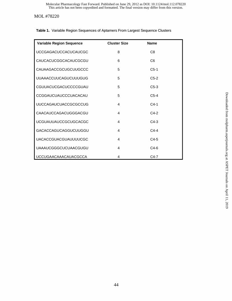
MOL #78220
44
Variable Region Sequence Cluster Size Name
UCCGAGACUCCACUCAUCGC 8 C8
CAUCACUCGGCACAUCGCGU 6 C6
CAUAAGACCGCUGCUUGCCC 5 C5-1
UUAAACCUUCAGUCUUUGUG 5 C5-2
CGUUACUCGACUCCCCGUAU 5 C5-3
CCGGAUCUAUCCCUACACAU 5 C5-4
UUCCAGAUCUACCGCGCCUG 4 C4-1
CAACAUCCAGACUGGGACGU 4 C4-2
UCGUAUUAUCCGCUGCACGC 4 C4-3
GACACCAGUCAGGUCUUGGU 4 C4-4
UACACCGUACGUAUUUUCGC 4 C4-5
UAAAUCGGGCUCUAACGUGU 4 C4-6
UCCUGAACAAACAUACGCCA 4 C4-7
Table 1.Table 1. Variable Region Sequences of Variable Region Sequences of AptamersAptamers From Largest Sequence ClustersFrom Largest Sequence Clusters
This article has not been copyedited and formatted. The final version may differ from this version.Molecular Pharmacology Fast Forward. Published on June 29, 2012 as DOI: 10.1124/mol.112.078220
at ASPE
T Journals on A
pril 11, 2019m
olpharm.aspetjournals.org
Dow
nloaded from

This article has not been copyedited and formatted. The final version may differ from this version.Molecular Pharmacology Fast Forward. Published on June 29, 2012 as DOI: 10.1124/mol.112.078220
at ASPE
T Journals on A
pril 11, 2019m
olpharm.aspetjournals.org
Dow
nloaded from

This article has not been copyedited and formatted. The final version may differ from this version.Molecular Pharmacology Fast Forward. Published on June 29, 2012 as DOI: 10.1124/mol.112.078220
at ASPE
T Journals on A
pril 11, 2019m
olpharm.aspetjournals.org
Dow
nloaded from

This article has not been copyedited and formatted. The final version may differ from this version.Molecular Pharmacology Fast Forward. Published on June 29, 2012 as DOI: 10.1124/mol.112.078220
at ASPE
T Journals on A
pril 11, 2019m
olpharm.aspetjournals.org
Dow
nloaded from
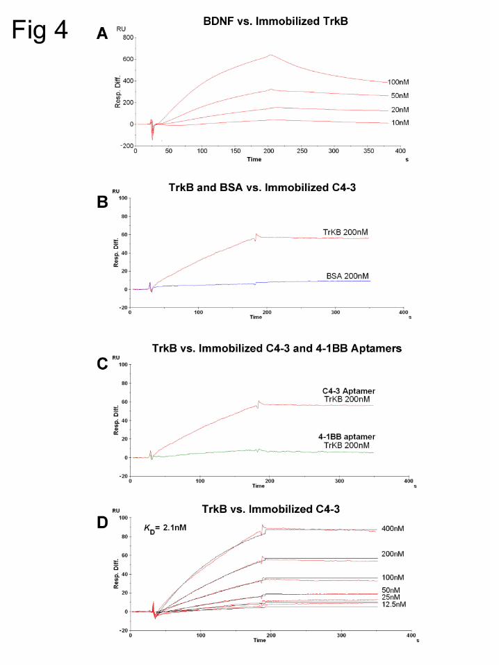
This article has not been copyedited and formatted. The final version may differ from this version.Molecular Pharmacology Fast Forward. Published on June 29, 2012 as DOI: 10.1124/mol.112.078220
at ASPE
T Journals on A
pril 11, 2019m
olpharm.aspetjournals.org
Dow
nloaded from

This article has not been copyedited and formatted. The final version may differ from this version.Molecular Pharmacology Fast Forward. Published on June 29, 2012 as DOI: 10.1124/mol.112.078220
at ASPE
T Journals on A
pril 11, 2019m
olpharm.aspetjournals.org
Dow
nloaded from

This article has not been copyedited and formatted. The final version may differ from this version.Molecular Pharmacology Fast Forward. Published on June 29, 2012 as DOI: 10.1124/mol.112.078220
at ASPE
T Journals on A
pril 11, 2019m
olpharm.aspetjournals.org
Dow
nloaded from

This article has not been copyedited and formatted. The final version may differ from this version.Molecular Pharmacology Fast Forward. Published on June 29, 2012 as DOI: 10.1124/mol.112.078220
at ASPE
T Journals on A
pril 11, 2019m
olpharm.aspetjournals.org
Dow
nloaded from

This article has not been copyedited and formatted. The final version may differ from this version.Molecular Pharmacology Fast Forward. Published on June 29, 2012 as DOI: 10.1124/mol.112.078220
at ASPE
T Journals on A
pril 11, 2019m
olpharm.aspetjournals.org
Dow
nloaded from

This article has not been copyedited and formatted. The final version may differ from this version.Molecular Pharmacology Fast Forward. Published on June 29, 2012 as DOI: 10.1124/mol.112.078220
at ASPE
T Journals on A
pril 11, 2019m
olpharm.aspetjournals.org
Dow
nloaded from