Title Page Development of Poly Unsaturated Fatty Acid...
31
JPET #234781 1 Title Page Development of Poly Unsaturated Fatty Acid Derivatives of Aspirin for Inhibition of Platelet Function Jahnabi Roy, Reheman Adili, Richard Kulmacz, Michael Holinstat, and Aditi Das Department of Comparative Biosciences (A.D.), Department of Biochemistry (A.D.), Center for Biophysics and Quantitative Biology (A.D.), Division of Nutritional Sciences (A.D.), Department of Bioengineering(A.D.), Department of Chemistry (J.R.), Beckman Institute for Advanced Science (A.D.), University of Illinois Urbana-Champaign, Urbana IL 61801. The Department of Internal Medicine, Texas Health Science Center at Houston, McGovern Medical School, Houston, TX 77225-0708 (R.K.), Department of Pharmacology (R.A., M.H.), Division of Cardiovascular Medicine (M.H.), University of Michigan Medical School, Ann Arbor, MI 48109 This article has not been copyedited and formatted. The final version may differ from this version. JPET Fast Forward. Published on August 3, 2016 as DOI: 10.1124/jpet.116.234781 at ASPET Journals on July 3, 2018 jpet.aspetjournals.org Downloaded from
Transcript of Title Page Development of Poly Unsaturated Fatty Acid...

JPET #234781
1
Title Page
Development of Poly Unsaturated Fatty Acid
Derivatives of Aspirin for Inhibition of Platelet
Function
Jahnabi Roy, Reheman Adili, Richard Kulmacz, Michael Holinstat, and Aditi Das
Department of Comparative Biosciences (A.D.), Department of Biochemistry (A.D.), Center for
Biophysics and Quantitative Biology (A.D.), Division of Nutritional Sciences (A.D.), Department
of Bioengineering(A.D.), Department of Chemistry (J.R.), Beckman Institute for Advanced
Science (A.D.), University of Illinois Urbana-Champaign, Urbana IL 61801. The Department of
Internal Medicine, Texas Health Science Center at Houston, McGovern Medical School,
Houston, TX 77225-0708 (R.K.), Department of Pharmacology (R.A., M.H.), Division of
Cardiovascular Medicine (M.H.), University of Michigan Medical School, Ann Arbor, MI 48109
This article has not been copyedited and formatted. The final version may differ from this version.JPET Fast Forward. Published on August 3, 2016 as DOI: 10.1124/jpet.116.234781
at ASPE
T Journals on July 3, 2018
jpet.aspetjournals.orgD
ownloaded from

JPET #234781
2
Running Title Page
Running Title: Aspirin anhydrides to reduce platelet aggregation
*Corresponding Author:
Aditi Das, Ph.D,
University of Illinois, Urbana-Champaign
3836 VMBSB, 2001 South Lincoln Avenue, Urbana IL 61802
Phone: 217-244-0630
Pages: 26
Tables: 0
Figures: 5
References: 22
Abstract: 170 words
Introduction: 714 words
Discussion: 941 words
List of Non-Standard Abbreviations
COX-1- Cyclooxygenase 1
TXAS- Thromboxane Synthase
TXA2 - Thromboxane A2
PGH2- Prostaglandin H2
DHA- Docosahexaenoic acid
EPA- Eicosapentaenoic acid
LA- Linoleic acid
AA- Arachidonic acid
Recommended section assignment: Cardiovascular
This article has not been copyedited and formatted. The final version may differ from this version.JPET Fast Forward. Published on August 3, 2016 as DOI: 10.1124/jpet.116.234781
at ASPE
T Journals on July 3, 2018
jpet.aspetjournals.orgD
ownloaded from
JPET #234781
3
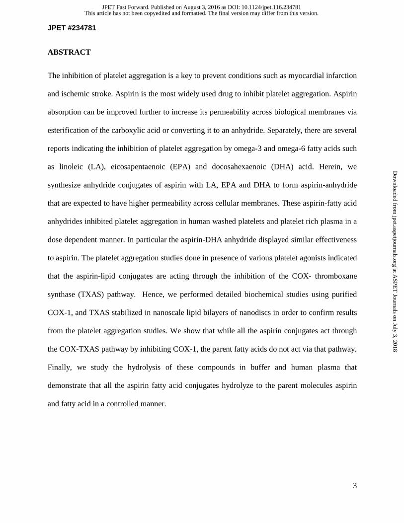
ABSTRACT
The inhibition of platelet aggregation is a key to prevent conditions such as myocardial infarction
and ischemic stroke. Aspirin is the most widely used drug to inhibit platelet aggregation. Aspirin
absorption can be improved further to increase its permeability across biological membranes via
esterification of the carboxylic acid or converting it to an anhydride. Separately, there are several
reports indicating the inhibition of platelet aggregation by omega-3 and omega-6 fatty acids such
as linoleic (LA), eicosapentaenoic (EPA) and docosahexaenoic (DHA) acid. Herein, we
synthesize anhydride conjugates of aspirin with LA, EPA and DHA to form aspirin-anhydride
that are expected to have higher permeability across cellular membranes. These aspirin-fatty acid
anhydrides inhibited platelet aggregation in human washed platelets and platelet rich plasma in a
dose dependent manner. In particular the aspirin-DHA anhydride displayed similar effectiveness
to aspirin. The platelet aggregation studies done in presence of various platelet agonists indicated
that the aspirin-lipid conjugates are acting through the inhibition of the COX- thromboxane
synthase (TXAS) pathway. Hence, we performed detailed biochemical studies using purified
COX-1, and TXAS stabilized in nanoscale lipid bilayers of nanodiscs in order to confirm results
from the platelet aggregation studies. We show that while all the aspirin conjugates act through
the COX-TXAS pathway by inhibiting COX-1, the parent fatty acids do not act via that pathway.
Finally, we study the hydrolysis of these compounds in buffer and human plasma that
demonstrate that all the aspirin fatty acid conjugates hydrolyze to the parent molecules aspirin
and fatty acid in a controlled manner.
This article has not been copyedited and formatted. The final version may differ from this version.JPET Fast Forward. Published on August 3, 2016 as DOI: 10.1124/jpet.116.234781
at ASPE
T Journals on July 3, 2018
jpet.aspetjournals.orgD
ownloaded from

JPET #234781
4
INTRODUCTION
Platelet adhesion and aggregation is important for maintaining normal hemostasis and is an
essential component in the morbidity and mortality associated with cardiovascular disease,
including myocardial infarction and ischemic stroke. Therefore, the ability to modulate platelet
function is of significant clinical importance. Aspirin is currently the most widely used
therapeutic for inhibition of platelet activation. Aspirin functions via irreversible acetylation of
platelet cyclooxygenase-1 (COX-1), resulting in inhibition of platelet-derived thromboxane A2
(TXA2) formation. Aspirin has poor permeability across biological membranes at physiological
pH due to the presence of free carboxylic acid, which is significantly ionized at this pH. The
result is poor absorption of drugs with carboxylic acid moieties through lipid membrane barriers.
Typically, non-steroidal anti-inflammatory drugs (NSAIDs) such as aspirin also cause gastric
toxicity as they inhibit COX-1 which is involved in maintaining the integrity of gastrointestinal
epithelium. The general approach to solve these side effects is to esterify the carboxylic acid to
produce lipophilic pro-drug forms. However, several aliphatic or aromatic esters of carboxylic
acid drugs are not sufficiently labile in vivo to ensure a suitably high rate and extent of
conversion from the esterified form. Additionally, esters are highly susceptible to enzymatic
hydrolysis in plasma and thus have differential rates of aspirin liberation in different individuals.
Moreover, aspirin has two major functional groups: the carboxylate end and the O-acetyl end. As
the O-acetyl end is the pharmacophore for the activity of aspirin, for an aspirin ester derivative
pro-drug to be effective, it must hydrolyze at the carboxylate end faster than the O-acetyl end
(Gilmer et al., 2002) (scheme 1). Therefore, in this work, we synthesize aspirin anhydride
derivatives with naturally occurring lipids and evaluate their platelet aggregation function.
This article has not been copyedited and formatted. The final version may differ from this version.JPET Fast Forward. Published on August 3, 2016 as DOI: 10.1124/jpet.116.234781
at ASPE
T Journals on July 3, 2018
jpet.aspetjournals.orgD
ownloaded from

JPET #234781
5
Recent work has focused on the modification of drug molecules with lipids or lipid derivatives as
a means to overcome their side effects as well as to facilitate their delivery. Specifically, addition
of a hydrophobic lipid tail allows the drug molecule to cross the hydrophobic plasma membrane
and enter the cell (Bradley et al., 2001). Herein we combine the two approaches of derivatizing
aspirin with poly-unsaturated fatty acids (PUFA) in order to keep aspirin in an unionized form in
order to facilitate facile transport across the plasma membrane. The “hybrid-drug” approach of
combining two drugs with similar therapeutic properties but different mechanisms has been
gaining interest (Sparatore et al., 2011). For instance, there are several reports in literature
indicating the inhibition of platelet aggregation by dietary fatty acids, chiefly omega-3 fatty acids
EPA and DHA and omega-6 fatty acid linoleic acid (LA) (Phang et al., 2013). The action of
these lipids is thought to be through competitive inhibition of COX-1-mediated arachidonic acid
metabolism. The metabolism of EPA and DHA by COX-1 leads to the formation of alternative
thromboxane-like molecules that are less potent than TXA2 molecules with regard to platelet
aggregation.
Taken together, herein we synthesize anhydride conjugates of dietary fatty acids - LA, EPA and
DHA with aspirin for the inhibition of platelet aggregation. We hypothesized that if the omega-3
fatty acids and aspirin are reducing platelet aggregation separately then the conjugated moiety
itself will have anti-platelet aggregatory properties through a synergistic or co-drug effect.
Additionally, these aspirin-lipid conjugates will prevent the deleterious effects of the free aspirin
carboxylate group. The aspirin-lipid anhydride bond will be more susceptible to hydrolysis
compared to O-acetyl end. Therefore, the compound will decompose to its carboxylic acid
counterpart at a predictable rate and pattern depending on the conjugated molecule, as shown
previously with related compounds (Mizrahi and Domb, 2009). Furthermore, anhydrides are
This article has not been copyedited and formatted. The final version may differ from this version.JPET Fast Forward. Published on August 3, 2016 as DOI: 10.1124/jpet.116.234781
at ASPE
T Journals on July 3, 2018
jpet.aspetjournals.orgD
ownloaded from

JPET #234781
6
typically less sensitive to enzymatic cleavage than esters or amides (Kumar et al., 2002). Thus,
the use of lipid anhydride conjugation is likely to increase bio-availability, be released in a
controlled manner, and reduce gastric and mucosal toxicity.
In summary, this work is the first report on application of aspirin anhydride using
polyunsaturated fatty acids for reduction in platelet aggregation. We demonstrate that anhydride
conjugates of aspirin with dietary omega-3 fatty acids are potent inhibitors of platelet
aggregation. Furthermore, we biochemically examine the effect of parent fatty acid on the COX-
1 mediated TXA2 formation study as the previous literature focused mostly on the dietary
supplementation of these fatty acids in controlled trial groups (Phang et al., 2013). Finally, we
perform hydrolysis studies to demonstrate that the aspirin-anhydride conjugates release aspirin
and free fatty acids at a controlled rate in both buffer and plasma.
MATERIALS AND METHODS
Materials: Ampicillin, arabinose, chloramphenicol, isopropyl β-D-1-thiogalactopyranoside
(IPTG) and Ni-NTA resin were bought from Gold Biotechnology. δ-Aminolevulinic acid (δ-
ALA) and hematin were obtained from Frontier Scientific. 1-palmitoyl-2-oleoyl-sn-glycero-3-
phosphocholine (POPC) was purchased from Avanti Polar Lipids. Aspirin was purchased from
Sigma Aldrich. Arachidonic acid, EPA, DHA and LA were obtained from Cayman Chemicals.
PAR4-AP (AYPGKF) and PAR1-AP (SFLLRN) were purchased from GL Biochem (Shanghai,
China). Thrombin was purchased from Enzyme Research Laboratories (South Bend, IN).
Collagen and ristocetin was purchased from Chorology Cooperation. U46619 and ADP were
purchased from Sigma Aldrich. University of Michigan Review Board approved studies and
written informed consent was obtained from all participants prior to blood collection.
This article has not been copyedited and formatted. The final version may differ from this version.JPET Fast Forward. Published on August 3, 2016 as DOI: 10.1124/jpet.116.234781
at ASPE
T Journals on July 3, 2018
jpet.aspetjournals.orgD
ownloaded from
JPET #234781
7
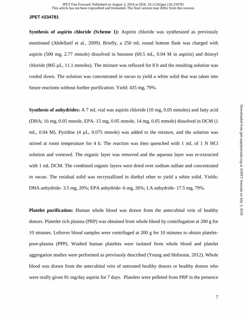
Synthesis of aspirin chloride (Scheme 1): Aspirin chloride was synthesized as previously
mentioned (Abdellatif et al., 2009). Briefly, a 250 mL round bottom flask was charged with
aspirin (500 mg, 2.77 mmole) dissolved in benzene (69.5 mL, 0.04 M in aspirin) and thinoyl
chloride (805 µL, 11.1 mmoles). The mixture was refluxed for 8 h and the resulting solution was
cooled down. The solution was concentrated in vacuo to yield a white solid that was taken into
future reactions without further purification. Yield: 435 mg, 79%.
Synthesis of anhydrides: A 7 mL vial was aspirin chloride (10 mg, 0.05 mmoles) and fatty acid
(DHA; 16 mg, 0.05 mmole, EPA- 15 mg, 0.05 mmole, 14 mg, 0.05 mmole) dissolved in DCM (1
mL, 0.04 M). Pyridine (4 µL, 0.075 mmole) was added to the mixture, and the solution was
stirred at room temperature for 4 h. The reaction was then quenched with 1 mL of 1 N HCl
solution and vortexed. The organic layer was removed and the aqueous layer was re-extracted
with 1 mL DCM. The combined organic layers were dried over sodium sulfate and concentrated
in vacuo. The residual solid was recrystallized in diethyl ether to yield a white solid. Yields:
DHA anhydride- 3.5 mg, 20%; EPA anhydride- 6 mg, 26%; LA anhydride- 17.5 mg, 79%.
Platelet purification: Human whole blood was drawn from the antecubital vein of healthy
donors. Platetlet rich plasma (PRP) was obtained from whole blood by centrifugation at 200 g for
10 minutes. Leftover blood samples were centrifuged at 200 g for 10 minutes to obtain platelet-
poor-plasma (PPP). Washed human platelets were isolated from whole blood and platelet
aggregation studies were performed as previously described (Yeung and Holinstat, 2012). Whole
blood was drawn from the antecubital vein of untreated healthy donors or healthy donors who
were orally given 81 mg/day aspirin for 7 days. Platelets were pelleted from PRP in the presence
This article has not been copyedited and formatted. The final version may differ from this version.JPET Fast Forward. Published on August 3, 2016 as DOI: 10.1124/jpet.116.234781
at ASPE
T Journals on July 3, 2018
jpet.aspetjournals.orgD
ownloaded from

JPET #234781
8
of ACD (2.5%) and apyrase (0.02 U/mL) by centrifugation at 2000 g for 10 minutes then
resuspended in Tyrodes buffer (12 mM NaHCO3, 127 mM NaCl, 5 mM KCl, 0.5 mM NaH2PO4,
1 mM MgCl2, 5 mM glucose, 10 mM HEPES) to a final concentration of 3.0 x 10⁸ platelets/mL.
Washed platelets (250 μL) were separately incubated with 2.5, 5, 10 μM or with same volume of
DMSO for 5 minutes.
Washed human platelet aggregation: Platelet aggregation was induced by 5 μM AA,
Thrombin (1 nM), PAR4-AP, PAR1-AP (SFLLRN), ADP (1 μM) or U46619 (1 μM) and change
in light transmission was recorded by eight channel platelet aggregometer (Chronolog) under
stirring at 1200 rpm at 37°C.
Platelet aggregation in PRP: Platelet concentration in PRP was adjusted to 3.0 x 10⁸
platelets/mL using PPP from same donor. Then, 10 μM of C1, C2, C3 compounds or DMSO in
same volume was separately incubated with 250uL of PRP for 5 minutes. Platelet aggregation
was induced by adding AA (5 μM) ADP (1 μM), collagen (2 μg/mL), Ristocetin (1 μM) or
U46619 (1 μM) and change in light transmission were recorded by platelet aggregometer as
described above.
Expression and purification of cyclooxygenase 1 (COX-1)
An 8- 10 g of Sf9 cell pellet from a 2L cell culture expression COX-1 was suspended in 40-45
ml of Buffer A (25 mM NaPO4 / 20 mM imidazole, pH 7.4 / 1 mM phenol) and 5 ml of 10x
Sigma protease inhibitor (P2714) was added. Cell clumps were resuspended by magnetic stirring.
This resuspension was homogenized by sonication and centrifuged at 100,000 × g for 1 hr and
This article has not been copyedited and formatted. The final version may differ from this version.JPET Fast Forward. Published on August 3, 2016 as DOI: 10.1124/jpet.116.234781
at ASPE
T Journals on July 3, 2018
jpet.aspetjournals.orgD
ownloaded from

JPET #234781
9
supernatant was removed. The pellet was resuspended in ~45 ml of Buffer B (25 mM NaPO4 /
100 mM NaCl / 20 mM imidazole / 0.1 mM phenol, pH 7.4) using with a Dounce homogenizer.
10% Tween 20 solution was added to a 1.5% final concentration and stirred for 1-2 hr followed
by centrifugation at 100,000 x g for 1 hr. The supernatant (S1) was removed and the pellet was
resuspended in ~20 ml Buffer B. S1 was mixed with 2.5-3 ml of Ni-NTA Agarose (Qiagen)
prewashed with Buffer C (25 mM NaPO4; 100 mM NaCl; 20 mM imidazole; 0.1 mM phenol, pH
7.4; 0.1% Tween 20) and shaked in cold room for 2 hours. The mixture was poured into column
(~1.5 x 10 cm) and the flow though was allowed to drain out. The column was washed with 10-
15 mL of Buffer C and then with 10-15 mL of Buffer D (25 mM NaPO4; 300 mM NaCl; 20 mM
imidazole; 0.1 mM phenol, pH 7.4; 0.1% Tween 20). The protein was eluted with 10 x 0.75 ml
aliquots of Buffer E (25 mM NaPO4; 100 mM NaCl; 200 mM imidazole; 0.1 mM phenol, pH
7.4; 0.1% Tween 20). The active fractions were pooled and concentrated. The buffer was
exchanged on a 10DG column eluted with 50 mM KPi, pH 7.2 / 50 mM NaCl / 0.01% NaN3 /
0.1% Tween 20 and collect 0.25 mL fractions. COX-1 activity each fraction was assayed and
active fractions were pooled. Protein was stored at -80 °C with 25%glycerol.
Expression and purification of thromboxane synthase: TXAS was expressed and purified as
previously mentioned (Das et al., 2014a). The gene for thromboxane synthase was obtained from
Origene and modified at the N-terminus for expression in E.coli as described. Briefly, the cells
were grown in terrific broth (TB) and were induced with 1 mM IPTG and 0.5 mM δ-ALA and 4
mg/L arabinose at O.D 1.2. They were grown for 44 hours at 26°C and 160 rpm. The harvested
cells were resuspended in Buffer A (0.1 M potassium phosphate pH 7.4, 10% glycerol, 0.1 M
sodium chloride) containing 2 mM magnesium chloride, 1 mM PMSF, 1 mg each of DNase and
This article has not been copyedited and formatted. The final version may differ from this version.JPET Fast Forward. Published on August 3, 2016 as DOI: 10.1124/jpet.116.234781
at ASPE
T Journals on July 3, 2018
jpet.aspetjournals.orgD
ownloaded from

JPET #234781
10
RNase for 1 hour and lysed 5 times for 1 min each using a sonicator. The solution was then
centrifuged at 35 K rpm for 1 hour and the pellet was resuspended in Buffer A containing 2%
Lubrol to solubilize TXAS. The re-suspended sample was centrifuged at 35,000 rpm for 1 hour
and the supernatant was loaded onto a Ni-NTA column. The column was incubated for 2 hours
with 5X column volume of buffer A containing 10 mM Histidine, 5 mM ATP, 10 mM MgCl2
and 150 mM KCl to separate any co-purifying GroEL (Joseph and Andreotti, 2008). TXAS was
eluted using Buffer A containing 0.2% Lubrol and 100 mM Histidine.
Assembly of TXAS-nanodiscs: TXAS-nanodiscs were assembled from a mixture of TXAS,
membrane scaffold protein (MSP1D1), cholate and POPC lipids by removing the detergents
using Amberlite (Bayburt et al., 2002). MSP1D1 was added to POPC (solubilized using sodium
cholate) in a ratio of 65:1 (lipids: MSP1D1) and the solution was incubated at 4oC for 1 hour on
a shaker. We thank Stephen G. Sligar for giving the MSP1D1 gene. TXAS was then added in a
ratio of 1:15 (TXAS: MSP1D1) and incubated another 1 hour. Amberlite was added to remove
detergents and initiate the formation of nanodiscs. The TXAS-nanodiscs were then purified using
size exclusion chromatography as previously mentioned (Das et al., 2014a).
Coupled assay with secondary lipids: TXAS converts PGH2 into TXA2, and HHT (12(L)-
hydroxy-5,8,10-heptadecatrienoic acid) and MDA (malondialdehyde) as a side reaction
(Diczfalusy et al., 1977). To measure overall effect of a secondary lipid on the metabolization of
AA, a coupled enzymatic assay was performed. Briefly, to COX-1 (10 nM) incubated with
hematin (0.5 µM) in tris buffer (0.1 M, pH= 7.4) was added TXAS (50 nM). To this was added
AA (20 µM) and secondary substrate (20 µM) and rate of MDA formation was monitored over 3
This article has not been copyedited and formatted. The final version may differ from this version.JPET Fast Forward. Published on August 3, 2016 as DOI: 10.1124/jpet.116.234781
at ASPE
T Journals on July 3, 2018
jpet.aspetjournals.orgD
ownloaded from

JPET #234781
11
minutes at 268 nm and HHT at 234 nm. Absorbance was then measured and the reaction mixture
was quenched in 300 µL of ethyl acetate. The amount of TXB2 formed was determined using
TXB2 EIA ELISA kit (Cayman).
RESULTS
Synthesis of aspirin anhydride derivatives: The synthesis of the fatty acid-aspirin derivatives
was performed as shown in Scheme 1. The free carboxylic acid in aspirin was first converted to
its corresponding acid chloride by reacting with thionyl chloride in the presence of amine base
by refluxing in benzene for 8 h. The activated acid chloride was taken without further
purification to react with the carboxylic acid in EPA, DHA and LA to form a mixed anhydride.
The mixed anhydride was purified by recrystallization in diethylether to give >99% purity by 1H-
NMR and HR-ESI mass spectrometry (Supplemental Table 1 and Supplemental Figures 7-9).
Lipid- aspirin conjugates inhibit AA induced platelet aggregation: The effect of fatty acid-
aspirin derivatives compounds C1, C2 and C3 on platelet function was studied in vitro using
platelet aggregation at differing concentrations from 2.5 µM to 10 µM. As shown in Figure 1,
arachidonic acid-induced (5 µM) platelet aggregation was inhibited by all three compounds in a
dose-dependent manner. However, these compounds varied in potency of inhibition with C3
being the most potent followed by C2 and C1. As seen from the aggregation curves, as compared
to the DMSO control, compound C1, LA anhydride, shows a slight decrease in platelet
aggregation with a significant decrease at 10 µM concentration (p= 0.0158). In contrast C2, the
EPA anhydride, shows a greater inhibition at lower concentrations with a significant decrease at
5 µM (p= 0.0176) and a further decrease at 10 µM (p ≤ 0.0001). Compound C3, DHA anhydride,
This article has not been copyedited and formatted. The final version may differ from this version.JPET Fast Forward. Published on August 3, 2016 as DOI: 10.1124/jpet.116.234781
at ASPE
T Journals on July 3, 2018
jpet.aspetjournals.orgD
ownloaded from

JPET #234781
12
shows most significant inhibition of platelet aggregation with a significant inhibition at 2.5 µM
(p= 0.0049) and 5 µM (p= 0.0011) and almost complete inhibition of aggregation at 10 µM (p=
0.0002). Similar results were observed for platelet rich plasma (PRP) (Figure 2). As seen in Fig
2A and 2B, at 10 µM concentration, compounds C2 and C3 show a significant decrease in
platelet aggregation (p= 0.1006 for C2 and p= 0.0003 for C3). However, C1 did not show a
significant decrease in aggregation in PRP. The effect of LA, EPA, and DHA on AA-induced
platelet aggregation can be seen in Supplemental Figure 4. While LA and EPA do not inhibit
platelet aggregation at concentrations of 2.5 μM, 5 μM, and 10 μM, DHA inhibits platelet
aggregation at 2.5 μM.
In addition to AA, compounds C1, C2 and C3 attenuated platelet aggregation induced by various
platelet agonists including thrombin, PAR4-AP, PAR1-AP, ADP or 46619. As seen in
Supplemental Figures 1 and 2, a slight albeit insignificant decrease was observed in platelet
aggregation induced by thrombin, PAR4-AP and PAR1-AP for all three compounds at 10 µM
concentration. No notable inhibition in platelet aggregation was observed in PRP stimulated with
ADP, collagen, ristocetin, or U46619 (Supplemental Figure 1 (D), (E) and S2 (B), (C), (D)).
Collectively, these observations indicated that the aspirin-lipid conjugates are acting through the
inhibition of the COX- thromboxane synthase pathway. In order to understand the potency of
inhibitory effects of fatty acid-aspirin derivatives compounds on human platelet function, we
compared the effectiveness of compound C3 against aspirin treated controls. Figure 2C shows
comparison of aspirin treated controls with compound C3 and DMSO control in washed
platelets. As seen in figure, 20 µM of compound C3 shows a similar level of inhibition as aspirin
treated controls, of platelet function in response to AA (p< 0.0001). The inhibition values are
This article has not been copyedited and formatted. The final version may differ from this version.JPET Fast Forward. Published on August 3, 2016 as DOI: 10.1124/jpet.116.234781
at ASPE
T Journals on July 3, 2018
jpet.aspetjournals.orgD
ownloaded from

JPET #234781
13
similar to aspirin treated controls where a treatment of 81 mg/day of aspirin was administered for
7 days. Thus, compound C3 at 20 µM concentration displays similar effectiveness to aspirin.
Inhibition of COX-1-thromboxane synthase activity by lipid-aspirin conjugates: Platelet
aggregometry data shows that the inhibition by these compounds is effective only when induced
by AA. Thus, the mechanism of action of the derivatives is likely through the COX-1-TXAS
pathway. To confirm the effects of the derivatives and their parent compounds on the COX-1-
TXAS enzyme system, a coupled activity assay was performed. In this assay, COX-1 converts
AA to prostaglandin H2, which is further converted by TXAS to thromboxane A2. TXA2 is a pro-
aggregatory molecule that initiates platelet aggregation. However, TXA2 is quickly converted to
TXB2 by hydrolysis of the endoperoxide bond to the diol form. TXB2 can be subsequently
measured.
We utilized nanodiscs to stabilize TXAS is a membrane protein, which tends to lose
functionality in aqueous buffers typically used for protein assays. Therefore, it was incorporated
into nanodiscs, lipid bilayers surrounded by membrane scaffold protein (MSP) (Supplemental
Figure 3(A)) Previously, we have shown that TXAS facilitates robust biophysical studies in
nanodiscs as compared to naked protein in detergent solution (Das et al., 2014b).
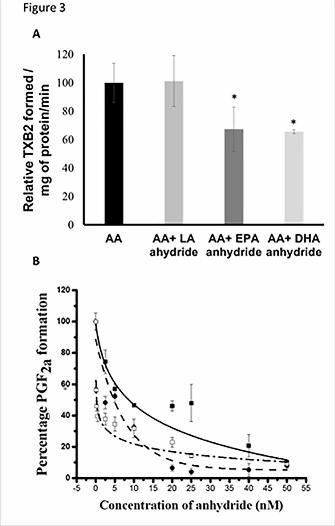
The anhydrides and their parent molecules were subjected to the coupled activity assay in a 1:1
ratio with the substrate molecule AA. As observed in Figure 3A, the EPA and DHA anhydrides
were potent in inhibiting the thromboxane formed in a coupled assay system to a similar extent.
The perceived reduction in TXA2 formation in both cases was about 30-35% of the control.
However, the LA anhydride did not show any significant decrease in the formation of
thromboxane. These observations are in agreement with the results obtained in the aggregometry
This article has not been copyedited and formatted. The final version may differ from this version.JPET Fast Forward. Published on August 3, 2016 as DOI: 10.1124/jpet.116.234781
at ASPE
T Journals on July 3, 2018
jpet.aspetjournals.orgD
ownloaded from
JPET #234781
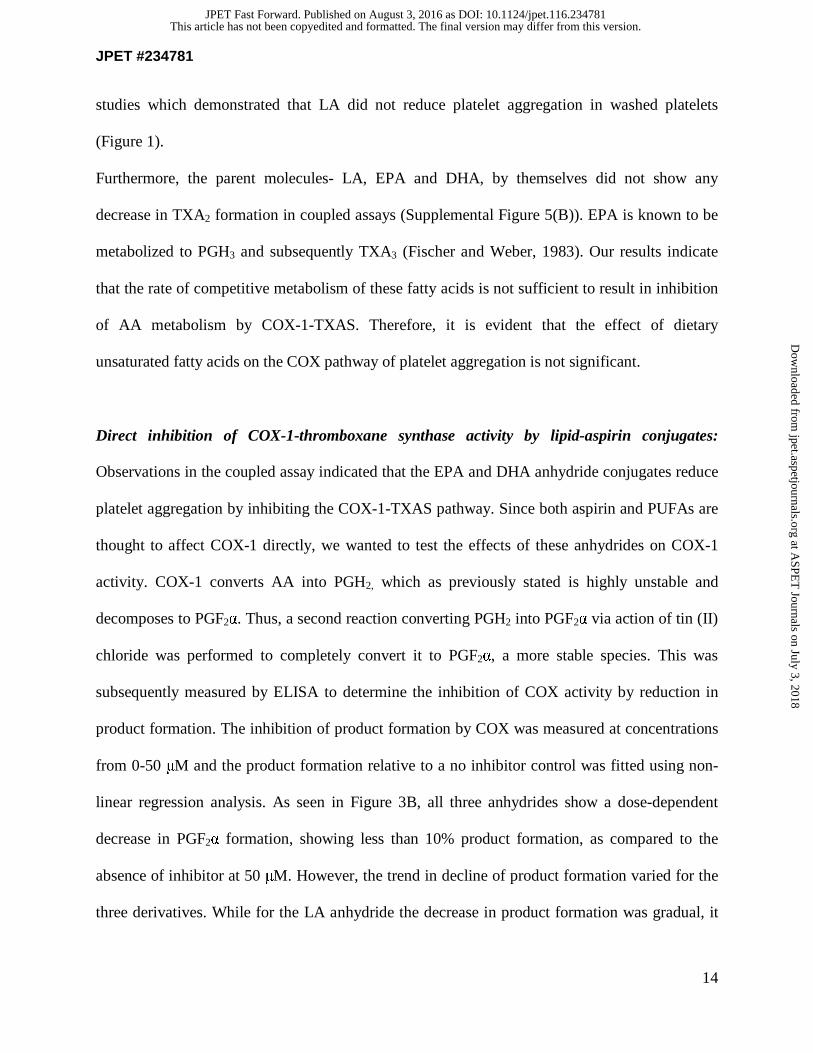
14
studies which demonstrated that LA did not reduce platelet aggregation in washed platelets
(Figure 1).
Furthermore, the parent molecules- LA, EPA and DHA, by themselves did not show any
decrease in TXA2 formation in coupled assays (Supplemental Figure 5(B)). EPA is known to be
metabolized to PGH3 and subsequently TXA3 (Fischer and Weber, 1983). Our results indicate
that the rate of competitive metabolism of these fatty acids is not sufficient to result in inhibition
of AA metabolism by COX-1-TXAS. Therefore, it is evident that the effect of dietary
unsaturated fatty acids on the COX pathway of platelet aggregation is not significant.
Direct inhibition of COX-1-thromboxane synthase activity by lipid-aspirin conjugates:
Observations in the coupled assay indicated that the EPA and DHA anhydride conjugates reduce
platelet aggregation by inhibiting the COX-1-TXAS pathway. Since both aspirin and PUFAs are
thought to affect COX-1 directly, we wanted to test the effects of these anhydrides on COX-1
activity. COX-1 converts AA into PGH2, which as previously stated is highly unstable and
decomposes to PGF2α. Thus, a second reaction converting PGH2 into PGF2α via action of tin (II)
chloride was performed to completely convert it to PGF2α, a more stable species. This was
subsequently measured by ELISA to determine the inhibition of COX activity by reduction in
product formation. The inhibition of product formation by COX was measured at concentrations
from 0-50 μM and the product formation relative to a no inhibitor control was fitted using non-
linear regression analysis. As seen in Figure 3B, all three anhydrides show a dose-dependent
decrease in PGF2α formation, showing less than 10% product formation, as compared to the
absence of inhibitor at 50 μM. However, the trend in decline of product formation varied for the
three derivatives. While for the LA anhydride the decrease in product formation was gradual, it
This article has not been copyedited and formatted. The final version may differ from this version.JPET Fast Forward. Published on August 3, 2016 as DOI: 10.1124/jpet.116.234781
at ASPE
T Journals on July 3, 2018
jpet.aspetjournals.orgD
ownloaded from

JPET #234781
15
was more dramatic initially for the EPA and DHA anhydrides. The EPA anhydride shows an
initial decline to 50% activity at 2.5 μM, and then gradually declines to >10% at 25 μM
concentration. The DHA anhydride shows an initial drop to less than 40% of total activity at 2.5
μM and then slowly declines to ~10% at 25 μM. Interestingly, both EPA and DHA anhydrides
show similar effects above 10 μM concentrations. The IC50 values for the compounds were
calculated to be 7.5 μM, 5.8 μM, and 2.1 μM for the LA anhydride, EPA anhydride, and DHA
anhydride, respectively.
Direct buffer hydrolysis of the lipid-aspirin conjugates: To test the lability of the anhydrides in
buffer, the rate of hydrolysis was measured by analyzing the remaining anhydride in phosphate
buffer saline (PBS) at 37°C at different time intervals. In all three cases, HPLC analysis of the
aliquot indicated the presence of the original parent compound, aspirin and the corresponding
fatty acid, indicating the release of the aspirin active moiety and not of salicylic acid
(Supplemental Figures 5-7). As observed in Figure 4A, all three compounds show pseudo-first
order kinetics of decomposition. The LA-aspirin anhydride shows a gradual decomposition over
30 mins, leading to decomposition of about 60% of the drug species into its individual
components. In contrast, the EPA-aspirin anhydride shows a sharp decomposition in the first 5
mins and plateaus around 60% decomposition after the initial hydrolysis. This trend provides an
explanation for the significant differences in the activities of EPA and LA anhydrides in the
COX activity assay as well as the COX-thromboxane synthase coupled activity assays which
were both carried out over shorter incubation times. The LA anhydride being liberated more
slowly has a lesser effect on the inhibition of COX-1 than the EPA anhydride, which releases the
This article has not been copyedited and formatted. The final version may differ from this version.JPET Fast Forward. Published on August 3, 2016 as DOI: 10.1124/jpet.116.234781
at ASPE
T Journals on July 3, 2018
jpet.aspetjournals.orgD
ownloaded from

JPET #234781
16
active species much faster. The DHA anhydride shows hydrolysis trends similar to LA anhydride
whereby it is more gradual over the initial 5 mins and plateaus to about 55% at 60 mins.
Plasma hydrolysis studies: To investigate the release of aspirin from the aspirin-fatty acid
mixed anhydride in plasma, plasma buffered with phosphate buffer (pH=7.4) was equilibrated at
37°C and the anhydride was added. At various time points aliquots were withdrawn and the
extracted products were analyzed by HPLC. Figure 4B represents the plasma hydrolysis patterns
of all three anhydrides. Similar to the buffer hydrolysis, pseudo-first order kinetics can be
observed in all three cases. All three compounds show a gradual decomposition over 2 hours
plateauing to about 35- 40% decomposition. Note that the plasma hydrolysis of DHA is at a
comparable range as EPA. However, as seen from Supplemental Figure 4, DHA is a much better
inhibitor of platelet aggregation in human blood samples, it can be deduced that the effect of
DHA and aspirin together on inhibiting aggregation is higher than EPA and aspirin in
combination.
DISCUSSION
Platelet aggregation is triggered by a number of primary activators leading to positive feedback
and potentiation of the activation signal. The cyclooxygenase-1 (COX-1)-thromboxane synthase
(TXAS) pathway is a well-established positive feedback signal in the platelet. In this pathway,
arachidonic acid (AA) is metabolized by COX-1 to prostaglandin H2 (PGH2) followed by
conversion of PGH2 to thromboxane A2 (TXA2), which is a pro-aggregatory molecule (Paul et
al., 1999). Activation of the platelet either through primary activation or feedback activation via
COX-1 leads to activation of the integrin GPIIb/IIIa resulting in platelet clot formation. The
This article has not been copyedited and formatted. The final version may differ from this version.JPET Fast Forward. Published on August 3, 2016 as DOI: 10.1124/jpet.116.234781
at ASPE
T Journals on July 3, 2018
jpet.aspetjournals.orgD
ownloaded from

JPET #234781
17
inhibition of platelet aggregation both ex vivo and in vivo can be aided by molecules inhibiting
the COX-1-dependent feedback pathway as well as by directly blocking GPIIb/IIIa integrins
(Yeung and Holinstat, 2012) .
Aspirin is the most popular NSAID for reducing pain, fever and inhibition of platelet function.
The free carboxylate group in aspirin stays as an ionized species at physiological pH, which is
poorly absorbed across cellular membranes. Hence, the derivatization of the free carboxylic acid,
to form more lipophilic species can facilitate transport across bio-membranes. Herein, we have
derivatized the carboxylate end of aspirin through anhydride formation with dietary fatty acids
including EPA and DHA (omega-3 fatty acids) as well as LA (omega-6 fatty acid) that have been
shown to reduce platelet aggregation through dietary supplementation in platelet aggregometry
studies. As compared to esters, anhydrides show more controlled patterns of hydrolysis and
decomposition that are enzyme independent. Therefore, we report the design of aspirin-poly
unsaturated fatty acid mixed anhydrides that would exist in a unionized, lipophilic state as well
as allow for a facile, more controlled release of the active species in plasma. Additionally, recent
studies on polyanhydrides as drug carriers show that anhydrides degrade in a controlled fashion
and are biocompatible with human body tissues, including the brain (Domb et al., 1999). One of
the rationale for the design of the hybrid co-drug is that the aspirin-fatty acid-anhydride pro-drug
would hydrolyze to release not one but two active species, which could both inhibit the COX
enzyme.
The synthesis of the mixed anhydrides were tested in platelet aggregometry studies to analyze
their anti-aggregatory effects. All three compounds displayed a concentration dependent
decrease in platelet aggregation. However, the DHA anhydride was most potent, almost
completely inhibiting aggregation at 10 µM concentration, followed by EPA anhydride and,
This article has not been copyedited and formatted. The final version may differ from this version.JPET Fast Forward. Published on August 3, 2016 as DOI: 10.1124/jpet.116.234781
at ASPE
T Journals on July 3, 2018
jpet.aspetjournals.orgD
ownloaded from

JPET #234781
18
finally, LA anhydride, which showed marginal decrease in the inhibition of aggregation. As
expected from the mechanism of inhibition of platelet aggregation studies, the decrease was only
observed in AA-induced platelet aggregation and not observed for ADP, collagen, U46619 or
thrombin, indicating that these molecules did not affect those pathways for platelet aggregation.
Furthermore, the inhibition of the DHA anhydride and aspirin-treated species showed
comparable inhibition potentials.
To understand the biochemical mechanism of action of these molecules, an activity assay was
performed on the COX-1 -thromboxane synthase system. It was observed that while the
reduction in TXA2 for EPA and DHA anhydride was significant, LA anhydride did not show
significant reduction, thereby following the similar trend of the platelet aggregometry studies.
More interestingly, however, the parent fatty acids showed no inhibition in thromboxane
formation (Fig S5(B)). Thus, it appears that while LA, EPA, and DHA may be able to compete
for metabolism against AA, this is likely not the chief mechanism of their action to prevent
platelet aggregation. Literature reports have suggested that omega-3 fatty acids reduce platelet
aggregation by signaling through other pathways (Abeywardena and Head, 2001). Furthermore,
a meta-analysis of the effect of omega-3 fatty acids on the inhibition of platelet aggregation
suggests that the effect of their metabolism is short-term. This is not sufficient to result in
significant changes in plasma lipid composition (Driss et al., 1984; Gao et al., 2013).
Additionally it is possible that this inhibition is mediated through collagen-induced pathways
(Tremoli et al., 1995).
Next, we determined the effect of the compounds on COX-1 activity and derived IC50 values for
the anhydrides. The values for the LA, EPA, and DHA anhydrides were obtained to be 7.1, 5.8,
and 2.1 μM, respectively. Literature reports suggest that the IC50 values of COX-1 inhibition for
This article has not been copyedited and formatted. The final version may differ from this version.JPET Fast Forward. Published on August 3, 2016 as DOI: 10.1124/jpet.116.234781
at ASPE
T Journals on July 3, 2018
jpet.aspetjournals.orgD
ownloaded from
JPET #234781
19
LA, EPA, and DHA are 93 μM, 13 μM and 15 μM, respectively. Furthermore, the IC50 value of
aspirin on COX-1 in purified enzyme system is 8 μM (Mitchell et al., 1993). These results
suggests a greater effect of the mixed anhydrides in comparison to their parent molecules.
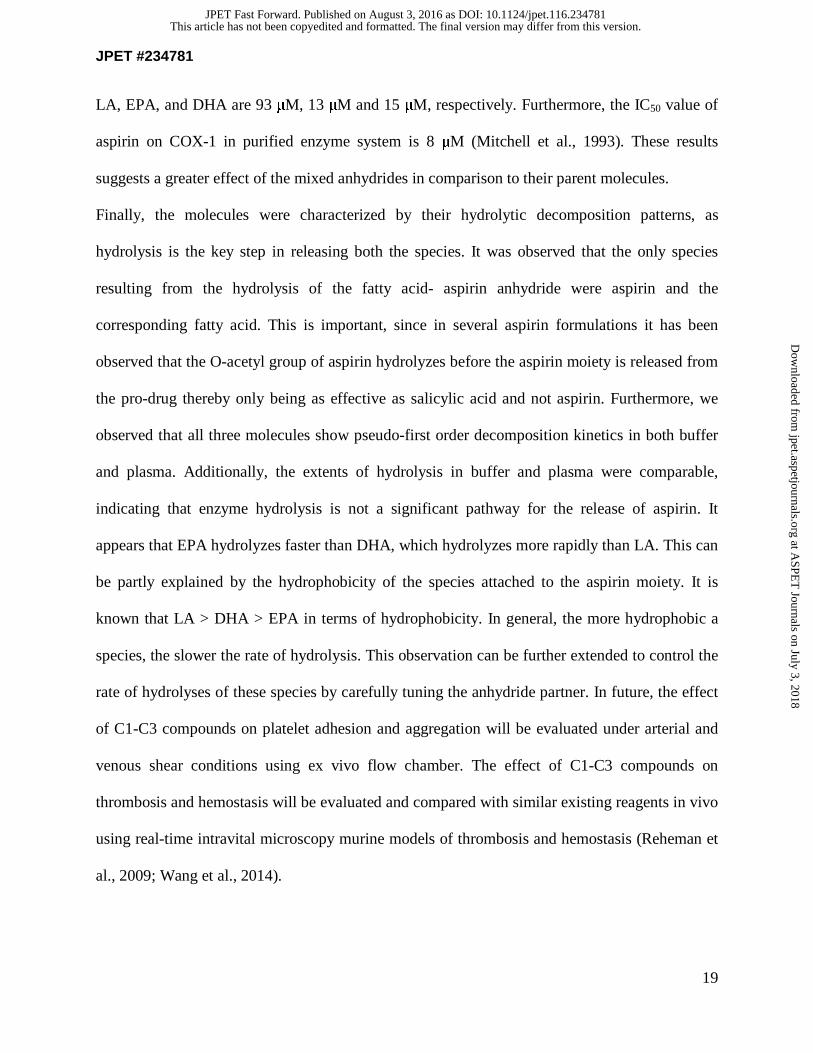
Finally, the molecules were characterized by their hydrolytic decomposition patterns, as
hydrolysis is the key step in releasing both the species. It was observed that the only species
resulting from the hydrolysis of the fatty acid- aspirin anhydride were aspirin and the
corresponding fatty acid. This is important, since in several aspirin formulations it has been
observed that the O-acetyl group of aspirin hydrolyzes before the aspirin moiety is released from
the pro-drug thereby only being as effective as salicylic acid and not aspirin. Furthermore, we
observed that all three molecules show pseudo-first order decomposition kinetics in both buffer
and plasma. Additionally, the extents of hydrolysis in buffer and plasma were comparable,
indicating that enzyme hydrolysis is not a significant pathway for the release of aspirin. It
appears that EPA hydrolyzes faster than DHA, which hydrolyzes more rapidly than LA. This can
be partly explained by the hydrophobicity of the species attached to the aspirin moiety. It is
known that LA > DHA > EPA in terms of hydrophobicity. In general, the more hydrophobic a
species, the slower the rate of hydrolysis. This observation can be further extended to control the
rate of hydrolyses of these species by carefully tuning the anhydride partner. In future, the effect
of C1-C3 compounds on platelet adhesion and aggregation will be evaluated under arterial and
venous shear conditions using ex vivo flow chamber. The effect of C1-C3 compounds on
thrombosis and hemostasis will be evaluated and compared with similar existing reagents in vivo
using real-time intravital microscopy murine models of thrombosis and hemostasis (Reheman et
al., 2009; Wang et al., 2014).
This article has not been copyedited and formatted. The final version may differ from this version.JPET Fast Forward. Published on August 3, 2016 as DOI: 10.1124/jpet.116.234781
at ASPE
T Journals on July 3, 2018
jpet.aspetjournals.orgD
ownloaded from
JPET #234781
20
CONCLUSION
We have synthesized anhydrides of aspirin and dietary fatty acids to explore a “hybrid-drug”
approach. Both aspirin and the fatty acids in this study are inhibitors of platelet aggregation and
this conjugation addresses the side effects of aspirin. We observed that all three compounds
individually inhibit platelet aggregation in a dose-dependent manner in the effectiveness order of
C3, C2, and C1. Additionally, we show that C3 is comparable to aspirin-treated samples.
Furthermore, we show that while all three molecules act through the COX-thromboxane synthase
pathway by inhibiting COX-1, the parent fatty acids do not act via that pathway. Finally,
hydrolysis studies of the drugs in buffer as well as plasma conditions confirm the release of
aspirin and fatty acid separately, instead of the salicylate moiety as is the case with several
aspirin pro-drugs.
Acknowledgement: We acknowledge Ms. Susan Zelasko for initial literature review, Mr. Daniel
McDougle and Ms. Navroop Gill for providing helpful comments on the manuscript.
AUTHORSHIP CONTRIBUTIONS:
Conceived Project: J. Roy and A. Das
Participated in Research Design: J. Roy, R. Adili, M. Holinstat and A. Das
Conducted Experiments: J. Roy and R. Adili
Contributed Reagents: J. Roy, R. Adili, R. Kulmacz, M. Holinstat and A. Das
Performed Data Analysis: J. Roy, R. Adili, M. Holinstat and A. Das
Wrote or significantly contributed to writing of manuscript: J. Roy, R. Adili, M. Holinstat and A.
Das
This article has not been copyedited and formatted. The final version may differ from this version.JPET Fast Forward. Published on August 3, 2016 as DOI: 10.1124/jpet.116.234781
at ASPE
T Journals on July 3, 2018
jpet.aspetjournals.orgD
ownloaded from

JPET #234781
21
REFERENCES:
Abdellatif KR, Chowdhury MA, Dong Y, Das D, Yu G, Velazquez CA, Suresh MR and Knaus
EE (2009) Dinitroglyceryl and diazen-1-ium-1,2-diolated nitric oxide donor ester
prodrugs of aspirin, indomethacin and ibuprofen: synthesis, biological evaluation and
nitric oxide release studies. Bioorg Med Chem Lett 19:3014-3018.
Abeywardena MY and Head RJ (2001) Longchain n-3 polyunsaturated fatty acids and blood
vessel function. Cardiovasc Res 52:361-371.
Bayburt TH, Grinkova YV and Sligar SG (2002) Self-assembly of discoidal phospholipid bilayer
nanoparticles with membrane scaffold proteins. Nano Letters 2:853-856.
Bradley MO, Swindell CS, Anthony FH, Witman PA, Devanesan P, Webb NL, Baker SD, Wolff
AC and Donehower RC (2001) Tumor targeting by conjugation of DHA to paclitaxel. J
Control Release 74:233-236.
Das A, Varma SS, Mularczyk C and Meling DD (2014a) Functional investigations of
thromboxane synthase (CYP5A1) in lipid bilayers of nanodiscs. Chembiochem 15:892-
899.
Das A, Varma SS, Mularczyk C and Meling DD (2014b) Functional Investigations of
Thromboxane Synthase (CYP5A1) in Lipid Bilayers of Nanodiscs. Chembiochem
15:892-899.
This article has not been copyedited and formatted. The final version may differ from this version.JPET Fast Forward. Published on August 3, 2016 as DOI: 10.1124/jpet.116.234781
at ASPE
T Journals on July 3, 2018
jpet.aspetjournals.orgD
ownloaded from

JPET #234781
22
Diczfalusy U, Falardeau P and Hammarstrom S (1977) Conversion of prostaglandin
endoperoxides to C17-hydroxy acids catalyzed by human platelet thromboxane synthase.
FEBS Lett 84:271-274.
Domb AJ, Israel ZH, Elmalak O, Teomim D and Bentolila A (1999) Preparation and
characterization of carmustine loaded polyanhydride wafers for treating brain tumors.
Pharm Res 16:762-765.
Driss F, Vericel E, Lagarde M, Dechavanne M and Darcet P (1984) Inhibition of platelet
aggregation and thromboxane synthesis after intake of small amount of icosapentaenoic
acid. Thromb Res 36:389-396.
Fischer S and Weber PC (1983) Thromboxane A3 (TXA3) is formed in human platelets after
dietary eicosapentaenoic acid (C20:5 omega 3). Biochem Biophys Res Commun
116:1091-1099.
Gao LG, Cao J, Mao QX, Lu XC, Zhou XL and Fan L (2013) Influence of omega-3
polyunsaturated fatty acid-supplementation on platelet aggregation in humans: a meta-
analysis of randomized controlled trials. Atherosclerosis 226:328-334.
Gilmer JF, Moriarty LM, Lally MN and Clancy JM (2002) Isosorbide-based aspirin prodrugs. II.
Hydrolysis kinetics of isosorbide diaspirinate. Eur J Pharm Sci 16:297-304.
Joseph RE and Andreotti AH (2008) Bacterial expression and purification of interleukin-2
tyrosine kinase: single step separation of the chaperonin impurity. Protein Expr Purif
60:194-197.
Kumar N, Langer RS and Domb AJ (2002) Polyanhydrides: an overview. Adv Drug Deliv Rev
54:889-910.
This article has not been copyedited and formatted. The final version may differ from this version.JPET Fast Forward. Published on August 3, 2016 as DOI: 10.1124/jpet.116.234781
at ASPE
T Journals on July 3, 2018
jpet.aspetjournals.orgD
ownloaded from

JPET #234781
23
Mitchell JA, Akarasereenont P, Thiemermann C, Flower RJ and Vane JR (1993) Selectivity of
nonsteroidal antiinflammatory drugs as inhibitors of constitutive and inducible
cyclooxygenase. Proc Natl Acad Sci U S A 90:11693-11697.
Mizrahi B and Domb AJ (2009) Anhydride prodrug of ibuprofen and acrylic polymers. AAPS
PharmSciTech 10:453-458.
Paul BZ, Jin J and Kunapuli SP (1999) Molecular mechanism of thromboxane A(2)-induced
platelet aggregation. Essential role for p2t(ac) and alpha(2a) receptors. J Biol Chem
274:29108-29114.
Phang M, Lincz LF and Garg ML (2013) Eicosapentaenoic and docosahexaenoic acid
supplementations reduce platelet aggregation and hemostatic markers differentially in
men and women. J Nutr 143:457-463.
Reheman A, Yang H, Zhu G, Jin W, He F, Spring CM, Bai X, Gross PL, Freedman J and Ni H
(2009) Plasma fibronectin depletion enhances platelet aggregation and thrombus
formation in mice lacking fibrinogen and von Willebrand factor. Blood 113:1809-1817.
Sparatore A, Santus G, Giustarini D, Rossi R and Del Soldato P (2011) Therapeutic potential of
new hydrogen sulfide-releasing hybrids. Expert Rev Clin Pharmacol 4:109-121.
Tremoli E, Maderna P, Marangoni F, Colli S, Eligini S, Catalano I, Angeli MT, Pazzucconi F,
Gianfranceschi G, Davi G and et al. (1995) Prolonged inhibition of platelet aggregation
after n-3 fatty acid ethyl ester ingestion by healthy volunteers. Am J Clin Nutr 61:607-
613.
Wang Y, Reheman A, Spring CM, Kalantari J, Marshall AH, Wolberg AS, Gross PL, Weitz JI,
Rand ML, Mosher DF, Freedman J and Ni H (2014) Plasma fibronectin supports
hemostasis and regulates thrombosis. J Clin Invest 124:4281-4293.
This article has not been copyedited and formatted. The final version may differ from this version.JPET Fast Forward. Published on August 3, 2016 as DOI: 10.1124/jpet.116.234781
at ASPE
T Journals on July 3, 2018
jpet.aspetjournals.orgD
ownloaded from

JPET #234781
24
Yeung J and Holinstat M (2012) Newer agents in antiplatelet therapy: a review. J Blood Med
3:33-42.
FOOTNOTES
FINANCIAL SUPPORT: This work was supported by American Heart Association (AHA)
Scientist Development grant [15SDG25760064] to AD, and [GM105671 and [HL114405] to
MH. We thank Graduate College Travel Award and Department of Chemistry Graduate
Fellowship (JR).
This article has not been copyedited and formatted. The final version may differ from this version.JPET Fast Forward. Published on August 3, 2016 as DOI: 10.1124/jpet.116.234781
at ASPE
T Journals on July 3, 2018
jpet.aspetjournals.orgD
ownloaded from

JPET #234781
25
LEGENDS FOR FIGURES
Scheme 1: Synthesis of aspirin-anhydride conjugate. Aspirin (acetylsalicylic acid) is first
converted to the activated acyl chloride by refluxing with thionyl chloride (SOCl2) in benzene.
This further reacts with the carboxylate on dietary fatty acids in the presence of pyridine in DCM
to give LA anhydride (C1) EPA anhydride (C2) and DHA anhydride (C3).
Figure 1: Effect of anhydrides on platelet aggregation in washed platelets by arachidonic acid.
The plots show the transmission of light versus time for various concentrations (2.5 μM, 5 μM
and 10 μM) of (A) LA anhydride (C1) (B) EPA anhydride (C2) and (C) DHA anhydride (C3),
and DMSO control. The reduction of light transmission indicates the inhibition of aggregation.
All compounds show a concentration dependent decrease in aggregation in the order of
effectivity LA anhydride<EPA anhydride<DHA anhydride. The results show data from n=5. (*
p< 0.05, ** p < 0.01, *** p < 0.001, **** p <0.0001)
This article has not been copyedited and formatted. The final version may differ from this version.JPET Fast Forward. Published on August 3, 2016 as DOI: 10.1124/jpet.116.234781
at ASPE
T Journals on July 3, 2018
jpet.aspetjournals.orgD
ownloaded from
JPET #234781
26
Figure 2: (A) Effect of 10 μM anhydrides on platelet aggregation as indicated by light
transmission in platelet rich plasma by 5 μM arachidonic acid. (B) All compounds show a
concentration dependent decrease in aggregation in the order of effectivity LA anhydride (C1) <
EPA anhydride (C2) < DHA anhydride (C3). The results show data from n=5. (C) Comparison
of the inhibitory effect of C3 compound with Aspirin. 20 μM C3 compound completely inhibited
the washed human platelet aggregation similar to Aspirin treated (81mg/day X 7days) tested by 5
μM AA.
Figure 3(A) Thromboxane formed in the presence of LA anhydride, EPA anhydride and DHA
anhydride in a coupled assay and AA control with no inhibitor. Results show that while no
significant reduction in thromboxane formation is observed in the presence of compound C1 (LA
anhydride), significant thromboxane reduction is seen in the presence of compound C2 (EPA
anhydride) and compound C3 (DHA anhydride). (B) Effect of C1 (solid square, solid line), C2
(solid circle, dashed line) and C3 (open square, dashed and dotted line) on COX activity from 0-
50μM. All anhydrides show a concentration dependent inhibition of COX-1. (* p <0.05)
Figure 4: (A) Buffer hydrolysis of aspirin-fatty acid anhydride conjugates. (B) Plasma
hydrolysis of aspirin-fatty acid-anhydrides: C3 (DHA anhydride) (dashed line, solid squares), C2
(EPA anhydride) (dotted line, solid circle) and C1 (LA anhydride) (solid line, open square). The
extent of hydrolysis is in the order C2 (EPA anhydride) > C3 (DHA anhydride) > C1 (LA
anhydride). All compounds achieve a plateau after an hour of hydrolysis.
This article has not been copyedited and formatted. The final version may differ from this version.JPET Fast Forward. Published on August 3, 2016 as DOI: 10.1124/jpet.116.234781
at ASPE
T Journals on July 3, 2018
jpet.aspetjournals.orgD
ownloaded from

This article has not been copyedited and formatted. The final version may differ from this version.JPET Fast Forward. Published on August 3, 2016 as DOI: 10.1124/jpet.116.234781
at ASPE
T Journals on July 3, 2018
jpet.aspetjournals.orgD
ownloaded from

This article has not been copyedited and formatted. The final version may differ from this version.JPET Fast Forward. Published on August 3, 2016 as DOI: 10.1124/jpet.116.234781
at ASPE
T Journals on July 3, 2018
jpet.aspetjournals.orgD
ownloaded from

This article has not been copyedited and formatted. The final version may differ from this version.JPET Fast Forward. Published on August 3, 2016 as DOI: 10.1124/jpet.116.234781
at ASPE
T Journals on July 3, 2018
jpet.aspetjournals.orgD
ownloaded from
This article has not been copyedited and formatted. The final version may differ from this version.JPET Fast Forward. Published on August 3, 2016 as DOI: 10.1124/jpet.116.234781
at ASPE
T Journals on July 3, 2018
jpet.aspetjournals.orgD
ownloaded from

This article has not been copyedited and formatted. The final version may differ from this version.JPET Fast Forward. Published on August 3, 2016 as DOI: 10.1124/jpet.116.234781
at ASPE
T Journals on July 3, 2018
jpet.aspetjournals.orgD
ownloaded from