Title of Presentation Myriad Pro, Bold, Shadow, 28pt · • Project Goal •Transmissibility - to...
34
Ryan A. Maddox, PhD Prion and Public Health Office July 10, 2017 Webinar Introduction Division of High-Consequence Pathogens and Pathology National Center for Emerging and Zoonotic Infectious Diseases
Transcript of Title of Presentation Myriad Pro, Bold, Shadow, 28pt · • Project Goal •Transmissibility - to...

Ryan A. Maddox, PhD
Prion and Public Health Office
July 10, 2017
Webinar Introduction
Division of High-Consequence Pathogens and Pathology
National Center for Emerging and Zoonotic Infectious Diseases

Today’s Presentation
Prion 2017 conference presentation by Dr. Stefanie
Czub
▪ Suggested great caution regarding human exposure to
CWD
▪ Convinced CDC attendees that there was a need for the
CWD community to be aware of findings
Thanks to:
▪ NASPHV
▪ Meri Phillips
▪ Call participants
▪ Dr. Czub

Human Exposure to CWD
CDC recommends that humans avoid exposure, if possible,
to transmissible spongiform encephalopathies (TSEs).
Foodborne Diseases Active Surveillance Network (FoodNet)
2006-2007 population survey (n=17,372)
▪ 18.5% of respondents had hunted deer or elk
▪ 67.4% had eaten deer or elk meat
CWD outbreak among free-ranging cervids expanding
▪ CWD in free-ranging cervids in 21 states and 2 Canadian
provinces
▪ Increasing number of people at risk of being exposed to CWD

Chronic Wasting Disease Among Free-Ranging Cervids by County, United States, June 2017

Surveillance
CDC conducts surveillance to help monitor for human
CWD.
▪ Investigates unusual cases of human prion disease
▪ Attempts to identify prion disease cases among persons
with an increased risk of exposure to the CWD agent
To date, we have no strong epidemiological evidence
for the occurrence of any case of human CWD.
▪ Potentially long incubation periods
▪ United Kingdom experience with BSE

The Study
Dr. Czub’s study:
▪ Uses cynomolgus macaques - close genetic similarity to
humans
▪ Mimics the types of exposure humans have with CWD
▪ Increases concerns about possible CWD risk to humans
▪ Is unlikely to be repeated by others in the near future due
to prohibitive cost

So without further delay…
Dr. Czub

CSTE/NASPHV WEBINAR, JULY 10 2017
Frist evidence of intracranial and peroraltransmission of Chronic Wasting Disease (CWD) into Cynomolgus macaques: a work in progress
Stefanie Czub, Walter Schulz-Schaeffer, Christiane Stahl-Hennig, Michael Beekes, Hermann Schaetzl and Dirk Motzkus
8

Project Specifics
– Start: April 2009
– Funding Agency: Alberta Prion Research Institute (APRI), Alberta Livestock & Meat Agency (ALMA)
– Funding volume: > $ 7.9 million
– Project Iead: Dr. Stefanie Czub UCVM/CFIA
– Collaborators: Drs. Dirk Motzkus/C. Stahl-Hennig(German Primate Center); Walter Schulz-Schaeffer (German CJD Reference Lab/UMG); Michael Beekes (Robert-Koch-Institute); Hermann Schaetzl (UCVM/UofC)
9

Ongoing
Ongoing
+ RT-QuiC
10

CWD Transmission into non-human Primates
• Project Goal
• Transmissibility - to investigate the zoonotic potential of CWD (1/5 goals)
• Clinical endpoint
• Experimental Design
• 21 macaques, different routes of challenge (n=18). 3 mock controls
• Intracranial – proof of concept
• Per oral – risk via consumption
• Skin scarification – risk via field dressing
• Intravenous – risk via blood transfusion
11

Animals & challenge material• 21 female cynomolgus macaques of Mauritian origin (age-matched) (Noveprim/Spain)
• With wild-type PrP , homozygous for methionine at codon 129 (PCR)
• CWD WTD challenge material:
confirmed disease status (rapid tests, PET-Blot)
2 distinct CWD isolates by biochemical (limited proteolysis – different Pk, pH & Guanidinium hydrochloride concentrations); conversion activity (PMCA); & structural analyses (Fourier transform infrared spectroscopy & atomic force microscopy) (M. Daus & M. Beekes, Prion 2010, p 133)
• CWD Elk challenge material:
confirmed disease status (rapid tests, IHC). Brain pool from 3 elk (132 MM homozygous) with clinical disease; pool determined to be CWD2
Titer 10 7.2 i.c. ID50/g brain Tg(CerPrP-M132)1536 & titer 10 7.0 i.c. ID 50/g brain (CerPrP-E226)5037 (Bian et al, JVirol, 2010)
• Macaque-macaque blood transfusion: non-clinical macaques as blood donors, challenged with CWD MD, elk & WTD. Infectious titers of challenge material ranged 1.0 X 10 6.0 i.c. ID 50/g to 2.0 x 10 9.0 i.c. ID 50/g brain in deer tg mice (Race et al, Emerging Infect Dis, 2009)
12
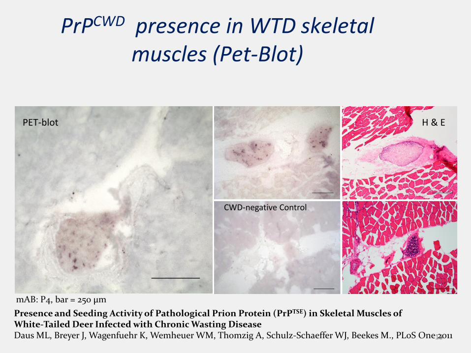
Presence and Seeding Activity of Pathological Prion Protein (PrPTSE) in Skeletal Muscles of White-Tailed Deer Infected with Chronic Wasting Disease Daus ML, Breyer J, Wagenfuehr K, Wemheuer WM, Thomzig A, Schulz-Schaeffer WJ, Beekes M., PLoS One 2011
mAB: P4, bar = 250 µm
CWD-negative Control
H & EPET-blot
PrPCWD presence in WTD skeletal muscles (Pet-Blot)
13

14
AnimalID
Date of inoculation Route of inoculation Inoculum Date ofautopsy
Lymphnodebiopsies
Years pi(05/2017)
AU242 2009/10/27 Ic steel wire mock control material 2015/06/30 - 5.7
AU153 2009/11/24 Ic steel wire CWD WTD 2012/09/10 - 2.8
AU500 2009/10/27 Ic steel wire CWD WTD - 7.6
AU519 2009/11/24 Ic steel wire CWD WTD 2015/01/21 - 5.2
AU308 2010/11/09 Ic steel wire CWD elk - 6.6
AU389 2010/11/09 Ic steel wire CWD elk 2015/05/04 - 4.5
AU520 2010/11/09 Ic steel wire CWD elk 2017/02/22 - 6.3
AU406 2009/07/07 Ic mock control - 7.6
AU408 2009/07/07 Ic 10 mg CWD WTD 2016/01/28 - 6.5
AU469 2009/07/07 Ic 10 mg CWD WTD 2016/06/06 - 6.9
AU398 2009/05/27 Skin scarification 1 ml 10 % CWD WTD - 7.10
AU451 2009/05/20 Skin scarification 1 ml 10 % CWD WTD - 7.10
AU315 2009/04/29 - 2010/07/20 oral 5 x 2mg mock control (WTD brain) - 8
AU467 2009/04/29 - 2010/07/20 oral 5 x 2 g CWD WTD brain 2015/03/04 - 5.8
AU243 2009/04/29 - 2010/07/20 oral 5 x 2 g CWD WTD brain - 8
AU316 2009/09/14 - 2012/11/02 oral ~5 kg CWD-WTD (repeatedly) 2017/03/10 - 7.4
AU385 2009/09/14 - 2012/11/06 oral ~5 kg CWD-WTD muscle (repeatedly) 2015/12/09 - 6.2
AU501 2009 /09/14- 2012/11/06 oral ~5 kg CWD-WTD muscle (repeatedly) 2015/02/01 - 5.4
AU456 2009/11/12 blood transfusion 14 ml plasma/buffy coat {RML#128+616 (elk)} 7.5
AU382 2009/11/16 blood transfusion 14 ml plasma/buffy coat {RML#144+116 (WTD} 7.5
AU390 2009/11/16 blood transfusion 9.5 ml plasma/buffy coat {RML#135 (mule deer)} 7.5

15
Animal # AU242 AU153 AU389 AU519 AU520 AU408 AU469 AU467 AU501 AU385 AU316
Years p.c. 5.7 2.8 4.5 5.2 6.3 6.5 6.9 5.8 5.4 6.2 7.4
Route i.c. steelwire
i.c.steelwire
i.c. steelwire
i.c. steel
wire
i.c. steelwire
i.c. i.c. oral oral oral oral
Inocul. Mock(-)
WTD+ Elk+ WTD+ ELK+ WTD+pool
WTD+pool
WTD+brain
WTD+muscle
WTD+muscle
WTD+muscle
Clinicalpresent.
wasting no(pm)
anxietyataxiatremorwasting
no(pm)
wasting no wasting wasting(died p.anest.)
anxietyataxiatremorwasting
apathyataxiatremorwasting
Ileus
Clinpath Glucose no Glucose no Glucose & HbA1c
Glucose Glucose & HbA1c
no no Glucose no
Animal info

IMMUNOHISTOCHEMISTRY & HISTOPATHOLOGY
Morphological assays
16

Proof-of-concept: i.c. sw; CWD elk, 4.5 years (AU389)
17
J Infect Dis. 2015;212(9):1459-1468. doi:10.1093/infdis/jiv232 (Holznagel , et al. )
Lumbar spinal cord, H&E; 6C2, x100

Proof-of-concept: ic sw; CWD WTD, 5.3 years (AU519)
18
GFAP H&E 6H4
Obex, x200x (GFAP, H&E) | 400x (6H4)

Per oral: CWD WTD brain, 5.8 years (AU467)
H&E 6H4 6H4
Lumbar Spinal Cord: x100 (H&E & 6H4)), x200 (6H4); DRG: x400 (6H4); Pons: x200 (H&E, 6H4); cerebellum x200 (6H4)

Per oral, CWD WTD muscle, 5.4 years: Weight, H&E &IHC (AU501)
-600 -400 -200 0 200 400 600 800 1000 1200 1400 1600 1800 20003.5
4.0
4.5
5.0
5.5
6.0
6.5
7.0
7.5
8.0
days before, during and post inoculation
bo
dy w
eig
ht [
kg
]
20Lumbar Spinal Cord: x2.5 (H&E & 6H4)), x200 (6H4); cerebellum x200 (6H4)

Per oral: CWD WTD muscle; 6.2 years (AU385)
21Spinal cord, H&E, x100, 6H4 IHC x25 & 200: Pons, H&E, x200; afferent nerve, 6H4, x200

AMYLOID SEEDING ACTIVITY
RT-QuIC
22

RT-QuIC: lumbar spinal cord (fluorescence curve & dot blot ROC characteristics)
23
Spinal Cord (Lumbar) w. Macaque rPrPc
***Mean Fluorescence
Time [h]
RF
U (T
hT
450/4
80)
0 10 20 30 40 50 60 70 80 90 100
1000
10000
TSE+ Controls TSE- Controls
CWD Challenged Macaques
MaqA4 BSE+(Cortex)
MaqA4 BSE+Lum.SpCd
DPZ#16825 DPZ#16828Au242
Au385 Au389 Au408 Au501 Au519
Elk#168(Cortex)
Elk#181(Cortex)
Tissues: Lumbar Spinal CordDilution: 1:90
rPrPc substrate: Macaque
ThT-signal time (hours)
Sam
ple
ID
0 10 20 30 40 50 60 70 80 90 100 110 120 130
Au519
Au501
Au408
Au389
Au385
Au242
DPZ#16828
DPZ#16825
MaqA4 BSE+
MaqA4 BSE+Ctx
Elk#181Ctx
Elk#168Ctx
A B
A: 100% Sens/Spec for BSE+ Macaque and DPZ/Au242 controlsB: 100% Sens., 94% Spec. for BSE+ Macaque and DPZ/Au242 controls

RT-QuIC: cervical spinal cord (fluorescence curve & dot blot ROC characteristics)
24
Spinal Cord (Cervical) w. Macaque rPrPc
***Mean Fluorescence
Time [h]
RF
U (T
hT
450/4
80)
0 10 20 30 40 50 60 70
1000
10000
A4(BSE+)Cortex
DPZ#16825 DPZ#16828Elk#168(Cortex)
TSE+ Controls TSE- Controls
A4(BSE+)Thor.SpCd
Au242
Au501 Au519Au385 Au389 Au408
CWD Challenged Macaques
Tissues: Cervical Spinal CordDilution: 1:90
rPrPc substrate: Macaque
ThT-signal time (hours)
Sam
ple
ID
0 10 20 30 40 50 60 70
Au519
Au501
Au408
Au389
Au385
Au242
DPZ#16828
DPZ#16825
Maq A4 (BSE+) Thor.SpCd
Maq A4 (BSE+) Cortex
Elk#181 CWD-
Elk#168 CWD+

RT-QuIC: mesenteric lymphnode(fluorescence curve & dot blot ROC characteristics)
25
Mesenteric Lymph Node w. Macaque rPrPc
***Mean Fluorescence
Time [h]
RF
U (T
hT
450/4
80)
0 10 20 30 40 50 60 70
1000
10000
TSE+ Controls
CWD Challenged Macaques
AU385 AU389 AU501 AU519
Elk#142MaqA4_BSE+ WTD 09-70006
TSE- Controls
AU242 DPZ#16825 DPZ#16828
Elk#137 WTD 09-71216
Tissues: Mesenteric Lymph NodeDilution: 1:30
rPrPc substrate: Macaque
ThT-signal time (hours)
Sam
ple
ID
0 10 20 30 40 50 60 70 80 90
AU519
AU501
AU389
AU385
AU242
DPZ#16825
DPZ#16828
MaqA4_BSE+
W09-71216_CWD-
Elk#137_CWD-
W09-70006_CWD+
Elk#142_CWD+

RESULTS
26

27
Animal # AU389 AU519 AU467 AU501 AU385
Years p.c. 4.5 5.2 5.9 5.4 6.3
Route i.c. steel
wirei.c. steel wire oral oral oral
Inocula Elk+ WTD+ WTD+ brain WTD+
muscle
WTD+
muscle
Clinical
disease
ataxia
anxiety
tremor
wasting
scheduled
PM
wasting(died
p.anesth.)
ataxia
anxiety
tremor
wasting
ataxia
apathy
tremor
wasting
H&E/IHC +
+
+
+
+
+
+
+
+
+
PMCA ip ip ip ip ip
QuIC + + + + +
Diabetes + - - - +
Results Summary

Proof-of-concept: i.c. sw; CWD elk, 4.5 years (AU389)
28
J Infect Dis. 2015;212(9):1459-1468. doi:10.1093/infdis/jiv232 (Holznagel , et al. )
Lumbar spinal cord, H&E; 6C2, x100

Spread & Distribution of C-type BSE (Ch. Hoffmann et al, 2011)
• 1) Peyer’s patches
• 2) Sympathetic Ganglion Chain
• 3) Obex
• 4) Vagal nerve
• 5) Spinal cord
29

30
Animal No. Route of inoculation Inoculum Years post inoculation
(07/2017)
AU500 i.c/ steel wire CWD WTD pool 7.8
AU308 i.c/ steel wire CWD elk 6.7
AU406 i.c. mock control 8.0
AU398 skin scarification CWD WTD pool 8.0
AU451 skin scarification CWD WTD pool 8.0
AU315 oral mock control 8.3
AU243 oral CWD WTD brain pool 8.3
AU456 blood transfusion plasma/buffy coat, RML elk 7.7
AU382 blood transfusion plasma/buffy coat , RML WTD 7.7
AU390 blood transfusion plasma/buffy coat , RML mule deer 7.7
CWD transmission project: remaining macaques

CWD Macaque Transmission Project: in July 2017
• 11/21 animals available for assessment
• 10 remaining animals with no clinical signs (incubation: 4.2 – 7.9 years) (2 mock controls)
• 3/11 animals with neurological signs
• 6/11 animals with wasting (6/11 animals with confirmed pre-clinical or clinical diabetes)
So far:
• In 5 animals: prion specific histopathologic lesions; PrPsc deposits &/or amyloid seeding. Read-out is ongoing (animal challenges; PET-BLOT; WB; PMCA)
• 2 animals are i.c challenged (steel wire); 3 animals are challenged per orally with pre-clinical CWD WTD muscle (2/3) or clinical CWD WTD brain (1/3)
• Post mortems of remaining animals scheduled in 2018 (potentially in absence of clinical disease)
31

Acknowledgments:• CFIA TSE Laboratory:
• Drs. S. Czub & R. Katoch: pathologists
• R. Anderson, S. Dudas, J. Gray, R. Quaghebeur, J. Yang: molecular group
• K. Colwell, Y. Fang, T. Pickles, K. Santiago Mateo: histology group
• Bianka Mussil (DPZ/Germany)
• Drs. B. Chesebro & B. Race (RML, Montana)
• Drs. JP Deslys & E. Comoy (CEA, France)
• Dr. T. Leighton (Canadian Cooperative Wildlife Health Center/SK & PrioNet Tissue Bank)
• Dr. M. Samuel (University of Wisconsin), Dr. Julie Langenberg
• Dr. J. Richt (Kansas State University)
Kevin Keough

Stefanie Czub, DVM, PhDManager pathology, TSE & virology
Head, Canadian & OIE Reference Laboratories for BSELethbridge Laboratory
Canadian Food Inspection Agency / Government of Canada
[email protected] Tel: 403-382-5549
33

Thank you for attending this webinar. If you have additional questions or comments concerning Chronic Wasting Disease or
other prion diseases please email :
Slides and the recording for this presentation will be posted to the CSTE webinar library