Title Correlation of external ear auricle formation with ...
20
RIGHT: URL: CITATION: AUTHOR(S): ISSUE DATE: TITLE: Correlation of external ear auricle formation with staging of human embryos Ozeki-Sato, Maimi; Yamada, Shigehito; Uwabe, Chigako; Ishizu, Koichi; Takakuwa, Tetsuya Ozeki-Sato, Maimi ...[et al]. Correlation of external ear auricle formation with staging of human embryos. 2016-03 http://hdl.handle.net/2433/216678 This is the accepted version of the following article: [Ozeki-Sato, M., Yamada, S., Uwabe, C., Ishizu, K., and Takakuwa, T. (2016) Correlation of external ear auricle formation with staging of human embryos. Congenital Anomalies, 56: 86–90. doi: 10.1111/cga.12140.], which has been published in final form at http://dx.doi.org/10.1002/ar.23099. This article may be used for non-commercial purposes in accordance with Wiley Terms and Conditions for Self-Archiving.; The full-text file will be mad ...
Transcript of Title Correlation of external ear auricle formation with ...
RIGHT:
URL:
CITATION:
AUTHOR(S):
ISSUE DATE:
TITLE:
Correlation of external ear auricleformation with staging of humanembryos
Ozeki-Sato, Maimi; Yamada, Shigehito; Uwabe,Chigako; Ishizu, Koichi; Takakuwa, Tetsuya
Ozeki-Sato, Maimi ...[et al]. Correlation of external ear auricle formationwith staging of human embryos.
2016-03
http://hdl.handle.net/2433/216678
This is the accepted version of the following article: [Ozeki-Sato, M., Yamada, S., Uwabe, C., Ishizu, K., and Takakuwa, T.(2016) Correlation of external ear auricle formation with staging of human embryos. Congenital Anomalies, 56: 86–90.doi: 10.1111/cga.12140.], which has been published in final form at http://dx.doi.org/10.1002/ar.23099. This article maybe used for non-commercial purposes in accordance with Wiley Terms and Conditions for Self-Archiving.; The full-textfile will be mad ...

1
SHORT COMMUNICATION
Correlation of external ear auricle formation with staging of human embryos
Ozeki
Running title: Auricles of human embryos
Maimi Ozeki1, Shigehito Yamada1,2, Chigako Uwabe2, Koichi Ishizu1, Tetsuya
Takakuwa1
1Human Health Science and 2Congenital Anomaly Research Center, Graduate School
of Medicine, Kyoto University, Kyoto, Japan
Corresponding author: Dr. Tetsuya Takakuwa
Human Health Science, Graduate School of Medicine, Kyoto University
Sakyo-ku Shogoin Kawahara-cyo 53, Kyoto 606-8507, Japan
E-mail: [email protected]; TEL/FAX: +81-75-751-3931
A Self-archived copy inKyoto University Research Information Repository
https://repository.kulib.kyoto-u.ac.jp
2
ABSTRACT
The formation of auricles in human embryos was evaluated between
Carnegie stage (CS)19 and CS23, and the findings were correlated across the stages.
The auricle was categorized into 11 steps according to Streeter’s criteria with
modifications. Mesenchyme cell condensation was observed at Step 7, and two layers
of cartilage consisting of the auricle were recognized at Step11. The representative
steps at each CS shifted from Step 3 to Step11 during CS16 and CS23, although
several steps overlapped between adjacent CSs. These results indicate that
observations of the auricle between CS19 and CS23 may be utilized for determining
embryo staging as convincing supportive evidence of external features reflecting the
internal histological structure, although other findings should also be taken into account.
Key Words: external ear, human embryo, Carnegie stage, auricle
A Self-archived copy inKyoto University Research Information Repository
https://repository.kulib.kyoto-u.ac.jp
3
INTRODUCTION
The external ear primordium arises on the mandibular and hyoid arches. At
this point, six hillocks of the branchial arch appear, which disappear by fusing to
establish the primitive auricles (Streeter 1922; O’Rahilly 1984). Since the morphology of
the auricle is very unique and easily observable externally, it has been incorporated into
assessments of the developmental staging of human embryos such as the Carnegie
stage (CS) system (O’Rahilly and Müller 1987). The CS system relies on detailed
morphology of the auricle from CS16 to CS18. Indeed, O’Rahilly and Müller (1987)
suggested that the findings of the auricles are one of the most prominent and reliable
factors for determining and discriminating between these three stages.
Streeter (1922) classified the formation of the auricles into 15 categories (A–
O) during embryonic development (5–33 mm in crown-rump length). Based on the CS
criteria (O’Rahilly and Müller 1987), categories C and D correspond to auricular
formation at CS16, category E corresponds to that at CS17, and categories G, H, and I
correspond to that at CS18 (Table 1, Figure 1). Although Streeter’s (1922)
categorization was not adapted to the CS system after CS19, six additional categories
(J–O) were nonetheless recognized that approximately correspond to these periods.
A Self-archived copy inKyoto University Research Information Repository
https://repository.kulib.kyoto-u.ac.jp
4
Only two ambiguous descriptions have been recognized (O’Rahilly and Müller 1987):
“the coalescence of parts in the pharyngeal region has altered the appearance of the
auricle, and the hillocks are less conspicuous” (p. 238) at CS19 and “the formation of
the auricle has progressed noticeably: the tragus and antitragus especially are
assuming a more definite form” (p. 259) at CS22.
However, as O’Rahilly and Müller (1987) themselves pointed out, these
descriptions are somewhat limited at the stage between CS19 and CS23 with respect to
both external and internal findings. In fact, there are only 27 pages dedicated to the
description of embryos between CS19 and CS23 in their book, whereas 79 pages are
focused on the description of CS14 to CS18. Therefore, accumulation of new
information on the external features corresponding to each stage may be useful to
improve the accuracy of staging as well as to improve the precision of studies
conducted during these periods. Nevertheless, it is necessary to take into account the
developmental status of various internal structures in order to precisely assign the stage
of a specimen after CS19. Considering this background, the aim of the present study
was to observe the formation of auricles between CS19 and CS23, and provide
quantitative data on the correlation between auricle formations and CSs.
A Self-archived copy inKyoto University Research Information Repository
https://repository.kulib.kyoto-u.ac.jp
5
MATERIALS AND METHODS
For external observations, 340 human embryo samples between CS16 and CS18, as
well as 498 samples between CS19 and CS23 were selected from the Kyoto Collection
(Nishimura et al. 1968; Shiota et al. 2007; Yamada et al. 2010). None of these samples
exhibited overt damage or anomalies. The morphology of the auricle was observed from
bilateral views.
The auricle was categorized into 11 steps according to the observations of Streeter
(1922) with minor modifications (Table 1, Figure 1). In brief, five categories were omitted
or combined from Streeter’s 16 categories (A–O). Category A included only the samples
before CS16. Category F was omitted because it was originally described as an
exceptional, abnormal case. Category M was combined with category L, which was
originally regarded as a variation of category L. Categories L and O were combined
because the discrimination between these categories was not apparent in the original
study, according to the following description “the transition from Category-N to -O brings
us to a condition that may be regarded as the definitive auricle” (p. 130). When the
A Self-archived copy inKyoto University Research Information Repository
https://repository.kulib.kyoto-u.ac.jp
6
steps differed between the left and right auricles in the embryo, the step at a more
progressed stage was defined as the step of the given sample. Each auricle was blindly
categorized twice by one author (M.O), and confirmed by another author (T.T.).
For histological observations, 33 organs from 17 embryo samples between Step 7
and Step 11 were used from the Kyoto Collection. Histological serial sections stained
with hematoxylin and eosin were observed microscopically by two authors
independently (M.O. and T.T.). The ethics committee of the Kyoto University Graduate
School and Faculty of Medicine approved this study (E986).
RESULTS AND DISCUSSION
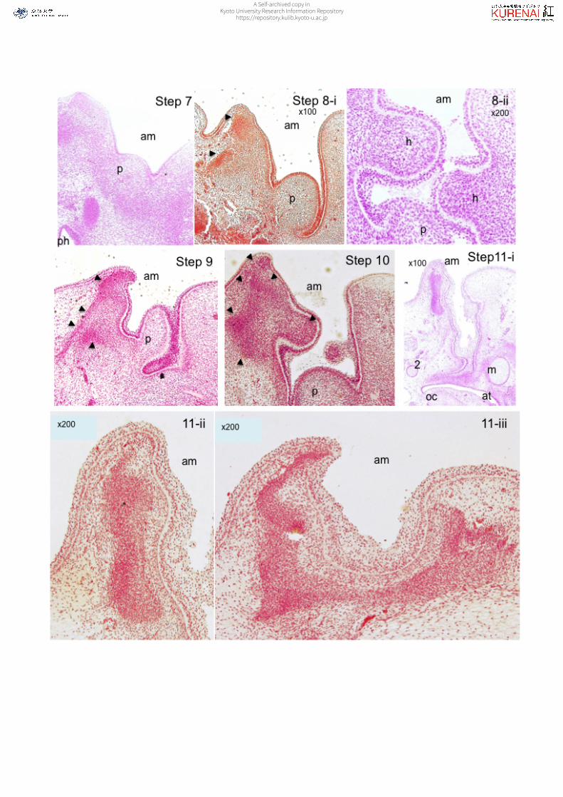
Vague condensation of the mesenchyme cells as pre-cartilage was
recognized around the external auditory meatus (EAM) at Step 7 (Figure 2). The meatal
plug was recognized on the bottom of the EAM at Step 7. The distinct border was first
recognized on the tip of the top of the raised margin (▲ in Step 8, Figure 2). Such
condensation of the mesenchyme cells was recognizable from one of 4 samples at Step
7 and in all samples at Step 8 (Table 2). The centers of condensation increased in
number as the steps proceeded (Steps 8-10). The hillocks were recognized as small
A Self-archived copy inKyoto University Research Information Repository
https://repository.kulib.kyoto-u.ac.jp
7
bulges rising into the EAM. Histologically, the cell density increased, but the border was
not distinct. The border of the pre-cartilage became distinct on the side further from the
EAM but not on the side closer to the EAM (step 9 in Figure 2).
A distinction between Step 10 and Step 11 was also revealed with the
histological observation. The cartilage was clearly recognizable in 5 of 6 samples at
Step 11 (Figure 2). The arrangements of the cells in the cartilage differed between the
side closer to and that further from the EAM: the inner cells run horizontally while the
outer cells run vertically. The extracellular matrix was scant compared with other
cartilage observed in the same Step 11 samples, such as Meckel’s cartilage and the
otic vesicles. The epithelium of the external auditory canal contained 1–2 columnar cells
until Step 7 (Table 2). The epithelium became stratified with 3–4 layers of cells in one of
4 samples at Step 7 and in all samples at Step 8. The stratified epithelium had over 4
layers at Steps 10 and 11. The histological observations indicate that the histological
changes during development may contribute to and influence the appearance of the
auricle at each step of its formation.
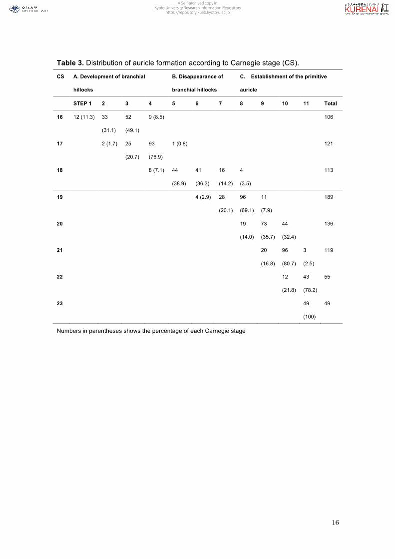
The representative steps to which most samples belonged at each CS shifted
from Step 3 to Step 11 progressing from CS16 to CS23 (Table 3). Thus, the
A Self-archived copy inKyoto University Research Information Repository
https://repository.kulib.kyoto-u.ac.jp
8
representative steps at each CS were different, except between CS22 and CS23 (i.e.,
Step 11). The auricles of the samples ranged broadly between 4 and 5 steps between
CS16 and CS18. On the other hand, the auricles of the samples ranged across less
than 4 steps between CS20 and CS23; namely, 3 steps at CS20 (Steps 8–10) and
CS21 (Steps 9–11), 2 steps at CS22 (Steps 10 and 11), and one step at CS23 (Step
11).
Overall, the range of steps to which the samples belonged overlapped between
adjacent CSs. This means that the determination of the CS does not depend on the
morphology of the auricle alone, but rather multiple findings of both the external and
internal morphology are required for appropriate staging. This was the case even
between CS16 and CS18, at which the findings of the auricle are considered to be the
most reliable information for appropriate staging (O’Rahilly and Müller 1987). However,
the CS can never be determined based on one absolute and dominant finding but
instead requires accumulated information from several observations as a whole. In
addition to the presence of auricular hillocks, the findings of limb buds, posture of the
body, and facial features (nasal tip and wing, eye lids, etc.) are included as crucial
findings for determining the stages between CS16 and CS18.
A Self-archived copy inKyoto University Research Information Repository
https://repository.kulib.kyoto-u.ac.jp
9
As for the later stages after CS19, detailed descriptions of external and internal
findings are still in progress. External findings for discriminating between CS19 and
CS23 were limited to the limb bud, posture of the limb, trunk, and head, eyelids, and the
superficial vascular plexus of the head. The descriptions of these features are vague
and/or do not always cover all stages between CS19 and CS23. For example, the
descriptions of the toe ray are the most detailed at CS19, as toe rays are prominent but
inter-digital notches have not yet appeared at that stage. However, this feature cannot
be used in later stages.
The present study provides information on the differentiation of the auricles in
a step-by-step manner, and demonstrates the correlation of these steps with CSs. The
important finding is that auricle formation may be accurately represented by the
histological features during development. These results indicate that observations of the
auricle at these developmental periods may be utilized to determining staging, providing
convincing supportive evidence of external embryonic features reflecting the internal
histological structure, although other findings should also be taken into account
simultaneously. These data may also be valuable for fetal three-dimensional
sonographic studies, where accurate depiction of the fetal ear is important given that
A Self-archived copy inKyoto University Research Information Repository
https://repository.kulib.kyoto-u.ac.jp
10
ear anomalies can be associated with complex syndromes (Merz et al, 1997, Shih et al,
1998).
Acknowledgements
This study was supported by Grant Nos. 25461642, 24119002, 26220004,
15H01119, 15K08134, 15H05270, 15H01121, and 15K15014 from the Japan
Society for the Promotion of Science.
Disclosures
None
References
Merz E, Weber G, Bahlmann F, Miric-Tesanic D. 1997. Application of transvaginal and
abdominal three-dimensional ultrasound for the detection or exclusion of
malformations of the fetal face. Ultrasound Obstet Gynecol 9:237-243.
A Self-archived copy inKyoto University Research Information Repository
https://repository.kulib.kyoto-u.ac.jp
11
Nishimura H, Takano K, Tanimura T, Yasuda M. 1968. Normal and abnormal
development of human embryos: first report of the analysis of 1,213 intact
embryos. Teratology 1:281-290.
O’Rahilly R. 1983. The timing and sequence of events in the development of the human
eye and ear during the embryonic period proper. Anat Embryol (Berl) 168: 87-99.
O’Rahilly R, Müller F. 1987. Developmental stages in human embryos: including a
revision of Streeter’s Horizons and a survey of the Carnegie Collection.
Washington, D.C.: Carnegie Institution of Washington 637. p. 239–251.
Shih JC, Shyu MK, Lee CN, Wu CH, Lin GJ, Hsieh FJ. 1998. Antenatal depiction of the
fetal ear with three-dimensional ultrasonography. Obstet Gynecol 91:500-505.
Shiota K, Yamada S, Nakatsu-Komatsu T, Uwabe C, Kose K, Matsuda Y, Haishi T,
Mizuta S, Matsuda T. 2007. Visualization of human prenatal development by
magnetic resonance imaging (MRI). Am J Med Genet A 143A: 3121-3126.
Streeter GL. 1922. Development of the auricle in the human embryo. Contrib Embryol
Carnegie Inst 14: 111-138.
A Self-archived copy inKyoto University Research Information Repository
https://repository.kulib.kyoto-u.ac.jp

12
Yamada S, Samtani RR, Lee ES, Lockett E, Uwabe C, Shiota K, Anderson SA, Lo CW.
2010. Developmental atlas of the early first trimester human embryo. Dev Dyn
239: 1585-1595.
A Self-archived copy inKyoto University Research Information Repository
https://repository.kulib.kyoto-u.ac.jp
13
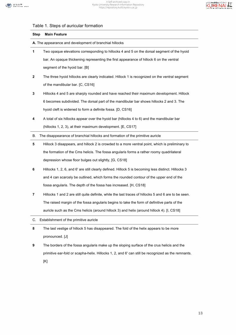
Table 1. Steps of auricular formation
Step Main Feature
A. The appearance and development of branchial hillocks
1 Two opaque elevations corresponding to hillocks 4 and 5 on the dorsal segment of the hyoid
bar. An opaque thickening representing the first appearance of hillock 6 on the ventral
segment of the hyoid bar. [B]
2 The three hyoid hillocks are clearly indicated. Hillock 1 is recognized on the ventral segment
of the mandibular bar. [C, CS16]
3 Hillocks 4 and 5 are sharply rounded and have reached their maximum development. Hillock
6 becomes subdivided. The dorsal part of the mandibular bar shows hillocks 2 and 3. The
hyoid cleft is widened to form a definite fossa. [D, CS16]
4 A total of six hillocks appear over the hyoid bar (hillocks 4 to 6) and the mandibular bar
(hillocks 1, 2, 3), at their maximum development. [E, CS17]
B. The disappearance of branchial hillocks and formation of the primitive auricle
5 Hillock 3 disappears, and hillock 2 is crowded to a more ventral point, which is preliminary to
the formation of the Cms helicis. The fossa angularis forms a rather roomy quadrilateral
depression whose floor bulges out slightly, [G, CS18]
6 Hillocks 1, 2, 6, and 6' are still clearly defined. Hillock 5 is becoming less distinct. Hillocks 3
and 4 can scarcely be outlined, which forms the rounded contour of the upper end of the
fossa angularis. The depth of the fossa has increased. [H, CS18]
7 Hillocks 1 and 2 are still quite definite, while the last traces of hillocks 5 and 6 are to be seen.
The raised margin of the fossa angularis begins to take the form of definitive parts of the
auricle such as the Cms helicis (around hillock 3) and helix (around hillock 4). [I, CS18]
C. Establishment of the primitive auricle
8 The last vestige of hillock 5 has disappeared. The fold of the helix appears to be more
pronounced. [J]
9 The borders of the fossa angularis make up the sloping surface of the crus helicis and the
primitive ear-fold or scapha-helix. Hillocks 1, 2, and 6' can still be recognized as the remnants.
[K]
A Self-archived copy inKyoto University Research Information Repository
https://repository.kulib.kyoto-u.ac.jp

14
Letters in brackets indicate the categories that correspond to Streeter's (1922) study and the Carnegie staging of
O'Rahilly and Müller (1987)
10 Hillocks 1 and 6' remain. The crus helicis is distinct and the primitive ear-fold is prominent.
There is the early form of the concha, divided by the crus into an upper and a lower half.[L,M]
11 Transition to a stage that may be regarded as the definitive auricle, namely the tragus,
antitragus, anthelix, scapha-helix, and, distinctly separate from the latter, the crus helicis is
recognized.[N,O]
A Self-archived copy inKyoto University Research Information Repository
https://repository.kulib.kyoto-u.ac.jp
15
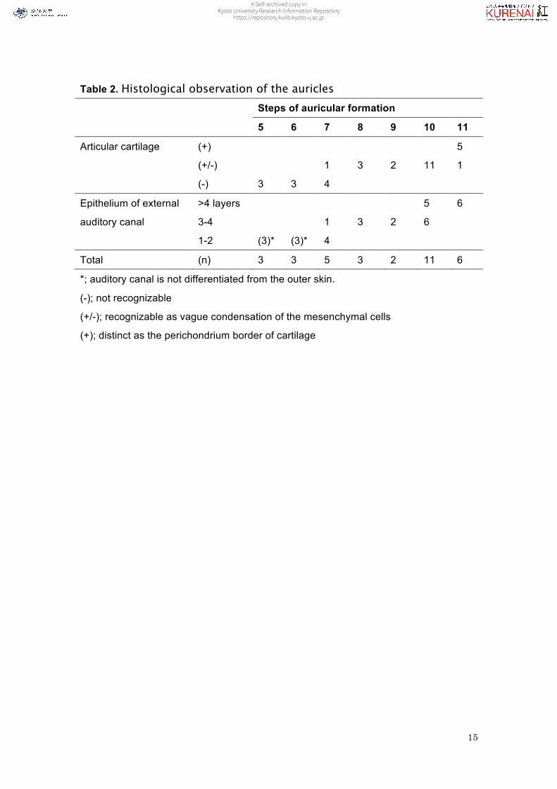
Table 2. Histological observation of the auricles
Steps of auricular formation
5 6 7 8 9 10 11
Articular cartilage (+) 5
(+/-) 1 3 2 11 1
(-) 3 3 4
Epithelium of external
auditory canal
>4 layers 5 6
3-4 1 3 2 6
1-2 (3)* (3)* 4
Total (n) 3 3 5 3 2 11 6
*; auditory canal is not differentiated from the outer skin.
(-); not recognizable
(+/-); recognizable as vague condensation of the mesenchymal cells
(+); distinct as the perichondrium border of cartilage
A Self-archived copy inKyoto University Research Information Repository
https://repository.kulib.kyoto-u.ac.jp
16
Table 3. Distribution of auricle formation according to Carnegie stage (CS).
CS A. Development of branchial
hillocks
B. Disappearance of
branchial hillocks
C. Establishment of the primitive
auricle
STEP 1 2 3 4 5 6 7 8 9 10 11 Total
16 12 (11.3) 33
(31.1)
52
(49.1)
9 (8.5) 106
17 2 (1.7) 25
(20.7)
93
(76.9)
1 (0.8) 121
18 8 (7.1) 44
(38.9)
41
(36.3)
16
(14.2)
4
(3.5)
113
19 4 (2.9) 28
(20.1)
96
(69.1)
11
(7.9)
189
20 19
(14.0)
73
(35.7)
44
(32.4)
136
21 20
(16.8)
96
(80.7)
3
(2.5)
119
22 12
(21.8)
43
(78.2)
55
23 49
(100)
49
Numbers in parentheses shows the percentage of each Carnegie stage
A Self-archived copy inKyoto University Research Information Repository
https://repository.kulib.kyoto-u.ac.jp
17
Figure legends
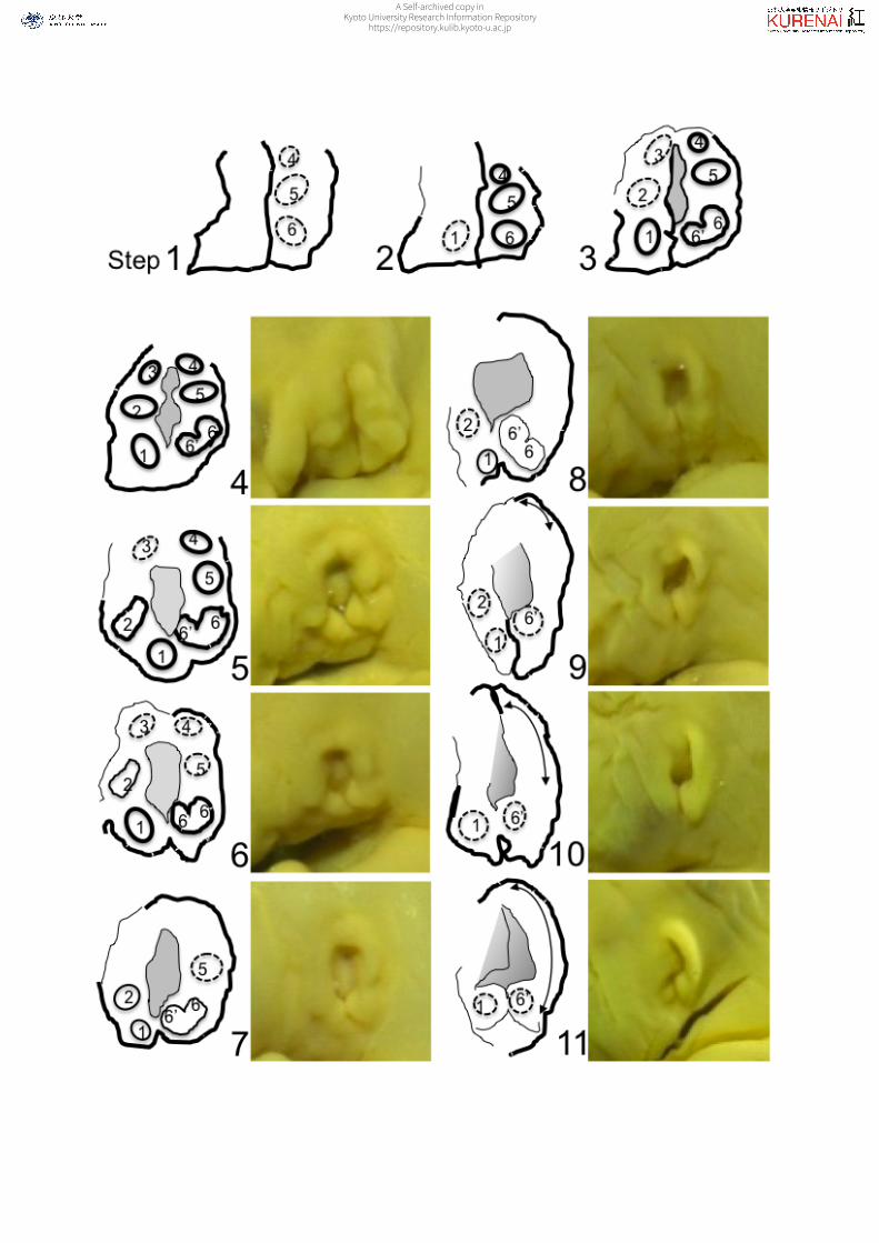
Figure 1. Representative findings of the auricles for all 11 steps
[Steps 1–4] Formation of the branchial hillocks until their maximum development; [Steps
5–7] Disappearance of the branchial hillocks; [Steps 8–11] Establishment of the
primitive auricle.
The illustration was drawn with reference to Figure 5 and the associated descriptions
found in Streeter (1922).
Figure 2. Histology of the auricles with hematoxylin and eosin staining from
Steps 7 to 11
am; external auditory meatus, h; auricular hillocks, p; auditory pluq, ph; pharynx, m;
Meckel’s cartilage, 2; cartilage of the 2nd arch, at; auditory tube, oc; otic capsule
A Self-archived copy inKyoto University Research Information Repository
https://repository.kulib.kyoto-u.ac.jp
A Self-archived copy inKyoto University Research Information Repository
https://repository.kulib.kyoto-u.ac.jp
A Self-archived copy inKyoto University Research Information Repository
https://repository.kulib.kyoto-u.ac.jp


