TISSUE REACTIONS PRODUCED BY CALCIUM …oem.bmj.com/content/oemed/15/3/168.full.pdf · wasas...
4
Brit. J. industr. Med., 1958, 15, 168. TISSUE REACTIONS PRODUCED BY CALCIUM FLUORIDE IN THE LUNGS OF RATS BY E. J. KING, M. YOGANATHAN, and G. NAGELSCHMIDT From the Postgraduate Medical School, London, and the Safety in Mines Research Establishment, Sheffield (RECEIVED FOR PUBLICATION NOVEMBER 8, 1957) In a previous paper (King, Mohanty, Harrison, and Nagelschmidt, 1953), we showed that removal of an amorphous " high-solubility layer" from the surface of quartz particles with hydrofluoric acid appeared to enhance their fibrosis-producing capa- city rather than to diminish it. Although the HF- treated quartz had been repeatedly washed, and even electro-dialyzed, it had at least as much, and usually more, fibrogenic capacity than the original quartz powder. We considered that this might be due to traces of hydrofluoric acid retained on the quartz particles. We thought it would be interesting, therefore, to test the effect of a relatively insoluble fluoride in the lungs of similar animals. Lung changes have been reported by Luton and Champeix (1951) in men working with calcium fluoride in France. Their radiographic changes were described as typical of pneumoconiosis, and, indeed, similar to silicosis. The disease had evolved very rapidly, six of the 19 cases having developed it in less than two years. In another group of workers, severe lung disease was also found, but the radio- graphic shadows were atypical and different from those of silicosis. The dust inhaled by the first group of workers contained 60 % of silica with 36 % calcium fluoride and small amounts of alu- minium and iron oxides. The second group inhaled dust which was mainly calcium fluoride (92-96%) with only 3-5 % silica. Luton, Champeix, and Faure (1953) considered that calcium fluoride may by itself injure the lung tissue and produce lesions, and that it certainly enhances the action of silica, resulting in a severe and rapidly-developing form of pneumoconiosis. Policard and Collet (1953) injected calcium fluoride intraperitoneally into rats, and found that after one to two months it gave a marked fibrotic reaction in the peritoneum, even more severe than that produced by quartz, but that, in contrast to quartz, it caused only an insignificant reaction in the lymph glands. Experimental Water-clear colourless crystals of calcium fluoride were ground to a fine powder and water-sedimented to give a 0-5-2,u fraction. The particle size distribution of the sample is given in Table 1. It contained less than 0-1 % of silica, and had a solubility in water at room temperature of 1-6 mg. CaF2 per 100 ml. TABLE 1 PARTICLE SIZE DISTRIBUTION AND ANALYSIS OF CALCIUM FLUORIDE DUST Size ( ) Percentage Percentage by Number by Mass 004-0-06 14-5 - 0 7 - 0-8 8-4 - 09 -0 11 8-1 - 0-12-0 16 6-6 - 0-17 - 0-23 5-5 0.2 0-24 - 0-32 7-6 0-5 033-045 90 1 7 0-46 - 0-64 15-0 8-2 065 -09 15-4 23-1 1 0 - 1-3 7-9 32-9 1-4 - 1-8 1-6 18 9 1-9 -2-6 04 14 5 Dvs* MMDt $r 0-98 1*0 1-6 £ nd3 * Dvs volume surface diameter, £.2 t MMD = Mass median diameter: Size at 50% mass on cumulative size distribution curve. a = geometrical standard deviation: ratio of diameters having 50% and 16-5 Y. mass or having 83-5 % and 50% mass. These are 1-7 and 15, mean 1t6. A group of 22 male rats of the black and white Medical Research Council strain (average weight 200 g.) was injected intratracheally with 50 mg. each of calcium fluoride. The animals were killed at regular intervals up to one year and the lung pathology studied. The collagen content of the lungs was determined in order to compare the values obtained with those from a group of rats injected intratracheally with 50 mg. of quartz (Stacy and King, 1954). The lymph glands were dissected and weighed. The preparation of the dust suspension for injection was as follows: A sample of the dust, 1 5 g., was weighed into a glass tissue grinder, and 30 ml. of sterile physio- logical saline added. The dust particles dispersed easily. 168 on 30 June 2018 by guest. Protected by copyright. http://oem.bmj.com/ Br J Ind Med: first published as 10.1136/oem.15.3.168 on 1 July 1958. Downloaded from
Transcript of TISSUE REACTIONS PRODUCED BY CALCIUM …oem.bmj.com/content/oemed/15/3/168.full.pdf · wasas...

Brit. J. industr. Med., 1958, 15, 168.
TISSUE REACTIONS PRODUCED BY CALCIUMFLUORIDE IN THE LUNGS OF RATS
BY
E. J. KING, M. YOGANATHAN, and G. NAGELSCHMIDT
From the Postgraduate Medical School, London, and the Safety in Mines Research Establishment, Sheffield
(RECEIVED FOR PUBLICATION NOVEMBER 8, 1957)
In a previous paper (King, Mohanty, Harrison,and Nagelschmidt, 1953), we showed that removalof an amorphous " high-solubility layer" from thesurface of quartz particles with hydrofluoric acidappeared to enhance their fibrosis-producing capa-city rather than to diminish it. Although the HF-treated quartz had been repeatedly washed, andeven electro-dialyzed, it had at least as much, andusually more, fibrogenic capacity than the originalquartz powder. We considered that this might bedue to traces of hydrofluoric acid retained on thequartz particles. We thought it would be interesting,therefore, to test the effect of a relatively insolublefluoride in the lungs of similar animals.Lung changes have been reported by Luton and
Champeix (1951) in men working with calciumfluoride in France. Their radiographic changes weredescribed as typical of pneumoconiosis, and, indeed,similar to silicosis. The disease had evolved veryrapidly, six of the 19 cases having developed it inless than two years. In another group of workers,severe lung disease was also found, but the radio-graphic shadows were atypical and different fromthose of silicosis. The dust inhaled by the firstgroup of workers contained 60% of silica with36% calcium fluoride and small amounts of alu-minium and iron oxides. The second group inhaleddust which was mainly calcium fluoride (92-96%)with only 3-5% silica. Luton, Champeix, andFaure (1953) considered that calcium fluoride mayby itself injure the lung tissue and produce lesions,and that it certainly enhances the action of silica,resulting in a severe and rapidly-developing form ofpneumoconiosis.
Policard and Collet (1953) injected calcium fluorideintraperitoneally into rats, and found that after oneto two months it gave a marked fibrotic reactionin the peritoneum, even more severe than thatproduced by quartz, but that, in contrast to quartz,it caused only an insignificant reaction in the lymphglands.
ExperimentalWater-clear colourless crystals of calcium fluoride
were ground to a fine powder and water-sedimented togive a 0-5-2,u fraction. The particle size distributionof the sample is given in Table 1. It contained less than0-1 % of silica, and had a solubility in water at roomtemperature of 1-6 mg. CaF2 per 100 ml.
TABLE 1PARTICLE SIZE DISTRIBUTION AND ANALYSIS OF
CALCIUM FLUORIDE DUST
Size ( ) Percentage Percentageby Number by Mass
004-0-06 14-5 -
0 7 - 0-8 8-4 -
09 -0 11 8-1 -
0-12-0 16 6-6 -
0-17 - 0-23 5-5 0.20-24 - 0-32 7-6 0-5033-045 90 1 70-46 - 0-64 15-0 8-2065-09 15-4 23-11 0 - 1-3 7-9 32-91-4 - 1-8 1-6 18 91-9 -2-6 04 14 5
Dvs*MMDt$r
0-981*01-6
£ nd3* Dvs volume surface diameter,£.2
t MMD = Mass median diameter:Size at 50% mass on cumulative size distribution curve.
a = geometrical standard deviation:ratio of diameters having 50% and 16-5 Y. mass or having83-5 % and 50% mass. These are 1-7 and 15, mean 1t6.
A group of 22 male rats of the black and white MedicalResearch Council strain (average weight 200 g.) wasinjected intratracheally with 50 mg. each of calciumfluoride. The animals were killed at regular intervalsup to one year and the lung pathology studied. Thecollagen content of the lungs was determined in order tocompare the values obtained with those from a groupof rats injected intratracheally with 50 mg. of quartz(Stacy and King, 1954). The lymph glands were dissectedand weighed.The preparation of the dust suspension for injection
was as follows: A sample of the dust, 1 5 g., was weighedinto a glass tissue grinder, and 30 ml. of sterile physio-logical saline added. The dust particles dispersed easily.
168
on 30 June 2018 by guest. Protected by copyright.
http://oem.bm
j.com/
Br J Ind M
ed: first published as 10.1136/oem.15.3.168 on 1 July 1958. D
ownloaded from

TISSUE REACTIONS PRODUCED BY CALCIUM FLUORIDE IN RATLUNGS 169
The suspension was transferred to a bottle, sterilized byautoclaving for 20 minutes and then placed on amechanical shaker until the time of injection.The animals were lightly anaesthetized with ether, and
were injected intratracheally with 1 ml. of the suspension(containing 50 mg. CaF2). The injections were made,as described in the previous paper (King et al., 1953),through a long blunt needle inserted through the mouthand down the trachea as far as the carina.The fate of the rats, whether they died or were killed,
and the duration of the experiment are shown in Table 2.
TABLE 2PATHOLOGICAL CHANGES IN LUNGS OF RATS GIVEN
50 MG. CALCIUM FLUORIDE
Days of Survival Mode of Death Grade of Fibrosis
30 Killed (2) 239 Died (2) 241 Died (1) 2 max.42 Died (1) 2 max.70 Killed (2) 2 max.81 Died (1) 390 Killed (2) 3*91 Died (1) 2105 Died (1) 2150 Killed (1) 2180 Killed (1) 2207 Died (1) -
210 Killed (1) 2275 Killed (1) 2300 Killed (1) 2330 Killed (1) 4*365 Killed (1) 3
* Sections reproduced in figures.Numbers in brackets indicate number dead or killed.Max. = maximum within the indicated grade of fibrosis
Grades of fibrosis: (1) lesions cellular, loose network of reticulin(2) cellular, thick compact reticulin, with or
without some collagen(3) slightly cellular, mainly collagen(4) completely acellular and wholly collagenous(5) lesions acellular, collagenous and confluent
Most of the deaths took place within the first 90 days.Two animals were killed each month until this time,thereafter one animal.The techniques of removing the lungs, fixation, em-
bedding, and sectioning were as previously described(King et al., 1953). Blocks were selected through thelong axes of both lungs at the level of the hilum andsections were stained with haematoxylin and eosin,impregnated with silver for reticulin and collagen, andmicro-incinerated to reveal the distribution of the dust.
Collagen analyses were performed on the driedpowdered remnants of each pair of lungs, to which wereadded the remainders of the blocks and their trimmingsafter suitably de-waxing with benzene. Results have beenexpressed as milligrams of collagen contained in eachpair of lungs. These figures are comparable, and aresubstantially correct, since only very small proportionsof the lungs were lost to analysis by inclusion in thesections.
ResultsSubpleural collections of dust were seen as
greyish-white foci, situated chiefly over the dorsalaspect of the lungs, early in the experiment. The3
hilar lymph nodes were enlarged to four or fivetimes normal size. By 40 days small areas of fibrosiswere seen scattered over both lungs. By 90 daysthey were 2 to 3mm. in diameter, and were hard.The animals which died during the first 90 daysshowed evidence of bronchopneumonic consolida-tion, widespread necrosis, and appearances simula-ting an acute pulmonary infection; but Gram-staining did not reveal the presence of organisms.The lymph nodes were greatly increased in size andwere about six times normal at 110 days. From thistime onward there appeared to be a regression in thesize of the lymph nodes, and at one year they weretwo or three times the normal size and very firm.Average weights of the nodes are given in Table 3together with those of control rats, (a) without dustand (b) with 50 mg. of quartz.
TABLE 3FRESH WEIGHTS OF LYMPH NODES IN CaF, EXPERIMENTCOMPARED WITH WEIGHTS WITH DOSE OF 50 MG. QUARTZ
AND WITHOUT DUST
Time after Node Weight (mg.)Injection(days) CaF, Quartz No Dust
0- 90 260 (12)* 240 (9) 32 (8)90-210 180 (4) 660 (12) 35 (14)270-365 80 (4) 1,100 (3) _
Numbers in brackets are number of rats.
Microscopically, there appeared to be a gooddust cell response by 30 days and a marked reactionin the peribronchial lymphoid tissue. Small discretelesions of irregular shape were mainly perivascularor peribronchial. But a large proportion of the dustwas lying free in clumps in the alveoli. At both30 and 40 days the lesions were seen to contain acompact network of reticulin with some collagen.Besides the fibrotic lesions some of the lungs showedevidence of patchy consolidation, with widespreadnecrosis and infiltration with acute inflammatorycells. No microorganisms could be found. Thiscondition was particularly evident in the animalsthat died, and we have assumed that it was broughtabout by the dust itself. At 60 days the nodularlesions were still scanty, cellular, and discrete. At90 days the reticulin network was largely replacedby collagen fibres, thick and compact (Fig. 1). (Thefibrosis produced by a similar dose of flint is shownin Fig. 2.) The very high concentration of dustwithin the lesions is shown in Fig. 3. Animals whichdied at 101 and 105 days showed marked necrosisand acute inflammatory cell infiltration, in additionto the fibrotic nodules, but no microorganisms weredemonstrated. Up to 330 days there was an increasein the number of nodules, but the maturity of theirfibrosis was not increased, except in one animal,where they were fully collagenous although smaller
on 30 June 2018 by guest. Protected by copyright.
http://oem.bm
j.com/
Br J Ind M
ed: first published as 10.1136/oem.15.3.168 on 1 July 1958. D
ownloaded from

BRITISH JOURNAL OF INDUSTRIAL MEDICINE
FiG. 1.-Compact, mainly collagenous nodules, in a ratlung, 90 days after 50 mg. calcium fluoride, Grade 3fibrosis, Silver impregnation, x 76.
FIG. 3.-Serial section to Fig. 1. Dark-ground illumina-tion, showing dense, centrally deposited dustcollections within the nodules. Micro-incineratedpreparation, x 76.
FiG. 2.-For comparison, rat lung 90 days after 50 mg.of flint. Grade 4 fibrosis.
FIG. 4.-Small, compact, wholly collagenous nodules,330 days after 50 mg. calcium fluoride. Grade 4fibrosis. Silver impregnation, x 76.
170
on 30 June 2018 by guest. Protected by copyright.
http://oem.bm
j.com/
Br J Ind M
ed: first published as 10.1136/oem.15.3.168 on 1 July 1958. D
ownloaded from
TISSUE REACTIONS PRODUCED BY CALCIUM FLUORIDE IN RATLUNGS
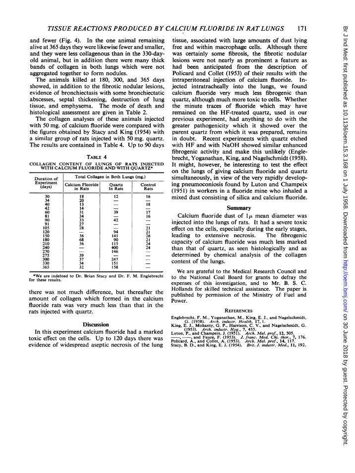
and fewer (Fig. 4). In the one animal remainingalive at 365 days they were likewise fewer and smaller,and they were less collagenous than in the 330-day-old animal, but in addition there were many thickbands of collagen in both lungs which were notaggregated together to form nodules.The animals killed at 180, 300, and 365 days
showed, in addition to the fibrotic nodular lesions,evidence of bronchiectasis with some bronchiectaticabscesses, septal thickening, destruction of lungtissue, and emphysema. The mode of death andhistological assessment are given in Table 2.The collagen analyses of these animals injected
with 50 mg. of calcium fluoride were compared withthe figures obtained by Stacy and King (1954) witha similar group of rats injected with 50 mg. quartz.The results are contained in Table 4. Up to 90 days
TABLE 4COLLAGEN CONTENT OF LUNGS OF RATS INJECTED
WITH CALCIUM FLUORIDE AND WITH QUARTZ*
Duration of Total Collagen in Both Lungs (mg.)Experiment Calcium Fluoride Quartz Control
(days) in Rats In Rats Rats
30 18 12 1634 20 - -
40 13 - 1842 14 - -60 31 39 1781 34 - 1690 33 42 -
91 27 - -
105 28 - 21120 - 94 21150 36 141 26180 68 90 21210 36 115 24240 - 400 24270 - 146 -
275 39 - -
300 37 247 -
330 34 151 -
365 32 158 -
*We are indebted to Dr. Brian Stacy and Dr. F. M. Englebrechtfor these results.
there was not much difference, but thereafter theamount of collagen which formed in the calciumfluoride rats was very much less than that in therats injected with quartz.
DiscussionIn this experiment calcium fluoride had a marked
toxic effect on the cells. Up to 120 days there wasevidence of widespread aseptic necrosis of the lung
tissue, associated with large amounts of dust lyingfree and within macrophage cells. Although therewas certainly some fibrosis, the fibrotic nodularlesions were not nearly as prominent a feature ashad been anticipated from the description ofPolicard and Collet (1953) of their results with theintraperitoneal injection of calcium fluoride. In-jected intratracheally into the lungs, we foundcalcium fluoride very much less fibrogenic thanquartz, although much more toxic to cells. Whetherthe minute traces of fluoride which may haveremained on the HF-treated quartz, used in ourprevious experiment, had anything to do with thegreater pathogenicity which it showed over theparent quartz from which it was prepared, remainsin doubt. Recent experiments with quartz etchedwith HF and with NaOH showed similar enhancedfibrogenic activity and make this unlikely (Engle-brecht, Yoganathan, King, and Nagelschmidt (1958).It might, however, be interesting to test the effecton the lungs of giving calcium fluoride and quartzsimultaneously, in view of the very rapidly develop-ing pneumoconiosis found by Luton and Champeix(1951) in workers in a fluoride mine who inhaled amixed dust consisting of silica and calcium fluoride.
SummaryCalcium fluoride dust of lut mean diameter was
injected into the lungs of rats. It had a severe toxiceffect on the cells, especially during the early stages,leading to extensive necrosis. The fibrogeniccapacity of calcium fluoride was much less markedthan that of quartz, as seen histologically and asdetermined by chemical analysis of the collagencontent of the lungs.
We are grateful to the Medical Research Council andto the National Coal Board for grants to defray theexpenses of this investigation, and to Mr. B. S. C.Hollands for skilled technical assistance. The paper ispublished by permission of the Ministry of Fuel andPower.
REFERENCESEnglebrecht, F. M., Yoganathan, M., King, E. J., and Nagelschmidt,
G. (1958). Arch. indusir. Health. 17, 1.King, E. J., Mohanty, G. P., Harrison, C. V., and Nagelschmidt, G.
(1953). Arch. industr. Hyg., 7, 455.Luton, P., and Champeix, J. (1951). Arch. Mal. prof., 12, 505.
, and Faure, F. (1953). J. franc. Med. Chi. thor., 7, 176.Policard, A., and Collet, A. (1953). Arch. Mal. prof., 14, 117.Stacy, B. D., and King, E. J. (1954). Brit. J. industr. Med., 11, 192.
171
on 30 June 2018 by guest. Protected by copyright.
http://oem.bm
j.com/
Br J Ind M
ed: first published as 10.1136/oem.15.3.168 on 1 July 1958. D
ownloaded from