Tissue culture of ornamental pot plant: A critical review...
30
Research review paper Tissue culture of ornamental pot plant: A critical review on present scenario and future prospects G.R. Rout a, ⁎ , A. Mohapatra a,1 , S. Mohan Jain b,2 a Plant Biotechnology Division, Regional Plant Resource Centre, Bhubaneswar-751015, India b International Atomic Energy Agency, FAO/IAEA Joint Division, Box-100, Vienna, Austria Received 18 October 2005; received in revised form 3 May 2006; accepted 14 May 2006 Available online 23 May 2006 Abstract Recent modern techniques of propagation have been developed which could help growers to meet the demand of the horticultural industry in the next century. An overview on the in vitro propagation via thin cell layer, meristem culture, regeneration via organogenesis and somatic embryogenesis is presented. Available methods for the transfer of genes could significantly simplify the breeding procedures and overcome some of the agronomic and environmental problems, which other wise would not be achievable through conventional propagation methods. The development and remarkable achievements with biotechnology in ornamental pot plants made during the three decades have been reviewed. The usefulness of the pot plants in commercial industry as well as propagation techniques, screening for various useful characteristics and selection of somaclonal variation is also discussed. © 2006 Elsevier Inc. All rights reserved. Keywords: Biotechnology; Genetic transformation; In vitro culture; Ornamental plants; Plant propagation Contents 1. Introduction ...................................................... 532 2. In vitro propagation .................................................. 533 2.1. Micropropagation via meristem culture or axillary bud/shoot tip culture .................... 533 2.2. Micropropagation via somatic embryogenesis.................................. 535 2.3. Micropropagation via thin cell layer ...................................... 537 2.4. Mechanization of in vitro plant propagation .................................. 538 3. Propagation of important pot plants .......................................... 540 3.1. Begonias .................................................... 540 3.2. Chrysanthemum ................................................. 541 3.3. Cyclamen .................................................... 542 3.4. Ficus spp..................................................... 542 Biotechnology Advances 24 (2006) 531 – 560 www.elsevier.com/locate/biotechadv ⁎ Corresponding author. Fax: +91 674 2550274. E-mail addresses: [email protected] (G.R. Rout), [email protected] (S.M. Jain). 1 Fax: +91 674 2550274. 2 Fax: +43 41 1 26007. 0734-9750/$ - see front matter © 2006 Elsevier Inc. All rights reserved. doi:10.1016/j.biotechadv.2006.05.001
Transcript of Tissue culture of ornamental pot plant: A critical review...
Biotechnology Advances 24 (2006) 531–560www.elsevier.com/locate/biotechadv
Research review paper
Tissue culture of ornamental pot plant: A critical reviewon present scenario and future prospects
G.R. Rout a,⁎, A. Mohapatra a,1, S. Mohan Jain b,2
a Plant Biotechnology Division, Regional Plant Resource Centre, Bhubaneswar-751015, Indiab International Atomic Energy Agency, FAO/IAEA Joint Division, Box-100, Vienna, Austria
Received 18 October 2005; received in revised form 3 May 2006; accepted 14 May 2006Available online 23 May 2006
Abstract
Recent modern techniques of propagation have been developed which could help growers to meet the demand of thehorticultural industry in the next century. An overview on the in vitro propagation via thin cell layer, meristem culture, regenerationvia organogenesis and somatic embryogenesis is presented. Available methods for the transfer of genes could significantly simplifythe breeding procedures and overcome some of the agronomic and environmental problems, which other wise would not beachievable through conventional propagation methods. The development and remarkable achievements with biotechnology inornamental pot plants made during the three decades have been reviewed. The usefulness of the pot plants in commercial industryas well as propagation techniques, screening for various useful characteristics and selection of somaclonal variation is alsodiscussed.© 2006 Elsevier Inc. All rights reserved.
Keywords: Biotechnology; Genetic transformation; In vitro culture; Ornamental plants; Plant propagation
Contents
1. Introduction . . . . . . . . . . . . . . . . . . . . . . . . . . . . . . . . . . . . . . . . . . . . . . . . . . . . . . 5322. In vitro propagation . . . . . . . . . . . . . . . . . . . . . . . . . . . . . . . . . . . . . . . . . . . . . . . . . . 533
2.1. Micropropagation via meristem culture or axillary bud/shoot tip culture . . . . . . . . . . . . . . . . . . . . 5332.2. Micropropagation via somatic embryogenesis. . . . . . . . . . . . . . . . . . . . . . . . . . . . . . . . . . 5352.3. Micropropagation via thin cell layer . . . . . . . . . . . . . . . . . . . . . . . . . . . . . . . . . . . . . . 5372.4. Mechanization of in vitro plant propagation . . . . . . . . . . . . . . . . . . . . . . . . . . . . . . . . . . 538
3. Propagation of important pot plants . . . . . . . . . . . . . . . . . . . . . . . . . . . . . . . . . . . . . . . . . . 5403.1. Begonias . . . . . . . . . . . . . . . . . . . . . . . . . . . . . . . . . . . . . . . . . . . . . . . . . . . . 5403.2. Chrysanthemum . . . . . . . . . . . . . . . . . . . . . . . . . . . . . . . . . . . . . . . . . . . . . . . . . 5413.3. Cyclamen . . . . . . . . . . . . . . . . . . . . . . . . . . . . . . . . . . . . . . . . . . . . . . . . . . . . 5423.4. Ficus spp. . . . . . . . . . . . . . . . . . . . . . . . . . . . . . . . . . . . . . . . . . . . . . . . . . . . . 542
⁎ Corresponding author. Fax: +91 674 2550274.E-mail addresses: [email protected] (G.R. Rout), [email protected] (S.M. Jain).
1 Fax: +91 674 2550274.2 Fax: +43 41 1 26007.
0734-9750/$ - see front matter © 2006 Elsevier Inc. All rights reserved.doi:10.1016/j.biotechadv.2006.05.001
Nomenclature
BA 6-benzylaminopurine2ip 6-(γ,γ-dimethylallylamine)purine2,4-D 2,4-dichlophenoxyacetic acidKn kinetinIAA indole-3-acetic acidIBA indole-3-butyric acidNAA 1-naphthaleneacetic acidMS Murashige and Skoog (1962)mediummediumPVP polyvinylpyrrolidoneTDZ thidiazuronTCL thin cell layer
532 G.R. Rout et al. / Biotechnology Advances 24 (2006) 531–560
3.5. Rose . . . . . . . . . . . . . . . . . . . . . . . . . . . . . . . . . . . . . . . . . . . . . . . . . . . . . . 5433.6. Saintpaulia . . . . . . . . . . . . . . . . . . . . . . . . . . . . . . . . . . . . . . . . . . . . . . . . . . . 5443.7. Yucca. . . . . . . . . . . . . . . . . . . . . . . . . . . . . . . . . . . . . . . . . . . . . . . . . . . . . . 546
4. Germplasm conservation . . . . . . . . . . . . . . . . . . . . . . . . . . . . . . . . . . . . . . . . . . . . . . . 5464.1. Clonal stability through in vitro culture . . . . . . . . . . . . . . . . . . . . . . . . . . . . . . . . . . . . 5464.2. Determination of genetic fidelity. . . . . . . . . . . . . . . . . . . . . . . . . . . . . . . . . . . . . . . . 547
5. Applications of in vitro propagation . . . . . . . . . . . . . . . . . . . . . . . . . . . . . . . . . . . . . . . . . 5475.1. In vitro mutagenesis . . . . . . . . . . . . . . . . . . . . . . . . . . . . . . . . . . . . . . . . . . . . . . 5475.2. Somaclonal variation. . . . . . . . . . . . . . . . . . . . . . . . . . . . . . . . . . . . . . . . . . . . . . 5485.3. Cryopreservation . . . . . . . . . . . . . . . . . . . . . . . . . . . . . . . . . . . . . . . . . . . . . . . . 5495.4. Genetic transformation . . . . . . . . . . . . . . . . . . . . . . . . . . . . . . . . . . . . . . . . . . . . . 549
6. Conclusion . . . . . . . . . . . . . . . . . . . . . . . . . . . . . . . . . . . . . . . . . . . . . . . . . . . . . . 551Acknowledgement . . . . . . . . . . . . . . . . . . . . . . . . . . . . . . . . . . . . . . . . . . . . . . . . . . . . . 551References . . . . . . . . . . . . . . . . . . . . . . . . . . . . . . . . . . . . . . . . . . . . . . . . . . . . . . . . . 552
1. Introduction
The commercial production of ornamental plants isgrowing worldwide. Its monetary value has significant-ly increased over the last two decades and there is agreat potential for continued further growth in bothdomestic and international markets (Jain, 2002). Majorpot plants such as Begonia, Ficus, Anthurium, Chry-santhemum, Rosa, Saintpaulia, and Spathiphyllum arebeing produced in the developed countries (Anony-mous, 2003). About 212.5 million plants including157 million ornamental plants amounting to 78% of thetotal production were reported (Pierik, 1991a,b). TheNetherlands dominates export of ornamental plantsincluding pot plants like Begonia, Ficus, Cyclamen,Philodendron, Saintpaulia, Spathiphyllum and Rhodo-dendron (O'Riordain, 1999; Anonymous, 2003). About156 ornamental genera are propagated through tissueculture in different commercial laboratories worldwide.
The shares of major producers are The Netherlands(33%), Japan (24%), Italy (11%), USA (12%), Thailand(10%) and others (14%). The major exporting countriesare The Netherlands (59%), Colombia (10%), Italy(16%), Israel (4%), Spain (2%), Kenya (1%) and others(18%). The four leading exporters (The Netherlands,Colombia, Italy and Israel) constitute about 80% of theworld market. The share of the developing countries ofAfrica, Asia and Latin America is less than 20%(Rajagopalan, 2000; Schiva, 2000) Planting material ofornamental plants is in great demand for commercialproduction as well as for domestic gardens andlandscaping. The better quality planting material is abasic need of growers for boosting productivity. Chebetet al. (2003) reported the use of biotechnologicalapproaches to improve horticultural crop production.The present review emphasizes the application ofbiotechnology on in vitro manipulation and propagationof ornamental pot plants.
533G.R. Rout et al. / Biotechnology Advances 24 (2006) 531–560
2. In vitro propagation
In vitro culture is one of the key tools of plantbiotechnology that exploits the totipotency nature of plantcells, a concept proposed by Haberlandt (1902) andunequivocally demonstrated, for the first time, by Stewardet al. (1958). Tissue culture is alternatively called cell,tissue and organ culture through in vitro condition(Debergh and Read, 1991). It can be employed forlarge-scale propagation of disease free clones and genepool conservation. Ornamental industry has appliedimmensely in vitro propagation approach for large-scaleplant multiplication of elite superior varieties. As a result,hundreds of plant tissue culture laboratories have come upworldwide, especially in the developing countries due tocheap labour costs. However, micropropagation technol-ogy is more costly than conventional propagationmethods, and unit cost per plant becomes unaffordablecompelling to adopt strategies to cut down the productioncost for lowering the cost per plant (IAEA-TECDOC-1384, 2004).
2.1. Micropropagation via meristem culture or axillarybud/shoot tip culture
In vitro propagation through meristem culture is thebest possible means of virus elimination and produces alarge numbers of plants in a short span of time. It is apowerful tool for large-scale propagation of horticulturalcrops including pot plants. The term ‘meristem culture’specifically means that a meristem with no leafprimordia or at most 1–2 leaf primordial which areexcised and cultured. The pathway of regenerationundergoes several steps. Starting with an isolatedexplant, with de-differentiation followed by re-differen-tiation and organization into meristematic centres. Uponfurther induction the cells can form unipolar structuresi.e. organogenesis, or bipolar structures called somaticembryogenesis. The organization into morphogeneticpatterns can take place directly on the isolated explant orcan be expressed only after callus formation, which iscalled indirect morphogenesis. When shoots are devel-oped directly from leaf or stem explants it refers to directmorphogenesis. Micropropagation is an alternativemethod of vegetative propagation, which is well suitedfor the multiplication of elite clones. It is accomplishedby several means, i.e., multiplication of shoots fromdifferent explants such as shoot tips or axillary buds ordirect formation of adventitious shoots or somaticembryos from tissues, organs or zygotic embryos. Thefirst significant use of plant tissue culture in ornamentalwas made during 1920s when orchid seeds were
germinated under laboratory conditions (Knudson,1922). Micropropagation generally involves four dis-tinct stages: initiation of cultures, shoot multiplication,rooting of in vitro grown shoots, and acclimatization.The first stage: culture initiation depends on explanttype or the physiological stage of the donor plant at thetime of excision. Explants from actively growing shootsare generally used for mass scale multiplication. Thesecond stage: shoot multiplication is crucial andachieved by using Plant Growth Regulators i.e. auxinand cytokinin. The third stage: the elongated shoots,derived from the multiplication stage, are subsequentlyrooted either ex vitro or in vitro. In some cases, thehighest root induction occurs from excised shoots in theliquid medium when compared with semi-solid medi-um. The fourth stage: acclimatization of in vitro grownplants is an important step in micropropagation.
In vitro plants are exposed to invariably controlledgrowth conditions such as high amount of organic andinorganic nutrients, Plant Growth Regulators, carbonsource, high humidity, low light and poor gaseousexchange. Although they may support rapid growth andmultiplication, the controlled conditions induce struc-tural and physiological changes in plants rendering themunfit to survive when transferred directly to the field.Thus, a gradual acclimatization from laboratory to fieldcondition is necessary. The plants are gradually shiftedfrom high humidity/low irradiance conditions to lowhumidity/high irradiance conditions, enabling them tosurvive under ‘adverse’ climatic conditions. Carbondioxide enrichment in the greenhouse for the cultivationof ornamental plants has a positive impact on produc-tion. Increased CO2 concentration also lessens waterstress of microcuttings by closing the stomata asreported by Matysiak and Nowak (1995). In CO2
enriched atmosphere (1200 μl/l) of the greenhouse andthe highest level of electrical conductivity(EC=2.8 mS cm) of the medium produced the bestgrowth of gerbera microcuttings taken from in vitroplants (Matysiak and Nowak, 2001). Photoautotropicmicropropagation of ornamental plants have beenreviewed (Kozai et al., 1988; Kozai, 1990a,b), and issuggested to use for reducing production costs, andautomation to use robots for micropropagation process(Kozai et al., 1988; Kozai, 1991a,b).
Many commercial ornamental plants are beingpropagated by in vitro culture on the culture mediumcontaining auxins and cytokinins (Preil, 2003; Rout andJain, 2004). Several different explants have been used fordirect shoot formation. Mayer (1956) succeeded first timeregeneration of Cyclamen shoots from tuber segments onMS medium supplemented with 10.7 μM NAA.
534 G.R. Rout et al. / Biotechnology Advances 24 (2006) 531–560
Furthermore, plants have been regenerated from leaftissues and petiole segments of Cyclamen (Geier, 1977;Geier et al., 1983; Schwenkel, 1991; Dillen et al., 1996),Heuchera sanguinea (Hosoki and Kajino, 2003), andBegonia (Takayama, 1983). In vitro clonal propagation ofDracaena deremensis has been reported by severalgroups (Debergh, 1975, 1976; Miller and Murashige,1976; Chua et al., 1981). The first report on shootmultiplication and rooting of rose (Rosa multiflora) wasmade by Elliott (1970) by using shoot tip explants andlater on followed by others (Hasegawa, 1979; Skirvin andChu, 1979; Rout et al., 1989). AboEl-Nil (1983) reviewedon the large-scale production of Pelargonium by usingdifferent explants. Atta-Alla et al. (1998) reported theshoot bud regeneration from leaf and petiole explants ofAnthurium parvispathum and subsequently establishmentin soil. Martin et al. (2003) succeeded in direct shoot budregeneration from lamina explants ofAnthurium andraea-num onMSmedium fortified with 1.11 μMBA, 1.14 μMIAA and 0.46 μM Kn. Furthermore, the regeneratedshoots were rooted on half-strength MS mediumsupplemented with 0.54 μM NAA and 0.93 μM Kn.Nearly 300 plantlets of each cultivar were transferred tosoil with 95% survival rate (Joseph et al., 2003). Thao etal. (2003) achieved shoots regenerated from petiole-derived callus of Alocasia micholitziana “Green velvel”onMSmedium fortified with 0.5 μMKn and 0.5 μM2,4-D. The regenerated shoots were rooted on hormone freeMS medium and subsequently established in the field.Skirvin et al. (1990) reported rapid method of shootmultiplication and rooting ofRosa hybrida cultivars. Nowshoot tip explant is being routinely used for themicropropagation of ornamental plants including Rhodo-dendron (Ettinger and Preece, 1985; McCown and Lloyd,1983; Brand and Kiyamoto, 1994a,b), Zantedeschiaalbomaculata (Chang et al., 2003) and Ebenus cretica(Hatzilazarou et al., 2001). Later on Brand and Kiyomoto(1997) accomplished shoot multiplication of Rhododen-dron “Montego” on woody plant medium (Lloyd andMcCown, 1980) supplemented with 10–50 μM 2ip. Thenumber of shoots increased with subsequent subcultureson the fresh culture medium. Micro-shoots were rooted inmoist sphagnum moss and vermiculite (3 :1 ratio), and90% microshoots survived and grown in the greenhouse.Several researchers have reported on clonal propagationof Spathiphyllum (Fonnesbech and Fonnesbech, 1979;Orlikowska et al., 1995; Wated et al., 1997).
Cytokinin alone in the culture medium induces shootformation in many plants. MS medium supplementedwith 3.0 mg/l BA was suitable for micropropagation ofFicus benjamina vars. Natasja and Starlight (Rzepka-Plevnes and Kurek, 2001). Jain (1997) micropropagated
Saintpaulia ionantha by culturing leaf disks on MSmedium containing 0.22–0.50 μM BA. Addition ofauxins with cytokinins becomes essential for shootinduction and multiplication depending on the planttype. In Petunia hybrida, mass shoot multiplication wasachieved on MS medium amended with 2.2 μMBA and5.7 μM IAA within 4 weeks of culture (Sharma andMitra, 1976). High concentration of cytokinins isunsuitable for shoot formation from leaf or petioleexplants in some ornamental pot plants. Takayama andMisawa (1981, 1982) used 1.3 μM BA or 4.6 μM Kn incombination with 5.4 μM NAA for shoot budregeneration from leaf, petiole or inflorescence seg-ments of Begonia species. Further, low concentration ofcytokinins also influences high rate of shoot budregeneration (Reuter and Bhandari, 1981; Bigot,1981a,b; Roest et al., 1981; Mikkelson and Sink,1978a,b; Welander, 1977, 1979, 1981; Simmonds,1984; Appelgren, 1976, 1985). The addition of 1–2 g/l activated charcoal in the culture medium increased therooting efficiency from excised shoots of Begonia×hiemalis (Bigot, 1981a,b). Activated charcoal seems toadsorb Plant Growth Regulators, prompting to betterresponse for rooting and even for shoot formation. Itseems Begonia has high endogenous cytokinin andauxins, and by adding activated charcoal in the mediumcertainly promotes organogenesis. Jain (1997) used twocytokinins (Kn and zeatin) for regeneration of plantletsof Begonia×elatior. He suggested that two cytokininsdid not affect the basic plant characteristics includingflower colour. Similarly, many reports indicated massmultiplication of Ficus species by adding cytokinins inthe culture medium (Gabryszewska and Rudnicki, 1997;Debergh and DeWael, 1997; Nobre and Romano, 1998).Demiralay et al. (1998) achieved shoot multiplication ofFicus carica var. Bursa Siyaki on MS medium contain-ing 1.0 mg/l BA and 89 mg/l phloroglucinol; shootmultiplication rate was 4.43 shoots/explant; and 68.33%rooting rate of the micropropagated shoots on rootingmedium containing 1 mg/l IBA. The rooting efficiencyenhanced by addition of 0.05% PVP in the culturemedium containing 2.5 μM IBA (Nobre and Romano,1998). The addition of PVP helps in oxidisingpolyphenols leached in the medium, and promoteshigh rate of organogenesis.
The quality of light also influences shoot induction.Gabryszewska and Rudnicki (1997) developed amicropropagation protocol for F. benjamina by usingshoot meristems; shoot numbers increased on MSmedium supplemented with 15 mg/l 2ip by red lighttreatment; and root initiation occurred in all lighttreatments (white, blue, green and red). However, the
535G.R. Rout et al. / Biotechnology Advances 24 (2006) 531–560
rooting and number of roots/shoot were highest in redlight on the medium having 0.5 mg/l IAA.
Liquid medium seems to be more effective for shootregeneration and root induction, which is due to betteraeration. Simmonds and Werry (1987) used liquidmedium for enhancing the micropropagation profile ofBegonia×hiemalis. Wated et al. (1997) comparedperformance of agar-solidified medium and interfacialmembrane rafts floating on liquid medium for shootmultiplication and root induction. The results showedshoot multiplication was highest on membrane raftsfloating on the liquid medium, and also plants rootedmuch better. Similarly, Osternack et al. (1999) suc-ceeded in inducing somatic embryogenesis and adven-titious shoots and roots from hypocotyl tissues ofEuphorbia pulcherrima on cytokinin containing medi-um. Subsequently, Preil (2003) noted that the regener-ation potential of isolated cells, tissue or organs and thecallus cultures is highly variable. Furthermore, petiolecross sections cultivated on auxin and cytokinincontaining medium give rise to adventitious shootsfrom epidermal cells and subepidermal cortex cells,never from pith cells of the central regions of the petiole.
The direct shoot bud formation without any callusphase from appropriate explants is of great success forlarge-scale clonal multiplication of desired clone allround the year to boost the commercial floriculture. Themicropropagation of major ornamental pot plants arepresented in Table 1.
2.2. Micropropagation via somatic embryogenesis
Somatic embryos, which are bipolar structures, arisefrom individual cells and have no vascular connectionwith the maternal tissue of the explant (Haccius, 1978).Embryos may develop directly from somatic cells(direct embryogenesis) or development of recognizableembryogenic structures is preceded by numerous,organized, non-embryogenic mitotic cycles (indirectembryogenesis). Somatic embryogenesis has a greatpotential for clonal multiplication. Under controlledenvironmental conditions, somatic embryos germinatereadily, similar to their seedling counterpart. Thecommercial application of somatic embryogenesis willbe accomplished only when the germination rate ofsomatic embryos is high up to 80–85%.
Considerable success has been achieved in inducingsomatic embryogenesis in ornamental pot plants likechrysanthemum (Dendrathema grandif lorum) (Mayand Trigiano, 1991; Tanaka et al., 2000), Cyclamenpersicum (Wicart et al., 1984; Pueschel et al., 2003),rose (R. hybrida) (Rout et al., 1991, Kim et al., 2003a),
Begonia gracilis (Castillo and Smith, 1997), S. ionanthacv. Benjamin (Murch et al., 2003), and E. pulcherrima(Osternack et al., 1999). In chrysanthemum, somaticembryos were produced from leaf mid-rib explants onmodified MS medium supplemented with 1.0 mg/l 2,4-D and 0.2 mg/l BA (May and Trigiano, 1991). Highestsomatic embryos were produced on the mediumcontaining 6–8% sucrose and kept in the darkness forfirst 28 days, followed by 10 days in the light. Twelvecultivars produced somatic embryos, but completeplantlets were recovered from only five cultivars.Castillo and Smith (1997) induced direct somaticembryogenesis from petiole and leaf blade explants ofB. gracilis on MS medium supplemented with 0.5 mg/l kinetin and 2% (v/v) coconut water. Somatic embryoswere obtained with greater frequency from petioleexplants than from leaf blade sections. Osternack et al.(1999) succeeded in achieving somatic embryos fromhypocotyl tissues of E. pulcherrima on MS mediumsupplemented with 2.0 mg/l IAA (Fig. 1). About1400 embryos were developed from 320 calli derivedfrom outer regions of the hypocotyls. However, only 8%developed normal plantlets. In most cases, shoots wererooted in hormone free medium. Both orientation of thepetiole explants and auxin transport system are crucialfactors for the induction of somatic embryogenesis ofSaintpaulia (Murch et al., 2003), and TDZ helped in thedevelopment of somatic embryos. Winkelmann et al.(1998) used cell suspension culture of Cyclamen forrapid development of somatic embryos, and later onfollowed by Hohe et al. (2001), who developed a large-scale propagation system of Cyclamen from embryo-genic cell suspension cultures. Bouman et al. (2001)reported that the efficiency of embryogenic callus ofCyclamen seems to be stable for more than 5 years;however, suspension cultures can lose embryogenicpotential after a number of subcultures. Therefore, it isnecessary to determine the number of subcultures beforeembryogenic cell suspensions lose their potential ofembryogenic nature. Pueschel et al. (2003) succeeded inplant regeneration via somatic embryogenesis of C.persicum and maintained the regeneration ability forprolonged period.
There are advantages and disadvantages of somaticembryogenesis in large-scale plant multiplication (Jain,2002). The major advantages are large-scale somaticembryo production in bioreactors, encapsulation, cryo-preservation, genetic transformation and clonal propa-gation. The major limitations are genotypic dependenceof somatic embryo production and poor germinationrate. Somatic embryogenesis in major ornamental potplants is presented in Table 2.
Table 1Micropropagation of major ornamental pot plants
Species/Cultivars Response References
Alocasia micholitziana ‘Green Velvet’ sbr, r Thao et al. (2003)Anthurium andraeanum sbr, r, pt Pierik et al., 1974; Pierik, 1976Anthurium patulum ads, r Eapen and Rao (1985)Anthurium scherzerianum (flamingo flower) ms, r, pt Liu and Xu (1992)Anthurium spp. ms, r, pt Matsumoto and Kuehnle (1997)Anthurium parvispathum ms, r, pt Atta-Alla et al. (1998)Anthurium andraeanum cvs. Tinora Red, Senator ads, r, pt Martin et al. (2003)Begonia×elatior cvs. Aphrodite Rose, Aphrodite Rose Pale, Nixe, Schwabenland Orange, Tacora ads, r, pt Bigot, 1981a,bBegonia×elatior cvs. Aphrodite Rosa, Claudis Mayer, Mayers Rote, Mayers Rosa ms, r, pt Reuter and Bhandari (1981)Begonia× elatior cvs. Krefeld Orange, Schwabenland Orange, Schwabenland Pink,Schwabenland Red
sbr, r, pt Takayama and Misawa (1982)
Begonia×hiemalis cv. Schwabenland Red sbr, r, pt Simmonds (1984)Begonia tuberhybrida sbr, r Peak and Cumming (1984)Begonia×elatior ms, r, pt Jain (1997)Dendranthema grandiflora cvs. Blue Bird, Montana, Meladion, Delaware ms, r, pt Wang and Ma (1978)Dendranthema grandiflora cv. Super Yellow ads, r, pt Lazar and Cachita (1983)Dendranthema hortorum cvs. Pink Camino, Super Yellow, Spider sbr, r, pt Gertsson and Andersson (1985)Dendranthema grandiflora cvs. Winter westland, Yellow westland, Dark westland, Snowdon,Yellow Snowdon, Altis, Blanche
ms, r, pt Ahmed (1986)
Dendranthema grandiflora ms, r, pt Kaul et al. (1990)Chrysanthemum coccineum sbr, r, pt Fujii and Shimzu (1990)Dendranthema grandiflora cv. Royal Purple sbr, r, pt Lu et al. (1990)Dendranthema grandiflora ads, r, pt Bhattacharya et al. (1990)Dendranthema maximum ads, r, pt Kumar and Kumar (1995)Dendranthema grandiflora cv. Deep Pink sbr, r, pt Rout et al. (1996)Dendranthema grandiflora ms, r, pt Mandal et al. (2000)Dendranthema grandiflora sbr, r, pt Teixeira de Silva and Fukai (2003b)Cyclamen persicum ads, r, pt Geier, 1977, 1978Cyclamen persicum ads, r, pt Ando and Murasaki (1983)Cyclamen persicum ads, r, pt Wainwright and Harwood (1985)Cyclamen persicum ads, r, pt Hawkes and Wainwright (1987)Dracaena deremensis cv. Warneckii ms, r Debergh (1975)Dracaena marginata Tricolour ms, r, pt Chua et al. (1981)Euphorbia pulcherrima ads, r, pt Langhe et al. (1974)Euphorbia fulgans ms, r, pt Zhang et al. (1987)Euphorbia pulcherrima cv. Angelika ads, r, pt Osternack et al. (1999)Ficus lyrata ms, r Debergh and DeWael (1997)Ficus religiosa sbr, r Narayan and Jaiswal (1986)Ficus benjamina cv. Golden King ms, r Gabryszewska and Rudnicki (1997)Ficus carica var. Bursa siyahi ms, r, pt Demiralay et al. (1998)Ficus carica cvs. Berbera, Lampa ms, r, pt Nobre and Romano (1998)Ficus religiosa ms, r, pt Deshpande et al. (1998)Ficus religiosa ms, r, pt Nagaraju et al. (1998)Ficus carica cv. Gular ms, r, pt Kumar et al. (1998)Ficus benjamina cvs. Natasja, Starlight ms, r, pt Rzepka-Plevnes and Kurek (2001)Petunia hybrida, Petunia inflata sbr, r, pt Rao et al. (1973)Petunia hybrida ms, r, pt Sharma and Mitra (1976)Pelargonium×hortorum ms, r Horst et al. (1976)Pelargonium spp. ms, r, pt Debergh and Maene (1977)Pelargonium spp. ms, r, pt Theiler (1977)Pelargonium zonale hybrid ms, r, pt Jelaska and Jelencic (1980)Rhododendron spp. ms, r Economou and Read (1984)Rhododendron spp. ms, r, pt Anderson (1984)Rhododendron spp. ms, r, pt Norton and Norton (1985)Rhododendron P.J.M. hybrid ms, r Ettinger and Preece (1985)Rhododendron ‘Montego’ ms, r, pt Brand and Kiyomoto (1997)Rosa hybrida cvs. Crimson Glory, Glenfiditch ms, r, pt Barve et al. (1984)Rosa hybrida cv. Amanda ms, r DeVries and Dubois (1988)
536 G.R. Rout et al. / Biotechnology Advances 24 (2006) 531–560
Table 1 (continued)
Species/Cultivars Response References
Rosa hybrida cv. Bridal Pink ads, r Burger et al. (1990)Rosa damascena ads, r, pt Ishiooka and Tanimoto (1990)Rosa hybrida cvs. Landora, Virgo, Happiness, Sea Pearl, Super Star, Queen-Elizabeth ms, r, pt Rout et al. (1990)Rosa hybrida cv. Landora ads, r, pt Rout et al. (1992)Rosa chinensis var. minima cvs. Debut, Ginny ms, r, pt Rogers and Smith (1992)Rosa chinensis var. minima (cvs. Baby Katie, Lavender Jewel, Red Sunblaze, Royal Sunblaze) ms, r, pt Chu et al. (1993)Hybrid tea ‘Dr. Verhage’ ms, r, pt Voyiatzi et al. (1995)Rosa multiflora ads, r, pt Rosu et al. (1995)Rosa hybrida ads, r, pt Van der Salm et al. (1996)Hybrid tea rose cv. Peace ms, r, pt Ara et al. (1997)Saintpaulia ionantha sbr, r, pt Starts and Cummings, 1976;
Grunewaldt,1977; Vazquez et al., 1977
Saintpaulia ionantha ms, r, pt Molgaard et al. (1991)Saintpaulia ionantha sbr, r, pt Hoshino et al. (1995)Saintpaulia ionantha 2 confusa hybrids sbr, r, pt Lo et al. (1997)Saintpaulia ionantha ms, r, pt Jain (1997)Saintpaulia ionantha cvs. Benjamin, William sbr, r, pt Mithila et al. (2003)Spathiphyllum cv. Clevelandii ms, r, pt Fonnesbech and Fonnesbech (1979)Spathiphyllum sbr, r, pt Orlikowska et al. (1995)Spathiphyllum floribundum cv. Petite Sbr, r Werbrouck and Debergh (1995)Spathiphyllum ‘Petite’ ms, r, pt Wated et al. (1997)Yucca aloifolia ms, r, pt Atta-Alla and Van Staden (1997)
Abbreviation: ads=adventitious shoot bud development, ms=multiple shoot, pt=plantlet formation, r=rooting, sbr=shoot bud regeneration.
537G.R. Rout et al. / Biotechnology Advances 24 (2006) 531–560
2.3. Micropropagation via thin cell layer
Thin cell layer (TCL) is a simple but effective systemthat relies on a small size explant derived from a limitedcell number of homogenous tissue. They are excisedlongitudinally or transversely from different organsranging from floral parts to root/rhizome of plants.Longitudinal TCL (lTCL) (0.5–1 mm wide and 5–10 mm long) is used when a definite cell type (epidermal,sub-epidermal, cortical, cambial or medullar cell) is to beanalysed. TCLs can be excised from stem, leaf, vein, floralstalk, petiole, pedicel, bulb-scale, etc.As for the transverseTCL (tTCL) (0.1–5 mm), other organs (leaf blade, root/rhizome, floral organs, meristems, stem node, etc.) can beused. The reduced cell number in TCL is important for thedevelopmental process or the morphogenetic programme,which can be altered by making changes in organ/tissueand size to be uniformly exposed to the medium (TranThanh Van, 1980).
Thin cell layer is the model systems and findapplications in higher plant tissue and organ culture andgenetic transformation (Teixeira da Silva, 2003a, 2005).Moreover, thin cell layer technology is a solution to manyof the issues currently hindering the efficient progress ofornamental and floricultural crop improvement, since itsolves the initial step i.e. plant regeneration problem. Thistechnology has also been effectively used in the micro-
propagation of various crops including floricultural crops(Tran Thanh Van and Bui, 2000; Fiore et al., 2002; Nhut etal., 2003a,b; Teixeira de Silva and Nhut, 2003a). Recently,Teixeira da Silva (2003a) published a detailed review onthe use of thin cell layer technology in ornamental plantmicropropagation and biotechnology, which highlightsorganogenesis and somatic embryogenesis for plantregeneration and genetic improvement via transformation.Mulin and Tran Thanh Van (1989) indicated that in vitroshoots and flowers were formed from thin epidermal cellsexcised from the first five internodes of basal floweringbranches inP. hybrida. Explants (1×10mm2) consisting of3–6 layers of subepidermal and epidermal cells producedvegetative buds within 2 weeks of culture. Ohki (1994)reported that 100–200 shoots per tTCL (transverse thin celllayer) explants were obtained from 0.3 to 0.5mmpetiole or3×3 mm2 lamina sections, respectively of S. ionanthawithin 4 weeks of culture. Over 70,000 plants wereproduced from a single leaf within 3–4 months. Gill et al.(1992) used tTCL hypocotyl explants (10 mm) of 1-week-old geranium (Pelargonium×hortorum) hybrid seedlingsfor induction of somatic embryogenesis. They observedthat the development of somatic embryoswas rapid and thenumber of embryos was about 8-fold higher than theculture of whole hypocotyl explants. Hsia and Korban(1996) achieved organogenic and embryogenic callus andsubsequent regeneration from lTCL (longitudinally thin
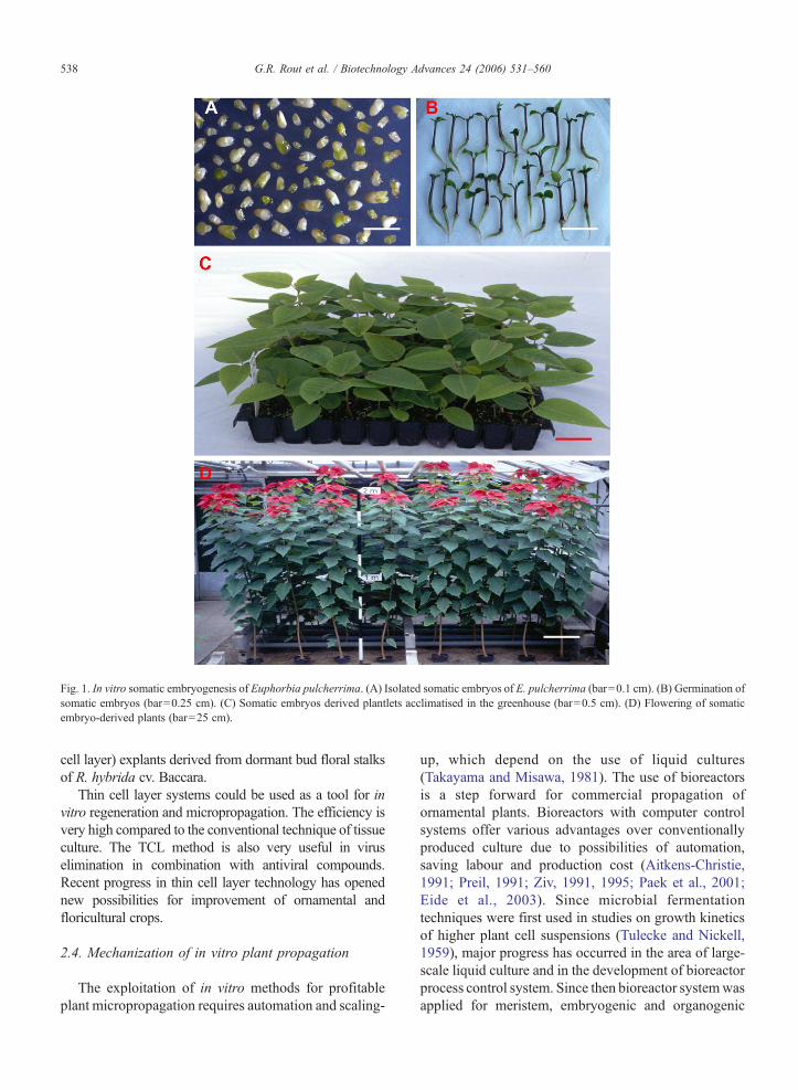
Fig. 1. In vitro somatic embryogenesis of Euphorbia pulcherrima. (A) Isolated somatic embryos of E. pulcherrima (bar=0.1 cm). (B) Germination ofsomatic embryos (bar=0.25 cm). (C) Somatic embryos derived plantlets acclimatised in the greenhouse (bar=0.5 cm). (D) Flowering of somaticembryo-derived plants (bar=25 cm).
538 G.R. Rout et al. / Biotechnology Advances 24 (2006) 531–560
cell layer) explants derived from dormant bud floral stalksof R. hybrida cv. Baccara.
Thin cell layer systems could be used as a tool for invitro regeneration and micropropagation. The efficiency isvery high compared to the conventional technique of tissueculture. The TCL method is also very useful in viruselimination in combination with antiviral compounds.Recent progress in thin cell layer technology has openednew possibilities for improvement of ornamental andfloricultural crops.
2.4. Mechanization of in vitro plant propagation
The exploitation of in vitro methods for profitableplant micropropagation requires automation and scaling-
up, which depend on the use of liquid cultures(Takayama and Misawa, 1981). The use of bioreactorsis a step forward for commercial propagation ofornamental plants. Bioreactors with computer controlsystems offer various advantages over conventionallyproduced culture due to possibilities of automation,saving labour and production cost (Aitkens-Christie,1991; Preil, 1991; Ziv, 1991, 1995; Paek et al., 2001;Eide et al., 2003). Since microbial fermentationtechniques were first used in studies on growth kineticsof higher plant cell suspensions (Tulecke and Nickell,1959), major progress has occurred in the area of large-scale liquid culture and in the development of bioreactorprocess control system. Since then bioreactor systemwasapplied for meristem, embryogenic and organogenic
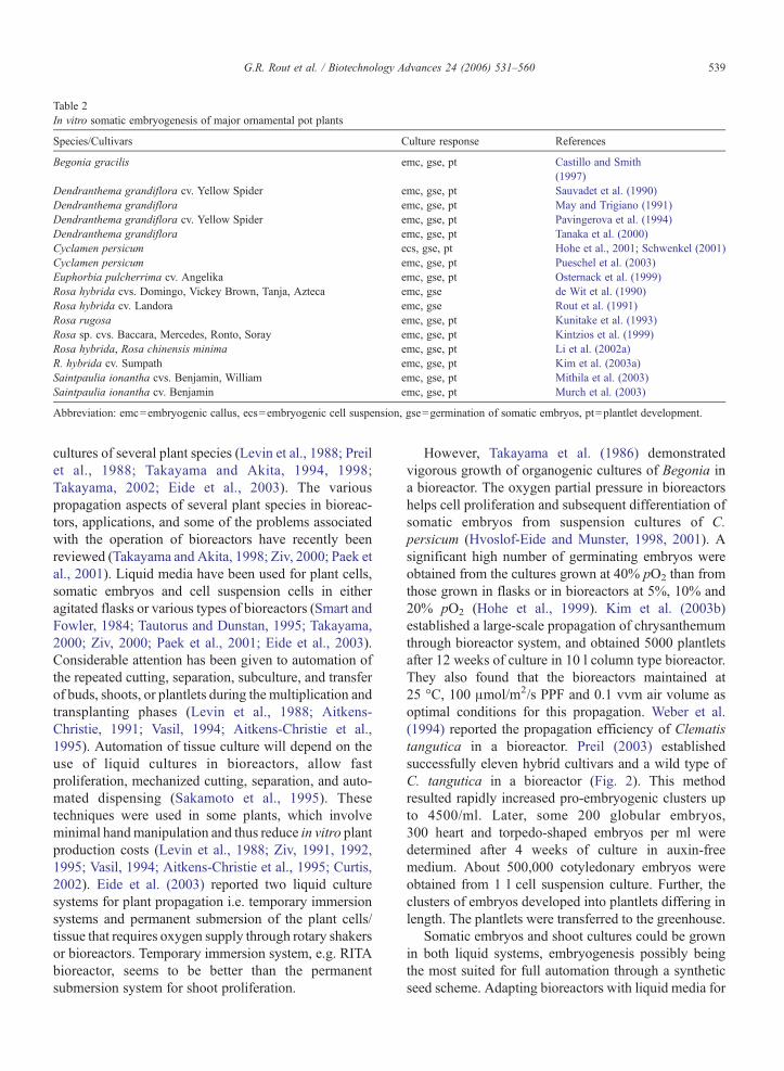
Table 2In vitro somatic embryogenesis of major ornamental pot plants
Species/Cultivars Culture response References
Begonia gracilis emc, gse, pt Castillo and Smith(1997)
Dendranthema grandiflora cv. Yellow Spider emc, gse, pt Sauvadet et al. (1990)Dendranthema grandiflora emc, gse, pt May and Trigiano (1991)Dendranthema grandiflora cv. Yellow Spider emc, gse, pt Pavingerova et al. (1994)Dendranthema grandiflora emc, gse, pt Tanaka et al. (2000)Cyclamen persicum ecs, gse, pt Hohe et al., 2001; Schwenkel (2001)Cyclamen persicum emc, gse, pt Pueschel et al. (2003)Euphorbia pulcherrima cv. Angelika emc, gse, pt Osternack et al. (1999)Rosa hybrida cvs. Domingo, Vickey Brown, Tanja, Azteca emc, gse de Wit et al. (1990)Rosa hybrida cv. Landora emc, gse Rout et al. (1991)Rosa rugosa emc, gse, pt Kunitake et al. (1993)Rosa sp. cvs. Baccara, Mercedes, Ronto, Soray emc, gse, pt Kintzios et al. (1999)Rosa hybrida, Rosa chinensis minima emc, gse, pt Li et al. (2002a)R. hybrida cv. Sumpath emc, gse, pt Kim et al. (2003a)Saintpaulia ionantha cvs. Benjamin, William emc, gse, pt Mithila et al. (2003)Saintpaulia ionantha cv. Benjamin emc, gse, pt Murch et al. (2003)
Abbreviation: emc=embryogenic callus, ecs=embryogenic cell suspension, gse=germination of somatic embryos, pt=plantlet development.
539G.R. Rout et al. / Biotechnology Advances 24 (2006) 531–560
cultures of several plant species (Levin et al., 1988; Preilet al., 1988; Takayama and Akita, 1994, 1998;Takayama, 2002; Eide et al., 2003). The variouspropagation aspects of several plant species in bioreac-tors, applications, and some of the problems associatedwith the operation of bioreactors have recently beenreviewed (Takayama and Akita, 1998; Ziv, 2000; Paek etal., 2001). Liquid media have been used for plant cells,somatic embryos and cell suspension cells in eitheragitated flasks or various types of bioreactors (Smart andFowler, 1984; Tautorus and Dunstan, 1995; Takayama,2000; Ziv, 2000; Paek et al., 2001; Eide et al., 2003).Considerable attention has been given to automation ofthe repeated cutting, separation, subculture, and transferof buds, shoots, or plantlets during the multiplication andtransplanting phases (Levin et al., 1988; Aitkens-Christie, 1991; Vasil, 1994; Aitkens-Christie et al.,1995). Automation of tissue culture will depend on theuse of liquid cultures in bioreactors, allow fastproliferation, mechanized cutting, separation, and auto-mated dispensing (Sakamoto et al., 1995). Thesetechniques were used in some plants, which involveminimal handmanipulation and thus reduce in vitro plantproduction costs (Levin et al., 1988; Ziv, 1991, 1992,1995; Vasil, 1994; Aitkens-Christie et al., 1995; Curtis,2002). Eide et al. (2003) reported two liquid culturesystems for plant propagation i.e. temporary immersionsystems and permanent submersion of the plant cells/tissue that requires oxygen supply through rotary shakersor bioreactors. Temporary immersion system, e.g. RITAbioreactor, seems to be better than the permanentsubmersion system for shoot proliferation.
However, Takayama et al. (1986) demonstratedvigorous growth of organogenic cultures of Begonia ina bioreactor. The oxygen partial pressure in bioreactorshelps cell proliferation and subsequent differentiation ofsomatic embryos from suspension cultures of C.persicum (Hvoslof-Eide and Munster, 1998, 2001). Asignificant high number of germinating embryos wereobtained from the cultures grown at 40% pO2 than fromthose grown in flasks or in bioreactors at 5%, 10% and20% pO2 (Hohe et al., 1999). Kim et al. (2003b)established a large-scale propagation of chrysanthemumthrough bioreactor system, and obtained 5000 plantletsafter 12 weeks of culture in 10 l column type bioreactor.They also found that the bioreactors maintained at25 °C, 100 μmol/m2/s PPF and 0.1 vvm air volume asoptimal conditions for this propagation. Weber et al.(1994) reported the propagation efficiency of Clematistangutica in a bioreactor. Preil (2003) establishedsuccessfully eleven hybrid cultivars and a wild type ofC. tangutica in a bioreactor (Fig. 2). This methodresulted rapidly increased pro-embryogenic clusters upto 4500/ml. Later, some 200 globular embryos,300 heart and torpedo-shaped embryos per ml weredetermined after 4 weeks of culture in auxin-freemedium. About 500,000 cotyledonary embryos wereobtained from 1 l cell suspension culture. Further, theclusters of embryos developed into plantlets differing inlength. The plantlets were transferred to the greenhouse.
Somatic embryos and shoot cultures could be grownin both liquid systems, embryogenesis possibly beingthe most suited for full automation through a syntheticseed scheme. Adapting bioreactors with liquid media for
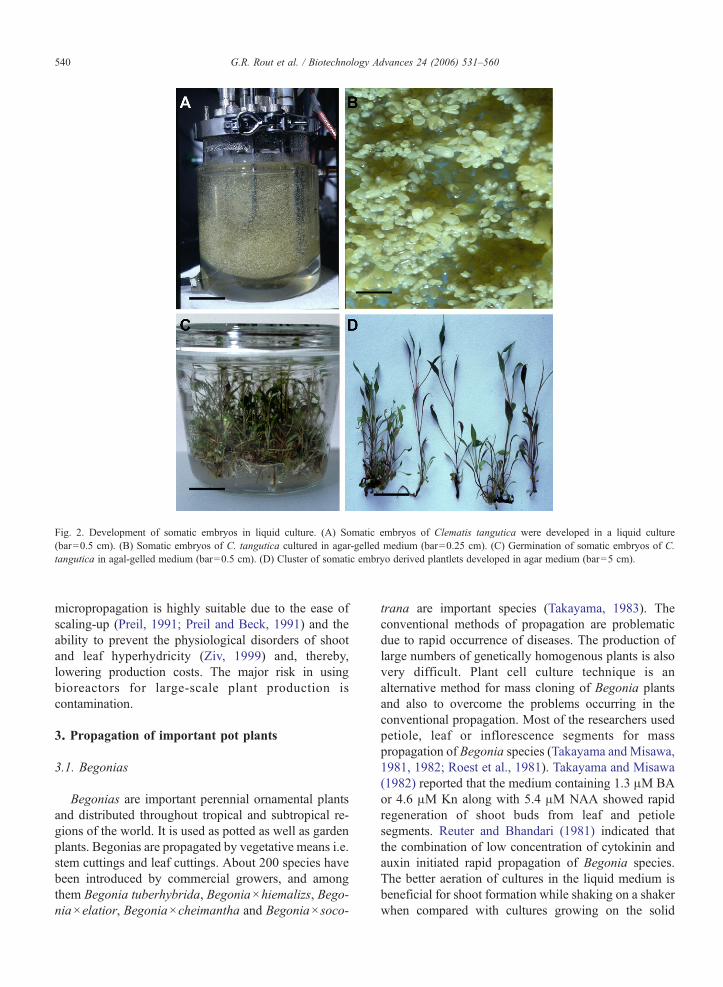
Fig. 2. Development of somatic embryos in liquid culture. (A) Somatic embryos of Clematis tangutica were developed in a liquid culture(bar=0.5 cm). (B) Somatic embryos of C. tangutica cultured in agar-gelled medium (bar=0.25 cm). (C) Germination of somatic embryos of C.tangutica in agal-gelled medium (bar=0.5 cm). (D) Cluster of somatic embryo derived plantlets developed in agar medium (bar=5 cm).
540 G.R. Rout et al. / Biotechnology Advances 24 (2006) 531–560
micropropagation is highly suitable due to the ease ofscaling-up (Preil, 1991; Preil and Beck, 1991) and theability to prevent the physiological disorders of shootand leaf hyperhydricity (Ziv, 1999) and, thereby,lowering production costs. The major risk in usingbioreactors for large-scale plant production iscontamination.
3. Propagation of important pot plants
3.1. Begonias
Begonias are important perennial ornamental plantsand distributed throughout tropical and subtropical re-gions of the world. It is used as potted as well as gardenplants. Begonias are propagated by vegetative means i.e.stem cuttings and leaf cuttings. About 200 species havebeen introduced by commercial growers, and amongthem Begonia tuberhybrida, Begonia×hiemalizs, Bego-nia×elatior, Begonia×cheimantha and Begonia× soco-
trana are important species (Takayama, 1983). Theconventional methods of propagation are problematicdue to rapid occurrence of diseases. The production oflarge numbers of genetically homogenous plants is alsovery difficult. Plant cell culture technique is analternative method for mass cloning of Begonia plantsand also to overcome the problems occurring in theconventional propagation. Most of the researchers usedpetiole, leaf or inflorescence segments for masspropagation of Begonia species (Takayama and Misawa,1981, 1982; Roest et al., 1981). Takayama and Misawa(1982) reported that the medium containing 1.3 μM BAor 4.6 μM Kn along with 5.4 μM NAA showed rapidregeneration of shoot buds from leaf and petiolesegments. Reuter and Bhandari (1981) indicated thatthe combination of low concentration of cytokinin andauxin initiated rapid propagation of Begonia species.The better aeration of cultures in the liquid medium isbeneficial for shoot formation while shaking on a shakerwhen compared with cultures growing on the solid
541G.R. Rout et al. / Biotechnology Advances 24 (2006) 531–560
medium. Takayama and Misawa (1982) developed aliquid culture system with shaking which helped thebuds to develop efficiently and quickly into plantlets.Also, reduction of growth hormones in the culturemedium benefits shoot culture, which is done by addingactivated charcoal in the culture medium. Bigot (1981a,b) reported that the addition of 1–2 g/l activatedcharcoal in the culture medium showed vigorous rootingfrom excised shoots of Begonias. Plant regenerationfrom leaf disk callus of Begonia×elatior was achievedon MS medium supplemented with 5.0 μM Kn and0.5 μM zeatin (Jain, 1997). He also reported that about84% of callus cultures showed shoot bud regenerationand rooting in vitro. Castillo and Smith (1997) reportedthe direct somatic embryogenesis in B. gracilis by usingmicro-cultured laminar segments and petioles. The rateof somatic embryogenesis induction was greater frompetiole explants than from leaf blade sections on MSmedium supplemented with 0.5 mg/l Kn and 2% (v/v)coconut water. The production of somatic embryos wassignificantly higher on responding laminar explants(60–70 embryos/leaf section) than on petioles (40–50embryos/petioles). Subsequently, somatic embryos weregerminated into plantlets (Castillo and Smith, 1997) andtransferred to the field.
3.2. Chrysanthemum
Chrysanthemum (Dendranthema grandiflora syn.Chrysanthemum morifolium Ramat.) is extensivelygrown as a pot plant as well as a cut flower worldwide.It is vegetatively propagated with cuttings and suckers.Breeding programmes have focussed on improvingvarious characteristics to enhance ornamental values,including flower colour, size and form, and productionquality. Although desirable traits have been introducedby classical breeding, there are limitations to thistechnique. Firstly, there is a limited gene pool. Secondly,distant crosses may be limited by incompatibility ordifferences in ploidy level between mutant parents.Thirdly, characteristics such as uniform growth andsynchronous flowering are polygenic. Hence sexualcrossing may alter the delicate balance of factorsdetermining plant growth and development. Plantbiotechnology offers an opportunity to develop newgermplasms and conservation. The techniques ofstimulating axillary branching or culturing nodal sec-tions in vitro are probably most commonly used inmicropropagation (Lawrence, 1981). A number offactors have influenced the induction of morphogenesisin chrysanthemum. Rout and Das (1997) have reviewedat length the recent developments of chrysanthemum
biotechnology. They emphasized on the application ofin vitro culture for mass-scale propagation and alsodiscussed the various possibilities for improvement ofchrysanthemum by using modern biotechnologicaltools. Recently, Teixeira da Silva (2003b) published adetailed review on tissue culture of chrysanthemum,which highlights organogenesis, thin cell layer, andsomatic embryogenesis for plant regeneration.
Prasad et al. (1983) reported that the rate of shootmultiplication is genotypic dependent in D. grandif lora.Datta et al. (2001) established a protocol using directshoot regeneration system from ray florets of 28genotypes. The regeneration frequency and averagenumber of shoots per explant varied among the cultivars.Shoot tip size also plays an important role in shootregeneration efficiency. Wang and Ma (1978) reportedthat shoot tip between 0.2 and 0.5 mm and shootmeristems between 0.1 and 0.2 mm diameter producedonly a single shoot. Larger explant (0.5–1.55 mmdiameter) formed multiple shoots. Mandal et al. (2000)used various explants for regeneration of D. grandifloraand regenerated new plants from mutated tissues. Liquidmedium has also proven beneficial in root induction inseveral plants, especially in some recalcitrant plants forrooting, due to better aeration of cultures. Roest andBokelmann (1975) successfully induced roots in theadventitious shoots of chrysanthemum in the liquid MSmedium containing 1.0 mg/l IAA. In general, shoots androots developed on a single medium containing 4.4 μMBA and 5.7 μM IAA. Rooting was achieved in 90%cultures of ‘Deep Pink’ rooted with about 2.0 klx (kilo-lux) of light, whereas higher light intensities (3.0 klx) gavea lower rooting percentage (Roberts et al., 1992; Rout etal., 1996). The rooted plants were successfully establishedin the soil (Rout et al., 1996; Roberts and Smith, 1990).Kim et al. (2003b) reported the propagation system andreduction of transplant production period. They reportedthat 5000 cuttings were obtained after 12 weeks of culturein 10-l column type bioreactor and subsequentlytransferred to the greenhouse with 100% survival.Belarmino and Gabon (1999) induced rapid multiplica-tion ofD. grandiflora onMSmedium supplemented with1.0 mg/l BA, 2.0 mg/l NAA and 10 mg/l gibberellic acid.Kumari et al. (2001) used cytokinins and auxins to scaleup the multiplication efficiency of chrysanthemum.Hosokawa et al. (2004) developed a new method toregenerate chrysanthemum plants from leaf primordia-free shoot apical meristem domes (LP-free SAMs) byestablishing the meristem dome on the cut surface of roottips of chrysanthemum or different plant species from theCompositae (cabbage). The highest shoot regenerationrate was observed with cabbage root tips.
542 G.R. Rout et al. / Biotechnology Advances 24 (2006) 531–560
Induction of somatic embryogenesis in chrysanthe-mum has been achieved by using leaf mid-rib explants(May and Trigiano, 1991), which depended on thephotoperiod and sucrose concentration. The highestnumber of somatic embryos was produced on themedium containing 9–18% sucrose, in the darkness forfirst 28 days of culture, followed by 10 days in the light.Twelve of the 23 cultivars evaluated produced somaticembryos, but complete plantlets were recovered onlyfrom five cultivars. The regenerated plants werephenotypically similar to parent plants in growth habit,leaf morphology and flower colour. Pavingerova et al.(1994) reported somatic embryogenesis and plantregeneration from transform calli of D. grandif lora.Tanaka et al. (2000) achieved the induction of somaticembryogenesis and plant regeneration in chrysanthe-mum from ray-floret explants by using IAA and kinetin.The somatic embryo derived plantlets were establishedin the greenhouse. The genotypic dependence remainsthe major limitation on the use of somatic embryogen-esis in chrysanthemum.
3.3. Cyclamen
Cyclamen belongs to the family Primulaceae, and isgrown as a pot plant. It is widely growing in Europe andvery popular in Germany. In addition to pot plants, tubersare produced as planting material and have commercialimportance. It is distributed in the Mediterranean regionand areas adjoining to the North and to the East.Cyclamen is propagated exclusively through seeds. It iscross-pollinated, and many cultivars are autotetraploid.Since repeated self-fertilization leads to inbreedingdepression, the traditional cultivars are maintained bycrossing selected plants of similar appearance. As aresult, uniformity is poor. Hence, in vitro clonalpropagation of Cyclamen has been widely studied asan alternate method for mass scale production of highquality planting material. In vitro clonal propagation ofCyclamen has been very well worked out. Mayer (1956)first used tuber segments on MS medium supplementedwith 1.1 μM NAA for shoot formation. Subsequently,Okumoto and Takabayashi (1969) and Pierik (1975)achieved shoot bud regeneration from tuber explants.Geier (1977) obtained shoot and root formation on themedium containing 14.3–28.6 μM IAA and 0.9–2.3 μMKn. He also compared types of explant on plantregeneration and observed less morphogenetic potentialin other plant parts as compared to tuber tissue. In vitrocloning of C. persicum through organogenesis has beenreported by different researchers (Geier, 1978; Geier etal., 1983; Schwenkel, 1991; Dillen et al., 1996).
Hoffmann and Preil (1987) established shoot budregeneration protocol in 13 genotypes of Cyclamen andsubsequently rooting. Similar genotypic-specific differ-ences in shoot bud regeneration from peduncle explantswere observed by Schwenkel and Grunewaldt (1988).Winkelmann et al. (1998) produced 90,000 plantlets from1 l of embryogenic cell suspension culture. Subsequently,Hohe et al. (2001) and Schwenkel (2001) also reportedclonal propagation of C. persicum by using embryogeniccell suspension culture. Pueschel et al. (2003) highlightedthe mass-scale propagation of C. persicum via somaticembryogenesis.
3.4. Ficus spp.
Genus Ficus has more than 800 species, and are usedas foliage plants including Ficus altissima, Ficusbenjamina, Ficus binnedijkii, Ficus elastica, Ficusmicrocarpa, Ficus pumila, Ficus retusa and Ficusrubiginosa. Ficus is one of the most popular indoorplants. It is native to India, Southeast Asia and NorthernAustralia. It is propagated either by air layering orrooting by stem cutting. Some varieties have appealingaesthetic appearances, and their performance is of highquality under interior low light conditions (Chen et al.,2001). The propagation however, is slow and limited.Hence, in vitro micropropagation of Ficus species hasbeen widely studied as an alternate method for mass-scale production of high quality planting material.Debergh and DeWael (1997) reported micropropagationof Ficus lyrata. Subsequently, Dijkshoorn-Dekker(1996) studied the influence of light and temperatureon propagation profile of F. benjamina. Propagation ofdifferent Ficus species by using shoot tips or axillarybud explants has been reported (Deshpande et al., 1998;Kumar et al., 1998; Demiralay et al., 1998; Nobre andRomano, 1998; Nagaraju et al., 1998). Deshpande et al.(1998) induced multiple shoots from nodal explants of35-year-old tree of Ficus religiosa on MS mediumsupplemented with 5.0 mg/l BA and 0.2 mg/l IBA, andobtained multiple shoots as well as rooting on MSmedium containing 1.5 mg/l BA and 1.5 mg/l Ads and1/2 MS plus 2.0 mg/l IBA and 1.0 mg/l NAA,respectively. Kumar et al. (1998) established micro-propagation protocol for F. carica cv. Gular by usingapical buds from 8-year-old trees, and succeeded ingetting multiple shoots and rooting in the liquid half-strength MS medium supplemented with 2.0 mg/l IAAand 0.2% activated charcoal. The micropropagatedplantlets were successfully established (68%) in soil.Rzepka-Plevnes and Kurek (2001) regenerated multipleshoots from nodal explants of F. benjamina on MS
543G.R. Rout et al. / Biotechnology Advances 24 (2006) 531–560
medium supplemented with 3.0 mg/l BA. The plantletsgrown in the medium with cytokinins were generallyshorter and developed shorter leaves as compared to thegrowth medium without cytokinin.
3.5. Rose
Rose is the most important cut flower as well as potplant. Roses attribute to great variation in flower andplant characteristics and to their wide adaptability tovaried agro-ecological conditions. The genetic resourcesof roses can be grouped into four categories: exoticvarieties, indigenously evolved varieties, native rosespecies and exotic species. Being an important com-mercial flower plant, systematic investigations havebeen carried out for its propagation and improvement inproduction both in quality and in quantity during the lastthree decades. Budding or grafting is done for thepropagation of roses. The breeding programmes arefocused on the improvement of various characteristics toenhance the ornamental value, including the flowercolour, size and keeping quality of the bloom and theresponse to various diseases. Although desirable traitswere introduced by conventional breeding, there werelimitations to this technique; firstly, because of thelimited gene pool, secondly, distant crosses were limitedby incompatibility or differences in ploidy levelbetween putative parents and thirdly, characteristicssuch as uniform growth and synchronous floweringwere polygenic. Plant tissue culture offers an opportu-nity to propagate roses in large scale.
In vitro mass multiplication of rose is successful bymicropropagation (Skirvin and Chu, 1979; Hasegawa,1979; Rout et al., 1989, 1990; Bressan et al., 1982;Arnold et al., 1995) and several reviews have beenwritten (Skirvin et al., 1990; Short and Roberts, 1991;Horn, 1992; Rout et al., 1999; Pati et al., 2006). Theyhave highlighted the role of growth regulators andphysical factors on shoot multiplication and rooting ofthe different cultivars of hybrid roses and also illustratedthe application of modern technology on improvement,conservation and documentation of roses. Skirvin andChu (1979) and Hasegawa (1979) reported a rapidmethod for shoot multiplication and rooting of hybridrose cultivars. Khosh-Khui and Sink (1982a,b) observedthe rate of shoot multiplication of R. hybrida, Rosadamascena and Rosa canina varied significantly duringdifferent subculture periods. By reducing the sucroseconcentration in the culture medium, the number ofmultiple shoots increased (Langford and Wainwright,1987). Similarly, size of the meristem (both shoot tipand nodal explant) of floribunda and miniature roses had
significant effect on shoot multiplication; on an average2.5–5.0 shoots were obtained per culture cycle,dependent on cultivars (Douglas et al., 1989). Thegrowth and multiplication of shoots increased byextending the culture period from 3 to 6 weeks (Chuet al., 1993). There are several factors affecting rosemicropropagation, which are: agar concentration(Ghashghaie et al., 1991), ethylene concentration(Kevers et al., 1992), growth room and vessel humidity(Sallanon and Maziere, 1992) and different types ofgelling agents (Podwyszynska and Olszewski, 1995).Kumar et al. (2001) developed an efficient protocol formicropropagation of R. damascena on MS mediumsupplemented with 1.0–2.5 μM TDZ. Pre-culturesoaking in thidiazuron improved the axillary shootproliferation in rose (Singh and Syamal, 2000). Carelliand Echeverrigaray (2002) developed an efficientprotocol for propagation of hybrid roses by using MSmedium amended with 3.0 mg/l BA and 0.5 mg/l NAA.The multiplication rate was 30.3 plantlets per explantafter 180 days. The addition of silver nitrate along withBA and IAA promoted the growth of the axillary shoots(Chakrabarty et al., 2000). The microshoots were rootedon growth medium supplemented with low concentra-tions of auxins (0.1 to 0.5 mg/l) and reduced concentra-tions of sucrose (2–2.5%) (Khosh-Khui and Sink,1982b,c; Rout et al., 1990; Arnold et al., 1995).
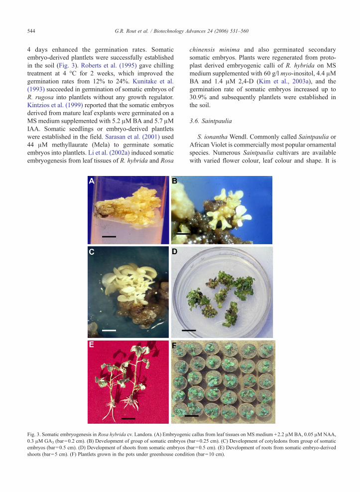
Somatic embryogenesis in rose has been successfullyaccomplished by using leaf, internode, filament ofstamen, root and zygotic embryo (Rout et al., 1991;Roberts et al., 1995; de Wit et al., 1990; Kunitake et al.,1993). Rout et al. (1991) induced embryogenic calli andlater on developed somatic embryos from 8-week-oldcallus, derived from immature leaf and stem segments ofR. hybrida acv. Landora. Medium amended with 2,4-Dhelped in the long-term maintenance of embryogeniccallus (Roberts et al., 1990; Matthews et al., 1991;Noriega and Sondahl, 1991). Kunitake et al. (1993)observed zygotic embryo-derived calli of Rosa rugosahas ability to develop somatic embryos on mediawithout exogenous growth regulators although embryo-genic potential did not persist after 6 months. Hsia andKorban (1996) reported low frequency rate of somaticembryogenesis from the rhizogenic callus of the cutrose. Noriega and Sondahl (1991) and Roberts et al.(1990) added ABA (abscisic acid) and GA3 in theculture medium for the germination of somatic embryos.By adding L-proline in the primary culture mediumfollowed by its removal from the regeneration medium,stimulated embryo development and reduced abnormal-ities (Rout et al., 1991). Furthermore, low temperatureexposure (8 °C) exposure to embryogenic calli for
544 G.R. Rout et al. / Biotechnology Advances 24 (2006) 531–560
4 days enhanced the germination rates. Somaticembryo-derived plantlets were successfully establishedin the soil (Fig. 3). Roberts et al. (1995) gave chillingtreatment at 4 °C for 2 weeks, which improved thegermination rates from 12% to 24%. Kunitake et al.(1993) succeeded in germination of somatic embryos ofR. rugosa into plantlets without any growth regulator.Kintzios et al. (1999) reported that the somatic embryosderived from mature leaf explants were germinated on aMS medium supplemented with 5.2 μMBA and 5.7 μMIAA. Somatic seedlings or embryo-derived plantletswere established in the field. Sarasan et al. (2001) used44 μM methyllaurate (Mela) to germinate somaticembryos into plantlets. Li et al. (2002a) induced somaticembryogenesis from leaf tissues of R. hybrida and Rosa
Fig. 3. Somatic embryogenesis in Rosa hybrida cv. Landora. (A) Embryogeni0.3 μM GA3 (bar=0.2 cm). (B) Development of group of somatic embryos (embryos (bar=0.5 cm). (D) Development of shoots from somatic embryos (shoots (bar=5 cm). (F) Plantlets grown in the pots under greenhouse condit
chinensis minima and also germinated secondarysomatic embryos. Plants were regenerated from proto-plast derived embryogenic calli of R. hybrida on MSmedium supplemented with 60 g/l myo-inositol, 4.4 μMBA and 1.4 μM 2,4-D (Kim et al., 2003a), and thegermination rate of somatic embryos increased up to30.9% and subsequently plantlets were established inthe soil.
3.6. Saintpaulia
S. ionantha Wendl. Commonly called Saintpaulia orAfrican Violet is commercially most popular ornamentalspecies. Numerous Saintpaulia cultivars are availablewith varied flower colour, leaf colour and shape. It is
c callus from leaf tissues on MS medium +2.2 μMBA, 0.05 μMNAA,bar=0.25 cm). (C) Development of cotyledons from group of somaticbar=0.5 cm). (E) Development of roots from somatic embryo-derivedion (bar=10 cm).
545G.R. Rout et al. / Biotechnology Advances 24 (2006) 531–560
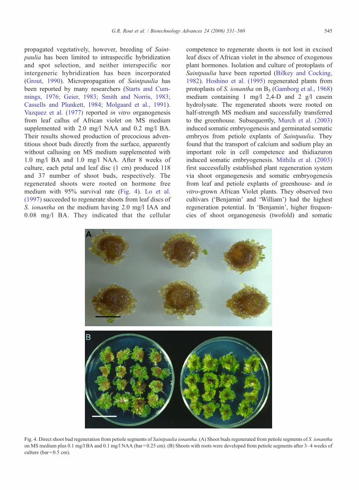
propagated vegetatively, however, breeding of Saint-paulia has been limited to intraspecific hybridizationand spot selection, and neither interspecific norintergeneric hybridization has been incorporated(Grout, 1990). Micropropagation of Saintpaulia hasbeen reported by many researchers (Starts and Cum-mings, 1976; Geier, 1983; Smith and Norris, 1983;Cassells and Plunkett, 1984; Molgaard et al., 1991).Vazquez et al. (1977) reported in vitro organogenesisfrom leaf callus of African violet on MS mediumsupplemented with 2.0 mg/l NAA and 0.2 mg/l BA.Their results showed production of precocious adven-titious shoot buds directly from the surface, apparentlywithout callusing on MS medium supplemented with1.0 mg/l BA and 1.0 mg/l NAA. After 8 weeks ofculture, each petal and leaf disc (1 cm) produced 118and 37 number of shoot buds, respectively. Theregenerated shoots were rooted on hormone freemedium with 95% survival rate (Fig. 4). Lo et al.(1997) succeeded to regenerate shoots from leaf discs ofS. ionantha on the medium having 2.0 mg/l IAA and0.08 mg/l BA. They indicated that the cellular
Fig. 4. Direct shoot bud regeneration from petiole segments of Saintpaulia ionon MS medium plus 0.1 mg/l BA and 0.1 mg/l NAA (bar=0.25 cm). (B) Shooculture (bar=0.5 cm).
competence to regenerate shoots is not lost in excisedleaf discs of African violet in the absence of exogenousplant hormones. Isolation and culture of protoplasts ofSaintpaulia have been reported (Bilkey and Cocking,1982). Hoshino et al. (1995) regenerated plants fromprotoplasts of S. ionantha on B5 (Gamborg et al., 1968)medium containing 1 mg/l 2,4-D and 2 g/l caseinhydrolysate. The regenerated shoots were rooted onhalf-strength MS medium and successfully transferredto the greenhouse. Subsequently, Murch et al. (2003)induced somatic embryogenesis and germinated somaticembryos from petiole explants of Saintpaulia. Theyfound that the transport of calcium and sodium play animportant role in cell competence and thidiazuroninduced somatic embryogenesis. Mithila et al. (2003)first successfully established plant regeneration systemvia shoot organogenesis and somatic embryogenesisfrom leaf and petiole explants of greenhouse- and invitro-grown African Violet plants. They observed twocultivars (‘Benjamin’ and ‘William’) had the highestregeneration potential. In ‘Benjamin’, higher frequen-cies of shoot organogenesis (twofold) and somatic
antha. (A) Shoot buds regenerated from petiole segments of S. ionanthats with roots were developed from petiole segments after 3–4 weeks of
546 G.R. Rout et al. / Biotechnology Advances 24 (2006) 531–560
embryogenesis (a 50% increase) were observed from invitro and greenhouse-grown plants. At a lower concen-tration of TDZ (2.5 μM), shoots organogenesis wasinduced, whereas at higher doses (5–10 μM) able toinduce somatic embryogenesis.
3.7. Yucca
Yucca, an important commercial ornamental potplant, has 42 species, and is native of North America.It has also great variety of uses. Many Yucca species areused as raw material for synthesizing steroidal com-pounds, such as cortisone and sex hormones (Romo deVivar, 1985). Yucca species are generally propagated byseeds, offsets and rhizome cuttings. It is a very slowgrowing plant and does not blossom every year. In vitroculture is an alternative technique to propagate on alarge-scale. Stohs et al. (1974) developed callus and cellsuspension from sprouts of seeds of Yucca glauca onMS medium, supplemented with 0.53 μM 2,4-D.Further, Meskhi et al. (1978) produced callus fromflowers of Yucca gloriosa on MS medium supplementedwith 2.6–5.3 μM 2,4-D. The callus was produced fromcoleoptile and leaf segments of Yucca filifera (Quinteroet al., 1982, 1987). Khanna and Purohit (1983)developed callus tissue from Yucca oloefolia leaves onMS medium amended with 5.3 μM 2,4-D. Eight-week-old callus showed an index growth of 1.7. The plantregeneration from callus tissues is not yet reported.Quintero (1983) reviewed the in vitro culture of Yuccaand synthesis of secondary compounds from callus andcell suspension culture. Subsequently, Atta-Alla andVan Staden (1997) succeeded to propagate Yuccaaloifolia by using shoot tip explants and the maximumnumber shoot production (6.6) was obtained from asingle shoot tip on MS medium supplemented with4.5 μM TDZ and 1.1 μM NAA. The proliferated shootsreadily rooted on half-strength MS medium containing2.5–4.9 μM IBA and 1% charcoal. The rooted plantswere successfully established in soil. In Yucca, shootregeneration is genotypic dependent, and still requiresrefinement of culture medium for increasing shootproduction. Temporary immersion system could be usedfor shoot and root production.
4. Germplasm conservation
4.1. Clonal stability through in vitro culture
Clonal stability of the micropropagated plants isessential for in vitro germplasm conservation. Manyresearchers reported plants derived from meristems were
more stable than the adventitious shoots derived fromcallus. Somaclonal variation was more common amongadventitious-shoot-derived plants in many ornamentalcrops like Chrysanthemum and Begonia (Skirvin, 1978;Bouman et al., 1995). Skirvin and Janick (1976) wereamong the first to emphasize the importance of clonalvariation in genotype improvement of horticulturalspecies. Subsequently, Thorpe and Harry (1997)emphasized that in vitro culture techniques have playedon important role in the breeding, production andimprovement of horticultural crops. Various types ofchanges were reported in cell cultures at phenotypes,karyotypic, physiological, biochemical and molecularlevel. Larkin and Scowcroft (1981) reviewed extensive-ly and reported the phenotypic variation among plantsregenerated after a passage through tissue and cellculture. Hasegawa (1980) found one ‘abnormal looking’plant amongst 600 tissue-culture-propagated plants ofhybrid rose. Martin et al. (1981) observed no variationamong 2125 rose plants raised in the field for 3 years.The lack of somaclonal variability suggests that rose isrelatively stable when propagated via axillary buds.Lloyd et al. (1988) reported that callus-derived shoots ofRosa persica×xanthina exhibited considerable degreeof variation in leaf shape. Malaure et al. (1991) observedshoots derived from ray florets of 16 cultivars ofchrysanthemum showed more variation than plantsregenerated from vegetative parts. However, in vitroselection and somaclonal variation are random process-es and yet have to be used to achieve specific goals inchrysanthemum improvement.
Somatic hybrids resulting from protoplast fusion alsoshow variation in morphology, cytology, fertility andothers. Ploidy level of parent is very important insomatic cell fusion work. Izhar and Tabib (1980)showed that the regenerated plants derived from leafmesophyll protoplasts of Petunia were diploid(2n=2x=14). Further, Izhar et al. (1983) observed thatover 1000 fertile somatic hybrids, derived from a fusionproduct of Petunia parodii and P. hybrida, weretetraploids. However, when protoplasts, isolated fromcell suspension cultures, were used as one of the fusionparents, the somatic hybrid plants were of a higherploidy level (2n=28) (Clark et al., 1986).
The selection of explant, age of the culture, genotype,culture conditions and method of plant regeneration arevery important features for genetic stability of theregenerated plants. Since most of somatic embryosoriginate from single cells, somaclonal variation amongregenerated plants can be minimised. Of course, there isalways a limit of number of subcultures before plantsshowing variation.
547G.R. Rout et al. / Biotechnology Advances 24 (2006) 531–560
4.2. Determination of genetic fidelity
The molecular markers have facilitated research ongenetic variation at the DNA level. The numerouspotential applications of DNA fingerprinting havebrought about their uses in plants such as in populationgenetics, parentage testing, and individual genotypeidentification and for shortening breeding programs(Ben-Meir et al., 1997). EST (Expressed Sequence Tags)database development, proteomics and expressionprofiling can be used to create unique database resourcesto identify genes that determine the quality (colour,flavour, phytonutrients) (Dandekar, 2003). He reportedthat ESTs represent closely related gene families couldbe used to define their function and to detect singlenucleotide polymorphisms (SNPs). Markers such asrestriction fragment length polymorphism (RFLPs) haverecently been used for molecular characterization oftissue culture-derived plants. Since its development,polymerase chain reaction (PCR) has revolutionizedmany standard molecular techniques, with modificationsof the original procedure designed to suit a number ofneeds. Random amplified polymorphic DNA (RAPD),arbitrarily primed PCR (AP-PCR), DNA amplificationfingerprinting (DAF), inter-simple sequence repeat(ISSR), sequence-tagged sites (STSs) and amplifiedfragment length polymorphism (AFLP) and many othersgenerate special classes of markers which are highlysensitive for genetic analysis of tissue culture-raisedplants (Rani and Raina, 2000). RAPD markers have alsobeen used to identify cultivars, to map importantagricultural traits and to construct genetic maps(Williams et al., 1990). For varietal identification,molecular markers have been useful especially RFLP,and amplified fragment length polymorphism (AFLP)(Rajapakse et al., 1992). Bouman et al. (1992) noticedRAPD polymorphism among micropropagated plants ofBegonia species. Debener and Mathiesch (1996) dem-onstrated RAPD markers for the construction of achromosome linkage map, using crosses between Rosamultif lora derived genotypes that differed in a range offloral and vegetative characters. Furthermore, Debener etal. (1997) used RAPD markers for parentage analysis ininterspecific crosses between different wild rose species.Vainstein et al. (1995) demonstrated that the probabilityof two offsprings from the crossing of similar rosegenotypes having identical DNA fingerprints is verylow. Ben-Meir et al. (1997) screened the hybrid rose withseven different horticultural traits through the RAPDmarker. Huang et al. (2000) studied the genetic analysisof chrysanthemum hybrids with RAPD markers andclassified them into seven types, i.e., markers shared
bands in both parents and offspring, in male and femaleparents, in male parent and offspring, in female parentand offspring, in the male parent only, in the femaleparent only, and markers were present in offspring only.Onlymale parent and offspringmarkers were suitable foridentifying the true male parent. Their results concludedthat there were no definite rules as to whether markers inoffspring were more similar to female or to male parentsby similarity analysis. Recently, Dandekar (2003)mentioned that the rapid identification of cultivar/progeny could be detect by using micro-arrays andsingle nucleotide polymorphisms (SNPs).
5. Applications of in vitro propagation
5.1. In vitro mutagenesis
Most of the available genetic variation used inbreeding programs has occurred naturally and exists ingermplasm collections of new and old cultivars, landrace and genotypes. This variation through crosses isrecombined to produce new and desired genes combina-tions (Maluszynski et al., 1995). Then existing germ-plasm fails to provide the desired recombinants, and it isnecessary to resort to other resources of variation. Sincespontaneous mutations occur with extremely lowfrequency, mutation induction techniques provide toolsfor the rapid creation and increase in variability in cropspecies. The impact of mutation techniques on cropimprovement has already been evaluated (www.iaea.org; Broertjes and Van Harten, 1978, 1988; Micke,1999). In vitro culture methods has facilitated the use ofmutation techniques for improvement of both seed andvegetatively propagated plants (Jain and Maluszynski,in press). In many vegetatively propagated cropsmutation induction in combination with in vitro culturetechniques may be the only effective method for plantimprovement (Jain, 2002). There has been considerablework done on induced mutations in roses, usingethylmethanesulphonate (Kaicker, 1982), ionizingradiations (Broertjes and Van Harten, 1978; Smilanskyet al., 1986). Benetka (1985) irradiated single budcuttings with 0, 20, 30, 40 and 60 Gy γ-rays andsubsequently observed four bud-propagated genera-tions. He found that 40 and 50 Gy were optimumdoses and that chimerism decreased with successivegenerations. Walther and Sauer (1986) used in vitrotechniques to increase plant variability by irradiationwith X-rays. Variability has been reported in differentchrysanthemum cultivars through physical or chemicalmutagenesis or low temperature tolerant mutants(Huttema et al., 1986). Nikaido and Onogawa (1989)
548 G.R. Rout et al. / Biotechnology Advances 24 (2006) 531–560
isolated mutants having higher levels of flavonoids andcarotenoids. Mandal et al. (2000) induced sectoralsomatic mutations in flower colour of chrysanthemumRoot cuttings were treated with gamma rays andcultured on agar-gelled MS medium supplementedwith cytokinin and auxin. Direct shoot organogenesiswas achieved within 2 weeks of culture on MS mediumsupplemented with 0.2 mg/l NAA and 0.5 mg/l BA.Shoot regenerated from mutated ray florets were rootedand transplanted in the field. The plants flowered andexhibited true to type in two successive generations.
By induced mutations a wide range mutants can beisolated including abiotic and biotic stresses. Preil et al.(1983) developed low temperature tolerant mutants of E.pulcherrima and Dendranthema from irradiated cellsuspension cultures by using X-irradiation (15 and20 Gy). Euphorbia mutants adapted better at lowtemperature in the greenhouse as compared to theparental cultivar. The Dendranthema mutants flowered7–10 days earlier than the original variety. Most of thelow-temperature tolerant mutants were obtained bysingle step selection procedure (Huttema et al., 1989,1991; Preil et al., 1991). During 1993, Japanese groupheaded by S. Nagatomi, developed six flower colourmutants of chrysanthemum by chronic irradiation (lowradiation dose treatment for longer period of time) ofplants (Nagatomi, 1993). Mandal et al. (2000) usedvarious explants for plant regeneration of D. grandif lo-ra, and regenerated new mutant plants from mutatedtissues. Latado et al. (2004) induced mutations inimmature floral pedicels of Dendranthema by ethyl-methane sulphonate (EMS) (0.77%) for 1 h and 45 minand developed adventitious buds through in vitro. Forty-eight mutants were identified from 910 plants, whichdeviated in petal colour. Most of them were phenotyp-ically uniform. Lamseejan et al. (2003) used chrysan-themum var. ‘Taihei’ for mutation induction with chronicand acute gamma irradiation treatment, and obtainedmutants with different traits such as flower colour, formand size. The mutation frequency for flower colour washigher than other traits. Six mutant varieties wereofficially registered with Kasestart University. Misra etal. (2004) developed two Dendranthema mutants by γ-irradiation (0.5 Gy). Both mutants were yellow but onehaving flat spoon shaped ray florets similar to theoriginal cultivar, while the other having tubular florets.Up to now, the mutation studies were helped to inducecolour variants of commercialized cultivars, similar tothose obtained by spontaneous mutation. The combina-tion of micropropagation and induced mutations candevelop and multiply elite mutants in a short period oftime in most of the ornamental plants.
5.2. Somaclonal variation
Somaclonal variation involves all forms of variationamong regenerated plants derived from tissue culture(Larkin and Scowcroft, 1981; Jain et al., 1998a; Jain andDe Klerk, 1998), such as: i) physical and morphologicalchanges in undifferentiated callus; ii) differences in theability to organize and form organs in vitro; iii) changesmanifested among differentiated plants; and iv) chro-mosomal changes. Somaclonal variation has beenreviewed at length (Skirvin, 1978; Scowcroft andLarkin, 1988), and has proven useful in plant improve-ment (Skirvin et al., 1993; Jain et al., 1998a,b; Jain andDe Klerk, 1998), and could be of much interest to thehorticultural breeders. In chrysanthemum, little varia-tion is observed in plants derived from shoot tips(Khalid et al., 1989). Most of the variation is observed inplants originating from protoplasts, which is termed asprotoclonal variation (Kawata and Oono, 1997; Jain,1997; Jain and De Klerk, 1998). Plants regeneratingfrom unorganized callus vary more than those fromorganised callus, whereas no or hardly any variationoccurs when plants are regenerated directly without anintermediate callus phase (Bouman and De Klerk,1996). Malaure et al. (1991) found somaclonal variationin plants regenerated from ray-florets of D. grandiflora.Subsequently, Ahloowalia (1992) developed 20 newvariants, which differed in height, leaf, flower shape andpetal size and curvature. Increase in variability forflowering date, plant height, plant width, number offlowers, and flower morphology was reported forChrysanthemum (Votruba and Kodyteck, 1988) andBegonia×elatior and S. ionantha (Jain, 1993a,b,c).Differential somaclonal variations were observed inSaintpaulia (2–10%), Dracaena (10%) and Chrysan-themum (60%) (Jain et al., 1998c). Jain (2001) reviewedthe variations occurred in tissue culture raised plants andtheir detection through molecular markers. Exploitationof somaclonal variation through callus culture mightbecome a source for new cultivars if this method iscombined with strategic and efficient in vitro selectionpressures (Gudin and Mouchotte, 1996). The selectedsomaclones should be genetically stable in seed andvegetatively propagated crops for routine induction ofgenetic variability through tissue culture, and this aspectshould be thoroughly checked before using them inregular crop improvement programs. Somaclonal vari-ation is unpredictable in nature and can be both heritable(genetic) and non-heritable (epigenetic) in regeneratedplants. DNA methylation causes genetic instability insomaclones, which probably comes from epigeneticchanges (Jain, 2001). Since somaclonal variation can
549G.R. Rout et al. / Biotechnology Advances 24 (2006) 531–560
broaden the genetic variation in number of crop plants, abroader range of plant characteristics can be altered,including plant height, yield, no. of flower/plant, earlyflowering, resistance to diseases, insects and pests andsalt. The reduction, and even the total loss ofregeneration ability, is a general phenomenon observedduring undifferentiated cell culture. The somaclonalvariation creates problem for micropropagators by theproduction of offtypes in clonally propagated plants.This can be controlled by reducing the subcultures andthe age of the cultures, depending on the plant species.Hirochika et al. (1996) reported that certain types ofretrotransposons are activated as the tissue cultures getolder and the regenerated plants show an increase inretrotransposon copy numbers leading to offtypes.
5.3. Cryopreservation
Cryopreservation of tissue and cells has been utilizedfor long-term storage of elite genetic material. In the mid1900s, the science of cryobiology improved rapidly withthe discovery of the beneficial effects of cryoprotectantsubstances that are added to cell freezing solutions. Priorto freezing, the cells must be treated with a cryoprotec-tant solution such as glycerol, dimethyl sulfoxide(DMSO) or ethylene glycol. These substances protectthe cells and their membranes from damage during thefreezing process. After the cells have been exposed tothe freezing medium containing the cryoprotectant, theymust be dehydrated so that the water inside the cells willnot form ice crystals that can also damage the cell/tissues. The cells/tissues are dehydrated and cooledslowly prior to plunging them into liquid nitrogen.
Sakai (1960) first reported the survival of planttissues after exposure to ultra low temperature −196 °Cand the significance of using DMSO as a cryoprotectant(Quatrano, 1968). Preservation of cultured plant tissuesin liquid nitrogen is efficient and appropriate method ofgermplasm conservation. Fukai et al. (1988) used cryo-preservation techniques to study the survival rates of 12species and two inter-specific hybrids of Chrysanthe-mum. Fukai and Oe (1990) established cryopreservationmethod for Chrysanthemum. Shoot tips were placed inMS medium supplemented with 0.1 mg/l BA, 1.0 mg/l NAA, 2% sucrose and 5% DMSO for 2 days, slowlycooled with a cryoprotectant solution (10% DMSO and3% sucrose) at a rate of 0.2 °C/min, then immersed andstored in liquid nitrogen. Shoot regeneration rate of thefrozen shoot tips varied from 94% to 100%, dependingon the species. Shoot tips of Chrysanthemum showedhigh viability even after 8 months of storage in the liquidnitrogen. Chartier-Hollis et al. (1996) reported that the
pre-freeze encapsulation and dehydration (with 0.5 Msucrose and 2 h exposure to silica gel) of shoot tips of R.multif lora helped in successful recovery from cryogenicstorage. Recently, Hitmi et al. (2000) developed asimple method for efficient cryopreservation of Chry-santhemum cinerariaefolium. The shoot tip explantswere treated with 0.55 M sucrose and 4 mM abscisicacid for 3 days and immersed in liquid nitrogen andsubsequently rapid warming was done at 4 °C. Thetreated shoot tips were cultured on solid nutrientmedium containing 11 μM NAA and 4.5 mM BA,about 75% shoot tips proliferated into plantlets. Thistechnique will greatly help in the in vitro conservation ofgermplasm on long-term basis, and that will lead to theestablishment of cryo-storage bank.
5.4. Genetic transformation
The study of genetic transformation of ornamentalplants will considerably enhance the existing efforts oftraditional and molecular breeding in generating newcultivars. Plant genetic transformation has assisted plantbreeders in crop improvement as well as in betterunderstanding of the basic mechanism involved in plantgene regulation (Wising et al., 1988). Gene transferenables the introduction of foreign genes, or specificallydesigned hybrid genes, into host plant genomes, thuscreating novel varieties with specifically designedcharacters including resistance to environmental stress,pest and disease (Ahmed and Sagi, 1983). Alternative totraditional plant breeding methods, the genetic changescan be made by using Agrobacterium-based gene vectors(Hutchinson et al., 1989; Hutchinson et al., 1992). Inchrysanthemum, genetic transformation by using Agro-bacterium based gene vectors have been reported (Ledgeret al., 1991; Firoozabady et al., 1991a; Van Wordragen etal., 1992; Lowe et al., 1993; Kudo et al., 2002). Urban etal. (1992) transformed three cultivars of D. grandif lorawith Agrobacterium EHA105 (pB1121). Subsequently,they developed an efficient, high frequency transforma-tion protocol for cvs. Iridon, Hekla and Polaris. Thetransformed shoots were rooted on medium containing50 mg/ml kanamycin. Seo et al. (2003) regenerated plantsefficiently from transformed leaf explants (Agrobacter-ium-mediated) of D. grandif lora Teixeira da Silva andFukai (2002a,b) tested four different methods of genetransfer for stable transgene expression in chrysanthe-mum. The results showed a 2- to 10-fold increase in stabletransformation efficiency rate, however, genotype depen-dence still plays an important role; other factors such aslow regeneration rate, variation in shoot regenerationcapacity on selection media are also critical.
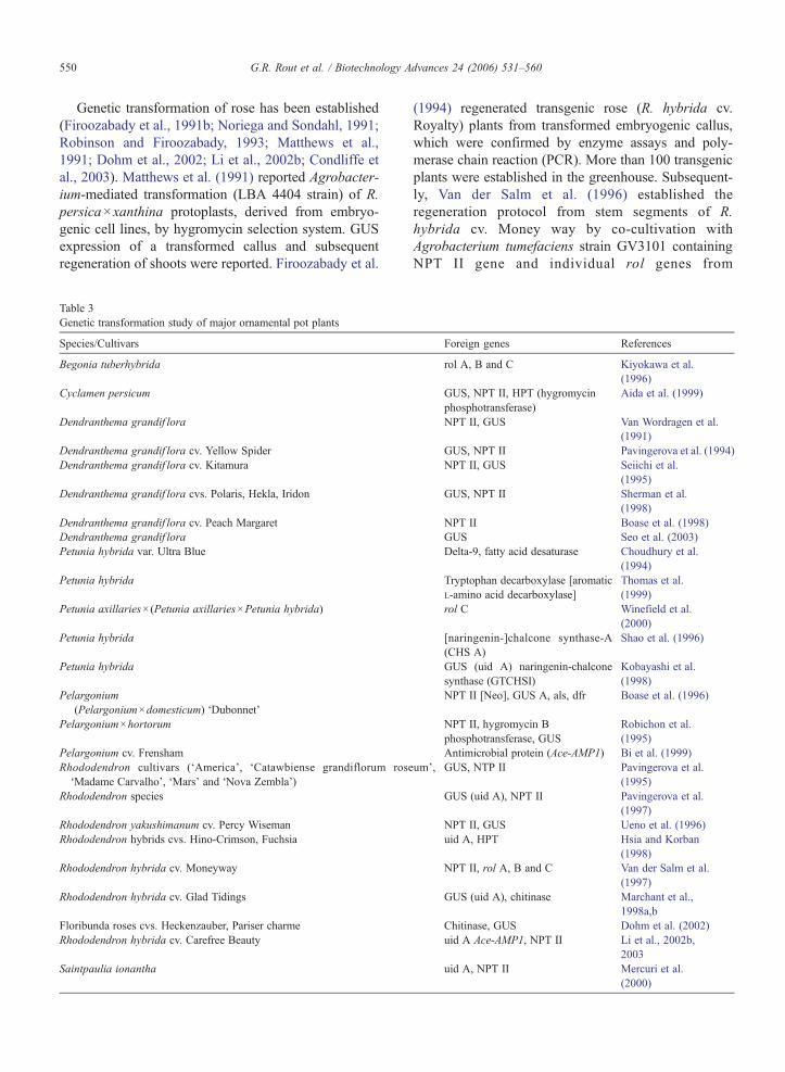
550 G.R. Rout et al. / Biotechnology Advances 24 (2006) 531–560
Genetic transformation of rose has been established(Firoozabady et al., 1991b; Noriega and Sondahl, 1991;Robinson and Firoozabady, 1993; Matthews et al.,1991; Dohm et al., 2002; Li et al., 2002b; Condliffe etal., 2003). Matthews et al. (1991) reported Agrobacter-ium-mediated transformation (LBA 4404 strain) of R.persica×xanthina protoplasts, derived from embryo-genic cell lines, by hygromycin selection system. GUSexpression of a transformed callus and subsequentregeneration of shoots were reported. Firoozabady et al.
Table 3Genetic transformation study of major ornamental pot plants
Species/Cultivars
Begonia tuberhybrida
Cyclamen persicum
Dendranthema grandif lora
Dendranthema grandif lora cv. Yellow SpiderDendranthema grandif lora cv. Kitamura
Dendranthema grandif lora cvs. Polaris, Hekla, Iridon
Dendranthema grandif lora cv. Peach MargaretDendranthema grandif loraPetunia hybrida var. Ultra Blue
Petunia hybrida
Petunia axillaries×(Petunia axillaries×Petunia hybrida)
Petunia hybrida
Petunia hybrida
Pelargonium(Pelargonium×domesticum) ‘Dubonnet’
Pelargonium×hortorum
Pelargonium cv. FrenshamRhododendron cultivars (‘America’, ‘Catawbiense grandiflorum rose‘Madame Carvalho’, ‘Mars’ and ‘Nova Zembla’)
Rhododendron species
Rhododendron yakushimanum cv. Percy WisemanRhododendron hybrids cvs. Hino-Crimson, Fuchsia
Rhododendron hybrida cv. Moneyway
Rhododendron hybrida cv. Glad Tidings
Floribunda roses cvs. Heckenzauber, Pariser charmeRhododendron hybrida cv. Carefree Beauty
Saintpaulia ionantha
(1994) regenerated transgenic rose (R. hybrida cv.Royalty) plants from transformed embryogenic callus,which were confirmed by enzyme assays and poly-merase chain reaction (PCR). More than 100 transgenicplants were established in the greenhouse. Subsequent-ly, Van der Salm et al. (1996) established theregeneration protocol from stem segments of R.hybrida cv. Money way by co-cultivation withAgrobacterium tumefaciens strain GV3101 containingNPT II gene and individual rol genes from
Foreign genes References
rol A, B and C Kiyokawa et al.(1996)
GUS, NPT II, HPT (hygromycinphosphotransferase)
Aida et al. (1999)
NPT II, GUS Van Wordragen et al.(1991)
GUS, NPT II Pavingerova et al. (1994)NPT II, GUS Seiichi et al.
(1995)GUS, NPT II Sherman et al.
(1998)NPT II Boase et al. (1998)GUS Seo et al. (2003)Delta-9, fatty acid desaturase Choudhury et al.
(1994)Tryptophan decarboxylase [aromaticL-amino acid decarboxylase]
Thomas et al.(1999)
rol C Winefield et al.(2000)
[naringenin-]chalcone synthase-A(CHS A)
Shao et al. (1996)
GUS (uid A) naringenin-chalconesynthase (GTCHSI)
Kobayashi et al.(1998)
NPT II [Neo], GUS A, als, dfr Boase et al. (1996)
NPT II, hygromycin Bphosphotransferase, GUS
Robichon et al.(1995)
Antimicrobial protein (Ace-AMP1) Bi et al. (1999)um’, GUS, NTP II Pavingerova et al.
(1995)GUS (uid A), NPT II Pavingerova et al.
(1997)NPT II, GUS Ueno et al. (1996)uid A, HPT Hsia and Korban
(1998)NPT II, rol A, B and C Van der Salm et al.
(1997)GUS (uid A), chitinase Marchant et al.,
1998a,bChitinase, GUS Dohm et al. (2002)uid A Ace-AMP1, NPT II Li et al., 2002b,
2003uid A, NPT II Mercuri et al.
(2000)
551G.R. Rout et al. / Biotechnology Advances 24 (2006) 531–560
Agrobacterium rhizogenesis. Marchant et al. (1998a,b)succeeded to introduce the chitinase gene in embryo-genic callus of R. hybrida cv. Glad Tidings throughbiolistic transformation. All transgenic plants weregrown in the greenhouse and morphologically true-to-type, with reduced blackspot disease by 13% to 43%.Subsequently, floribunda roses were transformed withgenes for antifungal proteins to reduce their suscepti-bility to fungal diseases (Dohm et al., 2002). Li et al.(2003) developed transgenic rose lines harbouring anantimicrobial protein gene (Ace-AMP1) to enhanceresistance to powdery mildew. They have confirmedthe stable integration of Ace-AMP1 and NPT II genesby Southern blotting. Condliffe et al. (2003) reportedthe optimised protocol for transformation (Agrobacter-ium-mediated) in different rose cultivars by using GUS(uidA2) gene. They found stable integration of thetransgene was confirmed at each stage of somaticembryogenesis and in regenerated plants.
Pavingerova et al. (1995) developed transgenicRhododendron cultivars by Agrobacterium-mediatedtransformation with GUS and NPT II gene as a selectablemarker gene. Hsia and Korban (1998) reported thesuccessful transformation of two cultivars of Rhododen-dron by using a helium pressure bombardment device.Aida et al. (1999) developed transgenic Cyclamen andsuccessfully transferred to greenhouse. Mercuri et al.(2000) established the Agrobacterium-mediated transfor-mation system in S. ionantha by using in vitro grownleaves and petioles explants and co-cultivation with twostrains (i.e., EHA105 (pKIWI105) and A281(pKIWI105)) carrying the genes uidA and NPT II. Thetransient transformation was confirmed by PCR andsouthern hybridization. Transgenic Petunia plants withrol C gene, which reduces plant height, leaf and flowersize, increased branching and decrease male and femalefertility has been reported (Winefield et al., 2000). Thegenetic transformation studies in ornamental pot plantsare presented in Table 3.
Thin cell layer (TCL) technology can be used forgenetic engineering and crop improvement. The un-transformed cells can be eliminated by using selectiveantibiotic medium, where the transformed cells willgrow faster. The cells harbouring selector gene withintheir genome proliferate on the selective medium. Thereports on the use of a thin cell layer system as an initialexplants for gene transfer are few, but those that existdemonstrate the effectiveness of introducing a gene intoan explants with defined cellular structure and with acontrolled regeneration program, allowing for theformation of non-chimeric transgenic plants (Teixeirada Silva, 2003a). Successful transformation of D.
grandiflora was done by using stem tTCLs or leafITCLs as an initial explants (Teixeira da Silva andFukai, 2002a,b).
6. Conclusion
Ornamental plants are produced mainly for theiraesthetic value, thus the propagation and improvementof quality attributes such as leaf types, flower colour,longevity and form, plant shape and architecture, andthe creation of novel variation are important economicgoals for floriculturists. Successful in vitro propagationof ornamental plants is now being used for commer-cialization. Many commercial laboratories and nationalinstitutes worldwide use in vitro culture system forrapid plant multiplication, germplasm conservation,elimination of pathogens, genetic manipulations, andfor secondary metabolite production (O'Riordain,1999). Annually, millions of ornamental plants areroutinely produced in vitro. The great potential ofmicropropagation for large-scale plant multiplicationcan be tapped by cutting down the cost of productionper plant by applying low-cost tissue culture, which isto adopt practices and proper use of equipment andresources to reduce the unit cost of micropropagule andplant production without compromising the quality.Bioreactor technology may cut down the cost of plantproduction provided proper precautions are taken toprevent contamination. Somatic embryogenesis facil-itates cryopreservation, synseed development, muta-tions, and genetic transformation. Plant transformationmethods and enhanced gene silencing technology caneffectively be used to evaluate and authenticate newlydiscovered endogenous genes to characterize theirfunction in plants as well as to genetically manipulatetrait quality and productivity (Dandekar, 2003). Recentprogress in genetic manipulation of plant cells hasopened new possibilities for improvement of ornamen-tal pot plants. In 2001, an Australian companyFlorigene became the first company in the world tosell genetically modified plants by mail order to thegeneral public for home garden use (Lu et al., 2003).They are still selling transgenic products of twocarnation types in Australia, Japan and USA.
Acknowledgement
The authors wish to acknowledge the Department ofForest and Environment, Government of Orissa forproviding the laboratory facilities. We also thank Prof.Dr. W. Preil, Federal Centre for Breeding Research onCultivated Plants, Institute for Ornamental Plant Breeding,
552 G.R. Rout et al. / Biotechnology Advances 24 (2006) 531–560
Bornkampsweg, Ahrensburg, Germany for providing theculture photographs ofClematis, Euphorbia and Saintpau-lia for publication.
References
AboEl-Nil MM.Geranium (Pelargonium). In: Ammirato PV, Evans DR,SharpWR,Bajaj YPS, editors. Hand book of plant cell culture, vol. 5.New York: McGraw Hill Publ. Co.; 1983. p. 439–60.
Ahloowalia BS. In vitro radiation induced mutants in Chrysanthemum.Mutat Breed Newsl 1992;39:6.
Ahmed HA. In vitro regeneration and propagation of meristem apicesof Chrysanthemum. Kert Egy Kozl 1986;50:199–214.
Ahmed KZ, Sagi F. Use of somaclonal variation and in vitro selectionfor induction of plant disease resistance: prospects and limitations.Acta Phytopathol Entomol Hung 1983;28:143–59.
Aida R, Hirose Y, Kishimoto S, Shibata M. Agrobacterium tume-faciens-mediated transformation of Cyclamen persicumMill. PlantSci 1999;148:1–7.
Aitkens-Christie J. Automation. In: Debergh PC, Zimmerman RH,editors. Micropropagation technology and application. TheNetherlands: Kluwer Acad. Publ.; 1991. p. 363–88.
Aitkens-Christie J, Kozai T, Takayama S. Automation in plant tissueculture: general introduction and overview. In: Aitken-Christie J,Kozai T, Smith MAL, editors. Automation and environmentalcontrol in plant tissue culture. The Netherlands: Kluwer Acad.Publ.; 1995. p. 1–18.
Anderson WC. A revised tissue culture medium for shoot multipli-cation of Rhododendron. J Am Soc Hortic Sci 1984;109:343–7.
Ando T, Murasaki K. In vitro propagation of Cyclamen by the use ofetiolated petioles. Tech Bull Fac Hort Chiba Univ 1983;32:1–5.
Anonymous.OmzettabelKamerplanten.VakblBloemist 2003;21a: 136–7.Appelgren M. Regeneration of Begonia hiemalis in vitro. Acta Hortic
1976;64:31–8.Appelgren M. Effect of supplementary light to mother plants on
adventitious shoot formation in flower peduncle segments ofBegonia×hiemalis. Sci Hortic 1985;25:77–83.
Ara KA, Hossain MM, Quasem MA, Ali M, Ahmed JU. Micrpropaga-tion of rose: Rosa sp. cv. Peace. Plant Tissue Cult 1997;7(2):135–42.
Arnold NP, Binns MR, Cloutier CD, Barthakur NN, Pellerin R.Auxins, salt concentrations and their interactions during in vitrorooting of winter-hardy and hybrid tea roses. Hortic Sci 1995;30(7):1436–40.
Atta-Alla H, Van Staden J. Micropropagation and establishment ofYucca aloifolia. Plant Cell Tissue Organ Cult 1997;48(3):209–12.
Atta-Alla H,McAlister B, Van Staden J. In vitro culture and establishmentof Anthurium parvispathum. S Afr J Bot 1998;64:296–8.
Barve DM, Iyer RS, Kendurkar S, Mascarenhas AF. An efficientmethod for rapid propagation of some budded rose varieties. IndianJ Hortic 1984;41:1–7.
Belarmino MM, Gabon CF. Low cost micropropagation of chrysan-themum (Chrysanthemum morifolium) through tissue culture.Philipp J Sci 1999;128(2):125–43.
Benetka V. Some experience of methodology with the isolation ofsomatic mutants in the rose cultivar “Sonia”. Acta Prubon1985;50:9–25.
Ben-Meir H, Scovel G, Ovadis M, Vainstein A. Molecular markers inthe breeding of ornamentals. Acta Hortic 1997;447:599–601.
Bhattacharya P, Dey S, Das N, Bhattacharya BC, Bhattacharya P. Rapidmass propagation of Chrysanthemum morifolium by callus derivedfrom stem and leaf explants. Plant Cell Rep 1990;9:439–42.
Bi YM, Cammue BPA, Goodwin PH, KrishnaRaj S, Saxena PK.Resistance to Botrytis cinerea in scented Geranium transformedwith a gene encoding the antimicrobial protein Ace-AMP1. PlantCell Rep 1999;18(10):835–40.
Bigot C. Multiplication vegetative in vitro de Begonia×hiemalis(Rieger et Schwabenland): I. Methodologie. Agronomie 1981a;1:433–40.
Bigot C. Multiplication vegetative in vitro de Begonia×hiemalis(Rieger et Schwabenland): II. Conformite des plantlets elevees enserre. Agronomie 1981b;1:441–7.
Bilkey PC, Cocking EC. A nonenzymatic method for the isolation ofprotoplasts from callus of Saintpaulia ionantha (African Violet).Z Pflanzenphysiol 1982;105:285–8.
Boase MR, Deroles SC, Winefield CS, Butcher SM, Borst NK, ButlerRC. Genetic transformation of regal Pelargonium (Pelargonium×domesticum “Dubonnet” by Agrobacterium tumefaciens. Plant Sci1996;121(1):47–61.
Boase MR, Bradley JM, Borst NK. Genetic transformation mediatedby Agrobacterium tumefaciens of florists “Peach Margaret”. InVitro Cell Dev Biol Plant 1998;34:46–51.
Bouman H, De Klerk GJ. Somaclonal variation in biotechnology ofornamental plants. In: Geneve R, Preece J, Merkle S, editors.Biotechnology of ornamental plants. UK: CAB International;1996. p. 165–83.
Bouman H, Kuijpers AM, De Klerk GJ. The influence of tissue culturemethods on somaclonal variation in Begonia. Physiol Plant1992;85:A45.
Bouman H, Kuijpers A, De Klerk GJ. Measurement of somaclonalvariation in Beginoa. Acta Hortic 1995;420:98–100.
Bouman H, Kuijpers AM, Hol T, Schoo W. Differences in growth andregeneration of suspension cultures derived from various calluslines of Cyclamen persicum cv. Purple Flamed. In: Schwenkel HG,editor. Reproduction of Cyclamen persicum Mill. through somaticembryogenesis using suspension culture systems. COST Action-Brussels: European Commission; 2001. p. 46–54.
Brand MH, Kiyamoto R. Behaviour of Rhododendron tissue affectedby tissue proliferation. In Vitro Cell Dev Biol 1994a;30A:72.
Brand MH, Kiyamoto R. Tissue proliferation apparently notlignotubers. Yank Nurs Q 1994b;3:5–6.
Brand MH, Kiyomoto R. The induction of tissue proliferation-likecharacteristics in in vitro cultures of Rhododendron `Monteg`.Hortic Sci 1997;32(6):989–94.
Bressan PH, Kim YJ, Hyndman SE, Hasegawa PM, Bressan RA.Factors affecting in vitro propagation of rose. J Am Soc Hortic Sci1982;107(6):979–90.
Broertjes C, Van Harten AM, editors. Application of MutationBreeding Methods on the Improvement of vegetatively propagatedcrops. The Netherlands: Elsevier Science Publ.; 1978. p. 325.
Broertjes C, Van Harten AM, editors. Applied mutation breeding forvegetatively propagated crops. Developments in crop science TheNetherlands: Elsevier Publ.; 1988. p. 197–204.
Burger DW, Liu L, Zary KW, Lee CI. Organogenesis and plantregeneration from immature embryos of Rosa hybrida L. Plant CellTissue Organ Cult 1990;21:147–52.
Carelli BP, Echeverrigaray S. An improved system for the in vitropropagation of rose cultivars. Sci Hortic 2002;92:69–74.
Cassells AC, Plunkett A. Production and growth analysis of plantsfrom leaf cuttings, and from tissue cultures of disks from matureleaves and young axenic leaves of African violet (Saintpauliaionantha Wendl.). Sci Hortic 1984;23:361–9.
Castillo B, Smith MAL. Direct somatic embryogenesis from Begoniagracilis explants. Plant Cell Rep 1997;16(6):385–8.
553G.R. Rout et al. / Biotechnology Advances 24 (2006) 531–560
Chakrabarty D, Mandal AK, Datta SK. In vitro propagation of rosecultivars. Ind J Plant Physiol 2000;5(2):189–92.
Chang HS, Chakrabarty D, Hahn EJ, Paek KY. Micropropagation ofcalla lily (Zantedeschia albomaculata) via in vitro shoot tipproliferation. In Vitro Cell Dev Biol Plant 2003;39:129–34.
Chartier-Hollis JM, Harris W, Lynch PT, Morisot A, Ricci P. Cryopre-servation of shoot tips of Rosa multiflora. Acta Hortic 1996;424:367–8.
Chebet DK, Okeno JA, Mathenge P. Biotechnological approaches toimprove horticultural crop production. Acta Hortic 2003;625: 473–7.
Chen J, Nell TA, Henny RJ, Robinson CA, Russell RD. Light levels ininfluencing production and subsequent interior performances ofFicus cultivars. Hortic Sci 2001;36:600 [abstract].
Choudhury ML, Chin CK, Polashock JJ, Martin CE. Agrobacterium-mediated transformation of Petunia hybrida with yeast Δ-9 fattyacid desaturase. Plant Growth Regul 1994;15:113–6.
Chu CY, Knight SL, Smith MAL. Effect of liquid culture on thegrowth and development of miniature rose (Rosa chinensis Jacq.`Minima`. Plant Cell Tissue Organ Cult 1993;32:329–34.
Chua BU, Kunisaki JT, Sagawa Y. In vitro propagation of Dracaenamarginata tricolour. Hortic Sci 1981;16:494.
Clark E, Izhar S, Hanson MR. Independent segregation of chloroplastDNA and cytoplasmic male sterility in Petunia somatic hybrids.Mol Gen Genet 1986;199:440–5.
Condliffe PC, Davey MR, Power JB, Koehorst-van Putten H, VisserPB. An optimised protocol for rose transformation applicable todifferent cultivars. Acta Hortic 2003;612:115–20.
Curtis WR. Application of bioreactor design principles to plantmicropropagation. Ist Int. Symp. ‘Liquid systems for in vitromicropropagation of plants’. 29th May – 2nd June, Norway; 2002.p. 58–9.
Dandekar AM. Techniques for manipulating quality and productivitytraits in horticultural crops. Acta Hortic 2003;625:293–305.
Datta SK, Chakrabarty D, Saxena M, Mandal AKA, Biswas AK.Direct shoot generation from florets of Chrysanthemum cultivars.Indian J Genet Plant Breed 2001;61(4):373–6.
Debener Th, Mathiesch L. Genetic analysis of molecular markerscrosses between diploid roses. Acta Hortic 1996;424:249–52.
Debener Th, Bartels C, Spethmann W. Parentage analysis interspecificcrosses between rose species with RAPD markers. Gartenbauwis-senschaft 1997;62:180–4.
Debergh P. Intensified vegetative multiplication of Dracaena dere-mensis. Acta Hortic 1975;54:83–92.
Debergh P. An in vitro technique for the vegetative multiplication ofchimeral plants ofDracaena andCordyline.ActaHortic 1976;64:17–9.
Debergh P, DeWael J. Mass propagation of Ficus lyrata. Acta Hortic1997;78:361–4.
Debergh HP, Maene L. Rapid clonal propagation of pathogen-freePelargonium plants starting from shoot tips and apical meristems.Acta Hortic 1977;78:449–54.
Debergh PC, Read PE. Micropropagation. In: Debergh PC, Zimmer-man RH, editors. Micropropagation. The Netherlands: KluwerAcad. Publ.; 1991. p. 1–13.
Demiralay A, Yalcin-Mendi Y, Aka-kacar Y, Cetiner S. In vitropropagation of Ficus carica L. var. Bursa siyahi through meristemculture. Acta Hortic 1998;480:165–7.
Deshpande SR, Josekutty PC, Prathapasenan G. Plant regenerationfrom axillary buds of a mature tree of Ficus religiosa. Plant CellRep 1998;17:571–3.
DeVries DP, Dubois LAM. The effect of BAP and IBA on sproutingand adventitious root formation of ‘Amanda’ rose single-node softwood cuttings. Sci Hortic 1988;34:115–21.
de Wit JC, Esendam HF, Horkanen JJ, Tuominen U. Somaticembryogenesis and regeneration of flowering plants in rose.Plant Cell Rep 1990;9:456–8.
Dijkshoorn-Dekker MWC. The influence of light and temperature onthe dynamic behaviour of Ficus benjamina ‘Exotica’. Acta Hortic1996;417:65–7.
Dillen W, Dijkstra I, Oud J. Shoot regeneration in long-term calluscultures derived from mature flowering plants of Cyclamenpersicum Mill. Plant Cell Rep 1996;15:545–8.
Dohm A, Ludwig C, Schilling D, Debener Th. Transformation of roseswith genes for antifungal proteins to reduce their susceptibility tofungal diseases. Acta Hortic 2002;572:105–11.
Douglas GC, Rutledge CB, Casey AD, Richardson DHS. Micro-propagation of floribunda ground cover and miniature roses. PlantCell Tissue Organ Cult 1989;19:55–64.
Eapen S, Rao PS. Regeneration of plant from Anthurium patulum. CurrSci 1985;54:284–6.
Economou AS, Read PE. In vitro shoot proliferation of Minnesotadeciduous azaleas. Hortic Sci 1984;19:60–1.
Eide AK, Munster C, Heyerdahl PH, Lyngved R, Olsen OAS. Liquidculture systems for plant propagation. Acta Hortic 2003;625:173–85.
Elliott EF. Axenic culture of meristem tips of Rosa multiflora. Planta1970;95:183–6.
Ettinger TL, Preece JE. Aseptic micropropagation of Rhododendron.P.J.M hybrids. J Hortic Sci 1985;60:269–74.
Fiore S, de Pasquale F, Carimi F, Sajeva M. Effect of 2,4-D and 4-CPPU on somatic embryogenesis from stigma and style transversethin cell layers of Citrus. Plant Cell Tissue Organ Cult 2002;68(1):57–63.
Firoozabady E, Lemieux CS, Moy YS, Moll B, Nicholas JA, RobinsonKEP. Genetic engineering of ornamental crops. In Vitro1991a;27:96A.
Firoozabady E, Noriega C, Sondahl MR, Robinson KEP. Genetictransformation of rose (Rosa hybrida cv. Royalty) via Agrobacter-ium tumefaciens. In Vitro 1991b;27:154A.
Firoozabady E, Moy Y, Courtneygutterson N, Robinson K. Regener-ation of transgenic rose (Rosa hybrida) plants from embryogenictissue. Biotechnology 1994;12:609–13.
Fonnesbech M, Fonnesbech A. In vitro propagation of Spathiphyllum.Sci Hortic 1979;10:21–5.
Fujii Y, Shimzu K. Regeneration of plants from stem and petals ofChrysanthemum coccineum. Plant Cell Rep 1990;8:625–7.
Fukai S, Oe M. Morphological observations of Chrysanthemum shoottips cultured after cryoprotection and freezing. J Jpn Soc Hortic Sci1990;59:383–7.
Fukai S, Morii M, Oe M. Storage of chrysanthemum (Dendrathemagrandiflorum Ramat.) plantlets in vitro. Plant Cell Tissue OrganCult 1988;5:20–5.
Gabryszewska E, Rudnicki RM. The effects of light quality on thegrowth and development of shoots and roots of Ficus benjamina invitro. Acta Hortic 1997;418:163–7.
Gamborg OL, Miller RA, Ojima K. Nutrient requirements ofsuspension culture of soybean root cells. Exp Cell Res 1968;50:151–8.
Geier T. Morphogenesis and plant regeneration from cultured organfragments of Cyclamen persicum. Acta Hortic 1977;78:167–74.
Geier T. Development of isolated rudimentary anthers from somaticanther callus of Cyclamen persicum Mill. Z Pflanzenphysiol1978;90:245–56.
Geier T. Induction and selection of mutants in tissue cultures ofGesneriaceae. Acta Hortic 1983;131:329–37.
554 G.R. Rout et al. / Biotechnology Advances 24 (2006) 531–560
Geier T, Kohlenbach HW, Reuther G. Cyclamen. In: Ammirato PV,Evans DR, Sharp WR, Bajaj YPS, editors. Hand book of plant cellculture, vol. 5. NewYork: McGraw-Hill Publ. Co.; 1983. p. 352–74.
Gertsson UE, Andersson E. Propagation of Chrysanthemum×hor-otorum and Philodendron scandens by tissue culture. Rapport,Institutionen for Tradgardsvetenskap. SitletSver Lantbr Univer;1985. p. 17.
Ghashghaie J, Brenckmann F, Saugier B. Effect of agar concentrationon water status and growth of rose plants cultured in vitro. PhysiolPlant 1991;82(1):73–8.
Gill R, Gerrath J, Saxena PK. High-frequency direct embryogenesis inthin layer cultures of hybrid seed geranium (Pelargonium). Can JBot 1992;71:408–13.
Grout BWW. African violet. In: Ammirato PV, Evans DA, Sharp WR,Bajaj YPS, editors. Handbook of plant cell culture, vol. 5. NewYork: MacGraw-Hill Publ. Co.; 1990. p. 181–205.
Grunewaldt J. Adventitious bud formation and plant regeneration inGesneriaceae in vitro. Gartenbauwissenschaft 1977;42:171–5.
Gudin S, Mouchotte J. Integrated research in rose improvement: abreeder's experience. Acta Hortic 1996;424:285–92.
Haberlandt G. Kulturversuche mit isollierten pflanzenzellen. S.B.Weisen Wien Naturwissenschaften, vol. 111. 1902. p. 69–92.
Haccius B. Question of unicellular origin of non-zygote embryos incallus cultures. Phytomorphology 1978;28:74–81.
Hasegawa PM. In vitro propagation of rose. Hortic Sci 1979;14:610–2.Hasegawa PM. Factors affecting shoot and root initiation from
cultured rose shoot tips. J Am Soc Hortic Sci 1980;105(2):216–20.Hatzilazarou S, Economou A, Antoniou T, Ralli P. Propagation of
Ebenus cretica L. by tissue culture. Propag Ornam Plants2001;1:25–7.
Hawkes HY, Wainwright H. In vitro organogenesis of Cyclamenpersicum Mill. seedling tissue. Acta Hortic 1987;212:711–4.
Hirochika H, Sugimoto K, Otsuki Y, Tsugawa H, Kanda M.Retrotransposons of rice involved in mutations induced by tissueculture. Proc Natl Acad Sci U S A 1996;93:7783–8.
Hitmi A, Barthomeuf C, Sallanon H. Cryopreservation of Chrysan-themum cinerariaefolium shoot tips. J Plant Physiol 2000;156:408–12.
Hoffmann M, Preil W. In vitro kultur von Blattexplantaten verschie-dener Cyclamen idiotypen. Gartenbauwissenschaft 1987;52:145–8.
Hohe A, Winkelmann T, Schwenkel HG. The effect of oxygen partialpressure in bioreactors on cell proliferation and subsequentdifferentiation of somatic embryos of Cyclamen persicum. PlantCell Tissue Organ Cult 1999;59(1):39–45.
Hohe A, Winkelmann T, Schwenkel HG. Development of somaticembryos of Cyclamen persicumMill. in liquid culture. Gartenbau-wissenschaft 2001;66:219–24.
Horn WAH. Micropropagation of rose. In: Bajaj YPS, editor.Biotechnology in agriculture and forestry, vol. 4. Germany:Springer Verlag; 1992. p. 320–4.
Horst RK, Smith SH, Horst HT, Oglevee WA. In vitro regeneration ofshoot and root growth from meristematic tips of Pelargonium×hortorum Bailey. Acta Hortic 1976;59:131–42.
Hoshino Y, Nakano M, Mii M. Plant regeneration from cellsuspension-derived protoplasts of Saintpaulia ionantha Wendl.Plant Cell Rep 1995;14:341–4.
Hosokawa M, Otake A, Sugawara Y, Hayashi T, Yazawa S. Rescue ofshoot apical meristem of chrysanthemum by culturing on root tips.Plant Cell Rep 2004;22:443–8.
Hosoki T, Kajino E. Shoot regeneration from petioles of coral bells(Heuchera sanguinea Engelm.) cultured in vitro, and subsequent
planting and flowering ex vitro. In Vitro Cell Dev Biol Plant2003;39:135–8.
Hsia CN, Korban SS. Factors affecting in vitro establishment and shootproliferation of Rosa hybrida L. and Rosa chinensis minima. InVitro Cell Dev Biol Plant 1996;32:217–22.
Hsia CN,Korban SS.Microprojectile mediated genetic transformation ofRhododendron hybrids. Am Rhododendr Soc J 1998;52(4):187–91.
Huang SC, Tsai CC, Sheu CS. Genetic analysis of Chrysanthemumhybrids based on RAPD molecular markers. Bot Bull Acad Sin2000;41(4):257–62.
Hutchinson JF, Miller R, Kaul V, Stevenson T, Richards D.Transformation of C. morifolium based on Agrobacterium genetransfer. J Cell Biochem 1989;26:261 [abstract].
Hutchinson JF, Kaul V, Maheswaran G, Moran JR, Graham MW,Richard D. Genetic improvement of floricultural crops usingbiotechnology. Aust J Bot 1992;40:765–87.
Huttema JBM, Gussenhoven G, Dons JJM, Broertjes C. Induction andselection of low temperature tolerant mutants of Chrysanthemummorifolium Ramat. Nuclear techniques and in vitro culture plantimprovement. Vienna, Austria: IAEA; 1986. p. 321–7.
Huttema JBM, Preil W, Gussenhoven GC, Schneidereit M. Methodsfor the selection of low-temperature tolerant mutants of Chrysan-themum morifolium Ramat. by using irradiated cell suspensioncultures: I. Selection of regenerants in vitro under subtropicaltemperature conditions. Plant Breed 1989;102:140–7.
Huttema JBM, Preil W, DeJong J. Methods for selection of low-temperature tolerant mutants of Chrysanthemum morifoliumRamat. using irradiated cell suspension cells: III. Comparison ofmutants selected with or without preselection in vitro at lowtemperature. Plant Breed 1991;107:135–40.
Hvoslof-Eide AK, Munster C. Somatic embryogenesis of Cyclamenpersicum Mill. in bioreactors. Proc Int Plant Propag Soc (IPPS)1998;47:377–82.
Hvoslof-Eide AK, Munster C. Light quality effects on somaticembryogenesis of Cyclamen persicum Mill. in bioreactors. In:Schwenkel HG, editor. Reproduction of Cyclamen persicum Mill.through somatic embryogenesis using suspensor culture systems.Report of working Group 2. COSTEuropean Commission; 2001.p. 79–84.
IAEA-TECDOC-1384. Low cost options for tissue culture technologyfor developing countries. Vienna: IAEA; 2004.
Ishiooka N, Tanimoto S. Plant regeneration from Bulgarian rose callus.Plant Cell Tissue Organ Cult 1990;22:197–9.
Izhar S, Tabib Y. Somatic hybridization in Petunia: II. Heteroplasmicstate in somatic hybrids followed by cytoplasmic segregation intomale sterile and male fertile lines. Theor Appl Genet 1980;57:241–6.
Izhar S, Schlichter M, Swartzberg D. Sorting out of cytoplasmicelements in somatic hybrids of Petunia and the prevalence of theheteroplasmon through several meiotic cycles. Mol Gen Genet1983;190:468–74.
Jain SM. Somaclonal variation in Begonia×elatior and Saintpauliaionantha L. Sci Hortic 1993a;54:221–31.
Jain SM. Studies on somaclonal variation in ornamental plants. ActaHortic 1993b;336:365–72.
Jain SM. Growth hormonal influence in somaclonal variation inornamental plants. Proc. of XVII Eucarpia Symp. on creatinggenomic variation in ornamentals; 1993c. p. 93–103.
Jain SM. Micropropagation of selected somaclones of Begonia andSaintpaulia. J Biosci 1997;22(5):585–92.
Jain SM. Tissue culture-derived variation in crop improvement.Euphytica 2001;118:153–66.
555G.R. Rout et al. / Biotechnology Advances 24 (2006) 531–560
Jain SM. Feeding the world with induced mutations and biotechnol-ogy. Proc. Int. Nuclear Conference 2002 – Global trends andPerspectives. Seminar 1: agriculture and bioscience. Bangi,Malaysia: MINT; 2002. p. 1–14.
Jain SM, De Klerk GJ. Somaclonal variation in breeding andpropagation of ornamental crops. Plant Tissue Cult Biotechnol1998;4(2):63–75.
Jain SM, Maluszynski M. Induced mutations and biotechnology inimproving crops. In: Mujib A, editor. In vitro applications in cropimprovement: recent progress. IBH-Oxford, India: in press.
Jain SM, Brar DS, Ahloowalia BS, editors. Somaclonal variation andinduced mutations in crop improvement. Dordrecht, The Nether-lands: Kluwer Acad. Publ.; 1998a. p. 603.
Jain SM, Ahloowalia BS, Veilleux RE. Somaclonal variation in cropplants. In: Jain SM, Brar DS, Ahloowalia, editors. Somaclonalvariation and induced mutations in crop improvement. Dordrecht,The Netherlands: Kluwer Acad. Publ.; 1998b. p. 203–18.
Jain SM, Buiatti M, Gimelli F, Saccardo F. Somaclonal variation inimproving ornamental plants. In: Jain SM, Brar DS, Ahloowalia BS,editors. Somaclonal variation and induced mutation in crop improve-ment. The Netherlands: Kluwer Acad. Publ.; 1998c. p. 81–105.
Jelaska S, Jelencic B. Plantlet regeneration from shoot tip culture ofPelargonium zonale hybrid. Acta Bot Croat 1980;39:59–63.
Joseph D, Martin KP, Madassery J, Philip VJ. In vitro propagation ofthree commercial cut flower cultivars of Anthurium andraeanumHort. Indian J Exp Biol 2003;41:154–9.
Kaicker US. Mutation breeding in roses. Ind Rose Ann Rep1982;2:35–42.
Kaul V, Miller RM, Hutchinson JF, Richards D. Shoot regenerationfrom stem and leaf explants of Dendrathema grandiflora Tzvelev(Syn. Chrysanthemum morifolium Ramat.). Plant Cell Tissue OrgCult 1990;21:21–30.
Kawata M, Oono K. Protoclonal variation in crop improvement. In:Jain SM, Brar DS, Ahloowalia BS, editors. Somaclonal variationand induced mutation for crop improvement. The Netherlands:Kluwer Acad. Publ.; 1997. p. 135–48.
Kevers C, Boyer N, Courduroux J, Gaspar T. The influence of ethyleneon proliferation and growth of rose shoot culture. Plant Cell TissueOrgan Cult 1992;28:175–81.
Khalid N, Davey MR, Power JB. An assessment of somaclonalvariation in Chrysanthemum morifolium: the generation of plantsof commercial value. Sci Hortic 1989;38:287–94.
Khanna SC, Purohit PV. Studies of steroidal sapogenins from Yuccaalaefolia L. In: Sen SK, Giles KL, editors. Basic Life Sciences,vol. 22. New York: Plenum Publ.; 1983. p. 65–9.
Khosh-Khui M, Sink KC.Micropropagation of new and old world rosespecies. J Hortic Sci 1982a;57(3):315–9.
Khosh-Khui M, Sink KC. Callus induction and culture of Rosa. SciHortic 1982b;17:361–70.
Khosh-Khui M, Sink KC. Rooting enhancement of Rosa hybrida fortissue culture propagation. Sci Hortic 1982c;17:371–6.
Kim SW, Oh SC, In DS, Liu JR. Plant regeneration of rose (Rosahybrida) from embryogenic cell derived protoplasts. Plant CellTissue Organ Cult 2003a;73(1):15–9.
Kim SJ, Hahn EJ, Paek KY, Murthy HN. Application of bioreactorculture for large scale production of Chrysanthemum transplants.Acta Hortic 2003b;625:187–91.
Kintzios S, Manos S, Makri O. Somatic embryogenesis from matureleaves of rose (Rosa sps). Plant Cell Rep 1999;18(6):467–72.
Kiyokawa S, Kikuchi Y, Kamada H, Harada H. Genetic transforma-tion of Begonia tuberhybrida by Ri rol genes. Plant Cell Rep1996;15(8): 606–9.
Knudson L. Flower production by orchid grown non-symbiotically.Bot Gaz 1922;89:192.
Kobayashi H, Oikawa Y, Koiwa H, Yamamura S. Flower-specific geneexpression directed by the promoter of a chalcone synthase genefrom Gentiana triflora in Petunia hybrida. Plant Sci 1998;131(2):173–80.
Kozai T. Autotropic (sugar free) tissue culture for promoting thegrowth of plantlets in vitro and for reducing biological contam-ination. Proc. Int. Symp. on application of biotechnology for smallindustries. Bangkok, Thailand; 1990a. p. 39–51.
Kozai T. Micropropagation under photoautotropic condition. In:Debergh P, Zimmerman RH, editors. Micropropagation: technol-ogy and application. The Netherlands: Kluwer Acad. Publ.; 1990b.p. 449–71.
Kozai T. Autotropic micropropagation. In: Bajaj YPS, editor. Biotech-nology in agriculture and forestry. . High-tech and micropropagation1. New York: Springer-Verlag; 1991a. p. 313–43.
Kozai T. Controlled environments in conventional and automatedmicropropagation. In: Levin R, Vasil IK, editors. Cell culture andsomatic cell genetics of plants. Scale-up and automation in planttissue culture. London: Acad. Press Inc.; 1991b. p. 213–30.
Kozai T, Kubota C, Watanabe I. Effect of basal medium compositionon the growth of carnation plantlets in auto- and mixotropic tissueculture. Acta Hortic 1988;230:159–66.
Kudo S, Shibata N, Kanno Y, Suzuki M. Transformation ofchrysanthemum [Dendranthema grandiflorum (Ramat.) Kitamura]via Agrobacterium tumefaciens. Acta Hortic 2002;572:139–47.
Kumar A, Kumar VA. High-frequency in vitro propagation inChrysanthemum maseimum. Indian Hortic 1995:37–8 [Jan–March].
Kumar V, Radha A, Kumar Chitta S. In vitro plant regeneration of fig(Ficus carica L. cv. Gular) using apical buds from mature trees.Plant Cell Rep 1998;17(9):717–20.
Kumar A, Sood A, Palni VT, Gupta AK, Palni LM. Micropropagationof Rosa damascena Mill. from mature bushes using thidiazuron.J Hortic Sci Biotechnol 2001;76:30–4.
Kumari M, Varghese TM, Mehta PK. Micropropagation of Chrysan-themum through shoot apex culture in two named varieties viz.Miss Universe and Snow Ball. Ann Agric Res 2001;11(3):371–6.
Kunitake H, Imamizo H, Mii H. Somatic embryogenesis and plantregeneration from immature seed-derived calli of rugosa rose(Rosa rugosa Thurb.). Plant Sci 1993;90:187–94.
Lamseejan S, Jompuk P, Deeseepan S. Improvement of Chrysanthe-mum var. “Taihei” through in vitro induced mutation with chronicand acute gamma rays. J Nucl Soc Thail 2003;4:2–13.
Langford PJ, Wainwright H. Effects of sucrose concentration on thephotosynthetic ability of rose shoots in vitro. Ann Bot 1987;60:633.
Langhe ED, Debergh P, van Rijk R. In vitro culture as a method forvegetative propagation of Euphorbia pulcherrima. Z Pflanzen-physiol 1974;71:271–4.
Larkin PJ, Scowcroft WR. Somaclonal variation – a novel source ofvariability from cell cultures for plant improvement. Theor ApplGenet 1981;60:197–214.
Latado RR, Adames AH, Neto AT. In vitro mutation of chrysanthe-mum (Dendranthema grandiflora Tzvelev) with ethylmethanesul-phonate (EMS) in immature floral pedicels. Plant Cell TissueOrgan Cult 2004;77:103–6.
Lawrence Jr RH. In vitro plant cloning system. Environ Exp Bot1981;21:289–300.
Lazar M, Cachita C. Micropropagation of Chrysanthemum: III.Chrysanthemum multiplication in vitro from capitulum explants.Prod Veg Hortic 1983;32:44–7.
556 G.R. Rout et al. / Biotechnology Advances 24 (2006) 531–560
Ledger SE, Deroles SC, Given NK. Regeneration and Agrobacterium-mediated transformation of chrysanthemum. Plant Cell Rep 1991;10:195–9.
Levin R, Gaba V, Tal B, Hirsch S, Denola D, Vasil IK. Automatedplant tissue culture for mass propagation. Biotechnology 1988;6:1035–40.
Li XQ, Krasnyanski SF, Korban SS. Somatic embryogenesis,secondary somatic embryogenesis, and shoot organogenesis inRosa. J Plant Physiol 2002a;159:313–9.
Li XQ, Krasnyanski S, Korban SS. Optimization of the uid A genetransfer into somatic embryos of rose via Agrobacteriumtumefaciens. Plant Physiol Biochem 2002b;40:453–9.
Li XQ, Gasic K, Cammue B, Broekaert W, Korban SS. Transgenic roselines harbouring an antimicrobial protein gene, Ace-AMP1, demon-strate enhanced resistance to powdery mildew (Sphaerothecapannosa). Planta 2003;218:226–32.
Liu CM, Xu ZH. An efficient procedure for micropropagation ofAnthurium scherzerianum Schott (flamingo flower). Chin J Bot1992;4:49–55.
Lloyd G, McCown B. Commercially feasible micropropagation ofmountain laurel,Kalmia latifolia, by use of shoot tip culture. CombProc Int Plant Propag Soc 1980;30:421–6.
Lloyd D, Roberts AV, Short KC. The induction of in vitro ofadventitious shoots in Rosa. Euphytica 1988;37:31–6.
Lo KH, Giles KL, Sawhney VK. Acquisition of competence for shootregeneration in leaf discs of Saintpaulia ionantha (African violet)cultured in vitro. Plant Cell Rep 1997;16(6):416–20.
Lowe JM, Davey MR, Power JB, Blundy KS. A study of somefactors affecting Agrobacterium transformation and plant regen-eration of Dendrathema grandiflora Tzvelev (Syn. Chrysanthe-mum morifolium Ramat.). Plant Cell Tissue Organ Cult 1993;33:171–80.
Lu CY, Nugent G, Wardley T. Efficient, direct plant regeneration fromstem segments of chrysanthemum (Chrysanthemum morifoliumRamat cv. Royal Purple). Plant Cell Rep 1990;8:733–6.
Lu CY, Chandler SF, Mason JG, Brugliera F. Florigene flowers: fromlaboratory to market. In: Vasil IK, editor. Plant biotechnology 2002and beyond. The Netherlands: Kluwer Acad. Publ.; 2003. p. 333–6.
Malaure RS, Barclay G, Power JB, Davey MR. The production ofnovel plants from florets of Chrysanthemum morifolium usingtissue culture: I. Shoot regeneration for ray florets and somaclonesvariation exhibited by the regenerated plants. J Plant Physiol1991;139:8–13.
Maluszynski M, Ahloowalia BS, Sigurbjornsson B. Application of invitro and in vivo mutation techniques for crop improvement.Euphytica 1995;85:303–15.
Mandal AKA, Chakrabarty D, Datta SK. In vitro isolation of solidnovel flower colour mutants from induced chimeric ray florets ofchrysanthemum. Euphytica 2000;114:9–12.
Marchant R, Power JB, Lucas JA, Davey MR. Biolistic transformationof rose (Rosa hybrida L). Ann Bot 1998a;81:109–14.
Marchant R, Davey MR, Lucas JA, Lamb CJ, Dixon RA, Power JB.Expression of a chitinase transgene in rose (Rosa hybrida L.)reduces development of blackspot disease (Diplocarpon rosaeWolf). Mol Breed 1998b;4:187–94.
Martin C, Carre M, Verney R. La multiplication vegetative in vitro desvegetaux ligneux cultivees: cas des rosiers. C R Acad Sci Paris III1981;293:175–7.
Martin KP, Joseph D, Madassery J, Phillip VJ. Direct shootregeneration from lamina explants of two commercial cut flowercultivars of Anthurium andraeanum Hort. In Vitro Cell Dev BiolPlant 2003;39:500–4.
Matsumoto TK, Kuehnle AR. Micropropagation of Anthurium. In:Bajaj YPS, editor. Biotechnology in agriculture and forestry 40:high-tech and micropropagation, vol. VI. New York: Springer-Verlag; 1997. p. 15–29.
Matthews D, Mottley J, Horan I, Roberts AV. A protoplast to plantsystem in roses. Plant Cell Tissue Organ Cult 1991;24:173–80.
Matysiak B, Nowak J. Acclimatization of ex vitro Homalomena‘Emerald Gem’ as affected by nutrient solution concentration andCO2 enrichment. Acta Hortic 1995;390:157–60.
Matysiak B, Nowak J. Carbon dioxide enrichment, light, and mineralnutrition for stimulation of growth of in vitro propagated Gerbera.Propag Ornam Plants 2001;1:20–4.
May RA, Trigiano RN. Somatic embryogenesis and plant regenerationfrom leaves of Dendrathema grandiflora. J Am Soc Hortic Sci1991;116:366–71.
Mayer L. Wachstum und organbildung an in vitro kultiviertensegmenten von Pelargonium zonale and Cyclamen persicum.Planta 1956;47:401–46.
McCown BH, Lloyd GB. A survey of the responses of Rhododendronto in vitro culture. Plant Cell Tissue Organ Cult 1983;2:77–85.
Mercuri A, De Benedetti L, Burchi G, Schiva T. Agrobacterium-mediated transformation of African violet. Plant Cell Tissue OrganCult 2000;60(1):39–46.
Meskhi AB, Gogoberidze MK, Katsitadze KP. Tissue culture of Yuccagloriosa. Chem Abstr 1978;89:358–9.
Micke A. Mutation in plant breeding. In: Siddiqui BA, Khan S, editors.Breeding in crop plants –mutations and in vitromutation breeding.New Delhi, India: Kalyani Publ.; 1999. p. 1–19.
Mikkelson EP, Sink Jr KC. In vitro propagation of Rieger Elatiorbegonias. Hortic Sci 1978a;13:242–4.
Mikkelson EP, Sink Jr KC. Histology of adventitious shoot and rootformation on leaf–petiole cuttings of Begonia×hiemalis FotschAphrodite Peach. Sci Hortic 1978b;8:179–92.
Miller LR, Murashige T. Tissue culture propagation of tropical foliageplants. In Vitro 1976;12(12):797–813.
Misra P, Datta SK, Chakrabarty D. Mutation in flower colour andshape of Chrysanthemum morifolium induced by γ-radiation. BiolPlant 2004;47(1):153–6.
Mithila J, Hall J, Victor JMR, Saxena PK. Thidiazuron induces shootorganogenesis at low concentrations and somatic embryogenesis athigh concentrations on leaf and petiole explants of African violet(Saintpaulia ionantha Wendl.). Plant Cell Rep 2003;21(5):408–14.
Molgaard JP, Roulund N, Deichmann V, Irgens-Moller L, Andersen SB,Farestveit B. In vitro multiplication of Saintpaulia ionantha Wendl.by homogenization of tissue culture. Sci Hortic 1991;48:285–92.
Mulin M, Tran Thanh Van K. Obtention of in vitro flowers from thinepidermal cell layers of Petunia hybrida (Hort.). Plant Sci1989;62:113–21.
Murashige T, Skoog T. A revised medium for rapid growth and bioas-says with tobacco tissue cultures. Plant Physiol 1962;15: 473–97.
Murch SJ, Victor JMR, Saxena PK. Auxin, calcium and sodium insomatic embryogenesis of African violet (Saintpaulia ionanthaWendl.) cv. Benjamin. Acta Hortic 2003;625:201–9.
Nagaraju S, Reddy SK, Farook SA. Propagation of Ficus reliosa L.from maxillary buds and shoot tips. Adv Plant Sci 1998;11(2):287–90.
Nagatomi S. Six mutant varieties of flower colours induced by floralorgan cultures of chronically irradiated plants in chrysanthemum.Inst Rad Breed Tech News, vol. 36. 1993.
Narayan P, Jaiswal VS. Differentiation of plantlets from leaf callus ofFicus religiosa. Indian J Exp Biol 1986;24:193–4.
557G.R. Rout et al. / Biotechnology Advances 24 (2006) 531–560
Nhut DT, Teixeira da Silva JA, Bui VL, Thorpe T, Tran Thanh Van K.Woody plant micropropagation and morphogenesis by thin celllayers. In: Nhut DT, Van Le B, Tran Thanh Van K, Thorpe T,editors. Thin cell layer culture system: regeneration and transfor-mation applications. Dordrecht, The Netherlands: Kluwer Acad.Publ.; 2003a. p. 473–93.
Nhut DT, Teixeira da Silva JA, Bui VL, Tran Thanh Van K. Thin celllayer (TCL) morphogenesis as a powerful tool in woody plant andfruit crop micropropagation and biotechnology, floral genetics andgenetic transformation. In: Jain SM, Ishii K, editors. Micropropa-gation of woody trees and fruits. Dordrecht, The Netherlands:Kluwer Acad. Publ.; 2003b. p. 783–814.
Nikaido T, Onogawa Y. Establishment of a non-chimaeric flowercolour mutation through in vitro cultures of florets from a sport onChrysanthemum with special references to the genetic backgroundof the mutation line obtained. Science Report of Faculty ofAgriculture, Ibaraki Univ., 1989. p. 63–7.
Nobre J, Romano A. In vitro cloning of Ficus carica L. adult trees.Acta Hortic 1998;480:161–4.
Noriega C, Sondahl MR. Somatic embryogenesis in hybrid tea roses.Bio/Technology 1991;9:991–3.
Norton ME, Norton CR. In vitro propagation of Ericaceae: a comparisonof the activity of the cytokinin N6-benzyladenine and N6-isopente-nyladenine in shoot proliferation. Sci Hortic 1985;27:335–40.
Ohki S. Scanning electron microscopy of shoot differentiation in vitrofrom leaf explants of the African violet. Plant Cell Tissue OrganCult 1994;36:157–62.
Okumoto H, Takabayashi S. Aseptic culture of Cyclamen tuber tissue.Effects of curing mode of inoculation, temperature on developmentof explants and percentage of microbial infection. J Jpn Soc HorticSci 1969;38:178–87.
O'Riordain F. Directory of European plant tissue culture laboratories,1996–97. COST Action, vol. 822. Brussels: Commission of theEuropean Communities; 1999.
Orlikowska T, Sabala I, Nowak E. Adventitious shoot regeneration onexplants of Anthurium,Codiaeum,Dieffenbachia,Gerbera, Rosa andSpathiphyllum for breeding purposes. Acta Hortic 1995;420:115–7.
Osternack N, Saare-Surminski K, Preil W, Lieberei R. Induction ofsomatic embryos, adventitious shoots and roots in hypocotylstissue of Euphorbia pulcherrima Willd. Ex Klotzsch: comparativestudies on embryogenic and organogenic competence. J Appl Bot1999;73:197–201.
Paek KY, Hahn EJ, Son SH. Application of bioreactors for large scalemicropropagation systems of plants. In vitro Cell Dev Biol Plant2001;37:149–57.
Pati PK, Rath SP, Sharma M, Sood A, Ahuja PS. In vitro propagationof rose – a review. Biotechnol Adv 2006;24:94–114.
Pavingerova D, Dostal J, Biskova R, Benetka V. Somatic embryo-genesis and Agrobacterium-mediated transformation of Chrysan-themum. Plant Sci 1994;97:95–101.
Pavingerova D, Biskova R, Niedermeierova H, Kodytek K. Agro-bacterium-mediated transformation of Rhododendrons. ActaHortic 1995;420:89–91.
Pavingerova D, Briza J, Kodytek K, Niedermeierova H. Transforma-tion of Rhododendron spp using Agrobacterium tumefacienswith aGUS-intron chimeric gene. Plant Sci 1997;122(2):165–71.
Peak DE, Cumming BG. In vitro propagation of Begonia× tuberhy-brida from leaf sections. Hortic Sci 1984;19:395–7.
Pierik RLM. Vegetative vermeerdering van Cyclamen Vakbl. Bloe-misterij 1975;30:13.
Pierik RLM. Anthurium andraeanum plantlets produced from callustissues cultivated in vitro. Physiol Plant 1976;37:80–2.
Pierik RLM. Micropropagation of ornamental plants. Acta Hortic1991a;289:45–53.
Pierik RLM. Commercial micropropagation in Western Europe andIsrael. In: Debergh PC, Zimmerman RH, editors. Micropropaga-tion. The Netherlands: Kluwer Acad. Publ.; 1991b. p. 155–65.
Pierik RLM, Steegmans HHM, van der Meys JAJ. Plantlet formation incallus tissues ofAnthurium andraeanumLind. Sci Hort 1974;2:193–8.
Podwyszynska M, Olszewski T. Influence of gelling agents on shootmultiplication and the uptake of macroelements by in vitroculture of rose, cordyline and Homalomena. Sci Hortic 1995;64:77–84.
Prasad RN, Sharma AK, Chaturvedi HC. Clonal multiplication ofChrysanthemum morifolium “Otome zakura” in long-term culture.Bangaladesh J Bot 1983;12:96–102.
PreilW. Application of bioreactors in plant propagation. In: Debergh PC,Zimmerman RH, editors. Micropropagation technology and appli-cation. The Netherlands: Kluwer Acad. Publ.; 1991. p. 425–45.
Preil W. Micropropagation of ornamental plants. In: Laimer M, RuckerW, editors. Plant tissue culture 100 years since GottliebHaberlandt. New York: Springer-Verlag; 2003. p. 115–33.
Preil W, Beck A. Somatic embryogenesis in bioreactor culture. ActaHortic 1991;289:179–92.
Preil W, Engelhardt M, Walther F. Breeding of low temperaturetolerant Poinsettia (Euphorbia pulcherrima) and Chrysanthemumby means of mutation induction in in vitro culture. Acta Hortic1983;131:345–51.
Preil W, Florek P, Wix U, Beck A. Towards mass propagation by use ofbioreactors. Acta Hortic 1988;226:99–105.
Preil W, Huittema JBM, DeJong J. Method of selection of low-temperature tolerant mutants of Chrysanthemum morifolium Ramat.using irradiated cell suspension cultures: II. Preselection in vitrounder low-temperature stress. Plant Breed 1991;107:131–4.
Pueschel AK, Schwenkel HG, Winkelmann. Inheritance of the ability forregeneration via somatic embryogenesis in Cyclamen persicum. PlantCell Tissue Organ Cult 2003;72:43–51.
Quatrano RS. Freeze preservation of cultured flax cells utilizingDMSO. Plant Physiol 1968;43:2057–61.
Quintero A. Yucca. In: Ammirato PV, Evans DR, Sharp WR, BajajYPS, editors. Hand book of plant cell culture. Ornamental Species.New York: McGraw-Hill Publishing Co.; 1983. p. 783–99.
Quintero A, Rosas V, Zamudio F, Capella S, Romo de Vivar A. Tissueculture of Yucca filifera cells. Identification of steroidal precursors.In: Fujiwara A, editor. Tissue culture 1982. Japan: Maruzen; 1982.p. 295–6.
Quintero A, Zamudio F, Rasas V, Capella S, Romode Vivar A.Sarsasapogenin in Yucca filifera callus culture. Rev LatinoamQuim 1987;18:24–8.
Rajagopalan C. Export potential of Indian floriculture and need ofpolicy environment. Floric Today 2000;9:29–33.
Rajapakse S, Hubbard M, Kelly JW, Abbott AGR, Ballard RE.Identification of rose cultivars by restriction fragment lengthpolymorphism. Sci Hortic 1992;52:237–45.
Rani V, Raina SN. Genetic fidelity of organized meristem-derivedmicropropagated plants: a critical reappraisal. In Vitro Cell DevBiol Plant 2000;36:319–30.
Rao PS, Handro W, Harada H. Hormonal control of differentiation ofshoots, roots and embryos in leaf and stem cultures of Petuniainflata and Petunia hybrida. Physiol Plant 1973;28:458–63.
Reuter G, Bhandari NN. Organogenesis and histogenesis ofadventitious organs induced on leaf blade segments of Begonia-elatior hybrids (Begonia×hiemalis) in tissue culture. Gartenbau-wissenschaft 1981;46:241–9.
558 G.R. Rout et al. / Biotechnology Advances 24 (2006) 531–560
Roberts AV, Smith EF. The propagation in vitro of chrysanthemum fortransplantation to soil: I. Protection of roots by cellulose plugs.Plant Cell Tissue Organ Cult 1990;21:129–32.
Roberts AV, Horan I, Mathews D, Mottley J. Protoplast technology andsomatic embryogenesis in Rosa. In: deJong J, editor. Integration ofin vitro techniques in ornamental plant breeding. Proc. Symp., 10–14th Nov, CPO centre for Plant Breeding Research. The Nether-lands: AAWageningen; 1990. p. 128–38.
Roberts AV, Walker S, Horan I, Smith EF, Mottley J. The effects ofgrowth retardants, humidity and lighting at stage III on stage IVofmicropropagation in chrysanthemum and rose. Acta Hortic1992;319:135–8.
Roberts AV, Yokoya K, Walker S, Mottley J. Somatic embryogenesisin Rosa spp. In: Jain SM, Gupta PK, Newton RJ, editors. Somaticembryogenesis in woody plants, vol. 2. The Netherlands: KluwerAcad. Publ.; 1995. p. 277–89.
Robichon MP, Renou JP, Jalouzot R. Genetic transformation ofPelargonium×hortorum. Plant Cell Rep 1995;15(1–2):63–7.
Robinson KEP, Firoozabady E. Transformation of floriculture crops. SciHortic 1993;55:83–99.
Roest S, Bokelmann GS. Vegetative propagation of Chrysanthemummorifolium Ramat. in vitro. Sci Hortic 1975;3:317–30.
Roest S, Van Bakel MAE, Bokelmann GS, Broertjes C. The use of anin vitro adventitious bud technique for mutation breeding ofBegonia×hiemalis. Euphytica 1981;30:381–8.
Rogers RM, Smith MAL. Consequences of in vitro and ex vitro rootinitiation for miniature rose production. J Hortic Sci 1992;67:535–40.
Romo de Vivar A. Productos naturales de la flora Mexicana. Mexico:Editorial Limusa; 1985. p. 194–202.
Rosu A, Skirvin RM, Bein A, Norton MA, Kushad M, Otterbacher AG.The development of putative adventitious shoots from a chimeralthornless rose (Rosa multiflora Thurb.ex J.Murr.) in vitro. J HorticSci 1995;70(6):901–7.
Rout GR, Das P. Recent trends in the biotechnology of Chrysanthe-mum: a critical review. Sci Hortic 1997;81:201–28.
Rout GR, Jain SM. Micropropagation of ornamental plants–cutflowers. Propag Ornam Plants 2004;4(2):3–28.
Rout GR, Debata BK, Das P. In vitro mass scale propagation of Rosahybrida cv. Landora. Curr Sci 1989;58(15):876–8.
Rout GR, Debata BK, Das P. In vitro clonal multiplication of roses.Proc Natl Acad Sci (India) 1990;60(3):311–8.
Rout GR, Debata BK, Das P. Somatic embryogenesis in callus culturesof Rosa hybrida L. cv. Landora. Plant Cell Tissue Organ Cult1991;27:65–9.
Rout GR, Debata BK, Das P. In vitro regeneration of shoots from calluscultures of Rosa hybrida cv. Landora. Indian J Exp Biol1992;30:15–8.
Rout GR, Palai SK, Panday P, Das P. Direct plant regeneration ofChrysanthemum morifolium Ramat. cv. Deep Pink: influence ofexplant source, age of explants, culture environment, carbohy-drates, nutritional factors and hormone regime. Proc Natl AcadSci (India) 1996;67:57–66.
Rout GR, Samantaray S, Mottley J, Das P. Biotechnology of the rose: areview of recent progress. Sci Hortic 1999;81:201–28.
Rzepka-Plevnes D, Kurek J. The influence of media composition onthe proliferation and morphology of Ficus benjamina plantlets.Acta Hortic 2001;560:473–6.
Sakai A. Survival of the twigs of woody plants at −196 °C. Nature1960;185:392–4.
Sakamoto Y, Onishi N, Hirosawa T. Delivery systems for tissue cultureby encapsulation. In: Christie JA, Kozai T, Smith MAL, editors.
Automation and environmental control in plant tissue culture. TheNetherlands: Kluwer Acad. Publ.; 1995. p. 215–43.
SallanonH,Maziere Y. Influence of growth room and vessel humidity onthe in vitro development of rose plants. Plant Cell Tissue Organ Cult1992;30:121–5.
Sarasan V, Roberts AV, Rout GR. Methyllaurate and 6-benzyladeninepromote the germination of somatic embryos of a hybrid rose.Plant Cell Rep 2001;20(3):183–6.
Sauvadet MA, Brochard P, Boccon-Gibod J. A protoplast-to-plantsystem in Chrysanthemum: differential responses among severalcommercial clones. Plant Cell Rep 1990;8:692–5.
Schiva T. Strategies for development of commercial floriculture inAsia and the Pacific. Report of the APO seminar, 2nd – 6th May,2000, New Delhi, India; 2000. p. 27–38.
Schwenkel H.G., Regeneration von Cyclamen persicum Mill. in vitround deren Integration in die Cyclamenzuchtung. Ph.D thesis, Univ.of Hannover, Germany: 1991.
Schwenkel HG. Development of a reproducible regenerationprotocol for Cyclamen. In: Schwenkel HG, editor. Reproductionof Cyclamen persicum Mill. through somatic embryogenesisusing suspension culture systems. COST ActionBrussels:European Commission; 2001. p. 8–11.
Schwenkel HG, Grunewaldt J. In vitro propagation of Cyclamenpersicum Mill. Acta Hortic 1988;226:659–62.
Scowcroft WR, Larkin PJ. Somaclonal variation. In: Bock G, March L,editors. Applications of plant cell and tissue culture. CIBAFoundation Symp.Chichester: Wiley; 1988. p. 21–35.
Seiichi F, DeJong J, Rademaker W. Efficient genetic transformation ofChrysanthemum (Dendranthema grandiflorum Ramat. Kitamura)using stem segments. Breed Sci 1995;45(2):179–84.
Seo SY, Choi DC, Kim JM, Lim HC, Kim HJ, Choi JS, et al. Plantregeneration from leaf explants and efficient Agrobacterium-mediated transformation system of Chrysanthemum (Den-dranthema grandiflorum). Acta Hortic 2003;625:403–9.
Shao Li, Li Y, Yang MZ, Song Y, Chen ZL. Gene expression ofchalcone synthase-A (CHSA) in flower colour alterations and malesterility of transgenic Petunia. Acta Bot Sin 1996;38(7):517–24.
Sharma AK, Mitra GC. In vitro culture of shoot apical meristem ofPetunia hybrida for mass production of plants. Indian J Exp Biol1976;14:348–50.
Sherman JM,Moyer JW, DaubME. A regeneration and Agrobacterium-mediated transformation system for genetically diverse Chrysanthe-mum cultivars. J Am Soc Hortic Sci 1998;123(2): 189–94.
Short KC, Roberts AV. Rosa species (roses): in vitro culture,micropropagation and production of secondary products. In: BajajYPS, editor. Biotechnology in agriculture and forestry. . Medicinaland Aromatic Plants: III. Berlin: Springer-Verlag; 1991. p. 376–97.
Simmonds J. Induction, growth and direct rooting of adventitiousshoots of Begonia×hiemalis. Plant Cell Tissue Organ Cult1984;3:283–9.
Simmonds J, Werry T. Liquid shake cultures for improved micro-propagation of Begonia×hiemalis. Hortic Sci 1987;22:122–4.
Singh SK, Syamal MM. Anti-auxin enhance Rosa hybrida L.micropropagation. Biol Plant 2000;43(2):279–81.
Skirvin RM. Natural and induced variation in tissue culture. Euphytica1978;27:241–66.
Skirvin RM, Chu MC. In vitro propagation of ‘Forever Your’ rose.Hortic Sci 1979;14(5):608–10.
Skirvin RM, Janick J. Tissue culture induced variation in scentedPelargonium spp. J Am Soc Hortic Sci 1976;101:281–90.
Skirvin RM, Chu MC, Young HJ, Rose. In: Ammirato PV, Evans DR,Sharp WR, Bajaj YPS editors. Hand Book of Plant Cell Culture.
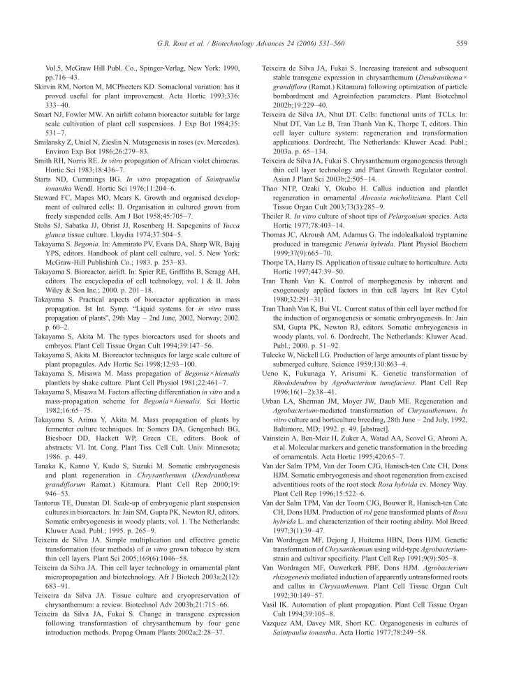
559G.R. Rout et al. / Biotechnology Advances 24 (2006) 531–560
Vol.5, McGraw Hill Publ. Co., Spinger-Verlag, New York: 1990,pp.716–43.
Skirvin RM, Norton M, MCPheeters KD. Somaclonal variation: has itproved useful for plant improvement. Acta Hortic 1993;336:333–40.
Smart NJ, Fowler MW. An airlift column bioreactor suitable for largescale cultivation of plant cell suspensions. J Exp Bot 1984;35:531–7.
Smilansky Z, Uniel N, Zieslin N. Mutagenesis in roses (cv. Mercedes).Environ Exp Bot 1986;26:279–83.
Smith RH, Norris RE. In vitro propagation of African violet chimeras.Hortic Sci 1983;18:436–7.
Starts ND, Cummings BG. In vitro propagation of Saintpauliaionantha Wendl. Hortic Sci 1976;11:204–6.
Steward FC, Mapes MO, Mears K. Growth and organised develop-ment of cultured cells: II. Organisation in cultured grown fromfreely suspended cells. Am J Bot 1958;45:705–7.
Stohs SJ, Sabatka JJ, Obrist JJ, Rosenberg H. Sapegenins of Yuccaglauca tissue culture. Lloydia 1974;37:504–5.
Takayama S. Begonia. In: Ammirato PV, Evans DA, Sharp WR, BajajYPS, editors. Handbook of plant cell culture, vol. 5. New York:McGraw-Hill Publishinh Co.; 1983. p. 253–83.
Takayama S. Bioreactor, airlift. In: Spier RE, Griffiths B, Scragg AH,editors. The encyclopedia of cell technology, vol. I & II. JohnWiley & Son Inc.; 2000. p. 201–18.
Takayama S. Practical aspects of bioreactor application in masspropagation. Ist Int. Symp. “Liquid systems for in vitro masspropagation of plants”, 29th May – 2nd June, 2002, Norway; 2002.p. 60–2.
Takayama S, Akita M. The types bioreactors used for shoots andembryos. Plant Cell Tissue Organ Cult 1994;39:147–56.
Takayama S, Akita M. Bioreactor techniques for large scale culture ofplant propagules. Adv Hortic Sci 1998;12:93–100.
Takayama S, Misawa M. Mass propagation of Begonia×hiemalisplantlets by shake culture. Plant Cell Physiol 1981;22:461–7.
Takayama S, Misawa M. Factors affecting differentiation in vitro and amass-propagation scheme for Begonia×hiemalis. Sci Hortic1982;16:65–75.
Takayama S, Arima Y, Akita M. Mass propagation of plants byfermenter culture techniques. In: Somers DA, Gengenbach BG,Biesboer DD, Hackett WP, Green CE, editors. Book ofabstracts: VI. Int. Cong. Plant Tiss. Cell Cult. Univ. Minnesota;1986. p. 449.
Tanaka K, Kanno Y, Kudo S, Suzuki M. Somatic embryogenesisand plant regeneration in Chrysanthemum (Dendranthemagrandiflorum Ramat.) Kitamura. Plant Cell Rep 2000;19:946–53.
Tautorus TE, Dunstan DI. Scale-up of embryogenic plant suspensioncultures in bioreactors. In: Jain SM, Gupta PK, Newton RJ, editors.Somatic embryogenesis in woody plants, vol. 1. The Netherlands:Kluwer Acad. Publ.; 1995. p. 265–9.
Teixeira de Silva JA. Simple multiplication and effective genetictransformation (four methods) of in vitro grown tobacco by sternthin cell layers. Plant Sci 2005;169(6):1046–58.
Teixeira da Silva JA. Thin cell layer technology in ornamental plantmicropropagation and biotechnology. Afr J Biotech 2003a;2(12):683–91.
Teixeira da Silva JA. Tissue culture and cryopreservation ofchrysanthemum: a review. Biotechnol Adv 2003b;21:715–66.
Teixeira da Silva JA, Fukai S. Change in transgene expressionfollowing transformastion of chrysanthemum by four geneintroduction methods. Propag Ornam Plants 2002a;2:28–37.
Teixeira de Silva JA, Fukai S. Increasing transient and subsequentstable transgene expression in chrysanthemum (Dendranthema×grandiflora (Ramat.) Kitamura) following optimization of particlebombardment and Agroinfection parameters. Plant Biotechnol2002b;19:229–40.
Teixeira de Silva JA, Nhut DT. Cells: functional units of TCLs. In:Nhut DT, Van Le B, Tran Thanh Van K, Thorpe T, editors. Thincell layer culture system: regeneration and transformationapplications. Dordrecht, The Netherlands: Kluwer Acad. Publ.;2003a. p. 65–134.
Teixeira de Silva JA, Fukai S. Chrysanthemum organogenesis throughthin cell layer technology and Plant Growth Regulator control.Asian J Plant Sci 2003b;2:505–14.
Thao NTP, Ozaki Y, Okubo H. Callus induction and plantletregeneration in ornamental Alocasia micholitziana. Plant CellTissue Organ Cult 2003;73(3):285–9.
Theiler R. In vitro culture of shoot tips of Pelargonium species. ActaHortic 1977;78:403–14.
Thomas JC, Akroush AM, Adamus G. The indolealkaloid tryptamineproduced in transgenic Petunia hybrida. Plant Physiol Biochem1999;37(9):665–70.
Thorpe TA, Harry IS. Application of tissue culture to horticulture. ActaHortic 1997;447:39–50.
Tran Thanh Van K. Control of morphogenesis by inherent andexogenously applied factors in thin cell layers. Int Rev Cytol1980;32:291–311.
Tran Thanh Van K, Bui VL. Current status of thin cell layer method forthe induction of organogenesis or somatic embryogenesis. In: JainSM, Gupta PK, Newton RJ, editors. Somatic embryogenesis inwoody plants, vol. 6. Dordrecht, The Netherlands: Kluwer Acad.Publ.; 2000. p. 51–92.
Tulecke W, Nickell LG. Production of large amounts of plant tissue bysubmerged culture. Science 1959;130:863–4.
Ueno K, Fukunaga Y, Arisumi K. Genetic transformation ofRhododendron by Agrobacterium tumefaciens. Plant Cell Rep1996;16(1–2):38–41.
Urban LA, Sherman JM, Moyer JW, Daub ME. Regeneration andAgrobacterium-mediated transformation of Chrysanthemum. Invitro culture and horticulture breeding, 28th June – 2nd July, 1992,Baltimore, MD; 1992. p. 49. [abstract].
Vainstein A, Ben-Meir H, Zuker A, Watad AA, Scovel G, Ahroni A,et al. Molecular markers and genetic transformation in the breedingof ornamentals. Acta Hortic 1995;420:65–7.
Van der Salm TPM, Van der Toorn CJG, Hanisch-ten Cate CH, DonsHJM. Somatic embryogenesis and shoot regeneration from excisedadventitious roots of the root stock Rosa hybrida cv. Money Way.Plant Cell Rep 1996;15:522–6.
Van der Salm TPM, Van der Toorn CJG, Bouwer R, Hanisch-ten CateCH, Dons HJM. Production of rol gene transformed plants of Rosahybrida L. and characterization of their rooting ability. Mol Breed1997;3(1):39–47.
Van Wordragen MF, Dejong J, Huitema HBN, Dons HJM. Genetictransformation ofChrysanthemum using wild-type Agrobacterium-strain and cultivar specificity. Plant Cell Rep 1991;9(9):505–8.
Van Wordragen MF, Ouwerkerk PBF, Dons HJM. Agrobacteriumrhizogenesismediated induction of apparently untransformed rootsand callus in Chrysanthemum. Plant Cell Tissue Organ Cult1992;30:149–57.
Vasil IK. Automation of plant propagation. Plant Cell Tissue OrganCult 1994;39:105–8.
Vazquez AM, Davey MR, Short KC. Organogenesis in cultures ofSaintpaulia ionantha. Acta Hortic 1977;78:249–58.
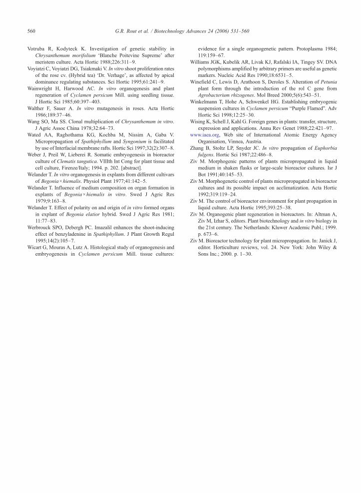
560 G.R. Rout et al. / Biotechnology Advances 24 (2006) 531–560
Votruba R, Kodyteck K. Investigation of genetic stability inChrysanthemum morifolium ‘Blanche Poitevine Supreme’ aftermeristem culture. Acta Hortic 1988;226:311–9.
Voyiatzi C, Voyiatzi DG, Tsiakmaki V. In vitro shoot proliferation ratesof the rose cv. (Hybrid tea) ‘Dr. Verhage’, as affected by apicaldominance regulating substances. Sci Hortic 1995;61:241–9.
Wainwright H, Harwood AC. In vitro organogenesis and plantregeneration of Cyclamen persicum Mill. using seedling tissue.J Hortic Sci 1985;60:397–403.
Walther F, Sauer A. In vitro mutagenesis in roses. Acta Hortic1986;189:37–46.
Wang SO, Ma SS. Clonal multiplication of Chrysanthemum in vitro.J Agric Assoc China 1978;32:64–73.
Wated AA, Raghothama KG, Kochba M, Nissim A, Gaba V.Micropropagation of Spathiphyllum and Syngonium is facilitatedby use of Interfacial membrane rafts. Hortic Sci 1997;32(2):307–8.
Weber J, Preil W, Lieberei R. Somatic embryogenesis in bioreactorculture of Clematis tangutica. VIIIth Int Cong for plant tissue andcell culture, Firenze/Italy; 1994. p. 202. [abstract].
Welander T. In vitro organogenesis in explants from different cultivarsof Begonia×hiemalis. Physiol Plant 1977;41:142–5.
Welander T. Influence of medium composition on organ formation inexplants of Begonia×hiemalis in vitro. Swed J Agric Res1979;9:163–8.
Welander T. Effect of polarity on and origin of in vitro formed organsin explant of Begonia elatior hybrid. Swed J Agric Res 1981;11:77–83.
Werbrouck SPO, Debergh PC. Imazalil enhances the shoot-inducingeffect of benzyladenine in Spathiphyllum. J Plant Growth Regul1995;14(2):105–7.
Wicart G, Mouras A, Lutz A. Histological study of organogenesis andembryogenesis in Cyclamen persicum Mill. tissue cultures:
evidence for a single organogenetic pattern. Protoplasma 1984;119:159–67.
Williams JGK, Kubelik AR, Livak KJ, Rafalski IA, Tingey SV. DNApolymorphisms amplified by arbitrary primers are useful as geneticmarkers. Nucleic Acid Res 1990;18:6531–5.
Winefield C, Lewis D, Arathoon S, Deroles S. Alteration of Petuniaplant form through the introduction of the rol C gene fromAgrobacterium rhizogenes. Mol Breed 2000;5(6):543–51.
Winkelmann T, Hohe A, Schwenkel HG. Establishing embryogenicsuspension cultures in Cyclamen persicum “Purple Flamed”. AdvHortic Sci 1998;12:25–30.
Wising K, Schell J, Kahl G. Foreign genes in plants: transfer, structure,expression and applications. Annu Rev Genet 1988;22:421–97.
www.iaea.org, Web site of International Atomic Energy AgencyOrganisation, Vinnea, Austria.
Zhang B, Stoltz LP, Snyder JC. In vitro propagation of Euphorbiafulgens. Hortic Sci 1987;22:486–8.
Ziv M. Morphogenic patterns of plants micropropagated in liquidmedium in shaken flasks or large-scale bioreactor cultures. Isr JBot 1991;40:145–53.
Ziv M. Morphogenetic control of plants micropropagated in bioreactorcultures and its possible impact on acclimatization. Acta Hortic1992;319:119–24.
Ziv M. The control of bioreactor environment for plant propagation inliquid culture. Acta Hortic 1995;393:25–38.
Ziv M. Organogenic plant regeneration in bioreactors. In: Altman A,Ziv M, Izhar S, editors. Plant biotechnology and in vitro biology inthe 21st century. The Netherlands: Kluwer Academic Publ.; 1999.p. 673–6.
Ziv M. Bioreactor technology for plant micropropagation. In: Janick J,editor. Horticulture reviews, vol. 24. New York: John Wiley &Sons Inc.; 2000. p. 1–30.