Tibial Shaft Fractures - Orthopaedic Trauma Association...
27
1 Boot Camp 2012 Boot Camp 2012 Tibial Shaft Fractures Tibial Shaft Fractures Philip Wolinsky University of California at Davis Tibial Shaft Tibial Shaft Fractures Fractures • Most common long bone Most common long bone fracture fracture • 492,000 fractures yearly 492,000 fractures yearly • Average 7.4 day hospital Average 7.4 day hospital stay stay Broad Broad Range Range of of Injuries Injuries • Low energy: Low energy: – Non Non-displaced displaced Simple patterns Simple patterns – Simple patterns Simple patterns – Heal reliably Heal reliably with simple with simple immobilization immobilization
-
Upload
vuongxuyen -
Category
Documents
-
view
223 -
download
2
Transcript of Tibial Shaft Fractures - Orthopaedic Trauma Association...

1
Boot Camp 2012Boot Camp 2012
Tibial Shaft FracturesTibial Shaft Fractures
pp
Philip WolinskyUniversity of California at Davis
Tibial Shaft Tibial Shaft FracturesFractures
•• Most common long bone Most common long bone fracturefracture
•• 492,000 fractures yearly492,000 fractures yearly
•• Average 7.4 day hospital Average 7.4 day hospital staystay
Broad Broad Range Range of of InjuriesInjuries
•• Low energy:Low energy:–– NonNon--displaceddisplaced
Simple patternsSimple patterns–– Simple patternsSimple patterns
–– Heal reliably Heal reliably with simple with simple immobilizationimmobilization

2
High Energy High Energy Fx’sFx’s
•• OOpen fracturespen fractures
•• Closed soft tissue Closed soft tissue injuriesinjuries
•• Bony Bony comminutioncomminution
High Energy High Energy Fx’sFx’s
•• Problems Problems obtaining obtaining union union
Tibi iTibi i•• Tibia is Tibia is subcutaneous, with a subcutaneous, with a relatively poor blood relatively poor blood supplysupply
History & Physical: Low History & Physical: Low EnergyEnergyMinimal Minimal softsoft--tissue injurytissue injury
•• Less complicated fracture Less complicated fracture patternpatternpp–– 76.5% closed76.5% closed
–– 53.5% mild soft53.5% mild soft--tissue tissue energy energy

3
History & Physical: High History & Physical: High EnergyEnergy
High High incidence of incidence of neurovascular neurovascular injury injury and open and open woundswounds
High suspicion for High suspicion for compartment compartment syndromesyndrome
Radiographic EvaluationRadiographic Evaluation
•• Full length AP and Full length AP and lateral Viewslateral Views
•• Include/check Include/check joint joint above & belowabove & below
Associated InjuriesAssociated Injuries
•• 30% of patients will have 30% of patients will have other other injuriesinjuries
–– Ipsilateral Fibula Ipsilateral Fibula ppFractureFracture
–– Foot & Ankle injuryFoot & Ankle injury
–– Ligamentous Ligamentous knee knee injuriesinjuries

4
ClassificationClassification•• By location:By location:
•• Proximal 1/3Proximal 1/3
•• Middle 1/3Middle 1/3
•• Distal 1/3Distal 1/3
•• By fracture By fracture pattern:pattern:
–– TransverseTransverse, oblique, , oblique, spiralspiral
ButterflyButterfly
ComminutedComminuted
AO/OTA ClassificationAO/OTA Classification
Hansen Hansen WinquistWinquist Classification:Classification:Degree Degree of of ComminutionComminution
•• 1: 1: minimal minimal comminutioncomminution
•• 2: 2: <50% <50%
•• 33:: >50%>50%
•• 4: 100% or 4: 100% or a a segmental segmental
fracturefracture
•• 5: 5: segmental bone segmental bone lossloss

5
Absolute OAbsolute Operative perative IIndicationsndications
•• Open Open fracturesfractures
•• Vascular injuryVascular injury
•• Compartment syndromeCompartment syndrome
Relative Relative Operative IndicationsOperative Indications
Floating Floating kneeknee
Intact fibulaIntact fibula
Segmental Segmental fracturefracture
S l d ft tiS l d ft tiSevere closed soft tissue Severe closed soft tissue injuriesinjuries
Multiple injuriesMultiple injuries
Ipsilateral limb injuriesIpsilateral limb injuries
IntraIntra--articular extensionarticular extension
Bilateral tibia fracturesBilateral tibia fractures
Acceptable AlignmentAcceptable Alignment
•• Defined as:Defined as:
•• <1 cm of shortening<1 cm of shortening
•• angulation < 5angulation < 5 7 degrees7 degrees•• angulation < 5angulation < 5--7 degrees7 degrees
•• rotational deformity <10 degreesrotational deformity <10 degrees
•• Based on minimal Based on minimal functional outcome datafunctional outcome data

6
Rotational Malalignment Tibial IMN’sRotational Malalignment Tibial IMN’s
•• 22 patients/CT scans22 patients/CT scans
•• 5/22 (22%) were off by5/22 (22%) were off by5/22 (22%) were off by 5/22 (22%) were off by more than 10 degreesmore than 10 degrees–– 3 were off by >= 15 3 were off by >= 15
degreesdegrees
Puloski et al JOT 18(7), 2004Puloski et al JOT 18(7), 2004
Closed Tibia FracturesClosed Tibia Fractures
Closed Closed FracturesFractures
•• “Standard” treatment for “Standard” treatment for “stable” closed tibia “stable” closed tibia fractures:fractures:
–– closed reductionclosed reduction–– closed reduction closed reduction
–– long leg castinglong leg casting
–– functional functional brace at about brace at about 33--6 weeks6 weeks
–– with early weight bearingwith early weight bearing

7
Cast bracing Cast bracing indications:indications: ““Stable” FracturesStable” Fractures
•• Closed transverse Closed transverse fractures that can fractures that can reducedreduced
•• Spiral, oblique, or Spiral, oblique, or comminuted fractures comminuted fractures with < 12 mm of with < 12 mm of initial shorteninginitial shortening
Functional BFunctional Bracing Relative Contraracing Relative Contra--IndicationsIndications
•• Intact fibulaIntact fibula
–– varus varus deformity > 5 deformity > 5 degreesdegrees likelylikelydegrees degrees likelylikely
•• Comminuted Comminuted fracturesfractures
–– take take longer to heallonger to heal
Sarmiento Sarmiento JBJS JBJS ‘‘8484
Closed Functional TreatmentClosed Functional Treatment
–– 1,000 1,000 TibialTibial FracturesFractures
–– 60% Lost to F/U60% Lost to F/U
Fracture CharacteristicsFracture Characteristics
–– All < 1.5cm shorteningAll < 1.5cm shortening
–– None with intact fibulaNone with intact fibula
–– Only 5% more than 8Only 5% more than 8°° varusvarus

8
Sarmiento Sarmiento JBJS JBJS ‘‘8484
Treatment CourseTreatment Course
–– Average 3.7 wks in long leg Average 3.7 wks in long leg castcast
–– Transition to functional Transition to functional ffracture braceracture brace
–– Early WBATEarly WBAT
–– Stable patterns!Stable patterns!
SarmientoSarmiento•• Union RateUnion Rate
–– 98.5%98.5%
•• Time to UnionTime to Union–– 18.1 weeks18.1 weeks
•• ShorteningShortening–– <1.4%<1.4%
•• Initial Shortening = Final Initial Shortening = Final ShorteningShortening
20 20 YO Walked into a cement plantarYO Walked into a cement plantar

9
InjuryInjury
Post CastPost Cast
Post CastingPost Casting

10
Unstable Unstable Fracture Fracture PPatternsatterns
•• In general “unstable” fractures need In general “unstable” fractures need operative stabilizationoperative stabilization
•• HighHigh--energy fractures:energy fractures:gg gygy–– higher incidence of delayed union higher incidence of delayed union
•• Severe closed soft tissue injurySevere closed soft tissue injury
•• Articular surface involvementArticular surface involvement
Definition: Unstable Fracture PatternsDefinition: Unstable Fracture Patterns
•• 100% displacement of the fracture on the 100% displacement of the fracture on the initial initial filmfilm
0%0% i ii i f hf h•• >50% >50% comminutioncomminution of the of the cortexcortex
•• Fibula Fibula fracture at the same level as the tibia fracture at the same level as the tibia fracturefracture
IMN vs. Closed Treatment of IMN vs. Closed Treatment of Isolated, closed, “unstable” Isolated, closed, “unstable” fracturesfracturesLiterature SummaryLiterature Summary
Treatment with an Treatment with an IMN IMN vsvs closed closed rxrx::
Sh i h li i h IMNSh i h li i h IMNShorter time to healing with IMNShorter time to healing with IMN
Higher union rate with IMNHigher union rate with IMN
Functional scores, general health status all Functional scores, general health status all favor IMNfavor IMN

11
IMN vs. Closed Treatment of IMN vs. Closed Treatment of Isolated, closed, “unstable” fracturesIsolated, closed, “unstable” fractures
Closed treatment:Closed treatment:–– Increased disabilityIncreased disability
–– 15% had hindfoot stiffness15% had hindfoot stiffness
–– 22% of those initially treated closed had an 22% of those initially treated closed had an operative procedure when reduction could not operative procedure when reduction could not be maintainedbe maintained
IM Nails IM Nails –– Bone et.al.Bone et.al.
Retrospective review 99 patientsRetrospective review 99 patients
CastCast NailNailCastCast NailNail
Time to unionTime to union 26 wks26 wks 18 wks18 wks
SFSF--36 36 74 74 8585
Knee score 89 Knee score 89 9696
Ankle score 84 Ankle score 84 97 97 Bone JBJS ‘97
Starting HoleStarting Hole
•• AP: Canal is just AP: Canal is just lateral to midlinelateral to midline
•• Lateral :Anterior Lateral :Anterior corner of the corner of the jointjoint
Tornetta, JOT 1999

12
Intramedullary Nailing (IMNIntramedullary Nailing (IMN))
•• Reamed interlocked nails:Reamed interlocked nails:–– High union ratesHigh union rates–– Low malunion ratesLow malunion rates–– Low infection ratesLow infection rates
•• Primary indication:Primary indication:•• diaphyseal fracturesdiaphyseal fractures
–– distal fractures within 4 cm of the ankle jointdistal fractures within 4 cm of the ankle joint
ReamedReamed NonNon--ReamedReamed
# pts. # pts. 73 73 6363
Nonunion Nonunion 4% 4% 11%11%
Ream or NotReam or Not??
MalunionMalunion 4% 4% 3%3%
Broken BoltsBroken Bolts 3% 3% 16%16%
Time to UnionTime to Union 16.7 16.7 wkswks 25.7 25.7 wkswks
Larsen JOT Larsen JOT ‘‘0404
CLOSEDCLOSED Fractures:Fractures:Reamed vs. Unreamed NailsReamed vs. Unreamed Nails
•• Prospective randomized studiesProspective randomized studies
•• NonreamedNonreamed nails:nails:I dI d ti t iti t i–– Increased Increased time to uniontime to union
–– Increased locking screw Increased locking screw breakage (old nails)breakage (old nails)
Knee pain and compartment syndrome rates were Knee pain and compartment syndrome rates were similarsimilar

13
Reamed Reamed vsvs Unreamed: Unreamed: SPRINTSPRINT
•• Possible benefit of reamed IM nails Possible benefit of reamed IM nails for for closed fracturesclosed fractures
•• No differenceNo difference forfor open fracturesopen fracturesNo difference No difference for for open fracturesopen fractures
•• Delaying reoperation for nonunion for at Delaying reoperation for nonunion for at least 6 months significantly lowers the need least 6 months significantly lowers the need for reoperation for reoperation
Bhandari M, et al JBJS, 2008
Knee pain s/p IMN Knee pain s/p IMN
•• Occurs in 10Occurs in 10-- 60% of patients60% of patients
•• No difference in knee pain if a patellarNo difference in knee pain if a patellarNo difference in knee pain if a patellar No difference in knee pain if a patellar tendon splitting approach is used vs. a tendon splitting approach is used vs. a parapatellar incisionparapatellar incision
•• Usually activity related and made worse by Usually activity related and made worse by kneelingkneeling
Knee pain s/p IMNKnee pain s/p IMN
•• In one study there was no correlation In one study there was no correlation between nail protrusion and knee painbetween nail protrusion and knee pain
•• 80% of patients had total or partial pain 80% of patients had total or partial pain relief with nail removalrelief with nail removal
•• Cause is unknownCause is unknown

14
Standard Standard ORIF: Tibial ShaftORIF: Tibial Shaft
•• Open reduction with Open reduction with wide exposures is usually wide exposures is usually avoided because avoided because of of infection and soft tissue infection and soft tissue complicationscomplicationscomplications.complications.
•• MIPPO MIPPO techniques:techniques:–– Distal and proximal Distal and proximal
1/3 fractures1/3 fractures
“MIPO” ORIF“MIPO” ORIF
•• Relative plating indications:Relative plating indications:
–– Fractures with extension into the ankle Fractures with extension into the ankle or knee jointor knee joint
–– Arterial injuries requiring repair Arterial injuries requiring repair (exposure (exposure may may already be done)already be done)
–– Proximal and distal 1/3 fractures Proximal and distal 1/3 fractures (increased incidence of deformity with (increased incidence of deformity with IMN’s)IMN’s)
External FixationExternal Fixation
•• Minimizes further disruption of the soft Minimizes further disruption of the soft tissue and blood supply of fracture tissue and blood supply of fracture fragmentsfragments
•• Current indications:Current indications:–– Initial Initial rxrx high grade high grade oopen fractures with pen fractures with
massive contaminationmassive contamination–– Damage control orthopedics:Damage control orthopedics:
•• “Sick” patients“Sick” patients•• “Sick limb”“Sick limb”

15
External FixationExternal Fixation
•• Complications: Complications: –– MalunionMalunion
–– Delayed and nonunionDelayed and nonunionDelayed and nonunionDelayed and nonunion
–– Pin tract infectionsPin tract infections
•• ***Higher rate of malunion compared with ***Higher rate of malunion compared with IMNIMN
Open Tibial Shaft FracturesOpen Tibial Shaft Fractures
Mechanism of InjuryMechanism of Injury
Lower Lower energy, torsional type injury (e.g., energy, torsional type injury (e.g., skiing)skiing)
More common with higher energy direct force More common with higher energy direct force (e.g. car bumper)(e.g. car bumper)

16
Priorities
ABC’S Assoc Injuries Tetanus Antibiotics Soft Tissue
Management/ Fixation
Long term issues
Physical ExaminationPhysical Examination
•• Given subcutaneous Given subcutaneous nature of tibia, deformity nature of tibia, deformity and open wound usually and open wound usually readily apparentreadily apparenty ppy pp
•• Circumferential inspection Circumferential inspection of soft tissue envelope, of soft tissue envelope, noting any lacerations, noting any lacerations, ecchymosis, swelling, and ecchymosis, swelling, and tissue turgiditytissue turgidity
Physical ExamPhysical ExamNeurologic Neurologic and vascular exam of extremity and vascular exam of extremity including ABIincluding ABI’’s if s if indicatedindicated
Wounds Wounds assessed once in ER, then covered with assessed once in ER, then covered with ,,sterile gauze dressing until treated in ORsterile gauze dressing until treated in OR-- digital digital camera / cell phone camera / cell phone
Wound classification after Wound classification after surgical surgical debridementdebridement

17
Classification of Open Tibia Classification of Open Tibia FracturesFractures
•• Gustilo and Anderson open fracture classification first published Gustilo and Anderson open fracture classification first published in 1976 and later modified in 1984in 1976 and later modified in 1984
•• In one study interobserver agreement on classification only 60%In one study interobserver agreement on classification only 60%
Limb SalvageLimb Salvage
•• Over all assessment of the limb and the Over all assessment of the limb and the patientpatient–– Associated injuriesAssociated injuries–– Age/ preAge/ pre--existing medical conditionsexisting medical conditions–– Degree of muscle damageDegree of muscle damage–– Bony injuryBony injury–– Vascular injuryVascular injury–– Plantar sensationPlantar sensation
•• Anatomic disruption!!!!!!!!!!!!!Anatomic disruption!!!!!!!!!!!!!
LEAP DataLEAP Data
•• Outcome at 2 and 7 years was the same for Outcome at 2 and 7 years was the same for amputees and salvaged limbsamputees and salvaged limbs
•• All patients were severely disabledAll patients were severely disabled
•• Salvage has a higher incidence of Salvage has a higher incidence of complications, more operations, and more complications, more operations, and more hospitalizationshospitalizations

18
Limb salvage scoring systemsLimb salvage scoring systems•• Low sensitivityLow sensitivity
•• High specificityHigh specificity
•• About 20% of amputations occur at scores About 20% of amputations occur at scores below the cutoff valuebelow the cutoff value
•• Do not use scoring systems alone to Do not use scoring systems alone to determine amputation vs. salvagedetermine amputation vs. salvage
Open FracturesOpen Fractures
•• Infection incidence depends onInfection incidence depends on::–– *Degree *Degree of soft tissue and bone injuryof soft tissue and bone injury
–– *Extent*Extent of contaminationof contaminationExtent Extent of contaminationof contamination
–– Timing/ use Timing/ use of antibioticsof antibiotics
–– Adequacy Adequacy of of debridmentdebridment
•• *not under surgeon control*not under surgeon control
Open Fracture TreatmentOpen Fracture Treatment
Surgical emergencySurgical emergency
ER ER wound care:wound care:
cover cover with a sterile dressingwith a sterile dressing
Debride wound and stabilize fracture in Debride wound and stabilize fracture in OROR
ReRe--debride every 48debride every 48--72 hours until the wound 72 hours until the wound is is healthy, if neededhealthy, if needed

19
AntibioticsAntibiotics
•• Closed, grade 1, grade II, grade IIIA open Closed, grade 1, grade II, grade IIIA open fractures: fractures: –– Cephalosporin for 24Cephalosporin for 24--48 hours48 hours
•• Grade IIIB and IIIC injuries:Grade IIIB and IIIC injuries:–– add amino glycosideadd amino glycoside
•• Anaerobic contamination (barnyard injury):Anaerobic contamination (barnyard injury):–– add penicillinadd penicillin
Open Fracture TreatmentOpen Fracture Treatment
•• AntibioticsAntibiotics: : 2424--72 72 hours initiallyhours initially
•• 24 hours after subsequent24 hours after subsequent debridmentsdebridments•• 24 hours after subsequent 24 hours after subsequent debridmentsdebridments
•• Soft tissue coverage should be obtained as Soft tissue coverage should be obtained as early as possibleearly as possible
Treatment of Soft Tissue InjuryTreatment of Soft Tissue Injury
•• Careful planning of skin Careful planning of skin incisionsincisions
•• Essential Essential to fully explore wound as even to fully explore wound as even Type 1 fractures can pull dirt/debris back Type 1 fractures can pull dirt/debris back ype actu es ca pu d t/deb s bacype actu es ca pu d t/deb s bacinto wound and on fracture into wound and on fracture endsends
•• All foreign material, necrotic muscle, All foreign material, necrotic muscle, unattached bone fragments, exposed fat and unattached bone fragments, exposed fat and fascia are debridedfascia are debrided

20
Large Fragments: What to do?
Infection Rates if retained - 21%
• Infection Rates if removed- 9%• Edwards CC, CORR, 1998
• Use to assist in determining length, rotation and alignment
Bone DefectsBone Defects
•• PMMA PMMA ––aminoglycoside +/aminoglycoside +/-- vancomycinvancomycin
•• Bead pouchBead pouch
•• Solid spacerSolid spacer
Bone Defects: PMMA SpacerBone Defects: PMMA SpacerMasqueletMasquelet AC, Reconstruction of the long bones by the induced AC, Reconstruction of the long bones by the induced
membrane and spongy membrane and spongy autograftautograft [French]. [French]. Ann Ann ChirChir PlastPlast EsthetEsthet 20002000

21
Soft Tissue CoverageSoft Tissue Coverage
•• Definitive coverage should be performed within 7Definitive coverage should be performed within 7--10 days if possible10 days if possible
•• Most type 1 wounds will heal by secondary intent Most type 1 wounds will heal by secondary intent or can be closed primarily or can be closed primarily Hohmann E, Comparison of delayed Hohmann E, Comparison of delayed and primary wound closure in the treatment of and primary wound closure in the treatment of open tibial fractures. open tibial fractures. Arch Arch Orthop Trauma Surg 2007Orthop Trauma Surg 2007
•• Delayed primary closure usually feasible for type Delayed primary closure usually feasible for type 2 and type 3a fractures2 and type 3a fractures
Soft Tissue CoverageSoft Tissue Coverage
•• Type 3b fractures require either local Type 3b fractures require either local advancement or rotation flap, splitadvancement or rotation flap, split--thickness skin graft, or free flapthickness skin graft, or free flap
•• STSG suitable for coverage of large STSG suitable for coverage of large defects with underlying viable muscledefects with underlying viable muscle
Soft Tissue CoverageSoft Tissue Coverage•• Proximal third tibia Proximal third tibia
fractures can be covered fractures can be covered with gastrocnemius with gastrocnemius rotation flaprotation flap
•• Middle third tibia Middle third tibia fractures can be covered fractures can be covered with soleus rotation flapwith soleus rotation flap
•• Distal third fractures Distal third fractures usually require free flap usually require free flap for coveragefor coverage

22
Stabilization of Open Tibia FracturesStabilization of Open Tibia Fractures
OOptions ptions depending on fracture pattern and soft depending on fracture pattern and soft tissue injury:tissue injury:
IM nailIM nail-- reamed vs. unreamedreamed vs. unreamed
External fixationExternal fixation
ORIFORIF
Unreamed IMN + Open FracturesUnreamed IMN + Open Fractures
Combined with aggressive debridmentCombined with aggressive debridment
•• Pooled data Pooled data
•• Grade 1: < 3% infectionGrade 1: < 3% infection
•• Grade II: 4%Grade II: 4%
•• Grade IIIA: 7%Grade IIIA: 7%
•• Grade IIIB: 17%Grade IIIB: 17%
•• Infection probably more related to degree of injury Infection probably more related to degree of injury rather than implantrather than implant

23
Reamed Reamed vsvs Unreamed: Unreamed: SPRINTSPRINT
•• Possible benefit of reamed IM nails Possible benefit of reamed IM nails for for closed closed fracturesfractures
•• No difference No difference for for open fracturesopen fractures
D l i ti f i f tD l i ti f i f t•• Delaying reoperation for nonunion for at Delaying reoperation for nonunion for at least 6 months significantly lowers the need least 6 months significantly lowers the need for reoperation for reoperation
BMPsBMPs
•• BMPBMP--2 (Infuse) FDA approval in subset of 2 (Infuse) FDA approval in subset of open tibia fractures open tibia fractures BESTT study group JBJS 84, 2002BESTT study group JBJS 84, 2002
•• Significant reduction in the incidence ofSignificant reduction in the incidence ofSignificant reduction in the incidence of Significant reduction in the incidence of secondary proceduressecondary procedures
•• Accelerated healingAccelerated healing
•• Lower infectionsLower infections
Compartment SyndromeCompartment Syndrome
24
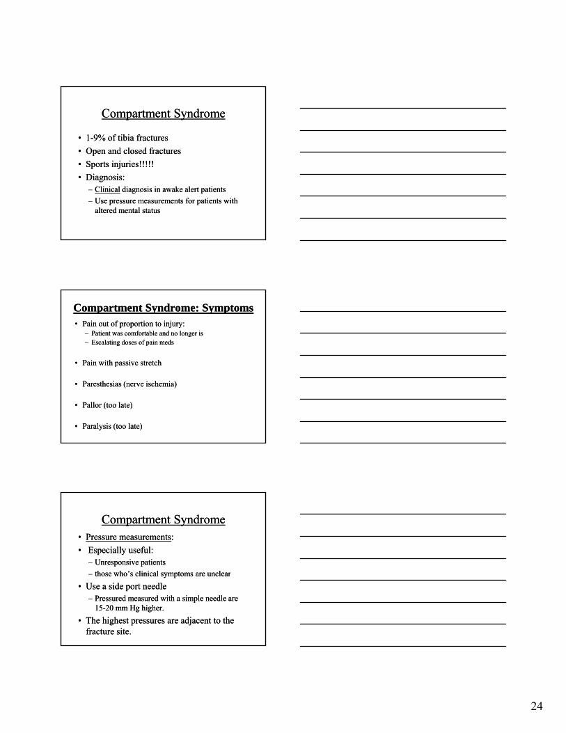
Compartment SyndromeCompartment Syndrome
•• 11--9% of tibia fractures9% of tibia fractures
•• Open and closed Open and closed fracturesfractures
•• Sports injuries!!!!!Sports injuries!!!!!•• Sports injuries!!!!!Sports injuries!!!!!
•• Diagnosis: Diagnosis: –– ClinicalClinical diagnosis in awake alert patientsdiagnosis in awake alert patients
–– Use Use pressure measurements for patients with pressure measurements for patients with altered mental statusaltered mental status
Compartment Syndrome: SymptomsCompartment Syndrome: Symptoms•• Pain out of proportion to Pain out of proportion to injury:injury:
–– Patient was comfortable and no longer isPatient was comfortable and no longer is–– Escalating doses of pain medsEscalating doses of pain meds
•• Pain with passive stretchPain with passive stretchpp
•• Paresthesias (nerve ischemia)Paresthesias (nerve ischemia)
•• Pallor Pallor (too late(too late))
•• Paralysis Paralysis (too late(too late))
Compartment SyndromeCompartment Syndrome•• Pressure measurementsPressure measurements::
•• Especially useful:Especially useful:–– Unresponsive patientsUnresponsive patients
–– those who’s clinical symptoms are unclearthose who’s clinical symptoms are unclear
•• Use a side port needleUse a side port needle–– Pressured measured with a simple needle are Pressured measured with a simple needle are
1515--20 mm Hg higher.20 mm Hg higher.
•• The highest pressures are adjacent to the The highest pressures are adjacent to the fracture site.fracture site.

25
Pressures Not UniformPressures Not Uniform
•• Highest at Fracture Highest at Fracture SiteSite
•• Highest Pressures Highest Pressures in:in:–– Deep PosteriorDeep Posterior
–– AnteriorAnterior
•• Heckman JBJS Heckman JBJS ’’7676
ICP ICP Threshold for Threshold for FasciotomyFasciotomy
•• Absolute pressure is unclear: 30Absolute pressure is unclear: 30-- 45 mm Hg45 mm Hg
•• ΔP: DBPΔP: DBP-- ICP <30 mm ICP <30 mm HgisHgis less than less than 30mm 30mm HgHg
Tibial CompartmentsTibial Compartments
•• AnteriorAnterior::–– Deep peroneal nerveDeep peroneal nerve
–– Sensation to 1Sensation to 1stst web web spacespacespacespace
•• LateralLateral::–– Sup peroneal nerveSup peroneal nerve
–– Sensation to dorsum of Sensation to dorsum of footfoot

26
Tibial CompartmentsTibial Compartments
•• Deep posteriorDeep posterior::–– Tibial nerveTibial nerve
–– Sensation to sole of footSensation to sole of foot
•• Superficial posteriorSuperficial posterior::–– Medial sural cutaneous Medial sural cutaneous
nervenerve
–– No predictable sensationNo predictable sensation
Management:#1Management:#1-- Split Casts and Split Casts and Bandages!Bandages!
•• Circumferential casts or dressing decrease Circumferential casts or dressing decrease the volume of a compartmentthe volume of a compartment
ManagementManagement
•• 44--compartment compartment fasciotomyfasciotomy
•• One (lateral) or two One (lateral) or two (medial and lateral) (medial and lateral) incisionsincisions

27
ManagementManagement•• Long Long skin and skin and fascialfascial
incisionsincisions
•• Leave themLeave them openopen•• Leave them Leave them open open ((vacvac))
•• Back to OR every 48 Back to OR every 48 hours or sohours or so
•• Closure vs. Closure vs. STSGSTSG
EndEnd