Thyroid Gland Anatomy - uomustansiriyah.edu.iq03_22... · Thyroid Lobes •The thyroid gland...
38
Thyroid Gland Anatomy
Transcript of Thyroid Gland Anatomy - uomustansiriyah.edu.iq03_22... · Thyroid Lobes •The thyroid gland...

Thyroid Gland Anatomy
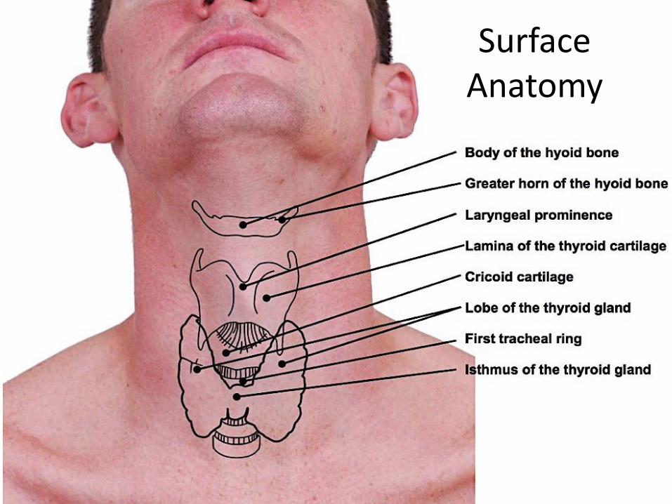
Surface Anatomy
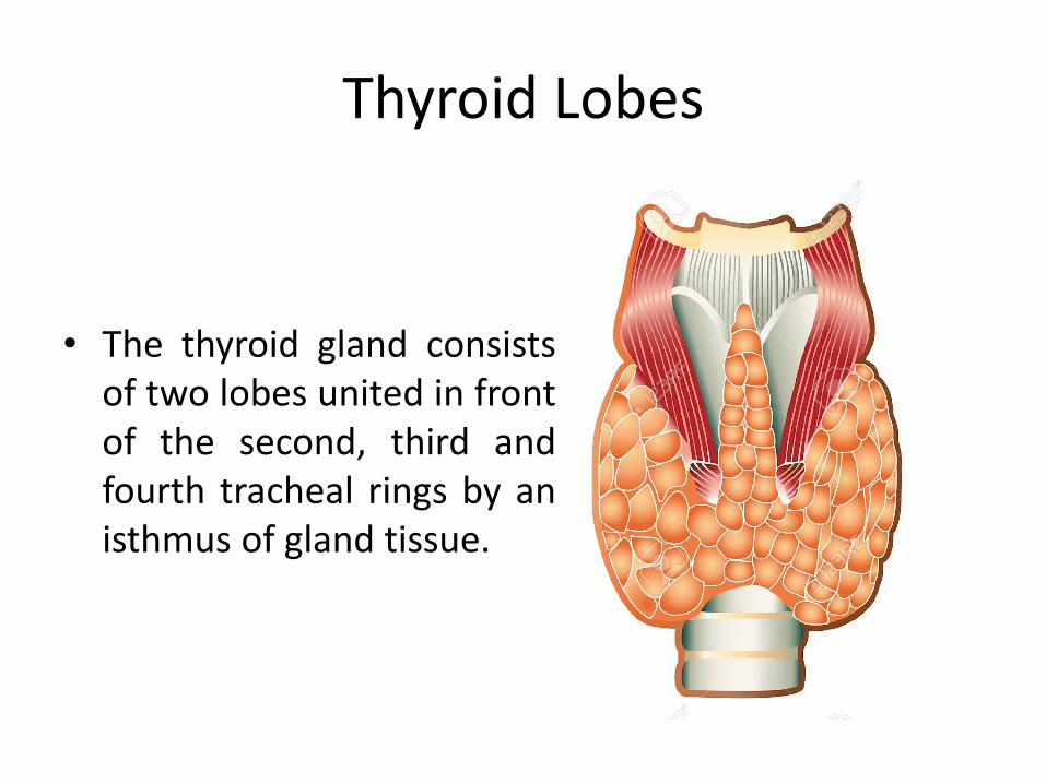
Thyroid Lobes
• The thyroid gland consistsof two lobes united in frontof the second, third andfourth tracheal rings by anisthmus of gland tissue.

Thyroid Gland Position
• It lies under cover of sternothyroid and sternohyoidmuscles on the side of the larynx and trachea
stern
oth
yro
id
stern
ohyoid
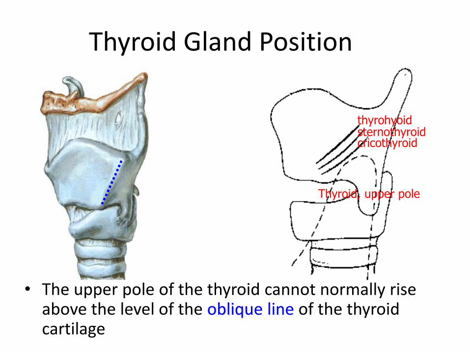
Thyroid Gland Position
• The upper pole of the thyroid cannot normally rise above the level of the oblique line of the thyroid cartilage
Thyroid, upper pole
sternothyroidthyrohyoid
cricothyroid

Thyroid Gland Position

Thyroid Gland Position
• The lower pole of the thyroid gland extends along the side of the trachea as low as the sixth tracheal ring
123456

Thyroid Gland Position
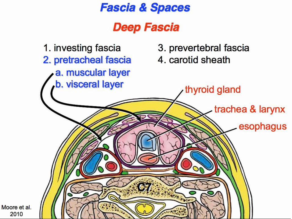

Pre-tracheal fascia attachment

Thyroid Blood Supply

Superior thyroid artery
• enters deep to sternothyroid
stern
oth
yro
id
Superior thyroid vessels

Inferior thyroid artery
• Ascends and turns medially at the level of the cricoid cartilage to enter the back of the gland some distance above the lower pole.

Inferior thyroid artery

Remember this!
• The inferior thyroid artery divides outside the pre-
tracheal fascia into four or five branches that pierce
the fascia separately to reach the lower pole of the
gland.
• The superior thyroid artery divides within the
pretracheal fascia.
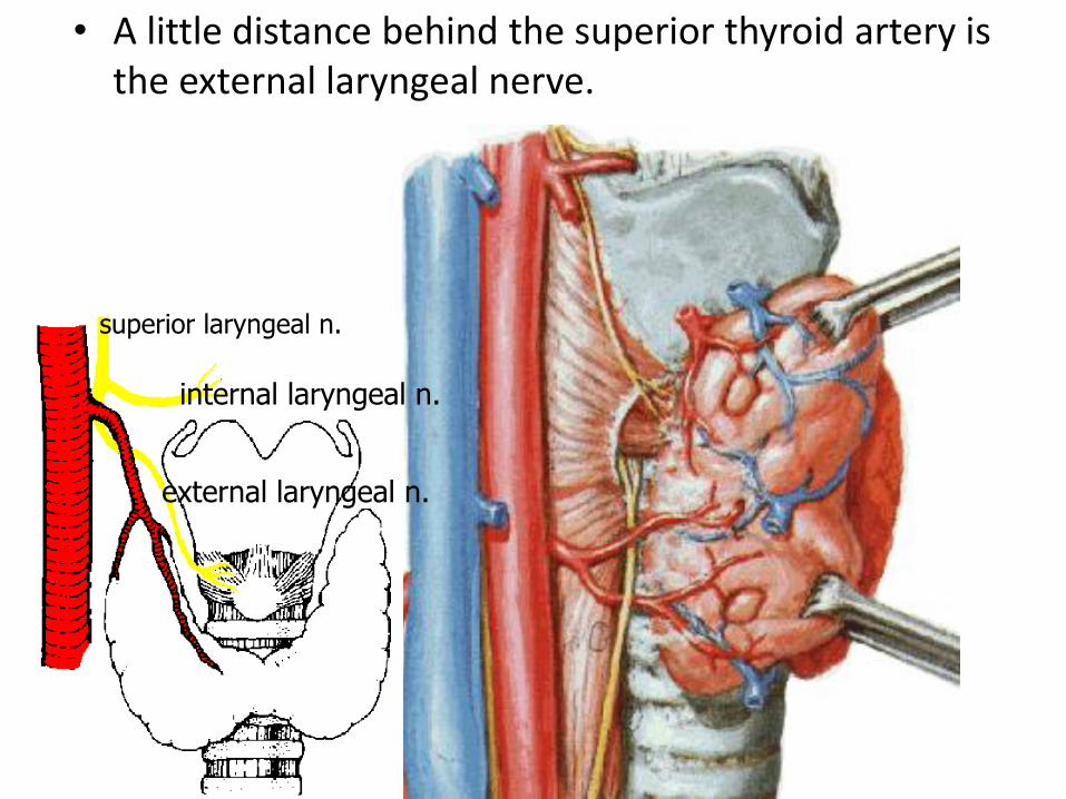
• A little distance behind the superior thyroid artery is the external laryngeal nerve.
external laryngeal n.
internal laryngeal n.
superior laryngeal n.

• The recurrent laryngeal nerve always lies behind the pre-tracheal fascia and if this structure remains intact during thyroidectomy the nerve will not have been divided

• Both thyroid arteries are related to nerves which must be avoided when tying the arteries.

• To avoid injury to the external laryngeal nerve, the superior thyroid artery is ligated and sectioned near the superior pole of the thyroid gland where it is not so closely related to the nerve as it is at its origin.

• hence the advisability of ligating the inferior thyroid artery well lateral to the glandbefore it begins to divide into its terminal branches.
• the inferior thyroid artery gives off esophageal and inferior laryngeal branches before its terminal distribution into the thyroid gland

Thyroidae ima artery
• In about 10% of individuals, an unpaired artery, the thyroidae ima is a small occasional artery from the brachiocephalic trunk, or left common carotid artery, or direct from the arch of the aorta

Thyroidae ima artery
• The possible presence of the thyroid ima artery must be remembered when incising the trachea inferior to the isthmus.
• As the thyroidae ima runs anterior to the trachea, it is a potential source of serious bleeding

Thyroid veins
• The veins are three in number on each side
• the superior thyroid vein from the upper pole follows the artery and enters the internal jugular vein or the common facial vein
Superior thyroid v.
Internal jugular v.

Thyroid veins
• The middle thyroid vein is short and wide, it enters the internal jugular vein middle thyroid v.
Internal jugular v.

Thyroid veins
• From the isthmus and lower pole of the gland the inferior thyroid veins form a plexus within the pre-tracheal fascia that descends in front of the trachea to reach the left brachiocephalic vein
inferior thyroid vv.
brachiocephalic v.

Inferior thyroid veins
• As the inferior thyroid veins cover the anterior surface of the trachea inferior to isthmus, they are potential sources of bleeding during tracheotomy (also remember the situation of the thyroidae ima artery).

Development of the thyroid gland
• After the tongue has developed, it can be seen that the point of outgrowth of the thyroglossal duct is the foramen cecum (of Morgagni) [Morgagni, Giovanni
Battista, 1682-1771, a Padua anatomist and pathologist, also known for hydatid of Morgagni (appendix testis) and anal columns (of Morgagni)].

Thyroglossal cyst
• cysts derived from the duct may also appear anywhere between the foramen cecum and the normal position in the midline of the neck
1. Beneath foramen cecum2. Floor of the mouth3. Suprahyoid4. Subhyoid5. On thyroid cartilage6. At level of cricoid cartilage

Thyroglossal cyst
• Can be diagnosed because characteristically it moves upwards as the patient puts his tongue out.

Lingual thyroid
• Rarely the thyroid fails to descend during development resulting in the development of a lingual thyroid

Ectopic thyroid
• Failure of descent mar result in a superior cervical thyroid in the region of the hyoid bone
• the thyroid may sometimes descended too far and be found in the superior mediastinum
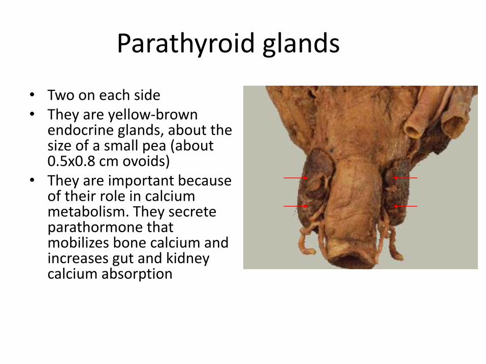
Parathyroid glands
• Two on each side• They are yellow-brown
endocrine glands, about the size of a small pea (about 0.5x0.8 cm ovoids)
• They are important because of their role in calcium metabolism. They secrete parathormone that mobilizes bone calcium and increases gut and kidney calcium absorption

Parathyroid glands
• Are located posterior to the thyroid gland between its capsule and fascial sheath
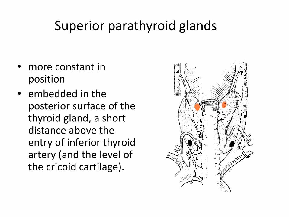
Superior parathyroid glands
• more constant in position
• embedded in the posterior surface of the thyroid gland, a short distance above the entry of inferior thyroid artery (and the level of the cricoid cartilage).

Inferior parathyroid glands
• variable in position
• usually embedded behind the lower pole but is often found elsewhere (they may even present in the superior mediastinum).

Parathyroid glands, blood supply
• The glands are usually supplied by the inferior thyroid arteries but may also be supplied by both superior and inferior thyroid arteries
posterior br.of superiorthyroid a.
inferiorthyroid a.

Parathyroid glands
• Awareness of the close relationship between the parathyroid glands and the thyroid gland is essential to prevent removal or damage of the parathyroid glands during thyroidectomy.