Thrombus (stationary clot) occludes a branch of the central retinal vein Blockage causes bleeding...
37
Retina
-
Upload
philip-henry -
Category
Documents
-
view
216 -
download
0
Transcript of Thrombus (stationary clot) occludes a branch of the central retinal vein Blockage causes bleeding...
Retina

Baby Picture of the Day!

Branch Retinal Vein Occlusion (BRVO)Thrombus (stationary
clot) occludes a branch of the central retinal vein
Blockage causes bleeding from that branch
Concerned about neovascularization afterward (due to ischemia)

Central Retinal Vein Occlusion (CRVO)Like a branch
retinal vein occlusion, but the whole central retinal vein is occluded
Bleeding all over retina

Branch Retinal Artery Occlusion (BRAO)Embolus
(traveling clot) occludes a branch of the central retinal artery

Central Retinal Artery Occlusion (CRAO)Like BRAO, but
the whole central retinal artery is occluded
Ischemia of entire retina
Macula is spared due to its dual blood supply (choroid)

Diabetic Retinopathy (DR)Biggest risk factor
= number of years with diabetes
Caused by damage to retinal capillaries
Non-proliferative DR = ischemia, hemorrhages, but no neovascularization
Proliferative DR = neovascularization
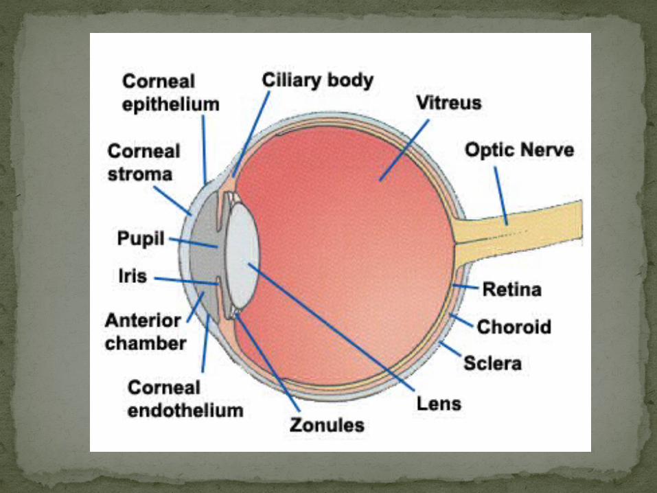
Hypertensive RetinopathyBilateral,
asymmetricNarrowing of
arteriolesA/V nicking (vein
presses on artery)IschemiaSwelling of optic
nerve headMacular star
(exudates)
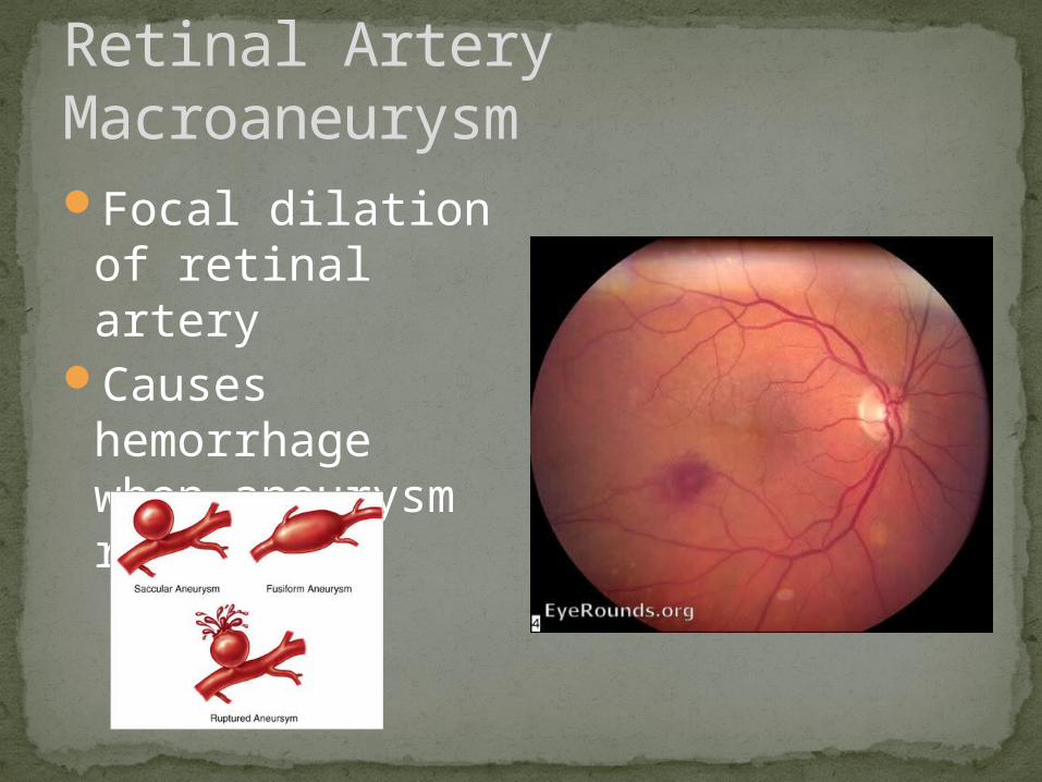
Retinal Artery MacroaneurysmFocal dilation of
retinal arteryCauses
hemorrhage when aneurysm ruptures

HIV RetinopathyIschemia (cotton
wool spots)Retinal
hemorrhagesAsymptomaticNot infectious

Interferon RetinopathyLooks like HIV retinopathyCaused by use of interferon (usually
for hepatitis)

Talc RetinopathyBilateralIV drug useDeposits near
maculaMay occlude
capillaries and cause ischemia

Retinopathy of Prematurity (ROP) / Retrolental FibroplasiaBabies born <36
weeksNasal vessels
form first, young ROP patients don’t have temporal vessels formed
Neovascularization can occur due to ischemia

Retinoblastoma#1 intraocular
malignancy in children
Tumor of developing retinal cells
Leukocoria

Congenital Hypertrophy of the RPE (CHRPE)BenignNon-progressive

Choroidal NevusPresent at birthNon-progressive
(usually)May progress to
melanomaUse red-free
filter (green light) to distinguish from CHRPE
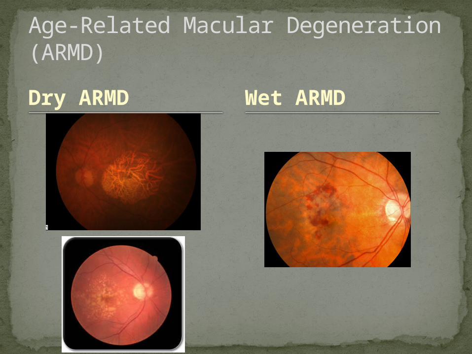
Dry ARMD
Age-Related Macular Degeneration (ARMD)
Wet ARMD

Acquired MaculopathiesCentral serous
choroidopathy (CSR)
HistoplasmosisPathological
myopia
Epiretinal membrane (ERM) / Macular pucker
Macular holeAlbinism

Central Serous Choroidopathy (CSR)Plasma
underneath the macula
Young men, high stress
May significantly reduce VA
Usually improve without treatment

Histoplasmosis“Histo belt” (Ohio-
Mississippi River Valley)
Fungus infectionAtrophy of optic
nerveLesions in
peripheral retinaMaculopathy with
possible neovascularization
Clear vitreous

Pathological MyopiaRx >6D ORAxial length >26mmProblems come
from the eye stretching to large size
#1: Posterior staphyloma = posterior retinal thinning/bulging

Epiretinal Membrane (ERM) / Macular PuckerPosterior vitreous
detachment (vitreous detaches from retina) pulls on retina, detaches, and leaves glial cells behind
Shiny membrane (cellophane)

Macular HoleHole caused by
vitreous pulling on retina
Round red spotSignificantly
reduces VA if full-thickness hole

AlbinismMelanin not
produced properlyOculocutaneous
(skin + eyes)Cutaneous (skin
only)VA reduced by foveal
hypoplasia (lack of development of fovea)
Photophobia

Hereditary Fundus DystrophiesRetinitis
Pigmentosa (RP)Stargardt’s
diseaseChoroideremiaCone Dystrophy
Best’s Disease (vitelliform dystrophy)
Gyrate atrophyLattice
Degeneration
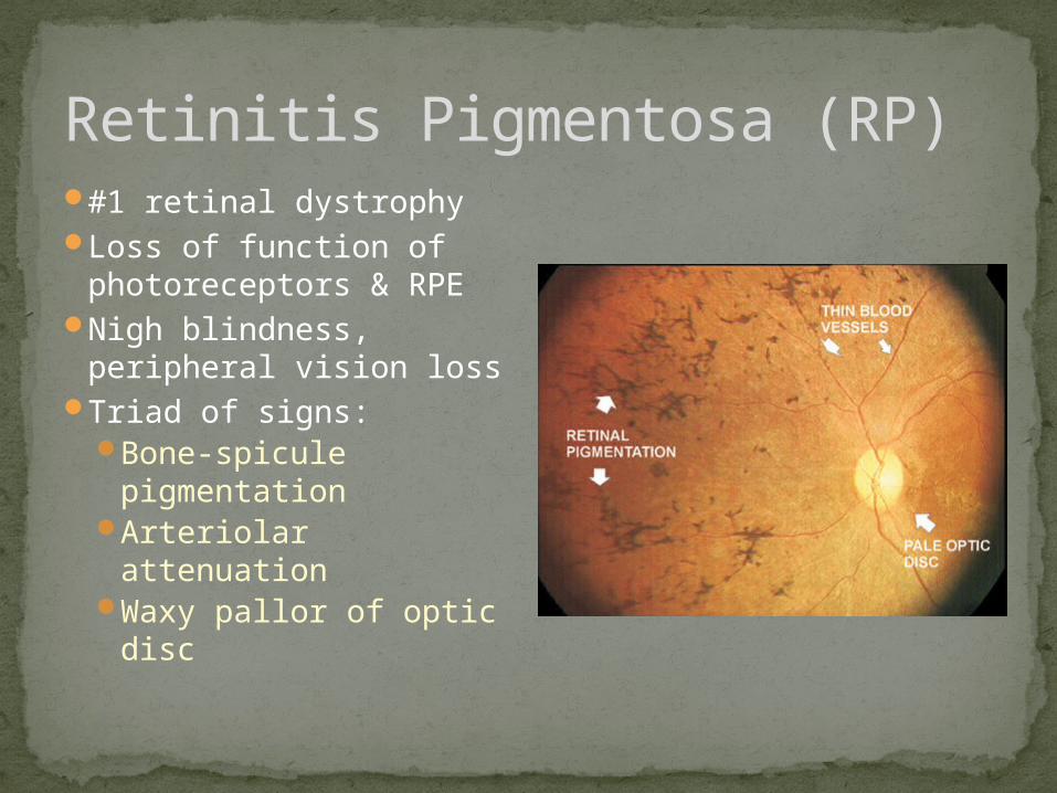
Retinitis Pigmentosa (RP)#1 retinal dystrophyLoss of function of
photoreceptors & RPENigh blindness,
peripheral vision lossTriad of signs:
Bone-spicule pigmentation
Arteriolar attenuation
Waxy pallor of optic disc

Stargardt’s Disease#1 hereditary
macular dystrophyAutosomal
recessive“Beaten bronze”
macula in late stages
Reduction of VA & color vision
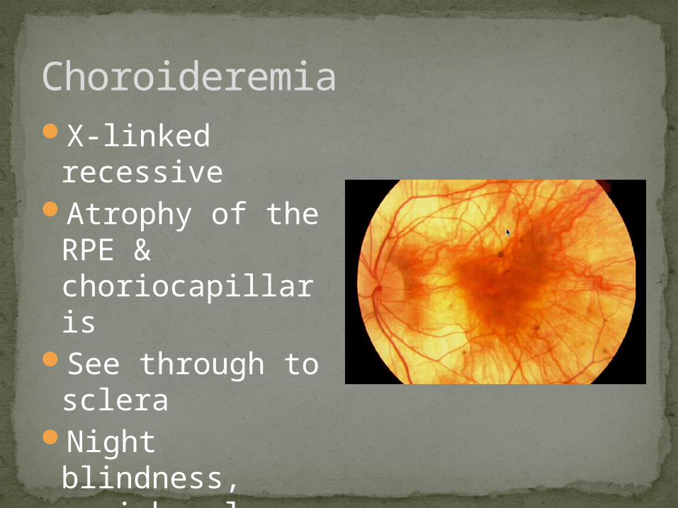
ChoroideremiaX-linked
recessiveAtrophy of the
RPE & choriocapillaris
See through to sclera
Night blindness, peripheral vision loss

Cone DystrophyYoung patientsUsually autosomal
dominantLoss of cone
photoreceptorsDecreased VA,
photophobia, color vision loss
Geographic atrophy of RPE, vessel attenuation, optic nerve pallor

Best’s Disease (Vitelliform Dystrophy)Autosomal
dominantMaterial
accumulates in RPE (“egg yolk”)
No symptoms early on, later reduced VA
Bilateral

Rhegmatogenous Retinal Detachments (RDs)Retinal
detachment caused by a hole or tear
Vitreous fluid gets into subretinal space and retina detaches

Exudative RDsDamage to RPE
causes fluid accumulation below the retina detachment
Ex: ARMD
RD caused by traction
Ex: proliferative retinopathy (neovascularization)
Non-Rhegmatogenous RDs
Tractional RDs

Lattice DegenerationPeripheral retinal
thinningSometimes
pigmentedFirmly adhere to
vitreous can cause retinal detachment if vitreous starts to pull away
Bilateral

Age-related Degenerative Retinoschisis
Toxocariasis
Choroidal/Retinal Degenerations

Age-Related Degenerative RetinoschisisSplitting of retina
between the outer plexiform layer & inner nuclear layer
Looks like a retinal detachment, but doesn’t move
No symptomsVisual field defect

ToxocariasisIntestinal
nematodeUnilateral
inflammationChorioretinal
scars