THROMBOLYSIS IN ACUTE MYOCARDIAL INFARCTION · THROMBOLYSIS IN ACUTE MYOCARDIAL INFARCTION ......
108
THROMBOLYSIS IN ACUTE MYOCARDIAL INFARCTION
-
Upload
hoangtuyen -
Category
Documents
-
view
243 -
download
0
Transcript of THROMBOLYSIS IN ACUTE MYOCARDIAL INFARCTION · THROMBOLYSIS IN ACUTE MYOCARDIAL INFARCTION ......

THROMBOLYSIS IN ACUTE MYOCARDIAL INFARCTION

THROMBOLYSIS IN ACUTE MYOCARDIAL INFARCTION
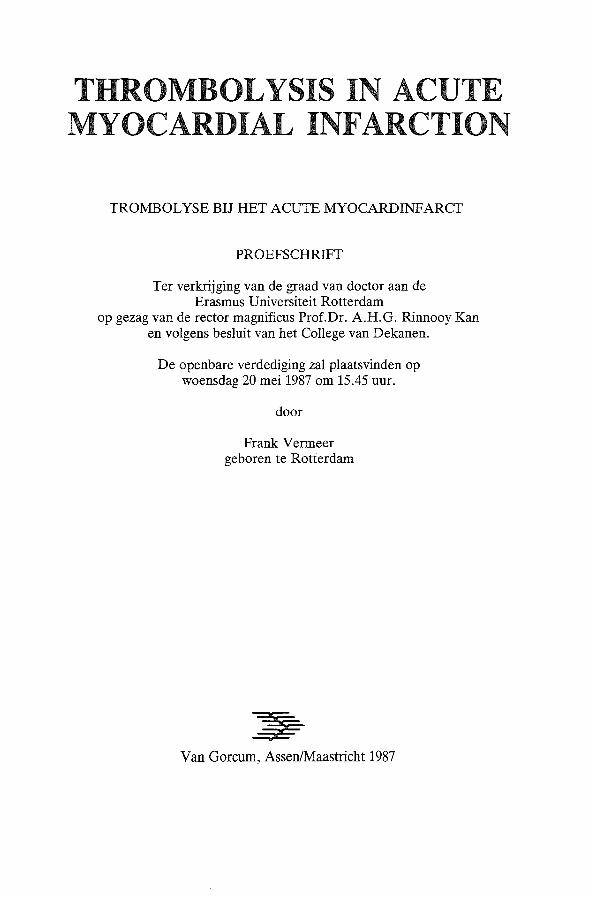
TROMBOL YSE BIJ HET ACUTE MYOCARDINFARCT
PROEFSCHRIFT
Ter verkrijging van de graad van doctor aan de Erasmus Universiteit Rotterdam
op gezag van de rector magnificus Prof.Dr. A.H.G. Rinnooy Kan en volgens besluit van het College van Dekanen.
De openbare verdediging zal plaatsvinden op woensdag 20 mei 1987 om 15.45 uur.
door
Frank Vermeer geboren te Rotterdam
Van Gorcum, Assen/Maastricht 1987
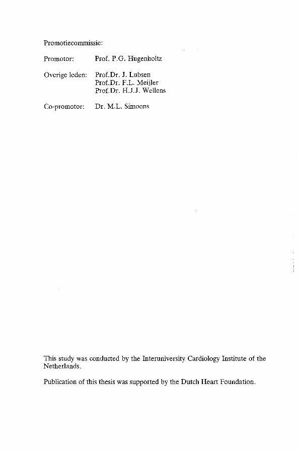
Promotiecommissie:
Promotor: Prof. P. G. Hugenholtz
Overige leden: Prof.Dr. J. Lubsen Prof.Dr. F.L. Meijler Prof.Dr. H.J.J. Wellens
Co-promotor: Dr. M.L. Simoons
This study was conducted by the Interuniversity Cardiology Institute of the Netherlands.
Publication of this thesis was supported by the Dutch Heart Foundation.

Proclaim in the name of the Lord, who created men? He created man from clots of blood.
Koran XCVI 1.1
To El, Tim, Margaux and ..


Acknowledgement
The multicentre trial described in this thesis would not have been possible without the cooperation of all those who were involved in the study in the five participating hospitals. I gratefully acknowledge the dedication of all attending physicians, the nursing staff and the staff of the catheterisation laboratories in the University Hospital Dijkzigt in Rotterdam, the University Hospital in Maastricht, the Hospital of the Free University in Amsterdam, the Zuiderziekenhuis in Rotterdam and the University Hospital in Leiden, and of everyone involved in the data analysis and the preparation of the manuscripts. Without their cooperation this study would not have yielded the succes it did.
Many others who are not mentioned in the list of collaborators below are known to have contributed to the multidisciplinar teamwork necessary for successful scientific work.
Participating centres and collaborators:
Thoraxcenter, Erasmus University and University Hospital Dijkzigt, Rotterdam: (M.J.B.M. van den Brand, F.J. van Dalen, P.J. de Feyter, P. Fioretti, P.G. Hugenholtz, P.W. Serruys, M.L. Simoons, H. Suryapranata, W. Wijns)
Department of Cardiology, Free University Amsterdam: (M.J. van Eenige, J.C.J. Res, J.P. Roos, F.C. Visser, F.W.A. Verheugt)
Department of Cardiology, Zuiderziekenhuis, Rotterdam: (D.C.A. van Hoogenhuyze, X.H. Krauss, H.A.C.M. Kruyssen, W.J. Remme, C.J. Storm)
Department of Cardiology, University Hospital Maastricht, University of Limburg, Maastricht: (F.W. Bar, S.H. Braat, P. Brugada, K. den Dulk, W.T. Hermens, L.V.M. Lejeune, L.A.H. Manders, M. Ramentol, H.J.J. Wellens, G. M. Willems, C. de Zwaan)
VII
Department of Cardiology, University Hospital, Leiden: (B. Buis, J. Engbers, A. van der Laarse, P.J. Senden, E.E. van der Wall)
Data processing center, Thoraxcenter, Erasmus University, Rotterdam: (A.J. Azar, B. Bos, S. van der Does, R.T. van Domburg, G.A. vanEs, J. Lubsen, J.P. van Mantgem, K. de Neef, M. Patijn, J. Planellas, N. Speelman, J.G.P. Tijssen, P. Vrijdag, A.A. Wagenaar, I.C.J. Zorn).
VIII

Table of contents
Chapter 1, Introduction . . . . . . . . . . . . . . . . . . . . . . . . . . . . . . . . . . . . . . 1
Chapter 2, Pharmacology . . . . . . . . . . . . . . . . . . . . . . . . . . . . . . . . . . . . . 4
Chapter 3, Study design . . . . . . . . . . . . . . . . . . . . . . . . . . . . . . . . . . . . . . 10
Chapter 4, Early thrombolysis in acute myocardial infarction: Limitation of infarct size and improved survival. By: ML Simoons, PW Serruys, M vd Brand, J Res, FWA Verheugt,
XH Krauss, WJ Remme, F Bar, C de Zwaan, A vd Laarse, F Vermeer, J Lubsen.
In: JAm Coll Cardiol1986; 7, 717-28 . . . . . . . . . . . . . . . . . . . . . . . . . 18
Chapter 5, Effects of early intracoronary streptokinase on infarct size estimated from cumulative enzyme release and on enzyme release rate: A randomized trial of 533 patients with acute myocardial infarction. By: AvdLaarse,FVermeer, WTHermens, GMWillems,KdeNeef,
ML Simoons, PW Serruys, J Res, FW A Verheugt, XH Krauss, F Bar, C de Zwaan, J Lubsen.
In: Am Heart J 1986; 112, 672-81 . . . . . . . . . . . . . . . . . . . . . . . . . . . . . 30
Chapter 6, Value of admission electrocardiogram in predicting outcome of thrombolytic therapy in acute myocardial infarction. By: FW Bar, F Vermeer, C de Zwaan, M Ramentol, S Braat, ML
Simoons, WT Hermens, A vd Laarse, FW A Verheugt, XH Krauss, HJJ Wellens.
In: Am J Cardiol1987; 59, 6-13 . . . . . . . . . . . . . . . . . . . . . . . . . . . . . . . 40
Chapter 7, Which patients benefit most from early thrombolytic therapy with intracoronary streptokinase? By: F Vermeer, ML Simoons, FW Bar, JGP Tijssen, RT van Dam
burg, PW Serruys, FWA Verheugt, JCJ Res, C de Zwaan, A vd Laarse, XH Krauss, J Lubsen, PG Hugenholtz.
In: Circ 1986; 74, 1379-89 . . . . . . . . . . . . . . . . . . . . . . . . . . . . . . . . . . . . 48
IX

Chapter 8, Cost benefit analysis of early thrombolytic therapy with intracoronary streptokinase. By: F Vermeer, ML Simoons, C de Zwaan, GA v Es, FW A Verheugt,
A vd Laarse, DCA van Hoogenhuyze, AJ Azar, FJ van Dalen, J Lubsen, PG Hugenholtz. ·
In slightly different version accepted for publication in Br Heart J . . . 59
Chapter 9, Value of PTCA performed immediately after successful thrombolysis with intracoronary streptokinase by: F Vermeer, ML Simoons, PJ de Feyter, FW Bar, H Suryapranata,
P Fioretti, PW Serruys, B Buis, JCJ Res, SH Braat, J Lubsen, PG Hugenholtz.
Submitted for publication . . . . . . . . . . . . . . . . . . . . . . . . . . . . . . . . . . . . . 72
Chapter 10, Summary of other reports from the trial . . . . . . . . . . . . . . 86
Chapter 11, Conclusions and recommendations . . . . . . . . . . . . . . . . . . 91
Summary . . . . . . . . . . . . . . . . . . . . . . . . . . . . . . . . . . . . . . . . . . . . . . . . . . . . 104
Samenvatting . . . . . . . . . . . . . . . . . . . . . . . . . . . . . . . . . . . . . . . . . . . . . . . . 107
Curriculum vitae . . . . . . . . . . . . . . . . . . . . . . . . . . . . . . . . . . . . . . . . . . . . . 110
X
CHAPTER 1
Introduction
In the first century BC Aristotle described that he detected fibers in the blood of killed animals, but not in the blood of hunted deer. He stated that the blood of such animals could not coagulate, not realising that the enhanced fibrinolytic state in the deer was caused by exhaustion (1). Therapeutic enhancement of the fibrinolytic system was first reported in 1933, when Tillet and Garner demonstrated that cultures of hemolytic streptococci had the capacity to liquify clotted human plasma (2). Christensen believed that streptococcal activity to be kinase-like and introduced the term streptokinase in 1945 (3). Streptokinase was for the first time used as a thrombolytic agent in 1959 when Fletcher demonstrated its capability to lyse venous thrombi in healthy volunteers (4). Also in 1959, Fletcher observed a low mortality in 22 patients with acute myocardial infarction after treatment with high doses of intravenous streptokinase ( 5). In later years the efficacy of intravenous streptokinase in the treatment of patients with myocardial infarction was tested in randomised clinical trials, but the results were not unequivocally in favour of this therapy, although beneficial effects were reported in some (6-11). In these trials early mortality appeared to be on average 5% lower in patients treated with streptokinase when compared to treatment with heparin. However, it remains difficult to draw conclusions from these studies since patients were admitted up to 48 hours after onset of symptoms, no intermediate measurements were made and the relation between treatment delay and mortality reduction was not studied. Moreover, the role of acute thrombotic obstruction of a coronary artery as the cause of myocardial infarction was not yet generally accepted (12). It was DeWood who settled the controversy whether the thrombotic occlusion of a coronary artery was the cause of myocardial infarction or the consequence of it by reporting that coronary angiography performed within hours after the onset of myocardial infarction showed obstruction of a coronary artery in more than 75% of the cases (13). Although intracoronary lysis with fibrinolytic agents had been shown earlier by Boucek and Chazov (14, 15), the clinical breakthrough came when Rentrop demonstrated in 1979 that an occluded coronary artery could be recanalised during coronary angiography by the intracoronary application of streptokinase (16). This led to the use of intracoronary streptokinase on a large scale in many centres, although benefits other than angiographically documented recanalisation had not been
1

proven. Consequently, warnings were sounded that until it became clear whether the potential benefits of acute recanalisation, in terms of infarct size limitation and mortality reduction, outweighted the risks of the procedure, widespread application should be avoided (17,18). To assess whether a strategy aimed at early reperfusion of an occluded coronary artery would be of benefit for patients with acute myocardial infarction, a randomised trial was initiated in 1981 in the Thoraxcenter in Rotterdam and later extended with four other hospitals under the auspices of the Interuniversity Cardiology Institute of the Netherlands. In this thesis parts of the results of this trial are presented.
The mechanisms of fibrinolysis and the pharmacological properties of thrombolytic agents are described in chapter 2. The design of the trial and the methodology used is discussed in chapter 3. The main results are presented in chapter 4, followed by detailed reports about the observed limitation of infarct size (chapter 5) and the value of the admission electrocardiogram to predict the outcome of thrombolytic therapy (chapter 6). Subgroup analysis was performed to identify which patients benefitted most from the therapy (chapter 7) and benefits were related to the costs of the intervention (chapter 8). The additional value of coronary angioplasty, performed immediately after successful thrombolysis in selected patients, was assessed in a matched pair analysis (chapter 9). A number of other observations from the trial are summarised in chapter 10. The results are discussed and compared with results of other clinical trials with thrombolytic therapy. Based on these results recommendations for application of thrombolytic therapy in acute myocardial infarction are presented (chapter 11).
References
1. Aristotle: De partibus animalum II, 4. 2. Tillet WS and Garner RL: The fibrinolytic activity of hemolytic streptococci. J Exp Med 1932;
58, 485-502. 3. Christensen LR and Macleod CM: A proteolytic enzyme of serum: characterization, activa
tion, and reaction with inhibitors. J Gen Physiol1945; 28, 559-83. 4. Fletcher AP, Alkjaersig N, and Sherry S: The maintenance of a sustained thrombolytic state in
man. I. Induction and effects. J Clin Invest 1959; 38, 1096-110. 5. Fletcher AP, Sherry S, and Alkjaersig N, et al: The maintenance of a sustained thrombolytic
state in man. II. Clinical observations on patients with myocardial infarction and other thromboembolic disorders. J Clin Invest 1959; 38, 1111-9.
6. Schmutzler R, Reckner F, Kortge P, et al: Zur thrombolytischen Therapie des frischen Herzinfarktes. Dtsch Med Wochenschr 1966; 91, 581-7.
7. European Working Party: Streptokinase in recent myocardial infarction: a controlled multicentre trial. Br Med J 1971; 3, 325-31.
8. Heikinheimo R, Ahrenberg P, Honkapohja H, et al: Fibrinolytic treatment in acute myocardial infarction. Acta Med Scan 1971; 189, 7-13.
9. Bett JHN, Biggs JC, Castalde P A, et al: Australian multicentre trial of streptokinase in acute myocardial infarction. Lancet 1973; 2, 57-60.
2

10. Aber CP, Bass NM, Berry CL, et al: Streptokinase in acute myocardial infarction: A controlled multicentre study in the United Kingdom. Br Med J 1976; 2, 1100-4.
11. European Cooperative Study Group for Streptokinase Treatment in Acute Myocardial Infarction: Streptokinase in acute myocardial infarction. N Engl J Med 1979; 301, 797-802.
12. Roberts WC: Coronary thrombosis and fatal myocardial ischemia. Circ 1974; 49, 1-3. 13. De Wood MA, Spores J, N otske MD, et al: Prevalence of total coronary occlusion during the
early hours of transmural myocardial infarction. N Engl J Med 1980; 303, 897-902. 14. Boucek RJ, Murphy WP J r: Segmental perfusion of the coronary arteries with fibrinolysin in
man following a myocardial infarction. Am J Cardiol1960; 6: 525-33. 15. Chazov EI, Mateeva LS, Mazaev AV, et al: Intracoronary administration of fibrinolysin in
acute myocardial infarction. Ter Arkh 1976; 48, 8-19. 16. Rentrop P, De Vivie ER, Karsch KR, et al: Acute myocardial infarction: intracoronary
application of nitroglycerin and streptokinase in combination with translurninal recanalization. Clin Cardiol1979; 5, 354-63.
17. Muller JE, Stone PH, MarkiesJE, et al: Let's not let the genie escape from the bottle- again. N Engl J Med 1981; 304, 1294-6.
18. Hugenholtz PG, Rentrop P: Thrombolytic therapy for acute myocardial infarction: quo vadis? A review of the recent literature. Eur Heart J 1982; 3: 395-403.
3
CHAPTER2
Pharmacology
The fibrinolytic system
Lesions of the endothelium of an artery trigger a cascade of enzyme reactions, finally leading to coagulation of blood by formation of fibrin. Opposing this event is the capacity of the fibrinolytic system to resolve blood clots in the circulation. Fibrin is fragmented by plasmin into soluble fibrin degradation products. Under normal circumstances the fibrinolytic system is in a state of "dynamic equilibrium" with the coagulation system in order to maintain an intact and patent vascular bed (1 ). In coronary artery disease, platelet aggregation and coagulation may occur on atherosclerotic lesions in the coronary arteries, leading to intravascular fibrin deposits (thrombosis), with eventual occlusion of a coronary artery and subsequent myocardial infarction. The primary goal of thrombolytic therapy is to enhance fibrinolytic activity when pathologic deposition of fibrin has occurred and naturally induced fibrinolysis by itself is insufficient to maintain patency of the vascular bed. The mechanisms of fibrinolysis and the role of thrombolytic agents are discussed below.
Mechanisms of fibrinolysis
Plasminogen, a single chain glycoprotein (molecular weight 92,000) can be converted into plasmin by cleavage of a protein bond. Plasmin has high affinity for fibrin and breaks it down rapidly (figure). Free plasmin in the circulating blood is in turn rapidly inactivated by a2-antiplasmin, a single chain glycoprotein with a molecular weight of 70,000. The conversion of plasminogen into plasmin is enhanced by plasminogen activators. Tissue plasminogen activator and single and double chain urokinase-type plasminogen activator all have been identified as naturally occurring glycoproteins which catalyse the conversion of plasminogen into plasmin. The release of plasminogen activators is under control of both central and local control mechanisms. Central control of plasminogen activator levels in blood is mediated by catecholamine release by either a nervous or a humoral pathway (2). Locally, plasminogen activators are probably released by the endothelium in response to local fibrin deposits.
4
Streptokinase
Streptokinase, a single chain protein with a molecular weight of 46,000, is formed by hemolytic streptococci and has antigenic properties. Different theories exist about its mode of action. A first theory implies that streptokinase diffuses into thrombi, where it forms equimolar complexes with fibrin-bound or penetrating plasminogen, and the resulting complexes further enhance the conversion of plasminogen into plasmin (3). A second theory states that complexes of streptokinase and plasminogen are formed in blood and diffuse into thrombi (4). A third theory suggests that fibrinolysis by plasminogen activator in the thrombus is enhanced after depletion of circulating a2-antiplasmin ( 5). Dosage schedules for treatment of patients with acute myocardial infarction vary, according to the presumed mode of action ( 6). The first theory favours a high concentration of streptokinase in the vicinity of the thrombus, which can be best achieved by selective intracoronary application. The other theories presume systemic effects of streptokinase preceding its thrombolytic activity, favouring intravenous administration. However, intracoronary administration of streptokinase leads to a higher patency rate of the infarct related coronary artery than intravenous streptokinase. Intravenous administration of streptokinase, in dosages varying from 500,000 U to 1,500,000 U given over one hour, induced recanalisation of an occluded coronary artery in 10 to 62% of patients with acute myocardial infarction (7). Intracoronary administration of streptokinase (250,000 U) led under similar circumstances to recanalisation rates of 60 to 79% (chapter 11). In the present study no differences in final patency i:ate were observed between patients treated with 250,000 U of intracoronary streptokinase and those in which this treatment was preceded by intravenous application of 500,000 U streptokinase, although initial patency rate rose from 18% to 41% (chapter 4). It may be concluded that although selective administration of streptokinase has systemic effects, its delivery in the vicinity of the thrombus is superior to systemic administration.
Because of the antigenic properties of streptokinase, short term corticosteroid therapy has been used in an effort to suppress immunologic reactions (chapter 4). However, in the GISSI trial (1,500,000 U of intravenous streptokinase) no steroids were given and allergic reactions were observed in only 2.5% of the patients (8). Patients who have suffered streptococci infections or have been treated previously with streptokinase may have high titers of antibodies against streptokinase. In those patients allergic reactions may occur and high dosages may then be required to saturate the antibodies. This is supported by our, unpublished, observation that in half of the patients with persistent occlusion after intracoronary administration of streptokinase there was no decrease in fibrinogen level. This finding is similar to that of others ( 6) and suggests that presently recommended dosages are insufficient in 10 to 20% of the patients, who neutralise streptokinase before thrombolysis has occur-
5

red. Furthermore, the use of repeated doses of streptokinase is not advocated within 6 to 12 months after its original use, because of possible immunologic reactions.
Urokinase
Urokinase, a glycoprotein with a molecular weight of 55,000 is a naturally occurring protein, produced in the kidney and excreted with urine. It enhances the conversion of plasminogen into plasmin by direct proteolytic action and not by forming a stable complex with plasminogen as streptokinase does. Its other farmacological effects are similar to that of streptokinase, although it does not exhibit antigenic properties (9). Dosages vary from 500,000 to 2,700,000 IU for intravenous administration and up to 700,000 IU for intracoronary use in patients with acute myocardial infarction (9-11). Since the production of urokinase is expensive compared to that of streptokinase, its use is presently restricted for those patients who have previously been treated with streptokinase, or when the latter compound is shown not to have effect during intracoronary administration.
Recombinant tissue-type plasminogen activator (rt-PA)
Tissue plasminogen activator, a glycoprotein with a molecular weight of 64,000, is present in practically all tissues. It has originally been isolated from human uterus (5). For investigational purposes it was subsequently produced by melanoma cell cultures, but recently recombinant DNA techniques have become available, leading to production on a large scale of recombinant tissuetype plasminogen activator. Rt-PA exhibits clot selectivity (see below) and its capacity to induce patency in the infarct related coronary artery was demonstrated to be superior to that of intravenous streptokinase with a lower incidence of bleeding complications (chapter 11). Effects of rt-PA on infarct size, left ventricular function and mortality in patients with acute myocardial infarction are currently subject to investigation in large, multicentre randomised trials. A dosage of 100 mg, given over three hours, is recommended for patients with acute myocardial infarction.
Anisoylated plasminogen-streptokinase activator complex (APSAC)
APSAC, an acylated complex of streptokinase and plasminogen, is inert and does not induce fibrinolysis. It becomes capable of inducing fibrinolysis after slow deacylation. APSAC does not exhibit clot selectivity, but due to the slow deacylation it has a longer half life (approximately 30 minutes) than the other thrombolytic agents (on average 10 minutes). In relatively small series was reported that an intravenous bolus of APSAC (30 mg) induced patency in 64 to 88% of patients with myocardial infarction with an incidence of bleeding complications comparable to that of streptokinase (chapter 11).
6
Recombinant single chain urokinase-type plasminogen activator (rscu-PA)
Single chain urokinase-type plasminogen activator was once considered to be an inactive precursor form of urokinase, but it is now known that the single chain form has plasminogen activator activity. Its clot selective fibrinolytic properties have been demonstrated in vitro (12), in animal experiments (13-15) and in patients with acute myocardial infarction (16-18). A bolus of 10 mg followed by intravenous infusion of 60 mg rscu-PA over one hour has induced patency of the infarct related coronary artery in 13 out of 17 patients (76%) without significant bleeding complications (17). In larger clinical trials, which are presently underway, the efficacy and safety of rscu-PA will have to be demonstrated before its use in patients with myocardial infarction can be recommended.
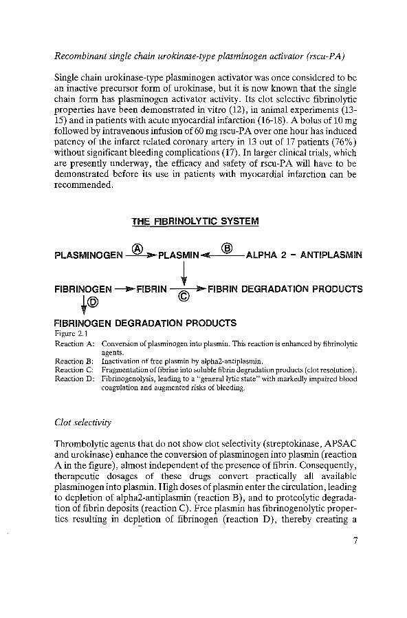
THE FIBRINOlYTIC SYSTEM
PlASMINOGEN @ ,... PlASMIN -c: @ AlPHA 2 - ANTIPlASMIN
i .. FIBRIN DEGRADATION PRODUCTS FIBRINOGEN _,...FIBRIN .@ FIBRINOGEN DEGRADATION PRODUCTS Figure 2.1 Reaction A: Conversion of plasminogen into plasmin. This reaction is enhanced by fibrinolytic
agents. Reaction B: Inactivation of free plasmin by alpha2-antiplasmin. Reaction C: Fragmentation offibrine into soluble fibrin degradation products (clot resolution). Reaction D: Fibrinogenolysis, leading to a "general lytic state" with markedly impaired blood
coagulation and augmented risks of bleeding.
Clot selectivity
Thrombolytic agents that do not show clot selectivity (streptokinase, APSAC and urokinase) enhance the conversion of plasminogen into plasmin (reaction A in the figure), almost independent of the presence of fibrin. Consequently, therapeutic dosages of these drugs convert practically all available plasminogen into plasmin. High doses of plasmin enter the circulation, leading to depletion of alpha2-antiplasmin (reaction B), and to proteolytic degradation of fibrin deposits (reaction C). Free plasmin has fibrinogenolytic properties resulting in dep~etion of fibrinogen (reaction D), thereby creating a
7

"general lytic state" in which blood coagulation is markedly impaired and bleeding risks are augmented. This general lytic state lasts for 24 to 48 hours, before alpha2-antiplasmin and fibrinogen levels return to normal (18). The recently developed clot selective thrombolytic agent rt-P A has a low affinity for plasminogen in the absence of fibrin, but the affinity and the rate of conversion of plasminogen into plasmin increase markedly (50 to 500 times) in the presence of fibrin (19). The background of the clot selectivity of rscu-PA is less well understood. The presence of a rscu-PA inhibitor in plasma has been postulated (20), while in another study a higher affinity of rscu-PA to fibrin bound plasminogen rather than to free plasminogen was observed (21). Because of clot selectivity systemic depletion of fibrinogen occurs less and plasminogen, alpha2-antiplasmin and fibrinogen levels in serum decrease only to a lesser extent than after use of streptokinase, APSAC or urokinase (22, 23).
Conclusions
Therapeutic enhancement of fibrinolysis may be beneficial when blood supply to tissue is menaced by thrombotic obstruction in the vascular bed. Nowadays, thrombolytic therapy is used or proposed in the treatment of acute myocardial infarction, unstable angina pectoris, peripheral artery thrombosis, pulmonary embolism and deep vein thrombosis. The major part of our present knowledge about the risks and benefits of thrombolytic therapy has been derived from studies with streptokinase and urokinase. It is only in the last decade that other thrombolytic agents have been developed which differ in their mode of action from streptokinase and urokinase. The efficacy, costs, and safety of these newer fibrinolytic agents should be compared with the presently available methods for thrombolytic therapy in acute myocardial infarction, before recommendations can be given as to what might be considered optimal therapy for these patients.
References
1. Fearnley GR: A concept of natural fibrinolysis. Lancet 1961; 1, 992-3. 2. D. Collen: On the regulation and control of fibrinolysis. Edward Kowalski Memorial Lecture.
Thromb Haemost 1980; 43, 77-89. 3. Chesterman CN, Allington MJ, Sharp AA: Relationship of plasminogen activator to fibrin.
Nature 1972; 238, 15-7. 4. DeutschE, Fischer M: Die Wirkung intravenos applizierter Streptokinase aufFibrinolyse und
Blutgerinnung. Thromb Diath Haemorrh 1960; 16, 38-50. 5. Brommer EJP: The level of extrinsic plasminogen activator (t-PA) during clotting as a
determinant of the rate of fibrinolysis; inefficiency of activators added afterwards. Thromb Research 1984; 34, 109-15.
6. Six AJ, Brommer EJP, Muller EJ, eta!: Activation of the fibrinolytic system during intracoronary streptokinase administration. JAm Coli Cardiol1987; 9, 189-96.
7. Rentrop KP: Thrombolytic therapy in patients with acute myocardial infarction. Circ 1985; 71, 627-31.
8
8. Gruppo Italiano per lo Studio della Streptochinasi nell' Infarto miocardico (GISSI): Effectiveness of intravenous thrombolytic treatment in acute myocardial infarction. Lancet 1986; 1, 397-401.
9. Tennant SN, Dixon J, Venable TC, eta!: Intracoronary thrombolysis in patients with acute myocardial infarction: comparison of the efficacy of urokinase with streptokinase. Circ 1984; 69, 756-60.
10. Barlow GH: Pharmacology of fibrinolytic agents. Progr Cardio Diseases 1979; 21, 315-26. 11. European Collaborative Study: Controlled trial of urokinase in myocardial infarction. Lancet
1975; 2, 624-6. 12. Gurewich V, Pannell R, Louie S, eta!: Effective and fibrin-specific clot lysis by a zymogen
precursor form of urokinase (pro-UK): a study in vitro and in two animal species. J Clin Invest 1984; 73, 1731-8.
13. Collen D, Stassen JM, Blaber M, et a!: Biological and thrombolytic properties of proenzyme and active forms of human urokinase. IlL Thrombolytic properties of natural and recombinant urokinase in rabbits with experimental jugular vein thrombosis. Thromb Haemost 1984; 52,27-32.
14. Collen D, Stump DC, Van de Werf F, et a!: Coronary thrombolysis with intravenously administered human pro-urokinase (pro-UK). Circ 1985; 72, 384-90.
15. Flam eng W, V anhaecke J, Stump DC, et a!: Coronary thrombolysis by intravenous infusion of recombinant single chain urokinase-type plasminogen activator or recombinant urokinase in baboons: effect on regional blood flow, infarct size and hemostasis. JAm Coli Cardiol1986; 8, 118-25.
16. Van de Werf F, Nobuhara M, Collen D: Coronary thrombolysis with human single-chain, urokinase-type plasminogen activator (pro-urokinase) in patients with acute myocardial infarction. Ann Intern Med 1986; 104, 345-52.
17. Van de Werf F, Vanhaecke J, De Geest H, eta!: Coronary thrombolysis with recombinant urokinase-type plasminogen activator in patients with acute myocardial infarction. Circ 1986; 74, 1066-70.
18. Bell WR: Streptokinase and urokinase in the treatment of pulmonary thromboemboli. From a national cooperative study. Thromb Haemost 1976; 35, 79-89.
19. Sobel BE, Gross RW, Robinson AK: Thrombolysis, clot selectivity, and kinetics. Circ 1984; 70, 160-4.
20. Pannel R, Gurewich V: Pro-urokinase: a study of its stability in plasma and of a mechanism for its selective fibrinolytic effect. Blood 1986; 67, 1215-23.
21. Lynen HR, Zamarron C, Blaber M, et a!: Activation of plasminogen by pro-urokinase. L Mechanism. J Bioi Chern 1986; 261, 1253-8.
22. Verstraete M, Bernard R, Bory M, eta!: Randomised trial of intravenous recombinant tissuetype plasminogen activator versus intravenous streptokinase in acute myocardial infarction. Lancet 1985; 1, 842-7.
23. Werf F vd, Bergmann SR, Fox KAA, et a!: Coronary thrombolysis with intravenously administered human tissue-type plasminogen activator produced by recombinant DNA technology. Circ 1984; 69, 605-10.
9
CHAPTER3
Study design
Status of thrombolytic therapy in 1981
In 1979 Rentrop and co-workers demonstrated that early recanalisation of an occluded coronary artery could be achieved with intracoronary infusion of streptokinase, a finding later confirmed by others (1-3). Also it was observed that successful reperfusion led to improvement in left ventricular function, and left ventricular ejection fraction appeared to be higher in successfully recanalised patients than in patients with persistent occlusion of the infarct related artery ( 4-6). Furthermore, data from animal experiments had indicated that reperfusion within hours after the onset of myocardial infarction could lead to limitation of infarct size (7). On the other hand, serious complications of the interventions were also reported, such as fatal complications during acute angiography, severe bleeding and intramyocardial hemorrhage (5, 8, 9). It was also pointed out that results of large scale clinical trials with intravenous streptokinase or urokinase had not been unequivocally in favour of thrombolytic therapy (10-13). Warnings were sounded that beneficial effects of thrombolytic therapy had not been proven and that the expected benefits should be compared with the potential risks of the procedure in randomised clinical trials before its use could be recommended (14,15). For this reason a large randomised clinical trial was planned at the Thoraxcenter in Rotterdam. Before the trial started three pilot studies were performed (9, 16).
Results of pilot studies
The pilot studies were performed in 1980 and 1981. In six patients recanalisation was attempted with intravenous niphedipine and intracoronary nitroglycerine. The infarct related artery was occluded in five patients and recanalisation was achieved in one. In nine patients recanalisation was attempted with intracoronary urokinase (250,000 U). An occluded coronary artery was successfully recanalised in two out of eight patients (25%). More favourable results were obtained in a study with intracoronary streptokinase (250,000 U), in which 37 patients were enrolled. Four patients died from irreversible cardiogenic shock during acute cardiac catheterisation. One patient with a large anterior infarction developed severe bradycardia followed by
10
asystole when after left ventriculography right coronary arteriography was performed prior to the initiation of thrombolytic therapy. Two patients with an occluded left anterior descending artery suddenly developed severe hypotension and asystole during intracoronary infusion of streptokinase. Angiography revealed embolisation of a part of the thrombus from the left anterior descending artery into the left circumflex artery. The fourth patient was known with severe three vessel disease with an occluded left anterior descending artery. This patient suffered an acute inferior wall infarction during hospitalisation. During intracoronary infusion of streptokinase in the occluded right coronary artery severe hypotension developed, followed by electromechanical dissociation and death (9). In 27 of the remaining 33 patients an occluded infarct related artery was found which could be reperfused in 19 patients (70%). It was further observed in that study that the time interval between initiation of thrombolytic therapy and angiographic documentation of vessel patency was related to the delay between onset of chest pain and initiation of therapy. Lysis time was less than 30 minutes when thrombolytic therapy was started within three hours after onset of symptoms and considerably longer when treatment delay exceeded four hours.
From these pilot studies several important lessons were learned. In the pilot studies the best results were obtained with intracoronary streptokinase in a dosage of 250,000 U. The preliminary results of the study with urokinase were disappointing. It appeared later, however, that the urokinase given contained mostly the low molecular weight form of urokinase with less activity than the high molecular weight form (17). Probably, the dosage given (250,000 U) was roughly equal to 100,000 Ploug Units, which can explain the low reperfusion rate observed (25% ). It was decided that streptokinase would be used in the randomised trial in a dosage of 4,000 U/min with a maximum of 250,000 U. Furthermore it was observed that the risks of acute angiography increased dramatically when contrast was injected into the non infarct-related artery or in the left ventricle before reperfusion was achieved, especially in patients with large anterior infarction. It was decided that angiography would begin with the infarct related coronary artery followed by intracoronary infusion of streptokinase. Left ventricular angiography would only be performed in patients with a clinically stable condition at the end of the procedure who had a left ventricular enddiastolic pressure below 35 mm Hg. The pilot studies proved the feasibility of a randomised trial. Based on the observed relation between lysis time and treatment delay it was decided that only patients admitted to the hospital within four hours after onset of chest pain would be enrolled in the present study.
Study design
The purpose of the randomised trial was to analyse the difference between two strategies in the treatment of myocardial infarction. A strategy aimed at early
11

reperfusion, including angiography and administration of intracoronary streptokinase, would be compared with conventional treatment of myocardial infarction without acute angiography and without administration of fibrinolytic therapy. Consequently, this was an open study. The random assignment was known to the investigators and comparisons between the effects of both strategies could be made while the trial was still in progress. At the start of the trial the organisers realised that thrombolytic therapy was still in its infancy. It was expected that improvements of the intervention might arise during the trial. Therefore, modifications of the protocol would be allowed, if during the trial insights how to achieve optimal reperfusion would alter. The modifications in the protocol will be referred to as policy decisions.
Patient selection and randomisation
Patients were eligible for the trial if admitted to the hospital within four hours after onset of chest pain and with electrocardiographic signs typical of acute myocardial infarction: ST segment elevation of at least 0.1 m V in one or more extremity leads, at least 0.2 m V in one or more precordial leads, or at least 0.2 m V ST segment depression in one or more precordial leads compatible with posterior infarction. Several categories of patients were excluded from the trial: patients who had previously been treated with streptokinase, since its repeated use might cause allergic reactions; patients with enhanced risks of bleeding, such as patients over 70 years of age, patients with a history of gastrointestinal bleeding, gastric or duodenal ulcer, hematuria or cerebral vascular accident within the last three months, patients with recent trauma including prolonged cardiac resuscitation, and female patients currently menstruating or pregnant. Furthermore, patients with previous bypass surgery in a vessel corresponding to the infarct location were excluded since it might be uncertain in which vessel streptokinase should be infused. Finally, patients who had previously been admitted to the study and patients in whom incomplete follow-up was anticipated were excluded from the study for methodological reasons. These exclusion criteria were rather strict since benefits of early thrombolytic therapy had not been proven. In summary, only patients with specific electrocardiographic signs of acute myocardial infarction without any indication of enhanced risks of complications were admitted to the study. Details about the treatment protocol are presented in chapter 4. Until May 1983 envelopes containing random treatment allocation were provided by the data centre and stored in the participating centres. Since June 1983 patients who met the inclusion criteria were registered by a central telephone answering service. The responsible physician provided administrative data including hospital name, patient initials, gender, date of birth, and clinical state. The answering service then opened a randomisation envelope and provided treatment allocation. Informed consent was sought from patients allocated to thrombolytic therapy only. This method, proposed by Zelen (18), is useful in
12
the evaluation of a new therapy. Randomisation took place before informed consent had been obtained. Patients allocated to thrombolytic therapy were informed about the possible benefits of early reperfusion and about the risks and inconveniences of acute coronary angiography and were asked for consent. Patients who refused consent were treated with conventional therapy, but included in the analysis according to original treatment allocation.
Interim analyses and policy decisions
Patient recruitment started June 1981 at the Thoraxcenter in Rotterdam. Preliminary data indicated that reocclusion of a coronary artery after successful thrombolysis was related to the severity of the residual stenosis (19). In September 1981 it was decided that immediate PTCA might be attempted as part of the recanalisation procedure in patients with a residual diameter stenosis of 70% or more in the infarct related coronary artery after successful thrombolysis.
In 1982 a second policy decision was taken that the trial would be extended with other hospitals to enhance patient recruitment. A consequence of this decision was that immediate PTCA would not be obligatory, since this procedure could be performed in only two hospitals. Participation in the trial was extended with three hospitals in 1983 (the Hospital of the Free University of Amsterdam, the Zuiderziekenhuis in Rotterdam and the University Hospital in Maastricht). A first interim analysis was performed in 1983, when 234 patients were admitted to the trial and results were presented from the first 150 patients admitted to the trial in the Thoraxcenter (20). These results indicated that final patency of the infarct related artery was obtained in 84% of the patients who underwent acute angiography, that thrombolytic therapy seemed to result in limitation of enzymatic infarct size measured by cumulative alphahydroxybutyrate dehydrogenase release (see chapter 5) and that left ventricular ejection fraction measured by contrast angiography was higher in the thrombolysis group than in the control group (table; group I). Three months mortality was not significantly different between the two treatment groups.
In 1983 other studies reported successful recanalisation by means of early intravenous infusion of streptokinase (21). Since it was the strategy of this trial to achieve optimal reperfusion, a third policy decision was taken in 1983 that acute catheterisation would be preceded by infusion of 500,000 U streptokinase intravenously over 20 minutes, to be given immediately upon hospital admission. Analysis of the data had shown a treatment delay of 60 minutes or more between admission to the coronary care unit and initiation of intracoronary infusion of streptokinase in the catheterisation laboratory. Furthermore, bleeding after intracoronary streptokinase had mainly been limited to the angiography puncture site and bleeding risks after pretreatment with intravenous streptokinase were considered acceptable. A second interim analysis was performed in 1984, in which the results were analysed from all
13

"""""' .j:>. Table 3.1 Interim analyses
Group I Group II Group I+II Group III All
Group I : Group II: Group III:
Three months n final median HBDH (U/1) median LVEF (%) mortality (%)
c T patency c T p c T p c T p
--74 76 84% 980 720 0.02 47 55 0.0002 14 11 0.6 76 76 82% 1120 790 0.006 49 56 0.07 9 8 0.9
150 152 83% 1040 760 0.0004 48 56 O.Dl 11 9 0.6 114 117 87% 1270 780 0.02 47 54 0.0001 15 4 0.007 264 269 85% 1100 770 0.0001 47 53 0.0001 13 7 0.02
The first 150 patients admitted to the trial in the Thoraxcenter (1981 to 1983). 135 patients admitted in 1983 in the other hospitals, and 17 patients admitted late 1983 in the Thoraxcenter. 231 patients admitted in 1984 and 1985 in all participating centres.
Abbreviations: n: number of patients; C: control group; T: allocated to thrombolytic therapy; HBDH: alpha-hydroxybutyrate dehydrogenase; LVEF: left ventriculair ejection fraction.
patients admitted to the study before the pretreatment with intravenous streptokinase was added to the protocol (table; group I+ II).
In 1984 it was decided that at least 200 more patients would have to be recruited after the addition of the intravenous pretreatment to the protocol. This number of patients was required to allow an analysis of the effects of this additional intervention. Late in 1984 the participation was extended with the University Hospital in Lei den. Patient recruitment ended March 15, 1985. The results of the trial are discussed in the next chapters.
Data analysis
Data were recorded with the Thoraxcenter Utility System (TUS), running under a DSM operating system on a PDP 11/70 computer. Parts of the data were converted to another PDP 11/70 computer running under a RSX operating system, on which access was available to a BMDP statistical software package. Data were expressed in most cases as median values with first and third quartiles, in some cases as means with standard deviation. Differences between groups were tested with Fisher's exact test, Mann Whitney's rank sum test or Student's T-test, when appropriate. Differences between survival curves were analysed with the Mantel-Cox test. Two-sided p values are reported.
Discussion
The strategy chosen for this trial was based on the comparison of two options in the treatment of acute myocardial infarction: an attempt at reperfusion of an occluded coronary artery or "standard" treatment without acute angiography. This resembles the decision the attending physician must make when a patient with acute myocardial infarction is admitted to the hospital. The decision whether or not to attempt reperfusion must be based on information available shortly after hospital admission (medical history, physical examination and electrocardiogram) and not on results of coronary angiography. So, the results of the present trial were analysed according to the intention-to-treat principle. In this trial informed consent was asked from patients allocated to thrombolytic therapy only. This differed from the "classical approach" whereinformed consent is asked prior to randomisation. However, it was considered unethical to discuss the potential benefits and risks of thrombolytic therapy with critically ill patients who would eventually not receive this therapy. The patients in the control group were treated according to the, at that time, standard therapy for acute myocardial infarction and no informed consent was asked for any new or "experimental" therapy. A consequence of this approach was that patients allocated to thrombolytic therapy who refused consent were treated conventionally but included in the analysis according to their original treatment allocation. This method of randomisation does not lead to loss of
15
statistical power compared to the "classical approach" where informed consent is asked before randomisation (22). It should be realised that exclusion from the analysis of patients who refused consent or in whom thrombolytic therapy was withheld for other reasons would introduce a bias into the data, since for example patients who refuse consent may well differ from patients who do give informed consent. In fact, two categories of patients often refused to undergo acute angiography: patients in whom chest pain had already markedly diminished and patients who felt very ill and "wanted to be left alone".
Much knowledge about thrombolytic therapy and the optimal way to achieve reperfusion became available while the trial was in progress. This information could not be neglected and modifications of the protocol were introduced. However, the main strategy of the trial, a comparison between attempted reperfusion and conventional therapy, was maintained during the entire study. Also, the results of the interim analyses showed a high grade of consistency. In all subgroups final patency was achieved in more than 80% of the patients who underwent acute angiography, a 30% limitation of enzymatic infarct size by thrombolytic therapy was observed, left ventricular ejection fraction was higher in the thrombolysis group than in the control group, and three months mortality was higher in the latter. Thus, the results of the present trial were not modified by the successive policy decisions. Only the statistical significance of the results reached higher values when more patients were admitted to the trial. The inference drawn from the results of this trial is discussed in chapter 11.
References
1. Rentrop P, De Vivie ER, Karsch KR, et al: Acute myocardial infarction: intracoronary application of nitroglycerin and streptokinase in combination with transluminal recanalization. Clin Card 1979; 5, 354-6.
2. Merx W, Dorr D, Rentrop P, et al: Evaluation of the effectiveness of intracoronary streptokinase infusion in acute myocardial infarction: postprocedure management and hospital course in 204 patients. Am Heart J 1981; 102, 1181-7.
3. Ganz W, Buchbinder N, Marcus H, et al: lntracoronary thrombolysis in evolving myocardial infarction. Am Heart J 1981; 101, 4-13.
4. Reduto LA, Smalling RW, Freund GC, et al: Intracoronary infusion of streptokinase in patients with acute myocardial infaction: effects of reperfusion on left ventricular performance. Am J Cardiol1981; 48, 403-9.
5. Mathey D, Kuck KH, Tilsner V, eta!: Nonsurgical coronary artery recanalization in acute transmural myocardial infarction. Circ 1981; 63, 489-97.
6. Rentrop P, Blanke H, Karsch KR, et al: Changes in left ventricular function after intracoronary streptokinase infusion in clinically evolving myocardial infarction. Am Heart J 1981; 102, 1188-93.
7. Reimer KA, Lowe JE, Rasmussen MM, eta!: The wavefront phenomenon of ischemic cell death; myocardial infarction size vs duration of coronary occlusion in dogs. Circ 1977; 56,785-99.
8. Bresnahan GF, Roberts R, Shell WE, et al: Deleterious effects due to hemorrhage after myocardial reperfusion. Am J Cardiol1974; 33, 82-6.
16
9. Serruys PW, Brand M vd, Hooghoudt TEH, et al: Coronary recanalization in acute myocardial infarction: immediate results and potential risks. Eur Heart J 1982; 3, 404-15.
10. European Working Party: Streptokinase in recent myocardial infarction: a controlled multicentre trial. Br Med J 1971; 3, 325-31.
11. European collaborative study. Controlled trial of urokinase in myocardial infarction. Lancet 1975; 2, 624-6.
12. Schroder R, Biamino G, Von Leitner ER, eta!: Intra venose Streptokinaseinfusion bei akutem Myokardinfarkt.Dtsch Med Wochenschrift 1981; 106, 294-301.
13. Duckert F: Thrombolytic therapy in myocardial infarction. Cardiovasc Dis 1979; 21,342-50. 14. Muller JE, Stone PH, Markies JE, eta!: Let's not let the genie escape from the bottle- again.
N Eng! J Med 1981; 304, 1294-6. 15. Hugenholtz PG, Rentrop P: Thrombolytic therapy for acute myocardial infarction: quo vadis?
A review of the recent literature. Eur Heart J 1982; 3, 395-403. 16. Fioretti P, Simoons ML, Serruys PW, eta!: Clinical course after attempted thrombolysis in
myocardial infarction. Results of pilot studies and preliminary data from a randomized trial. Eur Heart J 1982; 3, 422-32.
17. Lormeau JC, Goulay J, Vairel EG, et al: A comment on the activities of high and low molecular weight urokinase. In: Fibrinolysis: Current Fundamental and Clinical Concepts. London: Gaffney PJ and Balkuv-Ulitin S (Ed), Pub. Academic Press 1983.
18. Zelen M: A new design for randomized clinical trials. N Eng! J Med 1979; 300, 1242-5. 19. Serruys PW, Wijns W, Brand M vd, et al: Is transluminal coronary angioplasty mandatory
after successful thrombolysis? Br Heart J 1983; 50,257-65. 20. Simoons ML, N eef K de, Serruys PW, et al: Recanalization in acute myocardial infarction by
intracoronary infusion of streptokinase. Interim results of a randomized trial. In: New techniques in cardiology. Amsterdam: V. Manger Cats (Ed), 1984.
21. Schroder R, Biamino G, Von Leitner ER, et al: Intravenous shortterm infusion of streptokinase in acute myocardial infarction. Circ 1983; 67, 536-48.
22. Pocock SJ: Clinical trials, a practical approach. New York: Wiley (Ed), 1983.
17

CHAPTER4
COOPERATIVE STUDIES
Early Thrombolysis in Acute Myocardial Infarction: Limitation of Infarct Size and Improved Survival
MAARTEN L. SIMOONS, MD, FACC, PATRICK W. SERRUYS, MD,
MARCEL VAN DEN BRAND, MD, JAN RES, MD, FREEK W. A. VERHEUGT, MD, FACC,
X. HANNO KRAUSS, MD, WILLEM J. REMME, MD, FRITS BAR, MD, CHRIS DE ZWAAN, MD,
ARNOUD VANDER LAARSE, PHD, FRANK VERMEER, MD, JACOBUS LUBSEN, MD,
for the Working Group on Thrombolytic Therapy in Acute Myocardial Infarction of the Netherlands
Interuniversity Cardiology Institute*
Rorterdam. The Netherlands
The effect of thrombolysis in acute myocardial infarction on infarct size, left ventricular function, clinical course and patient survival was studied in a randomized trial comparing thrombolysis (269 patients) with conventional treatment (264 control patients). All 533 patients were admitted to the coronary care unit within 4 hours after the onset of symptoms related to the infarction. Baseline characteristics were similar in both groups. Informed consent was requested only of patients allocated to thrombolysis; no angiography was performed in 35. The infarct-related artery was patent in 65 patients and occluded in 169. Recanalization was achieved in 133 patients. The median time to angiographic documentation of vessel patency was 200 minutes after the onset of symptoms.
The clinical course in the coronary care unit was more favorable after thrombolysis. Infarct size, estimated from myocardial enzyme release, was 30% lower after thrombolysis. In patients admitted within 1 hour after the onset of symptoms the reduction of infarct size was 51%, in those admitted between I and 2 hours it was 31% and in those admitted later than 2 hours it was 13%. Left
Myocardial infarction in most patients results from a thrombotic occlusion of a major coronary artery. Recently, Rentrop and other investigators (1-4) have demonstrated that rapid recanalization can be achieved by intracoronary in-
*A listing of Participating Centers and Collaborators is presented at the end of the text.
From the Thoraxcenter, Erasmus University, Rotterdam, The Netherlands.
Manuscript received July 23, 1985: revised manuscript received December 2. 1985. accepted December 13, 1985.
Address for reprints: Maarten L. Simoons. MD. Thoraxcenter, BD 434. Erasmus University. PO Box 1738. 3000 DR Rotterdam. The Netherlands.
© 1986 by the American College of Cardiology
18
ventricular function measured by radionuclide angiography before hospital discharge was better after thrombolysis (ejection fraction 48 ± 15%) than in control patients (44 ± 15% ). Similar improvement was observed in patients with a first infarct only (thrombolysis 50 ± 14%, control subjects 46 ± 15% ), in patients with anterior infarction (thrombolysis 44 ± 16%, control subjects 35 ± 14%) and in those with inferior infarction (thrombolysis 52 ± 12%, control subjects 49 ± 12%)_ Similar results were obtained by contrast angiography.
Mortality was lower after thrombolysis. After 28 days 16 patients allocated to thrombolysis and 31 control patients had died. One year survival rates were 91 and 84%, respectively. On the other hand, nonfatal reinfarction occurred more frequently after thrombolysis (36 patients) than in control subjects (16 patients). Early thrombolysis by intracoronary streptokinase leads to a smaller infarct size estimated by enzyme release, preserves left ventricular function at the second week and leads to improved 1 year survivaL
(}Am Col/ Cardio/1986;7:717-28)
fusion of streptokinase in approximately 80% of patients. Because early editorials called for caution (5-7), we initiated in May I 981 a randomized trial to compare a straiegy aimed at early recanalization by intracoronary administration of streptokinase with conventional treatment in the coronary care unit. The primary objective was to study the effect of the intervention on mortality and morbidity after myocardial infarction. In addition we analyzed the effect of attempted thrombolysis on infarct size and left ventricular function measured by various methods.
Because the aim of thrombolysis is rapid restoration <Jf blood flow to the jeopardized myocardium to preserve eel-
0735-1097/86/$3.50

Table I. Distribution of Patients and Results of Angiography and Thrombolysis in the Four Participating Hospitals
Control Thrombolysis No Coronary Patency+
Group Group Angiography* o~o •~o
·~· Thorax center 118 119 13 24 69 13 St. Annada! 62 61 17 26 II Free University 46 47 10 19 Zuiderziekenhuis 28 33 II 15 Leiden University 10 3 4
Total 264 269 35 65 133 36
lntracoronary !50 !52 16 25 88 23 thrombolysis
Intravenous and 114 117 19 40 45 13 intracoronary thrombolysis
*Angiography was refused by or contraindicated in 35 patients. +The infarct-related vessel was open and remained open in 65 patients (0- OJ: recanalization of an occluded vessel was achieved in 133 patients (0- OJ. while the occlusion persisted in 36 patients (CP .._ Ol. Coronary angioplasty was attempted in 46 patients and succeeded in 44. Note the greater fraction of patients with an open infarct-related vessel at angiography after pretreatment with intravenous streptokinase (40 [41 9cj of98 patients) than in patients without such treatment (25 [ 189c] of 136 patients).
Table 2. Baseline Data in 533 Patients
Number of patients Male Age (yrl (mean :::!::: SO)
History
Angina longer than 4 weeb. Angina Jess than 4 weeb. Previous myocardial infarction Previous bypass surgery
Maintenance therapy None Anticoagulant thcr..tpy
Beta-blockers Calcium antagonists Long-acting nitrates Digoxin Diuretic drug~
Therapy before admis:-.ion None Analge:-.ic:-. Antiarrhythmic agents Beta-blockers Calcium antagonists Nitrates Resuscitation
Hemodynamic state Heart rate (beats/min) Systolic blood pressure (mm Hgl Diastolic blood pressure (mm Hg) Mild heart failure (no.) Acute congestive failure (no.) Shock (no.)
Control Group
264 224 !85) 55 ± 8
74 128! 91 134)
60 123! 8 !31
143 1541 II 141 64!241 31 1121 47 !18!
6 121 16 161
134 !51! 57 (22)
14!5)
17 161 24191
87 !331 5 (2)
74 ± 16 131 :::!::: 27 84 :::!::: 20 40 (!51
2!11 9 13)
Thrombolysis
Group
269 217 !811
56 :::!::: 9
69 126! 89 1331 56 !211
5 121
146 1541 20 (7)
62 1231 31 I 121 49 I lSI
8 !31 30 ill}
133 1491 57 !211 II 141
13 151 28 (101
79 1291 7 131
74 ± 17 131 ± 30 83 :!: 20 40 (15)
I 10.5)
II 141
The actual numbers in each group are presented: percentages are shown in parentheses.
lular integrity and function, time must be a crucial factor limiting the salutary effects of thrombolysis. When it became evident that preparation of the catheterization laboratory and introduction of the catheter delayed streptokinase infusion by approximately I hour, pretreatment with intravenous streptokinase was given to patients who entered the trial after December 31. 1983 (4,8-10). The intake was completed in March 1985 after entry of 533 patients. Details of the design of the study and preliminary data were reported in 1982 ( 11-13).
Recently. we reported ( 14) that short-term and I year survival rates were significantly improved after thrombolysis. The data presented in this final report demonstrate that improved survival after early thrombolysis in acute myocardial infarction is indeed associated with a reduction of infarct size and with preservation of global left ventricular function. Furthermore. regional wall motion after thrombolysis appeared to be better than in the control group (15).
Methods Patient selection. Patients were eligible for the trial if
they were admitted to one of the participating coronary care units within 4 hours after the onset of chest pain lasting 20 minutes or more and with electrocardiographic signs compatible with myocardial infarction (11-14). ST segment elevation of 0.2 mY or greater had to be present in one of the precordial leads or 0.1 mY in a limb lead, or both. despite treatment with oral or intravenous nitroglycerin or nifedipine. or both. In addition, patients were included with 0.2 mY or greater ST segment depression in precordial leads. compatible with posterior wall infarction.
19
100
Patency~
cath.labarrlval' *
80
50
20
50 120 180 300 350 420 min
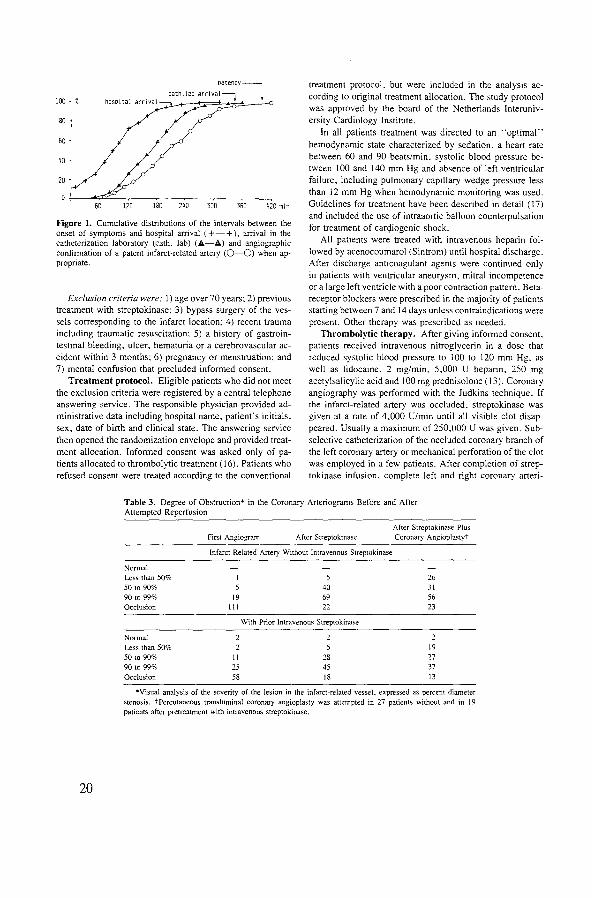
Figure 1. Cumulative distributions of the intervals between the onset of symptoms and hospital arrival ( +-+ ). arrival in the catheterization laboratory (cath. lab) (.~-A) and angiographic confirmation of a patent infarct-related artery (0-0) when appropriate.
Exclusion criteria were: I) age ov~r 70 years: 2) previous treatment with streptokinase: 3) bypass surgery of the vessels corresponding to the infarct location: 4) recent trauma including traumatic resuscitation: 5) a history of gastrointestinal bleeding, ulcer, hematuria or a cerebrovascular accident within 3 months: 6) pregnancy or menstruation: and 7) mental confusion that precluded informed consent.
Treatment protocol. Eligible patients who did not meet the exclusion criteria were registered by a central telephone answering service. The responsible physician provided administrative data including hospital name, patient's initials, sex. date of birth and clinical state. The answering service then opened the randomization envelope and provided treatment allocation. Informed consent was asked only of pa· tients allocated to thrombolytic treatment ( 16). Patients who refused consent were treated according to the conventional
treatment protocol. but were included in the analysis according to original treatment allocation. The study protocol was approved by the board of the Netherlands Interuniversity Cardiology Institute.
In all patients treatment was directed to an "optimal" hemodynamic state characterized by sedation, a heart rate between 60 and 90 beats/min. systolic blood pressure be· tween I 00 and 140 mm Hg and absence of left ventricular failure. including pulmonary capillary wedge pressure less than 12 mm Hg when hemodynamic monitoring was used. Guidelines for treatment have been described in detail ( 17) and included the use of intraaortic balloon counterpulsation for treatment of can;liogenic shock.
All patients were treated with intravenous heparin followed by acenocoumarol (Sintrom) until hospital discharge. After discharge anticoagulant agents were continued only in patients with ventricular aneurysm, mitral incompetence or a large left ventricle with a poor contraction pattern. Betareceptor blockers were prescribed in the majority of patients starting between 7 and 14 days unless contraindications were present. Other therapy was prescribed as needed.
Thrombolytic therapy. After giving informed consent, patients received intravenous nitroglycerin in a dose that reduced systolic blood pressure to I 00 to 120 mm Hg, as well as lidocaine. 2 mg/min, 5,000 U heparin, 250 mg acetylsalicylic acid and 100 mg prednisolone (13). Coronary angiography was performed with the Judkins technique. If the infarct-related artery was occluded, streptokinase was given at a rate of 4,000 U/min until all visible clot disappeared. Usually a maximum of 250,000 U was given. Subselective catheterization of the occluded coronary branch of the left coronary artery or mechanical perforation of the clot was employed in a few patients. After completion of streptokinase infusion, complete left and right coronary arteri-
Table 3. Degree of Obstruction* in the Coronary Arteriograms Before and After Attempted Reperfusion
20
Normal Less than 50% 50 to 90% 90 to 99% Occlusion
Normal Less than 50% 50 to 90% 90 to 99% Occlusion
First Angiogram After Streptokinao.;e
Infarct-Related Artery Without Intravenous Streptokinase
19 Ill
II 25 58
40 69 22
With Prior Intravenous Streptokinase
28 45 18
After Streptokinase Plus Coronary Angioplastyt
26 31 56 23
19 27 37
13
*Visual analysis of the severity of the lesion in the infarct-related vessel. expressed as percent diameter steno~is. tPercutaneous transluminal coronary angioplasty was attempted in 27 patients without and in 19 patients after pretreatment with intravenous streptokinase.
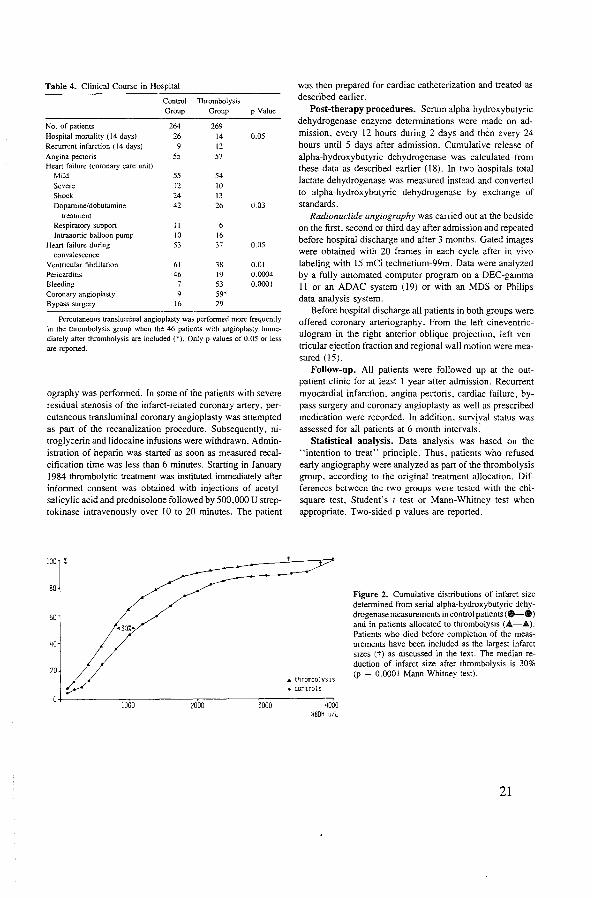
Table 4. Clinical Course in Hospital
Control Group
No. of patients 264
Hospital mortality (14 days) 26 Recurrent infarction ( 14 days) 9 Angina pectoris 55 Hean failure (coronary care unit)
Mild 55 Severe 12 Shock 24 Dopamine/dobutamine 42
treatment Respiratory support II lntraaortic balloon pump 10
Heart failure during 53 convalescence
Ventricular fibrillation 61 Pericarditis 46
Bleeding Coronary angioplasty Bypass surgery 16
Thrombolysis Group
269
14 12
57
54
10 13
26
16 37
38 19 53 59* 29
p Value
0.05
0.03
0.05
0.01 0.0004 0.0001
Percutaneous transluminal angioplasty was performed more frequently in the thrombolysis group when the 46 patients with angioplasry immediately after thrombolysis are included (*). Only p values of 0.05 or less are reported.
ography was performed. In some of the patients with severe residual stenosis of the infarct-related coronary artery, percutaneous transluminal coronary angioplasty was attempted as part of the recanalization procedure. Subsequently, nitroglycerin and lidocaine infusions were withdrawn. Administration of heparin was started as soon as measured recalcification time was less than 6 minutes. Starting in January 1984 thrombolytic treatment was instituted immediately after informed consent was obtained witb injections of acetylsalicylic acid and prednisolone followed by 500,000 U streptokinase intravenously over lO to 20 minutes. The patient
100 %
80
50
20
was then prepared for cardiac catheterization and treated as described earlier.
Post-therapy procedures. Serum alpha-hydroxybutyric dehydrogenase enzyme determinations were made on admission, every 12 hours during 2 days and then every 24 hours until 5 days after admission. Cumulative release of alpha-hydroxybutyric dehydrogenase was calculated from these data as described earlier (!8). In two hospitals total lactate dehydrogenase was measured instead and converted to alpha-hydroxybutyric dehydrogenase by exchange of standards.
Radionuclide angiography was carried out at the bedside on the first, second or third day after admission and repeated before hospital discharge and after 3 months. Gated images were obtained with 20 frames in each cycle after in vivo labeling witb 15 mCi technetium-99m. Data were analyzed by a fully automated computer program on a DEC-gamma II or an ADAC system ( 19) or with an MDS or Philips data analysis system.
Before hospital discharge all patients in both groups were offered coronary arteriography. From the left cineventriculogram in the right anterior oblique projection, left ventricular ejection fraction and regional wall motion were measured (15).
Follow-up. All patients were followed up at the outpatient clinic for at least l year after admission. Recurrent myocardial infarction. angina pectoris, cardiac failure, bypass surgery and coronary angioplasty as well as prescribed medication were recorded. In addition. survival status was assessed for all patients at 6 month intervals:
Statistical analysis. Data analysis was based on the "intention to treat" principle. Thus. patients who refused early angiography were analyzed as part of the thrombolysis group, according to the original treatment allocation. Differences between the two groups were tested with the chisquare test, Student's t test or Mann-Whitney test when appropriate. Two-sided p values are reported.
,. tnrombolysts • controls
Figure 2. Cumulative distributions of infarct size determined from serial alpha-hydroxybutyric dehydrogenase measurements in control patients ( e-e) and in patients allocated to thrombolysis (A-A). Patients who died before completion of the measurements have been included as the largest infarct sizes (t) as discussed in the text. The median reduction of infarct size after thrombolysis is 30% (p = 0.0001 Mann-Whitney test).
+---------~J~o~oo~--------~2~oo~o----------=Jo:'oo~---------~o HBDH u/l
21

more, similar results were obtained when the patients who died within 72 hours were not included in the analysis and when data from the five hospitals were analyzed separately. Median values for alpha-hydroxybutyric dehydrogenase infarct size in control and thrombolysis patients were I, I 00 and 770 U/liter, respectively. In patients with a first infarct these values were 1,140 and 790 U/liter, respectively, in anterior wall infarction 1,280 and 840 U/liter, respectively and in inferior wall infarction 970 and 670 U/liter, respectively. In Figure 3 alpha-hydroxybutyric dehydrogenase release is shown in relation to the interval between the onset of symptoms and hospital admission. In patients allocated to the control group, alpha-hydroxybutyric dehydrogenase release was independent of the interval between the onset of symptoms and admission. On the other hand, we found smaller enzyme release in patients allocated to thrombolysis within 2 hours after the onset of symptoms. These data indicate a 51% reduction of infarct size by thrombolysis in patients admitted within I hour, a 31% reduction of infarct size in those admitted between 1 and 2 hours and a 13% reduction in patients admitted between 2 and 4 hours after the onset of symptoms.
Left ventricular function (Table 5). Left ventricular ejection fraction was measured by radionuclide angiography between day 2 and day 4 in 418 patients and before hospital discharge in 361 patients. Missing data were equally distributed between the two treatment groups and were due to death, transfer to other hospitals. patient refusal, unavailability of the gamma camera or other administrative reasons. There was no change in global left ventricular ejection fraction between the second day and hospital discharge in the control group. In the thrombolysis group left ventricular ejection fraction before discharge was 3.7 ± 9.0% higher than the first measurement. Accordingly. ejection fraction after 10 to 20 days was approximately 4% higher when thrombolysis was compared with conventional treatment. This difference was significant in the whole group, in patients treated with intracoronary thrombolysis only, in pa-. tients with a first infarction and in those with anterior in-
Table 6. Clinical Follow-Up
Control Thrombolysis Group Group
No. of patients 264 269 Death 42 23 Reinfarction 16 36 Acute PTCA 46 Late PTCNCABG 40 62
farction. Similarly, a 6% greater ejection fraction was found during cardiac catheterization in the thrombolysis group. Again these differences were similar in patients with a first infarct only and in patients with anterior wall or inferior wall infarction on admission and in both treatment protocols (15).
Follow-up (Tables 6 and 7). Clinical follow-up ranged from I to 48 months after admission. There was a 45% reduction of mortality after thrombolysis. This was offset by a higher incidence of late reinfarction and more frequent performance of late coronary angioplasty or bypass surgery after thrombolysis. The reduction in mortality was present in all subgroups, and it was similar in all five hospitals.
The subgroup of patients without early angiography and those in whom recanalization failed fared worse than those in whom recanalization was achieved. On the other hand, there was only I death in 65 patients with a patent infarctrelated vessel at angiography and also I death in 46 patients in whom coronary angioplasty was performed immediately after thrombolysis. This particular patient underwent thrombolysis and angioplasty of the left anterior descending artery. Despite treatment with anticoagulant agents and nifedipine, he developed postinfarction angina. After 7 days the artery was reoccluded at the same site and coronary angioplasty was repeated. After 31 days the patient developed a new anteroseptal infarction and died from intractable cardiogenic shock.
Discussion The primary aim of the present study was the analysis
of the effect of early thrombolysis on the clinical course and survival of patients with acute myocardial infarction. The results demonstrate that thrombolysis in the first hours after the onset of infarction can reduce myocardial damage and thus preserve part of the function of the left ventricle and improve patient survival. In one other randomized trial (20,21) there was a similar improvement in survival, although left ventricular function and infarct size appeared
No Thrombolysis
Angiography* o~o e~o
·~· 35 65 133 36 8
4 21 13 31 18 28
*Patients who were allocated to the thrombolysis group but did not undergo acute angiography. Major complications (mortality and nonfatal recurrent infarction) and coronary artery bypass surgery (CABG) or percutaneous transluminal coronary angioplasty (PTCA) in patients allocated to conventional treatment (control) or thrombolysis. Symbols as in Table 1.
24
Table 7. Mortality
Total mortality
I ntracoronary thrombo!y~is
lntmvenous and
intracoronary
thromboly':.i:-.
First infarct only
Anterior wall infarction Inferior wall infun:tion
Thomxcenter SL Annada! Free Univer:-.ity Zuiderziekt:nhui:-. Ldden University
Control Group
42
23
19
26
25 17
20 Ill 6 6
Thrombolysi~ No Thrombolysis
Group Angiography o-o e-o ·-· 23
17
II
12 4 II 4
II 3 4 I
Mortality in patients admitted before or since January 19X4 and mortality data in the tive panicipating ho~pital:-.. Patienh allocated to thrombolytic treatment an: grouped according to the re.,.ulb of the intervention. Note that similur trend':. an;: present in all ':.ubgroups. Symbols as in Table I.
unaltered (22). while results of several smaller randomized trials were inconclusive (23-28). The difference between the results of the present trial and those of other studies can be explained hy differences in study design. by the shorter delay between the onset of symptoms and treatment and by its larger size.
Study design. The study was designed to compare a new method of treatment (thrombolysis) with the accepted mode of therapy. Informed consent was asked only from patients allocated to angiography and thrombolysis as proposed by Zelen ( 16). This design was chosen to prevent extensive discussions of the risk and potential benefits of acute angiography and thrombolysis in half of these critically ill patients who were eventually allocated to conventional coronary care unit treatment. Data analysis was based on original treatment allocation. Therefore. the 35 patients who did not undergo acute angiography were analyzed as part of the thrombolysis group. This subgroup included a few patients in shock who refused the intervention ·"because they wanted to be left alone.·· This is reflected by the relatively high mortality in this group (5 of 35 patients) n able 6). Because similar patients must be part of the control group. removal of this subgroup from the intervention group would falsely favor the effect of thrombolytic treatment. Yet it is evident that these deaths are not related to the thrombolytic therapy.
Possible limitations. The interpretation of this study might be questioned because of changes in the protocol in January !984. the inclusion of coronary angioplasty in some of the patients. missing data. the lack of coronary arteriography
on admission in the control group and the absence of direct measurements of baseline left ventricular function. These points will therefore be discussed in detail.
Because the aim of the trial was not to study the effect of intracoronary streptokinase itself, but rather to study the effect of early reperfusion. we decided to combine both intravenous and intracoronary thrombolysis in the later patients when it became apparent that the preparation of the catheterization laboratory. the introduction of catheters and the first angiogram delayed the administration of streptokinase by approximately I hour. while several reports (8-10) indicated that recanalization occurred in a considerable number of patients with administration of intravenous streptokinase.
Direct perforation of the thrombus was attempted in 5 patients and coronary angioplasty was performed in addition to the streptokinase infusion in 46 patients. This intervention was considered an integral part of the recanalization procedure because earlier observations indicated that patients with residual subtotal occlusion after thrombolysis are at increased risk for reocclusion. which would negate the effect of thrombolysis (3). Coronary angioplasty was not associated with complications. In fact. alpha-hydroxybutyric dehydrogenase release after coronary angioplasty was decreased and left ventricular ejection fraction was higher than in patients in whom only thrombolysis was carried out ( 15). Therefore. it is likely that the beneficial effects of thrombolysis in the present study would have been less apparent without additional coronary angioplasty.
No patients were lost to follow-up with respect to mor-
25
tality or major clinical events, which represent the major end points of the study. Missing data on left ventricular function were due to death of the patients, patient refusal, transfer to other hospitals, lack of technical facilities at the required time or intervening bypass surgery. It is unlikely that this would invalidate the results because missing data were equally distributed between the two groups and because similar differences were observed between patients allocated to thrombolysis and control patients in various subgroups. Also, there were no differences in baseline data between patients with or without measurements of left ventricular function.
In contrast with other trials (20-28). early angiography wa' not performed in our control group. Therefore. the coronary anatomy on admission could not be studied in these patients. This procedure was elected because acute angiography is not a part of conventional management of myocardial infarction. In fact, angiography might expose these patients to a small but pertinent risk, which could worsen prognosis in the control group ( 13). Similarly, determination of left ventricular function by radionuclide angiography was not attempted on admission because this would have delayed the intervention and thus diminish the possible salutary effects of recanalization:
Angiography in acute myocardial infarction. Earlier data from pilot studies performed at the Thoraxcenter ( 13) indicated that the risk of mortality. directly related to intracoronary infusion of streptokinase in patients with acute myocardial infarction. might be as high as 5'/f. However. despite 5 deaths during the catheterization procedure, 14 day mortality in patients allocated to thrombolysis was lower (14 deaths [5%[) than in the control group (26 deaths [10%[). In our experience the risk of the intervention is not so much associated with early angiography itself as with the actual recanalization. We thus agree with De Wood et al. (29). who reported that early angiography can be performed without excessive risk in patients with myocardial infarction. The 82% incidence of coronary occlusion at angiography and the 79% recanalization rate in patients treated with intracoronary streptokinase in the present study are consistent with other reports on early angiography and intracoronary thrombolysis ( l-4.20.23-28).
Myocardial enzyme release. Total alpha-hydroxybutyric dehydrogenase release in 72 hours was calculated as a measure of infarct size using a two compartment model (30). This model takes into account the fractional catabolic rate or clearance of the enzymes from the blood and is insensitive to faster washout after reperfusion (30,31 ). Earlier studies (32) demonstrated similar results when infarct size was estimated from different enzymes including creatine kinase. creatine kinase-ME isoenzyme. alpha-hydroxybutyric dehydrogenase and aspartate aminotransferase. In the present study, alpha-hydroxybutyric dehydrogenase release was measured because it can be computed accurately
26
from samples taken at 12 hour intervals, whereas creatine kinase should be sampled more frequently. Furthermore, the error in the computed total enzyme release due to biologic variations in the fractional catabolic rate of alphahydroxybutyric dehydrogenase is smaller than for creatine kinase, creatine kinase-ME isoenzyme or aspartate aminotransferase (32). Although estimation of infarct size by enzyme release is controversial, earlier studies (18) demonstrated a relation between total alpha-hydroxybutyric dehydrogenase release and the clinical sequelae of myocardial infarction. Furthermore, an autopsy study (33) in 84 patients who died of acute myocardial infarction demonstrated that peak levels of lactate dehydrogenase or thermostable lactate dehydrogenase correlated better with anatomic infarct size than did peak aspartate aminotransferase. Actually, the correlation between thermostable lactate dehydrogenase and anatomic infarct size (r = 0.79) was similar to that in a recent study (34) comparing anatomic infarct size with peak creatine kinase (r = 0. 79) or total creatine kinase infarct size (r = 0.86). Other investigators have used peak creatine kinase values (20.23) or peak lactate dehydrogenase measurements (27), which appeared not to be reduced after thrombolysis. In fact, peak creatine kinase in the Western Washington trial (20) was 30% higher in the streptokinase group than in control patients. However, such peak measurements are not reliable estimates of myocardial enzyme release because enzyme washout is faster after reperfusion. In the present study we observed a faster washout of alphahydroxybutyric dehydrogenase, lactate dehydrogenase and creatine kinase after thrombolysis.
In our study a 30% median reduction of alpha-hydroxybutyric dehydrogenase release was observed in the thrombolysis group. This supports the hypothesis that myocardial tissue can be salvaged by early thrombolysis. The estimation of alpha-hydroxybutyric dehydrogena'e release from lactate dehydrogenase measurements in two hospitals did not affect the difference between the thrombolysis and control groups. because similar differences between these two groups were observed in all five hospitals. Although these methods have not been validated in patients after coronary reperfusion, it is unlikely that myocardial salvage is overestimated by computation of alpha-hydroxybutyric dehydrogenase release. In fact. total creatine kinase release in dogs was approximately I 0% greater after reperfusion than without reperfusion at the same anatomic infarct size. whereas enzyme breakdown in the circulation was not affected by the presence of streptokinase (35). If the data from such studies apply to alphahydroxybutyric dehydrogenase release in patients with acute myocardial infarction. the actual salvage of myocardium would even be underestimated. The observed reduction of alpha-hydroxybutyric dehydrogenase release is in agreement with data reported by Anderson et al. (28). From their Figure 6 we computed a 40% reduction of total lactate dehydrogenase-! isoenzyme release in the thrombolysis group com-

726 SJMOONS ET AL. EARLY THROMBOLYSIS IN ACUTE MYOCARDIAL INFARCTION
pared with conventionally treated patients. Thus both studies are consistent with a reduction of enzymatic infarct size after thrombolysis.
Left ventricular function. Global left ventricular ejec· tion fraction was measured by contrast angiography and radionuclide angiography. Both methods showed a higher ejection fraction 2 weeks after thrombolysis than after conventional therapy (15). This improvement was seen in inferior wall as well as in anterior wall infarction and applied to patients with a first infarct as well as to those with recurrent infarction. Some differences between the two groups in left ventricular ejection fraction measured by radionuclide angiography were already apparent after 2 to 4 days (Table 5), although these differences were of borderline significance. Although no measurements of ejection fraction were performed on admission, we may presume that these data were similar in both groups because other baseline data were also evenly distributed. The data then indicate a gradual recovery of left ventricular function during the first 2 weeks after reperfusion (36). The differences in ejection fraction between the two groups of patients were small (4%). However, the global left ventricular ejection fraction is related to the function of both the infarcted myocardium and other areas. Thus, changes in the infarcted area may be underestimated because of compensatory changes elsewhere. The improvement in left ventricular function after thrombolysis is supported by analysis of regional wall motion and by the smaller end-diastolic and end-systolic volumes after thrombolysis, measured by contrast angiography (15).
Earlier experiments have demonstrated salvage of myocardial function after early coronary reperfusion in dogs (37-39). In addition, several studies reported a higher ejection fraction in patients with successful thrombolysis than in patients whose infarct-related coronary artery remained occluded (40-43) or in historical control patients (44). In other randomized trials, only Anderson et al. (28) reported a beneficial effect on ejection fraction after streptokinase treatment. Although the lack of a beneficial effect in other series may be due to the small number of patients studied, the different results between our trial and those of two other larger trials (20-23) can be explained by the longer delay until treatment in the latter studies because it is less likely that recanalization after 4 or 6 hours of occlusion will salvage significant amounts of myocardium in the majority of patients (38,39). In fact, the data from the present trial indicate that a marked reduction of infarct size can be achieved only in patients admitted within 2 hours after the onset of symptoms.
Clinical course and follow-up. Survival was improved significantly by early thrombolysis. Furthermore, there was a borderline significant reduction of heart failure and shock in patients allocated to thrombolysis. These data are in agreement with the observed reduction of infarct size and preservation of left ventricular function. Actually, the I year
JACC Vol. 7, No.4 April 1986:717-28
survival rates are very similar to those reported by investigators in the Western Washington Trial (21), although these investigators did not find differences in intermediary measurements, such as infarct size or left ventricular function (20).
Unfortunately, these beneficial effects of thrombolytic therapy on survival were offset by a higher incidence of reinfarction, particularly in patients with an inferior wall infarction (14). Because reinfarction occurred in some of the patients despite adequate anticoagulant therapy or after successful coronary angioplasty, further studies are warranted to determine the optimal mode of treatment after thrombolysis. Bleeding after thrombolytic treatment occurred frequently at puncture sites. but did not result in significant morbidity ( 45).
Conclusions. The aim of recanalization in patients with acute myocardial infarction is rapid restoration of blood flow to the jeopardized myocardium to preserve viable myocardial cells. Ultimately this should result in preservation of myocardial function and improved prognosis after the infarction. Although the measurements of infarct size and left ventricular function used in this study might be criticized, the consistency of the observations supports the true benefits of early thrombolysis in patients with myocardial infarction. In contrast with other studies that either were too small (24-27) or initiated treatment later after the onset of symptoms (20-23), we demonstrated a 30% smaller infarct size estimated from myocardial enzyme release as well as preservation of left ventricular function documented by contrast angiography and radionuclide angiography in patients allocated to early thrombolytic therapy, as well as a reduction. of early and late mortality. Future studies should investigate whether intracoronary administration of a thrombolytic agent is indeed mandatory, or whether similar results can be obtained by intravenous infusion of newer thrombolytic drugs such as the tissue plasminogen activator (46-48). Finally, strategies should be developed for early recognition of the symptoms of myocardial infarction by the patient and for immediate intervention, because myocardial salvage is only attainable if myocardial blood flow is restored within the first few hours of infarction.
Appendix
Participating Centers and Collaborators Thoraxcenter, Erasmus University and University Hospital Dijk
zigt, Rotterdam. Marcel J. B. M. vd Brand, MD, Pim J. de Feyter, MD, Paolo Fioretti, MD, Paul G. Hugenholtz, MD, Patrick W. Serruys, MD, Maarten L. Simoons, MD, William Wijns, MD.
Department of Cardiology, Free University, Amsterdam. Machiei 1. van Eenige, MSc, Jan C. 1. Res, MD, Jan P. Roos, MD, Freek W. A. Verheugt, MD, Frans C. Visser, MD, Ernst E. vd Wall, MD.
Department of Cardiology, Zuiderziekenhuis, Rotterdam. Diederik C. A. van Hoogenhuyze. MD. X. Hanno Krauss, MD. WillemJ. Remme, MD.
Department of Cardiology, St. Annadal HOspital, Maastricht. Frits
27
W. H. M. Bar. MD. Simon H. J. G. Braat, MD. Pedro Brugada. MD. K;.~.rd den Dulk. MD. Wim T. Hermens. MSc. Hein J. J. Wellens. MD. Chri:-. de Zwaan. MD.
Department or Cardiology, University Hospital. Leiden. Berend Buis. MD. Jo:-. Engbers. MD. Amoud vd Laarse. PhD.
Data processing center. Thoraxcenter, Erasmus University, Rotterdam. Sophie vd Doe:-.. Ron T. van Domburg. MSc. Jacobus Lubsen. PhD. Jan P. van Mantgem. MD. Karel J. de Neef. MD. Juan Planellas. MD, Frank Vermeer. MD. Anneke A. Wagenaar. loge C. J. Zorn.
References I. Rc:ntrop P. De Vivie ER. Kar:-.ch KR. Kreuzer H. Acute myocardial
infarction: intracoronary application or nitroglycerin and streptokinase in combination with transluminal recanalization. Clio Cardiol 1979;S;JS4-6.
2. Merx W. Dorr R. Rentrop P. ct al. Evaluation of the effectiveness of intr;.~.coronary :-;treptokina:-.e infusion in acute myocardial infarction: po:-.tproccdure management and hospital course in 204 patient:-.. Am Heart J 19XI:I02:IIXI-7.
J. St:rruy:-. PW. Wijn:-. W. Brand M v/d, eta!. b tmnsluminal coronary angioplasty mandatory after :-.ucce:-.sful thrombolysi:-."! Br Heart J 19XJ50:257-6S.
-l-. Rentrop KP. Thrombolytic therapy in patient:-. with acutt: myocardial infarction. Circulation 19X5:71 :627-J I.
5. Muller JE. Stone PH. Markie!'. JE. Braunwald E. Let':-. not let the genic c:-.cape from the bottlc~again. N Eng! J Med 19~ I :304: 1294-6.
6. Swan HJC. Editorial: thromboly:-.1:-. in acute myocardial infarction: treatment of the underlying coronary artery di:-.ea:-;c. Circulation 19X2;66:914-6.
7. Hugenh()ltl PG. Rentn)p P. Thromb()lytic therapy for acute my()Cardial infarction: quo vadi:-."! A review of the recent !itt:rature. Eur Heart J I YX2;3:395-403.
X. Roger WJ. Mantle JA. Hood WP, et al. Streptokinase in acute myocardial infarction. Circulation 19X3:6X: I 051-61.
9. Schroeder R. Biamino G. Von Leitner ER. et a!. lntravenou:-. shorttenn infusion of streptokinase in acute myocardial infarction. Circulation 1983;67:536--48.
10. Alderman EL. Jutzy KR. Berte LE. et al. Randomized comparison of intravenou:-. versu~ intracoronary :-;treptokinase for myocardial infarction. Am J Cardiol IYX4:54:14-9.
II. Simoon:-. ML. Wijn:-. W. Balakumar.m K. et al. The effect of intracoronary thromboly~b with streptokina:-.c on myocardial thallium distribution and left ventricular function assessed by blood-pool :-.cintigr<tphy. Eur Heart J \9X2:3:433-40.
12. Fioretti P. Simoon:-. ML. Scrruy:-. PW. Br..tnd M vld. Fe\s PW. Hugt:nholtz PG. Clinical course after attempted thromboly:-.is in myocardial infarction. Rc:sult:-. of pilot studie:-. and preliminary data from a randomized trial. Eur Heart J 19X2:3:422-J2.
IJ. Scrruy:-. PW. Brand M van den. Hooghoudt TEH. et al. Coronary recanalization in acute myocardial infarction: immt:diate results and potential ri!-.b. Eur Heart J 19X2:J:404-15.
14. Simoons ML. Serruys PW. Brand M v/d, et al. Improved survival after early thrombolysis in acute myocardial infarction. A randomized trial conducted by the Interuniversity Cardiology Institute in the Netherlands. Lancet 1985;2:57X-82.
15. Serruys PW. Simoons ML. Suryapranata H. et al. Preservation of global and regional left ventricular function after early thrombolysis in acute myocardial infarction. J Am Coli Cardiol 1986;7:729---42.
16. Zclen M. A new design for mndomized clinical trials. N Eng! J Med 1979:300:1242-5.
17. Simoons ML. Serruys PW. Fioretti P. Brand M v/d, Hugenholtz PG. Practical guidelines for treatment with beta-blockers and nitrates in patients with acute myocardial infarction. Eur Heart J 1983:4:129-35.
28
18. Laarse A v/d. Hennens WT. Hollaar L. et al. Assessment of myocardial damage in patients with acute myocardial infarction by serial measurement of serum alpha-hydroxybutyrate dehydrogenase levels. Am Heart J 1984;107:248-60.
19. Reiber JHC. Lie SP, Simoons ML. et al. Clinical validation of fully automated computation of ejection fraction from gated equilibrium bloodpool scintigrams. J Nucl Med 1983:24:1099-107.
20. Kennedy JW, Ritchie JL. Davis KB. Fritz JK. Western Washington randomized trial of intracoronary streptokinase in acute myocardial infarction. N Engl J Med 1983;309;1477-82.
21. Kennedy JW. Ritchie JL, Davis KB. Stadius ML, Maynard C. Fritz JK. The Western Washington randomized trial of intracoronary streptokinase trial in acute myocardial infarction. N Eng! J Med 1985;312: 1073-8.
22. RitchieJL. Davis KB. Williams DL, Caldwell J, Kennedy JW. Global and regional left ventricular function and tomographic radionuclide perfusion: The Western Washington intracoronary str.!ptokinase in my(~eardial infarction trial. Circulation 1984:70:867-75.
23. Rentrop KP. Feit F. Blanke H. et al. Effects of intracoronary streptokinase and intracoronary nitroglycerin infusion on coronary angiegraphic patterns and mortality in patients with acute myocardial infarction. N Engl J Med 1984;311:1457-63.
24. Khaja F. Walton JA, Brymer JF. et a!. lntracoronary fibrinolytic therJ.py in acute myocardial infarction. N Eng! J Med 19~3;308: 1305-11.
25. Anderson JL, Mar:-.hall HW. Bray BE. et al. A randomized trial of intracoronary streptokinase in the treatment of acute myocardial infarction. N Eng! J Med 19~3:308:1312-8.
26. Rogers WJ, Mantle JA. Hood WP. et al. Prospective randomized trial of intravenous and intracoronary streptokinase in acute myocardial infarction. Circulation 1983:68: I 051-61.
27. Leiboff RH. Katz RJ. Wasserman AG. et al. A randomized, angiographically controlled trial of intracoronary streptokinase in acute myocardial infarction. Am J Cardin! 1984;53:404-7.
2X. Anderson JL. Marshall HW. Askins JC. et al. A randomized trial of intmvenous and intracoronary streptokinase in patients with acute myocardial infarction. Circulation 1984;70:606-18.
29. De Wood MA. Spores J, Notske R, et al. Prevalence of total coronary occlu:-.ion during the early hours of trJ.rt->mural myocardial infarction. N Engl J Med 1980;30J:897-902.
30. Willems GM. Muijtjens AMM. Lambi FHH. Hennens WT. Estimation of circulatory parameters in patients with acute myocardi<:\1 infarction. Significance for calculation of enzymatic infarct size. Cardiovasc Res 1979:13:578-87.
31. Hennens WT, Willems GM. Visser MP. Quantification of circulating proteins. In: Theory and Applications Based on Analysis of Plasma Protein Leveb. Amsterdam: Martinus Nijhoff. 1982:125-36.
32. Willems GM. Visser MP. Krill MT A. Hennens WT. Quantitative analysis of pla"ma enzyme levels based upon simultaneous determination of different enzymes. Cardiova-;c Res 1982;16: 120-31.
33. Erhardt LR. Clinical and pathological observations in different types of acute myocardial infarction. Acta Med Scand 1974:560(suppl): 1-78.
34. Hackel DB. Reimer KA. ldekcr RE. et al. Comparison of enzymatic and anatomic estimates of myocardial infarct size in man. Circulation 1984;70;824-35.
35. Roberts R. Ishikawa Y. Enzymatic estimation of infarct size during reperfusion. Circulation 1983:68(suppll):l-83.
36. Ellis SG. Henschke CI. Sandor T. Wynne J, Braunwald E. Kloner RA. Time course of functional and biochemical recovery of myocardium salvaged by reperfusion. JAm Coil Cardiol 1983:1:1047-55.
37. Baughman KL. Maroko PR. Vatner SF. Effects of coronary artery reperfusion on myocardial infarct size and survival in conscious dogs. Circulation 1981:63:317-23.
38. Lavallee M, Cox D. Patrick TA. Vatner SF. Salvage of myocardial function by coronary artery reperfusion l, 2. and 3 hours after occlusion in conscious dogs. Circ Res 1983:53:235---47.
39. Bush LR. Buja M, Samowitz W, et al. Recovery of left ventricular segmental function after long-term reperfusion following temporary coronary occlusion in conscious dogs. Circ Res 1983:53:248-63.
40. Mathey DG, Rodewald G. Rentrop P. et al. Intracoronary streptokinase thrombolytic recanalization and subsequent surgical bypass of remaining atherosclerotic stenosis in acute myocardial infarction: complementary combined approach effecting reduced infarct size, preventing reinfarction, and improving left ventricular function. Am Heart J 1981;102:1194-201.
41. Smalling RW, Fuentes F. Matthews MW, et al. Sustained improvement in left ventricular function and mortality by intracoronary streptokinase administration during evolving myocardial infarction. Circulation 1983;68:131-8.
42. Rogers WJ, Hood WP. Mantle JA, et al. Return of left ventricular function after reperfusion in patients with myocardial infarction: importance of subtotal stenoses or intact collaterals. Circulation 1984:69:338--49.
43. Sheehan FH, Mathey DG, Schofer J, Krebber HJ, Dodge HT. Effect
of interventions in salvaging left ventricular function in acute myocardial infarction: a study of intracoronary streptokinase. Am J Cardia! 1983;52:431-8.
44. De Feyter PJ, Eenige MJ van, Wall EE van der. et a!. Effects of spontaneous and streptokinase-induced recanalization on left ventricular function after myocardia] infarction. Circulation 1983:67:1039-44.
45. Verheugt FWA, Eenige MJ van, Res JCJ, et al. Bleeding complications of intracoronary fibrinolytic therapy in acme myocardial infarction. Br Heart J 1985:54:455-9.
46. \Yerf F van de, Ludbrook PA, Bergmann SR. et al. Coronary thrombolysis with tissue-type plasminogen activator in patients with evolving myocardial infarction. N Eng! J Med 1984:310:609-13.
47. TIMI Study Group, The thrombolysis in myocardial infarction (TIM!) trial: phase l findings. N Eng! J Med 1984:310:932-6.
48. Verstraete M. Bory M, Collen D. et al- Randomized trial of intravenous recombinant tissue-type plasminogen activator versus intravenous streptokinase in acute myocardial infarction. Lancet 1985; 1: 842-7.
29
CHAPTERS
Effects of early intracoronary streptokinase on
infarct size estimated from cumulative enzyme
release and on enzyme release rate: A
randomized trial of 533 patients with acute
myocardial infarction·
The effects of early intracoronary streptokinase (SK) on enzymatic infarct size and rate of enzyme release were studied in a randomized multicenter trial. A total of 533 patients with acute myocardial infarction (AMI) were allocated to either the SK treatment group (n = 269) or the conventional (control) treatment group (n = 264). Enzymatic infarct size was represented by the cumulative quantity of a·hydroxybutyrate dehydrogenase (HBDH) released by the heart per liter of plasma in the first 72 hours. Rate of enzyme release was represented by the ratio of HBDH quantities released in 24 hours and 72 hours. On an "intention to treat" basis, the SK group had a smaller (by 30%; p = 0.0001) median enzymatic infarct size and a higher (by 35%; p = 0.0001) median rate of enzyme release than the control group. Limitation of infarct size was less apparent in patients treated with intracoronary SK only (25%) than in patients treated with intravenous plus intracoronary SK {34%). Compared to the control group, the enzyme release rate in patients treated with intracoronary SK only was slightly less (34%) than that in patients treated with intravenous plus intracoronary SK (38%). Patients with a patent infarct-related coronary artery at acute angiography had a median infarct size which was 55% (p = 0.0001) smaller than the median infarct size of the control group, and the median rate of enzyme release was 38'% (p = 0.001) higher than the median release rate of the control group. Patients with successful recanalization during intracoronary SK infusion had a median infarct size which was 31% (p = 0.002) smaller than the median infarct size of the control group and a median rate of enzyme release which was 42% (p = 0.0001) higher than the median release rate of the control group. Patients with persistent coronary occlusion in spite of thrombolytic therapy had a median infarct size which was 11% (NS) higher than the median infarct size of the control group, although the median rate of enzyme release was still 23% (p = 0.02) higher than the median release rate of the control group. It is concluded that thrombolysis in the early phase of AMI limits infarct size and that intracoronary SK treatment itself accelerates the process of enzyme release from infarcted myocardium, independent of the angiographic result. (AM HEART J 1986;112:672.)
A. van der Laarse, F. Vermeer,' W. T. Hermens,' G. M. Willems,' K. de Neef,'· 3
M. L. Simoons,1 P. W. Serruys,1 J. Res,' F. W. A. Verheugt,' X. H. Krauss,' F. Blir,S C. de Zwaan,' and J. Lubsen1• Leiden, The Netherlands Working group on thrombolytic therapy in acute myocardial infarction of the Netherlands Interuniversity Cardiology Institute.
From the Laboratory of Cardiobiochemistry, University Hospital Leiden, Leiden; and 1Thoraxcenter, Erasmus University and Hospital Dijkzigt, Rotterdam; lflept. of Biophysics, Biomedical Center, University of Limburg, Maastricht; 3Present address:Organon-International BV, OSS; •Dept. of Cardiology, Free University, Amsterdam; ~Dept. of Cardiology, Zuiderziekenhuis, Rotterdani: hDept. of Cardiology, St. Annadal Hospital, Maastricht.
Received for publication Feb. 3, 1986; accepted March 21, 1986.
Reprint requests: Dr. A. van der Laarse, Department of Cardiology, University Hospital Leiden, gebouw 1, CS-024, Rijnsburgerweg 10, 2333 AA Leiden, The Netherlands.
*Participating centers and collaborators: Thoraxcenter, Erasmus Universi-
30
ty and University Hospital Dijkzigt, Rotterdam (M. v/d Brand, P. de Feyter, P. Fioretti, P. G. Hugenholtz, P. W. Serruys, M. L. Simoons, W. Wijns); Department of Cardiology, Free University, Amsterdam (M. van Eenige, J. Res, J. Roos, F. Visser, F. W. A. Verheugt, E. E. v/d Wall); Department of Cardiology, Zuiderziekenhuis, Rotterdam (D. van Hoogenhuyze, X. H. Krauss, W. J. Remme, C. J. Storm, H. A. C. M. Kruyssen); Department of Cardiology, St. Annadal Hospital, Maastricht (F. Biir, S. Braat, P. Brugada, K. den Dulk, W. T. Hermens, M. Ramentol, H. J. J. Wellens, G. M. Willems, C. de Zwaan); Department of Cardiology, University Hospital, Leiden {B. Buis, J. Engbers, A. v/d Laarse); Data processing center, Thoraxcenter, Erasmus University Rotterdam (S. v/d Does, R. van Domburg, J. Lubsen, J. P. van Mantgem, K. de Neef, J. Planellas. F. Vermeer, A. Wagenaar, I. Zorn).
Both short- and long-term prognosis of patients who have had a myocardial infarction depends largely on "infarct size," or rather on the remaining functioning myocardial tissue.1
-' Thus it is likely that limitation of "infarct size" will lead to a better prognosis of infarct patients. Early recanalization of the occluded coronary artery in patients with acute myocardial infarction (AMI) has been proposed as a method for myocardial salvage.5• 6 We recently completed a study of 533 patients comparing early recanalization by intracoronary administration of streptokinase (SK) with conventional treatment. The results indicate that early recanalization does indeed limit "infarct size" (estimated from serial enzyme determinations), thereby preserving myocardial function (estimated by contrast angiography and radionuclide angiography). Finally, the 1-year survival rate in patients allocated to thrombolytic therapy was significantly improved compared to that of patients in the control group (91% vs 84% ).7·9
This report further details the effects of thrombolytic therapy on the estimated "infarct size" by calculation of total a-hydroxybutyrate dehydrogenase (HBDH) enzyme release and the effects of treatment on th.e rate of enzyme release. Limitation of infarct size was only apparent in patients with a patent infarct-related artery after the intervention. However, in contrast with earlier reports, 10·'3 the rate of enzyme release appeared to be accelerated by thrombolytic therapy independent of the angiegraphic results.
METHODS
Patient selection. The study, sponsored by the Netherlands Interuniversity Cardiology Institute, was initiated in one hospital in 1981 and later extended to five hospitals.'· 9·" All patients admitted to the participating coronary
care units were eligible for the trial if severe chest pain lasting more than 20 minutes was accompanied by ST segment elevation of 0.2 m V or greater in one or more precordial leads and/or 0.1 m V or greater in one or more limb leads. In addition, patients were included who had 0.2 m V ST segment depression in precordial leads indicative of posterior wall infarction. Patients up to the age of 70 years were included if they were admitted to the coronary care unit within four hours after the onset of symptoms. Exclusion criteria were: previous treatment with SK, bypass surgery of the infarct-related vessel, recent trauma, traumatic resuscitation, gastrointestinal bleeding, gastric or duodenal ulcer, hematuria or cerebrovascular accident within 3 months, pregnancy or menstruation, or inability to give informed consent.
Eligible patients were registered by a telephone answering service which then provided treatment allocation. After randomization, informed consent was requested
from patients allocated to the group receiving thrombolytic therapy only.15 Prior to cardiac catheterization patients received nitroglycerin, lidocaine (2 mg/min), heparin (5000 U), acetylsalicylic acid (250 mg), and prednisolone (100 mg) intravenously. Coronary arteriography was performed with preformed catheters from the femoral approach. If the infarct-related artery appeared occluded, SK was administered intracoronarily at the rate of 4000 U/min until all visible clots disappeared. Usually 250,000 U was given. In patients with persistent occlusion, subselective infusion or mechanical perforation of the thrombus was attempted on occasion, while percutaneous transluminal coronary angioplasty was performed in some of the patients with severe residual stenosis of the infarctrelated coronary artery.16 In order to reduce treatment delay, in patients admitted since January, 1984, 500,000 U of SK was administered intravenously, immediately after informed consent was obtained, as soon as possible, followed by intracoronary SK infusion during acute cardiac catheterization. 7
Measurement of serum enzyme activities. Samples of 5 ml of venous blood were obtained at admission, directly after the SK infusion procedure (if allocated to study treatment), every 12 hours for the first 2 days, and daily for the following 3 days. The blood samples were all owe< l to clot, and the activities of HBDH in the serum were measured in the coronary care units of Rotterdam Thoraxcenter, Maastricht St. Annada! Hospital, and the University Hospital Leiden, and the activity of lactate dehydrogenase (LDH) in the serum was measured in the Rotterdam Zuiderziekenhuis and the Amsterdam Free University Hospital. Because erythrocytes contain high activity levels of LDH-1 and LDH-2, hemolytic samples were discarded, and new samples were ordered.
Activity levels are expressed in micromoles of a-ketobutyrate converted (HBDH) or micromoles of pyruvate converted (LHD) per minute or liter of serum at 37° C. The autoanalyzers which measured HBDH activities were calibrated daily such that the HBDH activity of a test serum (Autonorm H, batch 227, Nyegaard & Co, Oslo, Norway) equaled 430 U/L. So calibrated, the mean reference value of HBDH activity in serum is 83 U/L. From the autoanalyzers, which measured LDH activities, the serum LDH activities were converted to HBDH activities via the following formulas: Amsterdam Free University Hospital HBDH = 1.106 X LDH + 16 (r = 0.96), and Rotterdam Zuiderziekenhuis HBDH = 0.473 X LDH - 5 (r = 0.99). These formulas were obtained after LDH and HBDH measurement of 10 dilutions of an infarct serum diluted with a pasteurized plasma protein solution (Central Laboratory of the Netherlands Red Cross Blood Transfusion Service, Amsterdam, the Netherlands) spanning an LDH range of 0 to 2660 U /L.
Calculation of cumulative HBDH release. Previously the two-compartment model was presented17 with which the movements of liberated myocardial proteins to and from intravascular and extravascular spaces can be described. This model has now been verified for a great number of proteins, mostly enzymes. Briefly, this model
31
Table I. Median values (M) and first and third quartiles (Q1,Q3) of calculated and estimated cumulative HBDH release of patients allocated to thrombolytic therapy and to conventional treatment (control subjects) groups
Streptokinase Control Patients n M
448 patients with HBDHQ72 calculatedt 230 770 495 patients with HBDHQ72 calculated 248 770
(448) or estimated including 17 patients without AMI (Q72 = 40 U/L) and 30 patients who died early (8 SK, 22 control, Q72 = 6000 U/L)t
29 patients with Q72 estimated by 14 860 1.2 X Q48
9 patients with Q72 estimated from 400 CK(MB) curves
All 533 patients with Q72§ 269 760
*Mann· Whitney test. tlndicated by HBDHQ72. tlndicated by HBDHQ72A. §Indicated by HBDHQ72B. AMI =acute myocardial infarction.
enables the calculation of the total quantity of enzyme which has entered the plasma up to timet (Q[t]) per liter of plasma. If the release of enzymes into plasma starts at timet= 0, Q(t) equals the quantity of enzyme present in 1 L of plasma (C,[t]) plus the quantity of enzyme present in the extravascular compartment per liter of plasma (C,[t]) plus the quantity of enzyme that has been eliminated from 1 L of plasma up to time t. Cumulative release of enzyme per liter of plasma up to time, t, Q(t), is represented by:
Q(t) = C' ,(t) + C',(t) + FCR J C' ,(r)dr (equation 1), in
which FCR is the fractional catabolic rate constant, and C' ,(t) and C' ,(t) are the enzyme concentrations at timet in plasma and the extravascular space, respectively. The accents added to C' ,(t) and C',(t) in equation 1 indicate that enzyme concentrations are corrected for the normal steady-state concentration (C,), that is, C' ,(t) = C,(t) -C,.
This implies that Q(t) does not include normal steadystate release of enzyme but only refers to the extra release of enzyme resulting from AMI. The term C',(t) can be calculate~ according to the formula: C' ,(t) = TER ·
e<-ERR·li.J e<ERR·•1 •. C',(r)dr (equation 2), in which
TER and ERR represent the transcapillary escape rate constant and the extravascular return rate constant, respectively." With the use of fixed mean values for FCR, TER, and ERR being 0.015 hr-1, 0.014 hr-1
, and 0.018 hr-', respectively, Q(t) was calculated from serial determinations of serum HBDH activities and the normal steadystate HBDH activity C, = 83 U/L. The total HBDH activity ultimately released by the infarcted myocardium is represented by HBDHQ72 as at 72 hours after onset of
32
Q, Q .• n M Q, Q, p*
450 1230 218 1020 580 1630 0.0001 410 1260 247 1100 580 1860 0.0001
600 1500 15 1290 910 1850 0.0001
200 500 1050 1250 0.0001
400 1250 264 1140 630 1860 0.0001
chest pain enzyme release rate is almost (~95%) completed by then."
Calculation of HBDH release rate. As a measure of the rate with which enzymes are liberated from the myocardium into the circulation, the ratio HBDHQ24/HBDHQ72 is used, that is, the ratio of the quantities released in the first 24 hours and the first 72 hours.
Statistical analysis. Differences between medians of two groups of data were tested with the Mann-Whitney rank-sum test. Differences with p values >0.05 (twosided) were considered not significant.
RESULTS
Between June, 1981, and March, 1985, a total of 533 patients were entered into the study. Two hundred sixty-four patients were allocated to the conventional treatment group and 269 patients to the thrombolytic therapy group. In spite of treatment allocation, no angiography was performed in 35 patients. 7• 8 In 111 of 136 patients catheterized without intravenous SK the infarct-related vessel was occluded (82%) and recanalization was achieved in 79% of these. After prior intravenous SK the infarct-related vessel was patent in 40 of 98 patients (41% ). Ultimately the infarct-related artery was patent at admission or recanalized in 198 patients (85%). The median time between onset of symptoms !lld angiographic documentation of a patent infarct-related vessel was 200 minutes.
Of 533 patients included in the study, measurements of HBDH (or LDH) were complete in 448 patients (84% ). The measurements were incomplete in 85 patients. In order to verify whether the results
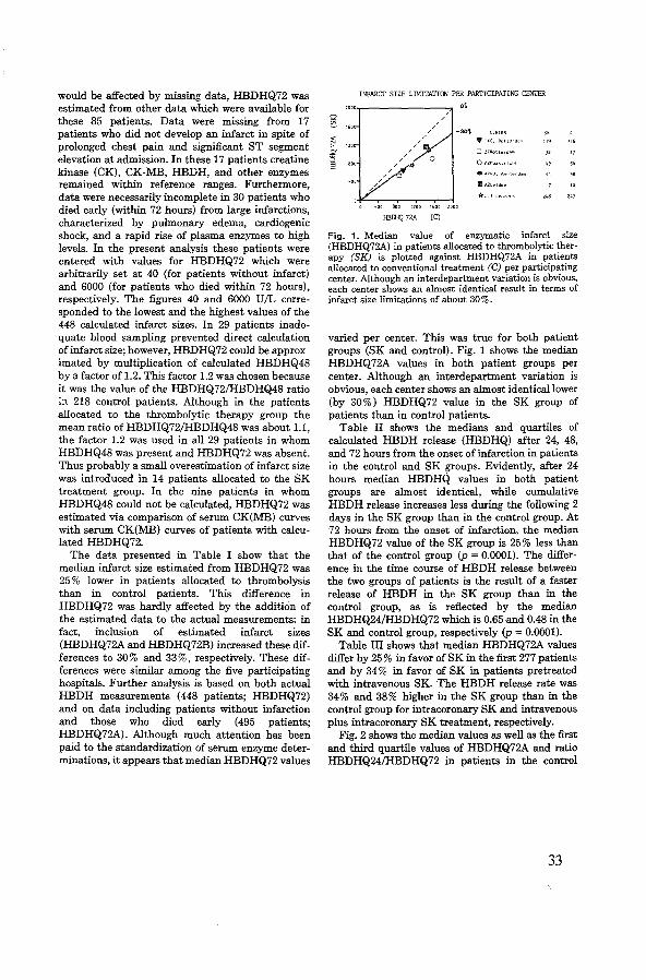
would be affected by missing data, HBDHQ72 was estimated from other data which were available for these 85 patients. Data were missing from 17 patients who did not develop an infarct in spite of prolonged chest pain and significant ST segment elevation at admission. In these 17 patients creatine kinase (CK), CK-MB, HBDH, and other enzymes remained within reference ranges. Furthermore, data were necessarily incomplete in 30 patients who died early (within 72 hours) from large infarctions, characterized by pulmonary edema, cardiogenic shock, and a rapid rise of plasma enzymes to high levels. In the present analysis these patients were entered with values for HBDHQ72 which were arbitrarily set at 40 (for patients without infarct) and 6000 (for patients who died within 72 hours), respectively. The figures 40 and 6000 U!L corresponded to the lowest and the highest values of the 448 calculated infarct sizes. In 29 patients inadequate blood sampling prevented direct calculation of infarct size; however, HBDHQ72 could be approximated by multiplication of calculated HBDHQ48 by a factor of 1.2. This factor 1.2 was chosen because it was the value of the HBDHQ72/HBDHQ48 ratio ;:-~ 218 control patients. Although in the patients allocated to the thrombolytic therapy group the mean ratio of HBDHQ72/HBDHQ48 was about 1.1, the factor 1.2 was used in all 29 patients in whom HBDHQ48 was present and HBDHQ72 was absent. Thus probably a small overestimation of infarct size was introduced in 14 patients allocated to the SK treatment group. In the nine patients in whom HBDHQ48 could not be calculated, HBDHQ72 was estimated via comparison of serum CK(MB) curves with serum CK(MB) curves of patients with calculated HBDHQ72.
The data presented in Table I show that the median infarct size estimated from HBDHQ72 was 25% lower in patients allocated to thrombolysis than in control patients. This difference in HBDHQ72 was hardly affected by the addition of the estimated data to the actual measurements: in fact, inclusion of estimated infarct sizes (HBDHQ72A and HBDHQ72B) increased these differences to 30% and 33%, respectively. These differences were similar among the five participating hospitals. Further analysis is based on both actual HBDH measurements (448 patients; HBDHQ72) and on data including patients without infarction and those who died early (495 patients; HBDHQ72A). Although much attention has been paid to the standardization of serum enzyme determinations, it appears that median HBDHQ72 values
INFARCI' SIZE LIMITATION PER PARTICIPATING CENTER
o\
-30'!.
600 1200 1600 2000
HBIEQ 7ZA (C)
OllRotl.,cd.,m
IIAZLeld"n
*"II ~el>!"r~
"' "
z48 247
Fig. 1. Median value of enzymatic infarct size (HBDHQ72A) in patients allocated to thrombolytic therapy (SK) is plotted against HBDHQ72A in patients allocated to conventional treatment (C) per participating center. Although an interdepartment variation is obvious, each center shows an almost identical result in terms of infarct size limitations of about 30%.
varied per center. This was true for both patient groups (SK and control). Fig. 1 shows the median HBDHQ72A values in both patient groups per center. Although an interdepartment variation is obvious, each center shows an almost identical lower (by 30%) HBDHQ72 value in the SK group of patients than in control patients.
Table II shows the medians and quartiles of calculated HBDH release (HBDHQ) after 24, 48, and 72 hours from the onset of infarction in patients in the control and SK groups. Evidently, after 24 hours median HBDHQ values in both patient groups are almost identical, while cumulative HBDH release increases less during the following 2 days in the SK group than in the control group. At 72 hours from the onset of infarction, the median HBDHQ72 value of the SK group is 25% less than that of the control group (p = 0.0001). The difference in the time course of HBDH release between the two groups of patients is the result of a faster release of HBDH in the SK group than in the control group, as is reflected by the median HBDHQ24/HBDHQ72 which is 0.65-and 0.48 in the SK and control group, respectively (p = 0.0001).
Table III shows that median HBDHQ72A values differ by 25% in favor of SK in the first 277 patients and by 34% in favor of SK in patients pretreated with intravenous SK. The HBDH release rate was 34% and 38% higher in the SK group than in the control group for intracoronary SK and intravenous plus intracoronary SK treatment, respectively.
Fig. 2 shows the median values as well as the first and third quartile values of HBDHQ72A and ratio HBDHQ24/HBDHQ72 in patients in the control
33

Table II. Median values (M) and first and third quartiles (Q1,Q3) of cumulative quantities of HBDH released per liter of plasma after 24, 48, 72, and 96 hours in patients allocated to thrombolytic therapy and to conventional treatment (control subjects) groups
Streptokinase Controls n M Q, Q, n M Q, Q, p*
HBDHQ24 252 460 260 780 236 490 300 730 NS HBDHQ48 245 680 420 1070 234 920 530 1390 0.003 HBDHQ72 230 770 450 1230 218 1020 580 1630 0.0001 HBDHQ96 187 780 450 1280 171 1050 620 1750 0.0006 HBDHQ24/HBDHQ72 230 0.65 0.55 0.76 218 0.48 0.39 0.58 0.0001
NS = not significant. *Mann-Whitney rank-sum test (two-sided).
Table Ill. Median values (M) and first and third quartiles (Q1,Q3) of enzymatic infarct size (HBDHQ72A) and enzyme release rate (HBDHQ24/HBDHQ72) in patients allocated to thrombolytic therapy and to conventional treatment (control subjects)*
Infarct size Streptokinase Control HBDHQ72A n M Q, Q, n M Q, Q, pf
All patients 248 770 410 1260 247 1100 580 1860 0.0001 lntracoronary SK 140 750 380 1220 137 1000 580 1700 0.001 Intravenous + intracoronary SK 108 810 450 1320 110 1220 640 2010 0.001
Enzyme release rate Streptokinase Control HBDHQ24/HBDHQ72 M Q, Q, n M Q, Q, Pt
All patients 230 0.65 0.55 0.76 218 0.48 0.39 0.58 0.0001 Intracoronary SK 127 0.67 0.57 0.76 121 0.50 0.39 0.61 0.0001 Intravenous + intracoronary SK 103 0.65 0.53 0.76 97 0.47 0.40 0.54 0.0001
"'Until 1984, SK was given intracoronarily only, and from January, 1984, on SK was given intravenously followed by intracoronarily. tMann-Whitney rank-sum test (two-sided).
and SK groups. In addition, the SK group is subdivided into four groups of patients: those without catheterization (35 patients), 65 with a patent infarct-related coronary artery at angiography, 133 patients with actual recanalization by thrombolytic therapy, and 36 patients in whom the artery remained occluded (Fig. 2).
Median infarct sizes of the patients without acute coronary angiography (910 U/L), as well as those of the patients in whom the infarct-related artery remained occluded (1220 U/L), were not significantly different from those of control patients (1100 U/L). In patients with recanalization during angiography median infarct size was 760 U/L, which is 31% lower than in control patients (p = 0.0001). The smallest infarcts were observed in 65 patients who already had a patent infarct-related artery at first angiography (median 500 U/L). In Fig. 2 the values of the ratio HBDHQ24/HBDHQ72 are presented, which are a measure of the rate of enzyme release. The control group and the patients in the SK group who did not undergo acute coronary
34
angiography had a relatively low median ratio HBDHQ24/HBDHQ72 (0.48 and 0.55, respectively) compared to patients with patent infarct-related coronary arteries (0.66), those with recanalization during angiography (0.68), and those with persistent occlusion (0.59).
In order to study whether the severity of the remaining stenosis of the infarct-related artery had any effect on infarct size or rate of enzyme release, patients were subdivided according to the visual interpretation of the degree of stenosis after recanalization. The results in Fig. 2 show no effect of the degree of opening of the infarct-related artery on infarct size.
Fig. 3 shows that in both anterior (including anteroseptal infarcts) and inferior (including posterior) infarcts cumulative HBDH release was lower and HBDH release rate was higher in patients allocated to SK treatment compared to control patients. When both groups are compared, median values of HBDHQ72A differ by 34% (p = 0.007) and by 31% (p = 0.002) for patients with anterior and
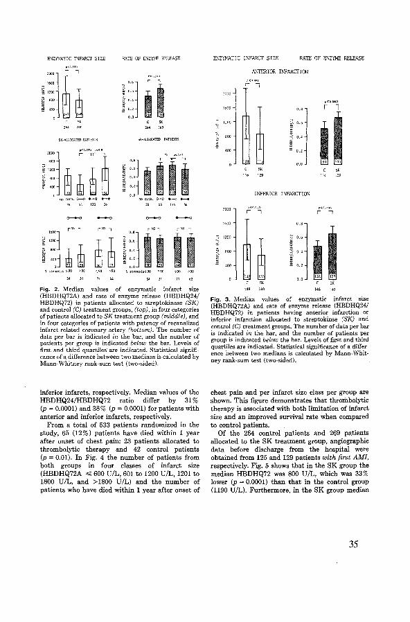
ENZYMATIC INFARCT SIZE RATE OF ENZYNE RELEASE
""l 1 0.6 II ~ 0.4
" ~ 0.2
o.o
"' '" SK-ALI.OCA.TID PATIE.\15 SK-ALUXATED PATIEl1TfS
no cnth. o--o .,._. no cath. o-o .-.-.o .._. t>S 133 3o l:S:S 36
o----o ..____, o----o ....__._, rNS -, r:-:s-, rNS-, rNS --,
""l "'l II I,:: ~ ~ B~
~ O,b II ::;;:- 0.4
il-~ 400 ~ a.z
0 ' 0.0 \ stenosts ~90 ~90 >90 \ stenosis~90 >90 :":;90
" 31 71 31 71
Fig. 2. Median values of enzymatic infarct size (HBDHQ72A) and rate of enzyme release (HBDHQ24/ HBDHQ72) in patients allocated to streptokinase (SK) and control (C) treatment groups, (top), in four categories of patients allocated to SK treatment group (middle), and in four categories of patients with patency of recanalized infarct-related coronary artery (bottom). The number of data per bar is indicated in the bar, and the number of patients per group is indicated below the bar. Levels of first and third quartiles are indicated. Statistical significance of a difference between two medians is calculated by Mann-Whitney rank-sum test (two-sided).
inferior infarcts, respectively. Median values of the HBDHQ24/HBDHQ72 ratio differ by 31% (p = 0.0001) and 38% (p = 0.0001) for patients with anterior and inferior infarcts, respectively.
From a total of 533 patients randomized in the study, 65 (12%) patients have died within 1 year after onset of chest pain: 23 patients allocated to thrombolytic therapy and 42 control patients (p = 0.01). In Fig. 4 the number of patients from both groups in four classes of infarct size (HBDHQ72A ~ 600 U/L, 601 to 1200 U/L, 1201 to 1800 U/L, and >1800 U/L) and the number of patients who have died within 1 year after onset of
ENZYMATIC INFARCT SIZE RATE OF ENZYME RELEASE
ANTERIOR INFARCTION
2000
p<~.~~OI
lt~OO O.!l r l
~ 12110 ~~ 0,(1
I I <
" ~ 800
i 0.4
400 0.2
lltl 11:. o.o SK SK
116 129 C9
INFERIOR INFARCTION
2000 r<•L.t•uo; r<<I,Ot1\L\
r 'l r I
10(10 0.8
~ 1200
~ [1 0.6
I ill
I < 800 ~ 0.4 ~
i;j 400 § 0.2
1l 0.0
SK c SK
148 140 148 140
Fig. 3. Median values of enzymatic infarct size (HBDHQ72A) and rate of enzyme release (HBDHQ24/ HBDHQ72) in patients having anterior infarction or inferior infarction allocated to streptokinse (SK) and control (C) treatment groups. The number of data per bar is indicated in the bar, and the number of patients per group is indicated below the bar. Levels of first and third quartiles are indicated. Statistical significance of a difference between two medians is calculated by Mann-Whitney rank-sum test (two-sided).
chest pain and per infarct size class per group are shown. This figure demonstrates that thrombolytic therapy is associated with both limitation of infarct size and an improved survival rate when compared to control patients.
Of the 264 control patients and 269 patients allocated to the SK treatment group, angiographic data before discharge from the hospital were obtained from 125 and 129 patients with first AMI, respectively. Fig. 5 shows that in the SK group the median HBDHQ72 was 800 U/L, which was 33% lower (p = 0.0001) than that in the control group (1190 U/L). Furthermore, in the SK group median
35

lnfarctalze c1asalflcatlon 0
072
1}01-IHOOU/1
great
SK lo.of p.atlenta 20 40 80 80 100 0
~diedlntyearfollowup
t·yearmortalltyla 9'1o 1-yearmortallty 1r1 11:> ...
L___p<001 ______J
Fig. 4. Mortality rates in four different classes of infarct size in patients allocated to thrombolytic therapy (SK) and in patients allocated to conventional treatment (C) groups.
left ventricular ejection fraction at discharge was 0.57, which was 14% higher (p = 0.0005) than that in the control group (0.50).
DISCUSSION
The aim of thrombolytic therapy is rapid restoration of blood flow to the jeopardized myocardium in order to preserve cellular integrity and function or, in other words, "limitation of infarct size." Thus thrombolytic therapy must either prevent the infarction or lead to an infarct with a size smaller than would occur in the absence of thrombolysisinduced reperfusion of the infarct-related coronary artery.
Indirectly, limitation of infarct size by early intracoronary SK infusion has been demonstrated to occur since regional and global left ventricular function as well as the mortality rate were markedly improved in treated patients compared to conventionally treated patients.'·• However, studies designed to investigate direct effects of thrombolytic therapy in terms of infarct size limitation are scarce. Therefore, in the study on thrombolytic therapy conducted by the Netherlands Interuniversity Cardiology Institute, measurement of enzymatic infarct size was incorporated. In 495 of 533 patients (93%) enrolled in the study quantitative information on infarct size (HBDHQ72A) was obtained. This study demonstrates that early thrombolysis does limit infarct size estimated by myocardial HBDH release. As expected, the smallest median HBDH infarct size was observed in patients with an open artery at angiography and, to a lesser extent, in patients with an occluded artery which later recanalized. Patients with a persistent occlusion in spite of thrombolytic therapy had the same infarct size as patients in the control group.
36
IN PATIENTS WITH FIRST AMI
LVEF at discharge
0. 70
0-60
I p<0.0005
0.50 L
0.40
T I 500
L_ ___)
p<O.OOOl
I 1000 1500
HBDIIQ72A (U/1)
Fig. 5. Relation between left ventricular ejection fraction (L VEF) determined before discharge from coronary care unit and enzymatic infarct size (HBDHQ72A) in 254 patients with first AMI allocated to streptokinase (SK) or control (C) treatment group. Per treatment group median values and first and third quartiles are presented. Statistical significance of a difference between two medians is calculated by Mann-Whitney rank-sum test (two-sided).
The method used to calculate infarct size in the present study is applicable to several cytoplasmic enzymes liberated from infarcted myocardium. 19
·22
Based on the assumption that different cytoplasmic (iso) enzymes are liberated from necrotic myocytes simultaneously, the different serum (iso)enzyme curves can be fully described by differences in their concentrations in normal myocytes and differences in fractional catabolic rate. Calculation of cumulative released quantities of these (iso)enzymes results in identical overlapping curves when activities are expressed in gram equivalents of myocardium." Thus the different cytoplasmic (iso)enzymes are released to the same extent percentagewise from the infarcted tissue. In infarcted animal hearts23
·25
and in infarcted human hearts26 the extent to which the infarcted myocardium looses its enzymes is approximately 80% to 85%.
The two-compartment model employed enables calculation of the cumulative activity released from the infarcted myocardium into the plasma space for each cytoplasmic (iso)enzyme. The method has proved its value for assessing myocardial damage induced by surgical and cardioplegic procedures27
•29
and for assessing the relationship between enzymat-
ic infarct size and the occurrence of pump failure, EOG changes, conduction and rhythm disturbances, global left ventricular function, wall motion abnormalities, and 1-year survival rate.17 Shell et al.30 in 1971, published a different method for calculating enzymatic infarct size, which has since then been used by a large number of investigators. This method is confined to CK and CK-MB, and is based on a one-compartment modeL As in the early reports they mention a recovery rate of 30% of the CK (or CK-MB) activity,30 and since 1975 only 15%,31 the cumulative CK(-MB) activity calculated on the basis of serial serum CK(-MB) activities has to be multiplied by 1/0.3 (early) or by 1/0.15 (since 1975) to obtain the activity of CK(-MB) released by the heart. The remaining part of the released CK(-MB) is degraded locally in the heart according to the authors." The factor representing recovery of CK liberated by necrotic myocytes in the circulation appears to be sensitive to the occurrence of reperfusion of infarcted myocardium. Roberts and Ishikawa" showed that two different relationships exist between accumulated CK release expressed in CK gram-equivalents of infarcted myocardium and anatomic infarct size during unperfused infarction and during infarction followed by early reperfusion. From reperfused myocardium greater CK release per gram of anatomic infarct size has been observed than from unreperfused myocardium.
Tamaki et al.,34 who also used the method of Shell et al.,30 observed that in patients with AMI successfully treated with intracoronary urokinase more CK-MB was released per equivalent infarct volume estimated by myocardial emission tomography with thallium-201 than in patients with AMI with unsuccessful thrombolysis. The discrepancies mentioned by these authors may reflect different recovery fractions of the enzyme in both situations, since CK as well as CK-MB are relatively unstable (iso) enzymes compared to aspartate aminotransferase and LDH (see also reference 25). However, the infarct size calculated by the method of Shell et al. 30
in patients with AMI correlated well with anatomic infarct size in patients who died from AMI,35 left ventricular ejection fraction in patients with first AMI,36
•37 and long-term survival rates.l-4. 38 Erhardt"
reported that in 43 patients with AMI the peak serum levels of LDH and heat-stable LDH (representing the combined activities ofLDH-1 and LDH-2) were well correlated (r = 0.73 and r = 0.79, respectively) with anatomic infarct size, measured at postmortem examination. Anderson et al./3 who measured serial activities of several (iso)enzymes in serum, calculated the integrated concentrations of serum LDH and serum LDH-1, and did not find
significant differences between the SK group and the control group. However, recalculation of released quantities of LDH and LDH-1, by means of the method used in the present study, yielded a smaller infarct size after thrombolysis of 20% and 40% with LDH and with LDH-1, respectively. This is in agreement with the better left ventricular ejection fraction after thrombolysis reported by Anderson et al." Enzyme release rates (Q24/Q72) were 0.49 and 0.77 measured with LDH and 0.46 and 0.70 measured with LDH-1 for control and SKtreated patients, respectively, which are indeed similar to the results of the present study.
Another advantage of calculation of infarct size from HBDH rather than from CK values is that HBDH-infarct size can be incorporated easily and routinely in trials on infarct size limitation as serum HBDH has to measured only twice a day during the first 2 days and once a day during the following 3 days at low cost. No assumption regarding the HBDH release function (f[t]) is incorporated in the calculation. Thus factors which increase (e.g., SK) or decrease (e.g., beta blockers) the HBDH release rate do not influence any parameter involved in the calculation. The quantity of the release as well as the rate at which HBDH is released are the outcome of the calculation, and can be used in rapidly releasing infarcts (reperfusion) as well as in slowly releasing infarcts (unreperfused infarcts) (see also reference 22).
The enzymatic method for quantitating myocardial infarct size has one major drawback since
. patients must survive the first 3 days to enable calculation of cumulative enzyme release. In order to prevent bias resulting from the higher mortality rate among patients in the control group, we included the patients who died early from extensive infarctions giving those a large value for HBDHQ72. Since this value was chosen arbitrarily non parametric statistical tests were used. Similarly, patients without infarction were included with a low value for HBDHQ72 (40 U/L).
Enzyme washout, as expressed as the HBDHQ24/ HBDHQ72 release ratio, was increased in all groups of patients after thrombolytic therapy. The latter finding is not completely understood. Shell et al.4° demonstrated that reperfusion of the infarcted myocardium significantly augments the release rate of CK-MB, while Ong et al.41 reported that in patients with AMI not treated with thrombolytic agents a rapid release rate of CK-MB correlated with improvement of global left ventricular function and regional left ventricular wall motion. These reports suggest that a rapid enzyme release rate is a manifestation of recanalization, either spontaneous or
37

CHAPTER6
Value of Admission Electrocardiogram in Predicting Outcome of Thrombolytic Therapy
in Acute Myocardial Infarction
A Randomized Trial Conducted by The Netherlands Interuniversity Cardiology Institute
FRITS W. BAR, MD, FRANK VERMEER, MD, CHRIS DE ZWAAN, MD, MERCEDES RAMENTOL, MD, SIMON BRAAT, MD, MAARTEN L. SIMOONS, MD,
WIM T. HERMENS, PhD, ARNOUD VAN DER LAARSE, PhD, FREEK W.A. VERHEUGT, MD, X. HANNO KRAUSS, MD,
and HEIN J.J. WELLENS, MD
To determine the value of the admission 12-Iead electrocardiogram to predict infarct size limitation by thrombolytic therapy, data were analyzed in 488 of 533 patients with acute myocardial infarction (AMI) from a randomized multicenter study. All patients had typical electrocardiographic changes diagnostic for an AMI and were admitted within 4 hours after the onset of chest pain; 245 patients were allocated to thrombolytic treatment and 243 to conventional treatment. Cumulative 72-hour release into plasma of myocardial a-hydroxybutyrate dehydrogenase (HBDH) was used as a measure of infarct size. In general, the amount of infarct limitation due to thrombolytic therapy was proportional to the size of the area at risk. Patients with new a waves, high aRS score and high ST -segment elevation or depression had the largest enzymatic infarct size in both treatment groups, irrespective of location of the AMI. Compared with conventionally treated patients, patients with anterior AMI treated with streptokinase
From the Department of Cardiology. University Hospital Maastricht, University of Limburg, Maastricht; Data Processing Center, Thoraxcenter. Erasmus University. Rotterdam: Thoraxcenter, Erasmus University and University Hospital Dykzigt, Rotterdam; Department of Biophysics, University of Limburg, Maastricht; Department of Cardiology, University of Leiden; Department of Cardiology, Free University, Amsterdam; and the Department of Cardiology, Zuiderziekenhuis, Rotterdam, The Netherlands. Manuscript received May 20, 1986; revised manuscript received August 8, 1986, accepted August 11, 1986.
Address for reprints: Frits W. Bar, MD, Department of Cardiology. University Hospital Maastricht, University of Limburg, P.O. Box 1918, 6201 BX Maastricht, The Netherlands.
40
had significant infarct size limitation ( 480 U/liter HBDH, 37%), and limitation was most prominent in those with a waves (820 U/liter HBDH) or high ST elevation (750 U/liter HBDH). Infarct size limitation in inferior AMI was less impressive (330 U/liter HBDH, 33%) and patients with high ST-segment elevation ( 460 U/liter HBDH) or marked contralateral ST-segment depression (430 U/liter HBDH) had the most notable infarct limitation. Independent of interval between onset of chest pain and admission, in both types of AMI no significant infarct limitation was seen in patients with low ST elevation in the absence of Q waves, while in those with high ST elevation, in the presence but especially in the absence of a waves, thrombolytic therapy was effective. Thus, thrombolytic therapy is most potent in patients with AMI admitted early after onset of chest pain who have electrocardiographically a large infarcted or ischemic area.
(Am J Cardiol1987;59:6-13)
Data from the study conducted by the Netherlands Interuniversity Cardiology Institute and by others indicate that in patients treated with streptokinase, reduction of the mortality rate1· 5 and preservation of left ventricular function4· 7 is related to limitation of enzymatic infarct size, which was estimated to be 30o/o.8
However, such treatment carries a risk of bleeding and increased need for blood transfusion. 9 It would be of great clinical value to be able to identify patients with acute myocardial infarction (AMI) most likely to benefit from thrombolytic therapy at the time of admission using the 12-lead electrocardiogram and time

of onset of chest pain. In a retrospective manner the present analysis was done to define such subgroups of patients.
Methods Study design: The study design has been de
scribed.3 Briefly, patients admitted to the coronary care unit with an AMI were randomized to conventional treatment (control subjects] or thrombolytic therapy. Inclusion criteria were: age 70 years or younger; typical chest pain for more than 20 minutes; admission to the coronary care unit within 4 hours of onset of symptoms; no contraindications to streptokinase therapy; and electrocardiographic changes diagnostic of AMI (;:::0.1 mV of ST-segment elevation at the J point in ;:::1 extremity lead or ;:::o.2 mV in ;:::1 precordial lead, or ;:::0.2 m V of ST -segment depression in ;:::1 precordial lead, indicating posterior wall infarction].
Informed consent was sought from patients allocated to thrombolytic therapy only.1° Conventional theraPY was given to patients in the control group. This included, in all patients, Thalamonal® (droperidol, fentanyl] and heparin followed by acenocoumarol (Sintrom®) administration until hospital discharge. Other drugs were given when indicated.11 Patients who received thrombolytic therapy but who refused consent were treated according to the conventional treatment protocol. but evaluated according to their original treatment allocation (intention to treat].'0 At first, patients who received thrombolytic therapy were treated with intracoronary streptokinase only. In later patients a combination of intravenous and intracoronary streptokinase was given because thrombolytic therapy appeared to be delayed by approximately 1 hour, which was needed for preparation of the catheterization. Furthermore, other studies12•13 suggested that the initial patency rate would increase to approximately 50o/o of patients. Intravenous streptokinase was given immediately after informed consent in a dosage of 500,000 U infused within 20 minutes. In patients in whom the infarct-related vessel was occluded or the open artery contained thrombus, intracoronary streptokinase was given up to a maximal dosage of 250,000 U in 1 hour.
Electrocardiographic criteria used for QRS pathology and ST-segment changes: For the present analysis 2 sets of criteria for QRS pathology were used. Further, the sum of ST-segment elevation and the sum of ST-segment depression were studied. QRS complexes were judged abnormal by the following criteria: (1] Extremity leads-Q wave more than 25o/o of the R wave or width of Q at least 0.04 second (lead III is excluded]. (2) Lead V1-Q wave was diagnosed if a QIR complex was seen. An isolated QS complex in lead V1 was interpreted as normal. (3) In leads V2 to V3-a Q wave was always interpreted as abnormal. (4) Lead V 4-the Q wave was abnormal when it was more than 0.1 mV deep or the Qin lead V4 was larger than in V5, or the Q was at least 0.02 second wide. (5) In lead V5-the Q wave was considered abnormal if more than 25o/o of the R wave, larger than the Q wave in V6
or at least 0.04 second wide. (6) Lead V6-Q wave more than 25o/o of the R wave or Q wave at least 0.04 second wide.
In addition, the QRS scoring system proposed by Wagner et al14 was applied. Patients with complete right bundle branch block or left axis deviation were not excluded. In these cases points attributed to height of the R wave in leadsV1 and V2 or the S wave in leads V4-V6 were neglected. Patients with a QRS score of 2 or less according to the criteria were not considered to have Q signs of AMI. Patients with previous AMI were excluded when presence or absence of Q waves and height of QRS score was determined because these electrocardiographic abnormalities have no relation to the current AMI.
ST-segment elevation and depression were measured at the J point. We calculated sum of the elevations and depressions in the anterior and inferior leads (Table I]. Leads I, aVL, V5 and V6 were assigned to the inferior leads in case of inferior or posterior AMI, provided that anterior infarction was absent15; otherwise these 4 leads were combined with the anterior leads. Based upon location of the ST-segment elevation on the electrocardiogram at the time of randomization, we separated our patients into 2 categories: those with anterior AMI (n = 220) and those with inferior AMI (n = 268) (Table I). For practical reasons degree of elevation or depression was measured in millimeters instead of millivolts (10 mm = 1 mV) (Fig. 1).
Sum of ST-segment elevation and depression was separated into 2 categories of equal size: In anterior AMI, ST-segment elevation was S12 mm or >12 mm and ST -segment depression S2 mm or >2 mm. In inferior AMI, ST elevation was S6 mm or >6 mm and ST depression in S4 mm or >4 mm. Electrocardiograms were read independently by 2 experienced electrocardiographers. In case of differences in interpretation,
TABLE I Location of Anterior and Inferior Electrocardiographic Abnormalities
ST elevation
Sum (mm) deter-mined in leads
Contralateral ST depression
Sum (mm) deter-mined in leads
Q wave
Anterior
Leads
Inferior
Leads
V 1-V" ll,lll.aVF
-if ST elevation\ /-if ST elevation v,-v,. ll,lll.aVF
I,AVL,v5.V6
-if no ST / '- -if ST depression abnormalities "'-v 1-V" in other leads v,-v, ll,lll.aVF (J,aVL,V5 ,V6 ) (l,aVL,V,V,) v,-v6 ll,lll.aVF
V,-V6 ll,lll,aVF
v,-v,. ll,aVF
{l,aVL,V5,V6) (l,aVL,V5 ,V6)
Anterior infarct: ST elevation in 1 or more of the leads V 1-V ,., and I, aVL, V 5 , V 6 (if ST elevation in V 1-V,. or no ST abnormalities in other leads).
Inferior infarct ST elevation in 1 or more of the leads II, Ill, aVF and I, aVL, V5, V6 (if ST elevation JJ, JIJ, aVF or ST depression V 1-V,.).
41

FIGURE 1. Twelve-lead electrocardiogram of a patient having an acute inferoposterior infarct (1 mV: 10 mm). The a wave In lead II
is approximately 40 ms, while the a in lead a VF Is more than 25% ot the R wave. According to the QRS scoring system the Q wave In lead Ills ~30 ms, the Q wave in lead aVF is ~40 ms and the R-wave
duration in lead V1 is ~40 ms and in lead V2 ~50 ms, resulting in 5 points. ST segment elevation-lead II, 2 mm; Ill, 4 mm; aVF, 3 mm;
V 8, 1 mm; sum ST elevation, 10 mm; contralateral ST depression: lead V1, 3 mm; V2, 5 mm; V3, 3 mm; sum ST depression, 11 mm.
they agreed after discussion. Adequate electrocardiographic tracings were available in 488 of 533 patients. Forty-five patients were excluded from the present analysis as explained in the Results section.
Enzymatic infarct size determination: Myocardial a-hydroxybutyrate dehydrogenase [HBDH] levels were measured on admission, every 12 hours during the first 2 days and then once daily up to the fifth day after admission. Enzymatic infarct size was determined from the cumulative release of HBDH within 72 hours after onset of symptoms, which reflects at least 95o/o of the total HBDH release.8•16 Missing values for HBDH release were substituted on the basis of clinical data [death] or other enzymatic data as described elsewhere.8
Left ventricular function: Radionuclide left ventricular ejection fraction was measured before hospital discharge. If radionuclide angiographic ejection fraction before discharge was missing, earlier or later nuclear studies or angiographic left ventricular ejection fraction was used, as described before. 5 Patients with previous AMI were excluded from analysis.
Statistical analysis: Because missing data on enzymatic infarct size were arbitrarily set at 6,000 or 40 U/liter, nonparametric tests were used for analysis. Differences between groups were tested with the Fisher exact test or the Mann-Whitney rank sum test when appropriate. Two-sided p values are reported. Multivariate linear regession analysis was performed to predict the effect of thrombolytic therapy on enzymatic infarct size and left ventricular ejection fraction.
42
Results One hundred fifty-two of the first 302 patients were
randomized to intracoronary streptokinase therapy and 150 patients to conventional treatment. Of the next 231 patients, 117 were assigned to intravenous and intracoronary streptokinase therapy. Gender (83% men), age (mean 55 years) and a history of infarction (22%) were equally distributed between control and thrombolysis groups.
Median interval between onset of symptoms and hospital admission was 90 minutes. Randomization was done at 115 minutes and patients receiving streptokinase arrived in the catheterization laboratory 165 minutes after symptom onset. Infusion of intracoronary streptokinase was initiated 195 minutes [range 55 to 375) after the onset of chest pain. In patients in whom the occluded vessel reopened during the intervention, recanalization was achieved after a median of 30 minutes.3
The electrocardiogram at the time of randomization was suitable for the present analysis in 488 of 533 patients. Forty-five patients were excluded: The reviewers judged the electrocardiograms of 40 patients inadequate for the present analysis, although the physician who admitted the patient to the trial interpreted the electrocardiogram as typical of AMI; the electrocardiographic diagnosis "true posterior infarct" depended on subjective interpretation. Therefore, 6 patients with ST-segment depression in leads V1-V2
only were excluded. Also, 4 patients were excluded because of complete left bundle branch block, WolffParkinson-White syndrome or third-degree atrioventricular block with ventricular escape rhythm. In 30 patients the electrocardiogram did not fulfill the criteria of ST-segment el-evation. The electrocardiogram of 5 patients could not \Je retrieved.
The 488 patients available for analysis had baseline characteristics similar to those of the total group of 533 patients. Two hundred forty-five patients were allocated to thrombolysis and 243 to conventional treatment. In 212 of the 245 patients allocated to streptokinase therapy, acute coronary angiography was performed. Thirty-three patients refused the intervention or were subsequently found to have a contraindication. At the end of the procedure patency was 85% after intracoronary and 86% after intravenous plus intracoronary streptokinase therapy.
Infarct size and electrocardiogram: Patients treated with intracoronary streptokinase and those treated with intravenous plus intracoronary streptokinase showed no significant differences in enzymatic infarct size [Table II]. Also, location of AMI was equally distributed. Therefore, we present results as from 1 group. Median enzymatic infarct size [cumulative HBDH release in 72 hours) was significantly smaller in the thrombolysis group for both anterior and inferior AMI than in the conventionally treated group [Table II]. Overall, infarct size was smaller with inferior AMI than with anterior AMI.
Streptokinase treatment resulted in significant limitation of infarct size irrespective of electrocardiographic findings [Fig. 2). However, enzymatic infarct
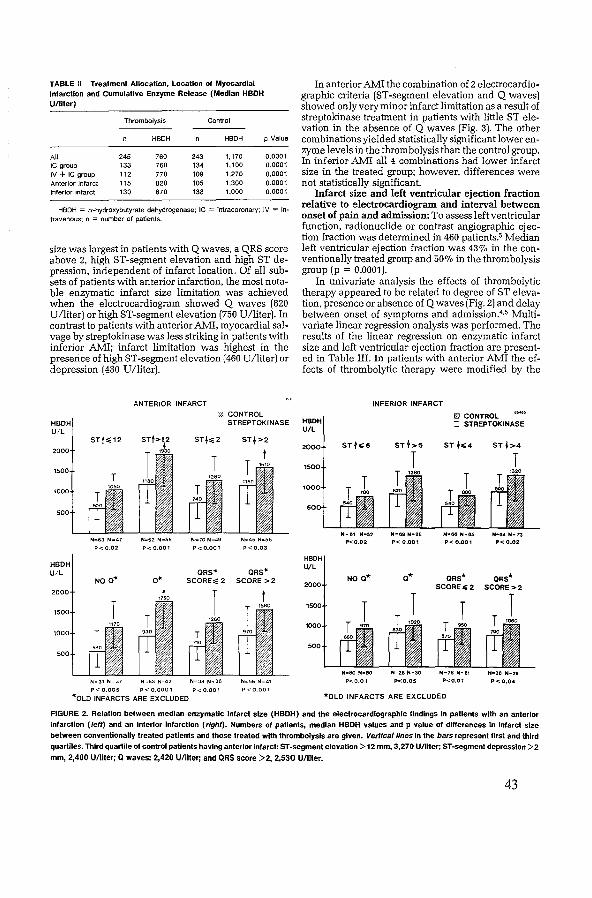
TABLE 11 Treatment Allocation, Location of Myocardial Infarction and Cumulative Enzyme Release (Median HBDH U/liter)
Thrombolysis Control
HBDH HBDH
All 245 760 243 1,170 JC group 133 760 134 1,100 IV+ IC group 112 770 109 1,270 Anterior infarct 115 820 105 1,300 Inferior infarct 130 670 138 1,000
p Value
0.0001 0.0001 0.0001 0.0001 0.0001
HBDH = a-hydroxybutyrate dehydrogenase; IC = intracoronary; IV = intravenous; n = number of patients.
size was largest in patients with Q waves, a QRS score above 2, high ST-segment elevation and high ST depression, independent of infarct location. Of all subsets of patients with anterior infarction, the most notable enzymatic infarct size limitation was achieved when the electrocardiogram showed Q waves (820 U/liter) or high ST-segment elevation (750 U/liter). In contrast to patients with anterior AMI, myocardial salvage by streptokinase was less striking in patients with inferior AMI; infarct limitation was highest in the presence of high ST-segment elevation (460 U/liter) or depression ( 430 U /liter).
ANTERIOR INFARCT 'ii. CONTROL
In anterior AMI the combination of 2 electrocardiographic criteria (ST-segment elevation and Q waves) showed only very minor infarct limitation as a result of streptokinase treatment in patients with little ST elevation in the absence of Q waves (Fig. 3). The other combinations yielded statistically significant lower enzyme levels in the thrombolysis than the control group. In inferior AMI all 4 combinations had lower infarct size in the treated group; however, differences were not statistically significant.
Infarct size and left ventricular ejection fraction relative to electrocardiogram and interval between onset of pain and admission: To assess left ventricular function, radionuclide or contrast angiographic ejection fraction was determined in 460 patients.5 Median left ventricular ejection fraction was 43o/o in the conventionally treated group and 50% in the thrombolysis group (p = 0.0001).
In univariate analysis the effects of thrombolytic therapy appeared to be related to degree of ST elevation, presence or absence of Q waves (Fig. 2) and delay between onset of symptoms and admission.4•5 Multivariate linear regression analysis was performed. The results of the linear regression on enzymatic infarct size and left ventricular ejection fraction are presented in Table III. In patients with anterior AMI the effects of thrombolytic therapy were modified by the
INFERIOR INFARCT
STREPTOKINASE HBDH 0 CONTROL 0 STREPTOKINASE
U/L STt>2
N=63 N"'47 N=52 N=58 N-=70 N.,49 N"'45 N=56
P< 0.02 P<0.001 P<0.001 P< 0.03
ORS"" ORS"" NO a• SCORE>; 2 SCORE >2
P< 0.005 P< 0.0001 P< 0.001
""OLD INFARCTS ARE EXCLUDED
N•C1 N•52 P<0.02
NO a*
N"80 N-80
P<0.01
N-eg Nc86
P< 0.001
o*
N=26 N•30
P<0.05
N•66 N•85
P< 0.001
ORS* SCOREC:2
N•78 N•61
P<0.01
*OLD INFARCTS ARE EXCLUDED
N·~ N•73 P< 0.02
FIGURE 2. Relation between median enzymatic Infarct size (HBDH) and the electrocardiographic findings In patients with an anterior
infarction (/eft) and an inferior Infarction (right). Numbers of patients, median HBOH values and p value of differences In Infarct size
between conventionally treated patients and those treated with thrombolysis are given. Vertical lines In the bars represent first and third
quartiles. Third quartile of control patients having anterior infarct: ST-segment elevation >12 mm, 3,270 U/llter; ST-segment depression >2
mm, 2,400 U/liler; Q waves: 2,420 U/liter; and QRS score >2, 2,530 U/llter.
43

ANTERIOR INFARCT_..
STt .012
NO a
N -21 N~20
NS
a
N~30 N~13
P<0.001
Ill CONTROL :::J STREPTOKINASE
STt > 12
NO a
N•10 N=17
P< 0.005 N-33 N,.29
P< 0.01
N-35 N=32 N=12 N .. 6 N~45 N-48 N .. 14 N-22 NS NS NS NS
"'OLD INFARCTS ARE EXCLUDED
FIGURE 3. Relation between median enzymatic infarct size and combination of presence or absence of a waves and low or high ST
segment elevation in anterior and inferior Infarction. Control patients with anterior Infarction had following third quartlles: No a waves+ ST elevation >12 mm, third quartile 3,310 U/liter; a waves + ST elevation >12 mm, third quartile 2,630 U/liter. For further explanation see Figure 2.
degree of ST elevation (p = 0.02 in covariance analysis), presence or absence of Q waves (p = 0.02) and admission delay (p = 0.2). In patients with inferior infarction these effect modifications were apparent, but did not reach statistical significance. A decision tree is presented in Figure 4, based on the results of the regression analysis on enzymatic infarct size.
Patients with anterior infarction (Fig. 4, left), little ST-segment elevation and no Q waves had only minimal infarct limitation after streptokinase administration. This was independent of admission delay. All other combinations showed smaller infarct size after thrombolysis. The most impressive infarct limitation was found in patients with high ST-segment elevation in the absence of Q waves (Table III). To a lesser degree also in inferior AMI (Fig. 4, right), patients with high ST elevation in the absence of Q waves benefited most from thrombolytic therapy. The only other combination with important limitation of infarct size was early admission, high ST elevation and Q waves. All other comparisons yielded only small differences in enzyme levels between patients treated with thrombolysis and those treated conventionally.
44
Discussion The present analysis of data shows that thromboly
tic therapy is most effective in terms of infarct size limitation in a subset of patients admitted early after onset of chest pain with signs of a large infarction (development of Q waves) or extensive ischemic area (high ST-segment elevation or marked ST depression). These observations are relevant for the clinical decision as to whether the patient should or should not receive thrombolytic treatment. Such an analysis has not been undertaken. Results of some studies'-2.17-19 of streptokinase therapy in patients with AMI agree with our results3-7 that thrombolytic therapy preserves left ventricular function or reduces mortality, but Rentrop et aJ2° could not show a positive effect of this type of treatment. The disparities in outcome of thrombolytic therapy can partially be explained by the small number of patients in most studies.
Enzymatic infarct size: In most trials peak CK-MB or lactic dehydrogenase enzyme levels for determination of infarct size were used1.20 This is another reason for the discrepancy in outcome of streptokinase trials. As pointed out by van den Laarse eta! and others,B.l6.21
peak enzyme levels cannot be used for that purpose. In contrast, cumulative HBDH release in the first 72 hours correlates well with left ventricular ejection fraction, development of heart failure, arrhythmias and intraventricular conduction disturbances, 16 and in-hospital death and death in the first year after AMI4 - 6·8 Erhardt22 found good correlation between peak thermostable lactic dehydrogenase release in plasma, which is identical to HBDH,8 and postmortem determined infarct size. We therefore selected for our study the cumulative release of myocardial HBDH in 72 hours.
Electrocardiogram: In many studies the electrocardiographic inclusion criteria have not been described, except for the remark that the electrocardiogram was typical for AMI. ST-segment elevation provides information on severity of ischemic injury, while Q waves are markers for infarct size.14·23 Therefore, in some studies,1·17·20 patients with QRS changes such as Q waves or loss of R wave in the infarcted area were excluded because the investigators believed that Q waves precluded salvage of myocardial tissue by thrombolysis. This conclusion appeared to be contradictory to the present observation that important salvage occurred in patients with Q waves.
ST segment: High ST-segment elevation and marked ST-segment depression on the admission electrocardiogram were related to infarct size. These patients had also largest infarct limitation after thrombolytic therapy. This was true for both anterior and inferior AMI. Berland et aJ24 reported similar findings in patients with inferior infarction.
The electrocardiogram during AMI can change dramatically within a short period. Because ST-segment abnormalities may vary considerably over time, in some patients the classification of the sum of ST elevation or depression would have been different if the electrocardiogram of randomization had been re-
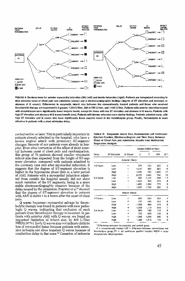
EFFECT DECISION DELAY ST~ELE- 0- EFFECT DECISION DELAY ST-ELE- a- THROM- TREE VATION WAVES THROM-
TREE VATION WAVES BOLYSIS THROMBOLYSIS BOLYSIS
THROMBOLYSIS 0ABSENT -o <ABSENT-0 []LOW<
<o~ _,_. < 0PRESENT -0 o .. 2H o .. 2H 0ABSENT -I§] <0ABSENT -II 0HIGH<
0HIGH 0PRESENT-CiJ
L:J OPRESENT-11
<0ABSENT -0 <DABSENT -0 I
[JLDW [JLDW < DPR5ENT-0 < o~-w [J2·4H
[]2-4H DABSENT-IS:i
<0ABSENT -II []HIGH< HBDH U/l
[]HIGH HBDH U/L
D PRESENT- [J [].;250 DPR5ENT-0 o .. 250 [] 260-500 ~260-500
11>500 [J>4H -o ll>soo []>4H -0?
FIGURE 4. Decision trees for anterior myocardial Infarction (MI) (teff) and Inferior Infarction (right). Patients are categorized according to time between onset of chest pain and admission (delay) and 2 electrocardiographic findings (degree of ST elevation and presence or
absence of Q waves). Differences In enzymatic Infarct size between the conventionally treated patients and those who received thrombolytic therapy are expressed in 3 groups: <250 U/liler, 260 to 500 U/llter, and >500 U/liler. Patients with anterior infarction treated with streptokinase have significantly lower enzyme levels, except for those with low ST elevation and absence of Q waves. Patients with
high ST elevation and absence ot Q waves benefit most. Patients with inferior Infarction have similar findings. Patients admitted early, with high ST elevation and Q waves also have significantly lower enzyme levels In the thrombolysis group. Finally, thrombolysis Is more effective In patients with a short admission delay.
corded earlier or later. This is particularly important in patients already admitted to the hospital, who have a severe anginal attack with prominent ST-segment changes. Seventy of our patients were already in hospital. Even after correction of the effect of short interval between onset of chest pain and randomization, this group of 70 patients showed smaller enzymatic infarct size than expected from the height of ST-segment elevation, compared with patients admitted to the coronary care unit after myocardial infarction. It suggests that the degree of ST-segment elevation is higher in the hyperacute phase than in a later period of AMI. Patients with a myocardial infarction admitted from outside the hospital usually did not show much variation of the ST segments, being in a more stable electrocardiographic situation because of the delay caused by the admission. Foerster et aJ25 showed that the degree of ST-segment elevation in patients with AMI is stable 1 to 4 hours after the onset of chest pain.
Q wave: Important myocardial salvage by thrombolytic therapy was found in patients with new pathologic Q waves, indicating that exclusion of such patients from thrombolytic therapy is incorrect. In patients with anterior AMI with Q waves, we found an important limitation of infarct size, by 820 U/liter HBDH (47% ). Early Q waves may not indicate definite loss of myocardial tissue because patients with extensive ischemia can show transient Q waves because of conduction delay in that zone.26 Cessation of ischemia
TABLE Ill Enzymatic Infarct Size, Radlonucllde Left Ventricular Ejection Fraction, Electrocardiogram and Time Delay Between Onset of Chest Pain and Admission: Results from Multivariate Regression Analysis
Median HBDH (U/Iiter)
Delay ST Elevation Q Waves c T Diff Ef•
Anterior Infarct
<2 hours Low 970 740 230 Low + 1,410 590 820 16 High 1,940 720 1.220 17
High + 2,070 1.330 740 15 2-4 hours Low 880 1.110 -230 -4
Low + 1.330 960 370 High 1.860 1,090 770 High + 1.990 1.700 290
Inferior Infarct
<2 hours Low 830 580 250 Low + 720 480 240 High 1.330 860 470 High + 1,450 1.110 340
2-4 hours Low 860 750 110 Low + 750 650 100 High 1,360 1.030 330 High + 1,480 1,310 170
"Differences between thrombolysis and control group. C = conventionally treated; Diff = difference between conventional and
thrombolysis group; EF = lett ventricular ejection fraction; H~DH = a-hy-droxybutyrate dehydrogenase.
45

by reopening the occluded coronary artery may prevent further damage in that area. Even after 2 hours thrombolytic therapy limited infarct size in the anterior wall, suggesting that large infarcts are still evolving.
The criteria we used for pathologic Q waves were strict. However, results did not differ if the QRS scoring system proposed by Wagner et al'4 was applied. This system was developed for infarct size estimation in patients with completed myocardial infarction, but appears to be helpful in AMI as well. No or minimal infarct limitation by thrombolytic therapy was found in patients without Q waves in combination with low ST-segment elevation. This was independent of time between onset of chest pain and admission. Such a finding suggests that the occluded artery reopened before treatment, preventing occurrence of high ST -segment elevation and Q waves, when beneficial effect of thrombolysis on infarction size can no longer be expected. Another explanation could be that such patients suffer only from a small infarct, in which case the effect of thrombolytic therapy is minor and not statistically significant.
Importance of time between onset of chest pain and admission: In the reported studiest-3.17-zo interval from onset of chest pain to intervention varied from 3 to 18 hours. An inverse relation between duration of occlusion and subsequent ventricular function has been shown in animal experiments.27 In humans, this relation was not always found.19·28 Most studies reported, however. that reperfusion within 4 hours after the onset of symptoms resulted in improvement of left ventricular function18·29 or a decrease in mortality rate, as shown in the GISSitriaJ.2 In the present trial, thrombolysis was started relatively early after onset of symptoms (median 195 minutes), while recanalization was achieved within 4 hours in most patients. This is probably the most important explanation as to why, in our study, streptokinase had such a beneficial effect. As expected from animal studies,27 we found prominent infarct size limitation in most patients arriving at the hospital within 2 hours after onset of chest pain. As shown by Simoons et al,4 in patients allocated to conventional treatment, HBDH release was independent of the interval between onset of symptoms and hospital admission. On the other hand, infarct size was reduced with 51 o/o in thrombolysis patients admitted within 1 hour, 31 o/o in those admitted between 1 and 2 hours, and only 13% in those arriving 2 to 4 hours after the start of complaints. In the patients coming to the coronary care unit between 2 and 4 hours after onset of chest pain, subgroups could be identified that did not show limitation of infarct size even after successful reperfusion. This indicates that a time limit of 4 hours is too long for some patients. Although it was apparent that time after onset of chest pain is an important determinant of the outcome of successful thrombolytic therapy, the exact time of onset of pain may be difficult to define. Especially in patients with several attacks of pain, time of onset of myocardial infarction cannot be determined accurately. Accepting these uncertainties, we found that in patients arriving at the coronary care unit between 2 and 4 hours after onset of chest pain,
46
thrombolytic therapy was less successful than in those admitted earlier. After AMI Q waves develop over time. In contrast to this, ST elevation is highest early. after the onset of AMI. In patients arriving 2 to 4 hours after the start of complaints with high ST -segment elevation and absence of Q waves, streptokinase was still effective. Incorrect judgment of the time of onset of AMI may explain why these patients still have such significant infarct size limitation.
Another argument for the validity of the electrocardiographic criteria was the correlation between median HBDH values and left ventricular ejection fraction in patients with anterior infarction (Table III). This correlation did not hold for patients with inferior infarction, because the distance of the infarcted area to the collimator is much greater than in anterior infarction. As discussed by Wackers,30 the contribution of the posteroinferior wall to ejection fraction will be less, resulting in overestimation of ventricular wall motion.
It was mentioned earlier that 33 of 245 patients refused the intervention or were subsequently found to have a contraindication for thrombolysis. They were evaluated according to their original treatment allocation.10 These 33 patients influence negatively the results of thrombolysis. If they would have been excluded, therapeutic success in terms of infarct size limitation would be greater.
Acknowledgment: We thank Viviane Lejeune and Adri van den Dool for their help in preparation of the manuscript.
References 1. Kennedy JW, Ritchie fL. Davis KB, Fritz JK. Western Washington random~ ized trial of intracoronory streptokinase in acute myocardial infarction. N Engl T Med 1985;312:1073-1078. 2. Gruppo italiano per Jo studio della streptochinasi nell' infarto miocardico (GISSI). Effectiveness of intravenous thrombolytic treatment in acute myo· cordial infarction. Lancet 1986;397-401. 3. Simoons ML, Serruys PW, van der Brand M, Bar FW, de Zwaan C. Res J, Verheugt FWA. Krauss XH, Remme Wf, Vermeer F, Lubsen J. Improved survival after early thrombolysis in acute myocardial infarction. Lancet 1985;578-582. 4. Simoons ML, Serruys PW, Brand M van der. Res J, Verheugt FW, Krauss XH, Remme Wf, Bar FW, deZwaan C. van der Laarse A, Vermeer F, Lubsen J. Thrombolysis in acute myocardial infarction: limitation of infarct size and improved survival. JACC 1986;7:717-728. 5. Vermeer F, Simoons ML, Biir FVV, Tijssen JCP, Domburg RT, Serruys PW, Verheugt FW, Res J, de Zwaan C, van der Laarse A, Krauss XH, Lubsen J, Hugenholtz PG. Which patients benefit most from early thrombolytic therapy with intracoronary streptokinase? Circulation, in press. 6. Serruys PW, Simoons ML. Suryapranata H, Vermeer F, Wijns W. van der Brand M, Bar FW. KraussXH, Remme WJ, Res J, Verheugt FW, Domburg RT, Lubsen J, Hugenholtz PG. Preservation of global and regional left ventricular function in acute myocardial infarction. JACC 1986;7:729-742. 7. Res[. Simoons ML. van der WallE, van Eenige MJ, Vermeer F. Verheugt FVV. Wijns W, Braat SH, Remme WJ, Serruys PW, Roos JP. Long term improvement in global left ventricular function after early thrombolytic therapy in acute myocardial infarction. Br Heart J, in press. 8. van der Laarse A. Vermeer F, Hermens WT, Willems GM, de Neef K. Simoons ML, Serruys PW, Res J, Verheugt FVV, KraussXH, Bar FVV, de Zwaan C. Lubscn J. Effects of early introcoronary streptokinase on cumulative enzyme release and enzyme release rote in patients with acute myocardial infarction. Am Heart J, in press. 9. Verheugt FWA, van Eenige MJ. Res J, Simoons ML, Serruys PW, Vermeer F, van Hoogenhuyze DCA. Remme WH. de Zwaan C. Bar FVV. Bleeding complications of intracoronary fibrinolytic therapy in acute myocardial infarction. Br Heart 11985;54:455-459. 10. Zelen MA. A new design for randomized clinical trials. N Engl J Med 1979;300:1242-1245. 11. Simoons ML. Serruys PW. Fioretti P. van den Brand M, Hugenholtz PG. Practical guidelines for treatment with beta blockers and nitrates in patients
with acute myocardial infarction. Eur Heart J 1983;4:suppl D:129-135. 12. SchrOder R, Biamino G. von Leitner ER, Linderer T, BrUggeman T. Heitz J, V6hringer HF, Wegscheider K. Intravenous short-term infusion of streptokinase in acute myocardial infarction. Circulation 1983;67:536-538. 13. Spann JF, Sherry S, Carabello BA, Maurer AH, Cooper EM. Coronary thrombolysis by intravenous streptokinase in acute myocardial infarction: acute and follow up studies. Am J Cardiol1984;53:655-661. 14. Wagner GS, Freye Cf, Palmeri ST. Roark SF, Stack NC, Ideker RE, Harrell FE, Selvester RH. Evaluation of a QRS scoring system for estimating myocardial infarct size. I. Specificity and observer agreement. Circulation 1982:65:342-347. 15. Bar FW, Brugada P, Dassen WR. WerfTvd, Wellens HJJ. Prognostic value of Q waves, RIS ratio, loss of R wave voltage, ST-T segment abnormalities, electrical axis, low voltage and notching: correlation of electrocardiogram and left ventriculogram. JACC 1984:4:17-27. 16. van der Laarse A, Hermens WT, Hollaar L. Jol M, Willems GM. Lemmers HEAS, Liem AH, Souveryn JHM, Oudhof JH, de Hooge J, Buis B, Arntzen ius AC, Assessment of myocardial damage in patients with acute myocardial infarction by serial measurement of serum alpha-hydroxybutyrate dehydrogenase levels. Am Heart 11984;107:248-260. 17. Mathey DC, Kuck KH, Tilsner V. Krebber H}. Bleifeld W. Nonsurgical coronary artery recanalization in acute transmural myocardial infarction. Circulation 1981;63:489-497. 18. Anderson fL, Marshal HW, Bray BE, Lutz JR. Frederick PR. Yanowitz FG, Datz FL. Klausna SC. Magan AD. A randomized trial of intracoronary streptokinase in the treatment of acute myocardial infarction. N Engl J Med 1983;308:1312-1318. 19. Smalling RW, Fuentes F, Freund GC, Reduto LA, Wanta-Matthews M. Gaeta JM, Walker W, Sterling R, Gould KL. Beneficial effects of intracoronary thrombolysis up to eighteen hours after onset of pain in evolving myocardial infarction. Am Heart 11982;104:912-920. 20. Rentrop KP. Feit F, Blanke H, Stecy P. Schneider R, Rey M. Horowitz S, Goldman M, Karsch K, Meilma H, Cohen M. Siegel S, Sanger J, Slater J, Gorlin R, Fox A, Fagerstrom R, Calhoun WF. Effect of intracoronary streptokinase and intracoronory nitroglycerin infusion on coronary angiographic patterns and mortality in patients with acute myocardial infarction. N EnglJ Med 1984;311:1457-1463. 21. Shell W, Mickle DK. Swan HfC. Effects of non-surgical myocardial reperfusion on plasma creatinekinase kinetics in man. Am Heart J 1983;106:665-669. 22. Erhardt LR. Clinical and pathological observations in different types of acute myocardial infarction. Acta Med Scand 1974;suppl1-78:560, 23. Muller JE, Maroko PR, Braunwald E. Evaluation of precordial electrocardiographic mopping as a means of assessing changes in myocardial ischemic
injury. Circulation 1975;52:16-27. 24. Berland J, Cribier A, Behar P, Letac B. Anterior ST depression in inferior myocardia! infarction: correlation with results of intracoronary thrombolysis. Am Heart J 1986;11:481-488. 25. Foerster JM, Vera Z, Janzen DA, Foerster SJ, Mason DT. Evaluation of precordial orthogonal vectorcardiographic lead ST segment magnitude in the assessment of myocardia! ischemic injury. Circulation 1977;55:728-732. 26. Durrer D, van Dam RT. Freud GE. Janse MJ, Meyler FL. Arzbf.icher R. Total excitation of the isolated human heart. Circulation 1970;41:895-912. 27. Baughman KL. Maroko PR, Vatner SF. Effects on coronary artery reperfusion on myocardial infarct size and survival in conscious dogs. Circulation 1981;63:317-323. 28. Rogers WJ, Hood WP, Mantle JA, Baxley WA, Kirklin JK, Zorn GL, Nath HP. Return of left ventricular function after reperfusion in patients with myocardial infarction: importance of subtotal stenoses or intact collaterals. Circulation 1984;69:338-349. 29. Schwarz F, Schuler G. Katus H, Hofman M, Manthey J, Tillmanns H, Mehmel HC, Kubler W. Intracoronary thrombolysis in acute myocardial infarction: duration of ischemia as a major determinant of late results after recanalization. Am T Cordial 1982;50:933-937. 30. Wackers F. Thallium 201 and technetium 99m-pyrophosphate myocardial imaging in the coronary care unit. In: Simoons ML, Reiber JHC, eds. Nuclear Imaging in Clinical Cardiology. The Hague: Martinus Nijhoff, 1980.
Appendix We thank all collaborators at the participating centers:
M.j.B.M. van den Brand, P.j. de Feyter, P. Fioretti, P.G. Hugenholtz. P.W. Serruys and W. Wijns. Thoraxcenter. University Hospital Dykzigt and Erasmus University, Rotterdam: M.j. van Eenige, J.P. Roos. J.C.j. Res. F.C. Visser, and E.E. vander Wall. Free University, Amsterdam; D.C.A. van Hoogenhuyze. H.A.C.M. Kruyssen, W.j. Remme. and C.J. Storm, Zuiderziekenhuis. Rotterdam; P. Brugada, K. den Dulk and G.M. Willems, University Hospital Maastricht; B. Buis and ).G. Engbers. University Hospital. Leiden; S. van der Does, R.T. van Domburg, j. Lubsen, J.P. van Mantgem, K.j. de Neef, J. Planellas, A.A. Wagenaar, and !.C.). Zorn, Data Processing Center. Erasmus University. Rotterdam.
47
CHAPTER 7
THERAPY AND PREVENTION CORONARY THROMBOLYSIS
Which patients benefit most from early thrombolytic therapy with intracoronary streptokinase? FRANK VERMEER. M.D., MAARTEN L. SIMOONS, M.D .. FRITS W. BAR, M.D., JAN G. P. TUSSEN. M.S .. RoN T. VAN DOMBURG, M.S., PATRICK W. SERRUYS, M.D., FREEK W. A. VERHEUGT, M.D .. JAN C. J. RES, M.D .. CHRIS DE ZWAAN, M.D .. ARNOUD VANDER LAARSE, PH.D., X. HANNO KRAUSS, M.D .. JACOBUS LUBSEN, PH.D., AND PAUL G. HuGENHOLTZ, M.D.
ABSTRACT The effect of thrombolysis in acute myocardial infarction on enzymatic infarct size. left ventricular function. and early mortality was studied in subsets of patients in a randomized trial. Early thrombolytic therapy with intracoronary streptokinase (!52 patients) or with intracoronary streptokinase preceded by intravenous streptokinase ( 117 patients) was compared with conventional treatment (264 patients). All 533 patients were admitted to the coronary care unit within 4 hr after onset of symptoms indicative of acute myocardial infarction. Four hundred eighty-eight patients were eligible for this detailed analysis. and 245 of these were allocated to thrombolytic therapy and 243 to conventional treatment. Early angiographic examinations were performed in 212 patients allocated to thrombolytic therapy. Patency of the infarct-related artery was achieved in 181 patients (85%). Enzymatic infarct size. as measured from cumulative a-hydroxybutyrate dehydrogenase release. was smaller in patients allocated to thrombolytic therapy (median 760 vs 1170 U/liter in control patients, p = .0001). Left ventricular ejection fraction measured by radionuc!ide angiography before discharge from the hospital was higher after thrombolytic therapy (median 50% vs 43% in control patients. p = .0001 ). Three month mortality was lower in patients allocated to thrombolytic therapy (6% vs 14% in the control group, p = .006). With the use of multivariate regression analysis. infarct size limitation, improvement in left ventricular ejection fraction. and three month mortality were predicted by sum of the ST segment elevation. time from onset of symptoms to admission, and Killip class at admission. Thrombolysis was most effective in patients admitted within 2 hr after onset of symptoms and in patients with a sum of STsegment elevation of 1.2 mV or more. On the other hand, no beneficial effects of streptokinase on enzymatic infarct size. left ventricular function, or mortality were observed in the subset of patients with a sum ofSTsegment elevation of less than 1.2 mV who were admitted between 2 and 4 hr after onset of symptoms. Circulation 74, No. 6, 1379-1389, 1986
IN APPROXIMATELY 80% to 90% of patients with acute myocardial infarction, intracoronary infusion of streptokinase given in the first hours after onset of chest pain leads to thrombolysis and recanalization of the occluded coronary artery.,_,
angiographically proven obstruction, without .major complications. The high patency rate after thrombolysis was associated with limitation of infarct size, higher left ventricular ejection fraction (L VEF), and improved survival in comparison with these variables in conventionally treated patients. The design of this study. clinical course in the hospital, complications of early catheterization, limitation of enzymatic infarct size, and improvement in left ventricular function and survival during the follow-up period have been described elsewhere .... ,
The multicenter randomized trial conducted by the Netherlands Interuniversity Cardiology Institute demonstrated early recanalization in 79% of patients with
From the Working Group on Thrombolytic Therapy in Acute Myo. cardia! Infarction of the Netherlands Interuniversity Cardiology Institute. Participating centers and collaborators are listed before the references.
Address for correspondence: F. Vermeer, M.D., Department of Cardiology, University Hospital Maastricht, University of Limburg. P.O. Box 19I8. 6201 BX Maastricht. The Netherlands.
Received April 15, 1986; revision accepted Aug. 21. 1986.
48
The aim of the present analysis was to define subsets of patients that benefitted most from early recanalization. Special attention was paid to patient characteristics available early after hospital admission, such as
history, the delay between onset of symptoms and admission, the extent of myocardial ischemia as reflected by the sum of ST segment elevation (lST) on the electrocardiogram (ECG), and the hemodynamic state of each patient.
Patient selection and methods Patients were eligible for the trial if they were admitted to one
of the participating coronary care units within 4 hr after the onset of chest pain and with electrocardiographic signs typical of acute myocardial infarction: ST segment elevation of at least 0.1 mY in one or more extremity leads orthat of at least 0.2 mY in one or more precordial leads. or ST segment depression of at least 0.2 mY in one or more precordial leads. compatible with posterior wall infarction.4 Exclusion criteria were age over 70 years. previous treatment with streptokinase, recent trauma (including prolonged resuscitation). enhanced risk of bleeding, pregnancy. bypass surgery of a vessel corresponding to the infarct location, and mental confusion that precluded the patient from giving informed consent. Patients who met the inclusion criteria were registered by a central telephone answering service and were allocated either to thrombolytic therapy or to conventional treatment. Informed consent was obtained from patients allocated to thrombolytic therapy only. Patients who refused consent or patients in whom thrombolytic therapy was withheld for other reasons received conventional treatment, but were included in the analysis according to original treatment allocation. 8
Early coronary angiographic examinations were performed only in patients allocated to thrombolytic therapy. after informed consent had been obtained (figure I). If the infarctrelated coronary artery appeared to be occluded, intracoronary streptokinase was given at a rate of 4000 U/min, until all visible clots had disappeared or the total of250,000 U of streptokinase had been given. In the second part of the study cardiac catheterization was preceded by infusion of 500,000 U of streptokinase over 20 min, administered intravenously to reduce treatment delay. After catheterization patients allocated to thrombolytic therapy were treated according to the same treatment protocol as the control group.
Serum a-hydroxybutyrate dehydrogenase (HBDH) levels were determined upon admission, every 12 hr for 2 days. and then daily up to the fifth day. Cumulative HBDH release was calculated from these data as described before!· 10 Radionuc!ide angiography was performed I to 3 days after admission. before discharge. and again after 3 months. Cardiac catheterization was offered to all patients before discharge. All patients were followed at the outpatient clinic for at least I year after admission.
For identification· of subgroups the following data. available at the moment of randomization, were considered: (I) history (age, gender, previous myocardial infarction. history of angina pectoris. time from onset of symptoms to admission). (2) results of physical examination (heart rate, blood pressure, Killip class), (3) electrocardiographic findings (location of the infarct, extent of ST segment elevation and depression).
All ECGs were reviewed independently by two experienced cardiologists. Consensus about the interpretation was always reached. The location of the infarct was defined as anterior if there was ST segment elevation in leads V 1 to V 4, and as inferior if there was ST segment elevation in leads II, Ill. and a YF. In the case of ST segment elevation in leads I, aYL. Y5 , and Y6•
the location of the infarct was defined as anterior. unless ST segment elevation was also present in leads 11.111, and aYF, or ST segment depression was present in leads V 1 to V 4 • In the
latter case the location of the infarct was defined as inferior. The 2ST on the ECG was defined for anterior infarcts as the sum of ST segment elevation in leads I. aYL. andY 1 toY 6 and that for inferior infarcts was defined as the sum of ST segment elevation in leads I, II, Ill. aYL, aYF, Y5, and Y6 and ST segment depression in leads Y 1 to Y 4•
Statistical analysis. Missing data for cumulative HBDH release and L YEF were supplemented by various means. as described in the results. Because arbitrarily chosen values for cumulative HBDH release and L VEF were used for some of the patients, nonparametric statistical tests were used. Differences between groups were tested with the Fisher exact test or the Mann-Whitney rank-sum test when appropriate. Two-sided p values are reported. Linear regression analysis was used to select the significant and clinically relevant predictors of enzymatic infarct size. LYEF. and 3 month mortality. A BMDP statistical software package was used. Baseline variables shown by univariate analysis to be related to cumulative HBDH release were entered stepwise into the regression model. Factors that modified the effects of thrombolytic therapy were included as interaction factors. 11 The same procedure was used to predict L YEF and 3 month mortality.
Results
Five hundred thirty-three patients were admitted to the trial between June 1981 and March 1985. Two
ADMISSION CCU SYMPTOMS < 4H
TYPICAL ECG
1 RANDOM I ZA Tl ON
/~ CONVENTIONAL INFORMED CONSENT
CARE C!V+)JC STREPTOKINASE
~/ CUMULATIVE ENZYME RELEASE
l CONTRAST ANGIOGRAPHY
RAD!ONUCL!DE ANGIOGRAPHY
1 LONG TERM FOLLOW UP
FIGURE 1. Flow chart of the study protocol. Informed· consent was obtained from patients allocated to thrombolytic therapy only. Patients who refused consent received conventional treatment, but were includ
ed in the analysis according to original treatment allocation. CCU =
coronary care unit; H = hours.
49
hundred sixty-four patients were allocated to conventional treatment and 269 to thrombolytic therapy; 152 of these were allocated to treatment with intracoronary streptokinase and 117 to intracoronary streptokinase preceded by intravenous streptokinase.
Forty-five patients were excluded from the present analysis because the ECG obtained at admission to the trial could not be interpreted in accordance with the definitions presented above. These patients were evenly distributed between the two treatment groups. Nineteen patients were excluded because they did not have the required amount of ST segment elevation on the ECG and 17 were excluded because they had only ST segment depression. Two patients with third-degree atrioventricular block and ventricular escape rhythm, one patient with left bundle branch block, and one patient with preexcitation due to an abnormal atrioventricular conduction pathway were also excluded. The ECGs from five patients could not be retrieved for review.
Table I lists the baseline and follow-up data from the patients admitted to the trial, including 488 patients included in this analysis and 45 patients excluded. One year follow-up was complete in all patients. All baseline characteristics were equally distributed between the two treatment groups. Patients allocated to thrombolytic therapy had lower cumulative HBDH release.
TABLE I Baseline and follow-up data
Baseline data
No. of patients
Males
Age (median)
Previous myocardial infarction
Anterior infarction
In hospital at onset of symptoms
Killip class III/IV on admission
LST (mV)
Median time since onset of symptoms (min)
Follow-up data
Median cumulative HBDH release (Ufl)
Median radionuclide angiographic LVEF on day 10--20 (o/c)
Median contrast angiographic LVEF on day 10-40 (%)
Total 3 month mortality
Total 1 year mortality
higher L VEFs as measured by radionuclide and contrast angiography, and improved survival compared with patients allocated to conventional treatment ..... From here on results are presented for patients included in this analysis only.
Early angiographic examinations were performed in 212 patients allocated to thrombolytic therapy (figure 2). Among patients treated with intracoronary streptokinase only, the infarct-related vessel was patent on the first angiogram in 22 of 118 patients ( 19%) and patency was achieved in 100 patients (85%). In the group pretreated with intravenous streptokinase initial vessel patency was observed in 39 of 94 ( 41%), and final patency was achieved in 81 patients (86%).
Effect of thrombolytic therapy on enzymatic infarct size.
Enzymatic infarct size was measured by cumulative HBDH release in the first 72 hr after onset of symptoms in 414 patients. To eliminate bias caused by missing data, infarct size was estimated in 7 4 patients. Twenty-seven patients who died within 72 hr all had extensive myocardial infarction and were assigned to the largest infarct category and I 0 patients without significant enzyme elevation were assigned to the group with the smallest infarct size. In 29 patients infarct size was estimated from cumulative HBDH release in the first 48 hr, and in eight patients it was estimated from cumulative creatine kinase release.
Patients included in Patients excluded from
this analysis this analysis
T c T c
245 243 24 21
197 204 20 20
57 56 61 55
45 54 II 6
116 105 13 II
29 41 10 3
10 11
1.2 1.3 0.2 0.1
95 85 50 90
760 1170 720 560
50 44 42 45
56 48 50 42
15 (6%) 33 (14%) 4 (17%1 I (5%)
22 (9%) 41 (17%) 4 (17%) 2 (10%)
All baseline characteristics were distributed evenly among groups. Patients allocated to thrombolytic therapy had smaller
enzymatic infarct size. higher L VEF. and improved survival compared with control patients.
T = allocated to thrombolysis group~ C = control group.
50

100%
0 - 0
75%
50% Ill - 0
25%
Ql - Ill
0% 83 118
IC
94
IV+IC sr ... L 2 "'2H
32 ShL 2 2-4H
66 ST<L 2 "'2H
31 ST<L 2 2-4H
FIGURE 2. Results of early angiography in subsets of patients. Early catheterization was offered to patients allocated to
thrombolytic therapy only. Final patency rate was lower in patients admitted 2 to 4 hr after onset of symptoms than in patients admitted within 2 hr after onset of symptoms. ST = sum ofST segment elevation on the ECG at admission (mV); H = hours: 0-0
= infarct-related vessel patent at first angiogram; •-o = infarct-related vessel opened during intracoronary infusion of
streptokinase; •-• = infarct related vessel remained occluded.
Median cumulative HBDH release was I I 70 U/Iiter in patients allocated to conventional treatment, and 760 U/liter in patients allocated to thrombolytic therapy (p = .0001). The difference in cumulative HBDH release between the two treatment groups (410 U/liter) represents limitation of infarct size by thrombolytic
TABLE 2
therapy. The largest differences in enzymatic infarct size were found in patients with high ~ST (table 2). The relationship between time from onset of symptoms to hospital admission and enzymatic infarct size is also given in table 2. Patients admitted to the hospital before onset of symptoms were considered a special
Cumulative HBDH release, LVEF. and 3 month mortality in subsets of patients eligible for this analysis
HBDH (median. U!l) LVEF (median. %) Mortality (%)
T c T c p value T c p value T c Risk difference
All 245 243 760 I 170 0.0001 50 43 0.0001 14 -8 (-13. -2)
Streptokinase ic only 133 134 760 1100 0.0001 50 41 0.0001 12 -4(-12.3)
Streptokinase iv + ic 112 109 770 1270 0.0001 50 44 0.006 4 15 -II (-20, -3)
Anterior infarct 116 105 820 1300 0.0001 44 32 0.0001 18 -10 (-20, -2)
Inferior infarct 129 138 670 1000 0.0001 55 49 0.0001 10 -6 (-12, I)
In hospital at onset of symptoms 29 41 400 970 0.004 56 44 0.004 12 -9 (-23. 6)
Admitted within 60 min 45 54 620 1310 0.0001 51 42 0.0006 4 18 -14 (-27. -I)
Admitted 61-120 min 99 90 760 1160 0.0003 50 41 0.002 6 II -5 (-14. 3)
Admitted 121-240 min 72 58 970 1320 0.06 47 49 0.5 14 -6 (-18. 6)
LSTO.I-0.6mV 36 38 500 780 0.005 57 50 0.03 -5(-17.5)
LST 0.7-1.1 mY 80 58 590 820 0.05 51 44 0.03 12 -5(-16.6)
LST 1.2-1.7 mY 61 68 800 1280 0.003 50 46 0.07 12 -4 (-15, 8)
LST >1.7 mY 68 79 1120 1770 0.0001 44 37 0.003 20 -14 ( -26, -4)
FirstMI 200 189 780 1150 0.0001 52 46 0.0001 -3 (-8, I)
Previous MI 45 54 710 1250 0.03 40 33 0.06 20 41 -21 (-38, -2)
Killip class I or II 235 232 760 1140 0.0001 50 43 0.0001 5 II -6 (-II, -I)
Killip class III or IV 10 II 1440 33!0 0.02 38 27 0.1 20 64 -44 (-73. -I)
Two-sided p values (Mann-Whitney rank-sum test) or risk differences with 95% confidence interval are reponed. Thrombolytic therapy led to
limitation of enzymatic infarct size. improvement in LVEF and reduction in 3 month mortality in all subsets of patients. Most prominent differences were in patients with high :EST. in patients in Killip class III or IV at admission, and in patients with an anterior infarct.
MI = myocardial infarction; other abbreviations are as in table I.
51
group. For patients admitted to the hospital after onset of symptoms there was no relationship between duration of symptoms and enzymatic infarct size in the control group, but limitation of infarct size by thrombolytic therapy decreased as the delay between onset of symptoms and admission lengthened. Limitation of infarct size was greater in patients with an anterior infarct (480 U/liter) than in patients with an inferior infarct (330 U/liter). Patients in Killip class III or IV at admission had extensive myocardial infarction and in these patients there was a substantial limitation of enzymatic infarct size by thrombolytic therapy.
Effect of thrombolytic therapy on left ventricular fnnction. L VEF was measured by radionuclide angiography I to 3 days after admission, before discharge, and again after 3 months, and by contrast angiography I 0 to 40 days after admission. Radionuclide angiographic L VEF before discharge was used to assess the improvement in left ventricular function induced by streptokinase (336 patients). If data on radionuclide angiographic L VEF before discharge were missing, measurements obtained after 3 months (56 patients), after I to 3 days ( 62 patients) or of contrast angiographic LVEF (six patients) were used. A LVEF ofO% was assigned to 24 patients who died within I 0 days after admission without undergoing angiography. Median L VEF was 43% in the control group and 50% in the streptokinase group (p = . 000 I). The largest differences in median L VEF between the two treatment groups were found in patients with an anterior infarct. in patients admitted within 2 hr after onset of symptoms, and in patients in Killip class III or IV at admission (table 2).
Effect of thrombolytic therapy on 3 month mortality. Three month mortality was higher in patients allocated to conventional treatment (14%) than in patients allocated to thrombolytic therapy (6%, p = .006). This difference in mortality was expressed as a negative risk difference (6% - 14% = - 8%) produced by thrombolytic therapy. The largest reduction in 3 month mortality was observed in patients admitted within I hr after onset of symptoms, in patients with lST greater than I. 7 m V, in patients pretreated with intravenous streptokinase, in patients with previous myocardial infarction, and in patients in Killip class III or IV at admission (table 2).
Multivariate analysis. Multivariate regression analysis was used to define the relative contributions of baseline factors to the effect of thrombolytic therapy on enzymatic infarct size, L VEF, and mortality. In the 27 patients who died within 72 hr, a cumulative HBDH release of 3500 U/liter was used in the computations.
52
In stepwise regression analysis the following baseline variables appeared to be relevant predictors of cumulative HBDH release, LVEF, and 3 month mortality: kST, allocation to thrombolytic therapy, location of the infarct, and Killip class at admission. The effect of thrombolytic therapy was modified by 1ST, delay in admission, and Killip class on admission. Other factors, including age, gender, history of angina, participating center, and allocation to intracoronary streptokinase or intravenous and intracoronary streptokinase did not contribute to the prediction of cumulative HBDH release, L VEF, or 3 month mortality when the above-mentioned variables were included in the regression model.
The results of the regression analysis are given in table 3. At the top, the coefficients for the baseline variables predicting cumulative HBDH release, LVEF, and 3 month mortality are given. The bottom of table 3 gives additional values for predictive variables for patients allocated to thrombolytic therapy, indica!ing the limitation of enzymatic infarct size, the improvement in LVEF, and the reduction in 3 month mortality as a result of thrombolytic therapy. From the coefficients for the variables used in the regression analysis, the predicted effect of thrombolytic therapy on enzymatic infarct size, L VEF, and mortality can be calculated. For example, a patient with an anterior infarction was admitted I hr after onset of symptoms
TABLE 3 Regression coefficients for the multivariate regression models pre~ dieting enzymatic infarct size, LVEF, and mortality
HBDH LVEF Monality
(U!l) (%) (%)
Intercept A 450 57 -4
~ST (each mY) 600 (70) -6 (I) 9 (3)
Admission delay (each hour) -10 (60) 0 (1) 0 (2)
In hospital -300 (150) I (3) -1 (6)
Killip class ill or IV 1230 (230) -13 (4) 49 (9)
Previous MI 200 (80) -12 (2) 8 (3)
Location Anterior 240 (70) -10 (1) 5 (3)
Additional value of predictive variables for patients allocated to thrombolytic therapy
Thrombolytic therapy
~ST (each m V)
Admission delay (each hour)
In hospital Killip class ill or IV
Standard error of the estimate
-300 (220) -200 (100)
100 (80)
130 (240) -630 (340)
740
9 (4)
2 (2)
-3 (3)
I (4)
2 (6)
13
3 (8)
-8 (4)
2 (3)
-3 (9)
-33 (13)
28
Standard errors for the regression coefficients are given in parentheses. M1 = myocardial infarction. "(Hypothetic) Val outcome variable if all independent variables are 0.
See text.
with a :SST of 1.5 m V. He was in Killip class II and had suffered a previous myocardial infarction. If he were not treated with streptokinase, predicted enzymatic infarct size would be 450 U/liter + 1.5 X 600 U/liter (:sST) - 10 U/liter (admission delay) + 200 U/liter (previous myocardial infarction) + 240 U/liter (anterior location) = 1780 U/liter. If the same patient were treated with streptokinase, enzymatic infarct size would be 1780 U/liter - 300 U/liter (allocation to thrombolytic therapy) - 1.5 X 200 U/liter (:SST) + 100 U/liter (admission delay) = 1280 U/liter. Predicted limitation of enzymatic infarct size by thrombolytic therapy would be 1780 U/liter- 1280 U/liter = 500 U/liter (95% confidence interval 360 to 640 U/liter). Similarly, predicted L VEF would be 26% and risk of mortality 22% if the patient were not treated with thrombolytic therapy, while predicted L VEF would increase to 35% and risk of mortality would be reduced to 15% after treatment with streptokinase. The expected improvement in L VEF induced by thrombolytic therapy would be 9% (95% confidence interval 7% to 11 %), and the risk difference for mortality -7% (95% confidence interval -2% to -12%).
The expected limitation of infarct size, improvement in LVEF, and reduction in mortality can be expressed as a function of :SST and time from onset of symptoms to admission. Corrections must be made for
2. 0
ST
mY
1.0
NO LIMITATION OF INFARCT SIZE BY THROMBOLYTIC THERAPY
patients in hospital at the time of onset of symptoms and patients in Killip class III or IV at admission. The predicted effect of thrombolysis on enzymatic infarct size is a limitation with 300 U/liter + 200 U/liter for each millivolt of :SST - I 00 U/liter for each hour delay between onset of symptoms and admission (fig. ure 3). The predicted improvement in LVEF as a result of thrombolysis is 9% + 2% for each millivolt of :SST - 3% for each hour after onset of symptoms. The risk difference for mortality after thrombolytic therapy is 3% - 8% for each millivolt of :SST + 2% for each hour between onset of symptoms and admission.
Since these computations are somewhat cumbersome, four subgroups of patients that can easily be recognized in clinical practice were defined: patients with :SST less than I .2m V, those with :SST of 1.2 m V or more, those with times from onset of symptoms to admission of less than 2 hr, and those with times from onset of between 2 and 4 hr. Patency rate at the end of the procedure was high in all these subsets of patients. although somewhat lower (68%) in patients with :SST less than I. 2 m V who were admitted between 2 and 4 hr after onset of symptoms (figure 2). Limitation of infarct size by thrombolytic therapy was greatest in patients with :SST of 1.2 mV or more and in patients admitted within 2 hr after onset of symptoms (figure 4). Improvement in LVEF was seen only in patients
0 w 1~ 100 MIN 240 TIME FROM ONSET OF SYMPTOMS TO ADMISSION
FIGURE 3. Limitation of infarct size by streptokinase. Results of the multivariate regression model. Predicted limitation of infarct size is a function ofi.ST and time from onset of symptoms to admission. ST = sum of ST segment elevation on the ECG
at admission (mV).
53

204 p = 0. 04 P = D. 3 p = 0.2
MORTALITY 16
17 ~STREPTD
iS% OcoNTRDLs
10 10% 9
8 7
5%
0%
ST ;>I. 2 ~2 HOURS
ST ;>I. 2 2-4 HOURS
ST <I. 2 ~2 HOURS
ST <I. 2 2-4 HOURS
FIGURE 6. Three month mortality, expressed as a percentage. in four subsets of patients in both treatment groups. Reduction in three month mortality was most prominent in patients with 1ST of 1.2 mY or more. ST = sum of ST segment elevation on the ECG at admission (mV).
treatment groups was not altered by the use of another L VEF measurement in the patients in whom radionuclide angiography was not performed before discharge. The 24 patients who died within 10 days after admission without angiography (22 of them died within 72 hr after admission) were included to prevent the bias described above in the discussion of cumulative HBDH release. To these patients a L VEF of 0% was assigned; no apparent differences with respect to the prediction of L VEF were found in the regression analysis when L VEF values of I 0% or 20% were assigned to these patients.
The effects of thrombolytic therapy on limitation of enzymatic infarct size, improvement in LVEF, and reduction in mortality were related to the same independent factors. No relevant changes in the value of these independent factors were found when the regression analyses were repeated without the inclusion of estimates for missing data on cumulative HBDH release and L VEF.
Pretreatment with intravenous streptokinase. During the course of this trial on early reperfusion by intravenous streptokinase other studies have been reported. 13
Since it was our aim to achieve reperfusion as quickly as possible, pretreatment with intravenous streptokinase was added to the protocol for patients allocated to thrombolytic therapy since January 1984. Earlier analysis suggested that, although intravenous pretreatment resulted in a larger fraction of patients with a patent infarct-related vessel at the first angiographic examination (figure 2), effects on infarct size and left ventricular function were similar with and without pretreatment. 6• 10 Accordingly, inclusion of pretreatment
56
in the regression model did not improve the prediction of cumulative HBDH release, LVEF, or 3 month mortality when the other predictors were included in the modeL Due to the low number of patients pretreated with intravenous streptokinase (n = 94), no conclusions can be drawn about the merits of this additional therapy from our data.
Comparison with other trials. Other randomized trials 17- 26 of intracoronary streptokinase have not shown consistent limitation of infarct size, improvement in left ventricular function, or reduction in early mortality. This discrepancy with our data can now be explained by the delays in admission and the different inclusion criteria in those trials. Time is a crucial factor in thrombolytic therapy after acute myocardial infarction. In nonrandomized trials it has been reported that recanalization leads to limitation of infarct size provided therapy is started within 4 hr after onset of symptoms, 27
• 28 although beneficial effects of intracoronary
streptokinase have been reported in individual patients treated up to 18 hr after the onset of chest pain. 29 In the present study beneficial effects of streptokinase in patients admitted between 2 and 4 hr after onset of chest pain were observed only in those with extensive myocardial ischemia, as reflected by a high ~ST, and in patients in Killip class III or IV at admission. Although recanalization was also observed in a high percentage (68%) of patients with low ~ST, this did not lead to limitation of enzymatic infarct size, improvement in LVEF, or reduction in mortality.
In the Western Washington trial, 17-
20 mean time to initiation of streptokinase infusion was 276 min, and patients with newly fonned Q waves or receiving

maintenance therapy for congestive heart failure were excluded. A significant reduction in early mortality was reported, although no beneficial effect on infarct size by thallium imaging or on left ventricular function was observed. Apparently this study included many patients who would not benefit from thrombolytic therapy according to our analysis, while patients with extensive ischemia leading to left ventricular failure, who might have benefitted from thrombolytic therapy, were excluded. In two other studies the infusion of streptokinase started on average more than 5 hr after the onset of symptoms and patients with signs of cardiogenic shock were excluded. 2 1.
22 No improvement in left ventricular function or reduction in mortality was observed. which is consistent with our observations. Among three relatively small studies beneficial effects of thrombolytic therapy were observed in only one, in which the mean time between onset of symptoms and admission was 160 min. 2,_
26 In most of these studies patients with new! y formed Q waves or signs of cardiagenic shock were excluded, and these groups of patients were shown to benefit greatly from thrombolytic therapy in the present trial. 30
ST segment elevation as predictor of the effects of thrombolytic therapy. In the regression analysis enzymatic infarct size was strongly related to 2ST. although ST segment elevation may vary considerably over longer periods of time in patients with acute myocardial infarction."· 32 The highest ST segment elevation is usually found within I hr after the onset of symptoms. In a group of patients V(ho are already in the hospital before the onset of symptoms infarct size may be overestimated by 2ST. This explains why "in hospital at the onset of symptoms" was included as an independent variable in the regression model. In patients admitted after the onset of symptoms no correction was necessary for the gradual decrease in ST segment elevation with time.
Contrary to other observations. 33 we noted that patients with inferior infarction and precordial ST segment depression generally had larger infarctions than patients without precordial ST segment depression. The regression coefficient for the total ST segment elevation was equal to the regression coefficient for the total precordial ST segment depression. so both were combined. Contralateral ST segment depression was not related to enzymatic infarct size in patients with anterior infarction, and therefore was not included in the regression model.
Which patients benefit most from thrombolytic therapy by intracoronary streptokinase? Thrombolytic therapy with intracoronary streptokinase led to significant limitation of infarct size. improvement in left ventricu-
Iar function. and reduction in mortality in patients with extensive myocardial ischemia, that is. in patients with a high 2ST, when thrombolytic therapy was started in the first few hours after onset of symptoms. Data from the GISSI trial34 and the ISAM study35 also indicate that beneficial effects from intravenous streptokinase dominate in patients admitted within 3 hr after onset of symptoms. Thus. it can now be recommended that thrombolytic therapy be offered to patients admitted within the first few hours after the onset of symptoms. and only to those patients who show extensive ST segment elevation on the ECG. Further studies presently underway both in the United States and in Europe should determine the relative values of intravenous and intracoronary treatment and of the use of streptokinase or other thrombolytic agents such as recombinant tissue-type plasminogen activator.
Participating centers and collaborators in the Working Group on Thrombolytic Therapy in Acute Myocardial Infarction
Thoraxcenter, Erasmus University and University Hospital Dijkzigt, Rotterdam: M. 1. B. M. van den Brand. P. 1. de Feyter, P. Fioretti. P. G. Hugenholtz. P. W. Serruys. M. L. Simoons, W. Wijns.
Department of Cardiology. Free University. Amsterdam: M. 1. van Eenige, 1. C. 1. Res. 1. P. Roos, F. C. Visser, F. W. A. Verheugt, E. E. van der Wall.
Department of Cardiology. Zuiderziekenhuis. Rotterdam: D. C. A. van Hoogenhuyze. X. H. Krauss. H. A. C. M. Kruyssen, W. 1. Remme, C. 1. Storm.
Department of Cardiology, University Hospital Maastricht. University of Limburg, Maastricht: F. W. Bar, S. H. 1. G. Braat, P. Brugada. K. den Dulk, W. T. Hermens, M. Ramento!, H. 1. 1. Wellens. G. M. Willems, C. de Zwaan.
Department of Cardiology, University Hospital, Leiden: B. Buis. 1. G. Engbers. A. van der Laarse.
Data processing center, Thoraxcenter. Erasmus University, Rotterdam: A. 1. Azar, B. Bos. S. van der Does, R. T. van Domburg, G. A. vanEs. 1. Lubsen, 1. P. van Mantgem, K. 1. de Neef. M. Patijn, 1. Planellas, 1. G. P. Tijssen, F. Vermeer. A. A. Wagenaar, I. C. 1. Zorn.
References 1. Merx W. DorrW. Rentrop P. Blanke H. Karsch KR, Mathey DG,
Kremer P. Rutsch W, Schmutzler H: Evaluation of the effectiveness of intracoronary streptokinase infusion in acute myocardial infarction: postprocedure management and hospital course in 204 patients. Am Heart 1 102: 1181. 1981
2. Weinstein J: Treatment of myocardial infarction with intracoronary streptokinase: efficacy and safety data from 209 United States cases in the Hoechst-Roussel registry. Am Heart J 104: 894, 1982
3. Rentrop P. Blanke H. Karsch KR, Rutsch W, Schartl M. Merx W, DOrr R, Mathey D. Kuck K: Changes in left ventricular function after intracoronary streptokinase infusion in clinically evolving myocardial infarction. Am Heart J 102: 1188. I 985
4. SimoonsML. Serruys PW, Brand M vd. BarF.Zwaan Cde. ResJ, Verheugt FW A. Krauss XH. Remme WJ, Venneer F, Lubsen J: Improved survival after early thrombolysis in acute myocardial infarction. A randomized trial conducted by the Interuniversity Cardiology Institute in The Netherlands. Lancet 2: 578, 1985
5. Simoons ML, Serruys PW, Brand M vd, Res J, Verheugt F\VA,
57

Krauss XH, Remme WJ. Bar F. Zwaan C de. Laarse A vd. Vermeer F. Lubsen J: Early thrombolysis in acute myocardial infarction: limitation of infarct size and improved survival. J Am Coli Cardiol 7: 717. 1986
6. Serruys PW. Simoons ML, SuryapranataH. Vermeer F. Wijns W. Brand M vd, Bar F. Krauss XH, Remme WJ, Res J, Verheugt FW A. Domburg R van. Lubsen J. Hugenholtz PG: Preservation of global and regional left ventricular function after early thrombolysis in acute myocardial infarction. JAm Coli Cardiel 7: 729, 1986
7. Verheugt FW A, Eenige MJ van, Res JCJ, Simoons ML. Serruys PW. Hoogenhuyze DCA van, Remme PJ. Zwaan C de, Bar F. Vermeer F: Bleeding complications of intracoronary fibrinolytic therapy in acute myocardial infarction. Assessment of risk in a randomized controlled triaL Br Heart J 54: 455. 1985
8. Zelen M: A new design for randomized clinical trials. N EnglJ Med 300: 1242. 1979
9. Vander Laarse A. Hermens WT. Hollaar L. Jol M. Willems GM, Lemmers HEAS, Liem AH, Souverijn JHM. Oudhof JH. Hooge J de, Buis B. Arntzenius AC: Assessment of myocardial damage in patients with acute myocardial infarction by serial measurements of serum a-hydroxy butyrate dehydrogenase levels. Am Heart J 107: 248. 1984
10. Vander Laarse A, Vermeer F. Hermens WT. Willems GM. de NeefK. Simoons ML. Serruys PW. Res J, Verheugt FW A. Krauss XH, Biir F. de Zwaan C. Lubsen J: Effects of early intracoronary streptokinase on infarct size estimated from cumulative enzyme release and on enzyme release rate. Am Heart J 1986 (in press}
11. Kleinbaum DG. Kupper LL: Applied regression analysis and other multivariable methods. Boston. 1978, Duxbury Press
12. Willems GM. Visser MP. Krill MTA. Hermens WT: Quantitative analysis of plasma enzyme levels based upon simultaneous determination of different enzymes. Cardiovasc Res 16: 120. 1982
13. SchrOder R, Biamino G. Von LeitnerER. LindererT. Briiggemann T. Heitz J, VOhringer HF. Wegscheider K: Intravenous short-term infusion of streptokinase in acute myocardial infarction. Circulation 67: 536. 1983
14. GanzW. Gefll. MaddahiJ, Berman D, Charuzi Y, Shah PK, Swan HJC: Nonsurgical reperfusion in evolving myocardial infarction. J Am Coli Cardioll: 1247. 1983
15. Roberts R. Ishikawa Y: Enzymatic estimation of infarct size during reperfusion. Circulation 68(suppl I): I-83, 1983
16. Willems GM. Dreessen VAB, de Zwaan C. Hermens WT: Estimation of fractional catabolic rates (FCR) of cardiac enzymes in man by multi-enzyme analysis. J Mol Cell Cardioll6(suppl2): 41. !984 (abstr)
17. Kennedy JW, Fritz JK. Ritchie JL: Streptokinase in acute myocardial infarction: Western Washington randomized trial - protocol and progress report. Am Heart J 104: 899, 1982
18. Kennedy JW. Ritchie JL, Davis KB, Fritz JK: Western Washington randomized trial of intracoronary streptokinase in acute myocardial infarction. N Eng! J Med 309: 14 77, 1983
19. Kennedy JW, Ritchie JL, Davis KB, Stadius ML, Maynard C. Fritz JK: The Western Washington randomized trial of intracoronary streptokinase in acute myocardial infarction. A 12-month follow-up report. N Eng! J Med 312: 1073. 1985
20. Ritchie JL. Davis KB, Williams DL. Caldwell J. Kennedy JW: Global and regional left ventricular function and tomographic radionuclide perfusion: The Western Washington intracoronary streptokinase in myocardial infarction trial. Circulation 70: 867. 1984
21. Rentrop KP. Feit F. Blanke H. Stecy P. Schneider R. Rey M, Horowitz S, Goldman M. Karsch K. Meilman H, Cohen M. Siegel
58
S, Sanger J, Slater J, Gorlin R, Fox A, Fagerstrom R, Ford Calhoun W: Effects of intracoronary streptokinase and intracoronary nitroglycerin infusion on coronary angiographic patterns and mortality in patients with acute myocardial infarction. N Engl J Med 311: 1457, 1984
22. Raizner AE. Tortoledo FA, Verani MS. van Reet RE. Young JB, Rickman FD. Cashion WR. Samuels DA. Pratt CM. Attar M. Rubin HS, Lewis JM, Klein MS. Roberts R: Intracoronary thrombolytic therapy in acute myocardial infarction: a prospective, randomized, controlled trial. Am J Cardiel 55: 301. 1985
23. Khaja F. Walton JA. Brymer JF, Lo E. Osterberger L. O'Neill WW. ColferHT. Weiss R, LeeT. Kurian T, Goldberg AD. Pitt B. Goldstein S: Intracoronary fibrinolytic therapy in acute myocardial infarction. A report of a prospective randomized trial. N Eng! J Med 308: 1305. 1983
24. Leiboff RH, Katz RJ, Wasserman AG. Bren BG. Schwartz H. Varghese PJ. Ross AM: A randomized angiographically controlled trial of intracoronary streptokinase in acute myocardial infarction. Am J Cardiol 53: 404, !984
25. Anderson JL. Marshall HW. Bray BE. Lutz JR. Frederick PR. Yanowitz FG, Datz FL. Klausner SC, Hagan AD: A randomized trial of intracoronary streptokinase in the treatment of acute myocardial infarction. N Eng! J Med 308: 1312. 1983
26. Anderson JL, Mcilvaine PM, Marshall HW. Bray BE. Yanowitz FG, Lutz JR, Menlove RL. Hagan AD: Long-term follow-up after intracoronary streptokinase for acute myocardial infarction: a randomized, controlled study. Am Heart J 108: 1402, 1984
27. Bergmann SR. Lerch RA. Fox KAA. Ludbrook PA. Welch MJ. Ter·Pogossian MM. Sobel BE: Temporal dependence of beneficial effects of coronary thrombolysis characterized by positron tomography. Am J Med 73: 573. 1982
28. Schwarz F. Schuler G. Katus H. Hofmann M, Mathey J, Till manns H. Mehmel JC. KUbler W: Intracoronary thrombolysis in acute myocardial infarction: duration of ischemia as a major determinant of late results after recanalization. Am J Cardiel 50: 933, 1982
29. Smalling RW, Fuentes F. Freund GC. Reduto LA. Wanta-Matthews M. Gaeta JM, Walker W. Sterling R, Gould KL: Beneficial effects of intracoronary thrombolysis up to eighteen hours after onset of pain in evolving myocardial infarction. Am Heart J 104: 912. !982
30. B:irFW, Vermeer F. de Zwaan C. Ramentol M, Braat S, Simoons ML. Hermens WT, Krauss XH, Wellens HJJ: Value of admission electrocardiogram in predicting outcome of thrombolytic therapy in acute myocardial infarction. Am J Cardiel 1987 (in press)
31. Selwyn AP, Ogunro EA. Shillingford JP: Natural history and evaluation of ST segment changes and MB-CK release in acute myocardial infarction. Br Heart J 39: 988, !977
32. Foerster JM, Vera Z, Janzen DA. Foerster SJ, Mason DT: Evaluation of precordial orthogonal vectorcardiographic lead ST.segment magnitude in the assessment of myocardial ischemia injury_ Circulation 55: 728. 1977
33. Cohen M, Blanke H. Karsh KR, HoltJ. Rentrop P: Implications of precordial ST segment depression during acute inferior myocardial infarction. Arteriographic and ventriculographic correlations dur· ing the acute phase. Br Heart J 52: 497, 1984
34. Gruppo Italiano per lo studio della streptochinasi nell 'infarto miocardico (GISSI}: Effectiveness of intravenous thrombolytic treatment in acute myocardial infarction. Lancet 1: 397. 1986
35. The ISAM study group: A prospective trial of intravenous streptokinase in acute myocardial infarction (I.S.A.M.). Mortality, mor~ bidity, and infarctsize at 2! days. N Eng! J Med 314: 1465. 1986

CHAPTERS
Cost benefit analysis of early thrombolytic therapy with intracoronary streptokinase
12 month follow-up report of the randomised multicentre trial conducted by the Netherlands Interuniversity Cardiology Institute.
Frank Vermeer, Maarten L. Simoons, Chris de Zwaan, Gerrit Anne vanEs, Freek W.A. Verheugt, Arnoud van der Laarse, Diederik C.A. van Hoogenhuyze, Aida J. Azar, Fred J. van Dalen, Jacobus Lubsen, Paul G. Hugenholtz
Working Group on Thrombolytic Therapy in Acute Myocardial Infarction of the Interuniversity Cardiology Institute, The Netherlands:
In slightly different version accepted for publication in Br Heart J.
Summary
Costs and benefits of early thrombolytic therapy with intracoronary streptokinase in acute myocardial infarction were compared in a randornised trial. All hospital admissions were recorded and functional class was assessed at visits to the out-patient clinic during 12 months follow-up of 269 patients allocated to thrombolytic therapy and 264 to conventional treatment. Mean survival during the first year was calculated for patients with inferior and with anterior infarction and adjusted for impaired quality of life in case of symptoms or hospital admission. In patients with inferior infarction mean survival was 337 days (out of 365 days) for patients allocated to thrombolytic therapy and 327 days for controls (p = 0.4). Quality adjusted survival was 7 days longer in the thrombolysis group (307 v 300 days in controls). In patients with anterior infarction mean survival was 35 days longer in the thrombolysis group compared with the control group (p = 0.02) and quality adjusted survival was 38 days longer (304 v 266 days in controls; p = 0.002).
Life expectancy was calculated for all patients. The gain in life expectancy by thrombolytic therapy was 0. 7 years for patients with inferior infarction, 2.4 years for patients with anterior infarction, and 3.6 years for the subset of patients with large anterior infarction who were admitted within two hours after onset of symptoms. Costs of medical therapy, including hospital stay, cardiac catheterisations, coronary angioplasty, and bypass surgery in the first year follow-up were higher in patients allocated to thrombolytic therapy (Dfl. 7,000 in inferior and Dfl. 9,000 in anterior infarction) than in conventionally treated patients. The additional costs per year of life gained were Dfl. 10,000 in inferior infarction, Dfl. 3,800 in anterior infarction, and only
59

Dfl. 1,900 in patients with large anterior infarction, admitted within two hours after onset of symptoms. Intracoronary thrombolysis can be recommended as a cost effective therapy in patients with extensive antero-septal infarction.
Introduction
In the randornised trial conducted by the Netherlands Interuniversity Cardiology Institute, patency of the infarct related coronary artery was achieved in 85% of the patients treated with intracoronary streptokinase. This was associated with limitation of enzymatic infarct size, improvement of global and regional ieft ventricular function, and improved survival ( 1-5). However, nonfatal reinfarction occurred more frequently and percutaneous transluminal coronary angioplasty or coronary artery bypass grafting were required more often in patients allocated to thrombolytic therapy than in the control group (1,2). The improved survival after early thrombolytic therapy on one hand, together with the higher incidence of non-fatal complications in these patients, makes it difficult to assess the true value of thrombolytic therapy in acute myocardial infarction. In order to obtain a complete picture of total mortality and morbidity the functional status of each patient was recorded at regular intervals during one year follow-up. Also the total medical costs were recorded in each patient. From these data mean survival was calculated and adjusted for impaired quality of life in case of symptoms or hospital admission. Furthermore the "additional costs" of thrombolytic therapy per "year of life gained" were computed. In patients with anterior infarction quality adjusted survival appeared to be significantly better in patients allocated to thrombolytic therapy. On the other hand, differences between both groups were limited in patients with inferior infarcts.
Patient selection and methods
Five hundred and thirty three patients entered the trial as described earlier (1). Patients were eligible for the trial if admitted to one of the five participating coronary care units within four hours after onset of chest pain and with electrocardiographic signs typical for acute myocardial infarction. Two hundred and sixty four patients were allocated to conventional therapy, 269 to thrombolytic therapy of whom 152 to intracoronary streptokinase and 117 to intracoronary streptokinase preceded by intravenous streptokinase. Informed consent was asked from patients allocated to thrombolytic therapy only. Patients who refused consent or patients in whom thrombolytic therapy was withheld for other reasons received conventional treatment, but were included in the analysis according to original treatment allocation (6). Acute coronary angiography was performed in 234 patients allocated to thrombolytic therapy. If the infarct related coronary artery appeared to be occluded, intracoronary streptokinase was given, usually 250,000 U. In the second part of the study
60

angiography was preceded by intravenous administration of 500,000 U streptokinase in order to reduce treatment delay. In 46 patients with severe residual stenosis of the infarct related coronary artery coronary angioplasty was attempted as part of the recanalisation procedure. After catheterisation patients allocated to thrombolytic therapy were treated according to the same treatment protocol as the control group (1).
All patients were followed at the out-patient clinic for at least one year after admission. The following data were recorded: - Functional class on the day of each visit to the out-patient clinic, according
to the criteria of the New York Heart Association (7). - All hospital admissions, including day of admission, day of discharge, and
reason for admission. - Functional class before hospital admission and functional class at discharge. - If a patient died: day and cause of death and functional class prior to death. From these data the functional status was defined for each patient at weekly intervals as the lowest of the following six mutually exclusive classifications: - Class I (criteria New York Heart Association), not hospitalised - Class II (criteria New York Heart Association), not hospitalised - Class III (criteria New York Heart Association), not hospitalised - Class IV (criteria New York Heart Association), not hospitalised - Hospitalised - Deceased. Changes of functional class were assumed to have occurred halfway between two subsequent visits to the out-patient clinic unless known otherwise. Mean number of days spent in each category was calculated for all patients. Mean survival was calculated for each group of patients as the mean time elapsed between admission to the study and death or end of follow-up (one year). Survival was adjusted for impaired quality of life by discount factors for days spent in Class II-IV or in hospital, as described under results (8). Costs of medical therapy the first year after acute myocardial infarction were calculated, taking into account the number of days in hospital, the increased costs for stay in a coronary care unit or surgical intensive care unit, costs- of acute coronary angiography including thrombolytic therapy with or without coronary angioplasty, costs of elective coronary angiography, elective coronary angioplasty, and bypass surgery.
Differences between groups were tested with Fisher's exact test, Mann Whitney's rank sum test, or Student's t-test when appropriate. Two sided p values are reported.
Results
One year follow-up was complete in all 533 patients admitted to the trial, duration of follow-up ranged from 1 to 4 years (mean 2 years). Baseline characteristics were distributed evenly between both treatment groups (table
61

Rj Table 8.1. Baseline and follow-up data
All patients Inferior MI Anterior MI c T p c T p c T p
Number of patients 264 269 148 139 116 130 Males 224 217 123 110 101 107 Previous myocardial infarction 60 56 31 28 29 28 Median admission delay (minutes) 90 90 90 90 90 90
Median cumulative HBDH release (U/1) 1100 770 0.0001 970 670 0.007 1280 840 0.005 Mean angiographic LVEF (day 10-40, %) 47 53 0.0001 49 57 0.0001 43 50 0.002 Total one year mortality 43 26 0.03 17 12 0.4 26 14 0.01 Recurrent myocardial infarction 14 36 0.001 9 26 0.001 5 10 0.3
C =control group; T =thrombolysis group; HBDH =alpha hydroxybutyrate dehydrogenase; LVEF =left ventricular ejection fraction. Two sided p values (Mann-Whitney's rank sum test or Fisher's exact test) are reported from follow-up data.

1). Patients allocated to thrombolytic therapy showed a 30% limitation of enzymatic infarct size (median a-hydroxybutyrate dehydrogenase release 770 VII v 1100 U/1 in controls, p = 0.0001), higher left ventricular ejection fraction by contrast angiography (53% v47%, p = 0.0001), improved one year survival (90% v 84%, p = 0.03), and a higher incidence of non-fatal reinfarction (13% v 5%, p = 0.001). Mean hospital stay during the first year was similar in both treatment groups, although coronary angioplasty and bypass surgery were performed more frequently in the thrombolysis group (table 2).
Table 8.2. Hospital admissions, hospital stay, PTCA, CABG, and cardiac catheterisations in patients with inferior and anterior infarction, and in the subgroup of patients with anterior infarction, admitted to the hospital within 2 hours after the onset of symptoms with extensive myocardial ischemia (see results).
Anterior infarction
lST;?;1.2mV Inferior Anterior admitted
within 2 hrs c T c T c T
Number of patients 148 139 116 130 50 45 Catheterisation + Sk 107 88 29 Catheterisation + Sk + PTCA 17 29 12 Elective catheterisation 120 99 85 100 35 38 Recurrent MI 9 26 5 10 2 4
Late PTCA 7 9 6 14 2 8 CABG 21 22 11 22 1 4 Other hospital admissions 41 39 33 36 18 16 Days in hospital (general ward) 2521 2310 2041 2190 791 689 Days in CCU 491 523 367 456 164 133 Days in surgical ICU 42 44 22 44 2 8
MI = mycardial infarction; Sk = streptokinase; PTCA = percutaneous transluminal coronary angioplasty; CABG = coronary artery bypass grafting; CCV = coronary care unit; ICU = intensive care unit; l ST = total ST segment elevation on the ECG made at admission to the trial.
The proportion of patients in each functional class at weekly intervals during one year follow-up is presented for patients with inferior infarction in figure 1. Patients allocated to thrombolytic therapy showed improved survival, compared with the control group as is evident by the larger area occupied by the survivors. Mean survival and average number of days spent in each category were calculated (table 3). Mean survival at one year follow-up after inferior infarction was 10 days longer in the thrombolysis group compared to the control group (p = 0.4), although less days were spent without symptoms (225 v 232 days in controls). In patients with anterior infarction differences in
63

survival between the two treatment groups were more pronounced than in inferior infarction. Mean survival was 35 days longer in the thrombolysis group (p = 0.02), with more days without symptoms (239 v 183 days in controls, p = 0.002), as illustrated by the larger area of class I after thrombolysis in figure 2.
CONTROLS INFERIOR STREPTO INFERIOR 100%
DECEASED
II1-IV 80%
60% 60%
CLASS I 40% 40% CLASS I
20% 20%
0%~---.----.----.--~ 0%W----,----.----.--~ 3 6 9 12 3 6 9 12
MONTHS MONTHS
Figure 8.1. Proportion of patients with inferior infarction in each functional class during the first year after myocardial infarction. In hospital is presented by the shaded area. NYHA class III and IV are combined.
To weigh the opposite differences in mortality and morbidity, especially in patients with inferior infarction, quality adjusted survival was calculated by means of an estimation of the impairment of quality of life in case of symptoms or hospital admission. Quality of life without symptoms (class I) was given 100%. Quality of life in class II was estimated as 90%, in class III or IV as 70%, and in hospital as 30% (8). Following these estimations quality adjusted mean survival for patients with inferior infarction was 307 days (out of 365 days) in the thrombolysis group and 300 days in the control group (table 3). In patients with anterior infarction quality adjusted survival was 38 days longer in the thrombolysis group than in controls (p = 0.008).
64
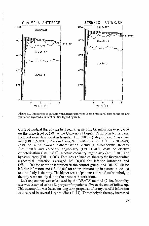
CONTROLS ANTERIOR STREPTO ANTERIOR 100% 100% DECEASED
DECEASED
I II-IV
80% 80%
II I-IV
60% 60%
CLASS I
40% 40%
CLASS I
20% 20%
0%~---.----.----r--~ O%w_ __ _,----,----.--~ 3 6 9 12 3 6 9 12
MONTHS MONTHS
Figure 8.2. Proportion of patients with anterior infarction in each functional class during the first year after myocardial infarction. See legend figure 8.1.
Costs of medical therapy the first year after myocardial infarction were based on the price level of 1984 at the University Hospital Dijkzigt in Rotterdam. Included were days spent in hospital (Dfl. 600/day), days in a coronary care unit (Dfl. 1,500/day), days in a surgical intensive care unit (Dfl. 2,500/day), costs of acute cardiac catheterisation including thrombolytic therapy (Dfl. 6,200) and coronary angioplasty (Dfl. 11,900), costs of elective catheterisation (Dfl. 2,600), elective coronary angioplasty (Dfl. 8,300) and bypass surgery (Dfl. 14,000). Total costs of medical therapy the first year after myocardial infarction averaged Dfl. 20,000 for inferior infarction and Dfl. 19,000 for anterior infarction in the control group, and Dfl. 27,000 for inferior infarction and Dfl. 28,000 for anterior infarction in patients allocated to thrombolytic therapy. The higher costs of patients allocated to thrombolytic therapy were mainly due to the acute catheterisation.
Life expectancy was calculated by the DEALE method (9,10). Mortality rate was assumed to be 6% per year for patients alive at the end of follow-up. This assumption was based on long term prognosis after myocardial infarction as observed in several large studies (11-14). Thrombolytic therapy increased
65
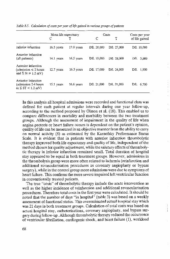
Table 8.5. Calculation of costs per year of life gained in various groups of patients
Mean life expectancy Costs Costs per year c T c T of life gained
Inferior infarction 16.3 years 17.0 years Dfl. 20,000 Dfl. 27,000 Dfl. 10,000
Anterior infarction (all patients) 14.1 years 16.5 years Dfl. 19,000 Dfl. 28,000 Dfl. 3,800
Anterior infarction (admission :,;;; 2 hours 12.7 years 16.3 years Dfl. 17,000 Dfl. 24,000 Dfl. 1,900 and :SSt;;, 1.2 mV)
Anterior infarction (admission 2-4 hours 15.1 years 16.6 years Dfl. 21,000 Dfl. 31,000 Dfl. 6,700 or :SST< 1.2 mV)
In this analysis all hospital admissions were recorded and functional class was defined for each patient at regular intervals during one year follow-up, according to the method proposed by Olsson et al. (18). This enabled us to compare differences in mortality and morbidity between the two treatment groups. Although the assessment of impairment in the quality of life when angina pectoris or heart failure occurs is dependent on the patient's opinion, quality of life can be measured in an objective manner from the ability to carry on normal activity (8) as estimated by the Karnofsky Performance Status Scale. It is evident that in patients with anterior infarction thrombolytic therapy improved both life expectancy and quality of life, independent of the method chosen for quality adjustment, while the salutary effects of thrombolytic therapy in inferior infarction remained small. Total duration of hospital stay appeared to be equal in both treatment groups. However, admissions in the thrombolysis group were more often related to ischemia (reinfarction and additional revascularisation procedures as coronary angioplasty or bypass surgery), while in the control group more admissions were due to symptoms of heart failure. This confirms the more severe impaired left ventricular function in conventionally treated patients.
The true "costs" of thrombolytic therapy include the acute intervention as well as the higher incidence of reinfarction and additional revascularisation procedures. Therefore total costs in the first year were calculated. It should be noted that the number of days "in hospital" (table 3) was based on a weekly assessment of functional status. This overestimated actual hospital stay which was 21 days in both treatment groups. Calculation of total costs was based on actual hospital stay, catheterisations, coronary angioplasty, and bypass surgery during follow-up. Although thrombolytic therapy reduced the occurrence of ventricular fibrillation, cardiogenic shock, and heart failure (1), workload
68
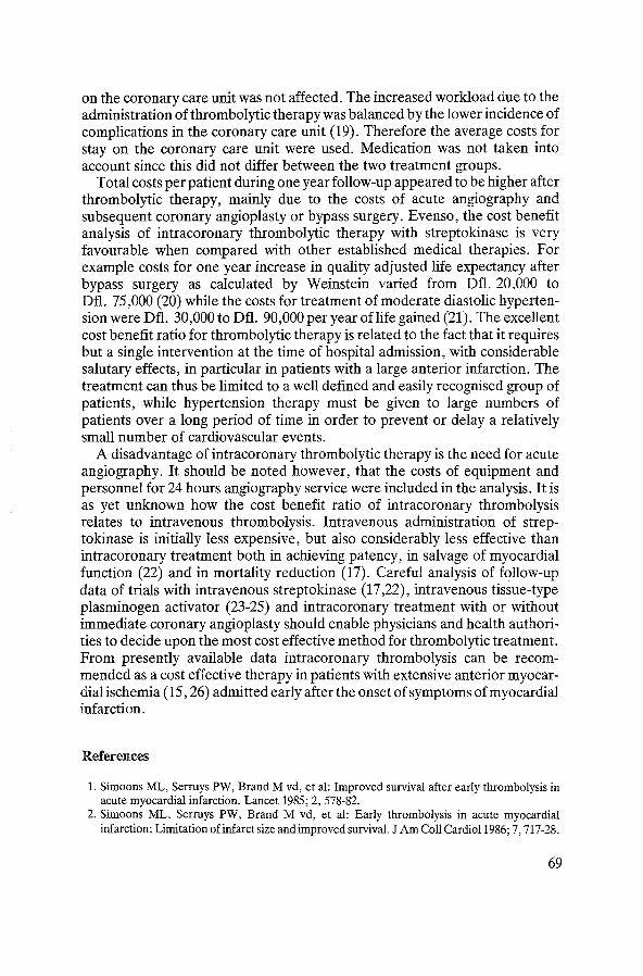
on the coronary care unit was not affected. The increased workload due to the administration of thrombolytic therapy was balanced by the lower incidence of complications in the coronary care unit (19). Therefore the average costs for stay on the coronary care unit were used. Medication was not taken into account since this did not differ between the two treatment groups.
Total costs per patient during one year follow-up appeared to be higher after thrombolytic therapy, mainly due to the costs of acute angiography and subsequent coronary angioplasty or bypass surgery. Evenso, the cost benefit analysis of intracoronary thrombolytic therapy with streptokinase is very favourable when compared with other established medical therapies. For example costs for one year increase in quality adjusted life expectancy after bypass surgery as calculated by Weinstein varied from Dfl. 20,000 to Dfl. 75,000 (20) while the costs for treatment of moderate diastolic hypertension were Dfl. 30,000 to Dfl. 90,000 per year oflife gained (21). The excellent cost benefit ratio for thrombolytic therapy is related to the fact that it requires but a single intervention at the time of hospital admission, with considerable salutary effects, in particular in patients with a large anterior infarction. The treatment can thus be limited to a well defined and easily recognised group of patients, while hypertension therapy must be given to large numbers of patients over a long period of time in order to prevent or delay a relatively small number of cardiovascular events.
A disadvantage of intracoronary thrombolytic therapy is the need for acute angiography. It should be noted however, that the costs of equipment and personnel for 24 hours angiography service were included in the analysis. It is as yet unknown how the cost benefit ratio of intracoronary thrombolysis relates to intravenous thrombolysis. Intravenous administration of streptokinase is initially less expensive, but also considerably less effective than intracoronary treatment both in achieving patency, in salvage of myocardial function (22) and in mortality reduction (17). Careful analysis of follow-up data of trials with intravenous streptokinase (17,22), intravenous tissue-type plasminogen activator (23-25) and intracoronary treatment with or without immediate coronary angioplasty should enable physicians and health authorities to decide upon the most cost effective method for thrombolytic treatment. From presently available data intracoronary thrombolysis can be recommended as a cost effective therapy in patients with extensive anterior myocardial ischemia (15, 26) admitted early after the onset of symptoms of myocardial infarction.
References
1. Simoons ML, Serruys PW, Brand M vd, et al: Improved survival after early thrombolysis in acute myocardial infarction. Lancet 1985; 2, 578-82.
2. Simoons ML, Serruys PW, Brand M vd, et al: Early thrombolysis in acute myocardial infarction: Limitation of infarct size and improved survival. JAm Coli Cardiol1986; 7, 717-28.
69
3. Serruys PW, Simoons ML, Suryapranata H, et a!: Preservation of global and regional left ventricular function after early thrombolysis in acute myocardial infarction. JAm Col! Cardiol 1986; 7, 729-42.
4. Laarse A vd, Vermeer F, Hermens WT, eta!: Effect of early intracoronary streptokinase on infarct size estimated from cumulative enzyme release and on enzyme release rate. Am Heart J 1986; 112, 672-81.
5. Res JCJ, Simoons ML, Wall EE vd, eta!: Long term improvement in global left ventricular function after early thrombolytic treatment in acute myocardial infarction. Br Heart J 1986; 56, 414-21.
6. Zelen M: A new design for randomised clinical trials. N Eng! J Med 1979; 300, 1242-5. 7. The criteria committee of the New York Heart Association, Inc.: Disease of the heart and
blood vessels; Nomenclature and criteria for diagnosis, 7th ed. Boston: Little, Brown 1973. 8. Yates JW, Chalmer B, McKegney FP: Evaluation of patients with advanced cancer using the
Karnofsky performance status. Cancer 1980; 45, 2220-4. 9. Beck JR, Kassirer JP, Pauker SG: A convenient approximation of life expectancy (The
"DEALE"). I. Validation of the method. Am J Med 1982; 73, 883-8. 10. Beck JR, Pauker SG, Gottlieb JE: A convenient approximation of life expectancy (The
"DEALE"). II. Use in medical decision-making. Am J Med 1982; 73, 889-97. 11. Norris RM, Barnaby PF, Brandt PWT, et a!: Prognosis after recovery from first acute
myocardial infarction: determinants of reinfarction and sudden death. Am J Cardiol1984; 53, 408-13.
12. Kannel WB, Sorlie P, McNamara PM: Prognosis after initial myocardial infarction: the Framingham study. Am J Cardiol1979; 44, 53-9.
13. Merrilees MA, Scott PJ, Norris RM: Prognosis after myocardial infarction: results of 15 year follow-up. Br Med J 1984; 288, 356-9.
14. Martin CA, Thompson PL, Armstrong BK, eta!: Long-term prognosis after recovery from myocardial infarction: a nine year follow-up of the Perth coronary register. Circ 1983; 68, 961-9.
15. Vermeer F, Simoons ML, Bar FW, eta!: Which patients benefit most from early thrombolytic therapy with intracoronary streptokinase? Circ 1986; 74, 1379-89.
16. Kennedy JW, Ritchie JL, Davis KB, eta!: The Western Washington randomized trial of intracoronary streptokinase in acute myocardial infarction. N Eng! J Med 1985; 312, 1973-8.
17. Gruppo Italiano per lo Studio della Streptochinasi nell'Infarto miocardico (GISSI): Effectiveness of intravenous thrombolytic treatment in acute myocardial infarction. Lancet 1986; 1, 397-401.
18. Olsson G, Lubsen J, Es GA, eta!: Quality of life after myocardial infarction; effect of chronic metoprolol treatment on mortality and morbidity. Br Med J 1986; 292, 1491-93.
19. Kint PP, Simoons ML, Vermeer F, eta!: Early thrombolysis in acute myocardial infarction does not increase CCU workload. Circ 1986; 74 II, 129 (abstract).
20. Weinstein MC, Stason WB: Cost effectiveness of coronary artery bypass surgery. Circ 1982; 66 III, 56-65.
21. Weinstein MC, Fineberg HV: Clinical decisions and limited resources. In: Clinical decision Analysis. Philadelphia: W.B. Saunders (Ed.), 1980.
22. Schroder R, Biamino G, Von Leitner ER, eta!: Intravenous short-term infusion of streptokinase in acute myocardial infarction. Circ 1983; 67, 536-48.
23. Verstraete M, Bory M, Collen D, eta!: Randomized trial of intravenous recombinant tissuetype plasminogen activator versus intravenous streptokinase in acute myocardial infarction. Lancet 1985; 1, 842-47.
24. Verstraete M, Brower RW, Collen D, eta!: Double-blind randomized trial of intravenous tissue-type plasminogen activator versus placebo in acute myocardial infarction. Lancet 1985; 2, 965-9.
25. TIMI Study Group: The thrombolysis in myocardial infarction (TIMI) trial: phase I findings. N Eng! J Med 1985; 310, 932-6.
70
26. Bar FW, Vermeer F, Simoons ML, et al: The value of the admission electrocardiogram to predict outcome of thrombolytic therapy in patients with acute myocardial infarction. Am J Cardiol1987; 59, 6-13.
71
CHAPTER9
Value of PTCA performed immediately after successful thrombolysis with intracoronary streptokinase
A matched pair analysis of the effect of PTCA in the randomized multicentre trial of intracoronary streptokinase conducted by the Interuniversity Cardiology Institute the Netherlands.
Frank Vermeer M.D., Maarten L. Simoons M.D., Pim J. de Feyter M.D., Frits W. Bar M.D., Harry Suryapranata M.D., Paolo Fioretti M.D., Patrick W. Serruys M.D., Beert Buis M.D., Jan C.J. Res M.D., Simon H. Braat M.D., Jacobus Lubsen Ph.D., Paul G. Hugenholtz M.D.
Submitted for publication
Abstract
Acute PTCA following successful thrombolysis with streptokinase was performed in 46 patients out of 533 patients enrolled in a multicentre randomized trial comparing a strategy aimed at early reperfusion with conventional therapy in patients with acute myocardial infarction. Additional effects of PTCA were compared with successful thrombolysis alone in a matched pair analysis. The indication for PTCA was significant residual obstruction after thrombolysis with insufficient distal run-off, as visualized during catheterization. Thirty six pairs of patients were formed who were identical with respect to the infarct related coronary artery, presence or absence of previous myocardial infarction, total ST segment elevation on the ECG at admission to the trial, and delay between onset of symptoms and hospital admission. All further hospital admissions were recorded and functional class was assessed at visits to the out patient clinic during three years follow-up. PTCA after successful thrombolysis did not lead to additional limitation of infarct size. However, improvement in L VEF, measured by radionuclide angiography, was 8% in the PTCA group vs 2% after successful thrombolysis alone (p = .005). Recurrent infarction within three years was 14% in the PTCA group versus 30% in the thrombolysis group (p = .05). In the PTCA group more time was spent without symptoms of angina or heart failure (mean 128 weeks out of 156 weeks) than in the thrombolysis group (102 weeks; p = .03). Prevention of recurrent ischemia by early PTCA was observed to the same extent in patients with anterior infarction and with inferior infarction.
It is concluded that immediate PTCA after successful thrombolysis prevents recurrent ischemia and improves quality of life. Thus PTCA can be recommended as an early procedure in patients who demonstrate a residual stenosis of 70% or more in the infarct related coronary artery with a poor distal run-off.
72
Introduction
A strategy aimed at early recanalization of an infarct related coronary artery with intracoronary streptokinase (1-5) will result in salvage of myocardial tissue (6-8), preservation of left ventricular function (9-10), and improved survival (5, 11-12). However, in many patients a severe stenosis of the infarct related artery remains after successful thrombolysis. This artery may reocclude in part of the patients and abolish the initial beneficial effects of thrombolytic therapy. Indeed, non-fatal reinfarction occurred more frequently in patients allocated to thrombolytic therapy than in conventionally treated patients (5, 12). In order to prevent reocclusion, but also to optimize coronary flow to the jeopardized myocardium, acute PTCA has been advocated by several authors (13-17), and the efficacy of PTCA early after thrombolytic therapy is currently investigated in randomized clinical trials (18-20). The purpose of the present analysis was to evaluate the influence of PTCA performed immediately after successful thrombolysis in part of the patients in the randomized trial conducted by the Interuniversity Cardiology Institute the Netherlands (ICIN), on left ventricular function, re-infarction, reocclusion, and on the necessity of subsequent revascularization procedures such as re-PTCA or coronary artery bypass grafting. In order to eliminate a possible selection bias, patients who underwent acute PTCA were compared with a matched group of patients with similar baseline characteristics in whom the lesion in the infarct related coronary artery was suitable for PTCA but who were admitted to those hospitals where PTCA could not be performed as part of the recanalization procedure.
Patient selection and methods
Patient selection and methods of the trial conducted by the ICIN have been described extensively before (5,14). Thrombolytic therapy was given after informed consent and consisted of intracoronary administration of streptokinase in the catheterization laboratory, usually 250,000 U. In patients admitted to the trial since January 1984 this was preceded by intravenous administration of 500,000 U streptokinase given upon admission, in order to reduce treatment delay. The indication for PTCA was significant residual obstruction after thrombolysis with insufficient distal run-off as visualized during catheterization. In addition all patients received acetyl salicylic acid 250 mg intravenously and heparin 20,000 U/24 hours followed by oral coumadin until hospital discharge.Niphedipine,(10 mg four times daily)was given to most patients after PTCA.
Total ST segment elevation was calculated as the sum of ST segment elevation in standard and precordial leads, as described before (11). Global left ventricular ejection fraction (L VEF) was measured by radionuclide angiography 1 to 3 days after admission and again after 10 to 20 days. All
73
patients were followed at regular intervals at the out-patient clinic for 20 to 60 months (mean 38 months) after admission. All hospital admissions were recorded and functional class was assessed according to the criteria of theN ew York Heart Association (NYHA).
From these data the functional status was defined for each patient at weekly intervals as the lowest of the following five mutually exclusive classifications: - Class I (NYHA), not hospitalized - Class II (NYHA), not hospitalized - Class III or IV (NYHA), not hospitalized - Hospitalized - Deceased. Mean number of weeks spent in each category was calculated for all patients. Mean survival was calculated as the mean time elapsed between admission to the study and death or end of follow-up (three years).
Matching
The patients in whom immediate PTCA was attempted (PTCA group) were matched with patients allocated to thrombolytic therapy in those participating centres where PTCA could not be performed as part of the recanalization procedure (Sk-only group). Matching was performed according to the following rules: - Coronary angiograms of all patients successfully treated with intracoronary
streptokinase and admitted to those participating centres where acute PTCA was not performed were reviewed by three cardiologists to assess whether the lesion in the infarct related coronary artery had been suitable for PTCA. Consensus about the suitability for PTCA was always reached. Only patients in whom the lesion was judged to have been suitable for PTCA were included in the next step of the matching procedure.
- All patients were grouped with regard to the infarct related coronary artery and the presence or absence of previous myocardial infarction.
- Within each group pairs of patients were selected with least difference in admission delay (maximum difference 30 minutes) and secondly least difference in total ST segment elevation (maximum difference 0.6 mV) on the ECG made at admission to the trial. Admission delay and total ST segment elevation were chosen as matching parameters since they determined the outcome of thrombolytic therapy as reported earlier (11).
In order to ascertain that differences between the PTCA group and the Sk -only group were not due to differences between the various hospitals, two matched control groups of patients allocated to conventional therapy were constructed. One control group was formed from patients allocated to conventional therapy in the hospitals where acute PTCA was performed (matched control group I), and the other from patients allocated to conventional therapy in hospitals without acute PTCA (matched control group II). Matching for these groups
74

Table 9.1. Baseline characteristics
Matched groups Matched control All groups
c TPTCA PTCA Sk-only CI en
No of patients 264 269 46 36 36 36 36 Males 224 217 39 29 29 28 26 Previous infarction 60 56 12 4 4 4 4 Anterior infarction 116 130 29 22 22 22 22 Age (median) 56 57 59 59 56 52 55 Admission delay (median; min) 90 90 90 90 105 90 100 l ST (median; mV) 1.2 1.1 1.3 1.2 1.1 1.2 1.3
Abbreviations: C: control group; T: allocated to thrombolysis; min: minutes; Sk: streptokinase; PTCA: percutaneous transluminaCcoronary angioplasty; CI: matched control group from hospitals with acute PTCA; CII: matched control group from hospitals with Sk-only; l ST: total STsegment elevation on the electrocardiogram made on admission to the trial.
Baseline characteristics were distributed evenly between the four matched groups.
was done according to the same rules as described for the PTCA group and the Sk-only group. Patients in the control groups with anterior infarction on the ECG were compared with patients in the thrombolysis group where the lesion was located in the left anterior descending artery, and patients with inferior infarction were compared with patients with a lesion in the right coronary artery.
Statistical analysis
Differences between groups were tested with Fisher's exact test or Student's T test when appropriate. Two sided p-values are reported.
Results
A total of 533 patients were admitted to the trial. Acute angiography was performed in 234 out of 269 patients allocated to thrombolytic therapy, 20 patients refused the intervention, and in the remaining 15 patients thrombolytic therapy was withheld for other reasons ( 5). PTCA was performed as part of the recanalization procedure in 46 patients admitted to two of the five participating hospitals, the Thoraxcenter in Rotterdam (44 out of 106 patients) and the Academic Hospital in Leiden (2 out of 9 patients) (6). PTCA was not successful in two patients since the artery reoccluded immediately after the procedure. Patency of the infarct related coronary artery as shown at angiography was achieved in 98 patients admitted to one of the three participating
75

Table 9.2. Results of coronary angiography (n = 234)
Matched groups PTCA Sk-only PTCA Sk-only
Number of patients 46 188 36 36
Infarct related artery LAD 29 74 22 22 RCA 15 74 14 14 LCX 2 38 0 0 bypass 0 2 0 0
Patency at the end of the procedure
occluded 2 34 2 0 91-99% 4 89 4 27 50-90% 4 54 4 8 <50% 36 11 26 1
Patency at second angiography patent 31 113 25 23 occluded 6 27 4 5 unknown 9 48 7 8
Abbreviations: PTCA: percutaneous transluminal coronary angioplasty; Sk: streptokinase; LAD: left anterior descending artery; RCA: right coronary artery; LCX: left circumflex artery.
PTCA led to a significant decrease in residual stenosis of the infarct related coronary artery.
centres where acute PTCA was not performed. Coronary angiograms were available for review from 86 of these 98 patients. The lesion in the infarct related coronary artery was on review judged to have been suitable for PTCA in 62 patients. These 62 patients were matched with the 46 patients in whom acute PTCA was actually carried out. Thirty six pairs of patients were formed following the matching rules as described in the methods section. Ten patients in whom acute PTCA was performed could not be matched because no pairs of patients could be formed which were identical with regard to the infarct related coronary artery and the presence or absence of previous myocardial infarction. Baseline characteristics were distributed evenly between the four matched groups (table 1).
Coronary angiography
Acute PTCA was performed more often when the lesion was located in the left anterior descending artery (29 out of 55 patients in the two hospitals with acute
76
Table 9.3. Patency of the infarct related coronary artery at second angiography (10-40 days) in patients with successful thrombolysis (n = 198)
All patients Matched groups PTCA Sk-only PTCA Sk-only
Number of patients 44 154 36 36
LAD patent 22 45 16 17 occluded 1 5 1 2 unknown 4 12 5 3
RCAorLCX patent 9 57 7 8 occluded 5 14 4 2 unknown 3 21 3 4
Abbreviations: See table 2.
Reocclusion rate was not influenced by PTCA.
PTCA; 53%) than in the right coronary artery (15 out of 40 patients; 38%) or the left circumflex artery (2 out of 19 patients; 11% ). Acute PTCA led to a significant decrease in residual stenosis (p = .001), although immediate reocclusion was observed in two patients (table 2). Reocclusion, defined as an occluded infarct related coronary artery at second angiography in patients with successful thrombolysis, was observed in 9 out of 57 patients (16%) in the two matched groups (table 3). Reocclusion rates were high when the lesion was located in the right coronary artery or left circumflex artery ( 6 out of 21 patients; 28%) and lower in case of a lesion in the left anterior descending artery (3 out of36 patients; 8%). Reocclusion rates were identical in the PTCA group and the Sk-only group (18% versus 14%).
Infarct size and left ventricular function
Enzymatic infarct size, measured by cumulative alpha-hydroxybutyrate dehydrogenase (HBDH) release (7) was 30% lower in patients allocated to thrombolytic therapy (median 770 U/1) than in the control group (median 1100 U/1, p = .0001). However, cumulative HBDH release in the PTCA group (median 760 U/1) did not differ from that in the Sk-only group (median 740 U/1). Improvement of left ventricular ejection fraction, measured by radionuclide angiography (6, 10) as well as by contrast angiography (9), was observed after thrombolytic therapy. Mean radionuclide LVEF after 1 to 3 days was 41% in the PTCA group and 48% in the Sk-only group. Mean L VEF after 10 to 20 days was 49% in the PTCA group and 50% in the Sk-only group.
77
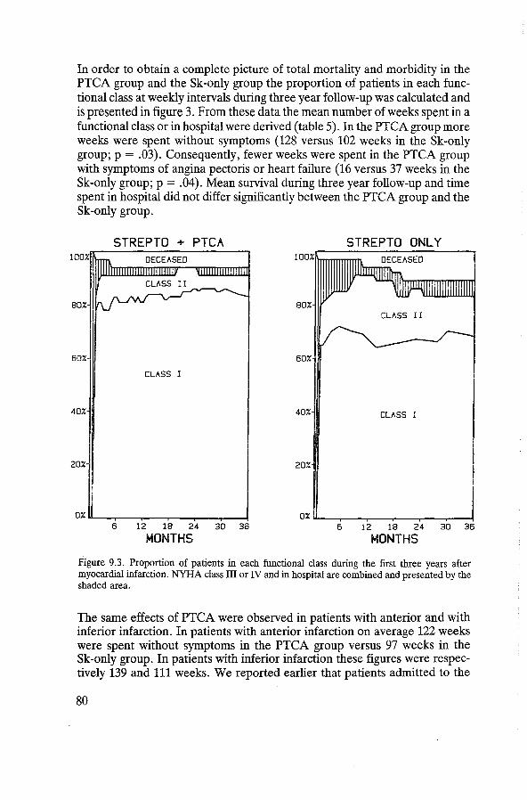
In order to obtain a complete picture of total mortality and morbidity in the PTCA group and the Sk-only group the proportion of patients in each functional class at weekly intervals during three year follow-up was calculated and is presented in figure 3. From these data the mean number of weeks spent in a functional class or in hospital were derived (table 5). In the PTCA group more weeks were spent without symptoms (128 versus 102 weeks in the Sk-only group; p = .03). Consequently, fewer weeks were spent in the PTCA group with symptoms of angina pectoris or heart failure (16 versus 37 weeks in the Sk-only group; p = .04). Mean survival during three year follow-up and time spent in hospital did not differ significantly between the PTCA group and the Sk-only group.
STREPTO + PTCA STREPTO ONLY
60% 60%
CLASS I
40% 40% CLASS I
20% 20%
6 12 18 24 30 36 6 12 18 24 30 36
MONTHS MONTHS
Figure 9.3. Proportion of patients in each functional class during the first three years after myocardial infarction. NYHA class III or IV and in hospital are combined and presented by the shaded area.
The same effects of PTCA were observed in patients with anterior and with inferior infarction. In patients with anterior infarction on average 122 weeks were spent without symptoms in the PTCA group versus 97 weeks in the Sk-only group. In patients with inferior infarction these figures were respectively 139 and 111 weeks. We reported earlier that patients admitted to the
80
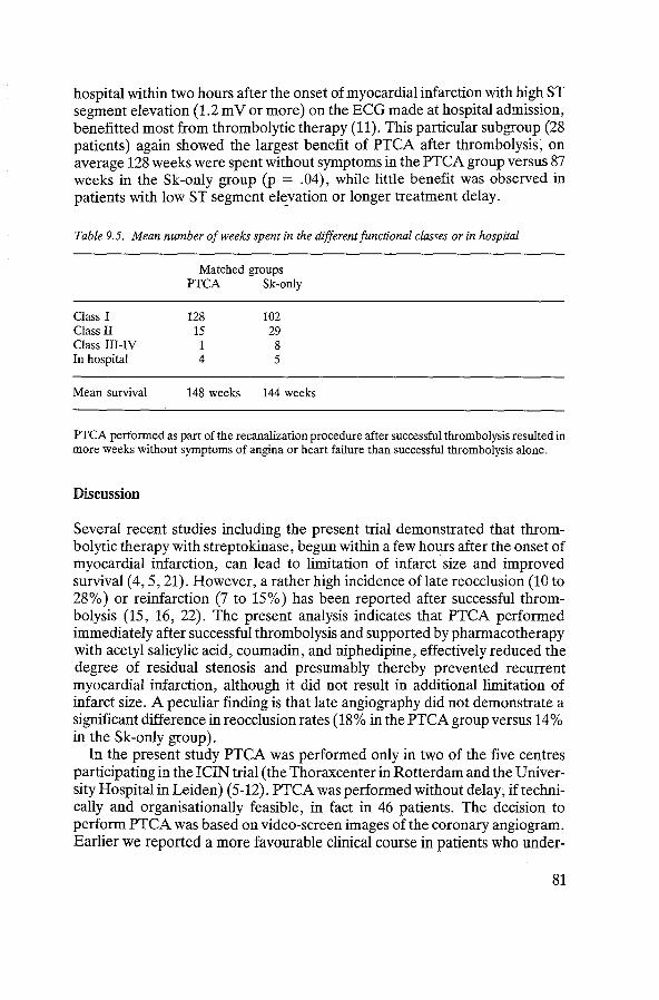
hospital within two hours after the onset of myocardial infarction with high ST segment elevation (1.2 m V or more) on the ECG made at hospital admission, benefitted most from thrombolytic therapy (11). This particular subgroup (28 patients) again showed the largest benefit of PTCA after thrombolysis, on average 128 weeks were spent without symptoms in the PTCA group versus 87 weeks in the Sk-only group (p = .04), while little benefit was observed in patients "'_ith low ST segment el~vation or longer treatment delay.
Table 9.5. Mean number of weeks spent in the different functional classes or in hospital
Class I Class II Class III-IV In hospital
Mean survival
Matched groups PTCA Sk-only
128 102 15 29 1 8 4 5
148 weeks 144 weeks
PTCA performed as part of the recanalization procedure after successful thrombolysis resulted in more weeks without symptoms of angina or heart failure than successful thrombolysis alone.
Discussion
Several recent studies including the present trial demonstrated that thrombolytic therapy with streptokinase, begun within a few hours after the onset of myocardial infarction, can lead to limitation of infarct size and improved survival ( 4, 5, 21). However, a rather high incidence of late reocclusion (10 to 28%) or reinfarction (7 to 15%) has been reported after successful thrombolysis (15, 16, 22). The present analysis indicates that PTCA performed immediately after successful thrombolysis and supported by pharmacotherapy with acetyl salicylic acid, coumadin, and niphedipine, effectively reduced the degree of residual stenosis and presumably thereby prevented recurrent myocardial infarction, although it did not result in additional limitation of infarct size. A peculiar finding is that late angiography did not demonstrate a significant difference in reocclusion rates (18% in the PTCA group versus 14% in the Sk-only group).
In the present study PTCA was performed only in two of the five centres participating in the ICIN trial (the Thoraxcenter in Rotterdam and the University Hospital in Lei den) (5-12). PTCA was performed without delay; if technically and organisationally feasible, in fact in 46 patients. The decision to perform PTCA was based on video-screen images of the coronary angiogram. Earli~r we reported a more favourable clinical course in patients who under-
81

went immediate PTCA in comparison with those without this additional intervention (9). However, since the results of the PTCA group might easily be biased by selection, a matched pair analysis was performed. Because beneficial effects of thrombolytic therapy are strongly influenced by the extent of myocardial ischemia upon admission (as reflected by total ST segment elevation) and by the rapidity of lysis onset (8, 11), these baseline characteristics were included in the matching procedure. In fact, the two matched groups turned out to be almost identical with regard to the most important prognostic factors.
It was found that reocclusion rates were higher when the lesion was located in the right coronary artery or in the circumflex artery than in the left anterior descending artery (table 3). The same observation was made in the ISAM study (22), and other smaller studies (15-16, 23-24). Surprisingly, we observed no reduction in reocclusion rate after acute PTCA despite its marked effect on the severity of the residual stenosis. Reocclusion rate after thrombolytic therapy is related to the degree of residual stenosis (23), but after PTCA it is probably more related to the anatomy of the residual lesion. Furthermore, many changes in coronary blood supply will have occurred (such as formation of collaterals) between the time of initial PTCA and late reobstruction explaining the apparent difference between increased reocclusion rate and decreased reinfarction rate. Also the pharmacotherapy (aspirin, niphedipine and heparin) may have influenced the outcome in an unpredictable manner. In the larger prospective studies of Erbel (25) and O'Neill (26, 27) acute PTCA led to reduction in reocclusion rates, but not as much as might be expected when the severity of the residual stenosis were the only important factor.
Temporary occlusion of a coronary artery leads to a depression in left ventricular function that may persist for several days, as is known from animal experiments and human observations (28-30). The left ventricular ejection fraction measured 1 to 3 days after the onset of myocardial infarction probably reflects the initially depressed state of left ventricular function. Thrombolytic therapy leads already to an improvement in left ventricular function when L VEF after 1 to 3 days is compared to L VEF at 10 to 20 days in the same patient (6, 10). This improvement in LVEF was more pronounced in the PTCA group than in the Sk-only group (figure 1), although L VEF after 10 to 20 days was almost equal in these two groups. Clearly, recovery from the stunned myocardium plays a major role here. From the data presented it remains unclear whether PTCA additionally improves left ventricular function or may have led to an initially more pronounced although temporary depression of left ventricular function. We believe that the former explanation is correct since in the study of O'Neill et al. (26) a greater improvement in left ventricular function was observed in the PTCA group, in whom the first radionuclide angiogram was performed prior to the initiation of thrombolytic therapy, a procedure lacking in the ICIN study.
Long term follow-up after successful thrombolysis followed by acute PTCA
82
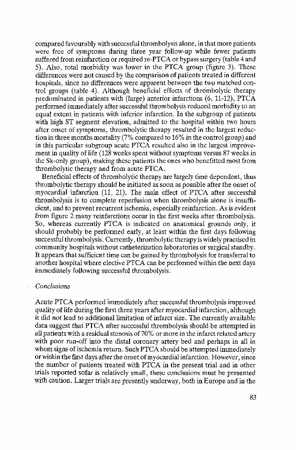
compared favourably with successful thrombolysis alone, in that more patients were free of symptoms during three year follow-up while fewer patients suffered from reinfarction or required re-PTCA or bypass surgery (table 4 and 5). Also, total morbidity was lower in the PTCA group (figure 3). These differences were not caused by the comparison of patients treated in different hospitals, since no differences were apparent between the two matched control groups (table 4). Although beneficial effects of thrombolytic therapy predominated in patients with (large) anterior infarctions (6, 11-12), PTCA performed immediately after successful thrombolysis reduced morbidity to an equal extent in patients with inferior infarction. In the subgroup of patients with high ST segment elevation, admitted to the hospital within two hours after onset of symptoms, thrombolytic therapy resulted in the largest reduction in three months mortality (7% compared to 16% in the control group) and in this particular subgroup acute PTCA resulted also in the largest improvement in quality of life (128 weeks spent without symptoms versus 87 weeks in the Sk-only group), making these patients the ones who benefitted most from thrombolytic therapy and from acute PTCA.
Beneficial effects of thrombolytic therapy are largely time dependent, thus thrombolytic therapy should be initiated as soon as possible after the onset of myocardial infarction (11, 21). The main effect of PTCA after successful thrombolysis is to complete reperfusion when thrombolysis alone is insufficient, and to prevent recurrent ischemia, especially reinfarction. As is evident from figure 2 many reinfarctions occur in the first weeks after thrombolysis. So, whereas currently PTCA is indicated on anatomical grounds only, it should probably be performed early, at least within the first days following successful thrombolysis. Currently, thrombolytic therapy is widely practised in community hospitals without catheterization laboratories or surgical standby. It appears that sufficient time can be gained by thrombolysis for transferral to another hospital where elective PTCA can be performed within the next days immediately following successful thrombolysis.
Conclusions
Acute PTCA performed immediately after successful thrombolysis improved quality of life during the first three years after myocardial infarction, although it did not lead to additional limitation of infarct size. The currently available data suggest that PTCA after successful thrombolysis should be attempted in all patients with a residual stenosis of 70% or more in the infarct related artery with poor run-off into the distal coronary artery bed and perhaps in all in whom signs of ischemia return. Such PTCA should be attempted immediately or within the first days after the onset of myocardial infarction. However, since the number of patients treated with PTCA in the present trial and in other trials reported sofar is relatively small, these conclusions must be presented with caution. Larger trials are presently underway, both in Europe and in the
83

United States, where the clinical course after acute PTCA and after delayed PTCA will be compared with thrombolytic therapy with recombinant tissuetype plasminogen activator alone. It may be expected that those studies will provide additional data regarding the optimal and practical timing of PTCA and the selection of patients in whom this additional procedure should be recommended.
References
1. Rentrop P, De Vivie ER, Karsch KR, et al: Acute myocardial infarction: intracoronary application of nitroglycerin and streptokinase in combination with transluminal recanalization. Clin Cardiol5: 354, 1979.
2. Merx W, Di:irr W, Rentrop P, et a!: Evaluation of the effectiveness of intracoronary streptokinase infusion in acute myocardial infarction: postprocedure management and hospital course in 204 patients. Am Heart J 102: 1181, 1981.
3. Weinstein J: Treatment of myocardial infarction with intracoronary streptokinase: Efficacy and safety data from 209 united states cases in the Hoechst-Roussel registry. Am Heart J 104: 894, 1982.
4. Kennedy JW, Ritchie JL, Davis KB, et al: Western Washington randomized trial of intracoronary streptokinase in acute myocardial infarction. N Eng! J Med 309: 1477, 1983.
5. Simoons ML, Serruys PW, Brand M vd, et al: Improved survival after early thrombolysis in acute myocardial infarction. A randomized trial conducted by the Interuniversity Cardiology Institute in The Netherlands. Lancet 2: 578, 1985.
6. Simoons ML, Serruys PW, Brand M vd, et a!: Early thrombolysis in acute myocardial infarction: Limitation of infarct size and improved survival. J Am Coli Cardiol 7: 717, 1986.
7. Laarse A vd, Vermeer F, Hermens WT, et al: Effects of early intracoronary streptokinase on infarct size estimated from cumulative enzyme release and on enzyme release rate. Am Heart J 112: 672, 1986.
8. Bar FW, Vermeer F, Zwaan C de, et al: Value of admission electrocardiogram in predicting outcome of thrombolytic therapy in acute myocardial infarction. Am J Cardiol59: 6, 1987.
9. Serruys PW, Simoons ML, Suryapranata H, et al: Preservation of global and regional left ventricular function after early thrombolysis in acute myocardial infarction. JAm Coli Cardiol 7: 729, 1986.
10. Res J, Simoons ML, WallE vd, eta!: Long term improvement in global left ventricular function after early thrombolytic therapy in acute myocardial infarction. Br Heart J 56: 414, 1986.
11. Vermeer F, Simoons ML, Bar FW, et al: Which patients benefit most from early thrombolytic therapy with intracoronary streptokinase? Circ 74: 1379, 1986.
12. Vermeer F, Simoons ML, Zwaan C de, et al: Cost benefit analysis of early thrombolytic therapy with intracoronary streptokinase. Br Heart J 1987 (in press).
13. Meyer J, Merx W, Schmitz H, eta!: Percutaneous transluminal coronary angioplasty immediately after intracoronary streptolysis of transmural infarction. Circ 66: 905, 1982.
14. Serruys PW, Wijns W, Brand M vd, et al: Is transluminal coronary angioplasty mandatory after successful thrombolysis? Br Heart J 50: 257, 1983.
15. Gold HK, Leinbach RC, Palacios IF, et al: Coronary reocclusion after selective administration of streptokinase. Circ 68 (suppl. I): 50, 1983.
16. Schroder R, Vohringer H, Linderer T, et al: Follow-up after coronary arterial reperfusion with intravenous streptokinase in relation to residual myocardial infarct artery narrowings. Am J Cardiol 55: 313, 1985.
17. Papapietro SE, MacLean WAH, Stanley A WH, et al: Percutaneous transluminal coronary angioplasty after intracoronary streptokinase in evolving acute myocardial infarction. Am J Cardiol55: 48, 1985.
84
18. Smalling RW, Nishikawa A, Merhige ME, et al: Intravenous tissue plasminogen activator followed by percutaneous transluminal coronary angioplasty in acute myocardial infarction: acute and follow-up quantitative angiographic analysis. Circ 74 (suppl II): 23, 1986 (abstract).
19. Topol EJ, Califf RM, George BS, et al: A multicenter randomized trial of intravenous recombinant tissue plasminogen activator and emergency coronary angioplasty for acute myocardial infarction: preliminary report from the TAMI study. Circ 74 (suppl II): 23, 1986 (abstract).
20. Collins GJ, Califf RM, Phillips HR, et al: Quantitative angiographic changes occurring within 24 hours and one week following treatment with tissue plasminogen activator in patients randomized to immediate and late angioplasty. Circ 74 (suppl II): 275, 1986 (abstract).
21. Gruppo Italiano per lo Studio della Streptochinasi nell'infarto miocardico (GISSI): Effectiveness of intravenous thrombolytic treatment in acute myocardial infarction. Lancet 1: 397, 1986.
22. Schroder R, Neuhaus KL, Leizorovics A, et al: A prospective placebo-controlled doubleblind multicenter trial of intravenous streptokinase in acute myocardial infarction (ISAM): Long-term mortality and morbidity. JAm Coli Cardiol9: 197, 1987.
23. Harrison DG, Ferguson DW, Collins SM, et al: Rethrombosis after reperfusion with streptokinase: Importance of geometry of residual lesions. Circ 72: 478, 1986.
24. Gash AK, Spann JF, Sherry S, et al: Factors influencing reocclusion after coronary thrombolysis. Circ 70 (suppl II): 256, 1984.
25. Erbel R, Pop T, Henrichs KJ, et al: Percutaneous transluminal coronary angioplasty after thrombolytic therapy: A prospective controlled randomized trial. JAm Coli Cardiol8: 485, 1986.
26. O'Neill W, Timmis GC, Bourdillon PD, et al: A prospective randomized clinical trial of intracoronary streptokinase versus coronary angioplasty for acute myocardial infarction. N Eng! J Med 314: 812, 1986.
27. Fung A Y, Lai P, Juni JE, et al: Prevention of subsequent exercise induced periinfarct ischemia by emergency coronary angioplasty in acute myocardial infarction: Comparison with intracoronary streptokinase. JAm Coli Cardiol 8: 496, 1986.
28. Lavallee M, Cox DA, Vatner SF: Effects of coronary artery reperfusion on recovery of regional myocardial function in conscious dogs. Eur Heart J 6 (supplE): 109, 1985.
29. Serruys PW, Wijns W, Brand M vd, et al: Left ventricular performance, regional blood flow, wall motion and lactate metabolism during transluminal angioplasty. Circulation 70: 25, 1984.
30. Wijns W, Serruys PW, Slager CJ, et al: Effect of coronary occlusion during percutaneous transluminal angioplasty on left ventricular chamber stiffnes and regional diastolic pressureradius relations. JAm Coli Cardiol 7: 455, 1986.
85

Chapter 10
Summary of other reports from the trial
In addition to the data presented in the chapters 4 to 9, other results of the trial have been analysed and presented in papers or abstracts. These reports are summarised in this chapter.
Clinical course
Patients allocated to thrombolytic therapy had a more favourable clinical course in hospital than the control group, except for a higher incidence of bleeding complications (chapter 4). This was equally true for patients allocated to intracoronary streptokinase only as for patients in whom angiography was preceded by intravenous administration of streptokinase (1, 2). Despite the more favourable clinical course in the thrombolysis group no significant differences were observed between patients allocated to thrombolytic therapy and the control group in hemodynamic measurements during the first 24 hours in the coronary care unit (3). The lower incidence of ventricular fibrillation in the thrombolysis group was remarkable, since a high incidence of reperfusion arrhythmias has been reported in animal experiments (4). In the thrombolysis group less patients had "spontaneous" ventricular fibrillation within 48 hours, but also the incidence of late ventricular fibrillation and ventricular fibrillation during treatment of cardiogenic shock was lower in these patients (5; table). The former may be due in part to treatment with lignocaine in the thrombolysis group, while the latter is in agreement with the reported limitation of infarct size and preservation of left ventricular function.
Table 10.1 Ventricular fibrillation
n Total VF Primary VF (<48 hours)* Catheter induced VF Late VF (>48 hours) Shock+ VF
* Median delay after symptoms
86
controls
264 60 (23%) 24 ( 9%) 9 ( 3%)
10(4%) 17 ( 7%)
3 hours
thrombolysis
269 36 (13%) 13 ( 5%) 15 ( 5%) 2 ( 1%) 6 ( 2%)
2 hours
Workload
Thrombolytic therapy did not affect the workload of the nursing staff in the coronary care unit. CCU workload, calculated with the Therapeutic Intervention Scoring System for 237 patients admitted to the Thorax center, was higher in the thrombolysis group on the day of hospital admission, but markedly lower on the second and third day after admission, due to fewer patients who required prolonged diuretic therapy and pulmonary catheters (6). On the other hand, workload was increased for the staff in the catheterisation laboratory, as reported earlier (7). The additional costs for this increased workload of the catheterisation staff were included in the cost benefit analysis (chapter 8).
Infarct size
Myocardial perfusion at rest and during exercise was measured by 201-Tl scintigraphy three months after the myocardial infarction. In a quantitative analysis of circumferential profiles of the thallium scintigrams from 118 patients admitted to the Thoraxcenter the infarct area size appeared to be 13% smaller in the thrombolysis group (8). Scintigraphically measured infarct size correlated well with enzymatic infarct size since in the same group of patients a 21% limitation of enzymatic infarct size was observed. Similar observations were made when thallium scintigrams of 236 patients with a first myocardial infarction from the five participating centres were analysed qualitatively (9, 10).
Left ventricular function
Global left ventricular ejection fraction, measured by radionuclide angiography, was significantly higher in the thrombolysis group at two days, two weeks and three months after admission (1i, 12). In addition, global and regional ejection fraction was assessed by contrast angiography in the right anterior oblique projection 10 to 40 days after admission. Global left ventricular ejection fraction was higher in the thrombolysis group (mean 53%) than in the control group (mean 47%; p = 0.0001), due to smaller end-systolic and enddiastolic volumes in the thrombolysis group. Wall motion analysis showed depressed values of regional contribution to ejection fraction in the "infarct zone" with at second angiography subsidence of the initially augmented functioning of the"non-infarct zone" The latter phenomenon was particularly prominent in patients who underwent the combined procedure of recanalisation and angioplasty. Segmental function in the "infarct zone" was poorest in patients whose coronary artery could not be recanalised and significantly better in patients who underwent successful thrombolysis. The highest increase (16%) in the value of the regional contribution to ejection fraction of
87
the "infarct zone" from the acute to the chronic stage was observed when angioplasty was performed as part of the recanalisation procedure (13, 14).
Location of the lesion
Final patency rate was high in all subsets of patients which were studied and appeared not to be related to the location of the lesion in the coronary artery (15). On the other hand, limitation of enzymatic infarct size was observed only in patients with a lesion in the left anterior descending artery or proximal in the right coronary artery and not in patients with a lesion in the left circumflex artery or distal in the right coronary artery ( 16). Patients with a lesion proximal in the right coronary artery could be distinguished from other patients with inferior infarction by ST segment elevation in lead V 4R on the admission electrocardiogram, as it was recorded from patients admitted to the University Hospital Maastricht and the Hospital of the Free University of Amsterdam (17).
Residual ischemia
In the majority of cases, a significant stenosis remained in the infarct related coronary artery after successful thrombolysis. This artery might reocclude in part of the patients and thus abolish the initial beneficial effects of thrombolytic therapy. Indeed, a high incidence of reocclusion of the infarct related artery or reinfarction within 30 days was observed after successful thrombolysis, namely 8% in patients with anterior infarction and 28% in those with inferior infarction (18). In the thrombolysis group more patients had to undergo coronary angioplasty or bypass surgery because of angina pectoris than in the control group (chapter 8). Coronary angioplasty, performed as part of the recanalisation procedure after successful thrombolysis in selected patients admitted to the Thoraxcenter or the University Hospital in Leiden, appeared to reduce reinfarction rates and led to a lower prevalence of angina pectoris when compared to successful thrombolysis alone, as assessed in the matched pair analysis (chapter 9). Surprisingly, exercise induced ischemia, assessed by 201-Tl scintigraphy three months after admission, was less prominent in the thrombolysis group than in the control group (8-10). It seems likely that after three months patients allocated to thrombolytic therapy are no longer more prone to recurrent ischemia and recurrent myocardial infarction than patients in the control group. Indeed, the higher incidence of recurrent infarction and additional revascularisation procedures in the thrombolysis group was only observed in the first three months after admission with no relevant difference between the two treatment groups in the incidence of these events during later follow-up (ranging from 20 to 60 months with a mean follow-up of 38 months).
88
Consistency of the results
The results of this trial might be criticised because of the modifications in the protocol while the trial was in progress, which were described in chapter 3. The inclusion of the pretreatment with intravenous streptokinase and the addition of coronary angioplasty did not affect the outcome of the trial. As becomes evident from the results of the interim analyses (see table in chapter 3), beneficial effects of early thrombolytic therapy were demonstrated in all subsets of patients. Limitation of enzymatic infarct size and improvement in left ventricular function by thrombolytic therapy were observed in the first 150 patients admitted to the trial in the Thoraxcenter, but also in patients admitted to the other hospitals, where immediate angioplasty was not performed as part of the recanalisation procedure. The additional value of acute angioplasty and the effects of the pretreatment with intravenous streptokinase were also analysed separately. In the latter subgroup the largest mortality reduction was observed (1). However, inclusion of the pretreatment in the multivariate regression model did not improve the prediction of the effects of intracoronary streptokinase on infarct size, left ventricular ejection fraction or three months mortality (chapter 7). Thus, it can be concluded that the better outcome of the patients allocated to thrombolytic therapy was for the greater part due to the effects of intracoronary streptokinase and cannot be ascribed only to effects of intravenous streptokinase or coronary angioplasty.
The additional reports summarised in this chapter support the outcome of the trial. All findings are in agreement with a smaller infarct size in the thrombolysis group, namely less heart failure in the coronary care unit and thereafter, a lower incidence of ventricular fibrillation and less patients with symptoms of pericarditis. As a consequence, these patients required less attention from the nursing staff, once the intervention was completed. The observed limitation of infarct size, measured by cumulative enzyme release and by 201-Tl scintigraphy, led to better left ventricular function, analysed with radionuclide and with contrast angiography, and to improved survival in the thrombolysis group. It is this consistency in the results that supports our conclusion that early thrombolytic therapy with intracoronary streptokinase is indeed beneficial in patients with acute myocardial infarction.
References
1. Simoons ML, Serruys PW, Brand M vd, et al: Improved survival after early thrombolysis in acute myocardial infarction. Lancet 1985;2,578-82.
2. Verheugt FW A, Eenige MJ van, Wall EE vd, et al: Bleeding complications of intracoronary fibrinolytic therapy in acute myocardial infarction. Br Heart J 1985;54,455-9.
3. Res J, Simoons ML, Verheugt F, et al: Thrombolysis in acute myocardial infarction; hemodynamic effects in the CCU. Circ 1984;70(II), 1318 (abstract).
4. Kaplinsky E, Ogawa S, Michelson El, et al: Instantaneous and delayed ventricular arrhythmias after reperfusion of acutely ischemic myocardium: evidence for multiple mechanisms. Circ 1983;63,333-40.
89

5. Vermeer F, Simoons ML, Lubsen J: Reduced frequency of ventricular fibrillation after early thrombolysis in acute myocardial infarction. Lancet 1986;2,1147-8.
6. Kint PP, Simoons ML, Vermeer F, eta!: Early thrombolysis in acute myocardial infarction does not increase CCU workload. Circ 1986;74(11),149(abstract).
7. Veldhof S: Van diagnostische naar therapeutische hartcatheterisatie-afdeling; financiele en organisatorische consequenties. Cordiaal1986;7 ,18-21.
8. Fioretti P, Vermeer F, Simoons ML, et a!: Limitation of scintigraphic infarct size and exercise induced ischemia after early thrombolyis. Circ 1986;74(11),506(abstract).
9. WallE vd, Vermeer F, Pol R vd, eta!: Sustained improvement of myocardial perfusion three months after thrombolysis assessed by thallium-201. J Am Col! Cardiol 1987;9,173A (abstract).
10. Wall EE vd, Res JCJ, Pol R vd, eta!: Sustained improvement of myocardial perfusion three months after early thrombolysis assessed by thallium-201 exercise scintigraphy. Submitted for publication.
11. Res JCJ, Simoons ML, WallE vd, eta!: Long term improvement in global left ventricular function after early thrombolytic treatment in acute myocardial infarction. Br Heart J 1986;56,414-21.
12. Wall EE vd, Res JCJ, Eenige MJ v, et al: Effects ofintracoronary thrombolysis on global left ventricular function assessed by an automated edge detection technique. J Nucl Med 1986;27,478-83.
13. Serruys PW, Simoons ML, Suryapranata H, eta!: Preservation of global and regional left ventricular function after early thrombolysis in acute myocardial infarction. JAm Coll Cardiol 1986;7' 729-42.
14. Suryapranata H, Serruys PW, Vermeer F, eta!: Value of immediate coronary angioplasty following intracoronary thrombolysis in acute myocardial infarction. Cathet Cardiovasc Diagn 1987(in press).
15. Bar FW, Vermeer F, Brand M vd, et a!: Success of thrombolytic therapy in relation to location of the lesion in the coronary artery. X. World congress of cardiology, Washington 1986;1825 (abstract).
16. Bar FW, Vermeer F, Serruys P, eta!: Effect of thrombolysis on enzymatic infarct size in relation to the location of the lesion in the coronary artery. X. World congress of cardiology, Washington 1986;1824 (abstract).
17. Bar FW, Res J, Braat SH, eta!: Location ofthe coronary lesion and importance oflead V 4R to predict success of thrombolytic therapy. Submitted for publication.
18. Fioretti P, Vermeer F, Simoons ML, eta!: La riocclusione coronarica dopo trombolisi con streptochinasi intracoronarica nell'infarto miocardico acuto. In: Corso di aggiomamento; cardio!ogia 1985. Milano: Librex (ed), 1985.
90

CHAPTER 11
Conclusions and recommendations
Other studies with intracoronary streptokinase
Effects of thrombolytic therapy with intracoronary streptokinase have been investigated in seven randomised clinical trials (1-8). Although successful recanalisation of an occluded coronary artery was observed in 60 to 80% of the patients, the majority of these studies did not demonstrate significant limitation of infarct size, improvement in left ventricular function or reduction in mortality (table 1). This discrepancy with the data of the ICIN study can be explained by differences in study size, treatment delay and inclusion criteria. In the Western Washington Trial, the second largest study with 250 patients, mean treatment delay was 280 minutes and patients with newly formed Q waves and those receiving maintenance therapy for congestive heart failure were excluded (1, 2). Apparently, this study included many patients who would not benefit from thrombolytic therapy according to our analysis (e.g., patients with a small ischemic area admitted to the hospital more than two hours after onset of symptoms; chapter 7), while patients with extensive ischemia leading to new Q waves, who might have benefitted from thrombolytic therapy (chapter 6), were excluded. In the Western Washington trial no differences in infarct size (measured by thallium imaging in a subset of patients) or in left ventricular ejection fraction were observed between the two treatment groups. One year mortality was lower in the thrombolysis group (11% vs 19% in the control group, risk difference -8% ). This difference was statistically not significant (p = 0.08), although the figures are similar to those from our larger trial. The studies of Khaja (3), Leiboff (4), Raizner (5) and Rentrop (6) included relatively few patients and had a long treatment delay. Accordingly, it became unlikely that a significant difference in left ventricular ejection fraction or mortality would be observed. On the other hand, in the study of Anderson 50 patients were included with a mean treatment delay of 240 minutes. Intracoronary streptokinase led to higher left ventricular ejection fraction and mortality reduction after 18 months follow-up (risk difference -11% ), albeit that the latter difference was statistically not significant due to the small size of the study. Infarct size, calculated at our institution from cumulative release of lactate dehydrogenase based on enzyme data provided by Anderson, appeared to be 40% smaller in the thrombolysis group than in
91

"' Table 11.1 Randomised clinical trials with intracoronary streptokinase N
n treatment recanalisation limitation LVEF (%) duration of mortality (%) Study c T delay rate(%) infarct c T p follow-up c T RD (95% CI)
(min) size (months)
Anderson 26 24 240 75 50% 39 47 0.04 18 19 8 -11 (-30, 8) ICIN 264 269 195 79 30% 47 53 0.0001 26 19 11 - 8 (-13, -1) Kennedy 116 134 280 69 0 46 46 12 15 8 - 7 (-15, 1) Khaja 20 20 300 60 49 51 10 20 5 -15 (-35, 5) Leiboff 18 22 240 69 40 43 11 6 9 + 3 (-13, 20) Raizner 35 29 330 72 54 46 14 days 6 14 + 8 (- 7, 23) Rentrop 61 63 350 74 6 10 21 +11 (- 2, 23)
Abbrevations: C: control group: T: thrombolysis group; min: minutes; L VEF: left ventricular ejection fraction RD (95% CI): risk difference with 95% confidence interval.

the control group, which is in agreement with the infarct size limitation observed in the ICIN study (chapter 5).
Studies with intravenous streptokinase
Effects of intravenous streptokinase on early or late mortality have been studied in many large clinical trials. An overview of their outcome has been presented by Yusuf et al (9). Most of the earlier trials with intravenous streptokinase did not demonstrate a significant reduction in mortality and effects on patency rate, infarct size or left ventricular function were not studied. The negative outcome of these trials can be explained by the fact that patients were admitted up to 48 hours after onset of symptoms and the relation between treatment delay and mortality reduction was not studied. In his overview Yusuf combined the data from all published randomised trials with intravenous streptokinase and concluded that early mortality was reduced by 15 to 20%. However, these conclusions are difficult to interpret, since one may not expect the same response to thrombolytic therapy in all groups of patients admitted up to 48 hours after onset of symptoms. Thus, this overview does not answer the question which patients with acute myocardial infarction are likely to benefit from intravenous streptokinase.
Table 11.2 Effects of intravenous (GISSI trial) vs intracoronary (ICIN study) streptokinase on early mortality
GISSI time to in hospital mortality (%) randomisation c T RD (95% CI)
<3 hours 12 9 -3 (-4, -1) 3-6 hours 14 12 -2 (-5, 0) 6-9 hours 14 13 -1 (-5, 2) 9-12 hours 14 16 +2 (-4, 8)
All 13 11 -2 (-3, -1)
ICIN time to hospital 14 days mortality (%) admission c T RD (95% CI)
<2 hours 11 5 -6 (-12, -1) 2-4 hours 6 6 0 (- 9, 9)
All 10 5 5 (- 9, 0)
Abbreviations: C: control group; T: allocated to thrombolytic therapy RD (95% CI) = risk difference with 95% confidence interval.
The first convincing results of the efficacy of intravenous streptokinase have come from the GISSI trial, in which 11,712 patients were enrolled and reduction in early mortality by treatment with 1,500,000 U streptokinase was
93

~ Table 11.4 Guidelines for the indications for thrombolytic therapy in patients with acute myocardial infarction.
Infarct location
Anterior*
Inferior*
Lateral/posterior
Admission delay
~ 2 hours
2-4 hours
4-6 hours
~ 2 hours
2-6 hours 0-6 hours
Thrombolytic therapy to be considered in patients with
all anterior infarcts
large anterior infarcts, defined as total ST segment elevation (sum of leads I, aVL, V1 to V6);;;,: 1.2 mV
unknown, probably only if total ST segment elevation>> 1.2 mV
large inferior infarcts, defined as total ST segment elevation (sum of leads I, II, III, aVL, aVF, V5, V6) > 0.6 mY or
total ST segment elevation+ depression (sum of leads V1 to V4);;;,: 1.2 mV or
STsegment elevation;;;,: 0.1 mVin lead V4R
unknown if minor benefits outweigh risks unknown; minor benefits possible
* anterior infarcts defined as ST segment elevation;;;,: 0.2 mV in two or more precordial leads
* inferior infarcts defined as ST segment elevation;;;,: 0.1 mV in two or more inferior leads

Which patients are candidates for thrombolytic therapy?
The interval between the onset of myocardial infarction and initiation of thrombolytic therapy largely influences the effects of reperfusion on infarct size, left ventricular function and mortality. Trials with a large time window produced negative results when thrombolytic therapy was compared to conventional treatment (table 1). Indeed it can be stated that thrombolytic therapy is not indicated in patients with acute myocardial infarction when treatment delay exceeds six hours. From all patients admitted within six hours after onset of symptoms in the GISSI trial and the ISAM study, major benefits from thrombolytic therapy were only observed in patients treated within three hours after onset of symptoms. In the ICIN study, the amount of myocardium at risk was also taken into consideration. Beneficial effects of intracoronary streptokinase on infarct size, left ventricular function and three months mortality were observed in patients admitted to the hospital within two hours after onset of symptoms and in patients with a large ischemic area (reflected by high degree of ST segment elevation on the admission electrocardiogram) admitted within four hours after onset of symptoms (chapter 7). However, improvement of long term survival was observed only in patients with anterior infarction (chapter 8). In patients with inferior infarction mortality during long term follow-up appeared to be reduced by thrombolytic therapy only to a minor extent in the ICIN study (9% in the thrombolysis group vs 11% in the control group during one year follow-up) and in the ISAM study (10% in the thrombolysis group vs 14% in the control group after a mean follow-up of 21 months), most likely due to the high incidence of recurrent infarction after thrombolysis in these patients. Based on these data, the following guidelines are presented for the use of thrombolytic therapy in patients with acute myocardial infarction and summarised in table 4.
Anterior infarction (at least 0.2 mV ST segment elevation in two or more precordial leads).
Beneficial effects have been demonstrated in patients admitted to the hospital within two hours after onset of symptoms and in patients with a large ischemic area (total ST segment elevation 1.2 m V or more) admitted within four hours after onset of symptoms. The largest benefit was observed in patients with extensive ischemia admitted within two hours after onset of symptoms. In other patients admitted within six hours after onset of symptoms it remains unclear whether benefits outweigh the risks of the procedure.
Inferior infarction (at least 0.1 mV ST segment elevation in two or more inferior leads).
97
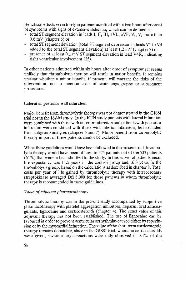
Beneficial effects seem likely in patients admitted within two hours after onset of symptoms with signs of extensive ischemia, which can be defined as: - total ST segment elevation in leads I, II, III, a VL, a VF, V5, V6 more than
0.6 mV (chapter 6) or - total ST segment deviation (total ST segment depression in leads V1 to V 4
added to the total ST segment elevation) at least 1.2 mV (chapter 7) or - presence of at least 0.1 mV ST segment elevation in lead V4R, indicating
right ventricular involvement (25).
In other patients admitted within six hours after onset of symptoms it seems unlikely that thrombolytic therapy will result in major benefit. It remains unclear whether a minor benefit, if present, will warrant the risks of the intervention, not to mention costs of acute angiography or subsequent procedures.
Lateral or posterior wall infarction
Major benefit from thrombolytic therapy was not demonstrated in the GISSI trial nor in the ISAM study. In the ICIN study patients with lateral infarction were combined with those with anterior infarction and patients with posterior infarction were combined with those with inferior infarction, but excluded from subgroup analyses (chapter 6 and 7). Minor benefit from thrombolytic therapy in part of these patients cannot be excluded.
When these guidelines would have been followed in the present trial thrombolytic therapy would have been offered to 325 patients out of the 533 patients (61%) that were in fact admitted to the study. In this subset of patients mean life expectancy was 14.5 years in the control group and 16.3 years in the thrombolysis group, based on the calculations as described in chapter 8. Total costs per year of life gained by thrombolytic therapy with intracoronary streptokinase averaged Dfl 5,000 for those patients in whom thrombolytic therapy is recommended in these guidelines.
Value of adjuvant pharmacotherapy
Thrombolytic therapy was in the present study accompanied by supportive pharmacotherapy with platelet aggregation inhibitors, heparin, oral anticoagulants, lignocaine and corticosteroids (chapter 4). The exact value of this adjuvant therapy has not been established. The use of lignocaine can be favoured in order to prevent ventricular arrhythmias caused either by reperfusion or by the myocardial infarction. The value of the short term corticosteroid therapy remains debatable, since in the GISSI trial, where no corticosteroids were given, severe allergic reactions were only observed in 0.1% of the
98
patients after treatment with streptokinase (10). The major problem lies in the prevention of reocclusion by pharmacotherapy. Even with the use of platelet aggregation inhibitors, heparin and oral anticoagulants a high incidence of reocclusion and reinfarction was observed in the present trial after successful thrombolysis, especially in patients with inferior infarction. Studies to define the optimal pharmacotherapeutic regimen to prevent reocclusion after successful thrombolysis are urgently needed. The applicability of thrombolytic agents in patients with inferior infarction will depend on the possibilities to prevent reocclusion in these patients.
Coronary angioplasty
Immediate coronary angioplasty after perforation of the thrombus in the infarct related coronary artery by a guidewire has been proposed by Rentrop et al. as an alternate approach to the use of thrombolytic therapy in patients with acute myocardial infarction (26). In a small study reported by O'Neill et al (24) patency of the infarct related artery was achieved in 24 out of 29 patients (83%) by this method (table 3). However, the inevitably longer treatment delay will restrict the applicability of this approach to patients in whom intravenous thrombolytic therapy is contraindicated. After successful or attempted thrombolysis coronary angioplasty may be of value in order to complete the reperfusion strategy. After successful thrombolysis severe stenosis may remain in the infarct related coronary artery. This artery may reocclude and abolish the beneficial effects of reperfusion. Indeed, reinfarction rates observed in patients with a residual stenosis of 70% or more after successful thrombolysis were as high as 30% during three years follow-up with most of the reinfarctions occurring within three months (chapter 9). The additional value of coronary angioplasty in those patients has been reported by others (27-30) and was also demonstrated in the present study. Coronary angioplasty appeared to be of value both in patients with anterior and with inferior infarction. In patients with anterior infarction reocclusion rates were relatively low (8% at second angiography), but reocclusion might lead to large anterior reinfarctions, while in patients with inferior infarction reocclusion rates were higher (28% at second angiography) but the area at risk was on average smaller than in patients with anterior infarction (chapter 9). Consequently, if acute angiography is not performed after attempted thrombolysis or when coronary angioplasty after successful thrombolysis is not feasible due to lack of surgical standby, coronary angiography should be carried out within the next days to assess the suitability for elective PTCA. When symptoms and signs of ischemia reappear this intervention should be carried out on a (semi-) urgent basis.
99
Recommendations
In patients with acute myocardial infarction in whom thrombolytic therapy can presently be recommended (table 4), the optimal approach begins with intravenous administration of streptokinase (500,000 U) as early as possible, usually immediately upon hospital admission, followed by acute angiography and intracoronary administration of streptokinase. Only when signs of reperfusion have appeared prior to angiography such as sudden relief of chest pain with marked decrease of ST segment elevation, can angiography be delayed. When after intracoronary thrombolysis a residual stenosis of 70% or more in the infarct related artery with poor run-off into the distal coronary artery bed is seen, immediate PTCA is recommended. When acute PTCA is not feasible or when acute angiography has not been performed following intravenous thrombolytic therapy, coronary angiography within the next days remains indicated to assess the suitability for elective PTCA.
Impact of these recommendations on health care
Following the guidelines presented before (table 4), thrombolytic therapy can be offered to 15 to 20% of all patients admitted to the hospital with acute myocardial infarction (31). Even in large hospitals in the Netherlands the number of patients treated with thrombolytic therapy will then not exceed 100 patients per hospital per year. When catheterisation laboratories are available on a 24 hour basis, total workload for their technical staff will only be increased to a minor extent. The costs for this increased workload were included in the cost benefit analysis (chapter 8) and the apparent benefits from thrombolytic therapy certainly warrant the additional costs (Dfl 5,000 per year of life gained) even in the present times when budgets available for health care are limited. However, many patients with acute myocardial infarction are admitted to community hospitals with limited or no possibilities for acute angiography. It is for this reason that Verstraete stated that intravenous thrombolytic therapy would be "the only way" (32). Furthermore, even if thrombolytic therapy would be restricted to intravenous administration in those hospitals, the number of subsequent catheterisations to be performed in adjacent large hospitals in order to assess the suitability for elective PTCA would lead to a major increase in workload in the catheterisation laboratories in those hospitals. The new generation of more effective thrombolytic agents (rt-PA, rscup A and APSAC) is currently being investigated in large clinical trials. Preliminary results have indicated that intravenous use of these agents does lead to successful thrombolysis in a higher percentage of patients than presently can be achieved with intravenous streptokinase (table 3). Part of these trials also adress the question whether PTCA is mandatory after successful thrombolysis, and whether this additional procedure should be performed immediately or can be deferred a few days. In our opinion the results of these trials will have
100
to be awaited before the required capacity of catheterisation laboratories can be estimated (see below).
Questions for the future
In this thesis the characteristics have been defined of those patients with acute myocardial infarction to whom early thrombolytic therapy would offer a major benefit. However, this concerns only 15 to 20% of all patients admitted to the hospital with acute myocardial infarction. It is still unclear whether early thrombolytic therapy will offer benefit to other patients with acute myocardial infarction. Presently, it seems unlikely that patients admitted to the hospital more than six hours after onset of symptoms will benefit from thrombolytic therapy, but effects of thrombolytic therapy should be investigated in other patients admitted within six hours after onset of symptoms than those in whom benefits were demonstrated (table 4). Especially patients admitted up to 24 hours after onset of symptoms with extensive ischemia (usually located anteriorly) or with signs of cardiogenic shock constitute a group of high risk patients who might derive major benefit from thrombolytic therapy (33). On the other hand, patients with less extensive ischemia admitted early after onset of symptoms and even patients admitted with unstable angina might also benefit from thrombolytic therapy, but in these low risk patients the possible benefits must be carefully weighted against the risks of the intervention in terms of major bleeding, stroke and consequences of eventual reocclusion. The indications for thrombolytic therapy, especially in patients with inferior infarction, will also depend on the possibilities to prevent reocclusion. If our observation that coronary angioplasty following successful thrombolysis and supported by pharmacotherapy effectively reduces reinfarction rate is confirmed in larger clinical trials (which are presently underway), more patients with acute myocardial infarction might be considered candidates for thrombolytic therapy.
It is likely that in the near future the new generation of thrombolytic agents (rt-PA, rsu-PA and APSAC) will become first choice for the intravenous initiation of thrombolytic therapy. Then, the indications for either acute or delayed angiography might be different from the present ones. Cost benefit analyses will be required to assess the additional value of intracoronary thrombolysis and coronary angioplasty after intravenous administration of rt-PA, rscu-PA or APSAC. The required capacity of catheterisation laboratories and facilities for coronary angioplasty and bypass surgery will depend on the results of those analyses. We expect that the guidelines presented in this thesis are applicable to all thrombolytic agents and that a stepwise approach including intravenous and possibly intracoronary treatment followed by coronary angioplasty in selected patients will prove to be the optimal strategy.
101
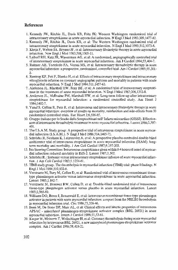
References
1. Kennedy JW, Ritchie JL, Davis KB, Fritz JK: Western Washington randomized trial of intracoronary streptokinase in acute myocardial infarction. N Eng! J Med 1983;309,1477-82.
2. Kennedy JW, Ritchie JL, Davis KB, et al: The Western Washington randomized trial of intracoronary streptokinase in acute myocardial infarction. N Eng! J Med 1985;312,1073-8.
3. Khaja F, Walton JA, Brymer JF, et al: Intracoronary fibrinolytic therapy in acute myocardial infarction. New Eng! J Med 1983;308,1305-11.
4. Leiboff RH, Katz JR, Wassermen AG, et al: A randomized, angiographically controlled trial of intracoronary streptokinase in acute myocardial infarction. Am J Cardiol1984;53,404-7.
5. Raizner AE, Tortoledo FA, Verani MS, et al: Intracoronary thrombolytic therapy in acute myocardial infarction: a prospective, randomized, controlled trial. Am J Cardio\1985;55,301-8.
6. Rentrop KP, Feit F, Blanke H, eta!: Effects of intracoronary streptokinase and intracoronary nitroglycerin infusion on coronary angiographic patterns and mortality in patients with acute myocardial infarction. N Eng! J Med 1984;311,1457-63.
7. Anderson JL, Marshall HW, Bray BE, et a!: A randomized trial of intra coronary streptokinase in the treatment of acute myocardial infarction. N Eng! J Med 1983;308,1312-8.
8. Anderson JL, Mcilvaine PM, Marshall HW, eta!: Long-term follow-up after intracoronary streptokinase for myocardial infarction: a randomized controlled study. Am Heart J 1984;1402-7.
9. Yusuf S, Collins R, Peto R, eta!: Intravenous and intracoronary fibrinolytic therapy in acute myocardial infarction: overview of results on mortality, reinfarction and side-effects from 33 randomized controlled trials. Eur Heart 1;6,556-85.
10. Gruppo italiano per lo Studio della Streptochinasi nell' Infarto miocardico ( GISSI). Effectiveness of intravenous thrombolytic treatment in acute myocardial infarction. Lancet 1986;2,397-401.
11. The I.S.A.M. Study group: A prospective trial of intravenous streptokinase in acute myocardial infarction (I.S.A.M.). N Engl J Med 1986;314,1465-71.
12. Schroder R, Neuhaus K, Leizorovicz A, et al: A prospective placebo-controlled double-blind multicenter trial of intravenous streptokinase in acute myocardial infarction (ISAM): longterm mortality and morbidity. JAm Col! Cardio\1987;9,197-203.
13. Isis Steering Committee: Intravenous streptokinase given within 0-4 hours of onset of myocardial infarction reduced mortality in ISIS-2. Lancet 1987;1,502.
14. Schroder R.: Systemic versus intracoronary streptokinase infusion of acute myocardial infarction. JAm Col! Cardiol1983;1,1254-61.
15. TIM! study group. The thrombolysis in myocardial infarction (TIM!) trial: phase I findings. N Eng! J Med 1984;310,932-6.
16. Verstraete M, Bory M, Collen D, et al: Randomised trial of intravenous recombinant tissuetype plasminogen activator versus intravenous streptokinase in acute myocardial infarction. Lancet 1985;1,842-7.
17. Verstraete M, Brouwer RW, Collen D, et al: Double-blind randomised trial of intravenous tissue-type plasminogen activator versus placebo in acute myocardial infarction. Lancet 1985;2,965-69.
18. Williams DO, Borer J, Braunwald E, eta!: Intravenous recombinant tissue-type plasminogen activator in patients with acute myocardial infarction: a report from the NHLBI thrombolysis in myocardial infarction trial. Circ 1986;73,338-46.
19. Been M, De Bono DP, Muir AL, eta!: Clinical effects and kinetic properties of intravenous APSAC - anisoylated plasminogen-streptokinase activator complex (BRL 26921) in acute myocardial infarction. Intern J Cardiol1986;11,53-61.
20. Kaspar W, Meinertz T, Wollschlager H, et al: Coronary thrombolysis during acute myocardial infarction by intravenous BRL 26921, a new anisoylated plasminogen-streptokinase activator complex. Am J Cardiol1986;58,418-21.
102
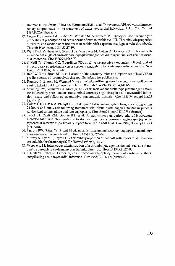
21. Bonnier JJRM, Swart JBRM de, Hoffmann JJML, eta!: Intravenous APSAC versus intracoronary streptokinase in the treatment of acute myocardial infarction. J Am Col! Cardiol 1987;9,62A(abstract).
22. Collen D, Stassen JM, Blaber M, Winkler M, Verstraete M.: Biological and thrombolytic properties of proenzyme and active forms of human urokinase - III. Thrombolytic properties of natural and recombinant urokinase in rabbits with experimental jugular vein thrombosis. Thromb Haemostas 1984;52,27-30.
23. Werf F vd, V anhaecke J, Geest H de, Verstraete M, Collen D.: Coronary thrombolysis with recombinant single-chain urokinase-type plasminogen activator in patients with acute myocardial infarction. Circ 1986;74,1066-70.
24. O'Neill W, Timmis GC, Bourdillon PD, et a!: A prospective randomized clinical trial of intracoronary streptokinase versus coronary angioplasty for acute myocardial infarction. New Eng! J Med 1986;314,812-8.
25. Bar FW, Res J, Braat SH, eta!: Location ofthe coronary lesion and importance oflead V 4 R to predict success of thrombolytic therapy. Submitted for publication.
26. Rentrop P, Blanke H, Wiegand V, eta!: Wiedereriiffnung verschlossener Kranzgefasse im akuten Infarkt mit Hilfe von Kathetem. Dtsch Med Wschr 1979;104,1401-5.
27. Smalling RW, Nishikawa A, Merhige ME, eta!: Intravenous tissue-type plasminogen activator followed by percutaneous transluminal coronary angioplasty in acute myocardial infarction: acute and follow-up quantitative angiographic analysis. Circ 1986;74 (suppl II),23 (abstract).
28. Collins GJ, Califf RM, Phillips HR, eta!: Quantitative angiographic changes occurring within 24 hours and one week following treatment with tissue plasminogen activator in patients randomized to immediate and late angioplasty. Circ 1986;74 (suppl II),275 (abstract).
29. Topol EJ, Califf RM, George BS, et a!: A multicenter randomized trial of intravenous recombinant tissue plasminogen activator and emergency coronary angioplasty for acute myocardial infarction: preliminary report from the TAMI trial. Circ 1986;74 (suppl II),23 (abstract).
30. Serruys PW, Wijns W, Brand M vd, eta!: Is transluminal coronary angioplasty mandatory after successful thrombolysis? Br Heart J 1983;50,257-65.
31. Murray N, Lyons J, Layton C, et a!: What proportion of patients with myocardial infarction are suitable for thrombolysis? Br Heart J 1987;57,144-7.
32. Verstraete M: Intravenous administration of a thrombolytic agent is the only realistic therapeutic approach in evolving myocardial infarction. Eur Heart J 1985;6,586-93.
33. O'Neill W, Erbel R, Laufer N, et a!: Coronary angioplasty therapy of cardiogenic shock complicating acute myocardial infarction. Circ 1985;72,III-309 (abstract).
103
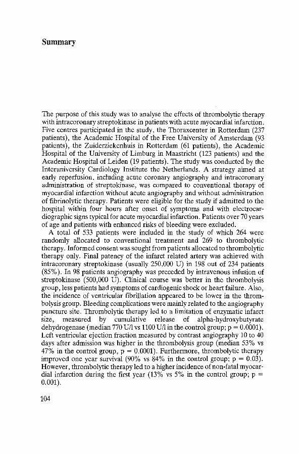
Summary
The purpose of this study was to analyse the effects of thrombolytic therapy with intracoronary streptokinase in patients with acute myocardial infarction. Five centres participated in the study, the Thorax center in Rotterdam (237 patients), the Academic Hospital of the Free University of Amsterdam (93 patients), the Zuiderziekenhuis in Rotterdam (61 patients), the Academic Hospital of the University of Limburg in Maastricht (123 patients) and the Academic Hospital of Leiden (19 patients). The study was conducted by the Interuniversity Cardiology Institute the Netherlands. A strategy aimed at early reperfusion, including acute coronary angiography and intracoronary administration of streptokinase, was compared to conventional therapy of myocardial infarction without acute angiography and without administration of fibrinolytic therapy. Patients were eligible for the study if admitted to the hospital within four hours after onset of symptoms and with electrocardiographic signs typical for acute myocardial infarction. Patients over 70 years of age and patients with enhanced risks of bleeding were excluded.
A total of 533 patients were included in the study of which 264 were randomly allocated to conventional treatment and 269 to thrombolytic therapy. Informed consent was sought from patients allocated to thrombolytic therapy only. Final patency of the infarct related artery was achieved with intracoronary streptokinase (usually 250,000 U) in 198 out of 234 patients (85% ). In 98 patients angiography was preceded by intravenous infusion of streptokinase (500,000 U). Clinical course was better in the thrombolysis group, less patients had symptoms of cardiogenic shock or heart failure. Also, the incidence of ventricular fibrillation appeared to be lower in the thrombolysis group. Bleeding complications were mainly related to the angiography puncture site. Thrombolytic therapy led to a limitation of enzymatic infarct size, measured by cumulative release of alpha-hydroxybutyrate dehydrogenase (median 770 U/1 vs 1100 U/1 in the control group; p = 0.0001). Left ventricular ejection fraction measured by contrast angiography 10 to 40 days after admission was higher in the thrombolysis group (median 53% vs 47% in the control group, p = 0.0001). Furthermore, thrombolytic therapy improved one year survival (90% vs 84% in the control group; p = 0.03). However, thrombolytic therapy led to a higher incidence of non-fatal myocardial infarction during the first year (13% vs 5% in the control group; p = 0.001).
104
In multivariate regression analysis it was observed that beneficial effects of thrombolytic therapy were related to the delay between onset of symptoms and hospital admission, to the extent of myocardial ischemia, and to the development of new Q waves on the admission ECG. Patients with extensive ischemia, admitted to the hospital within 2 hours after onset of symptoms, derived most benefit from thrombolytic therapy.
In a cost benefit analysis mortality and morbidity during one year follow-up were related to the costs of the procedure, of hospital admissions, and of additional interventions. In patients with inferior infarction thrombolytic therapy improved survival and quality oflife only to a minor extent. Total costs the first year averaged Dfl 20,000 for patients in the control group, and Dfl 27,000 for patients allocated to thrombolytic therapy. Total costs per year of life gained by thrombolytic therapy were D£110,000. In patients with anterior infarction thrombolytic therapy improved one year survival (89% vs 78% in the control group; p = 0.01) and improved quality of life during the first year follow-up, in that on average more time was spent without symptoms of angina pectoris or heart failure. Total costs averaged Dfl 28,000 in the thrombolysis group and D£121,000 in the control group. Total costs per year oflife gained by thrombolytic therapy therefore were Dfl3 ,800. The subgroup of patients with anterior infarction with extensive ischemia, admitted within 2 hours after onset of symptoms derived the largest benefit from thrombolytic therapy and total costs per year of life gained were only D£11,900.
The additional value of PTCA after successful thrombolysis was assessed in a matched pair analysis. Patients in whom PTCA was carried out as part of the recanalisation procedure were compared to patients with successful thrombolysis admitted to those hospitals where acute PTCA was not performed. PTCA performed immediately after successful thrombolysis additionally improved quality of life during three years follow-up, in that on average 128 weeks (out of 156 weeks) were spent without symptoms of angina pectoris or heart failure in the matched PTCA group and 102 weeks in a matched group of patients with successfull thrombolysis only (p = 0.03).
Thrombolytic therapy can presently be recommended in patients with anterior infarction admitted to the hospital within two hours after onset of symptoms and in patients with a large ischemic area as reflected by total ST segment elevation of 1.2 m V or more on the admission electrocardiogram, admitted two to four hours after onset of symptoms. In patients with inferior infarction beneficial effects of thrombolytic therapy seem only likely in patients admitted within two hours after onset of symptoms with signs of extensive ischemia on the admission electrocardiogram. It remains presently unclear whether early thrombolytic therapy will result in (minor) benefit in other patients with acute myocardial infarction.
The optimal approach begins with intravenous administration of streptokinase (500,000 U) followed by acute angiography and intracoronary administration of streptokinase (250,000 U), unless signs of reperfusion have
105
appeared prior to angiography. When after successful thrombolysis a residual stenosis of70% or more in the infarct related coronary artery with poor run-off into the distal coronary artery bed is seen, immediate PTCA is recommended. If acute angiography was not performed after thrombolysis or acute PTCA was not feasible, angiography should be carried out within the first days following successful thrombolysis to assess suitability for elective PTCA.
We expect this stepwise approach in the treatment of patients with acute myocardial infarction to be applicable to all thrombolytic agents, although the newer more effective thrombolytic agents may reduce the need for acute angiography and early PTCA. Large randomised clinical trials are presently underway, both in Europe and in the United States, to assess the efficacy of rt-P A, rscu-P A and APSAC in the treatment of patients with acute myocardial infarction and to determine the additional value of coronary angioplasty, either performed immediately or within the next days following successful thrombolysis.
106
Samenvatting
Deze studie had ten doel de effecten van een behandeling met intracoronair toegediende streptokinase bij patienten met een acuut hartinfarct te analyseren. Vijf ziekenhuizen namen aan deze studie deel, namelijk het Thoraxcentrum in Rotterdam (237 patienten), het Academisch ziekenhuis van de Vrije Universiteit van Amsterdam (93 patienten), het Zuiderziekenhuis in Rotterdam (61 patienten), het Academisch Ziekenhuis in Maastricht (123 patienten) en het Academisch Ziekenhuis Leiden (19 patienten). De studie werd verricht onder auspicien van het Interuniversitair Cardiologisch Instituut Nederland. Een behandeling gericht op vroegtijdige reperfusie met behulp van coronairangiografie en intracoronaire toediening van streptokinase werd vergeleken met een normale behandeling van patienten met een hartinfarct, dat wil zeggen een behandeling zonder acute angiografie en zonder toediening van fibrinolytica. Patienten kwamen aileen in aanmerking voor de studie indien zij werden opgenomen in het ziekenhuis binnen vier uur na het begin van de klachten. Het electrocardiogram moest duidelijk aangeven dater sprake was van een acuut hartinfarct. Patienten ouder dan 70 jaar en patienten met een verhoogd risico op bloedingen werden van deelname aan de studie uitgesloten.
In totaal werden 533 patienten in de studie opgenomen. Tweehonderdenvierenzestig patienten werden door randomisatie toegewezen aan de controlegroep, 269 aan de trombolytische behandeling. Toestemming voor deelname aan de studie werd aileen gevraagd aan patienten die waren toegewezen aan de behandeling met trombolytica. De aangedane coronairarterie was na intracoronaire toediening van streptokinase (meestal 250,000 E) doorgankelijk bij 198 van de 234 patienten (85%). Van deze 234 patienten hadden er 98 tevoren ook streptokinase intraveneus toegediend gekregen (500,000 E). Het klinische be loop van de patienten in de trombolysegroep was gunstiger dan van die in de controlegroep; met name kregen minder patienten in de trombolysegroep tekenen van decompensatio cordis of cardiogene shock. Ook trad ventrikelfibrilleren minder vaak op in de trombolysegroep. Bloedingen traden vrijwel aileen op random de insteekplaats van de catheter. De behandeling met streptokinase leidde tot een beperking van de enzymatische infarctgrootte, gemeten door mid del van de cumulatieve release van alpha-hydroxybutyraat dehydrogenase. De mediane release was 770 E/1 in de trombolyse-
107
groep en 1100 E/1 in de controlegroep (p = 0,0001). De linkerventrikel ejectiefractie (gemeten met behulp van contrastangio
grafie 10 tot 40 dagen na opname) was hoger in de trombolysegroep (mediaan 53% tegenover 47% in de controlegroep; p = 0,0001). De eenjaarsoverleving was ook beter in de trombolysegroep (90% tegenover 84% in de controlegroep; p = 0,03). De behandeling met streptokinase leidde wel tot een toename van het aantal recidief infarcten gedurende het eerste jaar (13% tegenover 5% in de controlegroep; p = 0,001).
De resultaten van multivariate regressieanalyse gaven aan dat de effecten van de behandeling met streptokinase afhingen van het tijdsverloop tussen het begin van de klachten en opname in het ziekenhuis, van de uitgebreidheid van de ischaemie en van het ontstaan van nieuwe Q golven op het electrocardiogram. Patienten met uitgebreide ischaemie, opgenomen binnen twee uur na het begin van de klachten, hadden de meeste baat bij de behandeling met streptokinase.
In een kostenbatenanalyse werden de effecten op de mortaliteit en de morbiditeit gerelateerd aan de kosten van de interventie, van de ziekenhuisopnames en van latere interventies. Bij patienten met een onderwandinfarct waren de gunstige effecten van de behandeling met streptokinase op de mortaliteit en de kwaliteit van het I even gering. De totale kosten bedroegen in het eerste jaar Fl20.000 voor de patienten in de controlegroep en Fl27.000 voor de patienten in de trombolysegroep. De extra kosten per gewonnen levensjaar waren gerniddeld Fl10.000. Bij patienten met een voorwandinfarct was de mortaliteit wel duidelijk lager in de trombolysegroep (11% tegenover 22% in de controlegroep; p = 0,01) en was ookde kwaliteitvanhetleven beter in de trombolysegroep in die zin, dat gemiddeld meer dagen werden doorgebracht zonder klachten van angina pectoris of decompensatio cordis. De totale kosten bedroegen Fl28. 000 in de trombolysegroep en Fl21. 000 in de controlegroep. De extra kosten per gewonnen levensj aar war en gemiddeld Fl3. 800. In de subgroep van patienten met uitgebreide ischaernie, opgenomen binnen twee uur na het begin van de klachten, werd het grootste voordeel van de behandeling met streptokinase gezien. In deze groep bedroegen de kosten per gewonnen levensjaar slechts Fll.900.
De effecten van PTCA na trombolyse werden geanalyseerd in een "matched pair" analyse. Patienten die een PTCA hadden ondergaan direct in aansluiting aan de behandeling met streptokinase werden vergeleken met patienten die met succes waren behandeld met streptokinase en die waren opgenomen in die ziekenhuizen waar geen acute PTCA werd verricht. PTCA, in aansluiting aan de behandeling met streptokinase, verbeterde de kwaliteit van het leven in die zin dat gerniddeld 128 weken (van de 156 weken) werden doorgebracht zonder klachten van angina pectoris of decompensatio cordis, tegen gerniddeld 102 we ken in de groep patienten die alleen met streptokinase was behandeld (p = 0,03).
Een behandeling met trombolytica kan thans worden geadviseerd bij pa-
108
tienten met een voorwandinfarct die zijn opgenomen in het ziekenhuis binnen twee uur na het begin van de klachten en bij patienten met uitgebreide ischaemie ( te zien op het electrocardiogram als een som van de ST eleva tie van minimaal 1,2 m V), opgenomen twee tot vier uur na het be ginn en van de klachten. Bij patienten met een onderwandinfarct zijn waarschijnlijk aileen gunstige effecten van een behandeling met trombolytica te verwachten bij patienten met uitgebreide ischaemie, opgenomen binnen twee uur na het begin van de klachten. Thans is het nog onduidelijk of andere patienten met een acuut hartinfarct mogelijk ook baat hebben bij deze behandeling.
Het volgende behandelingsschema wordt voorgesteld ten aanzien van het gebruik van trombolytica bij patienten met een acuut hartinfarct. De behandeling begint met intraveneuze toediening van 500.000 eenheden streptokinase, direct gevolgd door hartcatheterisatie en intracoronaire toediening van 250.000 eenheden streptokinase, tenzij tekenen van reperfusie zijn opgetreden voor het begin van de hartcatheterisatie. Wanneer na trombolyse een stenose van 70% of meer wordt gezien in de aangedane coronairarterie, bestaat er een indicatie voor acute PTCA. Indien acute catheterisatie na een trombolytische behandeling niet heeft plaatsgevonden of indien het niet mogelijk was een acute PTCA te verrichten, dient, indien mogelijk, angiografie plaats te vinden binnen enkele dagen na de trombolytische behandeling ten einde vast te stellen of er een indicatie bestaat voor electieve PTCA.
Deze stapsgewijze benadering van de trombolytische behandeling van patienten met een hartinfarct lijkt ons ook toepasbaar bij een behandeling met andere trombolytica, al zullen acute angiografie en PTCA wellicht minder vaak geindiceerd blijken te zijn na intraveneuze toediening van de nieuwe, effectievere trombolytica (rt-PA, rscu-PA en APSAC). De effectiviteit van deze middelen wordt thans onderzocht in grote gerandomiseerde studies, zowel in Europa als in de Verenigde Staten, waarbij tevens de waarde wordt bepaald van coronairangioplastiek, verricht direct in aansluiting aan een succesvolle behandeling met trombolytica dan wei binnen enkele dagen na deze behandeling.
109
Curriculum vitae
Frank Vermeer werd geboren op 3 april1952 te Rotterdam. In 1969 behaalde hij het eindexamen Gymnasium-B aan het Gymnasium Erasmianum te Rotterdam. De studie geneeskunde aan de Rijksuniversiteit te Leiden werd in 1977 afgesloten met het behalen van het artsexamen. Van 1977 tot 1978 was hij werkzaam als algemeen assistent geneeskundige in het ziekenhuis Bethlehem te Den Haag. In 1978 werd begonnen met de opleiding interne geneeskunde in het Diaconessenhuis te Voorburg, gevolgd door de opleiding cardiologie in het ziekenhuis Leyenburg te Den Haag. In 1983 volgde inschrijving in het specialistenregister als cardioloog. Van 1984 tot 1986 was hij als wetenschappelijk medewerker verbonden aan de Erasmus Universiteit te Rotterdam, aangesteld door het Interuniversitair Cardiologisch Instituut Nederland. Sinds 1986 is hij werkzaam als cardioloog in het Academisch Ziekenhuis Maastricht.
110