Thomas, F., Dawson, W., Lang, E., Burton, A., Bartlett, G ... · hpphppp, often denoted abcdefg....
10
Thomas, F., Dawson, W., Lang, E., Burton, A., Bartlett, G., Rhys, G., ... Woolfson, D. (2018). De Novo-Designed -Helical Barrels as Receptors for Small Molecules. ACS Synthetic Biology, 7(7), 1808-1816. https://doi.org/10.1021/acssynbio.8b00225 Peer reviewed version Link to published version (if available): 10.1021/acssynbio.8b00225 Link to publication record in Explore Bristol Research PDF-document This is the author accepted manuscript (AAM). The final published version (version of record) is available online via ACS at https://pubs.acs.org/doi/10.1021/acssynbio.8b00225 . Please refer to any applicable terms of use of the publisher. University of Bristol - Explore Bristol Research General rights This document is made available in accordance with publisher policies. Please cite only the published version using the reference above. Full terms of use are available: http://www.bristol.ac.uk/pure/about/ebr-terms
Transcript of Thomas, F., Dawson, W., Lang, E., Burton, A., Bartlett, G ... · hpphppp, often denoted abcdefg....
Thomas, F., Dawson, W., Lang, E., Burton, A., Bartlett, G., Rhys, G., ...Woolfson, D. (2018). De Novo-Designed -Helical Barrels as Receptors forSmall Molecules. ACS Synthetic Biology, 7(7), 1808-1816.https://doi.org/10.1021/acssynbio.8b00225
Peer reviewed version
Link to published version (if available):10.1021/acssynbio.8b00225
Link to publication record in Explore Bristol ResearchPDF-document
This is the author accepted manuscript (AAM). The final published version (version of record) is available onlinevia ACS at https://pubs.acs.org/doi/10.1021/acssynbio.8b00225 . Please refer to any applicable terms of use ofthe publisher.
University of Bristol - Explore Bristol ResearchGeneral rights
This document is made available in accordance with publisher policies. Please cite only the publishedversion using the reference above. Full terms of use are available:http://www.bristol.ac.uk/pure/about/ebr-terms
De novo designed α-helical barrels as receptors for small molecules
Franziska Thomas,a,b‡ William M. Dawson,a‡ Eric J.M. Lang,a,c Antony J. Burton,a,d Gail J. Bartlett,a Guto
G. Rhys,a Adrian J. Mulholland,a,c,e and Derek N. Woolfsona,c,f*.
aSchool of Chemistry, University of Bristol, Cantock’s Close, Bristol, BS8 1TS, UK.
bInstitute of Organic and Biomolecular Chemistry, Georg-August-Universität Göttingen, Tammannstr. 2, 37077 Göttingen,
Germany.
cBrisSynBio, University of Bristol, Life Sciences Building, Tyndall Avenue, Bristol, BS8 1TQ, UK.
dFrick Chemistry Laboratory, Princeton, NJ 084544, USA.
eCentre for Computational Chemistry, School of Chemistry, University of Bristol, Cantock’s Close, Bristol, BS8 1TS, UK. fSchool of Biochemistry, University of Bristol, Biomedical Sciences Building, University Walk, Brisol, BS8 1TD, UK
ABSTRACT: We describe de novo designed -helical barrels (HBs) that bind and discriminate between lipophilic biologically
active molecules. HBs have five or more α helices arranged around central hydrophobic channels the diameters of which scale with
oligomer state. We show that pentameric, hexameric and heptameric HBs bind the environmentally sensitive dye, 1,6-diphenyl-
hexatriene (DPH) in the M range and fluoresce. Displacement of the dye is used to report the binding of non-fluorescent molecules:
palmitic acid and retinol bind to all three HBs with sub-M inhibitor constants; farnesol binds the hexamer and heptamer; but -
carotene only binds to the heptamer. A co-crystal structure of the hexamer with farnesol reveals oriented binding in the centre of the
hydrophobic channel. Charged side chains engineered into the lumen of the heptamer facilitate binding of polar ligands: a glutamate
variant binds a cationic variant of DPH; and introducing lysine allows binding of the biosynthetically important farnesol diphosphate.
INTRODUCTION
Protein design has advanced sufficiently that it is now possible
to generate successfully a variety of stable protein structures
from first principles.1-2 This can be done using rules of thumb
that relate protein sequence to structure or by employing com-
putational methods.3-5 New challenges for this field of de novo
protein design include: (1) taking forays into the so-called dark
matter of protein space, i.e., designing entirely new protein
structures not observed in nature;6-7 (2) making the design meth-
ods open and accessible to others, particularly to non-specialist
users;8 and (3) building on these scaffolds and methods to de-
liver functional de novo proteins.9-10 Desired functions include
biomolecular recognition and sensing, and catalysis or enzyme-
like activities.11-12 A requirement for many such functions is
that the designed protein scaffolds bind and discriminate be-
tween various molecules, e.g., bioactive small molecules and
biological macromolecules.
In biological systems, protein-ligand dissociation constants
range from mM to fM.13 Moreover, many receptors respond to
multiple ligands across a range of binding affinities. One ad-
vantage of the one receptor-multiple ligand model is that it re-
moves the need to synthesize a specific receptor for every pos-
sible protein-ligand interaction. The olfactory network pro-
vides a natural example of such a system.14 In humans, genes
encoding circa 400 G-protein coupled receptors are able to dis-
tinguish up to 1 trillion different stimuli through low affinity
and relatively non-selective recognition of odorants.15
The design and engineering of new proteins that recognize and
bind peptides and folded proteins has achieved considerable
success, particularly using directed evolution to modify either
natural protein folds or de novo scaffolds generated by consen-
sus design.4, 12 However, the recognition and binding of small
molecules has proven more challenging.16-18 Two related issues
here are the generation of scaffolds that act as good receptors
for small molecules, and then the embellishment of these to dis-
criminate between what are often very similar molecules. The
small sizes and limited functional groups of the target molecules
often confound attempts to recognize, bind and distinguish
them. One solution is to adapt natural proteins with cavities
already evolved to bind small-molecule ligands.19-20 Here, we
add de novo proteins with central accessible channels to this
repertoire of small-molecule-binding proteins. These can be
adapted for the recognition, binding and release of ligands.
Our designs are based on de novo -helical coiled coils. The
vast majority of coiled coils are bundles with 2 – 4 helices
wrapped around a central superhelical axis leading to solid hy-
drophobic cores.21 These structures are directed by underlying
sequence repeats of hydrophobic (h) and polar (p) residues,
hpphppp, often denoted abcdefg. The 3,4-hydrophobic repeats
drive the folding of amphipathic helices, which assemble into
bundles to bury their hydrophobic, a+d faces. The helices
supercoil around each other because the 3.5-residue sequence
repeat and the 3.6-residue structural repeat do not match pre-
cisely. These relatively straightforward structural principles,
and the sequence-to-structure relationships that have followed,
have made coiled coils attractive targets for protein design.3
This has resulted in myriad de novo coiled-coil homo-dimers,
trimers and tetramers and reliable sets of de novo hetero-dimeric
and trimeric systems.22
Apart from a small number of examples,23-24 however, these
traditional coiled-coil dimers – tetramers do not have suitable
internal cavities or central channels to provide a basis for ligand
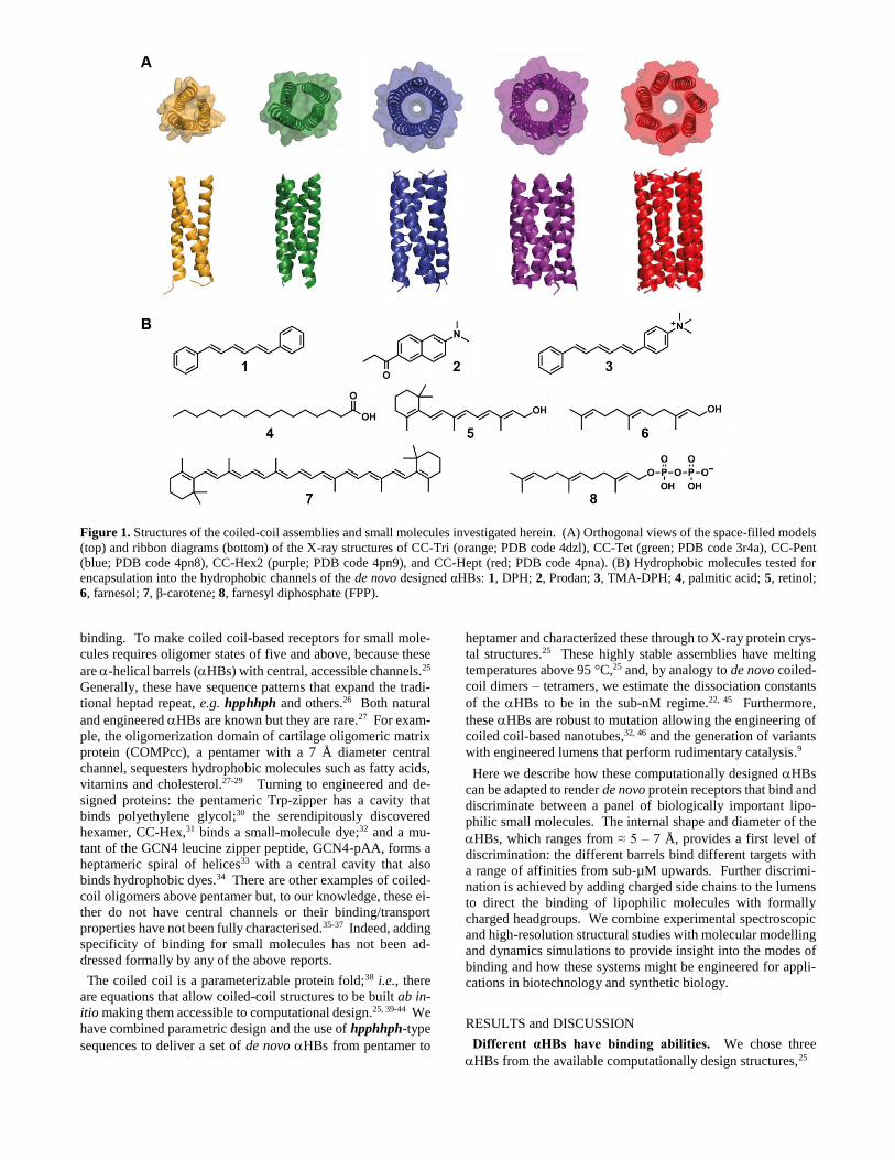
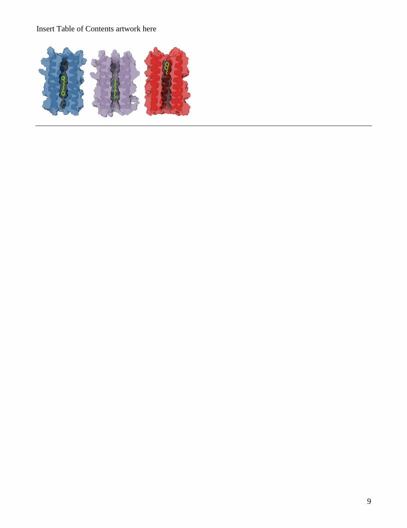
Figure 1. Structures of the coiled-coil assemblies and small molecules investigated herein. (A) Orthogonal views of the space-filled models
(top) and ribbon diagrams (bottom) of the X-ray structures of CC-Tri (orange; PDB code 4dzl), CC-Tet (green; PDB code 3r4a), CC-Pent
(blue; PDB code 4pn8), CC-Hex2 (purple; PDB code 4pn9), and CC-Hept (red; PDB code 4pna). (B) Hydrophobic molecules tested for
encapsulation into the hydrophobic channels of the de novo designed αHBs: 1, DPH; 2, Prodan; 3, TMA-DPH; 4, palmitic acid; 5, retinol;
6, farnesol; 7, β-carotene; 8, farnesyl diphosphate (FPP).
binding. To make coiled coil-based receptors for small mole-
cules requires oligomer states of five and above, because these
are -helical barrels (HBs) with central, accessible channels.25
Generally, these have sequence patterns that expand the tradi-
tional heptad repeat, e.g. hpphhph and others.26 Both natural
and engineered HBs are known but they are rare.27 For exam-
ple, the oligomerization domain of cartilage oligomeric matrix
protein (COMPcc), a pentamer with a 7 Å diameter central
channel, sequesters hydrophobic molecules such as fatty acids,
vitamins and cholesterol.27-29 Turning to engineered and de-
signed proteins: the pentameric Trp-zipper has a cavity that
binds polyethylene glycol;30 the serendipitously discovered
hexamer, CC-Hex,31 binds a small-molecule dye;32 and a mu-
tant of the GCN4 leucine zipper peptide, GCN4-pAA, forms a
heptameric spiral of helices33 with a central cavity that also
binds hydrophobic dyes.34 There are other examples of coiled-
coil oligomers above pentamer but, to our knowledge, these ei-
ther do not have central channels or their binding/transport
properties have not been fully characterised.35-37 Indeed, adding
specificity of binding for small molecules has not been ad-
dressed formally by any of the above reports.
The coiled coil is a parameterizable protein fold;38 i.e., there
are equations that allow coiled-coil structures to be built ab in-
itio making them accessible to computational design.25, 39-44 We
have combined parametric design and the use of hpphhph-type
sequences to deliver a set of de novo HBs from pentamer to
heptamer and characterized these through to X-ray protein crys-
tal structures.25 These highly stable assemblies have melting
temperatures above 95 °C,25 and, by analogy to de novo coiled-
coil dimers – tetramers, we estimate the dissociation constants
of the HBs to be in the sub-nM regime.22, 45 Furthermore,
these HBs are robust to mutation allowing the engineering of
coiled coil-based nanotubes,32, 46 and the generation of variants
with engineered lumens that perform rudimentary catalysis.9
Here we describe how these computationally designed HBs
can be adapted to render de novo protein receptors that bind and
discriminate between a panel of biologically important lipo-
philic small molecules. The internal shape and diameter of the
HBs, which ranges from ≈ 5 – 7 Å, provides a first level of
discrimination: the different barrels bind different targets with
a range of affinities from sub-µM upwards. Further discrimi-
nation is achieved by adding charged side chains to the lumens
to direct the binding of lipophilic molecules with formally
charged headgroups. We combine experimental spectroscopic
and high-resolution structural studies with molecular modelling
and dynamics simulations to provide insight into the modes of
binding and how these systems might be engineered for appli-
cations in biotechnology and synthetic biology.
RESULTS and DISCUSSION
Different αHBs have binding abilities. We chose three
HBs from the available computationally design structures,25
3
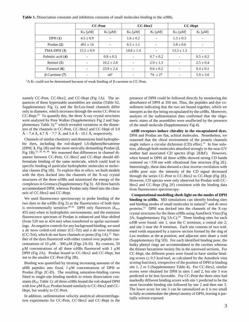
Table 1. Dissociation constants and inhibition constants of small molecules binding to the HBs.
CC-Pent CC-Hex2 CC-Hept
KD [μM] KI [μM] KD [μM] KI [μM] KD [μM] KI [μM]
DPH (1) 4.5 ± 0.9 - 1.6 ± 0.2 - 1.3 ± 0.3 -
Prodan (2) 401 ± 14 - 6.5 ± 1.1 - 5.8 ± 0.6 -
TMA-DPH (3) 15.5 ± 0.9 - 14.8 ± 1.4 - 13.3 ± 1.3 -
Palmitic acid (4) - 0.8 ± 0.3 - 0.7 ± 0.2 - 0.5 ± 0.2
Retinol (5) - 10.2 ± 2.8 - 2.9 ± 1.3 - 2.5 ± 0.4
Farnesol (6) - 23.9 ± 2.4 - 0.6 ± 0.2 - 0.4 ± 0.1
β-Carotene (7) - nd1 - 74 ± 27 - 5.9 ± 3.0
1A KI could not be determined because of weak binding of β-carotene to CC-Pent.
namely CC-Pent, CC-Hex2, and CC-Hept (Fig 1A). The se-
quences of these hyperstable assemblies are similar (Table S2,
Supplementary Fig 1), and the Ile/Leu-lined channels differ
only in diameter, which increases through the series CC-Pent to
CC-Hept.25 To quantify this, the three X-ray crystal structures
were analyzed by Pore Walker (Supplementary Fig 2 and Sup-
plementary Table 3),47 which revealed variations in the diame-
ters of the channels in CC-Pent, CC-Hex2 and CC-Hept of 3.0
Å – 7.4 Å, 4.7 Å – 7.7 Å, and 5.4 Å - 10.1 Å, respectively.
Channels of similar chemistry and dimensions bind hydropho-
bic dyes, including the rod-shaped 1,6-diphenylhexatriene
(DPH; 1, Fig 1B) and the more sterically demanding Prodan (2,
Fig 1B).32, 34, 46, 48 We reasoned that differences in channel di-
ameter between CC-Pent, CC-Hex2 and CC-Hept should dif-
ferentiate binding of the same molecule, which could lead to
specific binding of particular hydrophobic molecules or molec-
ular classes (Fig 1B). To explore this in silico, we built models
with the dyes docked into the channels of the X-ray crystal
structures of the three HBs and minimized the energy of the
complexes in Gromacs (Supplementary Fig 3). All three barrels
accommodated DPH, whereas Prodan only fitted into the chan-
nels of CC-Hex2 and CC-Hept.
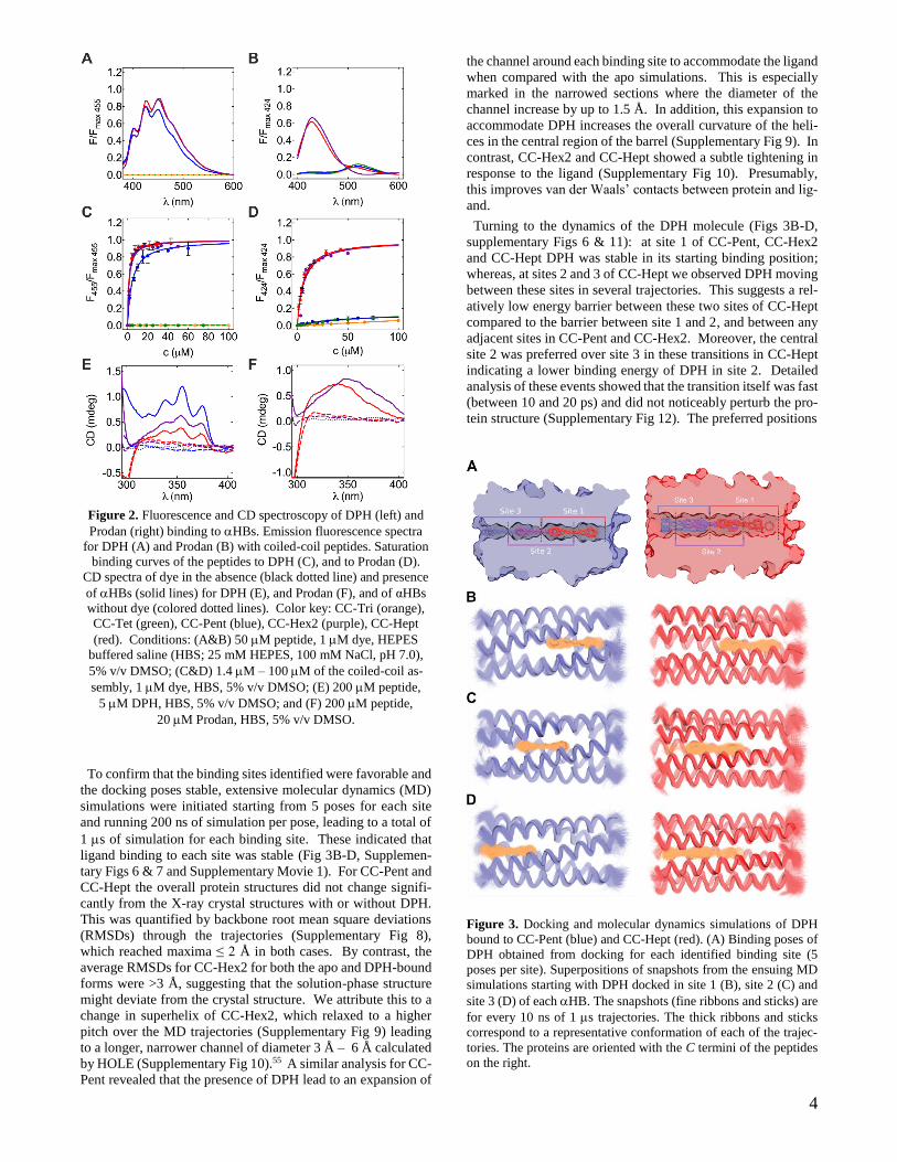
We used fluorescence spectroscopy to probe binding of the
two dyes to the HBs (Fig 2) as the fluorescence of both dyes
is environmentally sensitive:49-50 DPH only fluoresces (λmax
455 nm) when in hydrophobic environments; and the emission
fluorescence spectrum of Prodan is enhanced and blue-shifted
(from 520 nm to 424 nm) when transferred to apolar surround-
ings. As negative controls for any background binding, we used
a de novo coiled-coil trimer (CC-Tri) and a de novo tetramer
(CC-Tet), which do not have channels or pores (Fig 1A).22 Nei-
ther of the dyes fluoresced with either control over peptide con-
centrations of 10 M – 300 M (Figs 2A-D). By contrast, 50
M concentrations of all three HBs fluoresced with 1 M
DPH (Fig 2A). Prodan bound to CC-Hex2 and CC-Hept, but
not to the smaller CC-Pent (Fig 2B).
Binding was quantified by titrating increasing amounts of the
αHB peptides into fixed, 1 M concentrations of DPH or
Prodan (Figs 2C-D). The resulting saturation-binding curves
fitted to single-site binding models to return dissociation con-
stants (KD; Table 1): all three HBs bound the rod-shaped DPH
with low µM KDs; Prodan bound similarly to CC-Hex2 and CC-
Hept, but weakly to CC-Pent.
In addition, sedimentation velocity analytical ultracentrifuga-
tion experiments for CC-Pent, CC-Hex2 and CC-Hept in the
presence of DPH could be followed directly by monitoring the
absorbance of DPH at 350 nm. Thus, the peptides and dye co-
sediment indicating that the two are bound together, which we
interpret as the dye being encapsulated by the HBs. Moreover,
analysis of the sedimentation data confirmed that the oligo-
meric states of the assemblies were unaffected by the presence
of the small molecule (Supplementary Fig 4).
HB receptors induce chirality in the encapsulated dyes.
DPH and Prodan are flat, achiral molecules. Nonetheless, we
reasoned that the chiral environment of the protein channels
might induce a circular dichroism (CD) effect.51 In free solu-
tion, although both molecules absorbed strongly in the near-UV
neither had associated CD spectra (Figs 2E&F). However,
when bound to DPH all three HBs showed strong CD bands
centered on ≈350 nm with vibrational fine structure (Fig 2E).
Interestingly, these data showed a clear inverse correlation with
HBs pore size: the intensity of the CD signal decreased
through the series CC-Pent to CC-Hex2 to CC-Hept (Fig 2E).
However, CD spectra were only observed for Prodan with CC-
Hex2 and CC-Hept (Fig 2F) consistent with the binding data
from fluorescence spectroscopy.
Computational modelling sheds light on the modes of DPH
binding to HBs. MD simulations can identify binding sites
and binding modes of small molecules in natural52 and de novo
proteins.53 DPH was docked into the channels of the X-ray
crystal structures for the three HBs using AutoDock Vina (Fig
3A, Supplementary Fig 5A-C).54 Three binding sites for each
HB were found: site 1, near the C terminus; a central site 2;
and site 3 near the N terminus. Each site consists of two wid-
ened voids separated by a narrow section formed by the ring of
Leu residues at the a position; and adjacent sites share a void
(Supplementary Fig 5D). For each identified binding pose, the
bulky phenyl rings are accommodated in the cavities whereas
the thinner hexatriene moiety fits in the narrowed sections. For
CC-Hept, the different poses were found to have similar bind-
ing scores (≤ 0.5 kcal/mol, as calculated by the Autodock vina
scoring function), irrespective of the position of DPH in binding
site 1, 2 or 3 (Supplementary Table 4). For CC-Hex2, similar
scores were obtained for DPH in sites 1 and 2, but site 3 was
predicted to be less favorable. For CC-Pent the three sites had
markedly different binding scores with site 1 predicted to be the
most favorable binding site followed by site 2 and then site 3.
The lower score for site 3 can be rationalized as it is too small
to fully accommodate the phenyl moiety of DPH, leaving it par-
tially solvent exposed.
4
Figure 2. Fluorescence and CD spectroscopy of DPH (left) and
Prodan (right) binding to HBs. Emission fluorescence spectra
for DPH (A) and Prodan (B) with coiled-coil peptides. Saturation
binding curves of the peptides to DPH (C), and to Prodan (D).
CD spectra of dye in the absence (black dotted line) and presence
of HBs (solid lines) for DPH (E), and Prodan (F), and of αHBs
without dye (colored dotted lines). Color key: CC-Tri (orange),
CC-Tet (green), CC-Pent (blue), CC-Hex2 (purple), CC-Hept
(red). Conditions: (A&B) 50 M peptide, 1 M dye, HEPES
buffered saline (HBS; 25 mM HEPES, 100 mM NaCl, pH 7.0),
5% v/v DMSO; (C&D) 1.4 M – 100 M of the coiled-coil as-
sembly, 1 M dye, HBS, 5% v/v DMSO; (E) 200 M peptide,
5 M DPH, HBS, 5% v/v DMSO; and (F) 200 M peptide,
20 M Prodan, HBS, 5% v/v DMSO.
To confirm that the binding sites identified were favorable and
the docking poses stable, extensive molecular dynamics (MD)
simulations were initiated starting from 5 poses for each site
and running 200 ns of simulation per pose, leading to a total of
1 s of simulation for each binding site. These indicated that
ligand binding to each site was stable (Fig 3B-D, Supplemen-
tary Figs 6 & 7 and Supplementary Movie 1). For CC-Pent and
CC-Hept the overall protein structures did not change signifi-
cantly from the X-ray crystal structures with or without DPH.
This was quantified by backbone root mean square deviations
(RMSDs) through the trajectories (Supplementary Fig 8),
which reached maxima ≤ 2 Å in both cases. By contrast, the
average RMSDs for CC-Hex2 for both the apo and DPH-bound
forms were >3 Å, suggesting that the solution-phase structure
might deviate from the crystal structure. We attribute this to a
change in superhelix of CC-Hex2, which relaxed to a higher
pitch over the MD trajectories (Supplementary Fig 9) leading
to a longer, narrower channel of diameter 3 Å – 6 Å calculated
by HOLE (Supplementary Fig 10).55 A similar analysis for CC-
Pent revealed that the presence of DPH lead to an expansion of
the channel around each binding site to accommodate the ligand
when compared with the apo simulations. This is especially
marked in the narrowed sections where the diameter of the
channel increase by up to 1.5 Å. In addition, this expansion to
accommodate DPH increases the overall curvature of the heli-
ces in the central region of the barrel (Supplementary Fig 9). In
contrast, CC-Hex2 and CC-Hept showed a subtle tightening in
response to the ligand (Supplementary Fig 10). Presumably,
this improves van der Waals’ contacts between protein and lig-
and.
Turning to the dynamics of the DPH molecule (Figs 3B-D,
supplementary Figs 6 & 11): at site 1 of CC-Pent, CC-Hex2
and CC-Hept DPH was stable in its starting binding position;
whereas, at sites 2 and 3 of CC-Hept we observed DPH moving
between these sites in several trajectories. This suggests a rel-
atively low energy barrier between these two sites of CC-Hept
compared to the barrier between site 1 and 2, and between any
adjacent sites in CC-Pent and CC-Hex2. Moreover, the central
site 2 was preferred over site 3 in these transitions in CC-Hept
indicating a lower binding energy of DPH in site 2. Detailed
analysis of these events showed that the transition itself was fast
(between 10 and 20 ps) and did not noticeably perturb the pro-
tein structure (Supplementary Fig 12). The preferred positions
Figure 3. Docking and molecular dynamics simulations of DPH
bound to CC-Pent (blue) and CC-Hept (red). (A) Binding poses of
DPH obtained from docking for each identified binding site (5
poses per site). Superpositions of snapshots from the ensuing MD
simulations starting with DPH docked in site 1 (B), site 2 (C) and
site 3 (D) of each HB. The snapshots (fine ribbons and sticks) are
for every 10 ns of 1 s trajectories. The thick ribbons and sticks
correspond to a representative conformation of each of the trajec-
tories. The proteins are oriented with the C termini of the peptides
on the right.
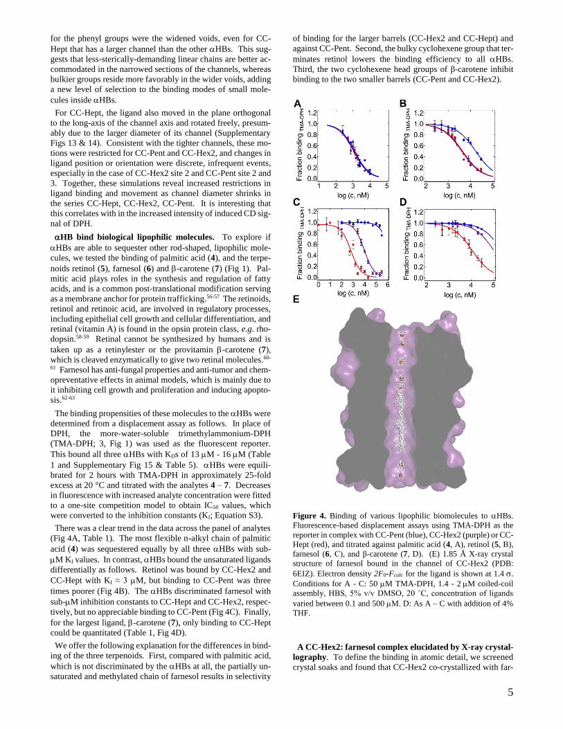
5
for the phenyl groups were the widened voids, even for CC-
Hept that has a larger channel than the other HBs. This sug-
gests that less-sterically-demanding linear chains are better ac-
commodated in the narrowed sections of the channels, whereas
bulkier groups reside more favorably in the wider voids, adding
a new level of selection to the binding modes of small mole-
cules inside HBs.
For CC-Hept, the ligand also moved in the plane orthogonal
to the long-axis of the channel axis and rotated freely, presum-
ably due to the larger diameter of its channel (Supplementary
Figs 13 & 14). Consistent with the tighter channels, these mo-
tions were restricted for CC-Pent and CC-Hex2, and changes in
ligand position or orientation were discrete, infrequent events,
especially in the case of CC-Hex2 site 2 and CC-Pent site 2 and
3. Together, these simulations reveal increased restrictions in
ligand binding and movement as channel diameter shrinks in
the series CC-Hept, CC-Hex2, CC-Pent. It is interesting that
this correlates with in the increased intensity of induced CD sig-
nal of DPH.
HB bind biological lipophilic molecules. To explore if
HBs are able to sequester other rod-shaped, lipophilic mole-
cules, we tested the binding of palmitic acid (4), and the terpe-
noids retinol (5), farnesol (6) and -carotene (7) (Fig 1). Pal-
mitic acid plays roles in the synthesis and regulation of fatty
acids, and is a common post-translational modification serving
as a membrane anchor for protein trafficking.56-57 The retinoids,
retinol and retinoic acid, are involved in regulatory processes,
including epithelial cell growth and cellular differentiation, and
retinal (vitamin A) is found in the opsin protein class, e.g. rho-
dopsin.58-59 Retinal cannot be synthesized by humans and is
taken up as a retinylester or the provitamin -carotene (7),
which is cleaved enzymatically to give two retinal molecules.60-
61 Farnesol has anti-fungal properties and anti-tumor and chem-
opreventative effects in animal models, which is mainly due to
it inhibiting cell growth and proliferation and inducing apopto-
sis.62-63
The binding propensities of these molecules to the HBs were
determined from a displacement assay as follows. In place of
DPH, the more-water-soluble trimethylammonium-DPH
(TMA-DPH; 3, Fig 1) was used as the fluorescent reporter.
This bound all three HBs with KDs of 13 M - 16 M (Table
1 and Supplementary Fig 15 & Table 5). HBs were equili-
brated for 2 hours with TMA-DPH in approximately 25-fold
excess at 20 °C and titrated with the analytes 4 – 7. Decreases
in fluorescence with increased analyte concentration were fitted
to a one-site competition model to obtain IC50 values, which
were converted to the inhibition constants (KI; Equation S3).
There was a clear trend in the data across the panel of analytes
(Fig 4A, Table 1). The most flexible n-alkyl chain of palmitic
acid (4) was sequestered equally by all three HBs with sub-
M KI values. In contrast, HBs bound the unsaturated ligands
differentially as follows. Retinol was bound by CC-Hex2 and
CC-Hept with KI ≈ 3 M, but binding to CC-Pent was three
times poorer (Fig 4B). The HBs discriminated farnesol with
sub-M inhibition constants to CC-Hept and CC-Hex2, respec-
tively, but no appreciable binding to CC-Pent (Fig 4C). Finally,
for the largest ligand, -carotene (7), only binding to CC-Hept
could be quantitated (Table 1, Fig 4D).
We offer the following explanation for the differences in bind-
ing of the three terpenoids. First, compared with palmitic acid,
which is not discriminated by the HBs at all, the partially un-
saturated and methylated chain of farnesol results in selectivity
of binding for the larger barrels (CC-Hex2 and CC-Hept) and
against CC-Pent. Second, the bulky cyclohexene group that ter-
minates retinol lowers the binding efficiency to all HBs.
Third, the two cyclohexene head groups of β-carotene inhibit
binding to the two smaller barrels (CC-Pent and CC-Hex2).
Figure 4. Binding of various lipophilic biomolecules to HBs.
Fluorescence-based displacement assays using TMA-DPH as the
reporter in complex with CC-Pent (blue), CC-Hex2 (purple) or CC-
Hept (red), and titrated against palmitic acid (4, A), retinol (5, B),
farnesol (6, C), and β-carotene (7, D). (E) 1.85 Å X-ray crystal
structure of farnesol bound in the channel of CC-Hex2 (PDB:
6EIZ). Electron density 2F0-Fcalc for the ligand is shown at 1.4 .
Conditions for A - C: 50 M TMA-DPH, 1.4 - 2 M coiled-coil
assembly, HBS, 5% v/v DMSO, 20 ˚C, concentration of ligands
varied between 0.1 and 500 M. D: As A – C with addition of 4%
THF.
A CC-Hex2: farnesol complex elucidated by X-ray crystal-
lography. To define the binding in atomic detail, we screened
crystal soaks and found that CC-Hex2 co-crystallized with far-
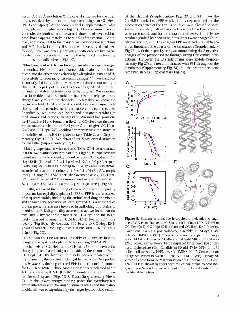
6
nesol. A 1.85 Å resolution X-ray crystal structure for the com-
plex was solved by molecular replacement using apo-CC-Hex2
(PDB code 4pn9)25 as the search model (Supplementary Table
6, Fig 4E, and Supplementary Fig 16). This confirmed the sin-
gle-molecule binding mode assumed above, and revealed far-
nesol bound approximately in the middle of the channel. More-
over, and in contrast to the many other X-ray crystal structures
and MD simulations of HBs that we have solved and per-
formed, there was density consistent with ordered hydrogen-
bonded water molecules connecting the hydroxyl functionality
of farnesol to bulk solvent (Fig 4E).
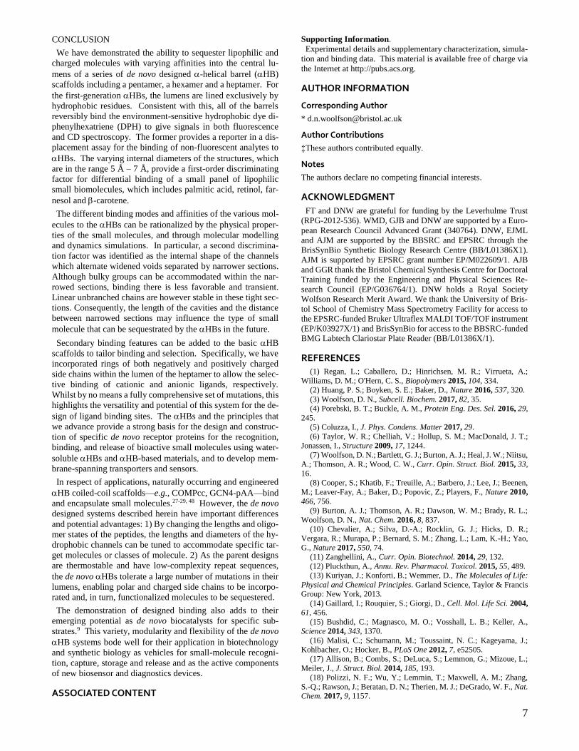
The lumens of HBs can be engineered to accept charged
molecules. Hydrophilic and charged side chains can be intro-
duced into the otherwise exclusively hydrophobic lumens of de
novo HBs without major structural changes.9, 31 For instance,
a robustly folded CC-Hept variant with three mutations per
chain, CC-Hept-Cys-His-Glu, has been designed and shows ru-
dimentary catalytic activity in ester hydrolysis.9 We reasoned
that ionizable residues could be included to help sequester
charged analytes into the channels. To test this, we chose the
larger scaffold, CC-Hept as it should tolerate charged side
chains and be receptive to larger, more-complex molecules.
Specifically, we introduced lysine and glutamate residues to
bind anions and cations, respectively. We modified positions
Ile-17 and Ile-24 and found that Ile-24 of CC-Hept was the most
robust towards substitution for Lys or Glu—to give CC-Hept-
I24K and CCHept-I24E—without compromising the structure
or stability of the HB (Supplementary Table 2, and Supple-
mentary Figs 17-22). We obtained an X-ray crystal structure
for the latter (Supplementary Fig 17).
Binding experiments with cationic TMA-DPH demonstrated
that the two variants discriminated this ligand as expected: the
ligand was relatively weakly bound by both CC-Hept and CC-
Hept-I24K (KD’s of 13.3 ± 1.3 M and 12.8 ± 0.6 M, respec-
tively, Fig 5A); whereas, binding to CC-Hept-I24E was almost
an order of magnitude tighter at 1.6 ± 0.1 M (Fig 5A, purple
trace). Using the TMA-DPH displacement assay, CC-Hept-
I24K and CC-Hept-I24E accommodated neutral farnesol with
KIs of 1.8 ± 0.3 M and 1.0 ± 0.04 M, respectively (Fig 5B).
Finally, we tested the binding of the anionic and biologically
important farnesyl diphosphate (8, FPP). FPP is the precursor
of sesquiterpenoids, including the antimalarial drug artemisinin
and squalene the precursor of sterols;64 and it is a substrate of
protein prenyltransferases involved in trafficking of proteins to
membranes.65 Using the displacement assay, we found that the
exclusively hydrophobic channel of CC-Hept and the nega-
tively charged channel of CC-Hept-I24E bound FPP only
weakly (Fig 5C). By contrast, FPP bound to CC-Hept-I24K
greater than ten times tighter with a measurable KI of 1.5 ±
0.2M (Fig 5C).
These data for FPP are most probably explained by binding
being driven by its hydrophobic tail displacing TMA-DPH from
the channels of CC-Hept and CC-Hept-I24E, and leaving the
charged diphosphate headgroup outside of the channel. With
CC-Hept-I24K the latter could also be accommodated within
the channel by the positively charged hepta-lysine. We probed
this in silico by docking charged FPP in the channel of a model
for CC-Hept-I24K. Three binding poses were selected and a
100 ns constant-pH MD (CpHMD) simulation at pH 7.0 was
run for each system (Figs 5D & E and Supplementary Movie
2). In the lowest-energy binding poses the pyrophosphate
group interacted with the ring of lysine residues and the hydro-
phobic tail was encapsulated by the larger hydrophobic section
of the channel (Supplementary Figs 23 and 24). For the
CpHMD simulations, FPP was kept fully deprotonated and the
protonation states of the Lys-24 residues were allowed to vary.
For approximately half of the simulation, 5 of the Lys residues
were protonated, and for the remainder either 6, 3 or 7 lysine
residues (ranked by decreasing prevalence) were charged (Sup-
plementary Fig 25). The charged FPP remained in a stable po-
sition throughout the course of the simulations (Supplementary
Fig 26), with the hepta-Lys ring accommodating the 3 negative
charges of the pyrophosphate through strong Coulombic inter-
actions. However, the Lys side chains were mobile (Supple-
mentary Fig 27) and not all interacted with FPP throughout the
simulation (Supplementary Fig 24); but the protein backbone
remained stable (Supplementary Fig 28).
Figure 5. Binding of bioactive hydrophobic molecules to engi-
neered CC-Hept channels. (A) Saturation binding of TMA-DPH to
CC-Hept (red), CC-Hept-I24K (blue) and CC-Hept-I24E (purple).
Conditions: 1.4 – 100 M coiled-coil assembly, 1 M dye, HBS,
5% v/v DMSO. (B&C) Fluorescence-based competition assays
with TMA-DPH bound to CC-Hept, CC-Hept-I24K, and CC-Hept-
I24E (colour key as above) being displaced by farnesol (B) or far-
nesol diphosphate (C). Conditions: 50 M TMA-DPH, 1.4 M
coiled-coil assembly, HBS, 5% v/v DMSO, 20 ˚C. Concentration
of ligands varied between 0.1 and 500 M. (D&E) Orthogonal
views of a pose from the MD simulation of FPP bound to CC-Hept-
I24K. FPP is shown in sticks with the carbon atoms colored ma-
genta; Lys-24 residues are represented by sticks with spheres for
the titratable protons.
7
CONCLUSION
We have demonstrated the ability to sequester lipophilic and
charged molecules with varying affinities into the central lu-
mens of a series of de novo designed -helical barrel (HB)
scaffolds including a pentamer, a hexamer and a heptamer. For
the first-generation HBs, the lumens are lined exclusively by
hydrophobic residues. Consistent with this, all of the barrels
reversibly bind the environment-sensitive hydrophobic dye di-
phenylhexatriene (DPH) to give signals in both fluorescence
and CD spectroscopy. The former provides a reporter in a dis-
placement assay for the binding of non-fluorescent analytes to
HBs. The varying internal diameters of the structures, which
are in the range 5 Å – 7 Å, provide a first-order discriminating
factor for differential binding of a small panel of lipophilic
small biomolecules, which includes palmitic acid, retinol, far-
nesol and -carotene.
The different binding modes and affinities of the various mol-
ecules to the HBs can be rationalized by the physical proper-
ties of the small molecules, and through molecular modelling
and dynamics simulations. In particular, a second discrimina-
tion factor was identified as the internal shape of the channels
which alternate widened voids separated by narrower sections.
Although bulky groups can be accommodated within the nar-
rowed sections, binding there is less favorable and transient.
Linear unbranched chains are however stable in these tight sec-
tions. Consequently, the length of the cavities and the distance
between narrowed sections may influence the type of small
molecule that can be sequestrated by the HBs in the future.
Secondary binding features can be added to the basic HB
scaffolds to tailor binding and selection. Specifically, we have
incorporated rings of both negatively and positively charged
side chains within the lumen of the heptamer to allow the selec-
tive binding of cationic and anionic ligands, respectively.
Whilst by no means a fully comprehensive set of mutations, this
highlights the versatility and potential of this system for the de-
sign of ligand binding sites. The HBs and the principles that
we advance provide a strong basis for the design and construc-
tion of specific de novo receptor proteins for the recognition,
binding, and release of bioactive small molecules using water-
soluble HBs and HB-based materials, and to develop mem-
brane-spanning transporters and sensors.
In respect of applications, naturally occurring and engineered
HB coiled-coil scaffolds—e.g., COMPcc, GCN4-pAA—bind
and encapsulate small molecules.27-29, 48 However, the de novo
designed systems described herein have important differences
and potential advantages: 1) By changing the lengths and oligo-
mer states of the peptides, the lengths and diameters of the hy-
drophobic channels can be tuned to accommodate specific tar-
get molecules or classes of molecule. 2) As the parent designs
are thermostable and have low-complexity repeat sequences,
the de novo HBs tolerate a large number of mutations in their
lumens, enabling polar and charged side chains to be incorpo-
rated and, in turn, functionalized molecules to be sequestered.
The demonstration of designed binding also adds to their
emerging potential as de novo biocatalysts for specific sub-
strates.9 This variety, modularity and flexibility of the de novo
HB systems bode well for their application in biotechnology
and synthetic biology as vehicles for small-molecule recogni-
tion, capture, storage and release and as the active components
of new biosensor and diagnostics devices.
ASSOCIATED CONTENT
Supporting Information.
Experimental details and supplementary characterization, simula-
tion and binding data. This material is available free of charge via
the Internet at http://pubs.acs.org.
AUTHOR INFORMATION
Corresponding Author
Author Contributions
‡These authors contributed equally.
Notes
The authors declare no competing financial interests.
ACKNOWLEDGMENT
FT and DNW are grateful for funding by the Leverhulme Trust
(RPG-2012-536). WMD, GJB and DNW are supported by a Euro-
pean Research Council Advanced Grant (340764). DNW, EJML
and AJM are supported by the BBSRC and EPSRC through the
BrisSynBio Synthetic Biology Research Centre (BB/L01386X1).
AJM is supported by EPSRC grant number EP/M022609/1. AJB
and GGR thank the Bristol Chemical Synthesis Centre for Doctoral
Training funded by the Engineering and Physical Sciences Re-
search Council (EP/G036764/1). DNW holds a Royal Society
Wolfson Research Merit Award. We thank the University of Bris-
tol School of Chemistry Mass Spectrometry Facility for access to
the EPSRC-funded Bruker Ultraflex MALDI TOF/TOF instrument
(EP/K03927X/1) and BrisSynBio for access to the BBSRC-funded
BMG Labtech Clariostar Plate Reader (BB/L01386X/1).
REFERENCES
(1) Regan, L.; Caballero, D.; Hinrichsen, M. R.; Virrueta, A.;
Williams, D. M.; O'Hern, C. S., Biopolymers 2015, 104, 334.
(2) Huang, P. S.; Boyken, S. E.; Baker, D., Nature 2016, 537, 320.
(3) Woolfson, D. N., Subcell. Biochem. 2017, 82, 35.
(4) Porebski, B. T.; Buckle, A. M., Protein Eng. Des. Sel. 2016, 29,
245.
(5) Coluzza, I., J. Phys. Condens. Matter 2017, 29.
(6) Taylor, W. R.; Chelliah, V.; Hollup, S. M.; MacDonald, J. T.;
Jonassen, I., Structure 2009, 17, 1244.
(7) Woolfson, D. N.; Bartlett, G. J.; Burton, A. J.; Heal, J. W.; Niitsu,
A.; Thomson, A. R.; Wood, C. W., Curr. Opin. Struct. Biol. 2015, 33,
16.
(8) Cooper, S.; Khatib, F.; Treuille, A.; Barbero, J.; Lee, J.; Beenen,
M.; Leaver-Fay, A.; Baker, D.; Popovic, Z.; Players, F., Nature 2010,
466, 756.
(9) Burton, A. J.; Thomson, A. R.; Dawson, W. M.; Brady, R. L.;
Woolfson, D. N., Nat. Chem. 2016, 8, 837.
(10) Chevalier, A.; Silva, D.-A.; Rocklin, G. J.; Hicks, D. R.;
Vergara, R.; Murapa, P.; Bernard, S. M.; Zhang, L.; Lam, K.-H.; Yao,
G., Nature 2017, 550, 74.
(11) Zanghellini, A., Curr. Opin. Biotechnol. 2014, 29, 132.
(12) Pluckthun, A., Annu. Rev. Pharmacol. Toxicol. 2015, 55, 489.
(13) Kuriyan, J.; Konforti, B.; Wemmer, D., The Molecules of Life:
Physical and Chemical Principles. Garland Science, Taylor & Francis
Group: New York, 2013.
(14) Gaillard, I.; Rouquier, S.; Giorgi, D., Cell. Mol. Life Sci. 2004,
61, 456.
(15) Bushdid, C.; Magnasco, M. O.; Vosshall, L. B.; Keller, A.,
Science 2014, 343, 1370.
(16) Malisi, C.; Schumann, M.; Toussaint, N. C.; Kageyama, J.;
Kohlbacher, O.; Hocker, B., PLoS One 2012, 7, e52505.
(17) Allison, B.; Combs, S.; DeLuca, S.; Lemmon, G.; Mizoue, L.;
Meiler, J., J. Struct. Biol. 2014, 185, 193.
(18) Polizzi, N. F.; Wu, Y.; Lemmin, T.; Maxwell, A. M.; Zhang,
S.-Q.; Rawson, J.; Beratan, D. N.; Therien, M. J.; DeGrado, W. F., Nat.
Chem. 2017, 9, 1157.

8
(19) Tinberg, C. E.; Khare, S. D.; Dou, J.; Doyle, L.; Nelson, J. W.;
Schena, A.; Jankowski, W.; Kalodimos, C. G.; Johnsson, K.; Stoddard,
B. L.; Baker, D., Nature 2013, 501, 212.
(20) Bick, M. J.; Greisen, P. J.; Morey, K. J.; Antunes, M. S.; La,
D.; Sankaran, B.; Reymond, L.; Johnsson, K.; Medford, J. I.; Baker,
D., eLife 2017, 6, e28909.
(21) Lupas, A. N.; Bassler, J.; Dunin-Horkawicz, S., The Structure
and Topology of α-Helical Coiled Coils. In Fibrous Proteins:
Structures and Mechanisms, Parry, D. A. D.; Squire, J. M., Eds.
Springer International Publishing: Cham, 2017; pp 95.
(22) Fletcher, J. M.; Boyle, A. L.; Bruning, M.; Bartlett, G. J.;
Vincent, T. L.; Zaccai, N. R.; Armstrong, C. T.; Bromley, E. H. C.;
Booth, P. J.; Brady, R. L.; Thomson, A. R.; Woolfson, D. N., ACS
Synth. Biol. 2012, 1, 240.
(23) Gonzalez, L.; Plecs, J. J.; Alber, T., Nat. Struct. Biol. 1996, 3,
510.
(24) Yadav, M. K.; Redman, J. E.; Leman, L. J.; Alvarez-Gutierrez,
J. M.; Zhang, Y. M.; Stout, C. D.; Ghadiri, M. R., Biochemistry 2005,
44, 9723.
(25) Thomson, A. R.; Wood, C. W.; Burton, A. J.; Bartlett, G. J.;
Sessions, R. B.; Brady, R. L.; Woolfson, D. N., Science 2014, 346, 485.
(26) Walshaw, J.; Woolfson, D. N., Protein Sci. 2001, 10, 668.
(27) Guo, Y.; Bozic, D.; Malashkevich, V. N.; Kammerer, R. A.;
Schulthess, T.; Enger, J., EMBO J. 1998, 17, 5265.
(28) Ozbek, S.; Engel, J.; Stetefeld, J., EMBO J. 2002, 21, 5960.
(29) MacFarlane, A. A.; Orriss, G.; Okun, N.; Meier, M.; Klonisch,
T.; Khajehpour, M.; Stetefeld, J., PLoS One 2012, 7.
(30) Liu, J.; Yong, W.; Deng, Y. Q.; Kallenbach, N. R.; Lu, M.,
Proc. Natl. Acad. Sci. U.S.A. 2004, 101, 16156.
(31) Zaccai, N. R.; Chi, B.; Thomson, A. R.; Boyle, A. L.; Bartlett,
G. J.; Bruning, M.; Linden, N.; Sessions, R. B.; Booth, P. J.; Brady, R.
L.; Woolfson, D. N., Nat. Chem. Biol. 2011, 7, 935.
(32) Burgess, N. C.; Sharp, T. H.; Thomas, F.; Wood, C. W.;
Thomson, A. R.; Zaccai, N. R.; Brady, R. L.; Serpell, L. C.; Woolfson,
D. N., J. Am. Chem. Soc. 2015, 137, 10554.
(33) Liu, J.; Zheng, Q.; Deng, Y. Q.; Cheng, C. S.; Kallenbach, N.
R.; Lu, M., Proc. Natl. Acad. Sci. U.S.A. 2006, 103, 15457.
(34) Xu, C. F.; Liu, R.; Mehta, A. K.; Guerrero-Ferreira, R. C.;
Wright, E. R.; Dunin-Horkawicz, S.; Morris, K.; Serpell, L. C.; Zuo,
X. B.; Wall, J. S.; Conticello, V. P., J. Am. Chem. Soc. 2013, 135,
15565.
(35) Spencer, R. K.; Hochbaum, A. I., Biochemistry 2016, 55, 3214.
(36) Spencer, R. K.; Hochbaum, A. I., Biochemistry 2017, 56, 5300.
(37) Lizatovic, R.; Aurelius, O.; Stenstrom, O.; Drakenberg, T.;
Akke, M.; Logan, D. T.; Andre, I., Structure 2016, 24, 946.
(38) Crick, F. H. C., Acta Crystallogr. 1953, 6, 689.
(39) Offer, G.; Sessions, R., J. Mol. Biol. 1995, 249, 967.
(40) Harbury, P. B.; Plecs, J. J.; Tidor, B.; Alber, T.; Kim, P. S.,
Science 1998, 282, 1462.
(41) Dunin-Horkawicz, S.; Lupas, A. N., J. Struct. Biol. 2010, 170,
226.
(42) Grigoryan, G.; DeGrado, W. F., J. Mol. Biol. 2011, 405, 1079.
(43) Huang, P. S.; Oberdorfer, G.; Xu, C. F.; Pei, X. Y.; Nannenga,
B. L.; Rogers, J. M.; DiMaio, F.; Gonen, T.; Luisi, B.; Baker, D.,
Science 2014, 346, 481.
(44) Wood, C. W.; Bruning, M.; Ibarra, A. A.; Bartlett, G. J.;
Thomson, A. R.; Sessions, R. B.; Brady, R. L.; Woolfson, D. N.,
Bioinformatics 2014, 30, 3029.
(45) Thomas, F.; Boyle, A. L.; Burton, A. J.; Woolfson, D. N., J.
Am. Chem. Soc. 2013, 135, 5161.
(46) Thomas, F.; Burgess, N. C.; Thomson, A. R.; Woolfson, D. N.,
Angew. Chem. Int. Ed. 2016, 55, 987.
(47) Steinbacher, S.; Bass, R.; Strop, P.; Rees, D. C., Curr. Top.
Membr. 2007, 58, 1.
(48) Hume, J.; Sun, J.; Jacquet, R.; Renfrew, P. D.; Martin, J. A.;
Bonneau, R.; Gilchrist, M. L.; Montclare, J. K., Biomacromolecules
2014, 15, 3503.
(49) Lentz, B. R., Chem. Phys. Lipids 1989, 50, 171.
(50) Parasassi, T.; Krasnowska, E. K.; Bagatolli, L.; Gratton, E., J.
Fluoresc. 1998, 8, 365.
(51) Sreerama, N.; Woody, R. W., Methods Enzymol. 2004, 383,
318.
(52) Ge, Y.; van der Kamp, M.; Malaisree, M.; Liu, D.; Liu, Y.;
Mulholland, A. J., J. Comput.-Aided Mol. Des. 2017, 1.
(53) Watkins, D. W.; Jenkins, J. M. X.; Grayson, K. J.; Wood, N.;
Steventon, J. W.; Le Vay, K. K.; Goodwin, M. I.; Mullen, A. S.; Bailey,
H. J.; Crump, M. P.; MacMillan, F.; Mulholland, A. J.; Cameron, G.;
Sessions, R. B.; Mann, S.; Anderson, J. L. R., Nat. Commun. 2017, 8,
358.
(54) Trott, O.; Olson, A. J., J. Comput. Chem. 2010, 31, 455.
(55) Smart, O. S.; Neduvelil, J. G.; Wang, X.; Wallace, B. A.;
Sansom, M. S. P., J. Mol. Graphics 1996, 14, 354.
(56) Wakil, S. J.; Stoops, J. K.; Joshi, V. C., Annu. Rev. Biochem.
1983, 52, 537.
(57) Linder, M. E.; Deschenes, R. J., Nat. Rev. Mol. Cell Biol. 2007,
8, 74.
(58) Blomhoff, R.; Blomhoff, H. K., J. Neurobiol. 2006, 66, 606.
(59) Bushue, N.; Wan, Y. J. Y., Adv. Drug Del. Rev. 2010, 62, 1285.
(60) Kloer, D. P.; Schulz, G. E., Cell. Mol. Life Sci. 2006, 63, 2291.
(61) Britton, G., FASEB J. 1995, 9, 1551.
(62) Albuquerque, P.; Casadevall, A., Med. Mycol. 2012, 50, 337.
(63) Joo, J. H.; Jetten, A. M., Cancer Lett. 2010, 287, 123.
(64) Grunler, J.; Ericsson, J.; Dallner, G., Biochim. Biophys. Acta
1994, 1212, 259.
(65) McTaggart, S. J., Cell. Mol. Life Sci. 2006, 63, 255.
9
Insert Table of Contents artwork here