This student paper was written as an assignment in the ... · hypoxia results in increased...
14
77:222 Spring 2003 Free Radicals in Biology and Medicine Page 0 This student paper was written as an assignment in the graduate course Free Radicals in Biology and Medicine (77:222, Spring 2003) offered by the Free Radical and Radiation Biology Program B-180 Med Labs The University of Iowa Iowa City, IA 52242-1181 Spring 2003 Term Instructors: GARRY R. BUETTNER, Ph.D. LARRY W. OBERLEY, Ph.D. with guest lectures from: Drs. Freya Q . Schafer, Douglas R. Spitz, and Frederick E. Domann The Fine Print: Because this is a paper written by a beginning student as an assignment, there are no guarantees that everything is absolutely correct and accurate. In view of the possibility of human error or changes in our knowledge due to continued research, neither the author nor The University of Iowa nor any other party who has been involved in the preparation or publication of this work warrants that the information contained herein is in every respect accurate or complete, and they are not responsible for any errors or omissions or for the results obtained from the use of such information. Readers are encouraged to confirm the information contained herein with other sources. All material contained in this paper is copyright of the author, or the owner of the source that the material was taken from. This work is not intended as a threat to the ownership of said copyrights.
Transcript of This student paper was written as an assignment in the ... · hypoxia results in increased...

77:222 Spring 2003 Free Radicals in Biology and Medicine Page 0
This student paper was written as an assignment in the graduate course
Free Radicals in Biology and Medicine
(77:222, Spring 2003)
offered by the
Free Radical and Radiation Biology Program
B-180 Med Labs The University of Iowa
Iowa City, IA 52242-1181 Spring 2003 Term
Instructors:
GARRY R. BUETTNER, Ph.D. LARRY W. OBERLEY, Ph.D.
with guest lectures from:
Drs. Freya Q . Schafer, Douglas R. Spitz, and Frederick E. Domann The Fine Print: Because this is a paper written by a beginning student as an assignment, there are no guarantees that everything is absolutely correct and accurate. In view of the possibility of human error or changes in our knowledge due to continued research, neither the author nor The University of Iowa nor any other party who has been involved in the preparation or publication of this work warrants that the information contained herein is in every respect accurate or complete, and they are not responsible for any errors or omissions or for the results obtained from the use of such information. Readers are encouraged to confirm the information contained herein with other sources. All material contained in this paper is copyright of the author, or the owner of the source that the material was taken from. This work is not intended as a threat to the ownership of said copyrights.

Ehab H. Sarsour HIF-1 1
Hypoxia-inducible factor 1 (HIF-1)
By
Ehab H. Sarsour
B-180 ML Free Radical and Radiation Biology Program
The University of Iowa Iowa City, IA 52242
For 77:222, Spring 2003
13 March 2003
Paper III
Abbreviations
ARNT Aryl hydrocarbon receptor nuclear translocator
FKBP Rapamycin-associated protein HIF-1 Hypoxia-inducible factor 1 IGF Insulin-like growth factor IUGR Intrauterine growth retardation NADPH Nicotinamide adenine
dinucleotide phosphate (reduced)
NF-κB Nuclear factor-κB NO• Nitric oxide NOS Nitric oxide synthase
P Partial pressure PI3K Phosphoinositol 3-kinase PTEN Phosphatase and tensin
homologue pVHL von Hippel–Lindau protein REF-1 Redox factor 1 ROS Reactive oxygen species TAD Transactivation domain TAD-C Carboxy-terminal TAD TAD-N Amino-terminal TAD VEGF Vascular endothelial growth
factor

Ehab H. Sarsour HIF-1 2
Table of Contents
1. Introduction………………………………………………………………………….3 2. Structure……………………………………………………………………………..3
3. Function and regulation……………………………………………………………...4 4. Hypoxia signaling pathways and HIF-1……………………………………………..6 5. HIF-1 in Disease and Cancer………………………………………….……………..8 6. Conclusion………………………………………………………………………….10
Abstract Hypoxia-inducible factor 1 (HIF-1) is a transcriptional activator regulated by oxygen
concentration that plays essential roles in mammalian physiology and disease pathogenesis.
Molecular oxygen is essential for the survival of all aerobic organisms; HIF-1 is essential for
maintaining the oxygen homeostasis in both normal and cancer cells. This review will
summarize some of what is currently known about HIF-1 including its structure, function, and
regulation under normal and pathological conditions.

Ehab H. Sarsour HIF-1 3
1. Introduction
Oxygen homeostasis represents an important organizing principle for human
development and physiology. The essential requirement for oxidative phosphorylation to
generate ATP is balanced by the risk of oxidative damage to cellular lipids, nucleic acids, and
proteins. As a result, cellular O2 concentrations are tightly regulated through response pathways
that affect the activity and expression of different cellular proteins [1]. HIF-1 plays a major role
in maintaining the cellular oxygen homeostasis. Oxygen concentrations regulate the expression
and transcriptional activity of HIF-1 [2]. HIF-1 is a basic helix-loop–helix protein consisting of
HIF-1α and HIF-1β subunits [3]. HIF-1 expression and HIF-1 transcriptional regulation of HIF-1
activity occurs at multiple levels. Whereas HIF-1α mRNA is constitutively expressed in tissue
culture cells, it is markedly induced by hypoxia or ischemia in vivo [4]. A wide spectrum of
studies have shown that HIF-1 is a major player in human diseases that involve O2 homeostasis
and hypoxia such cardiovascular disorders, pulmonary hypertension, pregnancy disorders, and
cancer.
2. Structure
HIF-1 is a heterodimer composed of HIF-1α and HIF-1β subunits [3](Figure 1). Whereas
HIF-1β is constitutively expressed, HIF-1α expression is induced in hypoxic cells with an
exponential increase in expression as cells are exposed to O2 concentrations of less than 6% [4],
which corresponds to a partial pressure (P) of O2 of approximately 40 mm Hg at sea level [3,5].
The amino-terminal half of HIF-1α (amino acids 1–390) is necessary and sufficient for
dimerization with HIF-1β and for DNA binding (Figure 1). HIF-1α is ubiquitinated and
subjected to proteasomal degradation in non-hypoxic cells [6–8]. A Pro–Ser–Thr rich protein

Ehab H. Sarsour HIF-1 4
stabilization domain is located between amino acids 429 and 608 of HIF-1α [7,9]. HIF-1α
protein have also two transactivation domains (TADs) in the carboxy-terminal half of HIF-1α
(amino acids 531–575 and 786–826) interact with coactivators such as CBP, p300, SRC-1 and
TIF2 (Figure 1) [10].
Figure 1. Structure HIF-1. The HIF-1α and HIF-1β subunits are shown with the basic helix-
loop-helix (bHLH) and PER-ARNT-SIM (PAS) domains that are required for dimerization and DNA binding. Also shown for HIF-1α are the amino-terminal (N) and carboxyterminal (C) nuclear localization signal (NLS) and TAD; the Pro–Ser–Thr rich protein stabilization domain (PSTD; also known as the oxygen-dependent degradation domain); and sites of interaction with VHL, and p300 and CBP. The double-headed arrow indicates that reduction of Cys800, which is mediated by thioredoxin (TRX) and redox factor 1 (REF-1), is required for the interaction of TAD-C with cofactor p300 or CBP. The relevant amino acides residues are indicated numerically. Adapted from [11].
3. Function and Regulation
Vascular endothelial growth factor (VEGF) plays an essential role in physiological
responses to reduced O2 availability (hypoxia). VEGF expression is induced when most cell
types are subjected to hypoxia, thus providing a mechanism by which tissue perfusion can be
optimized on demand. Hypoxia also results in the rapid accumulation of HIF-1α in the nucleus

Ehab H. Sarsour HIF-1 5
where it dimerizes with HIF-1β and binds to the core DNA sequence 5`-RCGTG-3` leading to
the transcriptional activation of VEGF and several dozen other known target genes (Table 1)[12].
Under hypoxic conditions, the fraction of HIF-1α that is ubiquitinated decreases
dramatically, resulting in an accumulation of the protein. The von Hippel-Lindau (VHL) protein
binds to the protein stabilization domain and plays a critical role in the ubiquitination of HIF-1α.
Exposure of cells to cobalt chloride or iron chelators (e.g. desferrioxamine) induces HIF-1α
expression and inhibits HIF-1α ubiquitination by dissociating VHL from HIF-1α [8,9,13]. p53
also interacts with HIF-1α and by doing so recruits the MDM2 ubiquitin protein ligase, which
reduces the induction of HIF-1α expression under hypoxic conditions [14]. Activation of the
signal transduction pathway involving phosphoinositolb3-kinase (PI3K) and the serine/threonine
kinases protein kinase B (AKT) and FKBP-rapamycin-associated protein (FRAP) has also been
shown to induce expression of HIF-1α protein and vascular endothelial growth factor (VEGF)
mRNA under non-hypoxic conditions [15]. In addition to increased steady-state levels of HIF-1α
protein in hypoxic cells, the two transactivation domains (TADs) in the carboxy-terminal half of
HIF-1α interact with coactivators such as CBP, p300, SRC-1 and TIF2 and activate HIF-1α
transcription [10].
Table 1. Direct HIF-1 target genes. Adapted from [12]
Glucose/Energy Metabolism and Cell Proliferation/Viability Adenylate Kinase 3, Aldolase A, Aldolase C, Enolase 1 (ENO1), Glucose Transporter 1, Glucose
Transporter 3, Glyceraldehyde-3-phosphate Dehydrogenase, Hexokinase 1, Hexokinase 2, Insulin-like Growth Factor 2 (IGF-2), IGF Binding Protein 1 (IGFBP-1), IGFBP-3, Lactate Dehydrogenase A, Phosphoglycerate Kinase 1, Pyruvate Kinase M, p21, Transforming Growth Factor _3(TGF_3)
Erythropoiesis and Iron Metabolism Ceruloplasmin, Erythropoietin, Transferrin, Transferrin Receptor
Vascular Development/Remodeling and Vasomotor Tone 1B-Adrenergic Receptor, Adrenomedullin, Endothelin-1, Heme Oxygenase 1, Nitric Oxide Synthase 2, Plasminogen Activator Inhibitor 1, Vascular Endothelial Growth Factor (VEGF), VEGF Receptor
FLT-1 Other
p35
Ehab H. Sarsour HIF-1 6
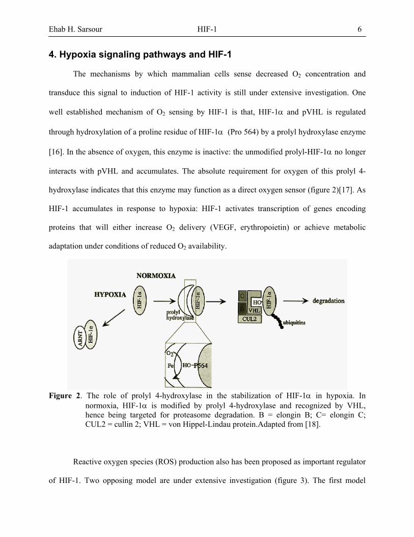
4. Hypoxia signaling pathways and HIF-1
The mechanisms by which mammalian cells sense decreased O2 concentration and
transduce this signal to induction of HIF-1 activity is still under extensive investigation. One
well established mechanism of O2 sensing by HIF-1 is that, HIF-1α and pVHL is regulated
through hydroxylation of a proline residue of HIF-1α (Pro 564) by a prolyl hydroxylase enzyme
[16]. In the absence of oxygen, this enzyme is inactive: the unmodified prolyl-HIF-1α no longer
interacts with pVHL and accumulates. The absolute requirement for oxygen of this prolyl 4-
hydroxylase indicates that this enzyme may function as a direct oxygen sensor (figure 2)[17]. As
HIF-1 accumulates in response to hypoxia: HIF-1 activates transcription of genes encoding
proteins that will either increase O2 delivery (VEGF, erythropoietin) or achieve metabolic
adaptation under conditions of reduced O2 availability.
Figure 2. The role of prolyl 4-hydroxylase in the stabilization of HIF-1α in hypoxia. In
normoxia, HIF-1α is modified by prolyl 4-hydroxylase and recognized by VHL, hence being targeted for proteasome degradation. B = elongin B; C= elongin C; CUL2 = cullin 2; VHL = von Hippel-Lindau protein.Adapted from [18].
Reactive oxygen species (ROS) production also has been proposed as important regulator
of HIF-1. Two opposing model are under extensive investigation (figure 3). The first model

Ehab H. Sarsour HIF-1 7
proposes that a NADPH oxidase converts O2 into ROS. A decrease in PO2 would result in
reduced formation of ROS, which would relieve the inhibition of signal transduction pathway
leading to HIF-1 activation (figure 3A). On the other hand, the second model suggests that,
hypoxia results in increased production of ROS in the mitochondria, where HIF-1 activation
directly correlated with changes in ROS (figure 3B). Many studies showed this correlation
between ROS production and HIF-1 regulation for example; antioxidants attenuated the ROS
signal and abolished the transcriptional activation response of HIF-1 [19-22].
Figure 3. Potential oxygen sensors involved in the stabilization of HIF-1α in hypoxia. (A) A
NADPH oxidase-like enzyme produces reactive oxygen species (ROS) in normoxia that induce HIF-1α degradation. (B) The mitochondrial respiratory chain produces ROS in hypoxia that induces HIF-1α stabilization. Adapted from [18].

Ehab H. Sarsour HIF-1 8
Nitric oxide (NO•) is another player in regulating HIF-1 activity and expression .NO• also
was found to have dual effects. On one hand, it was observed that different NO• donors and
overexpression of NOS upregulate HIF-1 activity in normoxia [23-25]. On the other hand, NO•
inhibits HIF-1 transcriptional activity in hypoxia [26].
5. HIF-1 in Disease and Cancer In diseases that involve O2 homeostasis impairment and hypoxia conditions, HIF-1 comes
as a main player and marker of such condition, also it becomes as a target for therapy strategies.
For example in myocardial ischemia atherosclerosis leads to arterial stenosis, impaired perfusion
of the downstream vascular bed, and ischemia. When oxygen and glucose deprivation
irreversibly affect myocardial viability, the end result is an infarction (heart attack). Myocardial
ischemia induces VEGF expression, HIF-1α mRNA and protein expression are induced and
precede VEGF expression during acute ischemia and early infarction in the human heart . Thus,
it is possible that variation in ischemia-induced HIF-1 activity may underlie the observed
variation in VEGF expression and represent an important risk factor for myocardial infarction. In
addition, therapeutic strategies designed to increase HIF-1α expression may promote
angiogenesis within ischemic myocardium [27,28]. On the opposite side, local inhibition of HIF-
1 activity in the lung might represent a therapeutic strategy for treating or preventing pulmonary
hypertension in at risk individuals [29]. During pregnancy a leading cause of fetal and neonatal
morbidity and mortality is intrauterine growth retardation (IUGR). Decreased placental
perfusion, resulting in placental and fetal hypoxia, is believed to be a major cause of IUGR.
Fetal and maternal insulin-like growth factors (IGFs) play an important role in regulating fetal
growth. IGF binding protein 1 (IGFBP-1) is a negative regulator of IGF activity. IGFBP-1

Ehab H. Sarsour HIF-1 9
expression, which is induced by hypoxia via a HIF-1 binding site in the gene promoter, is greatly
increased in the cord blood of newborn children with IUGR [30].
Hypoxia is an important selective force in the clonal evolution of tumors and HIF-1α is
overexpressed in common human cancers Increased expression of VEGF is essential for the
establishment of angiogenesis in most solid tumors. Many studies showed that, increased VEGF
expression is required to initiate and sustain tumor angiogenesis. Increased VEGF levels result
from the synergistic effects of tumor hypoxia and tumor-specific genetic alterations (mutations)
involving oncogenes and tumor suppressor genes. Increased VEGF expression results in the
formation of dysfunctional vasculature that cannot adequately perfuse the entire tumor. Cellular
adaptation to hypoxia is therefore a requirement of tumor progression independent of
angiogenesis. As a result, most solid tumors have the seemingly paradoxical characteristic that
poor clinical outcome is significantly correlated with both vascular density and tumor hypoxia.
An example for this correlation is that in human gliomas, there is a significant association
between tumor grade, vascularization, and HIF-1α overexpression [31].
Although microenvironmental tumor hypoxia is undoubtedly an important mechanism of
HIF activation in tumors, many studies in cell culture have demonstrated that other aspects of
cancer enhance the activation of HIF by hypoxia, or activate HIF by oxygen-independent
mechanisms. These studies have demonstrated activation of HIF in response to inactivation of a
number of different tumor suppressor genes, in response to activation of several different
oncogenes, and in response to activation of diverse growth factor pathways[32].

Ehab H. Sarsour HIF-1 10
6. Conclusion Studies and data presented above showed that HIF-1 is highly involved in different
biochemical, physiological and pathological aspects of human disease and cancer, and this make
HIF-1 a good candidate target for therapy. Also the new trend in the involvement of ROS in the
function and regulation of HIF-1, suggests that current research to develop new therapies for
cancer and human disease through manipulating and controlling the expression of many
antioxidant enzymes that alter ROS production, would be a very promising approach.

Ehab H. Sarsour HIF-1 11
6. References
1. Semenza GL. (1999) Perspectives on oxygen sensing. Cell. 98:281-284.
2. Wenger RH. (2000) Mammalian oxygen sensing, signalling and gene regulation. J Exp Biol. 203(8):1253-1263.
3. Wang GL, Jiang BH, Rue EA, Semenza GL. (1995) Hypoxia-inducible factor 1 is a
basic-helix-loop-helix-PAS heterodimer regulated by cellular O2 tension. Proc Natl Acad Sci. U. S. A. 92:5510-5514.
4. Yu AY, Frid MG, Shimoda LA, Wiener CM, Stenmark K, Semenza GL. (1998)
Temporal, spatial, and oxygen-regulated expression of hypoxia-inducible factor-1 in the lung. Am J Physiol. 275:L818-826.
5. Jiang BH, Semenza GL, Bauer C, Marti HH. (1996) Hypoxia-inducible factor 1 levels
vary exponentially over a physiologically relevant range of O2 tension. Am J Physiol. 271:C1172-1180.
6. Salceda S, Caro J. (1997) Hypoxia-inducible factor 1alpha (HIF-1alpha) protein is
rapidly degraded by the ubiquitin-proteasome system under normoxic conditions. Its stabilization by hypoxia depends on redox-induced changes. J Biol Chem. 272:22642-22647.
7. Huang LE, Gu J, Schau M, Bunn HF. (1998) Regulation of hypoxia-inducible factor
1alpha is mediated by an O2-dependent degradation domain via the ubiquitin-proteasome pathway. Proc Natl Acad Sci. U. S. A. 95:7987-7992.
8. Kallio PJ, Wilson WJ, O'Brien S, Makino Y, Poellinger L. (1999) Regulation of the
hypoxia-inducible transcription factor 1alpha by the ubiquitin-proteasome pathway. J Biol Chem. 274:6519-6525.
9. Sutter CH, Laughner E, Semenza GL. (2000) Hypoxia-inducible factor 1alpha protein
expression is controlled by oxygen-regulated ubiquitination that is disrupted by deletions and missense mutations. Proc Natl Acad Sci. U. S. A. 97:4748-4753.
10. Carrero P, Okamoto K, Coumailleau P, O'Brien S, Tanaka H, Poellinger L. (2000)
Redox-regulated recruitment of the transcriptional coactivators CREB-binding protein and SRC-1 to hypoxia-inducible factor 1alpha. Mol Cell Biol. 20:402-415.
11. Semenza GL. (1999) Regulation of mammalian O2 homeostasis by hypoxia-inducible
factor 1. Annu Rev Cell Dev Biol. 15:551-578.
12. Caniggia I, Mostachfi H, Winter J, Gassmann M, Lye SJ, Kuliszewski M, Post M. (2000) Hypoxia-inducible factor-1 mediates the biological effects of oxygen on human trophoblast differentiation through TGFbeta(3). J Clin Invest. 105:577-587.

Ehab H. Sarsour HIF-1 12
13. Maxwell PH, Wiesener MS, Chang GW, Clifford SC, Vaux EC, Cockman ME, Wykoff CC, Pugh CW, Maher ER, Ratcliffe PJ. (1999) The tumour suppressor protein VHL targets hypoxia-inducible factors for oxygen-dependent proteolysis. Nature. 399:271-275.
14. Ravi R, Mookerjee B, Bhujwalla ZM, Sutter CH, Artemov D, Zeng Q, Dillehay LE,
Madan A, Semenza GL, Bedi A. (2000) Regulation of tumor angiogenesis by p53-induced degradation of hypoxia-inducible factor 1alpha. Genes Dev. 14:34-44.
15. Zhong H, Chiles K, Feldser D, Laughner E, Hanrahan C, Georgescu MM, Simons JW,
Semenza GL. (2000) Modulation of hypoxia-inducible factor 1alpha expression by the epidermal growth factor/phosphatidylinositol 3-kinase/PTEN/AKT/FRAP pathway in human prostate cancer cells: implications for tumor angiogenesis and therapeutics. Cancer Res. 60:1541-1545.
16. Ivan M, Kondo K, Yang H, Kim W, Valiando J, Ohh M, Salic A, Asara JM, Lane WS,
Kaelin WG, Jr. (2001) HIFalpha targeted for VHL-mediated destruction by proline hydroxylation: implications for O2 sensing. Science. 292:464-468.
17. Jaakkola P, Mole DR, Tian YM, Wilson MI, Gielbert J, Gaskell SJ, Kriegsheim A,
Hebestreit HF, Mukherji M, Schofield CJ, Maxwell PH, Pugh CW, Ratcliffe PJ. (2001) Targeting of HIF-alpha to the von Hippel-Lindau ubiquitylation complex by O2-regulated prolyl hydroxylation. Science. 292:468-472.
18. Michels C, Minet E, Mollet D, Raes M. (2002) Regulation of gene expression by oxygen:
NF-κB and HIF-1, two extremes. Free Radic Biol Med. 33:1231-1242.
19. Chandel NS, Budinger GR, Choe SH, Schumacker PT. (1997) Cellular respiration during hypoxia. Role of cytochrome oxidase as the oxygen sensor in hepatocytes. J Biol Chem. 272:18808-18816.
20. Chandel NS, Maltepe E, Goldwasser E, Mathieu CE, Simon MC, Schumacker PT. (1998)
Mitochondrial reactive oxygen species trigger hypoxia-induced transcription. Proc Natl Acad Sci. U. S. A. 95:11715-11720.
21. Chandel NS, McClintock DS, Feliciano CE, Wood TM, Melendez JA, Rodriguez AM,
Schumacker PT. (2000) Reactive oxygen species generated at mitochondrial complex III stabilize hypoxia-inducible factor-1alpha during hypoxia: a mechanism of O2 sensing. J Biol Chem. 275:25130-25138.
22. Agani FH, Pichiule P, Chavez JC, LaManna JC. (2000) The role of mitochondria in the
regulation of hypoxia-inducible factor 1 expression during hypoxia. J Biol Chem. 275:35863-35867.

Ehab H. Sarsour HIF-1 13
23. Kimura H, Weisz A, Kurashima Y, Hashimoto K, Ogura T, D'Acquisto F, Addeo R, Makuuchi M, Esumi H. (2000) Hypoxia response element of the human vascular endothelial growth factor gene mediates transcriptional regulation by nitric oxide: control of hypoxia-inducible factor-1 activity by nitric oxide. Blood. 95:189-197.
24. Palmer LA, Gaston B, Johns RA. (2000) Normoxic stabilization of hypoxia-inducible
factor-1 expression and activity: redox-dependent effect of nitrogen oxides. Mol Pharmacol. 58:1197-1203.
25. Sandau KB, Fandrey J, Brune B. (2001) Accumulation of HIF-1alpha under the influence
of nitric oxide. Blood. 97:1009-1015.
26. Sogawa K, Numayama-Tsuruta K, Ema M, Abe M, Abe H, Fujii-Kuriyama Y. (1998) Inhibition of hypoxia-inducible factor 1 activity by nitric oxide donors in hypoxia. Proc Natl Acad Sci. U. S. A. 95:7368-7373.
27. Lee SH, Wolf PL, Escudero R, Deutsch R, Jamieson SW, Thistlethwaite PA. (2000)
Early expression of angiogenesis factors in acute myocardial ischemia and infarction. N Engl J Med. 342:626-633.
28. Li J, Post M, Volk R, Gao Y, Li M, Metais C, Sato K, Tsai J, Aird W, Rosenberg RD,
Hampton TG, Sellke F, Carmeliet P, Simons M. (2000) PR39, a peptide regulator of angiogenesis. Nat Med. 6:49-55.
29. Yu AY, Shimoda LA, Iyer NV, Huso DL, Sun X, McWilliams R, Beaty T, Sham JS,
Wiener CM, Sylvester JT, Semenza GL. (1999) Impaired physiological responses to chronic hypoxia in mice partially deficient for hypoxia-inducible factor 1alpha. J Clin Invest. 103:691-696.
30. Tazuke SI, Mazure NM, Sugawara J, Carland G, Faessen GH, Suen LF, Irwin JC, Powell
DR, Giaccia AJ, Giudice LC. (1998) Hypoxia stimulates insulin-like growth factor binding protein 1 (IGFBP-1) gene expression in HepG2 cells: a possible model for IGFBP-1 expression in fetal hypoxia. Proc Natl Acad Sci. U. S. A. 95:10188-10193.
31. Zagzag D, Zhong H, Scalzitti JM, Laughner E, Simons JW, Semenza GL. (2000)
Expression of hypoxia-inducible factor 1alpha in brain tumors: association with angiogenesis, invasion, and progression. Cancer. 88:2606-2618.
32. Semenza GL. (2000) Hypoxia, clonal selection, and the role of HIF-1 in tumor progression. Crit Rev Biochem Mol Biol. 35:71-103.