This PowerPoint presentation deals with basic stud farm...
36
Slide 1 This PowerPoint presentation deals with basic stud farm practices such as teasing, manipulation of estrus cycles and ovulation, early pregnancy diagnosis, monitoring of gestation and foaling.
Transcript of This PowerPoint presentation deals with basic stud farm...

Slide 1
This PowerPoint presentation deals with basic stud farm practices such as teasing, manipulation of estrus cycles and ovulation, early pregnancy diagnosis, monitoring of gestation and foaling.

Slide 2
This slide serves to illustrate several of the syndromes that we will be looking at in this presentation. Although examination of the genital system is paramount, a quick glance at the total presentation of the animal can be useful. For example, at the top left there is evidence of premature lactation which may indicate incipient abortion. The mare in the main image obviously has a severely and abnormally dependent abdomen. Notice that her mammary gland is almost obscured by the ventral swelling. She shows the harbingers of a ventral abdominal rupture. At lower left is a similar case where the ventral abdominal edema is even more noticeable.

Slide 3
Look at the external genitalia carefully. In some cases abnormalities will be obvious such as those shown here. The two images at left illustrate just two of the many variations in the “ambiguous” external genitalia that are seen in aneuploid horses. Aneuploidy refers to any condition where the chromosomal makeup of an animal is abnormal. On the right-hand side a very enlarged clitoris shows evidence of high circulating concentrations of androgens; in this case, due to a granulosa cell tumor.

Slide 4
One should be familiar with the fine anatomical structure of the clitoris. The composite image at top left shows one of several small clitoral sinuses that may harbor Taylorella equigenitalis, the gram- -1 bacterium that causes contagious equine metritis (CEM). The sinuses are so small that common swabs cannot fit into them. Therefore the small, wire swabs (ringed) must be used. It is important to remember that Taylorella equigenitalis is a rather fastidious bacterium so streptomycin is often added to the culture medium so that it is not overwhelmed by the growth of other bacteria. Unfortunately some strains of Taylorella equigenitalis are susceptible to streptomycin themselves (!) therefore culture is usually done on two media simultaneously; that containing streptomycin and a medium that is free of streptomycin. Unfortunately CEM reared its head again in North America in 2009, restricting the import and export of all equine products to and from the US, Europe and Canada. Therefore it is useful to be conversant on this subject, especially if one has clients who own horses.

Slide 5
Although a gloved hand examination will often reveal abnormalities of the vagina, speculum examination is also useful because one can see the condition of the vaginal mucosa as well. When performing a speculum examination is important to withdraw the speculum slowly, examining the vagina during that process. This is more important than examining the vagina as the speculum is inserted because structures such as partially persistent hymens and median walls of mullerian system can be displaced laterally during insertion. On the other hand, the structures moved into the line of sight when the speculum is withdrawn. The speculum is inserted at about a 75° angle as shown on the right-hand side. This allows it to pass over the ischial arch and into the vestibulum, then vagina. When the speculum is withdrawn is important to inspect the dorsal wall of the vagina and vestibule for the presence of varicose veins, and occasional finding in older mares. These are not very important except that they alarm owners who believe that their mares might be aborting. Small amounts of blood that leak from these vessels may also attract flies in summer.

Slide 6
This illustration shows exactly why one should perform a per vagina examination before breeding. On the left-hand side it appears as though the vagina has been divided into two. In fact, the dorsoventral band seen here is a remnant of the median wall of the mullerian system. Interestingly, below the dorsoventral band, one can see the opening to the urethra; a comparatively huge and distensible tube in the species. The image in the center shows a partially persistent hymen with two fenestrations. If a stallion had to breed this mare, an injury such as that seen at right, would be likely to occur (your last ever examination on that stud farm!).

Slide 7
A finger can be inserted into the cervix quite easily in most mares. However it is more difficult to do so during diestrus and when a mare is pregnant. The latter of course, is only done when one is terminating a pregnancy! Interestingly, some mares have amazingly tight cervixes. On vaginal examination, they present as very firm fingerlike extensions in the cranial vagina. As expected this is more common in maiden mares but it is occasionally found in old mares as well, even those with perfectly normal cervixes.

Slide 8
Even during artificial insemination, valuable information about the vagina and cervix can be obtained. In fact in breeds such as Standardbreds where artificial insemination is the norm, it is common to encounter vaginal abnormalities for the first time when a mare is inseminated!

Slide 9
The mare's uterus is approximately "T" shaped, has a well developed mesometrium and there is no intercornual ligament. Therefore "retraction" as it is known in cattle, is not practiced in mares.

Slide 10
This illustration shows the basic approach to transrectal palpation of the uterus in mares. Please refer to the video on this subject for more detail.

Slide 11
The ovaries of a mare of much larger than those of a cow and are covered by a thick membrane called the tunica albuginea which essentially prevents accurate palpation of structures within the ovary. This is one of the reasons why ultrasound is so useful for examining ovarian function in mares.

Slide 12
Even when a large structure such as the corpus hemorrhagicum seen here is present, it is not possible to palpate it with certainty. Again, ultrasound will reveal its presence easily.

Slide 13

Slide 14
Click this link for movie
Please click on this link to see a short video on transrectal palpation of the equine reproductive tract. If you have earphones, an audio commentary will be available to you.

Slide 15
More on ultrasound in pregnancy later…
If the endometrium is examined when there is a large amount of estrogen being produced, evidence of estrogen binding to the endometrium can be seen. When this occurs, the estrogen causes sodium retention which in turn results in water retention. This is visible as edema. During endoscopy of the uterus during estrus, endometrial edema is also obvious; compare the two small images at left in this composite image.

Slide 16
The series of images shows how accurately ovulation can be monitored in a mare. The echogenic mass seen in the image at lower right is clear evidence of a corpus hemorrhagicum. After a couple of days, this image will usually remain unchanged therefore it is often impossible to distinguish between a corpus hemorrhagicum and a corpus luteum using ultrasound.

Slide 17
Common diagnostic procedures that are done in cases of suspected infertility. Sometimes these procedures are also done for the purpose of pre-purchase examinations.

Slide 18
Although endometrial cultures and endometrial biopsies are invaluable to add to the diagnostic profile in cases of infertility, endometrial cytology is an extremely valuable tool as well. This is especially the case when ultrasonography shows the presence of fluid within the uterus during the breeding season. Cytolological examination of this fluid usually reveals that it is of a non-inflammatory nature, not requiring treatment with antibiotics.
Slide 19
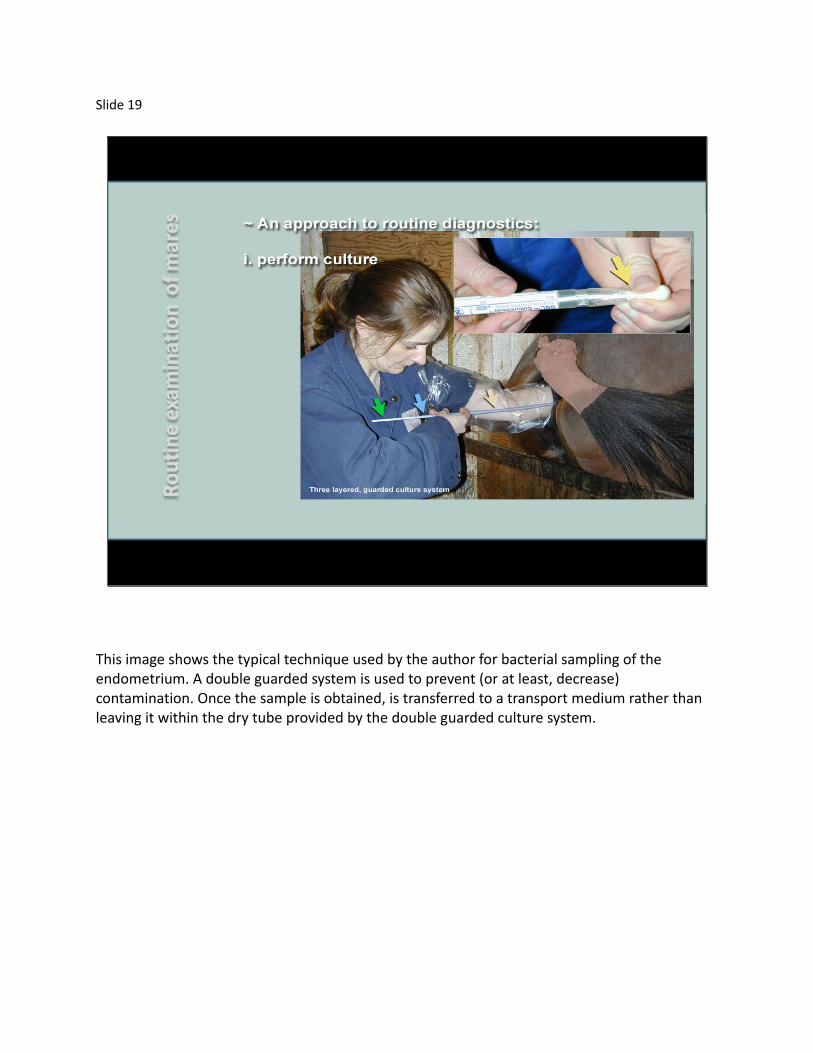
This image shows the typical technique used by the author for bacterial sampling of the endometrium. A double guarded system is used to prevent (or at least, decrease) contamination. Once the sample is obtained, is transferred to a transport medium rather than leaving it within the dry tube provided by the double guarded culture system.

Slide 20
To obtain excellent cytology samples, we use a modification of the human Pap smear brush. It is sterilized and inserted into the end of a standard fusette (a plastic infusion tube) which is then inserted into the outer tube of the double guarded culture system ( the outer tube is left in place after bacteriological sampling ). We do not use a swab to obtain cytology because it is inferior to this system.

Slide 21
The cytology brush is rolled out onto two slides which have been identified with the name of the mare and the date. One slide is marked "Diff quick" and the other, "Gram". If there is evidence of bacteria or inflammation on the "Diff quick" slide, the other slide is submitted for Gram staining. Usually this is unnecessary, so the majority of Gram slides are discarded. This is fortunate because Diff quick staining is rapid and can be done at the stud farm while the mare is still being examined.

Slide 22
After the culture and cytology samples have been obtained (culturing is always the first examination that is done) an endometrial biopsy is often taken. The biopsy punch is inserted through the cervix and into the uterus using a finger as a guide through the cervix. Although it is sometimes stated that one should biopsy the site of implantation, this is difficult to do and appears not to be essential. After placing the biopsy punch into the uterus (with the jaws of the punch closed), the hand is removed from the vagina and the same hand is inserted into the rectum to locate the jaws of the punch. The punch jaws are then opened so that the side of the jaws faces dorsoventral. Using very gentle pressure to insert the endometrium into the jaws of the punch, the jaws are closed and the biopsy is obtained. Several kinds of biopsy instruments are available, are working on approximately the same principle as described above.

Slide 23
After reading endometrial biopsies for more than 25 years, this author is convinced that more than 80 to 85% of all the endometrial biopsies taken will be essentially normal. In the image of a biopsy at left there is severe, chronic endometritis as is evidenced by the periglandular fibrosis. This degree of fibrosis is unusual. At right, there is evidence of a more acute case of endometritis. One can say this because of the relative absence of fibrous tissue and massive numbers of neutrophils. One should appreciate that there is always a resident population of lymphocytes and plasmacytes in the stratum compactum, providing protection through the generation of immunoglobulins and macrophages. Sometimes this is erroneously described as a mild diffuse lymphocytic to plasmacytic endometritis. Endometrial biopsies can reveal many things besides inflammatory processes. For example, hemosiderin can indicate that a mare has foaled before, the height of the mucosal epithelium may show that a mare is in estrus or diestrus. Also, the presence and density of endometrial

gland cross-sections can indicate if a mare is cycling and has recently been under the influence of progesterone. In essence therefore, a high density of gland cross-sections infers that a mare has an intact and functional hypothalamic-pituitary-ovarian axis.
Slide 24
Pregnancy diagnosis; and essential part of broodmare management.

Slide 25
One of the earliest signs of pregnancy is an increase in cervical tone. In fact, the cervix can be felt as a thin, pencil like structure is early as 14 of 15 days after a fertile ovulation. This is about the time that a mare would normally be returning to estrus if she were not pregnant. Uterine tone develops shortly afterwards and is still fairly obvious up until 55 or 60 days of gestation. One should remember that uterine tone usually indicates pregnancy in a mare and estrus in a cow!

Slide 26
Because the early equine embryo is not an elongated structure as it is in cattle, it moves about the uterus to convey the signals of early pregnancy recognition to the endometrium. It is spherical as shown.

Slide 27
In the absence of ultrasonography (like practicing without a thermometer!) one only has tentative evidence of pregnancy because of uterine tone to about 25 to 28 days of gestation when the fetal-placental unit is palpable at the junction between a uterine horn and the body of the uterus. However it must be emphasized that this is far too late for general pregnancy examinations because it does not allow for the management of twin pregnancies.

Slide 28
Before the advent of ultrasonography it was common to use eCG tests to confirm pregnancy in mares; especially if the veterinarian was not experienced in rectal palpation. Tests for eCG usually become positive at approximately 28 to 30 days of gestation and remains positive for up to 120 days or more. In fact, some mares remain positive throughout the rest of gestation because their endometrial cups are not rejected by the mare and remain functional. One should always remember that tests for eCG will remain positive even after an embryo has died as long as the pregnancy has advanced to a point where the endometrial cups have become well established i.e. after about 30 or 40 days of gestation. Therefore false positive test results will be obtained in such cases. When mares fail to return to estrus cycles after having lost pregnancies , these tests will be positive, showing that endometrial cups are still present. Typically, these mares cannot be rebred until the endometrial cups have stopped functioning.
Slide 29
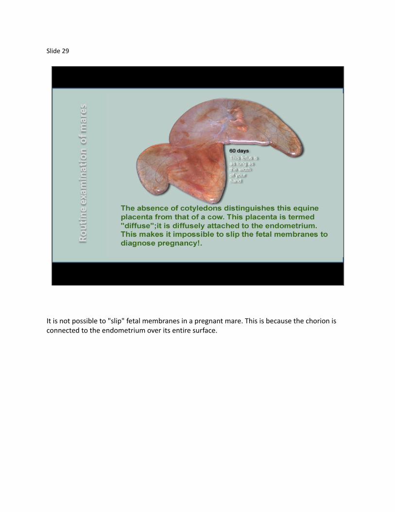
It is not possible to "slip" fetal membranes in a pregnant mare. This is because the chorion is connected to the endometrium over its entire surface.

Slide 30
During the last half of gestation, the placenta of most domestic species becomes a substantial source of estrogens, mostly estrone sulfate. This is actually a derivative of androgens that are produced by the fetal gonads (male or female). These androgens are then aromatized in the placenta to estrogens. The function of these estrogens is largely unknown but there may be important in mammary gland development. Estrone sulfate assays can be done on either serum or urine and are valuable for diagnosing pregnancy in wild equids such as zebras, Prezwalski horses and mustangs.

Slide 31
At about 5 to 7 months of gestation it is difficult to feel the fetus by transrectal palpation because it is so low within the abdomen. However it is easy to visualize the fetus using transabdominal ultrasonography during this time.

Slide 32
As mentioned elsewhere, ultrasonography is of cardinal importance in equine stud management, especially with regard to the diagnosis and monitoring of pregnancy.

Slide 33
Pregnancy is usually diagnosed at about 14 to 16 days after the time that an intact follicle was last seen. These words are carefully chosen; notice that we do not say "after the time of ovulation". This is because we usually examine brood mares every two or even three days (over weekends ) we can never be sure of when ovulation actually occurred. That may seem like a trivial point but it is actually of great importance because equine embryos stop moving within the uterus at about 16 days after the exact time of ovulation; important to bear in mind when dealing with twins. If we calculate the age of pregnancy from the time that a corpus hemorrhagicum is first observed, it is possible that the ovulation may have occurred anywhere between three minutes to three days before that observation; the corpus hemorrhagicum is identical on ultrasonography in both cases. Therefore it is possible to underestimate the real age of pregnancy by up to three days. That being the case, the embryo or embryos would have stopped moving within the uterus and attempts to reduce twin pregnancies would become more complicated and less successful.

Slide 34
When a mare is first examined at a stud farm, it is important to "map" all of the cystic structures within the uterus so that they will not cause confusion during early pregnancy diagnosis.

Slide 35
During early gestation is possible to use common and relatively inexpensive 3.5 to 7.5 MHz transducers for transrectal ultrasonography. However in mid- to advanced gestation, it is best to perform transabdominal ultrasonography using low-frequency (2 to 3 MHz) transducers for optimal results. Fortunately, some new ultrasound machines can provide frequencies that cover most of this range using a single transducer.

Slide 36
FINIS