(This is a sample cover image for this issue. The actual ...srbce.org/forum/sameer gupta and C....
15
(This is a sample cover image for this issue. The actual cover is not yet available at this time.) This article appeared in a journal published by Elsevier. The attached copy is furnished to the author for internal non-commercial research and education use, including for instruction at the authors institution and sharing with colleagues. Other uses, including reproduction and distribution, or selling or licensing copies, or posting to personal, institutional or third party websites are prohibited. In most cases authors are permitted to post their version of the article (e.g. in Word or Tex form) to their personal website or institutional repository. Authors requiring further information regarding Elsevier’s archiving and manuscript policies are encouraged to visit: http://www.elsevier.com/copyright
Transcript of (This is a sample cover image for this issue. The actual ...srbce.org/forum/sameer gupta and C....

(This is a sample cover image for this issue. The actual cover is not yet available at this time.)
This article appeared in a journal published by Elsevier. The attachedcopy is furnished to the author for internal non-commercial researchand education use, including for instruction at the authors institution
and sharing with colleagues.
Other uses, including reproduction and distribution, or selling orlicensing copies, or posting to personal, institutional or third party
websites are prohibited.
In most cases authors are permitted to post their version of thearticle (e.g. in Word or Tex form) to their personal website orinstitutional repository. Authors requiring further information
regarding Elsevier’s archiving and manuscript policies areencouraged to visit:
http://www.elsevier.com/copyright

Author's personal copy
Journal of Steroid Biochemistry & Molecular Biology 134 (2013) 23– 36
Contents lists available at SciVerse ScienceDirect
Journal of Steroid Biochemistry and Molecular Biology
jo u r n al hom epage: www.elsev ier .com/ locate / j sbmb
Physiological crosstalk between melatonin and glucocorticoid receptormodulates T-cell mediated immune responses in a wild tropical rodent,Funambulus pennanti
Sameer Gupta, Chandana Haldar ∗
Pineal Research Laboratory, Department of Zoology, Banaras Hindu University, Varanasi 221005, India
a r t i c l e i n f o
Article history:Received 10 May 2012Received in revised form 23 August 2012Accepted 12 September 2012
Keywords:Melatonin receptorsAntagonistsGlucocorticoidsImmunitySeasonal stressWild rodent
a b s t r a c t
Immunoenhancing attributes of melatonin (Mel) on the immunocompromised state induced by gluco-corticoid is well known, but the involvement of their receptors in the modulation of immunity has neverbeen studied in any rodent. The present study explores the role of Mel and its receptors (MT1 and MT2) inamelioration of immunocompromised state induced by a synthetic glucocorticoid, dexamethasone (Dex)in a tropical rodent Funambulus pennanti. Immune parameters viz. DTH response, Lymphocyte prolifer-ation, cytokine (IL-2) and antibody production were assessed following pretreatment of Mel and Dexalone or in combination. Mel enhanced the IL-2 production, thymic and splenic lymphocyte prolifera-tion thereby increasing T helper cell associated immune responses and anti-KLH-IgG production. MT1and MT2 receptor expression was downregulated following Dex treatment while glucocorticoid recep-tors (GR) expression was downregulated in Mel treated groups suggesting that the immunomodulatoryeffects of glucocorticoids and Mel are mediated via their receptors. To gain further insights on the role ofMel receptors, we used nonselective melatonin receptor antagonist luzindole which resulted in reversalof most of the immunomodulatory actions of Mel. Therefore, it may be suggested that a physiologicalcross talk exist between Mel and GR which is of high adaptive significance in wild animals for balancingthe immunity during ecologically stressful conditions.
© 2012 Elsevier Ltd. All rights reserved.
1. Introduction
Chronobiotic neurohormone melatonin plays a pivotal role inmodulation of seasonal reproduction and immunity in mammals[1–4]. Being active at cellular level interacting with various func-tional proteins and scavenging free radicals, melatonin also actsvia high affinity G protein coupled receptors (GPCRs). Till date twohigh affinity melatonin membrane receptors (MT1 and MT2) havebeen cloned and characterized in mammals [5–7]. These are ubiq-uitously located on immunocompetent cells and lymphoid tissueslike thymus and spleen [8–10].
Glucocorticoid, especially corticosterone is a hallmark of stressin rodents which evokes adaptive physiological strategies andhelps animal to cope up with stressful environmental conditions.Glucocorticoids exhibit their biological actions via two receptors,belonging to the nuclear hormone receptor superfamily, viz. gluco-corticoid receptor (GR) and mineralocorticoid receptor (MR). GR islow affinity glucocorticoid receptor and have two isomers/splicing
∗ Corresponding author. Tel.: +91 542 2307149x209/6702535;fax: +91 542 2368174/2575093; mobile: +91 9415222261.
E-mail address: [email protected] (C. Haldar).
variants. It is mainly active during the periods of abundant gluco-corticoid secretion such as circadian peak or systemic inflammation[11,12]. In immune system glucocorticoid effects are primarilymediated by GR [13,14].
Melatonin and glucocorticoid acts in concert to integrate theseasonal changes at the level of immune system that are equippedwith the melatonin and glucocorticoid receptors. In general glu-cocorticoids are known for immunoinhibitory effect on T cell andNK cell function there by hampering the immune status [14] whilemelatonin is well known for its immunoenhancing attributes [15].Interaction between glucocorticoids and the immune functionshave been explored mostly in relation to stress [16], and glucocor-ticoids are considered to mediate the immunosuppressive effectsof stress. Therefore, the present study has been planned in wildseasonal breeder as they experience more stress in terms of eco-factors (extreme conditions of temperature, scarcity of food), socialpressure and avoiding being killed by predators. Although, gluco-corticoids are necessary for the maintenance of overall metabolismwhich ensures survival during periods of stress, their inhibitory andfunctionally suppressive effects on the immune system is consid-erably less understood.
Glucocorticoid studies are clinically relevant because syn-thetic glucocorticoids are used as potent anti-inflammatory and
0960-0760/$ – see front matter © 2012 Elsevier Ltd. All rights reserved.http://dx.doi.org/10.1016/j.jsbmb.2012.09.013
Author's personal copy
24 S. Gupta, C. Haldar / Journal of Steroid Biochemistry & Molecular Biology 134 (2013) 23– 36
immunosuppressive agents for numerous functions associatedwith host defense [17]. Dexamethasone is one of the commer-cially available synthetic glucocorticoid which has a wide rangeof use in clinical conditions like chronic asthma [18], rheumatoidarthritis [19], and tissue/organ transplantation [20], which is basedon empirical evidences of its efficacy, while surprisingly little isunderstood concerning the mechanisms by which glucocorticoidssuppress immune function.
Earlier reports suggest that dexamethasone treatment in vivoaltered lymphoid tissue mass, morphology and resulted indecreased circulating immune cell number in tropical squirrelFunambulus pennanti [21]. However, till date the receptor medi-ated modulation of immune function by melatonin and adrenalsteroids has never been studied in any wild rodent. The presentstudy explores the role of melatonin and its membrane recep-tors (MT1/MT2) in regulation of GR induced immunosuppressionin a wild, tropical, seasonally breeding rodent F. pennanti. Fur-thermore, in order to delineate whether the antagonizing effectof melatonin on glucocorticoid induced stress on immune status ismediated by membrane bound melatonin receptor or is indepen-dently regulated by melatonin, we treated the splenocytes in vitrowith luzindole, a nonselective melatonin receptor MT1/MT2 antag-onist.
2. Materials and methods
All the experiments were conducted in accordance with Institu-tional practice and with in the framework of experimental animals(Scientific Procedure) Act 2007, of the Committee for the Purposeof Supervision and Control on Experiments on Animals (CPSCEA),Government of India, on animal welfare.
2.1. Animals and maintenance
The experiment was performed on healthy young adult malesquirrels F. pennanti approximately of same age (as judged by theircranium diameter, incisor length and body weight (120 ± 5 g) asreported earlier) [22]. The experiment started in the last week ofAugust and was continued till October. During this period the envi-ronmental day length in Varanasi (latitude 25◦18′N and longitude83◦1′E) is approximately 12 h light and 12 h dark (Temp. minimum28 ◦C − maximum 37 ◦C) and biotic and abiotic stress are minimumin nature. The squirrels were collected from the vicinity of Varanasiduring the last week of August. The squirrels were weighed andkept in wire net cages (25′′ × 25′′ × 30′′ in size) in a room havingphotoperiod of 12 light:12 dark and temperature 25 ± 2 ◦C for briefperiod of acclimatization for 10 days prior to treatment with dexa-methasone. All the experimental protocols were continued for 45days. After completion of treatments the animals were sacrificedduring third week of October. During experiment the animals werefed with soaked gram seeds (Cicer arietinum), seasonal fruits, nutsand water ad libitum. The experimental groups were divided asfollows:
2.2. Experimental plan I: in vivo
For in vivo experiment the squirrels were randomly divided intofour groups each containing 15 animals (n = 15/group) and treat-ment given was as follows:
Group I: Vehicle (normal saline) treated (Con)Group II: Melatonin treated (Mel); 25 �g/100 g body wt./dayGroup III: Dexamethasone treated (Dex); 60 �g/100 g bodywt./dayGroup IV: Melatonin + Dexamethasone treated (Mel + Dex)
2.2.1. Drugs and treatmentsMelatonin, dexamethasone, mitogen Concanavalin A (Con A),
luzindole, oxazolone and keyhole limpet hemocyanin (KLH; anti-gen) were all purchased from Sigma–Aldrich Chemicals, St. Louis,MO, USA). Melatonin and dexamethasone were first dissolved infew drops of ethanol and then diluted with normal saline up todesired concentration and injected. Melatonin was injected sub-cutaneously during evening hours (between 17:30–18:00 h IST;approximately 1–1.5 h before sunset), dexamethasone was injectedintramuscularly; the control animals were injected with the vehi-cle, normal saline in the same amount. All the injections/doseswere selected as reported earlier [21,23], and were continuedfor 45 days. Experiment was performed in three sets. Second setof squirrels (n = 5) was subjected for delayed type hypersensitiv-ity (DTH) response study while the third set (n = 5) was used forthe study of humoral immune response in terms of anti-KLH-IgGproduction.
2.2.2. Sample collectionAfter 24 h of the last injection the squirrels were weighed and
sacrificed under deep ether anesthesia inhalation. All the sacrificeswere made during the night time between (20:00–22:00 h, IST).The blood was collected by cardiac puncture in heparinized tubesand centrifuged at 3000 rpm for 20 min at 4 ◦C. Plasma was keptat −20 ◦C till the hormonal and other humoral estimations wereperformed. Spleen and thymus were dissected out on ice, weighedand processed for the assay of blastogenic response in terms oflymphocyte proliferation and also a part of it was kept for receptorassay and other biochemical estimations.
2.2.3. Isolation and culture of Peripheral Blood Mononuclear Cells(PBMC)
The PBMC were isolated from heparinized blood collected bycardiac puncture (n = 5), by density gradient centrifugation, anadapted method of isolating mononuclear cells by Boyum [24].Lymphocyte separation media was used according to manufac-turer’s instruction (HiSepTM LSM 1084, HiMedia, Mumbai, India).Briefly, the white band at the plasma-ficoll (Hisep, Himedia)interphase was collected and washed twice with PBS and finallysuspended in RPMI 1640 (Himedia, India) supplemented with 10%fetal calf serum and 100 units of penicillin and streptomycin (SigmaAldrich, USA). The cell concentration was adjusted to 1 × 10⁷ viablecells/ml with RPMI 1640. MTT assay was performed as mentionedin the following section.
2.2.3.1. MTT assay for thymocyte, splenocyte and peripheral bloodmononuclear cells (PBMC) proliferation. Cell-mediated immunefunction was assessed by measuring splenocyte, thymocyte andPBMC proliferation in response to the T-cell specific mitogen,Concanavalin A (Con A), using a colorimetric assay based onthe reduction of tetrazolium salt 3-(4,5-dimethylthiazol-2-yl)-2,5-diphenyltetrazolium bromide (MTT) [25]. Spleen and thymus ofsquirrels were removed in sterile condition and a single-cell sus-pension was prepared by mincing and grinding them betweensterile frosted glass slides. Erythrocytes were lysed by hypotonicshock using equal volume of cold ammonium chloride tris buffer(tris hydroxymethylene aminomethane, SRL, Mumbai, India); 0.5%tris buffer and 0.84% NH4Cl mixed in 1:10 ratio; pH 7.2). Thissingle cell suspension along with ice cold culture medium (RPMI-1640 supplemented with 1% penicillin (5000 U/ml) streptomycin(100 �g/ml), 1% l-glutamine (2 mM/ml), 0.1% 2-mercaptoethanol(5 × 10⁻² M/ml), and 10% heat-inactivated fetal calf serum). Thecell suspension was washed thrice. The cells were counted using ahemacytometer and viability was determined by trypan blue exclu-sion method. Viable cells (which exceeded 95%) were adjusted to1 × 107 cells/ml in culture medium, and 100 �l aliquots of each cell

Author's personal copy
S. Gupta, C. Haldar / Journal of Steroid Biochemistry & Molecular Biology 134 (2013) 23– 36 25
suspension were added to the wells of sterile flat-bottom 96-wellculture plates. 50 �l/well Concanavalin A (Con A, Sigma–Aldrich, St.Louis, USA) was added to the culture medium at the concentrationof 10 �g/ml to yield a final volume of 150 �l/well (each in dupli-cate). Finally 50 �l of complete medium was added to make the finalvolume of 200 �l/well (each in duplicate). Plates were incubated at37 ◦C with 5% CO2 for 69 h prior to addition of 10 �l of MTT (SRL,Bombay, India; 5 mg/ml in phosphate buffered saline) per well fol-lowed by an additional incubation for 4 h. At 72 h, 150 �l of acidifiedpropanol (0.04 mol/l HCL in isopropanol) was added to each cultureand the optical density (OD) of each well was determined with amicroplate reader (ELx-800, Biotek Instruments, Winooski VT, USA)equipped with a 570 nm wavelength filter. Mean OD values foreach set of duplicates were used in subsequent statistical analyses.Response was calculated as percent stimulation ratio (%SR) repre-senting the ratio of absorbance of mitogen stimulated cultures tocontrol cultures [23].
2.2.4. Western blot analysis for MT1, MT2 and GRThe western blot analysis was performed according to method
published elsewhere [10]. Briefly, the spleen and thymus were diss-ected out in chilled PBS. PBMC were separated from the blood ofthe squirrels as described in previous section. Tissue/cells werehomogenized and lysed in RIPA buffer containing aprotinin, sodiumorthovanadate and phenyl methylsulphonyl fluoride (PMSF). Theprotein content of the lysates were quantified using the Brad-ford method [26]. The aliquots containing 70 �g of protein forlymphoid organs and 40 �g protein for PBMC was resolved on12% SDS-polyacryl-amide gel electrophoresis (PAGE) for melatoninmembrane receptor MT1 and MT2 and 10% SDS-PAGE for GR fol-lowed by electrotransfer (Biometra, Germany) on nitrocellulose(Boiscience, Keene, NH, USA) for 1 h. Nitrocellulose membraneswere blocked for 60 min in Tris-buffered saline (TBS; Tris 50 mM,pH 7.5, NaCl 150 mM) containing 5% fat free dry milk and wereincubated with melatonin receptor MT1 and MT2 and glucocor-ticoid receptor (GR) antibodies (Mel1aR, R-18, Mel1bR, T-18, andGR, M-20, sc 1004, Santa Cruz Biotechnology, Santa Cruz, CA, USA).All the antibodies i.e. MT1, MT2 and GR were used at a dilutionof 1:200. The membranes were then washed thrice with TBS. Theimmunodetection was carried out using horseradish peroxidase(HRP) conjugated secondary antibody (donkey antigoat IgG-HRPfor melatonin receptors and donkey antirabbit IgG-HRP for GR;diluted 1:1000). Finally, the blots were washed thrice with TBSand developed with Super Signal West Pico Chemiluminescent sub-strate (34080, Thermo Scientific, USA). Further, the nitrocellulosemembranes were then stripped with stripping buffer (10% sodiumazide) and were immunostained with �-actin antibodies in 1:1000dilutions (A-2228; Sigma–Aldrich, USA) as internal loading control.Immunodetection of �-actin was performed with anti-mouse IgG-HRP (1:1000). Bands were quantified by measurement of opticaldensity using Scion Image Analysis Software (Scion Corporation).Values were expressed as the ratio of the density of the specificsignal to the �-actin signal [10]. The ratio of density was calculatedwith respect to �-actin (house-keeping gene) and expressed as %band intensity of MT1, MT2 and GR.
2.2.5. Radioimmunoassay (RIA) of melatoninThe radioimmunoassay (RIA) of melatonin was performed
following the method of Rollag and Niswender [27] using Guild-hey antisera (Guildhey, Surrey, UK). Details of the method andvalidation have been published elsewhere [28]. The intra- andinter-assay variation for melatonin was 9% and 15%, respectively.The sensitivity for melatonin RIA was 18–20 pg/ml. The recovery ofmelatonin RIA was 92%.
2.2.6. Corticosterone ELISACorticosterone was measured using an ELISA kit (kind gift from
Prof. T. G. Srivastava, National Institute for Health and Family Wel-fare, New Delhi, India) according to the manufacturer’s instructions.Plasma samples were thawed, and applied to a microplate. Fol-lowing incubation, the plates were read on a microplate readerat 405 nm, and values were determined by extrapolation from astandard curve. The recovery, accuracy, and sensitivity of the cor-ticosterone assay were 95%, 99.1%, and 0.27 �g/dl, respectively.Inter- and intra-assay variation was 3.38–5.56% and 5.69–7.84%,respectively. All samples were assayed in a single lot and samples,standards, and replicates were assayed in duplicate.
2.2.7. ELISA for IL-2Sandwich ELISA was performed to quantify plasma level of IL-
2 in all the four groups according to manufacturers instruction(Immunotech, France) Intra assay variation was between 3.3 and7.2% and inter assay variation was between 6.2 and 8.2%; sensitiv-ity; 5 pg/ml and recovery was between 80 and 132%.
2.2.8. Delayed-type hypersensitivity (DTH) responseAfter 45 days of treatment, DTH was induced by application of
antigen oxazolone (Sigma, St Louis, MO, USA) to the ear pinna of thesquirrel. Following the initial immunization by applying 100 �l ofOxazolone [5% (w/v) in 4:1, acetone:olive oil] for two consecutivedays to shaved dorsal region. On day 6 the baseline ear thicknesswas measured using vernier calliper and oxazolone challenge wasinduced by applying 50 �l of oxazolone [0.5% (w/v) in 4:1, ace-tone:olive oil] to the skin of the dorsal surface of right ear pinna.Left ear pinna was treated with the vehicle solution alone. Earswellings were measured for three consecutive days to record themaximum DTH response which eventually was observed on the2nd day. The DTH response was expressed as change in percentthickness and was measured by comparing differences betweenoxazolone treated and vehicle treated ear pinna [3,29].
2.2.9. Anti-KLH-IgG estimation for humoral immune statusThe anti-KLH-IgG estimation assay was performed following the
method of [30], with some modifications which has been publishedelsewhere [29]. Briefly, 24 h after the last injection the third experi-mental set of the squirrels received single subcutaneous injection of150 �g of KLH (Sigma–Aldrich) in 0.1 ml of sterile normal saline (0day). The squirrels were sacrificed and trunk blood was collected onthe days 5 and 10 (post immunization). These periods were chosento capture basal (5 days) and peak (10 days) levels of IgG. The bloodwas allowed to clot; after 1 h, the clots were removed and sampleswere centrifuged (at 4 ◦C) for 30 min at 700 × g; serum aspiratedand aliquots were stored at −20 ◦C until assayed for anti-KLH-IgG.
2.2.9.1. ELISA for anti-KLH-IgG. The humoral immune status wasassessed by measuring serum anti-KLH-IgG concentrations usingELISA [29,30]. In brief, the microtiter plates were coated with KLHantigen (0.5 mg/ml in sodium bicarbonate buffer) by overnightincubation at 4 ◦C. Next day the plates were washed with phosphatebuffered saline containing 0.05% Tween 20 (PBST), blocked with0.5% non-fat dry milk in PBST overnight at 4 ◦C and washed againwith PBST. The serum samples were diluted 1:50, 1:100, 1:200,1:400 and 1:800 in PBST, and finally 150 �l of each serum dilutionwas loaded in duplicate in antigen coated microtiter plate. Simi-larly the positive control (squirrels those have prior been exposedto KLH showing robust immune reaction) and negative control sam-ples (squirrels never exposed to KLH) were diluted and loaded inwells in duplicate. The plates were sealed and incubated at 37 ◦Cfor 3 h followed by PBST wash. Secondary antibody (alkaline phos-phatase conjugated, anti-mouse IgG; SC-2320, Santa Cruz Biotec,USA) was added to each well in a dilution of 1:500; and plates were
Author's personal copy
26 S. Gupta, C. Haldar / Journal of Steroid Biochemistry & Molecular Biology 134 (2013) 23– 36
incubated again at 37 ◦C for 1 h followed by washing with PBST.150 �l of p-nitrophenyl phosphate (enzyme substrate) 1 mg/ml indiethanolamine buffer (Sigma–Aldrich) was added to each well andthe plates were kept in dark, the enzyme-substrate reaction wasterminated by adding 50 �l of 1.5 M NaOH to each well. The opti-cal density (OD) of each well was determined using a microplatereader equipped with a 405 nm filter (ELx-800, Biotek Instruments,Winooski, VT, USA) and average OD was calculated for each sam-ple in duplicate. The mean for each sample was calculated andexpressed as a percentage of the positive control mean (% platepositive).
2.3. In vitro study
For in vitro study five squirrels were sacrificed (n = 5), spleno-cyte (and thymocyte) culture was performed in two experimentalgroups with four and five sets respectively each with quadruplicate.The groups were as follows.
2.3.1. Experimental set I: splenocytes and thymocytes cultureGroup I – Control (Con)Group II – Melatonin supplemented (Mel; 500 pg/ml)Group III – Dexamethasone supplemented (Dex; 2 �M)Group IV – Melatonin + Dexamethasone co supplementation (Mel;500 pg/ml + Dex; 2 �M)
2.3.2. Experimental set II: splenocytes cultureIn order to find out the role of melatonin via melatonin
receptors if any in alleviation of glucocorticoid induced immuno-compromised state splenocytes were cultured under five differentgroups.
Group I – Control (Con)Group II – Dexamethasone supplemented (Dex); 2 �MGroup III – Dexamethasone + Luzindole (Dex; 2 �M + Luz; 5 �M)Group IV – Mealtonin + Dexamethasone co supplementation(Mel; 500 pg/ml + Dex; 2 �M)Group V – Melatonin + Dexamethasone + Luzindole (Mel + Dex+ Luz)
Splenocyte and thymocytes proliferation was assessed usingMTT assay method [21]. Further, immunoblot analysis of thecultured splenocytes and thymocytes of experimental set I formelatonin receptor and GR was carried out and culture super-natants were also assessed for IL-2 production in all the groups. Forexperimental set II only %SR and GR expression were monitored insplenocytes following treatment.
2.3.3. MTT assay for splenocyte and thymocyte proliferationThe cells were isolated as explained in the previous section. The
viable cells (which exceeded 95%) were adjusted to 1 × 10⁷ cells/mlwith culture medium and 50 �l of cell suspension was added to thewells of sterile flat bottom, 96 well culture plate. Con A (SigmaAldrich) was added to the culture medium at a concentration of10 �g/ml and finally 50 �l of the mitogen was added to yield afinal volume of 100 �l/well each in quadruplicate. Melatonin, dexa-methasone and luzindole were all dissolved in absolute ethanol andthen diluted with RPMI-1640 to standard stock solution to be used.One hundred microliters of dexamethasone (2 �M) or melatonin(500 pg/ml) or 50 �l of each dexamethasone, melatonin and luzin-dole at a concentration of 5 �M was added to each well ultimatelyto yield a final volume of 200 �l/well (each in quadruplicate). Thecontrol cultures comprised of the same sets of treatment except forCon A. Melatonin, dexamethasone, Con A and luzindole were addedto the plates prior to the addition of cells. Plates were incubated inhumidified CO2 incubator at 37 ◦C. Harvesting of cultures for having
final OD were processed as described in the earlier in vivo sectionof the manuscript.
2.3.4. Western blot analysis of splenocytes and thymocytesThe splenocytes (and thymocytes) were collected under sterile
conditions using routine method as explained in previous sec-tions. The cells were suspended in RPMI-1640 at a concentrationof 1 × 10⁷ cells/ml. Then, 100 �l of cell suspension, 100 �l of mela-tonin (500 pg/ml), dexamethasone (2 �M) or both and RPMI-1640medium were added to each well of a 24-well culture plate to makea final volume of 1 ml. For antagonist part luzindole (5 �M), mela-tonin, dexamethasone or all the components along with RPMI-1640were added to make up the final volume to 1 ml. After 48 h of incu-bation at 37 ◦C with 5% CO2 the cells were collected and centrifugedat 2500 rpm for 10 min. The cell pallet was washed twice withPBS (0.1 M, pH 7.4) and finally the homogenate was made usinglysis buffer and protein content in the cell lysates was quantified.Aliquots containing 30 �g of protein were resolved on 12% SDS-PAGE for MT1 and MT2 and 40 �g aliquots were resolved on 10%SDS-PAGE for GR. Further details regarding western blot has alreadybeen given in the previous section of the manuscript.
2.3.5. IL-2 production and analysis in culture supernatantIL-2 level was estimated from culture supernatant of activated
splenocytes and thymocytes. Splenocytes and thymocytes werecollected by a routine method under sterile condition as explainedabove. The cells were suspended in RPMI-1640 medium at aconcentration of 1 × 10⁷ cells/ml. Then, 100 �l of cell suspension,100 �l of melatonin (500 pg/ml), 100 �l of dexamethasone (2 �M)or both, luzindole (5 �M; [19]), 100 �l of Con A with a final concen-tration of 10 �g/ml and RPMI-1640 medium were added to eachwell of a 24-well culture plate to make a final volume of 1 ml in eachwell. The cultures were centrifuged (500 × g, 10 min) after incuba-tion at 37 ◦C with 5% CO2 for 48 h. Then, the supernatants werecollected and preserved at −20 ◦C for IL-2 estimation. SandwichELISA was performed to quantify the level of IL-2 in culture super-natants according to the manufacturer’s instructions (Immunotech,France).
3. Statistical analyses
Statistical analysis of the data was performed using SPSS 17.0(SPSS Corp., USA) with one-way ANOVA followed by multiple com-parisons by the Duncan’s multiple range tests. The differences wereconsidered statistically significant when p < 0.05.
4. Results
4.1. In vivo study
4.1.1. Lymphocyte proliferation index of spleen, thymus andPBMC
Percent stimulation ratio (%SR) representing the change incell mediated immune status (T-helper cells) in response to a Tcell specific mitogen Concanavalin A challenge, presented a sig-nificant (p < 0.01) increase in splenocyte, thymocyte and PBMCproliferation following melatonin treatment, when compared withsaline treated control groups. A significant decrease (p < 0.01) wasobserved in splenic and thymic lymphocyte proliferation alongwith PBMC following dexamethasone treatment. Melatonin treat-ment along with dexamethasone however, significantly (p < 0.01)increased splenocytes, thymocytes and PBMC proliferation com-pared to dexamethasone treated group (Fig. 1A).

Author's personal copy
S. Gupta, C. Haldar / Journal of Steroid Biochemistry & Molecular Biology 134 (2013) 23– 36 27
Fig. 1. Effect of melatonin and dexamethasone treatment on proliferation of lymphocyte and mononuclear cells stimulated with Con A. (A) Blastogenic response (%SR) ofsplenocytes, thymocytes and PBMC following in vivo melatonin and dexamethasone administration. IL-2 levels in plasma (B) and in culture supernatant of PBMC (C) of Con,Mel, Dex and Mel + Dex treated F. pennanti. Values are expressed as mean, vertical bar on histograms represents ± SEM, n = 5. Significance of difference; **p < 0.01, Con vs. allexperimental groups and #p < 0.01, Dex vs. Mel + Dex.
4.1.2. Western blot analysisTo check the expression of melatonin receptor subtypes (MT1
and MT2) and GR in lymphoid tissues i.e. spleen and thymusof F. pennanti at translational level, Western blot analysis wasperformed. Melatonin receptor protein was detected as a sin-gle band between 35 and 40 kDa corresponding to the reportedmolecular mass of the receptor protein [10,23,31]. Single band forGR protein was detected between 80–100 kDa in the lymphoidtissues.
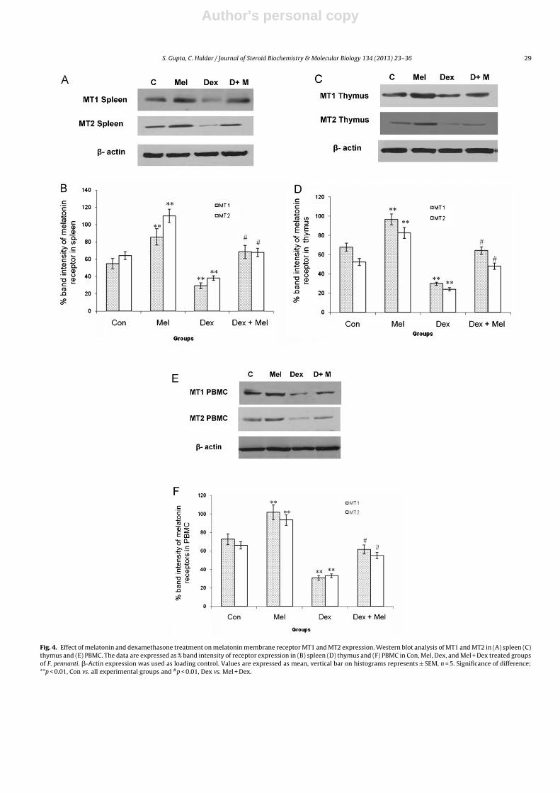
Following the in vivo administration of melatonin and dexa-methasone injections, both the melatonin membrane receptors(MT1 and MT2) presented similar expression pattern in spleenand thymus. A significant (p < 0.01) increase was observed in MT1and MT2 receptor expression following melatonin injection in bothspleen and thymus when compared to vehicle treated controlgroup. Dexamethasone treatment, on the other hand significantly(p < 0.01) decreased the melatonin membrane receptor MT1 andMT2 expression in spleen and thymus. Melatonin treatment along
Fig. 2. Effect of melatonin and dexamethasone treatment on plasma melatonin and corticosterone concentration. (A) Plasma melatonin (pg/ml) and (B) corticosterone level(ng/ml) following in vivo treatment of Mel, Dex and Mel + Dex in F. pennanti. Values are expressed as mean, vertical bar on histograms represents ± SEM, n = 5. Significanceof difference; **p < 0.01, Con vs. all experimental groups and #p < 0.01, Dex vs. Mel + Dex.
Author's personal copy
28 S. Gupta, C. Haldar / Journal of Steroid Biochemistry & Molecular Biology 134 (2013) 23– 36
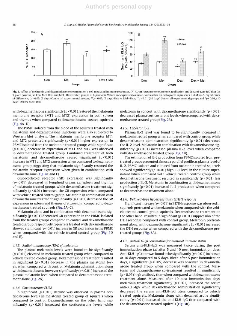
Fig. 3. Effect of melatonin and dexamethasone treatment on T cell mediated immune responses. (A) %DTH response to oxazolone application and (B) anti-KLH-IgG titer (as% plate positive) in Con, Mel, Dex, and Mel + Dex treated groups of F. pennanti. Values are expressed as mean, vertical bar on histograms represents ± SEM, n = 5. Significanceof difference; *p < 0.05, (5 days) Con vs. all experimental groups; ##p < 0.05, (5 days) Dex vs. Mel + Dex; **p < 0.01, (10 days) Con vs. all experimental groups and #p < 0.01, (10days) Dex vs. Mel + Dex.
with dexamethasone significantly (p < 0.01) restored the melatoninmembrane receptor (MT1 and MT2) expression in both spleenand thymus when compared to dexamethasone treated squirrels(Fig. 4A–D).
The PBMC isolated from the blood of the squirrels treated withmelatonin and dexamethasone injections were also subjected toWestern blot analysis. The melatonin membrane receptor MT1and MT2 presented significantly (p < 0.01) higher expression inPBMC isolated from the melatonin treated group; while significant(p < 0.01) decrease in expression of MT1 and MT2 was observedin dexamethasone treated group. Combined treatment of bothmelatonin and dexamethasone caused significant (p < 0.01)increase in MT1 and MT2 expression when compared to dexameth-asone group suggesting that melatonin significantly restored themelatonin receptor expression when given in combination withdexamethasone (Fig. 4E and F).
Glucocorticoid receptor (GR) expression was significantly(p < 0.01) decreased in lymphoid organs i.e. spleen and thymusof melatonin treated groups while dexamethasone treatment sig-nificantly (p < 0.01) increased the GR expression when comparedwith vehicle treated control group. Melatonin in combination withdexamethasone treatment significantly (p < 0.01) decreased the GRexpression in spleen and thymus of F. pennanti compared to dexa-methasone treated squirrels (Fig. 5A–C).
Melatonin alone and in combination with dexamethasone sig-nificantly (p < 0.01) decreased GR expression in the PBMC isolatedfrom the treated groups compared to control and dexamethasonetreated group respectively. Squirrels treated with dexamethasoneshowed significant (p < 0.01) increase in GR expression in the PBMCwhen compared with the vehicle treated control group (Fig. 5Dand E).
4.1.3. Radioimmunoassay (RIA) of melatoninThe plasma melatonin levels were found to be significantly
(p < 0.01) elevated in melatonin treated group when compared tovehicle treated control group. Dexamethasone treatment resultedin significant (p < 0.01) decrease in the plasma melatonin lev-els when compared with control. Melatonin administration alongwith dexamethasone however significantly (p < 0.01) increased theplasma melatonin level when compared to dexamethasone treat-ment alone (Fig. 2A).
4.1.4. Corticosterone ELISAA significant (p < 0.01) decline was observed in plasma cor-
ticosterone levels in melatonin treated group of squirrels whencompared to control. Dexamethasone, on the other hand sig-nificantly (p < 0.01) increased the corticosterone levels while
melatonin in concert with dexamethasone significantly (p < 0.01)decreased plasma corticosterone levels when compared with dexa-methasone treated group (Fig. 2B).
4.1.5. ELISA for IL-2Plasma IL-2 level was found to be significantly increased in
melatonin treated group when compared with control group whiledexamethasone administration significantly (p < 0.01) decreasedthe IL-2 level. Melatonin in combination with dexamethasone sig-nificantly (p < 0.01) increased plasma IL-2 level when comparedwith dexamethasone treated group (Fig. 1B).
The estimation of IL-2 production from PBMC isolated from pre-treated groups presented almost a parallel profile as plasma level ofIL-2. PBMC isolated and cultured from melatonin treated squirrelsshowed significantly (p < 0.01) high IL-2 level in the culture super-natant when compared with vehicle treated control group whiledexamethasone treatment resulted in significantly (p < 0.01) lessproduction of IL-2. Melatonin in combination with dexamethasonesignificantly (p < 0.01) increased IL-2 production when comparedto dexamethasone treatment alone.
4.1.6. Delayed-type hypersensitivity (DTH) responseSignificant increase (p < 0.01) in % DTH response was observed in
squirrels pretreated with melatonin when compared with the vehi-cle treated control group squirrels. Dexamethasone treatment onthe other hand, resulted in significant (p < 0.01) suppression of theDTH response compared with control group. Melatonin pretreat-ment along with dexamethasone significantly (p < 0.01) increasedthe DTH response when compared with the dexamethasone pre-treated groups (Fig. 3A).
4.1.7. Anti-KLH-IgG estimation for humoral immune statusSerum anti-KLH-IgG was measured twice during the post
immunization phase i.e. after 5 and 10 post immunization days.Anti-KLH-IgG titer was found to be significantly (p < 0.01) increasedat 10 days compared to 5 days. Bleed after 5 post immunizationdays, a significant (p < 0.05) decrease was observed in dexameth-asone treated group when compared with the control. Mela-tonin and dexamethasone co-treatment resulted in significantly(p < 0.05) high antibody titer when compared with dexamethasonetreatment alone. Measured after 10 post immunization days,melatonin treatment significantly (p < 0.01) increased the serumanti-KLH-IgG while dexamethasone administration significantlydecreased the serum anti-KLH-IgG titers compared to vehicletreated squirrels. Melatonin along with dexamethasone signifi-cantly (p < 0.01) increased the anti-KLH-IgG titer compared withthe dexamethasone treated squirrels (Fig. 3B).
Author's personal copy
S. Gupta, C. Haldar / Journal of Steroid Biochemistry & Molecular Biology 134 (2013) 23– 36 29
Fig. 4. Effect of melatonin and dexamethasone treatment on melatonin membrane receptor MT1 and MT2 expression. Western blot analysis of MT1 and MT2 in (A) spleen (C)thymus and (E) PBMC. The data are expressed as % band intensity of receptor expression in (B) spleen (D) thymus and (F) PBMC in Con, Mel, Dex, and Mel + Dex treated groupsof F. pennanti. �-Actin expression was used as loading control. Values are expressed as mean, vertical bar on histograms represents ± SEM, n = 5. Significance of difference;**p < 0.01, Con vs. all experimental groups and #p < 0.01, Dex vs. Mel + Dex.

Author's personal copy
30 S. Gupta, C. Haldar / Journal of Steroid Biochemistry & Molecular Biology 134 (2013) 23– 36
Fig. 5. Effect of melatonin and dexamethasone treatment on Glucocorticoid receptor (GR) expression. Western blot analysis of GR in (A) spleen (B) thymus and (D) PBMC.The data are expressed as % band intensity of GR expression in (C) lymphoid organs (spleen and thymus) and (E) PBMC in Con, Mel, Dex, and Mel + Dex treated groups ofF. pennanti. �-Actin expression was used as loading control. Values are expressed as mean, vertical bar on histograms represents ± SEM, n = 5. Significance of difference;**p < 0.01, Con vs. all experimental groups and #p < 0.01, Dex vs. Mel + Dex.

Author's personal copy
S. Gupta, C. Haldar / Journal of Steroid Biochemistry & Molecular Biology 134 (2013) 23– 36 31
Fig. 6. Effect of in vitro melatonin and dexamethasone supplementation on splenic and thymic lymphocytes stimulated with Con A. (A) Blastogenic response (%SR) ofsplenocytes and thymocytes (B) IL-2 concentration in culture supernatant of splenocytes and thymocytes after in vitro supplementation with Mel, Dex, and Mel + Dex. Valuesare expressed as mean, vertical bar on histograms represents ± SEM, n = 5. Significance of difference; **p < 0.01, Con vs. all experimental groups and #p < 0.01, Dex vs. Mel + Dex.
4.2. Results: in vitro study
4.2.1. Splenocyte and thymocyte proliferationSplenocyte and thymocyte proliferation measured in terms of
% stimulation ratio was increased significantly (p < 0.01) follow-ing melatonin supplementation, while dexamethasone decreasedit significantly (p < 0.01) when compared with the control plates.Co-supplementation of melatonin along with dexamethasonesignificantly (p < 0.01) increased the splenocyte and thymocyteproliferation when compared with dexamethasone supplementedplates suggesting for the restorative effects of melatonin on the ConA challenged splenic and thymic lymphocyte proliferation (Fig. 6A).
4.2.2. ELISA of IL-2 in culture supernatantIL-2 levels in the culture supernatant followed the pattern
of splenic and thymic lymphocyte proliferation in responseto in vitro treatment. Melatonin supplementation significantly(p < 0.01) increased the IL-2 level in culture supernatant when com-pared with control group while, dexamethasone supplementationsignificantly (p < 0.01) decreased the IL-2 production. Supple-mentation of melatonin along with dexamethasone significantly
increased (p < 0.01) the IL-2 level in the culture supernatants whencompared to dexamethasone supplemented groups (Fig. 6B).
4.2.3. Western blot analysis of splenocytes and thymocytesMelatonin membrane receptor MT1 and MT2 expression was
observed to significantly increase (p < 0.01) in both splenocytes andthymocytes supplemented with melatonin when compared withthe control group. Dexamethasone however, significantly (p < 0.01)reduced and down regulated the MT1 and MT2 receptor expressionin cultured splenocytes and thymocytes compared to control. Mela-tonin supplementation with dexamethasone significantly (p < 0.01)upregulated the MT1 and MT2 receptor expression when comparedwith dexamethasone alone treated group (Fig. 7A–D).
Gulcocorticoid receptor expression on the other hand decreasedsignificantly (p < 0.01) following the melatonin supplementationin splenocyte as well as thymocytes when compared withcontrol. Dexamethasone supplementation however, significantly(p < 0.01) increased the GR expression. Co-supplementation ofmelatonin along with dexamethasone resulted in significant(p < 0.01) decrease in the expression of GR in both splenocyteand thymocyte when compared with the dexamethasone treatedgroups (Fig. 8A–C).

Author's personal copy
32 S. Gupta, C. Haldar / Journal of Steroid Biochemistry & Molecular Biology 134 (2013) 23– 36
Fig. 7. Effect of in vitro melatonin and dexamethasone supplementation on melatonin membrane receptor MT1 and MT2 expression. Western blot analysis of MT1 and MT2in (A) splenocytes and (C) thymocytes. The data are expressed as % band intensity of receptor expression in (B) splenocytes and (D) thymocytes after Mel, Dex, and Mel + Dexsupplementation. �-Actin expression was used as loading control. Values are expressed as mean, vertical bar on histograms represents ± SEM, n = 5. Significance of difference;**p < 0.01, Con vs. all experimental groups and #p < 0.01, Dex vs. Mel + Dex.
4.3. In vitro: splenocytes treated with luzindole
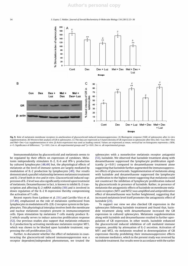
Splenocyte proliferation assessed as % stimulation ratio, wassignificantly (p < 0.01) decreased by dexamethasone and luzindoletreatment when compared with the control and dexamethasonetreated group. Co supplementation of luzindole along with dexa-methasone and melatonin further significantly (p < 0.01) decreasedthe splenocyte proliferation when compared to control and dexa-methasone treated group. Dexamethasone treatment along withmelatonin significantly (p < 0.01) increased the splenocyte prolifer-ation when compared to dexamethasone treated group alone butshowed a non-significant (p > 0.05) decrease when compared withthe control group (Fig. 9A).
4.3.1. Western blot analysis of GR in splenocytes treated withluzindole
Dexamethasone alone or in combination with luzindole signif-icantly (p < 0.01) upregulated GR expression when compared withthe control group. Melatonin treatment along with dexamethasonesignificantly decreased the GR expression when compared to thedexamethasone treated group, while luzindole supplementationalong with dexamethasone and melatonin further increased theGR expression significantly (p < 0.01) when compared to control ordexamethasone treated group of splenocytes (Fig. 9B and C).
5. Discussion
The present study highlights the role of melatonin membranereceptors (MT1 and MT2), that mediates the immunoenhancingrole of melatonin while counteracting the immunocompromisedstate induced by corticosterone in a wild tropical rodent F. pennanti.
Melatonin administration upregulated the expression of its mem-brane receptors (MT1 and MT2) while, dexamethasone treatmentdecreased it significantly. Dexamethasone increased the GR expres-sion and diminished T-helper cell mediated immune functions bydecreasing the lymphocyte proliferation which was successfullyrestored upon melatonin administration.
The splenocyte, thymocyte and circulatory mononuclear cells(PBMC) proliferation in response to T cell specific mitogen Con Awas enhanced by treatment of melatonin in vivo as well as in vitro.Melatonin administration perhaps mimicked the short day likeconditions by raising the plasma level of melatonin and at thesame time significantly (p < 0.01) upregulating the expression ofmelatonin membrane receptors MT1 and MT2. High melatoninlevel thereby counteracted the circulatory corticosterone level andsimultaneously decreased the GR expression which resulted inenhanced T cell mediated (%SR and %DTH) and humoral immuneresponse in seasonally breeding rodent F. pennanti. Our results arein line with the earlier report of Konakchieva et al. [32], suggestingthat melatonin administration was associated with diminishing thecorticosterone secretion thus protecting from the glucocorticoidinduced deteriorations.
Previous reports suggest that mitogen induced lymphocyte pro-liferation was significantly depressed following the treatment ofcorticosteroid, either in vivo or in vitro [21,33,34]. Melatonin on thecontrary is known to play an important role in immunomodulationby rescuing the immune cells from the immunocompromis-ing effects of environmental stressors which ultimately leadsto elevation of plasma corticosterone level [3,21,35,36]. Theseimmunomodulatory effects of melatonin might dependent onactivation of membrane melatonin receptors (MT1/MT2) by mela-tonin. There could be two possibilities for the increased melatonin

Author's personal copy
S. Gupta, C. Haldar / Journal of Steroid Biochemistry & Molecular Biology 134 (2013) 23– 36 33
Fig. 8. Effect of in vitro melatonin and dexamethasone supplementation on GR expression. Western blot analysis of GR in (A) splenocytes and (B) thymocytes. The dataare expressed as % band intensity of GR expression (C) in lymphoidal cells after Mel, Dex, and Mel + Dex supplementation. �-Actin expression was used as loading control.Values are expressed as mean, vertical bar on histograms represents ± SEM, n = 5. Significance of difference; **p < 0.01, Con vs. all experimental groups and #p < 0.01, Dex vs.Mel + Dex.
membrane receptor expression. First, melatonin injection ele-vated circulatory melatonin level that might have enhancedthe melatonin biosynthesis in lymphoid organs and circulatorymononuclear cells, which might have upregulated the receptorexpression (MT1 and MT2) in lymphoid tissues. Our result getssupport from previous reports [37–39] suggesting additive roleof endogenous melatonin on extrapineal sources in amplifyingthe tissue level of melatonin. However, in the present study wehave not measured the tissue level of melatonin to verify theabove hypothesis. Since, we have already reported upregulationof MT1 and MT2 receptor following injection of physiological doseof melatonin [23]. Therefore, the second possibility that increasedperipheral melatonin level might have upregulated the melatoninmembrane receptors expression independently, thereby rescuingthe immune repertoire by suppressing the apoptotic and antipro-liferative effects of glucocorticoids. However, to be more precisein interpretation of dynamics of melatonin membrane receptorexpression, further study needs to be undertaken by focusing onextrapineal biosynthesis of melatonin, following exogenous mela-tonin injection.
Delayed type hypersesitivity response, a Th1 type reaction is adirect measure of antigen specific cell mediated immune response.Melatonin treated squirrels showed robust DTH response whilethe dexamethasone treatment significantly (p < 0.01) decreasedthe DTH response compared with control and as noted earlierby Dhabhar and McEwen [40], suggesting that chronically ele-vated corticosterone or dexamethasone administration tends tosuppress DTH response. DTH reaction has been demonstrated to bemediated primarily by CD4+ T-helper cells [41,42], secreting IL-2
and IFN-� [43]. So we measured IL-2 production which was foundto be inhibited in the dexamethasone treated group that might havesuppressed the generation of antigen specific T cells in the squirrelsthus, decreasing the DTH response.
Production of anti-KLH-IgG is a T cell dependent phenomenon.Immunization with KLH in the dexamethasone pretreated groupsresulted in suppression of circulatory anti-KLH-IgG which wasrestored in the groups pretreated with melatonin and dexametha-sone. Melatonin pretreatment was found to be immunostimulatoryand considerably increased the antibody titer. As KLH is a Tcell specific antigen, so stress like levels of corticosterones afterdexamethasone treatment might have resulted in reduction in pro-liferation of anti-KLH T cells [44], resulting in reduced anti-KLH-IgGantibody level.
Melatonin may influence immune function by altering thecirculatory concentrations of other hormones such as gonadaland adrenal steroids [21,32,45,46], or by activation of mela-tonin receptors present on the cell surface of lymphoid organs[23,47]. We observed that physiological concentration of mela-tonin successfully counteracted the immunosuppressive effects ofglucocorticoids by up regulating the expression of melatonin mem-brane receptors thus modulating the cell mediated immunity onone hand and down turning the sensitivity of immunocompetentcells towards glucocorticoid on other, by attenuating GR expressionin lymphocytes. The observation implies for a possible involvementof melatonin in an adaptive bidirectional “trade off” commu-nication between neuroendocrine hypothalamo–hypophyseal–adrenal (HPA) axis and immune system in this tropicalrodent.
Author's personal copy
34 S. Gupta, C. Haldar / Journal of Steroid Biochemistry & Molecular Biology 134 (2013) 23– 36
Fig. 9. Role of melatonin membrane receptors in amelioration of glucocorticoid induced immunosuppression. (A) Blastogenic response (%SR) of splenocytes after in vitrosupplementation. (B) Western blot analysis of GR in splenocytes. (C) The data are expressed as % band intensity of GR expression in splenocyte after Dex, Dex + Luz, Mel + Dexand Mel + Dex + Luz supplementation in vitro. �-Actin expression was used as loading control. Values are expressed as mean, vertical bar on histograms represents ± SEM,n = 5. Significance of difference; **p < 0.01, Con vs. all experimental groups and #p < 0.01, Dex vs. all experimental groups.
Immunomodulation by glucocorticoid and melatonin seems tobe regulated by their effects on expression of cytokines. Mela-tonin independently stimulates IL-2, IL-6 and IFN-� productionby cultured lymphocytes [48,49] but, the physiological effects ofmelatonin at the level of immune system are largely mediated bymodulation of IL-2 production by lymphocytes [49]. Our resultsdemonstrated a parallel relationship between melatonin treatmentand IL-2 level both in vivo and in vitro. Glucocorticoid induced sup-pression of IL-2 level was also significantly restored upon treatmentof melatonin. Dexamethasone in fact, is known to inhibit IL-2 tran-scription and affecting IL-2 mRNA stability [50] and is involved indown regulation of the IL-2 R expression thereby compromisingthe activation of T cells.
Recent reports from Lardone et al. [51] and Carrillo-Vico et al.[37,49], emphasized on the role of melatonin synthesized fromlymphocytes in modulation of IL-2/IL-2 receptor system in the lym-phocytes. This physiological interplay involves both the membraneand nuclear melatonin receptors present in immunocompetentcells. Upon stimulation by melatonin T cells mainly produce IL-2 which usually serves to induce autocrine proliferation response[52]. Our previous studies also support that melatonin enhancesIL-2 production by activating membrane melatonin receptor MT1,which was shown to be blocked upon luzindole treatment, sup-pressing the cell proliferation [23].
Further, to document whether the effect of melatonin in coun-teracting the glucocorticoid induced immunosuppression was areceptor dependent/independent phenomenon, we treated the
splenocytes with a nonselective melatonin receptor antagonist[53], luzindole. We observed that luzindole treatment along withdexamethasone suppressed the lymphocyte proliferation signif-icantly (p < 0.01) compared to dexamethasone treatment alonesuggesting that luzindole further augmented the immunosuppress-ive effects of glucocorticoids. Supplementation of melatonin alongwith luzindole and dexamethasone suppressed the lymphocyteproliferation to the highest extent suggesting that melatonin couldnot counteract the inhibition of lymphocyte proliferation inducedby glucocorticoids in presence of luzindole. Rather, in presence ofmelatonin the antagonistic effects of luzindole on membrane mela-tonin receptors (MT1 and MT2) was amplified and antiproliferativeeffect of dexamethasone was further heightened, suggesting thatincreased melatonin level itself promotes the antagonistic effect ofluzindole [23].
To support our view we also checked GR expression in thesplenocytes following luzindole treatment and found that, luzin-dole treatment along with dexamethasone increased the GRexpression in cultured splenocytes. Melatonin supplementationalong with luzindole and dexamethasone resulted in further upre-gulation of GR expression. In other words, luzindole enhancedthe glucocorticoid induced inhibition of cell mediated immuneresponse, possibly by attenuation of IL-2 secretion. Activation ofMT1 and MT2, via melatonin resulted in downregulation of GRexpression thereby prohibiting their immunosuppressive effectson immune mediators, which in the present study was reversed byluzindole treatment. Our results were in accordance with the earlier

Author's personal copy
S. Gupta, C. Haldar / Journal of Steroid Biochemistry & Molecular Biology 134 (2013) 23– 36 35
report of Persengiev [54], which suggested that functional domainsof melatonin receptor molecule MT1 are involved in regulation ofGR induced transcription. However, our observations contrastedthe most recent findings of Presman et al. [55] suggesting luzindoleas an antiglucocorticoid in new born hamster kidney cells (BHK21).
The antagonizing effects of melatonin upon GR expression arealso evident from the reports where chronic melatonin treat-ment resulted in considerable alterations in binding activity ofGR leading to its differential sub-cellular localization [56,57]. Themolecular mechanisms behind these interactions are still poorlyunderstood however, it is proposed that melatonin attenuates thetranscriptional activity of GR [54]. Recently, melatonin has alsobeen proposed to impair the dissociation of HSP90 from GR whichretards its nuclear translocation and hence its biological activ-ity [56]. However, Presman et al. [56] reported inhibitory effectof melatonin on the GR translocation to be a tissue specific phe-nomenon as the inhibitory effect of melatonin on GR activitywas not observed in mouse fibroblasts L929, HC11 mouse mam-mary epithelial cells or Cos-7 kidney cells. In addition, there arereports which suggest that melatonin does not inhibits nucleartranslocation of GR, it rather impedes the interaction of GR withtranscriptional intermediary factor-2 (TIF-2) [55] that leads to sup-pression of GR induced transcriptional activity. Most importantly,all of these reports culminate to a common assumption that mela-tonin can be helpful in counteracting the deleterious effects ofglucocorticoids on central nervous system and immuno-competentcells.
In conclusion, our study suggests the existence of a reciprocal‘cross talk’ between two hormones, of antagonistic nature, mela-tonin and corticosterone. The reciprocal interactions noted amongtheir receptors may critically account for a dynamic and intri-cate physiological response required for maintenance of seasonalimmune homeostasis in wild conditions. Our view gets supportfrom previous reports of Sainz et al. [58], where melatonin whilemediating its effects down regulated the GR expression in dexa-methasone treated thymocytes while glucocorticoid always tendedto upregulate the same. Thus, the pineal gland via its hormone andreceptors seems to play a significant role in regulation of the HPA-lymphoid axis, maintaining an immunological link for significantadaptability in the wild species and thus increasing their chancesof survival against all the ecological odds.
Acknowledgements
Authors thank University Grants Commission and Council forScientific and Industrial Research, New Delhi, for financial supportto Mr. Sameer Gupta in form of SRF. Instrument gift from Alexandarvon Humboldt foundation, Bonn, Germany is gratefully acknowl-edged. Authors wish to thank Dr. Raise Ahmad, CSIR-RA, for varioushelp during manuscript preparation.
References
[1] R.J. Reiter, Pineal melatonin: cell biology of its synthesis and its physiologicalinteractions, Endocrine Reviews 12 (1991) 151–180.
[2] R.J. Nelson, G.E. Demas, S.L. Klein, L.J. Kriegsfeld, The influence of season, pho-toperiod, and pineal melatonin on immune function, Journal of Pineal Research19 (1995) 149–165.
[3] R. Ahmad, C. Haldar, Photoperiod–testicular–immune interaction in a seasonalbreeder Indian palm squirrel Funambulus pennanti during the reproduc-tively inactive and active phases, Journal of Neuroendocrinology 21 (2009)2–9.
[4] R.J. Reiter, D.X. Tan, L.C. Manchester, S.D. Paredes, J.C. Mayo, R.M. Sainz,Melatonin and reproduction revisited, Biology of Reproduction 81 (2009)445–456.
[5] M.L. Dubocovich, M. Markowska, Functional MT1 and MT2 melatonin receptorsin mammals, Endocrine 27 (2005) 101–110.
[6] S.M. Reppert, D.R. Weaver, T. Ebisawa, Cloning and characterization of amammalian melatonin receptor that mediates reproductive and circadianresponses, Neuron 13 (1994) 1177–1185.
[7] S.M. Reppert, C. Godson, C.D. Mahle, D.R. Weaver, S.A. Slaugenhaupt, J.F. Gusella,Molecular characterization of a second melatonin receptor expressed in humanretina and brain: the Mel 1b melatonin receptor, Proceedings of the NationalAcademy of Sciences of the United States of America 92 (1995) 8734–8738.
[8] J.R. Calvo, M. Rafii-El-Idrissi, D. Pozo, J.M. Guerrero, Immunomodulatory role ofmelatonin: specific binding sites in human and rodent lymphoid cells, Journalof Pineal Research 18 (1995) 19–126.
[9] A. Carrillo-Vico, A. Garcia-Perganeda, L. Naji, J.R. Calvo, M.P. Romero, J.M. Guer-rero, Expression of membrane and nuclear melatonin receptor mRNA andprotein in the mouse immune system, Cellular and Molecular Life Sciences60 (2003) 2272–2278.
[10] R. Ahmad, C. Haldar, Photoperiodic regulation of melatonin receptor MT1 andMT2 expression dynamics in spleen and thymus of a tropical rodent Funam-bulus pennanti during reproductively active and inactive phase, ChronobiologyInternational 27 (2010) 446–462.
[11] E.R. De Kloet, E. Vreugdenhil, M.S. Oitzl, M. Joëls, Brain corticosteroid receptorbalance in health and disease, Endocrine Reviews 19 (1998) 269–301.
[12] R.M. Sapolsky, L.M. Romero, A.U. Munck, How do glucocorticoids influencestress responses? Integrating permissive, suppressive, stimulatory, and prepar-ative actions, Endocrine Reviews 21 (2000) 55–89.
[13] J.I. Webster, L. Tonelli, E.M. Sternberg, Neuroendocrine regulation of immunity,Annual Review of Immunology 20 (2002) 125–163.
[14] J.D. Ashwell, F.W. Lu, M.S. Vacchio, Glucocorticoids in T cell development andfunction, Annual Review of Immunology 18 (2000) 309–345.
[15] G.J. Maestroni, The immunoneuroendocrine role of melatonin, Journal of PinealResearch 14 (1993) 1–10.
[16] C. Barriga, M.I. Martín, R. Tabla, E. Ortega, A.B. Rodríguez, Circadian rhythm ofmelatonin, corticosterone and phagocytosis: effect of stress, Journal of PinealResearch 30 (2001) 180–187.
[17] A.S. Fauci, D.C. Dale, The effect of in vivo hydrocortisone on subpopulations ofhuman lymphocytes, Journal of Clinical Investigation 53 (1974) 240–246.
[18] P.J. Barnes, Anti-inflammatory mechanisms of glucocorticoids, BiochemicalSociety Transactions 23 (1995) 940–945.
[19] B.W. Kirkham, M.M. Corkill, S.C. Davison, G.S. Panayi, Response to glucocorti-coid treatment in rheumatoid arthritis: in vitro cell mediated immune assaypredicts in vivo responses, Journal of Rheumatology 18 (1991) 821–825.
[20] M.N. Scherer, B. Banas, K. Mantouvalou, A. Schnitzbauer, A. Obed, B.K. Krämer,H.J. Schlitt, Current concepts and perspectives of immunosuppression in organtransplantation, Langenbeck’s Archives of Surgery 392 (2007) 511–523.
[21] C. Haldar, S. Rai, R. Singh, Melatonin blocks dexamethasone-induced immuno-suppression in a seasonally breeding rodent Indian palm squirrel, Funambuluspennanti, Steroids 69 (2004) 367–377.
[22] K.S. Bishnupuri, C. Haldar, Maternal transfer of melatonin alters the growth andsexual maturation of young Indian palm squirrel, F. pennanti, Biological Signalsand Receptors 10 (2001) 317–325.
[23] R. Ahmad, C. Haldar, S. Gupta, Melatonin membrane receptor type MT1modulates cell mediated immunity in seasonally breeding tropical rodentFunambulus pennanti, NeuroImmunoModulation 19 (2012) 50–59.
[24] A. Boyum, Isolation of mononuclear cells and granulocytes from human bloodIsolation of mononuclear cells by one centrifugation, and of granulocytes bycombining centrifugation and sedimentation at 1 × g, Scandinavian Journal ofClinical and Laboratory Investigation 97 (1968) 77–89.
[25] T. Mosmann, Rapid colorimetric assay for cellular growth and survival: applica-tion to proliferation and cytotoxicity assays, Journal of Immunological Methods65 (1983) 55–63.
[26] M.M. Bradford, A rapid and sensitive method for the quantitation of microgramquantities of protein utilizing the principle of protein-dye binding, AnalyticalBiochemistry 72 (1976) 248–254.
[27] M.D. Rollag, G.D. Niswender, Radioimmunoassay of melatonin in sheepexposed to different light regimes, Endocrinology 98 (1976) 482–488.
[28] C. Haldar, S. Dubey, Diurnal variations in circulating estradiol, testosterone,melatonin and harderian gland porphyrin concentration in Indian palm squir-rel, Funambulus pennanti, Indian Journal of Experimental Biology 34 (1996)695–697.
[29] R. Ahmad, C. Haldar, Immune responses to lipopolysaccharide challenge in atropical rodent (Funambulus pennanti): photoperiod entrainment and sex dif-ferences, Stress 15 (2012) 172–183.
[30] G.E. Demas, V. Chefer, M.I. Talan, R.J. Nelson, Metabolic costs of mounting anantigen-stimulated immune response in adult and aged C57BL/6J mice, Amer-ican Journal of Physiology 273 (1997) 1631–1637.
[31] Y. Song, C.W. Chan, G.M. Brown, S.F. Pang, M. Silverman, Studies of the renalaction of melatonin: evidence that the effects are mediated by 37 kDa receptorsof the Mel1a subtype localized primarily to the basolateral membrane of theproximal tubule, FASEB Journal 11 (1997) 93–100.
[32] R. Konakchieva, Y. Mitev, O.F.X. Almeida, V.K. Patchev, Chronic mela-tonin treatment counteracts glucocorticoid-induced dysregulation of thehypothalamic–pituitary–adrenal axis in the rat, Neuroendocrinology 67 (1998)171–180.
[33] D. Gordon, A.M. Nouri, Comparison of the inhibition by glucocorticosteroids andcyclosporin A of mitogen-stimulated human lymphocyte proliferation, Clinicaland Experimental Immunology 44 (1981) 287–294.
[34] T.R. Cupps, A.S. Fauci, Corticosteroid-mediated immunoregulation in man,Immunological Reviews 65 (1982) 133–155.
[35] R.J. Nelson, G.E. Demas, S.L. Klein, Photoperiodic mediation of seasonal breedingand immune function in rodents: a multifactorial approach, Integrative andComparative Biology 38 (1998) 226–237.
Author's personal copy
36 S. Gupta, C. Haldar / Journal of Steroid Biochemistry & Molecular Biology 134 (2013) 23– 36
[36] D.L. Drazen, A.M. Jasnow, R.J. Nelson, G.E. Demas, Exposure to short days, but notshort-term melatonin, enhances humoral immunity of male Syrian hamsters(Mesocricetus auratus), Journal of Pineal Research 33 (2002) 118–124.
[37] A. Carrillo-Vico, J.R. Calvo, P. Abreu, P.J. Lardone, S. Garcia-Maurino, R.J. Reiter,J.M. Guerrero, Evidence of melatonin synthesis by human lymphocytes andits physiological significance: possible role as intracrine, autocrine, and/orparacrine substance, FASEB Journal 18 (2004) 537–539.
[38] M.C. Naranjo, J.M. Guerrero, A. Rubio, P.J. Lardone, A. Carrillo-Vico, M.P.Carrascosa-Salmoral, S. Jiménez-Jorge, M.V. Arellano, S.R. Leal-Noval, M. Leal,E. Lissen, P. Molinero, Melatonin biosynthesis in the thymus of humans andrats, Cellular and Molecular Life Sciences 64 (2007) 781–790.
[39] D.X. Tan, L.C. Manchester, R.J. Reiter, W.B. Qi, M. Zhang, S.T. Weintraub, J. Cabr-era, R.M. Sainz, J.C. Mayo, Identification of highly elevated levels of melatoninin bone marrow: its origin and significance, Biochimica et Biophysica Acta 18(1999) 206–214.
[40] F.S. Dhabhar, B.S. McEwen, Enhancing versus suppressive effects of stress hor-mones on skin immune function, Proceedings of the National Academy ofSciences of the United States of America 96 (1999) 1059–1064.
[41] T. Diamanstein, R. Eckert, H.-D. Volk, J.-W. Kupier-Weglinski, Reversal byinterferon-� of inhibition of delayed-type hypersensitivity induction by anti-CD4 or anti-interleukin 2 receptor (CD25) monoclonal antibodies. Evidence forthe physiological role of the CD4+ Th1+ subset in mice, European Journal ofImmunology 181 (1988) 2101–2103.
[42] V.E. Kelley, P. Bacha, O. Pankewycz, J.C. Nichols, J.R. Murphy, T.B. Strom, Inter-leukin 2-diphtheria toxin fusion protein can abolish cell-mediated immunityin vivo, Proceedings of the National Academy of Sciences of the United Statesof America 85 (1988) 3980–3984.
[43] S. Grabbe, T. Schwarz, Immunoregulatory mechanisms involved in elicitationof allergic contact hypersensitivity, Immunology Today 19 (1998) 37–44.
[44] M. Fleshner, T. Deak, K.T. Nguyen, L.R. Watkins, S.F. Maier, Endogenous gluco-corticoids play a positive regulatory role in the anti-keyhole limpet hemocyaninin vivo antibody response, Journal of Immunology 15 (2001) 3813–3819.
[45] N.J. Olsen, W.J. Kovacs, Gonadal steroids and immunity, Endocrine Reviews 17(1996) 369–384.
[46] R. Ahmad, C. Haldar, Melatonin and androgen receptor expression interplaymodulates cell-mediated immunity in tropical rodent Funambulus pennanti:an in vivo and in vitro study, Scandinavian Journal of Immunology 71 (2010)420–430.
[47] A.M. Poon, E.A. Ayre, Y. Song, S.F. Pang, Melatonin implant decreases the densityof 2[125I]iodomelatonin binding sites in the chicken spleen, Biological Signals3 (1994) 278–287.
[48] S. Garcia-Maurino, M.G. Gonzalez-Haba, J.R. Calvo, M. Rafii-El-Idrissi, V.Sanchez-Margalet, R. Goberna, J.M. Guerrero, Melatonin enhances IL-2, IL-6,and IFN-gamma production by human circulating CD4+ cells: a possible nuclearreceptor-mediated mechanism involving T helper type 1 lymphocytes andmonocytes, Journal of Immunology 15 (1997) 574–581.
[49] A. Carrillo-Vico, S. Garcia-Maurino, J.R. Calvo, J.M. Guerrero, Melatonincounteracts the inhibitory effect of PGE2 on IL-2 production in humanlymphocytes via its mt1 membrane receptor, FASEB Journal 17 (2003)755–757.
[50] J.P. Northrop, G.R. Crabtree, P.S. Mattila, Negative regulation of interleukin 2transcription by the glucocorticoid receptor, Journal of Experimental Medicine175 (1992) 1235–1245.
[51] P.J. Lardone, A. Carrillo-Vico, P. Molinero, A. Rubio, J.M. Guerrero, A novelinterplay between membrane and nuclear melatonin receptors in human lym-phocytes: significance in IL-2 production, Cellular and Molecular Life Sciences66 (2009) 516–525.
[52] S.L. Swain, L.M. Bradley, M. Croft, S. Tonkonogy, G. Atkins, A.D. Weinberg, D.D.Duncan, S.M. Hedrick, R.W. Dutton, G. Huston, Helper T-cell subsets: pheno-type, function and the role of lymphokines in regulating their development,Immunological Reviews 123 (1991) 115–144.
[53] D.L. Drazen, D. Bilu, S.D. Bilbo, R.J. Nelson, Melatonin enhancement of spleno-cyte proliferation is attenuated by luzindole, a melatonin receptor antagonist,American Journal of Physiology – Regulatory, Integrative and ComparativePhysiology 280 (2001) 1476–1482.
[54] S.P. Persengiev, Multiple domains of melatonin receptor are involved in the reg-ulation of glucocorticoid receptor-induced gene expression, Journal of SteroidBiochemistry and Molecular Biology 68 (1999) 181–187.
[55] D.M. Presman, V. Levi, O.P. Pignataro, A. Pecci, Melatonin inhibitsglucocorticoid-dependent GR-TIF2 interaction in newborn hamsterkidney (BHK) cells, Molecular and Cellular Endocrinology 26 (2012)214–221.
[56] D.M. Presman, E. Hoijman, N.R. Ceballos, M.D. Galigniana, A. Pecci, Melatonininhibits glucocorticoid receptor nuclear translocation in mouse thymocytes,Endocrinology 147 (2006) 5452–5459.
[57] S.P. Persengiev, I.I. Kondova, Tissue-specifc modulation of rat glucocorti-coid receptor binding activity by melatonin, Experientia 49 (1993) 332–334.
[58] R.M. Sainz, J.C. Mayo, R.J. Reiter, I. Antolin, M.M. Esteban, C. Rodrigue, Mela-tonin regulates glucocorticoid receptor: an answer to its antiapoptotic actionin thymus, FASEB Journal 13 (1999) 1547–1556.