“This book or part thereof may not be reproduced by
208
Transcript of “This book or part thereof may not be reproduced by

ii
“This book or part thereof may not be reproduced by any person or agency in any manner.”
Laboratory Medicine – IIStudent Handbook, Class–XII
Price : `
First Edition :
Copies :
Paper Used : 80 GSM CBSE Water Mark White Maplitho
Published By : The Secretary, Central Board of Secondary Education, Shiksha Kendra, 2, Community Centre, Preet Vihar, Delhi-110092
Design & Layout : Public Printing (Delhi) Service C-80, Okhla Industrial Area, Phase-I, New Delhi-110020
Printed By :


v
Preface
I am very pleased to present the first edition of the text book in for the laboratory part of medical diagnostics.This is a new vocational course from Central board of Secondary Education.This is a part of the dream project of our Prime minister Mr.NarendraModiji
For skilling India. This is to ensure that all students who pass their 12th board examina-tions shall have the capability to get gainful employment because of their skill set.
This book has been written by Competent persons actively working in the various field of laboratory medicine –which includes histopathology,cytology,hematology,clinicalpathol-ogy,microbiology,blood banking etc. They are professors ,writers ,practising doctors and academicians.
This book shall have contents that shall cover the complete course curriculum for classes 11th to 12 th for the areas of Medical Diagnostics.Thewriteup of the book is fairly sim-ple and shall help the student update his knowledge in the subject including all recent developments.He shall be able to self judge /asses his own competency through a set of questions given for self assessment.
I would like to thank CBSE vocational Unit ,who had been the driving force behind the development of this book. who has pains taxingly devoted so much of her time inensuring that it comes along in this fine form. My team of subject expert specially to mention from Safdarjung Hospitals .The current text has been prepared keeping in view the current requirements of the students and the latest updates in the relevant areas in a concise mannerusing simple language for increasing the comprehension.
Constructive and helpful suggestions from readers for the improvement of the book are welcome.
Chairman, CBSE

vi
Acknowledgements
• ANITA KARWAL, IAS, Chairperson , CBSE
Editing & Coordination
• Dr. BiswajitSaha, Director (Voctional), CBSE
Content Developed by
• Dr Juhee Chandra -Lab Director at Mediaid Diagnostics
• Dr.Rashmi Vohra -Director Vohra Diagnostics
• Dr Geetika Khanna, Professor of Pathology, CIO Laboratory, VMMC & Safdarjung Hospital.
• Dr. Neeraj JainMBBS. MD.Director, Jain Diagnostics

vii
ContentsUnit – 1 Investigation Urine & Faeces Analysis 1
Unit – 2 Body Fluids 27
Unit – 3 Haematology Lab (Procen & Investigation) 35
Unit – 4 Blood Bank and Transfusion 82
Unit – 5 Histopathology (Lab Procen) 104
Unit – 6 Cytopathology 184

1
OVERVIEWThis unit will provide the student information about the scope of and the organization of a clinical pathology laboratory. It will help to understand the relevant terms, procedures and working of equipments pertaining to urine and faeces investigations.
Organization of a Clinical pathology Laboratory:
The personnel needs of a laboratory depends on overall work load and the different types of materials to be processed. Assuming that the laboratory consists of routine sections, the employees would include a laboratory head, skilled employees in the form of technicians to supervise the different sections, and unskilled employees in the form of laboratory assistants. Related areas omitted in this example should have close communications with the other departments, but maintain separate and distinct supervision.
The Chief of the Laboratory should be a trained pathologist.
The technicians should have a diploma in medical laboratory technology from a recognized institution. They are responsible for specimen collection, preparation and test validations.
Support Staff include clerical and secretarial workers in the laboratory. Physical Infrastructure of the laboratory must be well designed and conveniently located to enable the professional and support personnel to perform their duties effectively. It must contain four definitely separated areas:• Reception.• Specimen collection room.• Processing area.• Reporting room.
KNOWLEDGE AND SKILL OUTCOMESi. To understand the scope of clinical pathology.ii. To know the organizational structure of a clinical pathology laboratory.iii. To know the relevant terms, procedures and working of equipments pertaining to clinical
pathology.
UNIT - 1INVESTIGATION URINE & FAECES ANALYSIS

2
RESOURCE MATERIALSi. a. Text Book of Medical Laboratory Technology, Praful B. Godkar, First edition.ii. b. urine Analyzer insert.iii. c. A Textbook of Biochemistry by Harbans Lal.
DURATION
LEARNING OUTCOMESAfter completing this unit the students should be able to1. Demonstrate knowledge, comprehension, and application of general techniques in the
areas of:
• Specimen accessioning.
• Manual methods.
• Routine general examination.
• Microscopic examination.
2. Set-up, operate and maintain routine instruments.
3. Solve basic problems associated with reagents and methods of general techniques.
4. Apply principles of lab safety in completing all laboratory work.
5. Ensure quality control while performing general procedures.
1.1 INTRODUCTION TO urine examinationUrine is one of the most easily obtained specimens examined in the laboratory, and examination of the urine not only provides information about the functioning of the kidneys and possible abnormalities of the urinary tract, but may also lead to the diagnosis of various systemic diseases of the human body which are reflected by the presence of several abnormal substances in the urine.
1.2 PURPOSE OF THE EXAMINATIONTo perform complete urine examination
1.3 LIS OF EQUIPMENT REQUIRED FOR SETTING UP OF A LABORATORY1. Microscope2. Centrifuge
3
3. Urine strips4. Spirit lamp5. Testtubes6. Reagents for various manual tests slides/coverslips etc.
COLLECTION OF SPECIMENa) Early morning urine
The best urine specimen for routine analysis is collected in the morning. It is usually concentrated and has an acid pH. Casts and cells are poorly preserved in dilute or alkaline urine and traces of dissolved substances such as protein and sugar can be missed if the urine is very dilute.b) Random urine
This specimen can also collected at any time and is convenient for the patient and is suitable for most screening purposes.c) Preservative Used
For routine analysis, no preservative is required but the urine is best examined fresh. Bacterial growth will ruin a specimen if analysis delayed for more than 3 hours. Refrigeration is the best way to preserve it if analysis is delayed. Refrigeration for more than 24 hours is not recommended.d) Container for urine collection
The container used must be thoroughly clean and free from any detergent or disinfectant residue since the oxidants contained in such cleaning agents may cause the test areas for glucose and blood to indicate false positive results. After the urine is collected, the container should preferable be sealed.
1. CLINICAL SIGNIFICANCE
Routine urine examination is performed mainly for two purposes
1.1 To find out metabolic or endocrine disorders in the body (e.g. Normal urine does not contain bilirubin or sugar. Presence of bilirubin in urine, indicates metabolic disturbance of bilirubin and sugar in urine is a indicator of diabetes mellitus i.e. deficiency of insulin, an endocrine disorder.
1.2 To detect intrinsic conditions that adversely affect kidney or urinary tract. Diseased kidneys cannot function normally in regulating the volume and composition of body fluids and also in maintaining acid-base balance and homeostasis.

4
1.3 Structural elements such as leukocytes, red blood cells and casts from the lower urinary tract may appear in urine. Substances normally retained by kidneys or excreted by kidneys in small mounts may also appear in large quantities and substances normally excreted maybe retained which is indicated by increased values in the blood e.g. Urea, Creatinine
3. The expected changes in the composition of urine stored at room temperature are as follows :-
a. Lysis of red blood cells by hypotonic urineb. Decomposition of castsc. Bacterial multiplicationD. Decrease in glucose level, due to bacterial growthe. Formation of ammonia from urea by the action of bacteria (and the nature of urine
changes to alkaline)
4. METHODS
The various aspects considered in the complete examination of urine are as follows :-4.1 Physical examination of urine4.2 Chemical examination of urine4.3 Microscopic examination of urine
5.1 PHYS Following aspects are studies for the physical examination
5.1.a Volume (Optional)5.1.b Colour5.1.c Appearance5.1.d Odour5.1.e Specific gravity5.1.f Reaction (pH)
5.1.a VOLUMEA. For an adult, the normal average daily volume of urine is about 1200-1500 ml. More
urine formation takes during the day than at night. However the normal range for 24 hours may be 600-2000 ml.
B. Polyuria is an abnormal increase in excretion of urine volume (>2500ml) as in the diabetes mellitus and diabetes insipidus.
5
C. Oliguria is a decrease in urine excretion (<500ml). The term anuria means the complete suppression of urine formation in spite of high fluid intake.
CLINICAL IMPORTANCEA) POLYURIA :-Abnormal increase in urine volume > 2500ml/24 hours as in diabetes mellitus
and diabetes insipidus.B) OLIGURIA :-Decrease in urine volume <500ml/24 hours. Observed in renal and post-renal
conditions.C) ANURIA :- Complete suppression of urine formation as in renal failure.
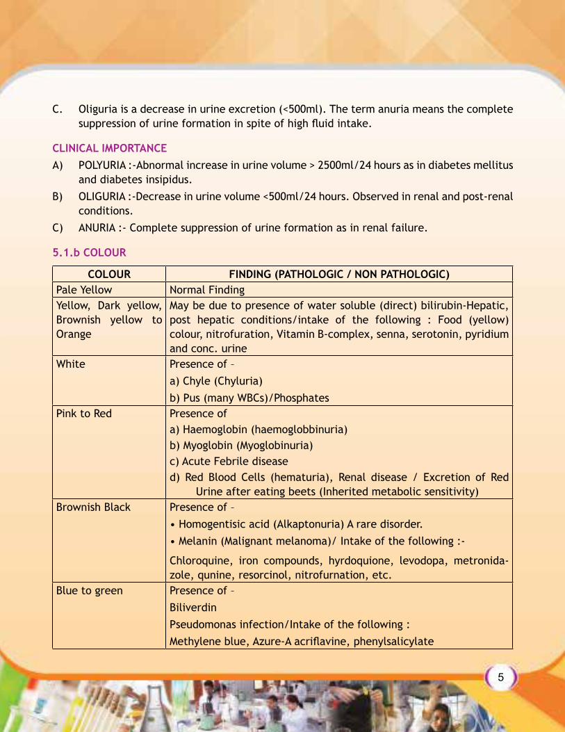
5.1.b COLOUR
COLOUR FINDING (PATHOLOGIC / NON PATHOLOGIC)Pale Yellow Normal FindingYellow, Dark yellow, Brownish yellow to Orange
May be due to presence of water soluble (direct) bilirubin-Hepatic, post hepatic conditions/intake of the following : Food (yellow) colour, nitrofuration, Vitamin B-complex, senna, serotonin, pyridium and conc. urine
White Presence of –a) Chyle (Chyluria)b) Pus (many WBCs)/Phosphates
Pink to Red Presence ofa) Haemoglobin (haemoglobbinuria)b) Myoglobin (Myoglobinuria)c) Acute Febrile diseased) Red Blood Cells (hematuria), Renal disease / Excretion of Red
Urine after eating beets (Inherited metabolic sensitivity)Brownish Black Presence of –
• Homogentisic acid (Alkaptonuria) A rare disorder.• Melanin (Malignant melanoma)/ Intake of the following :-
Chloroquine, iron compounds, hyrdoquione, levodopa, metronida-zole, qunine, resorcinol, nitrofurnation, etc.
Blue to green Presence of –BiliverdinPseudomonas infection/Intake of the following :Methylene blue, Azure-A acriflavine, phenylsalicylate

6
5.1.cAPPEARANCE
Normal urine colour varies from light yellow to deep amber. Urine colour sometimes may vary depending upon the diet and fluids, if any, consumed by the patient. The colour of urine is sometimes related to a pigment called “urochrome”. The degree of colour also depends on whether the specimen is concentrated of dilute.
Normal urine is usually clear. If the pH is alkaline, may be observed due to the precipitation of phosphates. Such urine should be centrifuged before analysis. Turbidity due to presence of chyle (chylomicrons) cannot be centrifuged, but required filtration using a special cellulose filter having <o.1 mm diameter and is confirmed with the help of ether test which dissolves the chylomicrons.a. Normal urine is usually clear :it may appear cloudy if amphorous phosphates are present
in alkaline urine or amorphous urates in acidic urine. Amorphous phosphates form white precipitate which dissolves when heated.
b. Urine may appear cloudy or turbid by the presence of leucocytes and epithelial cells.c. This can be confirmed by microscopic examination. Bacteria can also cause cloudiness
and mucous gives hazy appearance to urine. Fat and chyle give urine a milky colour. Presence of RBC may give urine turbid and smoky appearance.
5.1.dODOUR
Normal fresh urine has a mild odour of ammonia. Presence of ketone bodies gives urine a sweety or fruity smell. Contaminated urine with bacterial may give pungent shell due to the formation of ammonia. The urine of a infant with phenylketonuria gives musty odour.
TEST DONE ON DX URINE ANALYZERa) pHb) Specific Gravityc) Glucosed) Bilirubine) Urobiliinogenf) Ketonesg) Nitriteh) Blood Leucocytes
QDX urine test reagent strips for urine analysis and dip & read test strips are used as an in vitro diagnostics aid using urine specimen. The strips can provide qualitative & semi-quantitative determination.

7
PROCEDURE1. Remove the set strip from the bottle after checking the expiry date and re-cap bottle
immediately.2. In case of discolouration or darkening of the reagent areas do not use the strip.3. Dip the test strip completely for no more than I sec. in fresh well mixed uncentrifuged
urine.4. Remove the strip along the rim of the container to remove excess urine.5. Blot one side of the test strip on absorbent paper on one side to remove remaining urine
and prevent missing of chemicals.6. Hold the strip up horizontally and compare the colours developed with the colour chart
on the bottle label.7. The reading time is 60-120 sec is critical for optimal results.
STORAGE
The strips must be kept in the original bottle only and should not be used beyond the expiry date, each strip can be used only once. Dessicant should not be removed from the body. The strip should be stored at temperature between 2O C-300 C.
5.1.eSPECIFIC GRAVITY
METHOD :- not there in material
PRINCIPAL :- Electrolyte (MX) in the form of salt in urine the reacts with Polymethylvinyl Ether and Maleic Acid (OOH) which are weak acid ionic exchanger. The reaction produces Hydrogenous ionogen which reacts with the pH indicator and produces a color change.
Result
Normal range in urine 1.020-1.030
Visual test range 1.00-1.030
Instrument test range 1.005-1.030
CLINICAL SIGNIFICANCEA. Specific gravity at a constant temperature is the ratio of the weight of the volume of the
urine to the weight of same volume of distilled water. The specific gravity determination of urine is used to measure the concentrating and diluting power of the kidneys. The specific gravity of urine varies throughout the day and the normal range for random urine specimen is 1.003 to 1.035. The range for a 24 hour urine specimen is 1.015 to 1.030.

8
B. Polyuria is observed in both diabetes mellitus and diabetes insipidus. But in diabetes insipidus, the specific gravity of urine is low while in diabetes mellitus the specific gravity is high due to presence of glucose in the urine.
C. HYPERSTHENURIA :- It is the condition in which orine has high specific gravity, High specific gravity of urine is observed in various conditions such as dehydration, eclampsia, proteinuria, diabetes mellitus and lipoid nephrosis.
D. ISOSTHENURIA :- Excretion of urine with fixed specific gravity of 1.010. It is the indicator of poor tubular reabsorption.
E. HYPOSTHENURIA :-Urine excretion with constantly low specific gravity (1.0007). It is found in pyelonephritis, hypertension, protein malnutrition and diabetes insipidus. Diuretic medicines, and natural diuretics such as alcohol and coffee also cause excretion of urine with low specific gravity.
5.1.fREACTION (pH)
MANUAL METHOD:
Use a narrow range pH paper, In some clinical situations, measurements of approximate pH within+0.5 pH units using a narrow-rang pH paper may be very helpful.(a) PROCEDURE
Using pH paper
Put a drop of urine on a portion of pH indicator paper. The color obtained is compared with a standard chart. For checking the reliability of the pH paper cross check the pH of buffer solutions of known pH values having acidic and alkaline pH ranges.
Result :-Normal urine pH ranges from 4.5 to 8.0. The pH values are reported for example as 6.0 if pH paper is used shows as 6.1.
Interpretation and quality control
Urine pH is usually acidic in normal people, especially non vegetarians and is usually alkaline in vegetarians.
An early morning urine pH < 5.5 indicates that renal tubular acidification mechanism is intact. As a quality control measure, use certified reference buffers (commercial source), one in acidic rang, say, pH 4.0 and the other in alkaline range, preferably pH 9.2 to check the reliability of the pH paper used.
Always use a pH indicator paper before the date of expiry. Do not use outdated pH papers. Always close the bottle containing the pH paper tightly.

9
ANALYSER METHOD :-
PRINICIPLE :- This test is based on the double indicators (Methyl red and Bromothymol blue) which give colors ranging from red orange through green to blue covering the urine pH range of 5-9.a) Results
Record the reading from the instrument display and enter the values in the specific register.
Urine pH : Normal Range : 6-7
QDX urine Strip’s Measuring Range :
Visual Test range :- 5-8.5
Instrument Test Range :- 5-9
CLINICAL SIGNIFICANCEA. The freshly voided normal urine is usually slightly acidic and its pH may range from 4.6-
7.0. A high protein intake ingestion of acidic fruits produces acidic urine. Respiratory acidosis, metabolic acidosis (diabetes ketosis, starvation, severe diarrhea) produces acidic urine. Urinary tract infections caused by Escherichia Coli result in acidic urine.
In respiratory alkalosis (Hyperventilation) and in metabolic alkalosis (excessive vomiting) alkaline urine is excreted. A diet that is high in vegetables and citrus fruits causes an alkaline urine. Urinary infections caused by Proteus and Pseudomonas may cause alkaline urine.
5.2 CHEMICAL EXAMINATION OF URINE FOR ABNORMAL CONSTITUENTSThe routine chemical analysis for abnormal constituents of urine includes following investigations :a) Glucoseb) Ketone bodiesc) Proteinsd) Bloode) Bile saltsf) Bile pigmentsg) Urobilinogenh) Nitrite

10
5.2.aGLUCOSE
Manual Method :-
Sugar : Benedict’s Testa) PRINCIPLE :- Urinary sugars when boiled in Benedict’s Reagent reduce copper sulphate
to reddish cuprous oxide precipitate in hot alkaline medium, the intensity of which is proportional to the amount of sugar present in the urine. The results are reported as 1+, 2+, etc depending upon the colour and intensity of the cuprous oxide precipitate.
b) Reagent :- Dissolve 17.3 g of crystalline copper sulphate in about 800 ml of distilled water, then add 100 g of sodium carbonate, mix to dissolve and finally add 175 g of sodium citrate. Mix coloured bottle at 250-350C. Stable for one year.
c) Procedure :- To 5 ml of Benedict’s reagent taken in an 18 x 150 mm glass tube, add 8 drops (0.5ml) of urine, mix well and boil for 2-3 minute over the flame. Cool the tube and observe for any colour change.
Determination of glucose in urine by using QDX Urine Analyzer
PRINCIPLE :- Glucose is oxidized by Glucose oxidase to form glucuronic acid and hydrogen peroxide. Hydrogen peroxide releases neo-ecotypes oxide (O) under the action of peroxidase (O) oxidizes potassium iodide which produces the color change.
Sensitivity 50-100mg/dl. Visual and instrument tests range : Negative – 100mg/dl
Reactive Ingredients : 1.7% w/w glucose oxidase (microbial.123 U); 0.2% w/w peroxidase (horseradish 203 U); 0.1% w/w potassium iodine; 71.8% w/w buffer, 26.2% w/w nonreactive ingredients.
INTERPRETATION OF THE RESULTSNo change in the original colour of Benedicts’s solution – NegativeSolution appears pale green and slightly cloudy – TraceDefinity cloudy green 1+0.5%Yellow to orange precipitate 2+-01%Orange precipitate 3+-1.5%Brick Red precipitate 4+ (≥2g/dl)
False positive reactions are known to occur due to presence of non-carbohydrate substances like ascorbic acid, homogentisic acid, creatinine and uric acid.
Reducing sugars like lactose, galactose, fructose and pentoses will also give a positive reaction. The dipstick technique is specific for glucose and eliminates the false positive reaction due to the substances mentioned above.

11
CLINICAL SIGNIFICANCE
The normal renal threshold for glucose is 180mg/dl. When the glucose exceeds the normal threshold the renal tubules cannot reabsorb all the filtered glucose and then glycosuria occurs. Glycosuria is seen in following conditions.a. Diabetes Mellitusb. Endocrine Disorder – Diabetes Mellitus, Cushing’s syndrome, Pancreatic tumors,
Hyperthyroidism, & Hyperpituitarism.c. Phaeochromocytoma, Carcinoma of Pancreas, Pancreatitis.d. Central Nervous System Disorders, Brain tumors, asphyxia, burns, infection, Myocardial
infarction.e. Liver disease, Glycogen storage disease, obesity.f. Pregnancy : Reduced threshold for Glucose.g. Aged : Glucose intolerance.h. Glycosuria without huperglycemia-renal tubular dysfunction
OTHER Renal glycosuria, Alimentary glycosuria.
5.2.(b)KETONE BODIES
MUNUAL METHOD
Ketone bodies-Rothera’s test
The three main ketone bodies are acetone, acetoacetic acid (diacetic acid) and betahydroxy butyricacid. Testing for ketone bodies should be done on fresh urine or the specimen kept at 4.c.
Principal
Acetone and acetoacetic acid react with sodium nitroprusside in the presence of alkali to produce a purple color.
Sensitivity: 10-20 mg/dl, Visual Test Range, Negative-300mg/dl, Instrument Test Range: Negative-150mg/dl.
Reactive ingredients: 5.7% w/w sodium nitroprusside: 29.9% w/w nonreactive ingredients. 64.4% w/w buffer.
Procedure
To 5.0 ml of urine taken in 18 x 150mm glass tube. Saturate it with solid ammonium sulphate. Add 0.5 ml of 1% sodium nitroprusside solution. Mix well. Add ammonia solution along the side of the tube so that it layers on top of the urine.

12
Result
If acetone and diacetic acid are present, then apurple (permanganate calomel red) colour will form at the junction of the two layers within 30-60 seconds. The result can be graded from trace to 3+ based on the intensity of the colour formed as detailed below.
No change in colour-Negative
Pinkish rang - +
Red ring - ++
Deep purple ring - +++
Interpretation add quality control
Ketone bodies are intermediary products of fat metabolism and their presence in the blood and then in the urine are indications that the metabolism is dis disordered or incomplete. This is associated with metabolic acidosis. This occurs in poorly controlled diabetes mellitus and also in starvation.
Normal urine does not contain methyl ketone. Weak false positive reactions may occur if the urine contains L-dopa and phenyl pyruvic acid.
If there is suspicion of a false positive test, heat the urine in a test tube in a Bunsen burner flame for one minute, allow to cool and repeat the Rothera’s test. Heated urine.
5.2.(h)URINE FOR NITRITE using QDX Urine Analyzer
PRINCIPAL :- Nitrite in urine & aromatic amino sulphanilamide are diazotized to from diazonium compound. The diazonium compound reacts with tetra hydro benzo quinolin 3 phenol causing the color change.Normal range in urine zeroVisual & Instrument test range – NegativeSensitivity – 0.25-0.4 mg/dlAbnormal value means1. UTI2. Bacterial infection
MICROSCOPY
PURPOSE OF THE EXAMINATION
To perform microscopic examination of urine.

13
CLINICAL SIGNIFICANCE
The microscopic examination is a valuable diagnostic tool for the detection and evaluation of renal and urinary tract disorders and other systemic diseases.
METHOD
PRINCIPAL
The microscopic elements present in urine are collected in the form of deposits by centrifugation. A small drop of the sediment is examined by making a cover slip preparation under microscope.
REQUIREMENTSa. Test tubesb. Slidesc. Cover slipd. Pipettes/Dropperse. Microscopef. Centrifuge machine
SPECIMEN
Freshly collected midstream clean catch urine.
PROCEDUREa. Shake the urine container well and take about 5 ml of urine in the centrifuge tube.b. Centrifuge the tube for 5 minutes at 2500 rpm.c. Discard the supernatant quickly and completely into another tube (clear supernatant
can be used for proteins determination) Resuspend the deposit by shaking the tube.d. Take a clean glassslide and mark it with patient identification number.e. Please one drop of deposit on a slide: cover it with a cover slip.f. Observe the slide under low power objective and then under high power objective lens
of the microscope by lowering the condenser to minimize light.g. Record the findings.
MICROSCOPIC FINDINGS
The various findings can be observed by microscopy on the sediment may be as follows.
LEUCOCYTES – can also be detected by analyser method

14
Normal pus cells in urine : 2:3/hpf
Abnormal finding: 5/hpf
2% acetic acid can be added to the slide to accentuate the nuclei of leucocytes.
BY ANALYSER METHOD
Normal range in urine 0-10 WBC’s/ul
Measuring range 0-500 WBC’s/UL
Sensitivity 5-15 Lenko/ul
EPITHELIAL Cells – Squamous, tubular, transitional epithelial cells
Normal ; male : 2-3/hpf: Femal : 2-5/hpf
Abnormal :>5/hpf
ERYTHROCYTES – Presence of RBC’s with intact membrane or dysmorphic RBC with half moon shape or irregular shape with crenated margins. Yeasts cells can be mistaken for RBC’s Yeast
Normal : 1-2/hpf
Normal : Absent
The various abnormal casts found in urine specimen are as follows.a. Granular castsb. Hyaline castsc. Red cell castsd. White cell castse. Epithelial cell castsf. Waxy castsg. Fatty acids casts
CRYSTALS
CRYSTALS FOUND IN ACIDIC URINEa. Uric acid crystals: are rosette shaped Can be present normally. Also seen in gout, chronic
nephritisb. Calcium oxalate crystals: are envelope shaped Can be present after ingestion of
tomatoes, spinach, oranges, also seen in diabetes milletus, liver diseasesc. Amorphous urates: have no clinical significanced. Sodium urates: have no clinical significance

15
e. Calcium sulphate : are thin colorless needles – have no clinical significancef. Hippuric acid : are elongated prisms or plates-have no clinical significanceg. Cystine : are colorless hexagonal plates with equal or unepual sides-seen in cystinosish. Tyrosine : are fine refractile needles, occurring in clusters or sheaves-seen in tyrosinosisi. Leucine : are oily, refractile spheroids: Seen in severe haepatitis, maple syrup disease.j. Cholesterol : are transparent plates with notched corners seen in nephritis, nephritic
condition, chyluria.
CRYSTALS FOUNDI N ALKALINE URINEa. Tripe phosphate (ammonium magnesium phosphate) : are colorless prisms with 3-6 sides
seen in normal urine, chronic cystitis, pyelitisb. Amorphous phosphate : are present in granular form – have no clinical significancec. Calcium carbonate : are colorless in the form spherical, dumbbell shape or granular
form have no clinical significanced. Ammonium biurates : are yellow brown spherical bodies with or without irregular
spindles
MUCUS THREADS
Normally-absent
Abnormally : in UTI
OVAL FAT BODIES AND FAT DROPLETS
Normally-absent
Abnormally in nephritic syndrome, diabetes milletus, chronic glomerulonephritis, fat embo-lism
SPERMATOZOA
Normally – after coitus
BACTERIA:
Normally-absent
Abnormally-UTI
YEAST CELLS-yeast cells are ovoid and often with buds
Abnormally : UTI (ESPECIALLY IN DAIBETICS)i) PARASITES Normally absent. The parasites which can be seen are

16
j) a. Trichomonas vaginalis trophozoitesk) b. Enterobius vermicularis oval) c. Schistosoma haematobium ovam) n) ARTIFACTS THAT CAN BE SEEN IN URINE:o) 1. STRACH CRYSTALSp) 2. FIBRESq) 3. OIL DROPLETSr) 4. HAIRs) 5. AIR BUBBLESt) 6. TALCUM POWDER PARTICLESu) v) 6. POST EXAMINATION SAMPLE STORAGE
Urine specimens are stored at room temperature for 24 hrs. after which they are discarded following rule of biohazardous waste management.
STANDARD OPERATING PROCEDURE ON STOOL ROUNTINE EXAMINATION
1. PURPOSE OF THE EXAMINATION
To perform routine stool examination
2. CLINICAL SIGNIFICANCE
Most of the parasites and bacterial pathogens causing gastrointestinal infections primarily involve the intestine. The laboratory diagnosis if gastrointestinal infections is mainly based on examination of stood specimens.
3. METHODS
The various aspect involved in the stood examination are as follows
3.1 Gross or physical examination – color, consistency, Blood, Mucus and Adult Parasites or body parts of parasites.
3.1 Gross or physical examination – color, consistency, Blood, Mucus and Adult Parasites or body parts of parasites.

17
3.2 Chemical examination – Reaction (Ph) occult blood Mucus and Adult Parasites or body parts of parasites.
3.3 Microscopy –Trophozoites and cysts of protozoa’s Larvae and Ova of Nematodes and Cestodes, Plant cells, meat fibers, Crystals, Fat globules, Pus cells Erythrocytes, bacteria and yeast cells.
4. SPECIMEN
4.1 Stool specimen is collected for the diagnosis of GIT infections/other.
Gastrointestinal diseases e.g. Steatorrhoea.
4.2 Following precautions must be taken before collection stool specimen for routine examination & culture.
a. Stool should be collected prior to antibiotics : barium meal or mineral oil is given to the patient.
b. Do not contaminate faeces with urine.
c. Collect at least 3 specimens on 3 consecutive days.
e. in case of delay in analysis mix the stood specimen with transport media like
(Amies transport medium or buffered glycerol saline), while for parasitic Examination stool can be collected in 10% formal saline (3 parts formal saline and one part stood)or PVA (poly vinyl Alcohol).
f. Formal saline preserves helminthic eggs & larvae, while PVA is an excellent preservative for protozoan trophozoite stage.
g. before collection stood for occult blood test, the patient is asked to avoid for 3 days, the following. Red meat (Beef, lamb & liver) Vitamin C excess of 250mg/day, citrus fruits & juices. High peroxidase containing fruits & vegetables including turnips, radish, Horseradishes, broccoli, & Cauliflower.
5. PROCEDURE
5.1 GROSS OR PHYSICAL EXAMINATION : Observe the stood for
5.2 Color : Light to dark brown, bright red, Black, Clay colored, Fresh blood, white colored
5.3 Consistency : Well formed, Solid, Semisolid, Liquid, Rice water stools, Pale, Bulky, Frothy and mucoid.
5.4 Blood

18
5.5 Mucus
5.6 Adult Parasites or body parts of parasites
5.7 CHEMICAL EXAMINATION OF STOOD
A. REACTION (pH)
5.8 CLINICAL SIGNIFICANCE
Normally stool is slightly acidic, Neutral or Slightly alkaline. (Range-5.8 to 7.5) Check the pH of stood using pH paper strip and note down the findings.
5.9 METHOD
pH paper strip method
6. PRINCIPAL
Indicators used in the test area are methyl red (pH range 4.4 to 6.2 color change from red to yellow) and bromothymol blue (pH range 8.0 to 9.6, color change from yellow to blue). When test strip is touched to the stood sample, color of the strip changes according to the pH of the stood.6.1 REQUIREMENTS
a. pH paper srips (ranging from pH 2.0-10.5)6.2 PROCEDURE
a. Dip the test strip in stool sample
b. Remove the strip from the sample and observe the color of the test strip (changes form orange to yellow and green through blue depending on the pH of the stood)
c. Compare the color with the corresponding color chart on bottle
d. Record the findings in the specific register.6.3 RESULTS AND INTERPRETATION
Yellow color (no change in the color of the strip) Acidic Green through blue-AlkalineB. STOOL OCCULT BLOOD Theperoxidase activity of haemoglobin decomposes H2 O2 and liberates actives O2
which oxidizes the organic compound benizidine to give blue color.
6.1 HEMOSPOT test cardsDropper bottle containing developer solution Sample applicatorsPositive control

19
6.2 STORAGE/STABILITY
Store the reagent at 20-30Oc, Cool please away from direct sunlight, fluorescent light U.V rays and moisture. Not refrigerate.The reagents and test cards are stable till the expiry date mentioned on the label.
6.3 REAGENT PREPARATION
HEMOSPOT testcards consisting of a filter paper impregnated with the guaiac resin (Reactive surface), the Developer solution and the positive control and ready to use.
7. REQUIREMENTS1. Mixing stix2. Surgical hand gloves3. Face mask
8. PROCEDURE1. Piece the nozzle of the developer solution with a rust fee sharp pin or needle Retrieve
the required number of test Label the cards with correct patient identity. Open the sample application window labeled A and B respectively, to expose the reactive surfaces of the test card.
2. By using the sample applicator provided in the kit spread a very thin layer of stool on the reactive surfaces on the window A similarly on window B from a different part of the stool.
3. Wait until the smeared sample has dried completely.4. Turn over the test card.5. Open the result window add one drop of developer to fields RA and RB (the reverse side
of the sample smeared on the sample application windows respectively).6. Observe for color change exactly at two minutes.7. Even if one of the field’s has a blue color, the test is positive for occult blood.
9. RESULTS AND INTERPRETATION1. Negative For Occult Blood. No blue color indicates absence of occult blood in the stool.2. Positive for occult blood.
a. Trace blue colouration indicates presence of approximately 5mg/dl of occult blood in the stool.
b. Strong blue colouration indicates significantly more than 5 mg/dl of occult blood in the stool.

20
Results are entered in specific registers, against specific samples, interpreted and released accordingly.
C. REDUCING SUBSTANCES IN STOOL BY BENEDICT’S TEST
1. CLINICAL SIGNIFICANCE
Causes of reducing substances in stool are as follows
Lactose intolerance (lactase deficiency in rotavirus infection of upper small intestine, leads to passage of lactose in stool)
2. PRINCIPAL
When benedicts reagent is heated with stool specimen the glucose present in stool reduces cupric to cuprous ions in Benedict’s reagent. Alkaline medium provided in the reaction by so-dium carbonate present in the reagent. The original blue color of Benedicts reagent changes to green, yellow, orange or brick red color according to the concentration of glucose present in stool.
3. REAGENT
Benedict’s reagent3.1 Composition
a. Copper sulphate
b. Na2CO3 (anhydrous)
4. REQUIREMENTS
a. Test tube
b. Test tube holder
c. Pipette
d. Bunsen burner
e. Centriguge
f. Dropper
g. Surgical hand gloves
5. PROCEDURE
a. Add 1 ml of stool to 2 ml of normal saline and mix thoroughly.

21
b. Pipette 5 ml of Benedict’s reagent in a clean test tube and heart it in a Bunsen burner flame.
c. Then add sample 7-8 drops of saline suspension of stool sample to the tube Boil for 3-5 minutes and then cool the test tube.
d. Observe the color of the mixture and interpret the result.
6. RESULTS6.1 Color change Reducing substances
1. No change in color (Blue) Absent
2. Color change to brick red Present
7. MICROSCOPIC EXAMINATION OF STOOL7.1 APPLICATION
Sline & iodine wet mount for ova & cyst is simple & rapid method for diagnosis of gastrointestinal infection caused by either protozoa or helminthes.7.2 PRINCIPAL
Saline preparation of stool specimen helps to demonstrate live protozoal & helminthic forms. While in iodine preparation the cysts of protozoa & helminthic eggs are stained brown & can by detected easily.7.3 REAGENTS
a. Normal Saline (0.9%)
b. Lugol’s iodine – (source-Himedia Laboratiories)7.4 REQUIREMENTS
a. Slides
b. Cover slip
c. Droppers
d. microscope
e. Mixing sticks7.5 PROCEDURE
a. Take a clean grit free slide.
b. Put one drop of normal saline oat one side of the slide & a drop of Lugol’s iodine on another side of the slide.

22
c. Add on one drop of liquid stool to each of the normal saline & iodine drop. In case of solid or semisolid specimen mix small portion of stool to each of the drop.(Use separate applicator for mixing stool specimen in saline & iodine drop).
d. Cover the preparation with clean, grit free cover slip. Separate cover slips are used for saline & iodine preparation.
e. Observe both the preparation for presence of ova or cyst or live parasites under low & high power objectives of the microscope.
Safety in the laboratory• Treat all biological materials used in the laboratory as potentially infectious and
pathogenic to humans.• Laboratory coats must be worn by laboratory personnel at all times.• All open cuts on hands and other exposed skin surfaces must be covered by gloves.• Long hair should be tied back neatly, away from the shoulders.
Figure 5.1 Student Microscope (Courtesy Thermoshandon)

23
• The lab should be well-ventilated and should strictly follow the regulations governing the acceptable limits. If solvents are used during practical sessions, the exhaust fan must be switched on.
• Proper disposal of hazardous wastes is a must. Disinfection by using 1% hypo and keeing it for a minimum of 20 min before discarding
• Every instrument used in the laboratory should meet electrical safety specifications and have written instructions regarding its use.
• It is advisable that flammable materials are stored with utmost care in appropriate storage cabinets that are designed for this purpose.
• Fire safety procedures are to be strictly adhered to. Safety equipment including first aid kits, fire extinguishers, fire blankets and fire alarms should be within easy access. A shower and eyewash should be.
1.10 Self-Assessment1. Enumerate the steps in specimen receiving and accessioning.2. Enlist the list of equipment required for setting up of a clinical pathology laboratory.3. Outline the steps in routine processing of urine specimens.4. What is automated processing?5. What the various aspects of urine examination?6. Why is volume of urine important?7. What do the different colour of urine signify?8. What chemical testing is done for urine when done manually?9. How will you do a quality control for urine examination.10. What is the method of disposal or urine samples.11. How are stool samples transported to the laboratory?12. What precautions are taken for stool occult blood sample collection?13. How will you identify the various crystals and casts found in urine?14. How will you identify the various ova / cyst found in stool Microscopy?

24
Sputum definition
It is a secretion that is produced in the lungs and the bronchi (tubes that carry the air to the lung), also is known as phlegm 2. This mucus-like secretion may become infected, bloodstained, or contain abnormal cells that may lead to a diagnosis S
Tracheobronchial sections are an inconstant mixture of plasma, water, electrolytes and mucin 4. As these mixture pas though the lower and upper respiratory tract, they become contaminated with cellular exfoliations, nasal and salivary gland secretions and normal bacterial flora of the oral cavity
Sputum collection
Drinking a lot of water and other fluids the night before the test may help to get the sample 2. To be asked to cough deeply and spit any sputum in a sterile cup 3. The sputum is ten taken to the laboratory 4. There, it is placed in a special substance (medium) under conditions that allow the organisms to grow
Sputum smear findings
This slide shows typical buccal squamous epithelial cells which are much larger than polymorphonuclear leukocytes (PMN) and take up most of the field in a high power view ●This cell is covered with chains of Gram positive cocci typical of normal oral flora such as peptostreptococci. (oil immersion, 1000x)
Physical properties of sputum1 Appearance ●It may be described as liquid (serous), mucoil, purulent, bloody or
combination of theses2 Color ●Its color is determined by the material contained, and often color can indicate
the pathological process ●Yellow color indicates pus and epithelial cells are present3 Odor ●Usually no odor is present in normal and pathological sputum, but if bacterial
decomposition has been taken place within the body or after expectoration, a variety of odor will be present
Miscellaneous findings1 Cheesy Masses ●These are fragments of necrotic pulmonary tissue seen in such disease
as pulmonary gangrene or tuberculosis2 Bronchial Casts ●These are branching tree like casts of bronchi whose size varies with
that of bronchi in which they are formed ●They are composed of fibrin and are white or gray color

25
3 Broncholiths (Lung Stones) ● They are formed by calcification of necrotic or infected tissues ● Chronic tuberculosis is the most common casue for their formation.
4 Dietrich’s Plugs ● They are frequently observed in putrid bronchitis and bronchiectasis ● They are composed of cellular debris, fatty acids, crystals, fat globules and bacteria
HOW TO COLLECT A SAMPLE OF YOUR SPUTUMIf you are very sick, you may already be in the hospital. If so, the bedside nurse will help you cough up sputum to send to the laboratory for the test. If you have trouble coughing up sputum on your own, the nurse may have you breathe steam.
If you are sick at home, you will need to collect the sputum sample yourself.
Keep in mind that sputum from deep inside your lungs isn’t the same as saliva. Sputum is mucus, and is usually colored and thick in consistency, especially when there is an infection in the lungs. Saliva comes from your mouth and is clear-colored and thin.
Plan to collect sputum first thing in the morning. This makes the test more accurate. Do not eat or drink anything in the morning before collecting your sput The sample can be refrigerated for up to 24 hours if needed. Do not freeze it or store it at room temperature.
Abnormal results mean that bacteria and white blood cells were seen in the sputum sample. The bacteria found will be either Gram-positive, or Gram-negative.
Common Gram-positive bacteria found by the test include:• Staphylococcus• Streptococcus• Bacillus• Listeria• Enterococcus• Clostridium
Common Gram-negative bacteria found by the test include:• Cyanobacteria• Spirochaetes• Green sulfur bacteria• Certain types of Proteobacteria
A normal test result means that there were very few white blood cells and no bacteria seen in the sputum sample.

26
Sputum stain for mycobacteria is a laboratory test performed on a sample of the patient’s sputum (phlegm). It is also known as an acid-fast bacillus stain (AFB) or a tuberculosis (TB) smear. The test is commonly ordered by a doctor to find out if a patient has tuberculosis (TB) or another type of mycobacterial infection. ZN stain is done. This has been given in microbiology.
If your test results are normal (negative), this means no mycobacterial organisms were found.
If the test is abnormal, it means the stain is positive for one of the following organisms :• Mycobacterium tuberculosis• Mycobacterium leprae• Nontuberculous bacteria• Other acid-fast bacteria5. Foreign Bodies ● In childer, they can be any small object a child may place it into his
mouth ● In adults, they are either food particles or gastric contents aspirated during convulsion, during intoxication or operative anesthesia
6 Parasites ●They are extremely rare, but ascaris lumbricoides may be seen rarely.
Composition of sputum
Sputum Chemical Composition
Sold 5%, Water 95%
Solids are
DNA, Enzymes, a-antitrypsin, LDH, lysozyme, lactoferrin Lipids Proteins, Carbohydrates

27
Overview
In this unit, we discuss various body fluids present in human body, their analysis and changes observed in these fluids in different diseases.
Various body fluids presents are :2.1 Edema 2.2 Cerebrospinal Fluid2.3 Pleural fluid2.4 Peritoneal fluids2.5 Pericardial fluids2.6 Semen 2.7 Synovial fluid
Knowledge and skill outcomesa) Anatomical site of fluids and physiological propertiesb) Analysis of fluidsc) Variations in different diseases
Unit 2-Body Fluids Outcomes2.1 Edema2.2 Cerebrospinal Fluid Understand Anatomical site, Physiological properties,
pathological changes and estimation of cell counts (where relevant)
2.3 Pleural fluid 2.4 Peritoneal fluid2.5 Pericardial fluid2.6 Semen Method of collection, physical properties counts, morphology,
biochemical estimation and interpretation2.7 Synovial Fluid Understand Anatomical site, Physiological properties,
pathological changes and estimation of cell counts (where relevant)
UNIT - 2bOdY FLUIdS

28
2.1 EDEMAIt means swelling. It is defined as abnormal and excessive accumulation of fluid in the interstitial tissue spaces and serous cavities.
Following six mechanisms are responsible for edema.1. Decreased plasma oncotic pressure 2. Increased capillary hydrostatic pressure3. Lymphatic obstruction4. Tissue factors (increased oncotic pressure of interstitial fluid, and decreased tissue
tension).5. Increased capillary permeability6. Sodium and water retention.
List difference between Transudate and Exudate
Transudate Exudate1) Filtrate of blood plasma No change in capillary Permeability
Inflamed tissue oedema Increased capillary Permeability
2) Few cells Many cells3) Fluid Protein/Serum Protein<0.5 >0.54) Fluid LDH/Serum LDH<0.6 >0.65) Specific Gravity-Low High6) Examples- Congestive Heart Failure Examples- TB Malignant effusion
2.2 CERBROSPINAL FLUID [C.S.F]Three membranes or meninges cover the brain and spinal cord These are from out side to inside dura mater, arachnoid mater and pia mater which lies directly in contact with the brain. Most of the CSF is formed by the Choroid Plexus and circulates around the brain and spinal cord. It has a turnover rate of 20ml/hr.
Function of CSF
(A) Physical examinationA Appearance: Normal Cerebro Spinal Fluid. The specific gravity is 1.003-1.008. Normally
the cells present are all lymphocytes, and their number is less than 5 per Cubicmm.
CSF is colour less, clear and any colour is abnormal. The most common cause of an abnormal colour is the presence of blood. This may come from trauma occurring during lumbar puncture.
29
In this case the first few drops will be the most heavily contaminated, and if the first 1 or 2 ml are collected separately the fluid collected after this may be almost clear. If there has been subarachnoid haemorrhage into the C.S.F. there will be blood throughout. If the fluid is bloody, centrifuge the specimen after taking a well mixed sample for cytologic study to see colour of fluid itself. A yellow colour in the C.S.F. is called Xanthochromia and may be due to haemorrhage some time before, the red blood cells in the C.S.F. having haemolysed and the hemoglobin liberated and slowly converted into bilirubin. The fluid is also often yellow when the spinal canal is blocked by a tumor, perhaps in part due to a great increase in protein level.
Turbidity: Turbidity is seen when there is a great increase in the number of cells i.e. to 400-500 polymorphs per cu.mm or more or when large numbers of organisms are present, e.g. in pneumococcal meningitis. Small numbers of R.B.C. cause smoky or opalescent appearance. Fibrin clots may form on standing in pathological fluids containing fibrinogen, which is usually only found when the protein is greatly increased. C.S.F. from patients with spinal tumour sometimes sets solid on standing. In tuberculous meningitis if the fluid is allowed to stand overnight a delicate clot like a cobweb often forms. This may take up the tubercle bacilli, which are more easily seen if the web is stained and examined microscopically. Hence it is useful, if possible, to leave part of the specimen to stand overnight while examining the rest immediately.
B. Cytological Examination:
Normal CSF contains very few cells; usually only 0-5 white blood cells per cu.mm and all those being small mononuclear cells – lymphocytes. Because there are so few cells, the fluid is often examined in the counting chamber undiluted. If the CSF appears cloudy one can make 1:20 dilution. 0.05 ml of CSF is added to 9.95ml of CSF diluting fluid. (2 percent v/v ascetic acid with 5 drops of 3gm/dl methylene blue. New bauer counting chamber is charged and left for about 5 min. to let the cells settle down.
Calculations
Leukocytes in CSF / per cumm (µ1)
= Cells counted 0.9
If CSF is diluted (1:20) then the calculation is
Leukocytes in CSF / per cumm (µ1)
= Cells counted x 20 0.9

30
Differential Count:1. Leishman stained smear: Here we make a smear of the centrifuged, deposit – and after
it has dried, we stain it with Leishman stain or preferably a dilute solution of methylene blue. Cells in C.S.F. are commonly lymphocytes and neutrophils
Normal Cell Count In C.S.F: Adults 0-5 cells/cu.mm
Neonates 0-30 cells/cu.mm
Critical Value:>30 cells / cu.mm for any age.
Exercise1. Describe the physical properties of normal cereberospinal fluid?2. List the various causes of change in colour of cereberospinal fluid?3. Match the following (a) Xanthochromiat (1) Traumatic collection of CSF(b) Blood in CSF (2) Tuberculosis(c) Cobweb formalities (3) Yellow colour of CSF
2.3 & 2.4 PLEURAL AND PERITONEAL FLUIDSThese fluids are found around lungs (Pleural ) and in abdominal and pelvic cavities (Peritoneal)
The processing and reporting of the pleural and peritoneal fluids is essentially the same as that of CSF. The total cell count is done on Neubauer chamber using similar dilutions and the differential count is done on stained smears using same procedure.
The stains used may be Giemsa / leishman’s / pap / H&E
FLUID CYTOLOGY :
The pleura and peritoneum is lined by mesothelium, hence mesothelial cells are normally found in the fluid.
Lymphocytic predominance is commonly seen in Tuberculosis.
Polymorphic (neutrophilic) predominance is suggestive of Bacterial infections.
Exercise1. Give one reason for increased lymphocytes and neutrophils respectively in pleural fluid.
2.5 Pericardial FluidThis is the fluid around the heart. Normally it is clear and straw coloured and about 20-50 ml in volume. It is analysis is similar to other body fluids. Pericardial fluid may be increased

31
in volume pathologically in case of congestive heart failure. Bloody pericardial fluid may be seen in traumatic tap, tuberculosis, bacterial pericarditis ets.
Exercise1. List causes of bloody pericardial fluid.
2.6 SEMEN ANALYSISSemen is a fluid formed by testes and accessory male reproductive organs. It is composed of spermatozoa suspended in seminal plasma which provides a suitable nutrient medium and activates the spermatozoa to greater motility.
Method of collection: Specimens should be collected after a 3 day period of abstinence. Smoking and alcohol intake should be avoided during this period. Specimens should be collected in the laboratory itself. A clean and dry wide mouth bottle (50ml) is the recommended container for the collection of sample. The semen is collected by the patient and is delivered as soon as possible after collection, preferably within thirty minutes. The specimen liquefies usually within 15-30 minutes. It should be examined as soon as possible after liquefaction has taken place.
GROSS EXAMINATION :
Physical Characteristics :
Freshly ejaculated semen is a highly viscid, opaque, white or gray white coagulum with distinct musty or acrid odor. The coagulum will spontaneously liquefy within 10 to 20 mins to form a translucent, turbid, viscous fluid which is mildly alkaline with a pH of about 7.7. The specimen of normal viscosity can be poured drop by drop.
Coagulation and Liquefaction: Liquefaction should be complete within ‘30’mins.
Volume: Normal semen volume averages 2 to 6 ml
Chemical Examination:
Fructose Examination:
Fructose Test: (Qualitative):
Selewinoff’s test:
Selwinoff’s reagent:
Resorcinol - 50 mgs
D.H2O - 70 cc
Conc. HCl - 30 ml

32
PROCEDURE:
1. To ‘2’ ml of Selwinoff’s reagent add 0.3 ml of semen and heat in boiling water bath for ‘5’ min. A deep reddish color develops if fructose is present. And reported as positive / negative fructose test.
QUALITY CONTROL:
To be cross checked by a Sr. Technician / or duplicate test.
REFERENCE RANGE:
Normal study (WHO guidelines 1992)
Sperm count : 20-150 million / mlc
Sperm motility : >50% forward progressive motility (grade a+b)
Or
>25% rapid progressive motility ( grade a)
Sperm morphology >30% Normal morphology
Volume : 2 to 6 ml
Viscosity : Normal / Absent
Ph : 7.2 -8.0
Sperm vitality / viability : > 75 % alive
WBC (absolute count) : < 1 million / ml
INTERPRETATION :
Normozoospermia : Normal study
Oligozoospermia : sperm count < 20 millions / ml
Asthenozoospermia : sperm motility < grade a or a+b
Oligoasthenospermia : count and motility less than normal
Azoospermia : absent sperms in the sample.
Microscopic Examination:
Sperm Count:
Following liquefaction of the semen, the spermatozoa can be counted in a hemocytometer chamber following dilution with diluting fluid in 1:20 dilution. (0.38 ml of diluting fluid and 0.02ml of the sample). After charging the chamber ‘2’min are allowed for immobilized sperms to settle. The spermatozoa in ‘4’sq.mm are counted under low power magnification.

33
Diluting fluid:
Sodium bi carbonate -5 gm
Formalin (neutral) -1 ml
Distilled water -99 ml
Sperms counted in 4 Sq. X 10 X 20 X 1000
Calculation = --------------------------------------------------------
4E) Motility:
To evaluate motility, a small drop of liquefied semen is placed on a microscopic slide and then covered with a cover slip and examined under the microscope with low and high power magnification. Motility can be evaluated by scanning several fields with a high dry objective, until a total of at least “200” spermatozoa have been observed. It is essential to focus entire depth of a given field so as to include non-motile sperm that have settled to the bottom of the medium. The sperm motility is graded asa) Rapid Progressive motility: This is the motility along a linear track, covering a distance
of at least20 micro mt./ sec ( half the length of the spermatozoa).b) Slow or sluggish progressive motility: This is the non linear motilityc) Non- progressive motility: spermatozoa move their tails but do not move forward.d) Immotility (non- motile).F) Sperm Morphology:Is evaluated by performing differential counts of morphologically
normal and abnormal spermatozoa types on stained smears. Smears are prepared on clean, dry slides like blood films and the films are air dried and stained with Giemsa / Leishman stain. ‘200’ spermatozoa should be examined under oil immersion and percentage of abnormal forms are recorded. Some abnormalities which may be seen in sperm are marked. Increase in head, size, tail, short tail, mid piece defects etc.
In addition to sperm morphology, the presence of RBC’s, leucocytes and epithelial cells should be noted. Immature cells of germ line must be differentiated from macrophages and WBC’s.
Exercise
1. What are the precautions to be taken while collecting semen sample?
2. Describe the physical characteristics of a normal semen sample.
3. Describe the procedure for evaluating motility of semen sample.
34
4. Describe the fructose test in semen.
5. What is azoospermia?
2.7 SYNOVIAL FLUIDThis fluid is present around the joints and is produced by the synovial cells in the membrane lining of the joint spaces.It acts as a lubricant and adhesive, and provides nutrients for the avascular articular cartilage.
Large joints like knee joint contain not more than 4.0 ml of fluid. It is difficult to collect sample, unless there is an effusion.
Recommended tests on synovial fluid which give precise information of the disease are :
ROUTINE TESTS :
Gross examination (color, Clarity)
Total and differential leucocyte counts
Gram’s stain and bacterial culture (aerobic and anaerobic)
Crystal examination with polarizing microscope
Other useful tests include:
Fungal and acid fast stains and culture
PCR for mycobacterial DNA
Uric acid etc.
Exercise1. What is synovial fluid what is its function?2. List any four investigations performed on synovial fluid?
LIST OF REFERENCES
Textbook of Medical Laboratory Technology (Second Edition) - Praful B.Godkar & Darshan P. Godkar
INTRODUCTION TO HEAMATOLOGY• Haematology, is the branch of medicine, that is concerned with the study of blood, the
blood-forming organs, and blood diseases.• It includes the study of causes, diagnosis, treatment, out come, and prevention of blood
diseases.• Both – Production of blood and its components are affected by blood disease.

35
Overview3.1 Introduction3.2 Haematology Lab Instruments3.3 Collection of Blood samples3.4 Preparation of Blood smears3.5 Reagents – Preparation and their uses3.6 Staining methods3.7 Measurements or Quantitative Analysis3.8 Anaemia3.9 Haemostasis3.10 Bone Marrow Aspiration / Biopsy3.11 Lab safety
3.1 INTRODUCTION The Haematology laboratory was started few centuries ago when blood cells were measured, counted and examined manually with the aid of stains and microscope. Now a day however blood samples are commonly analyzed by multi parameter, automated analysers. Automation has increased precision and accuracy in the identification, classification and counting of cells
Laboratory investigation of hemostasis has also advanced significantly. From the earlier evaluation which included platelet count, bleeding time etc., now it has moved on to include Prothrombin Time with INR, Partial Thromboplastin Time, Thrombin Time, Fibrinogen, individual coagulation factor assays, platelet function study on automated instruments.
Test performed in Haematology lab includes.1. CBC – HB, TLC,DLC, RBC, PCV, PLATELET COUNT, MCV, MCH, MCHC2. Erythrocyte Sedimentation Rate (ESR)3. Peripheral smear for morphology4. Bone Marrow morophological study5. Coagulation study
UNIT - 3hAEmATOLOGY LAb
(Procen & Investigation)

36
6. Immuno flowcytometric analysis e.g. cell identification in leukemia.7. Other tests – LE cell, test for haemolytic anemia(e.g. Osmotic fragility test)
3.2 HEMATOLOGY LAB INSTRUMENTS
(A) CENTRIFUGE :
This instrument is used to separate a solution or mixture into sediment and supernatant by using required speed. Some precautions while using it are. a) The buckets should be balanced equally with correct
weight and size of the tubes .b) Centrifuge should always be covered when in use. c) It should be kept on firm and hard base.
(B) MICROSCOPE :
This instrument helps us to examine tiny objects which cannot be visualized with the naked eye. It is a delicate instrument and needs utmost care. a) Cleaning of objective and eyepiece should be done regularly
and they should be kept free from dust. The optical part is cleaned to remove grease using soft cloth or lens paper.
b) Hold the microscope firmly while moving it to prevent the lenses from dropping down.
c) Exposure to sunlight should be avoided and it should be kept at room temperature.
d) After one uses oil immersion, one must always clean the oil from the objective.
(C) AUTOMATED CELL COUNTERS :
These can be semiautomated or fully automated and are of two types (3 part and 5 part)
These are multichannel instruments used for cell counting and are based on the principles of electrical impedance, light scattering and flowcytometry. The cell counts in blood include RBC, WBC and PLATELETS, along with measurement of Hemoglobin and RBC indices. Sophisticated instruments can also detect abnormal cells.
Proper care and handling by trained staff is mandatory which includes: a) A quality control programme which is run everyday

37
b) Annual calibration and regular decontamination by the manufacturing company. c) Daily maintenance
COAGULATION ANALYSER
Coagulation tests like Prothrombin time, thromboplastin time, thrombin time, fibrinogen etc and coagulation factor assays are performed on these analysers Maintenance of instrument requires cleaning, daily Quality Control runs, maintaining optimum temperatures for room and instrument.
Exercise 1. What are the precautions one should take while using a centrifuge?2. What is the principle behind automated cell counters?
3.3 COLLECTION OF BLOOD SAMPLES
3.3.1 Anticoagulants
Anticoagulants are chemicals which when mixed with blood prevent clotting of blood. This is important since whole blood (or unclotted blood) is required for many investigations.
It would be helpful to revise the clotting mechanism briefly before we study about the various anticoagulants. Thromboplastin released in blood converts prothrombin to thrombin. This conversion also requires the presence of calcium ions. The thrombin so formed, then further converts fibrinogen (soluble) into fibrin clot (insoluble). This reaction too requires the presence of calcium Ions. With this overview, we can now study the various anticoagulants, their mechanism of action and their uses.
1. E.D.T.A (ethylenediaminetetra-acetic acid)
EDTA acts by chelating calciun molecules in blood. Potassium, sodium. and Lithium salts of EDTA can be used. However the recommended salt is dipotassium salt at a concentration of 1.5+/- 0.25 mg /ml of blood. Tripotassium salt produces some changes in RBC parameters like an increase in MCV and a decrease in PCV after some time of keeping blood. Such changes are negligible with dipottasium salt. EDTA in excess of 2mg/ml may produce changes in RBCs irrespective of the salt used. An increased MCHC and a decreased PCV (by manual method ) may be seen. Excessive EDTA may also lead to a spuriously high platelet count (platelets may swell and then disintegrate into fragments which are counted as platelets)
2. TRISODIUM CITRATE
This anticoagulant is best used for coagulation studies in 1: 9 ratios (1 vol of sodium citrate

38
(32g/l) to 9 vol of blood). For ESR (Westergren’s method), 1 vol of sodium citrate solution is added to 4 vol of blood.
3. Heparin
One may use lithium or sodium salt of heparin for gas analysis and biochemistry test. Chance of RBC lysis is minimum with this anticoagulant and hence it is best used for osmotic fragility test and immunophenotyping. However there are certain disadvantages with heparin-it gives the background a faint blue colouration in Romanowsky stained smears particularly when abnormal protiens are present. It also causes clumping of leucocytes and platelets and so can lead to their erroneous estimation.
4. Double oxalates – These are a combination of ammonium (3 parts) and potassium oxalate (2 parts).
Their anticoagulant action is due to their capacity to precipitate out calcium as insoluble oxalate. This anticoagulant is not generally used for routine haematology investigations.
3.3.2 Specimen Collection:
Proper blood sample collection is the first step to ensure reliable and accurate results from clinical laboratory testing.
Methods: 1. By Venipuncture 2. By Skin puncture 3. From indwelling catheters
3.3.2.1 Venipuncture:
The most common technique used to obtain a blood specimen is venipuncture. There are two ways to collect blood. 1. Syringe method. 2. Evacuated tube collection system.
The patient is first identified by name, OP/IP number or any other unique ID number.
The veins of the antecubital fossa are usually selected for routine venipuncture.
3.3.2.2 Tourniquet application: It should never be left for more than 1 minute. For prolonged application re- apply tourniquet after the site has been cleaned and just prior to insertion of needle.

39
Fig. a
3.3.2.3 Cleansing the venipuncure site:
The site is clensed thoroughly with 70% isopropanol from inside out. Dry the area with sterile guaze or allow it to air dry.
COLLECTION OF BLOOD:
When the tourniquet is tightened, veins become prominent. Tourniquet should not be applied for prolonged period as this could lead to haemoconcentration. If the veins are not visible the patient is asked to exercise the fingers or the forearm by flexion and extension. Thumb of left hand is placed over the vein just below the point of entrance and slight traction is applied to fix the vein. The syringe is taken in the right hand and the needle with the bevel uppermost is inserted at an angle of about 300 to the skin and pushed in firmly and steadily, care being taken not to pass right through the vein to avoid haematoma formation. When the needle has entered the vein, a slight pull on the piston is applied to draw blood into the syringe. When the necessary amount has been withdrawn, the tourniquet is released and the needle is quickly withdrawn. A piece of cotton wool is placed over the puncture and pressure is applied over it. The patient is then asked to keep the pressure for a while to prevent bleeding from the wound. The blood collected is immediately transferred to an appropriate container after first removing the needle from the syringe. When collecting in multiple tubes, correct order is very important. The following order is recommended. [1] Sterile blood culture tubes. [2] Tubes containing anticoagulants [Collecting Citrated specimens first followed by Heparin,
EDTA and Oxalate / Flouride] [3] Non additive tubes.

40
Fig b Fig c
Evacuated Tube collection system
Collection of samples in evacuated tube is becoming popular now a day. One requires glass or plastic tube under defined vaccum, needle holder and a needle for such a system. The tubes are available with and without anticoagulant. The rear end of the needle can pierce the cap of the evacuated tube and multiple tubes can be filled one after other.
2. Skin puncture:
It may sometimes be difficult to obtain venous blood sample. (e.g.in obese and in newborns). In such cases skin puncture can be done and capillary blood collected. Usually only small amounts of blood can be collected by this technique and so a limited number of tests can be performed. Preferred method of blood collection still remains venous blood.
Finger or ear lobe is usually selected for sample collection. Palmer surface of distal digits of third or fourth finger, 2 - 3 mm lateral to the nail-bed is selected for finger prick. In case of infants one can select the area over the heel or the great toe. The central planter area and the posterior curvature should not be selected as there is risk of injury or infection to the underlying bones. A stab is made by a sterile lancet after cleaning the selected site (by alcohol or spirit). The first drop of blood is wiped away and thereafter blood is collected. There should be free flow of blood. One should remember not to squeeze the cut or else erroneous results will be obtained. After adequate sample has been collected, a dry cotton swab is pressed on the cut till bleeding stops.

41
Fig d
Hb,PCV and RBC are higher in capillary blood than venous blood, however platelet count can be lower in capillary blood( as platelets may adhere to the skin puncture site.)
3. Collection from Indwelling catheters:
This method is used only in certain special situations.
Collection of blood in a patient who has an I/V line When an intravenous solution is being administered in a patient’s arm, blood should be drawn from the opposite arm. If an intravenous infusion is running in both arms, samples may be drawn after the intravenous infusion is turned off for at least two minutes before venipuncture and applying the tourniquet below the intravenous infusion site.
Advantage of Evacuated Tube system1. Adequate sample is ensured (vaccum in the tube controls the amount of blood entering
the tube.)2. Correct ratio of anticoagulant to blood is ensured.3. This is a closed system and spillage of blood and hence any Bio-hazard is thus avoided.4. Large amounts of blood (in multiple tubes) can be collected with minimum discomfort
to patient.
Exercise1. What is an anti-coagulant?2. Which is the most commonly used anticoagulant for routine haematological Studies?3. Which anticoagulant is commonly used for osmotic fragility test and why?4. What are the disadvantages of using heparin as an anticoagulant?5. What is the preferred site for routine venipuncture?6. Why should the tourniquet not be left for more than 1 minute during sample collection?7. Explain the process of sample collection by venipuncture?8. Enlist the order of sample collection tubes when transferring blood after venipuncture?9. Enumerate few advantages of using evacuated tube system for blood collection.

42
Fig. e
3.4 PREPARATION OF BLOOD SMEARSBlood films should be made as early as possible after collection of blood sample.
METHODS OF MAKING A BLOOD FILM;
EDTA blood or fresh blood without any anticoagulant can be used for making blood films. One should make blood films as soon as possible after collection of blood. Clean glass slides (75mm x 25mm and around 1 mm in thickness) are taken and a drop of blood is put on the slide about 1cm from one end in the centre line of slide. A spreader is then placed in front of the drop. At an angle of 300 to the slide. Spreader is then moved back so that the drop of blood spreads out along the line of contact. Next the Spreader is moved forward with steady movement so that a film about 3 cm in length is made.
Labeling of slides is then done.
Characteristics of a Proper Wedge Film:
The well – prepared blood film should have the following characteristic:1. Two third the length of the glass slide should be covered by
the film.2. Film should be narrower than the slide for better examination
of side edges.3. A homogeneous spread should be displayed with a gradual
transition from thick to thin areas and with no deformities.4. It should end in a slightly curved feathered end.5. The film should be thin to allow proper fixation during the staining procedure. Thick
areas appear dark green or gray or are washed off during staining.6. It should contain at least 10 low – power fields in which 50% of the erythrocytes do not
overlap. Single erythrocytes should have a well preserved central pale area.
Exercise1. Describe method of making a good blood smear?2. What are the characteristic of a good wedge film?

43
3.5 REAGENTS – PREPARATION AND THEIR USES ‘Romanowsky’ stains are made up of combinations of acid and basic dyes and various types are available e.g. Leishman’s, Giemsa’s and Wright’s Jenner’s stains. Methylene blue is used as the basic stain, and eosin as the acid stain. Some stains use toluidine blue and azure II.
Leishman’s stain:
Preparation:
0.15gms of powdered stain is dissolved in 100ml of acetone free methyl alcohol. Crystals are grounded to powder in a mortar and alcohol is added a little at a time until the stain is dissolved. The stain can be used in an hour but improves with time provided it is kept in a glass stoppered bottle.
Giemsa Stain:
Preparation:
This stain is prepared from Giemsa Powder.
Giemsa Powder: 0.3 gms
Glycerine : 25.0ml
Acetone – free Methyl alcohol: 25.0ml.
The solution is diluted before use by adding 1 ml. to 10 ml. [or 1 drop/ml] of dist. water.
Wright’s Stain:
Preparation:
Dissolve 0.2gms of stain powder in 100ml of Acetone free methyl Alcohol. The solution is allowed to stand for few days before using.
Field’s stain: Usually used for staining thick blood film. Thick blood film is required to detect parasites like microfilaria and malaria parasite.
Preparation:
Solution A
Methylene blue 0.8gms
Azure 0.5gms
Disodium Hydrogen phosphate

44
[Anhydrous] 5.0gms
Pot. Dihydrogen phosphate
[Anhydrous] 6.25gms
Distilled water 500.00 ml.
Solution B
Eosin [Yellow Eosin, water soluble] 1gms.
Disodium hydrogen phosphate [Anhydrous] 5 Gms.
Pot. Dihydrogen phosphate [Anhydrous] 6.25gms
Distilled water 500.00ml
Preparation of solution:
The phosphate salts are first dissolved in water and then the stains are added. Azure I is grounded in a mortar with phosphate solution to dissolve it well. Then it is kept for 24 hours and filtered before being used. The stain should be filtered again if a scum appears on the surface of the stain or if the dye precipitates on the stained film. The stains are kept in covered jars of such a size that the depth of the solution is maintained at about 3 inches, the level being maintained by the addition of fresh stain as necessary. Eosin solution should be discarded if it becomes greenish.
Buffer Water: - pH – 7.2
DILUTING FLUIDS
A. RBC Diluting fluid: 1. Dacie’s formol citrate Solution
1ml of 40% formaldehyde
900ml of 3% w/v trisodium citrate.
This is Cheap, easy to make, and red cell shape is maintained. Formal dehyde prevents growth of bacteria and fungus.
2. Hayem’s fluid:
Mercuric chloride – 0.5gms – Prevents growth of bacteria and fungus.
Sodium chloride – 1.0gms
Sodium sulphate - 5.0gms

45
Distilled water – 200ml
The fluid has to be renewed frequently to avoid RBC clumping.
3. Toisson’s fluid:
Sodium chloride -1.0gms
Sodium sulphate – 8.0gms
Methyl Violet - 0.025gms
Neutral Glycerine- 30ml
Distilled water – 200ml
Because of Glycerine, RBC tend to settle on the surface of the counting chamber very slowly
4. Gower’s Solution:
Sodium Sulphate – 12.5gms
Acetic Acid - 33.3ml
Distilled water - 200ml
Rouleaux formation is inhibited in this fluid (perhaps more than others.)
B. WBC Diluting fluid: 1. Turks Diluting fluid:
Glacial acetic acid 1.5ml
1% solution of Gentian violet in water 1ml
Distilled water 98ml
Thymol – (pinch) – prevent the growth of fungus.
Gentian violet stains the nuclei of leukocytes. Glacial acetic acid lyses the red cells.
2. Hingleman’s solution:
Yellow eosin 0.5gms
95% phenol 0.5ml
Formalin 0.5ml
Distilled water 99ml
This fluid is used for staining Eosinophils.

46
C. Platelet Diluting fluid:
Formol Citrate solution:
1% formalin in 3% tri sodium citrate.
0 -2 drops of 1% Brilliant Cresyl blue – to stain the platelets
Exercise1. What are Romanowsky stains? 2. What is the use of Field stain?3. Which is a better RBC diluting fluid if one wants to prevent rouleux formation?4. What is the function of glacial acetic acid in Turk’s (WBC) fluid?
3.6 STAINING METHODS
STAINING OF THE BLOOD FILMS:
Romanowsky stains:
The blood cells contain acid as well as basic structures. As already mentioned the stain has two type of dye – Basic dye and an Acid dye. Basic substances are acidophilic [phile = like] and so stain with the acid dye. Acidic substances are basophilic and stain with the basic dye. Nucleic acids of the nuclei are basophilic and stain blue. The acidic basophil granules contain acidic heparin and other substances and stain blue. Haemoglobin is basic and thus acidophilic, staining red.
Steps for staining: 1. Preparation of the blood film (using wedge technique ) 2. Air drying the smear 3. Fixation of the smear
Cells must first be fixed to the slide with pure Acetone free methonal alone or in solution with the dye. Fixation prevents the RBC from hemolyzing, prevents degenerative autolytic changes in the cells and allows storing the smears for longer duration. No staining occurs during this step. After fixation, addition of a buffer solution changes the pH of the solution and ionizes the reactants to initiate staining process. 4. Staining: It can be done two ways: - • Manual – By rack method or by dip method.
Rack Method: This uses rods overlying a sink. Glass slides are held in horizontal position on the rods during staining.

47
Technique: 1. Films should be air dried 2. Stain solution [Leishman etc.,] is spread over the slide till the top surface is flooded. 3. Wait for 2 to 3 minutes 4. Add double the volume of buffer water Mix by rocking or blowing with a help of a Pasteur pipette and wait for 7 to 10 minutes.
Then washing is done by floodeding the film with distilled water. This should be completed in 2 to 3 sec. If washing is prolonged, the stain will get removed.
5. The staining mixture is cleaned from the back of the slide with the help of a tissue or gauze and the slide is air dried by standing in a rack.
Criteria for a good stain:
The well stained film is reddish brown
Microscopically RBCs are stained pink, WBC nuclei are purple blue and platelets are purple blue.
Some of the problems encountered during staining:
Excessive blue stain is seen with Thick film, prolonged staining, inadequate washing and too high an alkalinity of stain or diluent. Remedial actions are Using less stain or more diluents,staining for a shorter time and changing to a buffer with a low pH
Excessive pink stain is seen with Insufficient staining, Prolonged washing, Mounting the cover slips before drying, too high an acidity of stain or buffer or methyl alcohol and the dye with improper polychromes (Try another lot)
Precipitate on the film occurs with Dirty slides, Drying of film during staining procedure, Improper washing of the slides, Improper Filtration of the stain and presence of dust on the slide or smear.
Exercise1. What is the principle behind staining is romanowsky stains?2. Write briefly about the steps in staining the blood films?3. Describe the criteria for a well stained blood film?4. What are the reasons for excessive blue stain?5. What precautions one must keep in mind to prevent precipitate on the stained film?

48
3.7 MEASUREMENTS OR QUANTITATIVE ANALYSIS
The Haemocytometer:
This is an instrument used for counting blood cells. It consists of two pipettes and a counting chamber. The pipette with a smaller bulb is used for W.B.C. counts, while the one with the larger bulb is used for R.B.C. counts. The counting chamber that is most commonly used is the Levy Chamber with the IMPROVED NEUBAUER ruling.
3.7.1 R.B.C COUNT
Method: 1. Red cell counting is performed with the pipette with the large bulb. This has a red glass
bead inside it. It has three marks, 0.5, 1 and 101. The blood is drawn up to the mark 0.5 tip is wiped clear followed by the diluting fluid which is drawn up to the 101 mark. The pipette is rotated rapidly between the fingers to allow the fluid to mix well. The glass bead helps in mixing of the fluid and the blood.
The dilution of blood contained is 1/200. 2. Charge the Counting Chamber: 3. Allow the cells to settle for 2 to 3 minutes and then count the RBCs in the central
square. 4. With the help of low power [10X] the ruling is first focused and then the counting is done
under the 40X objective. 5. The basic formula to be used for calculation is as follows:
No. of cells / cu.mm = No. of cells counted X Dilution Chamber depth X chamber area
Referance range: varies with age and gender.
Adult male: 4.5 - 5.5 millions /cumm
Adult female: 3.8 – 4.8 millions /cumm
Erythrocytosis:
3.7.2 W.B.C. COUNT 1. The same principle used for Total R.B.C. count also applies here W.B.C. are present in
much smaller numbers than R.B.C., therefore the dilution required is much less. 2. The leukocyte pipette is smaller than the red cell pipette and is marked 0.5, 1 and 11. 3. Steps:

49
1. The special W.B.C. pipette is used.
2. Blood is drawn up the 0.5 mark and the tip is wiped clean.
3. Diluting fluid is drawn to the 11 mark, [ making a dilution of 1 in 20]
4. The pipette is well shaken at right angles to its axis to mix the fluid and cells.
5. After discarding the first few drops, the count chamber is charged as described earlier.
The cells are counted using either the Low Power [10 x objective] lens and 10x eyepiece or [40x objective] lens and a 5 x eyepiece.
The cells in the four large corner squares of the Neubauer ruling [each 1 mm. square] are counted.
Basic formula:-
Total cells in cu.mm = No. of white cells counted X Dilution factor Depth factor X Area counted
Referance range (for WBC COUNT ): 4 – 10 x 10³ / cumm
Leucocytosis : is the count higher than the ref. range. It is seen in infection, injury, cancers etc.
Leucopenia : is the count lower than ref. range. It is seen in aplastic anemia, sepsis, megaloblastic anemia etc.
3.7.3 PLATELET COUNT
PLATELETS:
They are small and colorless and moderately refractile, in unstained preparations. They may be oval, spherical or elongated in shape and do not have any nucleus. Accurate platelet count requires far greater care because of the nature of platelets. It is not uncommon to obtain a falsely low count or a falsely high count. To avoid it syringes and EDTA tubes should be perfectly clean, diluting fluid must be fresh, kept in a glass stoppered bottle at 20 – 40OC and filtered daily before use.
DILUTING FLUID
Formol citrate solution which is 1 percent formalin in 3 percent trisodium citrate solution is used. One or two drops of 1 percent brilliant cresyl blue may be added. The fluid is stored in the refrigerator or else made fresh before use.
METHOD: 0.02 ml. [20cumm] of the blood should be diluted with 1.98ml of the diluting fluid.

50
It is mixed well for 2 minutes before charging the Improved Neubauer Counting Chamber. Charged chamber is placed in petridish with some moist filter paper (this prevents drying and keeps the air moist under the petridish). It is left undisturbed for 10 minutes. Platelets settle on the surface of the counting chamber. Using the 40x objective and 10 X eye piece with the condensor racked down the platelets are seen as highly refractile particles. Count the platelets in one or more sq. mm. It is important for greater accuracy to count at least 100 platelets.
Calculation: -
Total cells / cu.mm = No. of cells counted x Dilution factor Area counted x Depth factor
Referance range for platelet count: 150 – 400 x10³ / cumm
Thrombocytopenia: A decrease in platelet count is called thrombocytopenia.
It is seen in following conditions: ITP, Megaloblastic anemia, aplastic anemia, acute leukemia etc.
Thrombocytosis : An increase in platelet count is called as thrombocytosis.
It is seen in Iron deficiency anemia, after trauma, essential thrombocythemia etc.
3.7.4 RETICULOCYTE COUNT
PRINCIPLE:
Reticulocyte is a juvenile RBC, contains small amounts of RNA and ribosome and detected by incubating with supravital stains like 1% Brilliant cresyl blue or 1% New Methylene blue.
SAMPLE:
EDTA blood.
REAGENTS AND EQUIPMENTS:
Brilliant Cresyl Blue - 1gm
Normal saline - 100 ml.
PROCEDURE:
Add equal volumes of well mixed blood and freshly filtered reagent, mix and incubate the tube at 37OC for 15 to 20 min. Make thin smear from well mixed fluid.
Air dry and count the number of Reticulocyte for 1000 RBC’s under oil Immersion.

51
CALCULATION:
Retic % = N × 100 1000
QUALITY CONTROL:
Duplicate measurements and checking with peripheral smear and RBC morphology.
INTERPRETATION:
Reticulocytosis (increased reticulocyte count) is seen in iron deficiency anemia on treatment,
Megaloblastic anemia on treatment and Hemolytic anemias.
Reticulocytopenia (decreased reticulocyte count) is observed in Aplastic anemia and PRCA (Pure red cell Aplasia)
Errors may occur if
Reticulocytes are counted in less than 1000 RBC’s, there is delay in counting and Pappenheimer /Heinz bodies are confused with Reticulocytes and erroneously counted.
REFERENCE RANGE: 1. Adults: 0.5 – 2.5% 2. Infants (upto 1 yr) & full term (cord blood) : 2.0 - 5% 3. Children > 1yr: 0.5 – 2.5%
3.7.5 ABSOLUTE EOSINOPHIL COUNT
Eosinophilia : If the absolute eosinophil count is more than 440 / ul it is called Eosinophilia.
Some conditions in which eosinophillia is seen :1. Allergic reactions 2. Parasitic infections. 3. Brucellosis 4. Certain Leukemias.
Eosinopenia : If the count is less than 40 / ul is called Eosinopenia.
CONDITIONS:1. Hyperadrenalism [Cushings disease] 2. Shock 3. Administration of Adrenocoticosterioids [ACTH]

52
SPECIMEN REQUIRED
EDTA blood.
PRINCIPLE
Dilution of Blood is done in a special diluting fluid which stains the eosinophils and removes the red cells. These cell are counted under low power (10X) in a known volume of fluid with the help of a neubauer counting chamber.
REQUIREMENTS 1) Microscope2) Improved Neubauer chamber or Fuch Rosenthal counting chamber3) Diluting fluid : (Hingleman’s solution)
PROCEDURE 1) Pipette 0.36 ml of diluting fluid in a test tube.2) Add 0.04 ml of blood (Hb pipette may be used twice).3) Mix and keep for 10 minutes.4) After thorough mixing charge the counting chamber.5) Let it stand under a moist petri dish for about 2 to 3 minutes.6) Count the cells under low power objective with reduced light.If improved neubauer counting chamber is used, count cells in all nine squares
CALCULATIONS
Total number of eosinophils, cu mm (µl)
= Number of cells counted X 10 (dilution) 0.9 (volume of fluid)
NOTE a) Dilution = 10b) Volume of fluid = area counted X depth= 9 sq.mm X 0.1= 0.9
PRECAUTIONS- Eosinophils disintegrate in the diluting fluid, hence the count should not be delayed for
more than 30 minutes after diluting the blood.

53
REFERENCE RANGE: 40-440 cells / ul
ADDITIONAL INFORMATION If total leucocyte count and differential leucocyte count are known then one can calculate absolute count as follows
% Eosinophils = Absolute count X 100 Total leukocyte count
Exercise1. What can be the sources of eerror while doing manual RBC count?2. What is the normal range for total leucocyte count?3. What is leucocytosis?4. What are the causes of falsely high manual platelet count?5. In a manual platelet count, why should we leave the counting chamber for some time
after charging?6. What is the function of Brilliant Cresyl Blue in Formol-CItrate solution used for estimating
platelet count?7. What is thrombocytopenia? Enumerate two conditions in which it is seen.8. What are reticulocytes? How is a reticulocyte count done?9. What are causes of increased reticulocyte count?10. What can be the sources of error in reticulocyte counting?11. What is the normal range of Absolute eosinophil count? 12. List few causes of eosinophillia?
3.7.6 HEMOGLOBIN ESTIMATION
Hemoglobin is a metaloprotein with the primary function of carrying oxygen from lungs to tissue and carbondi-oxide from tissues to lungs. Each molecule of haemoglobin has four polypeptide chains,each chain having one heme group. Hemoglobin estimation is done generally to detect anemias. It is also used for the diagnosis of primary and secondary polycythemia.
Hemoglobin estimation: Hemoglobin may be estimated by various methods based on different principles like Colorimetric Method, Specific gravity Method, Chemical method and Gasometric method
1. Sahli Method or Acid Haematin Method:
Principle: Haemoglobin is converted to acid haematin by the addition of N/10 or 0.1 N hydrochloric acid and the resulting brown colour is compared with standard brown glass

54
reference blocks. The intensity of the brown colour depends on the amount of acid haematin, which in turn, proportional to amount of haemoglobin in the blood sample. Protein, lipid, bilirubin, methaemoglobin, carboxy – haemoglobin and sulfhaemoglobin influence the depth of colour. Acid haematin is in colloidal suspension and so cannot be used in the photometer or colorimeter which required optically clear solutions.
The Sahli Haemoglobinometer consists of a standard brown glass mounted on a comparator and graduated tube. A special pipette to measure out 20 cu.mm of blood is supplied with the instrument. The graduation on the tube varies with the different modifications. The original ones show 17.3gms. as equal to 100 per cent. The tubes commonly used now are square with graduations in per cent on one side and grams per 100ml. on the other.
Method: Place N/10 (made by mixing 1ml conc. HCL and 99ml of distilled water) hydrochloric acid in the tube up to the lowest mark. Draw blood up to the 20 cu.mm mark in the pipette and transfer it to the acid in the tube. Rinse the pipette well by drawing up some of the acid and re – expressing it. Mix the acid and blood by shaking the tube well, and allow the tube to stand for at least 10 minutes to allow the brown colour to develop. (About 98 percent of the colour has developed at the end of 10 minutes. Then the solution is diluted with distilled water by adding a few drops at a time until the colour of this solution matches with the glass plates in the comparator. The solution is mixed well after each addition of distilled water with the glass rod provided. The matching should be only against natural light. The level of the fluid is noted at its lower meniscus after taking out the glass rod and the reading corresponding to this level on the scale is read in grams per 100 ml. If only a percentage is given on the tube, this should be converted into grams percent. Carboxy Hb, meth Hb and sulf Hb are not converted to acid haemetin by this method and hence not measured. The brown colour so formed is not stable and so one should not delay in taking the reading.
2. Colorimetric Method:
This is based on measuring the colour of hemoglobin or it’s derivate. They are all based on Beers Law, which states that the optical density [depth of colour] of a coloured solution is directly proportional to the concentration of the coloured material in the solution.
Cyanmeth Hemoglobin Method: This is the preferred method of determining haemoglobin in most parts of the country. It is possible to make up stable known standards for comparison.
Principle: The haemoglobin is first converted to methaemoglobin and then to cyanmethaemoglobin by the addition of Sodium or Potassium cyanide and potassium ferricyanide.

55
Reagents:
Cyanmethaemoglobin solution.
Sodium Bicarbonate 1gm
Potassium ferricyanide 200gms
Potassum cyanide 50 mg
Make up to 1000ml with distilled H2O Store solution in a brown bottle in the refrigerator.
Standard Haemoglobin solution:
Preparation of standard curve that can be used to determine Grams per cent of Haemoglobin of whole blood. [1] Pipette 5ml of standard Haemoglobin solution into cuvette. [2] Pipette 2.5ml of standard Harmoglobin solution into a second cuvette and exactly 2.5ml
of Cyanmethaemoglobin reagent. [3] Prepare a blank by adding 5 ml or cyanmethaemoglobin reagent to a third cuvette. [4] Place blank in the instrument. Set the wave length at 540 or use a 540nm filter [Green].
Set transmission at 100 per cent or zero optical density. [5] Record values of the diluted and undiluted standards read against the blank [6] Prepare a graph: Use optical density values and the equivalent gm. per cent Hb. values
to plot construction of graph. [7] Determination of Haemoglobin:
1. Pipette 5 ml. of cyanmethaemoglobin reagent into cuvette.
2. Add 20µl of whole blood.
3. Read optical density and record value form chart.
Note – Except sulf Hb, all forms of Haemoglobin are converted to cyanmeth Hb and hence measured by this method.
REFERENCE RANGE:
Males: 13 to 17g/dl
Women: 12 to 15 g/dl
Exercise 1. Describe briefly Acid Haematin Method for the estimation of Haemoglobin.2. Which is the preferred method for Haemoglobin estimation and why? Describe it briefly.

56
3.7.7 PCV & ERYTHROCYTE INDICES
Definition of PCV [Haematocrit]:
It is defined as the volume of packed red cells in a given sample of blood and is expressed as a percentage of the total volume of blood.
It is used as screening test for anemia. Along with estimation of hemoglobin and RBC counts it enables the calculation of absolute indices.
Methods: 1. Wintrobe’s method2. Automated cell counter method3. Micro haematocrit method
Wintrobe’s method:
The Wintrobe“s tube is 11cms long glass tube with and internal diameter of 2.5mm and is calibrated from 1mm to 105 – 110mm. and holds 1ml of blood.
Sample required:
Venous blood collected in EDTA.
Method:
Fill the Wintrobe’s tube with blood using Pastuer pipette, upto 100mm mark starting from the bottom and gradually withdrawing the pipette as blood is expressed to avoid air bubbles.
Centrifuge the tube at 3000 rpm for 30minutes
Note the upper level of the column of red cells. This gives the PCV and is expressed as percent of total volume of blood.
After centrifugation 3 definite layers can be seen. 1. Lower most is the Layer of packed red cells.2. Thin layer of WBC and plateles is just above the red cells and is called buffycoat. 3. Plasma forms the uppermost layer.
REFERENCE RANGE:
Male: 40 – 50 %
Female: 36 – 46 %
Figure 1

57
ERYTHROCYTE INDICES
Based on the results of hemoglobin, PCV and total red cell count several indices are derived, which give quantitative information about the red blood cells. These are called absolute values or Erythrocyte indices.
Three basic indices are : 1. M.C.V. [Mean corpuscular volume]
It is the average cell volume of red blood cells
M.C.V = P.C.V x 10 R.B.C. in millions
This is expressed in femtolitre
Reference range: [Normocytic] : 83 – 101 fl
Clinical application:
Microcytic :less than 80 fl : Low MCV is found in Iron deficiency anemia and Thalassemias.
Macrocytic : more than 100 fl.: High MCV is found in conditions like Megaloblastic anemia because of Vitamin B12 and Folate deficiency etc. 2. M.C.H [Mean corpuscular hemoglobin]
It signifies the average haemoglobin in a red cell.
M.C.H = Hb [grams/dl] x 10 R.B.C. in millions
This is expressed in Picograms.
Reference ranges:[Normochromia] 27-32 Picograms.
Clinical application:
Decreased value of MCH i.e. less than 26pg is Hypochromia and is found in Iron deficiency and thalassemias & increased value of M.C.H more than 32pg is seen in macrocytic anaemia.3. M.C.H.C [Mean corpus cular Hemoglobin concentration]
It is the average concentration of Hb in a red blood cell.
M.C.H.C = Hb [grams/dL] X100 PCV
Reference ranges: 31.5-34.5 [grams/dL]

58
Clinical application:
High value of MCHC is seen in Spherocytosis & decreased value of MCHC is found in hypochromic anaemia.
Exercise1. Describe Wintrobe’s tube and explain the Wintrobe’s method for the estimation of PCV.2. What are various Red Cell Indices? Give their clinical significance.
3.7.8 ERYTHROCYTE SEDIMENTATION RATE [E.S.R.]Sedimentation is defined as settlement of red cells to the bottom with an upper plasma layer when anti coagulated blood is kept undisturbed for a period of time. There are three stages in which this occurs: 1) The stage of aggregation - This is the first stage when the red cells form rouleaux and is
the most important stage in sedimentation. 2) Stage of sedimentation - is the phase of actual falling of the cells, the larger the
aggregates formed in stage I, the faster the rate of fall. This is related to both weights to surface area.
3) The stage of packing - is the final one when individual cells and aggregates slow down due to crowding.
The distance the cells have fallen in a given period of time is measured and reported. The reading is usually taken after one hour.
There are two commonly used methods of measuring the E.S.R.1) Westergren’s Method2) Wintrobe’s Method
Westergren Method: Is the recommended method.
SAMPLE
Fasting blood sample is collected in trisodium citrate (32g/l). Four volumes of blood is added to one volume of tri sodium citrate solution.
(Blood can also be collected in EDTA and diluted as one volume of tri sodium citrate solution (32g/l) to four volumes of blood).
REQUIREMENTS 1) Westergren’s ESR tube – It is straight glass / transparent plastic tube 30cm in length,
about 2.55mm in diameter and graduated over lower 20cm.

59
2) Stand for holding the tube3) Timer or watch
PROCEDURE1) Fill the westergren tube exactly upto zero mark by means of a rubber bulb (avoid air
bubbles).2) Place the tube upright in the stand.It should fit evenly into the groove of the stand.3) Allow the tube to stand for exactly one hour(at room temperature – 18-25OC)4) Exactly after one hour, level to which the red cell column has fallen is noted.5) The results are reported in terms of mm/after 1st hour.
The test should preferably be done within four hours of collection of samples.
Wash the tubes as early as possible, under running tap water. Rinse in deionized water and dry in the incubator between 40OC - 50OC.
Factors Influencing Sedimentation:1. Fibrinogen, Globulin , Cholesterol: These factors increase the sedimentation rate2. RBC count - Higher the RBC count lower the ESR3. Sex: It is generally greater in women.4. Pregnancy – An increased ESR is seen from the 3rd month of pregnancy and returns to
normal 4 weeks after delivery
Laboratory factors which influence ESR:1. Temperature : ESR is increased at higher temperature2. Time: The sedimentation is maximum in first 4 hrs of collection of sample hence test
should preferably do within this time. 3. Anticoagulant: Heparin, Oxalate, are not suitable. Citrate in 3.8 percent solution is
preferable4. Length of the ESR tube: ESR is greater with longer tubes.5. Inclination of the tube: Deviation from the vertical increases the ESR. A 3 degree tilt
from vertical can lead to an increase in ESR by as much as 30%.
Importance of ESR:1. ESR is not diagnostic of any specific disease.2. It is used to check the progress of the disease.3. ESR is markedly raised in tuberculosis, Rheumatic fever etc.,

60
REFERENCE RANGE IN HEALTH : (20+-3o C)
Age (yrs) 95% upper limit (mm/hr)
Men
17 -50 Yrs10 or less
51 – 60 Yrs12 or less
61 – 70 Yrs14 or less
> 70 Yrsabout 30 or less
Women
17 – 50 Yrs 12 or less
51 -60 Yrs19 or less
61 – 70 Yrs20 or less
> 70 Yrs about 35 or less
Exercise1. What is ESR? What are the stages in ESR?2. What is the preferred method for estimation of ESR? Describe it briefly.3. What are the factors influencing ESR?
3.7.9 DIFFERENTIAL COUNTIt is the estimation of the percentage of different types of WBC’s in blood.
SAMPLE:
Sample is collected in EDTA.
METHOD:
Manual method: done on a stained slide
EQUIPMENT: • Microscope • Differential cell counter • Slides • Spreader slides

61
fig. f
REAGENTS: • Leishman’ s stain • Phosphate buffer
TECHNIQUE
For the differential count, the best area of the smear is where red cells show some overlap.
An uneven distribution of WBC ’s is seen with the central thicker portion being occupied by smaller cells like lymphocytes and the edges and the tail being occupied by larger cells like monocytes, eosinophils etc.
REFERENCE RANGE: • Neutrophils : 40-80% (2000 -7000)• Lymphocytes : 20-40% (1000 -3000) • Eosinophils : 1-6% (40-440) • Monocytes : 2-10% (200-1000) • Basophils : 1-2% (20-100) • Qualities of good film. - There should be a thick and thin portion and a gradual transition
form one to another. - The film should have a smooth even appearance, and be free from ridges, waves and holes. - In an optimal thickness film, there should be some overlap of red cells in much of the film and even distribution and separation of red cells towards the tail.
• 200 cell count is done by two independent observers, each on two films prepared from same sample
CORRECTING THE COUNT FOR NUCLEATED RBCs:
When nucleated RBCs are present in perepheral blood, they can give rise to falsely high TLC values. Hence a correction has to be done to get the actual TLC.• Care is taken to differentiate small lymphocytes
from NRBCs • When NRBCs are present in significant no. Eg. >
5/100 WBCs, their absolute No. is expressed as other nucleated cells and a correction reported as follows:
Corrected WBCs = Total WBC X 100 100+Number of NRBC/100WBC

62
fig.g
fig.h
OBSERVATIONS
Neutrophils: (Polymorphonuclear Leukocyte)
Measures about 12 μm in diameter, nucleus stains deep purple in color, lobes connected by delicate filament. The number of lobes varies from two to five. Cytoplasm is colorless and contains tiny, tan to pink granules. Constitute 40 - 80% of WBC in adults.
Eosinophils:
Measures about 13 μm in diameter. Cytoplasm contains large round or oval orangeophilic / bright red granules. The cytoplasm is colorless. The nuclear lobes are less deeply stained, usually bi or tri lobed. (Often spectacle shaped). Constitute 1- 6% of WBC in adults
Basophil:
Nucleus is less segmented usually indented or partially lobulated, the granules are large, deep purple, often mask the nucleus. Constitute 0 - 1 % of WBCs in adults.
Monocyte:
Is the largest among the normal blood cells in adults ,14 - 20 μm in size, contain single nucleus, partially lobulated, deeply indented or horse shoe shaped, round or oval with delicate lacy chromatin surrounded by ground glass / gray blue cytoplasm and contains fine red to purple granules. Constitute 2 - 10 % of WBCs.
Lymphocytes:
Lymphocytes are small mononuclear cells with a thin rim of cytoplasm. Nuclei are uniform in size and slightly larger than RBCs. Nuclear chromatin is homogenous with clumping at the periphery.
Sources of Errors while performing a differential count - Mechanical errors - Error of random distribution. - Under / over staining / washing etc.

63
Variation in leucocytes:
When the total leucocyte countis more than 10,000/cumm, it is called leucocytosis. On the other hand if the total leucocyte count is below 4000/cumm, it is termed as leucopenia.
Variation in specific leucocytes:
NEUTROPHILS
Neutrophilia: It refers to an increase in the number/percentage of neutrophils in the blood.
It is seen commonly in:
Acute infections
Acute blood loss
Corticosteroid therapy
Chronic myloid leukemia
Pregnancy
Heavy exercise
Neutropenia: refers to a decrease in the number of neutrophils in the blood.
It is seen in:
Viral infections e.g.measles, dengue e.t.c.
Supression of bone marrow by irrradiation or by drugs
Aplastic anaemia, megaloblastic anaemia
Eosinophilia: In a condition wherein the number / percentage of eosinophils is increased in blood.
It is seen in: (1) Allergic disorders: Eg. Bronchial asthma, Urticaria, Drug sensitivity, seasonal rhinitis
(hay fever) etc. (2) Skin disorders: Eczema, Pemphigus and Atopic dermatitis.(3) Parasitic infestations (with tissue invasion) Eg. Trichinosis, Tape worm,) (4) Pulmonary infiltration with Eosinophilia syndrome.(5) Blood disease: CML, hodgkin’s disease, polycythaemia vera. (6) Miscellaneous: After splenectomy, sarcoidosis.

64
Basophilis
An increase in basophils in the peripheral blood above the normal reference range is called basophilia.
It is seen in 1. Allergic reactions2. CML 3. Hypothyroidism 4. Polycythemia vera 5. Following splenectomy 6. Chronic Idiopathic Myelofibrosis
Monocytes:
Monocytosis It is an increase in monocyte number / percentage count above normal reference range
It is seen in: 1. Bacterial infections like tuberculosis, syphilis.2. Rickettsial and Protozoal infections like malaria, kala-azar.3. Viral infections.4. Haematopoietic diseases like CMML, Myeloproliferative disorders, monocytic leukemia,
multiple myeloma.5. Sarcoidosis, ulcerative colitis.6. Connective tissue disorders.7. During recovery from acute infections.
Lymphocytes
Lymphocytosis:
It is an increase in lymphocyte count above normal reference range
It is seen in 1. Tuberculosis 2. Cytomeglovirus infection 3. Infectious Mononucleosis

65
4. Brucelecellosis 5. Chronic Lymphocytic leukemia6. Lymphoma
Exercise1. Describe the morphology of a polymorphonuclear leucocyte? List a few conditions where
neutrophillia is seen.2. Describe an eosinophil. Enumerate few conditions in which eosinophilia are seen?3. Describe a lymphocyte. 4. Enumerate few conditions in which lymphocytosis may be seen?
3.7.10 LE CELLIn autaoimmune disorders (like SLE) antinuclear antibodies appear in the serum of the patient. These antibodies have the capacity to lyse the nuclei of neutrophils and then pahagocytosed by other normal neutrophils. The cell membrane however needs to be broken down (chemically or mechanically) for these antibodies to act on the nuclei. A buffy coat smear is prepared which is stained by leishman stain and LE cells are looked for. These are neutrophils with a spherical large pale purple homogenous opaque mass (LE body) in their cytoplasm. Nuclei of the neutrophils usually pushed to the periphery. Rarely an eosinophil or a monocyte may be the ingesting cell.
Sample type: Whole blood
Materials Required: Glass Beads, Centrifuge,Rubber Bungs, Centrifuge,Microscope, test tubes (10x 100mm), Glass slides, Leishman stain, Vortex Mixer.
Procedure:
5ml of blood is taken into glass test tube. After adding 5 rubber beads the test tube is stopperd.The test tube is rotated on a vortex mixer at 50 rpm for 30 minutes.The tubes are placed at 37OC for 10 minutes. The contents of the tube are transferred to a Wintrobe tube which is centrifuged for 30 minutes at 3000RPM. Smears are made from the buffy coat, dried, fixed in methanol and then stained using Lieshman Stain. Examination of films is done under high power and then under oil immersion.
Interpretation:
One should report LE cell positive only if one can find several LE cells.
Both false positive and false negative results can occur.

66
Now a days specific and sensitive immunological methods are available for the detection of these antibodies and hence those tests have superseeded the LE Cell test.
Exercise1. Describe a LE cell?
3.7.11. DETERMINATION OF OSMOTIC FRAGILITY OF RED BLOOD CELLSStructure of red cell membrane is such that the membrane restricts entry of solutions but allows water to pass through it so if red cells are placed in a hypotonic solutions (where concentration of sodium is less <0.55% w/v saline) then endosmosis takes place leading to swelling of red cells and ultimately haemolysis .If the red cells are kept in isotonic solution,then the red cells show no change. In this test decreasing concentrations of sodium chloride are made (0.9% w/v to o %) and red cells are suspended in these different concentrations. With the help of a photometer, the degree of haemolysis is measured and a fragility curve is plotted.
Increased Osmotic fragility is seen in conditions such as hereditary spherocytosis.
Decresed Osmotic fragility is seen in conditions seen as iron deficiency and thalassemia.
Exercise
What is the principle behind osmotic fragility test? Write in brief.
3.8 ANAEMIA
3.8.1 HAEMOPOISIS
The production of blood cells from Haematopoitic stem cells is called Haematopoisis.
The cell of origin is called the pluripotent stem cell.The Stem cells have the capacity to proliferate and produce more stem cells.The stem cells also have the capacity to differentite into progenitor cells.The progenitor cells are of two types- Common lymphoid progenitor cells and Common myloid progenitor cells.The common lymphoid progenitor cell gives rise to precursors for B cells, T cells and natural killer cells. On the other hand the common myloid progenitor cell gives rise to three types of committed stem cells (also called CFU or colony forming units) which can differentiate along the erythroid/ megakaryocytic, granulocyte/ macrophage and eosinophillic pathways. The commited stem cells divide and ultimately differentiate into the precursors of variuous mature cells like myeloblast,proerythroblast,monoblast,eosinophiloblast, basophiloblast and megakaryoblast. From these will then arise mature cells -neutrophil, RBCs, monocyte/macrophage,

67
eosinophils,basophils, and platelets. Cytokines or haematopoitic growth factors help in the proliferation and differentiation of stem cells. Haematopoisis starts in the third week of intra-uterine life in the yolk sac From 3rd month of intra-uterine life, haematopoisis starts in the liver and continues till shortly before birth.From fourth month, haematopoisis also starts in the bone marrow. At birth, haematopoisis is almost restricted to the marrow and that in liver almost subsides.Marrow throughout the skeleton is active till the age of puberty, however only the marrow in the ribs,skull,vertebrae,pelvis and proximal regions of humerus and femur retains activity and is red by the age of 18. Marrow in rest of the bones becomes inactive.
In the bone marrow, under the influence of erythropoitin, the comitted stem cells divide and differentiate into proerythroblast.These are the first cells which can be recognised as belonging to the erythroid series.These then give rise to basophillic erythroblast which give rise to polychromatic erythroblast which further gives rise to orthochromatic erythroblast which ultimately gives rise to reticulocyte. Reticulocytes are released in blood circulation and mature into red blood cells. As the cells pass from one stage to other the amount of haemoglobin in the cell increases, the cell size decreases and the nucleus becomes smaller. Reticulocytes are anucleate cells, similar to RBCs, except that they have polyribosomes in their cytoplasm.
ERYTHROCYTES
These are anucleate cells
They are biconcave in shape which allows for greater flexibility.
Size:Their diameter varies from 6.0 - 8.5 mm ( In well stained smears they are roughly the same size the size of the nucleus of a small lymphocyte). Their outer perephery is thicker than the inner portion and so in well stained films the central area shows one third pallor.
Life Span of RBCs is 120 days.
Function of RBCs is to carry oxygen and carbon di-oxide.
3.8.2 ANAEMIA - APPROACH
Anemia – Anemia is defined as decrease in oxygen carrying capacity of blood. In practice, decrease in hemoglobin is considered as anemia. Evaluation of Anemia is based on clinical history, examination and lab findings.
One should talk to the patient and ask about any history of exposure to drugs,chemicals,any change in bowel habits,fever,kidney dysfunction,early graying of hair or skin changes. Family history of bleeding disorders should also be asked. Sometimes patient may also complain of breathlessness, tiredness and fainting spells.

68
After a thorough history, one should examine the patient and look for jaundice, any lymph node enlargement, spleen or liver enlargement, changes in nails and sternal tenderness.
Tests are very essential in establishing the diagnosis of Anaemia. Laboratory test shows low haemoglobin. In addition one may also find low TLC, low Platelet Count and abnormal cells. Retic Count may be increased or decreased depending on the cause of anaemia.
Based on absolute values, anaemia can be classified as:
Microcytic, Macrocytic, Normocytic.
In microcytic anaemias, MCV < 80 fl.It may be accompanied by low MCH and MCHC and then is called microcytic hypochromic anaemia. If one examines the perepheral blood film, microcytic cells are seen. Common clinical conditions where this is seen are Iron deficiency anaemia and thalassemia.
In macrocytic anemias on the other hand, MCV > 100fl. There is usully an increased MCH with a normal MCHC. Perepheral blood examination in such cases shows macrocytes. Macrocytic anaemia could be because of vitamin B12 and/or folic acid deficiency (then it is labelled as megaloblastic anaemia)
Or it could be due to other causes like liver disease, alcohol intake, hypothyroidism, aplastic anaemia and accelerated erythropioisis. Some drugs such as cytotoxic drugs, immunosupressents and anticonvulsants can also cause macrocytic anaemias.
In Normocytic anaemias, the MCV is normal. These are usually accompanied by normal MCH and MCHC. There may be however a reduction in RBC Count. Perepheral blood film reveals relatively normal appearing red cells. Such anaemias are found in chronic diseases and after acute blood loss.
A brief mention is made here of Haemolytic anaemias. These anaemias are marked by incresed red cell destruction. Various test such as the reticulocyte count serum unconjugated billirubin, serum LDH, serum haptoglobin ,Urine haemoglobin, urine haemosiderin and urine urobilinogen help detect such anaemias.Conditions where they are found are hereditery spherocytosis, autoimmune immune haemolytic anaemias,G6PD deficiency e.t.c.
Exercise1. What is haematopoisis?2. List all the organs where all haematopoisis takes place in the intrauterine life?3. What are the stages in the development of red blood cells?4. Describe the red blood cells briefly?5. What is anaemia? How does one classify it based on absolute indices?6. Fill in the blanks:

69
A. Microcytes are found in _______________.B. In macrocytic anaemias MCV is ________________.C. Deficiency of Vitamin B 12 leads to _________________ anaemia.D. Serum unconjugated billirubin is increased in _________________anaemia.E. In haemolytic anaemias the reticulocyte counts is____________________.F. Haemolytic anaemia is seen in conditions such as________________and______________.G. Excessive alcohol intake usually leads to _______________anaemia.
3.9 HAEMOSTASISHuman beings have their own mechanism to keep blood in fluid state physiologically and save themselves from the dangers of thrombosis and haemorrhage. Injury to the blood vessel starts repair mechanism or thrombogenesis.
The various components are discussed below;
BLOOD VESSEL: The integrity of blood vessel wall helps in normal blood flow. An intact endothelium maintains the flow of blood and saves from thrombogenic influence of subendothelium and releases a few anti –thrombotic factors.
Damage to vessel exposes the subendothelial connective tissue which is thrombogenic and has important role in initiating coagulations as well as thrombosis.
PLATELETS: Endothelial cell injury, plays an important role through Platelet adhesion, Platelet release reaction and Platelet aggregation
COAGULATION SYSTEM: It serves to convert Plasma fibrinogen into solid mass of fibrin. The coagulation system is involved in haemostatic process as well as in thrombus formation .
The mechanisms are as follows :
Intrinsic pathway : Contact with abnormal surface leads to activation of factor XII and the sequential interactions of factors XI, IX, VIII and finally factor X, alongwith calcium ions (factor IV) and platelet factor 3.
Extrinsic pathway : Damage to tissue results in the release of tissue factor or thromboplastin.Tissue factor on interaction with factor VII activates factor X.
Common pathway: It begins where both intrinsic and extrinsic pathways converge to activate factor X which forms a complex with factor Va and platelet factor 3, in the presence of calcium ions. This complex activates prothrombin to thrombin which then converts fibrinogen to fibrin.The monomeric fibrin so formed is polymerized to insoluble fibrin by activation of factor XIII.

70
Sample required
32g/l tri sodium Citrate at a ratio of 9 parts blood to 1 part anticoagulant is used for all coagulation tests.
EDTA or heparin cannot be used for coagulation tests as EDTA irreversibly chelates Ca ions and heparin activates antithrombin which inhibits coagulation
Tests for hemostasis(a) Screening coagulation tests(b) Confirmatory Tests
(a) Screening tests
Peripheral smear are especially useful in (Bernards Soulier syndrome, leukemia, thrombocytopenia)
Complete Blood Count (BSS, Leukemias, thrombocytopenia)
Prothrombin Time (PT)
Activated Partial Thromboplastin Time (APTT / PTTK)
TT

71
FXIII screening
Prothrombin time measures the deficiency of all vitamin K dependent coagulation factors and also the integrity of extrinsic pathway.
Normal values 10 to 12 seconds (when recombinant human thromboplastin is used for assay).
Prothrombin Time (PT) could be abnormal in congenital Factor VII deficiency and in certain Acquired conditions such as liver disease, malignancy, oral anticoagulant therapy and disseminated intravascular coagulation.
APTT (Activated Partial Thromboplastin Time) on the other hand measures the integrity of intrinsic pathway.
Normal values: 26 - 40 seconds.
A prolonged or abnormal APTT is seen in patients of liver disease, disseminated intravascular coagulation, on anticoagulant therapy and patients having deficiency of any coagulation factor except factor VII.
(b) Confirmatory tests are based on results of screening tests and clinical findings in patients. Tests generally required are
- Factor VII assay- Factor IX assay- Von. willebrand factor- Mixing studies- Platelet function tests
3.9.1. BLEEDING TIME
Bleeding time by Ivy method
It s a Screening test to detect any vascular defect of the vessel wall or any abnormality of platelet number and function.
Materials Required ;1) Spirit Swab2) Sterile disposable blood lancets3) Sphygmomanometer4) Filter paper - 1mm5) Stopwatch

72
• BP cuff is placed on the patient’s arm about 2 to 3 inches above the elbow joint. Pressure is increased to 40 mm Hg. This pressure is kept for the entire procedure.
• An area is selected on the volar surface of the forearm ( devoid of any superficial viens) and cleaned with spirit swab. The area is allowed to dry.
• 2 skin punctures, 5 - 10 cm apart 2.5 mm deep, 1mm wide are made and stop watch started.
• Blood is blotted from each puncture site on a piece of filter paper every 15seconds. The filter paper should not touch the wound. (as this may interfere with the process of platelet plug formation)
• When bleeding stops, the watch is stopped, time noted and BP cuff released.• Bleeding times of the two puncture sites are noted and average of the two results are
reported.
Interpretation:
Prolonged Bleeding time if seen in following condition1. Low Platelet Count- in conditions like ITP (Idiopathic Thrombocytopenic Purpura)2. Platelet functional disorders like thrombasthenia, uraemia, and myeloproliferative
disorders.3. Vascular Abnormalities like Ehler-Danlos Syndrome.
Reference Range:
2 to 7 minutes.
Exercise1. Explain the ivy method for the estimation of bleeding time ?2. Enumerate few conditions where bleeding time may be raised.3. Why one must take care not to touch the wound with the filter paper while blotting
blood?
3.9.2.CLOTTING TIME BY LEE WHITE METHODIt is a screening test to measure the efficiency of all stages of intrinsic pathway of coagulation.
EQUIPMENT: Water bath / Dry bath 37OCTest tubes 15 x 125 mm Stop watch

73
Disposable syringe with 21 to 22 G needle Cotton & spiritMethod - Lee white
SAMPLE: Fresh whole blood 4 ml.
PROCEDURE:
About 2 ml of blood is collected and dispensed in two test tubes (1ml each). The stopwatch is started immediately.
The test tubes are kept at 370OC after putting cotton plugs.
After about 3 minutes, take out one tube ,tilt it gently by 450OC and observe whether blood has clotted( the test tube can be inverted without the blood spilling).Repeat the procedure every 30 sec till the blood clots and record the time.
Confirm the observation by checking the second tube.
(one can further observe the clotted blood for clot retraction and clot lysis time)
Sources of error:
Volume of blood less than 1 ml.
Inaccurate temperature of water-bath.
Agitation of the specimen.
REFERENCE RANGE: 5-12 min.
INTERPRETATION:
Prolonged Clotting time observed: 1. Patient on heparin 2. DIC 3. Severe Hemophiliacs
Decreased Clotting time: 1. Hypercoagulable states
Exercise1. Briefly explain the Lee-White method for clotting time?
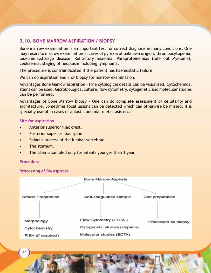
74
3.10. BONE MARROW ASPIRATION / BIOPSYBone marrow examination is an important test for correct diagnosis in many conditions. One may resort to marrow examination in cases of pyrexia of unknown origion, thrombocytopenia, leukumeia,storage disease, Refractory anaemia, Paraproteinemias (rule out Myeloma), Leukaemia, staging of neoplasm including lymphoma.
The procedure is contraindicated if the patient has haemostatic failure.
We can do aspiration and / or biopsy for marrow examination.
Advantages Bone Marrow aspiration – Fine cytological details can be visualised, Cytochemical stains can be used, Microbiological culture, flow cytometry, cytogenetic and molecular studies can be performed.
Advantages of Bone Marrow Biopsy – One can do complete assessment of cellularity and architecture. Sometimes focal lesions can be detected which can otherwise be missed. It is specially useful in cases of aplastic anemia, metastasis etc.
Site for aspiration.• Anterior superior iliac crest.• Posterior superior iliac spine.• Spinous process of the lumbar vertebrae. • The sternum. • The tibia is sampled only for infants younger than 1 year.
Procedure
Processing of BM aspirate

75
Once the bone marrow aspirate is collected.
Smears are prepared for morphology and cytochemistry and sample is collected in EDTA tube for flow cytometry and molecular studies. Sample is also collected in heparin tubes for cytogentic studies clot, if present, is collected in formalin and processed as biopsy.
Processing of BM Biopsy
Bone marrow biopsy is performed by Jamshidi needle and core of bone marrow obtained is kept for fixation in formalin and then for decalcification. Imprint smears are also prepared for morphological correlations.
Than it is processed as paraffin embidding followed by hematoxylin - eosin standing on thin sections.
Decalcification• 10%NITRIC ACID• HYDROCHLORIC ACID (HCL)• FORMIC ACID• EDTA
Embedding• Paraffin
Now days disposable needle is performed for aspiration and biopsy.
Exercise1. List a few diseases in which bone marrow examination is indicated?2. What are the various sites for bone marrow aspiration?

76
3.11 LAB SAFETY
3.11.1. BIOMEDICAL WASTE MANAGEMENT (BMWM)
Biomedical waste is waste generated by healh care establishments and lab during diagnosis, treatment or research.
CLASSIFICATION OF HOSPITAL WASTE
IMPORTANCE OF BMWM
Extremely hazardous• If it is not managed properly it can lead to serious health and environmental problems.
Therefore it Should be segregated, collected, stored, transported and disposed off properly to prevent the transmission of disease from patient to patient, from patient to health worker and vice versa and also to prevent injury to the health care workers and workers in support services, while handling biomedical waste. It is also required to prevent a general exposure to the harmful effects of the cytotoxic, genotoxic and chemical biomedical waste as much as possible. Disorganized management of hospital waste exposes community and surroundings to infection, toxic effects and injury. It is crucial that all medical waste materials are segregated at the point of generation, appropriately treated and disposed off safely.
People at risk are??
Healthcare staff
Patients
Vistors/attenders to healthcare setting
Bio medical waste Handlers
General public

77
• Waste should be collected and segregation at the site of generation itself and waste is separated into various specified categories as per its nature. Harmful and infected material shall be separated from harmless and non-contagious waste and specifically coloured waste containers and plastic bags are mandatory for this purpose.
TRANSPORTATION OF BMW(A) Within the hospital
Waste routes must be designed to avoid the passage of waste through patient care areas. Separate time should be earmarked for transportation dedicated wheeled containers, trolleys or carts should be used trolleys or carts should be thoroughly cleaned and disinfected in the event of any spillage. The wheeled containers should be designed that the waste can be easily loaded, remains secured during transportation, does not have any sharp edges and easy to clean and disinfect
(B) Outside the hospital
Untreated biomedical waste should be transported only in such vehicles as may be authorized for the purpose by the competent authority as specified by the Government under the Motor Vehicle Act, 1998.
The containers for transportation must be labeled with “Biomedical Waste” symbol on both sides and rear part
Personnel involved in BMWM shall near gloves, glasses, lab coat, boots and masks and also shall be immunized against tetanus & Hepatitis B.
3.11.1 PERSONNEL PROTECTIONIt is the responsibility of every lab to establish and maintain a safe working environment. There shall be a named person ultimately responsible for the safety of personnel at work and others who may be affected by it. All hazards shall be identified and measures shall be taken to eliminate them or reduce to as low level as practicable by
Substitution,
Containment and
Use of personal protections equipments.
Safety is the primary consideration and cost should be of secondary importance.
General Safety:
It Includes

78
Proper use of personal protective clothing/equipment (e.g., gloves, gowns, masks, eye protectors, etc.).Bio-medical wastes shall be disposed off properly along work.Proper handling of spillages (blood and other body fluids).
All occupational injuries or illnesses require medical treatment (except first aid)
Fire Safety
Laboratory shall
(1) have an automatic fire extinguishing system at all required places.
In all cases, a fire-bell, public address system, or other alarm system must be audible in all sections.
Electrical Safety• All laboratory instruments and appliances shall beAdequately grounded and • Checked for current leakage (before initial use, after repair or modification, and when
a problem is suspected)
This Task may be delegated to biomedical and electrical engineers
Chemical Safety• The lab shall have a comprehensive signage and labeling system in use.• Material safety data sheets (MSDS) shall be on file for each hazardous chemical.• Each laboratory shall have a written plan to reduce or eliminate mercury.• Chemical fume hoods shall be checked annually for proper function.• Piped eyewash fountains or the equivalent shall be present and should be checked
weekly.
Microbiological Hazards• The laboratory should have policies and procedures for assessing the occupational risk
associated with exposure to infectious agents handled in the microbiology laboratory.
Level II bio-safety cabinet should be used for working with infectious agents.
Waste Disposal– All solid and liquid waste must be disposed in compliance with applicable local, state,
and national regulations.– All sharps, especially those contaminated with potentially infectious materials, must be
properly discarded in puncture-resistant containers with tightly fitted lids.

79
Disaster Management– The laboratory must have documented procedure on “Internal and External Disaster
Preparedness.” It should be followed in the event of a catastrophe such as fire, flood, electrical outage,
or spill of hazardous volatiles (internal disaster), or atornado, earthquake, or other mass-casualty situation (external disaster) and lab shall have
– Proper evacuation plan
Ergonomics• “Ergonomics is an applied science concerned with the design of workplaces, tools, and
tasks that match the physiological, anatomical, and psychological characteristics and capabilities of the worker.”
• “The Goal of ergonomics is to ‘fit the job to the person,’ rather than making the person fit the job.” Ergotec
PERSONNEL PROTECTION
Safety of personnel is of primary importance and both management as well as staff are responsible for it. There is international standards ISO 15190 for medical lab safety in which all management and personnel responsibilities are stipulated.
Personnel Protective equipment (PPE) : has been discussed previously
Infection Control Measures Like • Cleaning, Hand Wash, Personnel Protection Equipments (PPE),Disinfection & Sterilization,
Vaccination, Awareness & Training Programme shall be followed appropriately and according to documented procedure.
Housekeeping
Housekeeping is defined as the provision of a clean, comfortable and safe environment for the patients and Laboratory staff.
Purpose:• The regular and routine cleaning of all surfaces and maintaining a high level of hygiene
in the facility. Areas of the Laboratory shall be classified according to the varying need for cleaning.

80
3.11.2. HAND HYGIENEOne of the most effective and simple measure for preventing infections is proper hand washing. It prevents transmission of infection between Health care worker’s (HCW’s) and patients in the hospital and so plays a very important role in hospital infection control
Decontamination of hands is important - before having direct contact with patients, after contact with patient intact skin, after contact with body fluids or excretions, before donning sterile gloves, after removing gloves, after contact with inanimate objects, before eating and after using rest room.
Hand Hygiene Technique
When washing hands with soap and water.• Wet hands first with water• Apply soap solution to hands and rub hands together vigorously covering all surfaces of
hands and fingers. After that rinse hands with water and dry thoroughly with a disposable towel
3.11.3. MANAGEMENT OF SPILLS
Introduction
Spilled blood and body fluids/substances and infective agents may be encountered in many situations in Hospitals & Laboratories. These must be attended to immediately.
All body fluid spillage must be cleaned up effectively and immediately. Disposable gloves and aprons should be worn by the person handling the spill. It is advisable to cover wounds and lesions on exposed skin with a waterproof dressing. Mops are never used to clear up body fluid spillages. Hypochlorite solution must be prepared and used within 24 hours. One should ensure that there is adequate ventilation is when using Hypochlorite to clean areas.
SPILL PROCEDURE• Gloves and Appropriate Personal Protective Equipment (PPE) like - Protective Eyeware,
Lab Coats, Masks and Face Shields where Splashing is Likely shall be used.• Any broken glass or sharp objects from the spill shall be removed using mechanical
means like forceps, hemostats, needle-nose pliers, broom and dust pan. • Sharps / broken glass shall not be removed by hand.• Spill is contained by covering with paper towels and carefully pouring appropriate disin-
fectant solution (1:10 to 1:100 dilution of Sodium hypochlorite) around and on the spill.

81
• Care shall be taken not to splash disinfectant solution or create aerosols while pouring.• Paper towels are removed and process shall be repeated until all visual soilage is removed• Cleaned area is re-wet with disinfectant and air dried• All contaminated paper towels in a “red bag” or an autoclave bag for appropriate
disposal (autoclaving, off site treatment, etc.)• Then all PPE are removed and immediately hands washed • A 1:100 dilution (500--615 ppm available chlorine) is used to decontaminate nonporous
surfaces after cleaning a spill of either blood or body fluids in patient-care settings• If a spill involves large amounts of blood or body fluids, or if a blood or culture spill
occurs in the laboratory, a 1:10 dilution (5,000--6,150 ppm available chlorine) shall be used for the first application of germicide before cleaning
Spills on the body
Splashes of body fluid or blood on to the skin must be washed off immediately with soap and water.
Exercise1. When all should one decontaminate hands?2. Describe briefly the spill procedure?
LIST OF REFERENCES(1) Practical Haematology (Tenth edition) - Dacie and Lewis(2) Textbook of Medical Laboratory Technology (Second Edition) - Praful B. Godkar & Darshan
P. Godkar(3) Pathologic Basis of Disease (Seventh Edition) - Robbins and Cotran

82
Unit overview:1) This unit will provide the students information about the scope of blood bank and the
various topics related to blood transfusion.2) It helps to understand the relevant terms, procedure and working of equipment 7
personnel as relevant to a blood bank.
Knowledge & Skill Outcomes1) To understand the scope of blood banking.2) To understand the organizational structure of a blood bank.3) To know the relevant term, procedure and working of equipment and personnel as
relevant to a blood bank.4) The responsibility of personnel working in a blood bank.
Learning Outcomes :-
After completing this unit the students should be able to demonstrate knowledge and shall be able to comprehense and apply the techniques related to :-1. Safe Collection2. Donor Selection3. Grouping of Blood4. Grouping associated problem solving5. Transfusion related problems
Material and equipment
1. Equipment for haemoglobin estimation :
a. Haemoglobinometer
HEMOCUE Hb 201 + ANALYSER :-
Used for measuring the hb of blood donors before they donate blood
2. Equipment for blood collection
UNIT - 4bLOOd bANk ANd TRANSFUSION

83
Blood donor couches
Blood collection monitor
Blood bags
Tube sealera. blood donor couch : They are specialized couches so that the donor feels comfortable
while blood donation. Generally there is a provision for giving a head low position when the donor is having giddiness.
b. blood collection bags : either 350 ml or 450 ml bags, there are different varieties of bags they are single, double, triple or quadruple.
Single bag : does not have any satellite bags double, triple and quadruple bags have one, two and three satellite bags respectively quadruple bags are also called buffy coat bags and are used for separating the buffy coat while preparing the blood components the bags contain the anticoagulant solution : CPD / PCD – A / SAGM
Citrate, Dextrose, Phosphate, Saline, Adenine Glucose, Mannitol The anticoagulant in 350 ml bags is 49 ml and in 450 ml bags in 63 mlc. blood collection monitor : used for the actual collection of blood. It can collect the
desired volume of blood from the donor which is either 350 or 450 ml. It has a visible volume display and flow rate also. There is a audible alarm if the flow of blood is slow. There is a provision for automatic clamping when the volume is reached. After collection this once again gives an audible alarm.
d. tube sealer : used for clamping the segment of the blood bag after it is collected. Available in either fixed sealer or a mobile sealer. Mobile sealer can be taken anywhere for sealing and is used in blood collection camps.
3. equipment for blood component preparation
Electronic weighing machine
Refrigerated centrifuge
Plasma expressor
Cell separator
Sterile connecting devicea. electronic weighing machine : for weighing the bags accurately.b. refrigerated centrifuge : bucket handle typed of centrifuge to hold the collected bags
with a provision for a wide range of temperature is preferred.

84
The main unit is built on a sturdy metal frame resting on castors and enclosed by sheet metal, which has an electrical interlock. Rotor consists of 4 to 6 buckets.
Features of cold centrifuge :1. Digital speed indicator2. Stepless speed relulator with ‘O’ start switch3. Digital automatic timer4. Dynamic break5. Digital temperature indicator cum controller6. Unbalance cut out switch7. Brush wear indicator8. Hermatically Sealed refrigeration unit9. Temperature Range 0OC to 300OCc. plasma expressor : to manually express the plasmad. cell separator
Cell separator is a instrument used into separate what ever components required for the patients.
But in our blood bank we are using mainly for the whatever the large amount of components, platelets required.
Ex. Platelet’s are required it is available to use this procedure. This procedure is called as “A-pheresis”.
Two types of process can be done by using cell separator.1. Continuous flow process2. Interrupted flow process
Continuous flow : It is a two arm procedure where in blood is drawn from one arm. The components are separated in a cart rid & the remaining cells & plasma flow back to the donor through the other area. Here the volume of blood which is outside the body is very small.
Interrupted flow : This is a one arm process. One line is connected to the donor the blood will be coming out after processing components will be separator, remaining required plasma & RBC’s will be reinfused back to the donor with same line and this process will takes little longer time than the continuous flow.
e.sterile connecting device : used to connect ends of two different segments in asterile manner. Widely used for separation of small volumes of blood for paediatric transfusion, buffy coat pooling and lab side leukodepletion.

85
4. equipment used for storage of blood
Blood storage cabinets
Plasma storage cabinets
Ultra low freezers
Cryoprecipitate bath
Platelet agitator and incubator
Plasma thawing bath
Blood storage cabinets 2-6 deg c+21. Blood storage cabinets are constructed of double walls with exterior made of powdered
steel steel and the inner is made of stainless steel. It is provided with number of compartments having transparent Plexiglas doors.
2. The cabinets are provided with pull out type trays for proper storage of blood bags.3. The control comprises of fully automatic digital temperature indications/ controls with
off/on switches indicating lamps with protective fuse.4. It also incorporates electronic safety circuit which gives audio visual alarm in case of
power failure.5. Temperature variation is +/-2C. In case the temperature increases more than 4C or if
the door is open protecting. The precious blood bags inside the cabinets.6. The alarm is set off thereby protecting the temperature maintenance.
IMPORTANCE :1. Uniform temperature between 2OC – 6OC (in AC room)2. Stainless steel inner chamber 3. Digital temperature indicater cum controller with audio visual alarm4. Inside acrylic door to avoid temperature loss5. Full view glass doors for observation without disturbing the inside conditions
Plasma storage cabinets (-20 deg to -30 deg c)
The plasma storage cabinet is designed storing of plasma and related blood components at temperatures upto – 20OC.
All models feature integrated control panel with digital temperature indicator cum controller and temperature recorder. Constructed of double walls, the exterior is made of powder coated sheet steel finish while the inner chamber is made of stainless steel.

86
The unit is mounted on castor wheels for ease of mobility. Plasma Freezers are available in horizontal and vertical versions.
SPECIAL FEATURES :- Low temperature up to -35OC- Stainless steel inner chamber- Extremely efficient insulation to minimise heat loss- Digital temperature indicator cum controller with audio visual alarm- Inside acrylic door to avoid temperature loss
Ultra low freezers (-80 deg c)• Ultra low freezers are constructed of double walls, the exterior is made of sheet steel
and finished with acrylic enamel paint. The inner chamber is prepared by stainless steel.• The unit is mounted by on castor wheels for ease of mobility.• The ultra low freezers are specially designed and suitable for blood bank research
laboratories.• The ultra low freezers are high-tech unite incorporating revolutionary cooling system
giving advantage to the users of rapid pull down of temperature.
SPECIAL FEATURES :- Stainless steel inner chamber- Extremely efficient insulation to minimize heat loss- High tech solid state digital indicator com-controller- Counter balanced door- Inside acrylic doors to avoid temperature loss
Cryoprecipitate bath :
Cryoprecipitate baths are used for quick thawing of Plasma at + 4OC / + 37OC and improves yields over conventional air thawing methods.- Cryoprecipitate bath is fabricated out of sheet with a stainless steel inner chamber.- Refrigeration is achieved by means of hermetically sealed compressor.- The temperature is controlled by a digital indicator cum controller.
Special features :- Suitable for 15 plasma bags with rack holder.- Direct digital temperature indicator cum controller.

87
- Stainless steel AISI 304 tank resistant to corrosion.- Circulating pump to ensure uniform temperature within the bath.
Platelet agitator and incubator :22 deg c specifically designed to maintain donor platelets in an even suspension throughout the blood plasma. Platelet Agitator agitates at the fixed speed of 70 to 80 strokes per minute.
Plasma thawing bath : It is provided with a metallic basket which can hold the plasma bags upright so that the ports are maintained above the water level. Side to side movement is maintained and the speed is adjustable. The temperature is maintained at 37OC with the help of a thermostat. The water bath is provided with a see through fiber glass lid.
5. Equipment and regents for testing of blood :
ABO grouping : cross matching : either test tubes or gel cards or micrplates – depending on the protocol of the blood bank
Infectitious disease testing : ELISA reader and washer : either automatic or semi automatic
Random access analyzer
Equipment for malaria and VDRL testing : respective reagents and strips
Centrifuges to centrifuge the blood samples
Records and Computer systems to enable documentation
Other equipment :a Autoclave :
An autoclave is used to sterilize various materials in the laboratory by steam sterilization method. Blood units that are sero positive or expired are autoclaved at 120 deg for 20 minutes.b microscope
Introduction : A microscope magnifies the image of an object. The modern compound microscope is the most important apparatus in a laboratory. It is a precision instrument and needs careful handling.
The light microscope use white light, either the external sunlight or the internal tungsten filament lamp, as the source of illumination. As viewed under the microscope, objects look dark or coloured, contrasted against a lighted background. In case of the dark field microscope, a special dark field condenser is used, that lights up the object, like stars against a dark sky. This is limited use in the microbiology laboratory for observing spirochetes. The fluorescent microscope uses a special ultraviolet lamp as the source of illumination. A fluorescent dye is attached to the object through laboratory procedures, which glows when exposed to the ultraviolet radiation. Fluorescent antibody testing requires this kind of microscope.

88
Parts of the microscope are :1. The support system consists of the base or foot rest which also holds the light source2. The tube or the arm holds the optical system and also the coarse/fine adjustments.3. The objectives are at the lower end of the tube and the eye pieces are at the upper end.4. The objectives are attached to the revolving nose piece and can be shifted. The
objectives are 10x (low power) 40x (high power) and 100x (oil immersion).5. Below the objectives is the stage which holds the object/slide. The stage has a central
hole through which the light passes. The stage may be fixed or movable. Below the stage is the iris/diaphragm which focuses the light.
6. The eye pieces are also of varying magnification; the commonly used one is 10 xs. The eye pieces are situated in the binocular tube.
Principles of microscope:
The magnification system is the optical component of the microscope. That magnifies the ob-ject placed on the stage. It is done by the combination of two series of lenses. The objective and the eye piece. The objective stays near the object of the tube. The low power objective (10x) is smallest in size, usually with a green ring on the objective for easy identification and when the image is in focus, the low power objective has the largest working distance (5 to 6mm). The working distance is the distance between the front lens and the objective slide. The working distance decreases with increasing magnification and is 0.5 to 1.5mm in case of the high power and 0.15 to 0.20mm in case of the oil-immersion objective. The eye piece magnifies put on a screen.
The total magnification of the object is the multiplication value of the magnifying powers of the objective and the eye piece. Thus with 10x magnification of the eye piece, the low power objective gives 10x10=100x total magnification. The high power 10x40=400x magnification and for the oil-immersion lens, 10x100=1000x magnification. The cost of the lens increases with higher magnification.
When using oil immersion a drop of cedar wood oil is applied directly on the smear.
c.incubator
Incubators are temperature controlled chamber’s which are well insulated. Necessary for various investigations where body temperature. 30OC incubation is required.
USE :
These are mainly used for1. Determination of enzyme’s in the specimen by end point reaction methods.2. Determination of glucose, urea, uric acid etc., by enzymatic methods.

89
3. Growing microorganisms on various culture media.4. Right temperature for immune antigen reaction.
d.hot air oven
use :
Hot air oven is mainly used for the following purposes.1. Dry sterilization of syringes & needles.2. Preparation of anticoagulated bulbs.3. Drying of glass ware.4. Heating of chemicals used for the preparation of primary standards.
Principle :
When electricity is passed through the heating coils, electrical energy is converted to heat energy.
Components :1. Double walled heavy gauge aluminum chamber.2. Thermal insulation by thick glass-wool between the working chamber and the outer mild
steel casting.3. Thermostat.4. Adjustable ventilator.5. Perforated metal shelves.6. Thermometer.
Temperature Range :-40 OC to 300 OC
The main unit is built on a sturdy metal frame resting on castors and enclosed by sheet metal, which has an electrical interlock. Rotor consists of 4 to 6 bucket’s which can add blood bags i.e., either single, double (or) Triple.
e.Clean air cabinet : (laminar flow cabinet)
70% of the air in recirculated through filters. So that the working area is bathed in clean (almost sterile) air. The air flow carries along any aerosols/particles produced in the course of the work and these are removed by the filters. Some of the air (30%) is exhausted to atmosphere and is replaced by a curtain of room air which enters at the working face. This cabinet is fitted with a UV lamp. The filters need to be cleaned periodically and swabbed every day with disinfectant.

90
Records in blood bank1. Blood donor record : it shall indicate the foll : Serial number, date of bleeding, name, address and signature of donor, age weight, hb,
blood grouping, blood pressure, medical examination, bag number, patient’s details incase of replacement donor, category of donation (voluntary / replacement), deferral records, sign of medical officer.
2. Master record for blood and components : Bag serial number, date of collection, date of expiry, quantity in ml, ABO / Rh, results
of testing for HIV, HBsAg, HCV, VDRL, malarial parasite, irregular antibodies if any, name and address of the donor, utilization issue number, components prepared or discarded, sign of the medical officer.
3. Issue register : serial number, date and time of issue, bag serial number, ABO, Rh, total quantity in ml, name and address of the recipient, group of recipient, unit/institution, details of crossmatching report, indication for transfusion, components issued, quantity issued, signature of the issuing person.
4. Records of the ACD, CPD, CPD-A, SAGM bags having details of the manufacturer, batch number, date of supply, results of testing.
5. Register for diagnostic kits and reagents used : name of the kits, reagents, batch number, date of expiry and date of use.
6. Transfusion adverse reaction reports :the reports and the investigations for all transfusion adverse reactions are maintained.
7. Records of purchase, use and stock in hand of disposable needles, syringes, blood bags, all the records must be maintained for a minimum of 5 years.
Laws of genetics
Genetics is the study of inheritance – the transmission of characteristics from parents to offspring. In 1865, Gregor Mendel did some experiments with garden peas thereby bringing to light the science of genetics. His studies led to the basic understanding of how genetic traits are passed to each generation.
Humans have 46 chromosomes, in which 22 pairs are autosomes and one set of sex chromosomes (XX and YY).
As a cell divides it reproduces the chromosomes so that all the daughter cells are identical to the parent cell. This process is known as mitosis.
In one set of experiments Mendel cultivated sweet peas until they bred off spring with flowers of all one color (e.g., red or white only). He then cross-bred these two plants and obtained a

91
first filial generation that were all red flowered. When plants from this generation were bred with each other, they produced red and white flower in a ratio of 3:1. When the traits are co dominant, both are expressed equally. This type of codominant expression is important in blood group genetics such as inheritance of AB group. This is the law of inhereitance.
The next law of Mendel is the law of independent assortment. This means that factors for different characteristics are inherited independently from each other (if they reside on different chromosomes).
Deoxyribonucleic acid (DNA)
Human chromosomes are composed of linear strands of deoxyribonucleic acid (DNA) wound around proteins called histones. This complex of DNA and proteins is called chromatin. It is this wrapping and condensing of DNA that allows so much genetic material to be stored in a small piece of the chromosome.
DNA is composed of four nitrogenous bases, a molecule of deoxyribose, and one phosphate group. The four nitrogen-containing bases are the purines adenine (A) and guanine (G) and the pyrimidines thymine (T) and cytosine (C). Each base can bind to a deoxyribose sugar to form a nucleoside; the addition of the phosphate group makes the compound a nucleotide.
Ribonucleic acid (RNA)
Composed of nucleotides AND usually exists as a single strand. Other differences include the substitution of ribose for deoxyribose and the nitrogenous base uracil for thymine. Ribose differs from deoxyribose by the presence of a hydroxyl group at the I carbon. Uracil differs from thymine by its lack of a methyl group.
Basics of Immunology
Blood groups are inherited characters which give rise to antigen-antibody systems. Blood group antigens are chemically either glycolipids or glycoproteins and are immunogenic expressions of the components of the red cell membrane. They have a polysaccharide component responsible for the specificity and an aminoacid fraction that determines the antigenicity.
Antigen :
An antigen is any substance, either protein or non-protein but when introduced into an animal causes the production of another specific substance called antibody. The antibody reacts specifically with the antigen.
In blood banking red cell antigens are of importance.
92
Antibody:
Antibodies are serum proteins, more specifically immunoglobulins. Antibodies are recognized by their interaction with antigens. In blood group serology this interaction is usually detected by agglutination of red blood cells.
Naturally occurring antibodies :
The presence of antibody in serum is not always because of exposure to antigenic stimulus by red cells. Some foreign substances like bacteria and plants (containing red cell like substances) are capable of causing antibody production and these antibodies are called naturally occurring antibodies. These are commonly of IgM type, occur in serum without any specific antigenic stimulus, e.g. anti-A, anti-B, and anti- Wra. These antibodies are present in individuals who lack that antigen.
Immune antibodies:
Immune antibodies are IgG. They develop due to immunization following pregnancy, previous transfusion or deliberate injection of immunogenic material. In some instances immunogenic event is unknown.
The antigen-antibody reactions In-vitro1. Agglutination :is defined as clumping of particles that have antigen on their surface and
is brought about by anti-bodies. This forms the basis of blood grouping tests.2. Hemolysis :Rupture of red cells with release of intracellular haemoglobin can occur if
the Antibody has the property of hemolysin. It requires presence of complement.3. Precipitation : is the formation of visible insoluble complex when soluble antibody
reacts with soluble antigen.4. Enzyme linked immunosorbent assay (ELISA) :Here an enzyme label is used and a colour
reaction that takes place in presence of a substrate denotes the presence of antigen/antibody as the case may be. This is the principle sued in Transfusion transmitted disease tests.
Factors affection agglutination reaction:I. The antigen (Ag) –The red cells should be properly stored to achieve optimum reactivity.
If the cells are frozen the reactivity is better preserved. The reactivity is better in fresh cells. Thus with dilute cell suspension, increase in sensitivity is achieved, as concentration of antibody in solution does not fall even when all cells are saturated with antibody. One drop (2-5%) of cell suspension is therefore found ideal to be added to two drops of serum.

93
Need to wash the cells :1. Washing improves reactivity.2. Removes plasma that contains fibrinogen and forms clot when mixed with serum giving
false positive.3. Plasma can cause roulaeux formation.4. Anticoagulant present in plasma is anticomplementary and inhibits complement binding
reactions.5. Plasma contains blood group substances that can neutralize that reaction.II. The antibody (Ab) from one individual may react differently at different times and sera
from different source with same type of antibody react differently. The stored sera lose reactivity and hence the importance of the batch numbers and expiry dates. The speed with which a particular antibody combines with the antigen forming a stable complex is called avidity, which is variable. The reactivity of Ag/Ab depends also on titre. The titre of the antibody is the highest dilution of serum with a positive reaction. Very high titre sera are likely to give prozone effect. The prozone is the inhibition of the reaction in presence of excess antibody.
III. Factors depending on reaction condition :1. Incubation time :Upto 60 minutes is adequate for all blood groups.2. Incubation temperature :depends on type of antibody.3. Centrifugation : should be adequate to produce a cell button with a clear supernatant
but without packing cells tightly so that they are difficult to dislodge. Over centrifugation leads to false positive.
4. Ionic strength :low ionic strength i.e. low concentration of dissolved salts increases the amount of body binding to cell.
5. pH: Normal blood pH is about 7.42.6. antigen-antibody ratio : The ratio of antigen and antibody in the mixture should be
optimum.7. Zeta potential :Erythrocytes have a genitive electrical charge. The electrical activity of
this ionic cloud is called Zeta potential.
Agglutination is the endstage of all grouping reactions and this can be carried out on slides, tiles with well microtitre plates, capillaries or tubes. The tubes are most advantageous.
Macroscopically : After centrifugation, the cells should be gently dispersed to observe for agglutination.
94
There are many causes for false positive reaction such as :1. Autoagglutination2. Bacterial contamination3. Polyagglutination4. Chemicals e.g. detergents5. Cell mixtures6. Leukocytosis ie wbc count7. Rouleaux formation-caused by:
(i) Presence of large molecular weight substances e.g. fibrinogen, dextran, Polyvi-nylpyrrolidone (PVP)
(ii) Abnormal proteins-e.g. multiple myeloma.
Causes of false negative :1. Deterioration of antigen on storage2. Chemical contamination3. Inhibition due to improper washing of cells4. Absence of complement5. Thick cell suspension6. Agammaglobulinemia7. Poor technique of reading
ABO Blood group systemLandsteiner discovered the ABO group antigens in 1900 and since then this is one of the most important discoveries in the field of medicine.
The importance of blood grouping :1. Safe blood transfusion2. Organ transplant especially liver, heart and kidney3. Medicolegal and forensic, paternity disputes4. Immunology and genetics
Landsteiner’s law
The reciprocal relation between ABO antigens and antibodies is called Landsteiner’s law

95
Cell grouping serum grouping blood groupanti-A anti-B anti-AB A cells B cells
+ - + - + A- + + + - B- - - + + O+ + + - - AB
The incidence of ABO groups in India
A 27%
B 31%
O 34%
AB 8%
Inheritance of ABO blood groups : A B antigens are inheretited as codominant fashion. Each individual inherits two ABO genes from each parent and hey determine the ABO antigen present on their red cells. O is an amorph and does not produce A or B substance. The serological typing reveals the phenotype and the family studies help to reveal the genotype.
Phenotype genotype
A AA
AO
B BB
BO
AB AB
O OO
Biochemistry of ABO system
The A,B antigens are glycoproteins. Each molecule of a precursor is made up of a peptide and a sugar. The blood group specificity is determined by the terminal sugar
Terminal sugar antigen
L Fucose H
N Acetylgalactosamine A
D Galactose B

96
Transferases :enzymes which assemble individual sugars into chains forming the antigen As each sugar is added on a new structure is formed which acts a substrate for another transferase. The A gene specified transferase adds N acetyl galatctosamine, the B gene transferase adds N acetyl galactose to the H gene. The H gene transferase is the L focosyl transferease. In the AB individual 2 different sugars are added to different chains of the same red cells.
Secretor states: The A, B and H substances on erythrocyte surfaces are lipopolyccharides, not water soluble but they can be dissolved in fat solvents, such as alcohol and chloroform. They are most clearly detectable in saliva and also identified in some other body fluids. These people are referred to as secretors. The secretor characteristic is inherited in the classic fashion by a dominant gene designated se. Itsallesle se has no effect. Hence noon secretors are sese while SeSe and Sese are secretors. The gene is independent of ABO and Hh.
Concept of H: H substance is the precursor substance from which A and B substances are formed by action of the genes A and B. All red cells have some H. but the amount varies according to the blood group. O cell have the maximum and it diminishes in the following order. A2, A1B.
In 1952 Bhende, Bhatia and Deshpande identified a new blood group called Bombay group. The red cells appear to be group O, but their serum contains a powerful antibody reacting with all other red cells except those of the same group. The cells do not contain H antigen
CONCEPT OF H
A GENE > A AND H ANTIGENS
H SUBSTANCE-B GENE—B AND H
O GENE>H
PRECURSOR________
SUBSTANCE
PRECURSORABO GENE
SUBSTANCE INEFFECTIVE Oh
The red cells and secretions of such a person lack H substance and the serum has anti-H Antibody. Since His absent, there is no substrate on which A or B genes can act, even if present, hence, genetically A or B group can result, but the expression of these genes and their secretor status is prevented by the absence of H. these ABO genes when present, even though not expressed, are inherited and transmitted normally. It is suggested that an inhibitor gene is involved to suppress A and B antigens. The presence of this h gene in homozygous

97
condition result in suppression of all A, B and H antigens on the red cell and saliva. The ABO and secretor genes are however fully expressed in individuals, heterozygous for the H-h gene. The H-h genes are not located on the same chromosome carrying ABO genes. Thus this phenotype is called Oh. Family studies are helpful in confirming the Oh phenotype. In India this is more common compared to other countries. The presence in India is more amongst Marathas of south west district of Maharashtra. The frequency in India is 1:13000.
The phenotype is easily detected by its failure to react with anit-H or positive reaction with all Oh group are no secretors of ABHantigens, Le (a+b-) and rarely Le (a-b-).
Due to the presence of anti H, the patient cannot be transfused with any other blood except Bombay blood.
Recently blood with weak A or weak B, in individuals with antibodies of H antigen is reported and this is explained on the basis of partial suppression and is called Ah-Bh phenotype or Para Bombay type.
ABO antibodies :
A and B antibodies are naturally occurring antibodies present in the serum of persons who lack the corresponding antigen. At birth these antibodies are not developed. A and B like substances are widely distributes in nature, in plant and animal tissues and in bacteria. Every one is exposed at he time of birth to these substances and by second or third month develop the antibodies. Hence the test on the serum of a new born or an infant unto 6 months are not reliable. In old age again the level of anti-A and anti-B is significantly reduced.
Sub groups in ABO System
Subgroups of A:
The cells which are agglutinated by anti-A but not by anti-A1 are A2 antigen and those agglutinated by both are A1 antigen cells. Anti-A1 occurs in the serum of approximate 1 to 8% of A2 and 22-35% of A2B individuals. The weaker subgroup of A are based ona. The reactivity of red cells with anti-A, anti-AB, anti-H and anti-A1.b. The presence or absence or absence of anti-A1 in the serum andc. The secretion of A and H substance by the secretors.
The few important ones are as follows:
Anti-A Anti-AB Anti-A1 Anti-H Substance in saliva
A int ++++ ++++ ++ +++ A & H

98
A 3 ++ ++ 0 ++++ A & HA X (A4/A5/ A6/A0A2)
Mixed field 0/+w
+/++ 0 ++++ H
Am +W +W 0 ++++ A & H
These subgroup are at times difficult to detect, and they pose a problem in case of the donor grouping. A donor unit of weak A may be grouped as O and if issued to and O groupindividual could cause a transfusion reaction. For recipient’s grouping if it is missed, it does not matter, as the recipient can receive O group blood without any ill effects.
Subgroups of B
The B group antigen also has similar weaker variants which are much less common (B2, B int, B3 Bx, Bm).
It has been noticed that the strength of the ABO antigens changes in the disease state. The weakening of A antigen has been reported in cases of leukemia, the depression is related to the course of the disease and thus during remission the reactivity appears to be normal. Report of suppression of A antigen in aplastic anemis, Hodgkin’s disease and in osteogenic sarcoma have been reported.
The other fascination phenomenon noticed is the acquired B antigen. Many cases have been reported, associated with carcinoma of colon and rectum, appendicitis, peritonitis, bacter-emia and urinary tract infections. It sis presumed that the change is caused by the pathogenic bacteria which modify the red cell membrane or the bacterial lipopolysaccharides which are similar to B group antigen (especially of E. coli O86) get passively adsorbed on the red cells.
Modifications of ABH antigens during the course of pregnancy have also been reported. The loss of antigenicity is more towards the delivery. Women on contraceptives for over a period of 9 months also show a similar loss of antigens. The loss is apparently 20% for A and B and 35% for A1 and H antigens. The L-D-galactosyl transferase enzymes in sera of B group women are found to decrease in pregnant women reaching their lowest at term. The changes are also observed during the menstrual cycle which relate to the hormonal levels.
PRACTICAL IMPORTANCE OF OTHER BLOOD GROUPS
I blood gr system
Discovered in 1956
All adult red cells have I antigen and cord blood has I antigen
By the time the infant is 18 months old the red cells have I antigens

99
Clinical significance :
Anti-I antibody is usually a cold antibody, occasionally reacts at 37 deg to cause hemolytic disease of newborn or hemolytic reaction
Duffy system
Duffy system contains two antigens Fya and Fyb
Plasmodium vivax infection does not affect red cells lacking Fya and Fyb
antiFya and Fyb antibodies are IgG and react at 37 deg c and cause hemolytic reaction and hemolytic disease of the newborn (HTR and HDN)
duffy antigens are classified asFy (a+b-)Fy (a+b+)Fy (a-b+)Fy (a-b-)
Kidd blood group system
Two antigens Jka and Jkb
The antibodies may be either IgG or IgM and may cause HTR or HDN
P system
Three antigens P1, P2 Pk are present for most of the population
Few case of HDN have been attributed to anti-P1
Hydatid cysts contain P1 substance
The donath Landsteiner autoantibody found in cases of PCH (Paroxysmal Cold Hemoglobinuria) shows anti-P1 specificity
MNSs system
Anti-M and anti-N are cold antibodies rarely they may react at 37 deg and cause HTR or HDN
S and salso may react at 37 deg and cause HTR
LEWIS system
Two main antigens are Le a, and Le b and the phenotypes are
Le (a+b+), Le (a-b+), Le (a+b-), Le (a-b-)

100
Secretors are either Le a+b+ or Le a-b+
Non secretors do not have the Le b antigen
KELL SYSTEM
The kell system was discovered by coomb’s Mourant Race in 1946. The kell antigen is strongly immunogenic. It is known to cause many transfusion reactions and Haemolytic disease of the new born. Incidence of Kell antigen in India is reported as 0.3 to 0.4%
Antibodies of this kell system anti K and antic are always immune and are best detected by indirect antiglobulin test.
LUTHERAN SYSTEM
The original antigens described were Lua and Lub.
Anti Lub has been reported to be mainly IgA. Anti Lua has not been incriminated as a cause of hemolytic disease of the new born or transfusion reaction.
Rh NULL
Rh Null is a clinically significant member or Rh system which is responsible for Haemolytic anemia in individuals who posses them.
Rh null people are usually observed when Rh positive blood is transfused. In some cases of pregnancy.
-D-
Only –D- antigen is found and these cells lack Cc and Ee.
For transfusion the same phenotype only will be compatible. Transfusion of other phenotypes where C/c and E/e will result in antibody formation and hemolysis.
Rhesus blood group systemLand Steiner and Wiener discovered this system in 1940 after Levine and Stetson in 1939 reported an irregular anti body in a mother which was later shown to be anti-D.
Rh antigens are found only on red cells. The Rh antibodies usually develop only in absence of Rh antigen, by a known stimulus, e.g. transfusion or pregnancy.
Rho (D) is the most immunogenic factor and therefore unless specified Rh positive and negative denote D positive or D negative. The incidence of Rh negative amongst India’s is approximately 5%.
Rhesus system is comparatively more complex. Wiener postulated that there are multiple allelic genes.

101
There are five main antigens reported and the various gene combinations are as follows:
The Rh genes are found to reside on chromosomes 1; and they behave as autosomal dominant characters
Fisher WienerCDE R1Cde R2cDe R0CDE RzCde RCde r’cdE r”Cde rY
For determining genotypes five different antisera D, C, c, E and e should be used. In case of D negative it is easy to denote genotype but in case of D positive it is relatively difficult to differentiate homozygous and heterozygous without help of tables of gene frequencies, as serologically anti-d is non existent.
D is the weak expression of D antigen. The cells which are not immediately agglutinated by anti-D sera cannot be easily classified as D Negative because some of these agglutinate after addition of antiglobulin sera. This weak reactivity is termed as Du. The genetically transmissible Du is more common in blacks and is transmitted in Mendelian dominant pattern of inheritance. The gene in this case appears to be Ro (cDe) and this is referred as low grade Du represents CDe gene which is due to the position effect and is commonly seen in whites, this is referred to as high grade Du.
The Du positive cells are likely to elicit an immune response in D negative individuals and the Du cells could be destroyed if the recipient is already immunized. Therefore Du positive donor is treated as D positive, and a recipient is treated as D negative. Hemolytic disease of the newborn has also been reported in a D negative mother with D antibodies due to earlier Du positive baby.
Rh antibody titresAn Rh negative mother with an Rh positive foetus may get alloimmunised with anti-D and cause hemolytic disease of the new born. Rh antibody titres are done in the antenatal period to rule out such a condition.

102
PRINCIPLE :
Titration is a semi-quantitative technique of measuring the concentration of an antibody in a serum. The titre of an antibody is usually determined by testing two fold serial dilution of the serum against selected red cells.
SAMPLE :
4 to 5 ml clotted blood.
REAGENTS : SALINE
AGH
Pooled Cells.
METHOD :
Label a row of tubes according to serum dilution 1 to 10 (1 : 2 to 1 : 5 12)
Place 1 volume (0.1 ml) or 1 drop of saline in all tubes except the first.
Add 1 volume (0.1ml) or 1 drop of serum to tubes 1 and 2 so that the first tube contains neat serum (1:1) and 2nd tube has 1 volume of serum in volume of saline (1:2). Using a clean pipette mix the contents of tube 2 (1 : 2 dilution) without forming any bubbles and transfer one volume of mixture to tube 3 (1:4).
Continue the same process through all dilutions, Remove I volume from last tube and save for use if further dilutions are required.
Add 1 volume of 2-5% saline suspended appropriate red cells to each tube. (For Rh antibody titration use ‘O’ positive pooled cells).
Mix well and incubate at RT for 60 minutes (IgM antibodies) and centrifuge all tubes at 1000 rpm for 1 minute.
Gently dislodge the cell button and record results using grades of agglutination reaction.
The last tube showing positive reaction is considered as the titre of the antibody.
For detection of IgG antibodies : arrange a 2nd row of tubes with the same serial dilution.
Incubate at 37 C. Centrifuge and remove supernatant, incubate at 37C for 45 minutes. Wash with saline thrice.
Arrange fresh tubes and add 1 drop of AHG and add the corresponding washed cells. Incubate at room temperature for 5 minutes, spin at 1000 rpm for 1 minute and look for clumping.

103
INTERPRETATION :
If there is clumping in first row of test tubes, it indicates the presence of saline antibodies or IgM.
If there is clumping in the second row of test tubes in indicates the presence of IgG antibodies.
The tube which shows minimum clumping shows the titration value; for eg: if third tube shows clumping then the titration value is 1 in 8 dilution positive.
RESULT REPORTING :
--------------------------------------------------------------------
Titre 1:1 1:2 1:4 1:8 1:16 1:32 1:64 1:128 1:256 1:512
--------------------------------------------------------------------
Reaction 4+ 4+ 4+ 3+ 2+ 2+ 1+ - - -
--------------------------------------------------------------------
Question :-Q1. Why Rh antibody titres done in pregnant ladies?

104
5.0 UNIT OVERVIEW AND DESCRIPTION
• OVERVIEW
This unit will provide the student information about the scope of histopathology and the organizational structure of a histopathology laboratory. It will help to understand the relevant terms, procedures and working of equipments pertaining to histopathology.
Organization of a Histopathology Laboratory:
The staff to be deployedby a laboratory depends on overall work load and the different types of histopathology materials to be processed. Assuming that the laboratory consists of routine histology and special/ immunohistochemical staining sections, the employees would include a laboratory head, skilled employees in the form of histotechnicians to supervise the different sections, and unskilled employees in the form of laboratory assistants. Related areas omitted in this example should have close communications with the histology department, but maintain separate and distinct supervision.
The Chief of the Laboratory should be a trained histopathologist.
The Histotechnicians should at least have a diploma in medical laboratory technology from a recognized institution. They are responsible for specimen collection, preparation and staining.
Support Staff include attendants and clerical workers in the laboratory.
The infrastructure of the laboratory must be well designed,adequately spaced and comfortable to enable the professional and support personnel to perform their duties optimally. It must contain four defined areas:● Reception.● Specimen collection room.● Processing and staining area.● Reporting room.
• KNOWLEDGE AND SKILL OUTCOMESi) To understand the scope of histopathology.
UNIT - 5hISTOPAThOLOGY
(Lab Procen)

105
ii) To know the organizational structure of a histopathology laboratory.iii) To know the relevant terms, procedures and working of equipments pertaining to
histopathology.
• RESOURCE MATERIALS 1. John D. Bancroft, Marilyn Gamble. Theory and Practice of Histological Techniques. 6th
ed. Churchill Livingston; Elsevier; 2008.2. Kim S Suvarna, Christopher Layton, John D. Bancroft. Bancroft’s Theory and Practice of
Histological Techniques: Expert Consult: Online and Print, 7th ed. Churchill Livingston; Elsevier; 2012.
3. Derek C. ALlen, Iain R Cameron (Editors). Histopathology Specimens: Clinical, Pathological and Laboratory Aspects. 2nd ed. Springer;2012.
• DURATION –TO BE SPECIFIED BY CBSE
• LEARNING OUTCOMES
After completing this unit the students should be able to1. Demonstrate knowledge, comprehension, and application of general techniques in the
areas of:
• Specimen accessioning.
• Grossing methods.
• Fixation techniques.
• Processing of tissues.
• Microtomy.
• Routine and special staining.
• Cover slipping, labeling and release of slides.
• Storage and archiving of slides and blocks.
• Processing of specialized tissue like bone.2. Assemble, operate and maintain routine histology instruments.3. Provide solutions for basic problems related to methods of general histology techniques.4. Apply principles of lab safety in completing all laboratory work.5. Ensure quality control while performing general histology procedures.

106
5.1 INTRODUCTION TO HISTOPATHOLOGY Histopathology is a branch of pathology which deals with the tissue diagnosis of disease. A histopathological diagnosis can be made on the basis of either biopsy material taken from the patient, or from autopsy material. Tissue diagnosis helps in establishing the cause and mechanism of death by natural causes, as well as, unnatural ones (as in criminal cases or forensic pathology). It also aids in monitoring disease progression or response to treatment.
The former (biopsy material) constitutes the bulk of a histopathologist’s workload. In any patient subjected to histopathological examination the final diagnosis is made by the histopathologist and this largely determines the future clinical management of the patient.
5.2 SCOPE OF HISTOPATHOLOGY Histopathology is used for demonstration of morphological alterations in disease. The differences between diseased and normal tissues are often minor or imperceptible the majority of the methods involved in histopathology may be used for both normal and diseased tissue. The specialized methodology used for preserving and preparing tissue is known as processing. In order to study tissues with a microscope they must be cut into sections thin enough to be translucent. In other words tissue must be prepared in such a manner that it is one to two cells thick to enable easy visualization.
It is difficult to recognize the various components of cells and tissues on a light microscope, without differential staining. The stains may react chemically or physically to create as wide a variation as is possible. The staining methods can be altered to suit the needs of the examiner in order to accentuate certain tissues or organelles.
5.3 LIST OF EQUIPMENT REQUIRED FOR SETTING UP OF A HISTOPATHOLOGY LABORATORY
1. Microscope2. Grossing station and instruments3. Tissue processor4. Tissue embedder5. Microtome6. Incubator7. Hot air oven 8. Tissue floatation bath9. Manual staining equipment/ Automated slide stainer10. Refrigerator / Block cooling tray

107
11. Laminar flow12. Manual immunohistochemistry staining equipment/automated immunohistochemistry
stainer13. Magnetic stirrer14. Microwave oven15. Mechanical/Analytical balance16. Centrifuge17. pH meter18. Image analysis system
5.4 HANDLING OF THE SPECIMENA histopathology specimen can be transported in glass, plastic or metal containers in 10% formalin. If formalin is not readily available, one can refrigerate the specimen at 40oC to slow down autolysis. The specimen hardens after fixation so the container should be large enough for its easy removal. Fresh material is required for the following purposes:1. Frozen section2. Tissue culture3. Molecular analysis and other research
5.5 STEPS IN TISSUE PROCESSING1. Receipt of samples and identification2. Specimen entry and acknowledgement3. Grossing4. Tissue processing5. Embedding6. Section cutting7. Staining (Haematoxylin & Eosin and special stains)8. Typing and dispatch of reports9. Confidentially of reports10. Critical test reports/ Quality control11. Amendment of reports12. Issue of slides13. Review of outside slides14. Maintenance of records/ digital archiving
Figure 5.1 : Student Microscope
108
5.6 ROUTINE TECHNIQUES IN HISTOPATHOLOGY
Specimen Receiving1. All surgical pathology specimens received in the laboratory should have an accompany-
ingrequisition form that gives detailed patient information and history (patient name, medical record number, age and sex, address, date and time of collection, requesting physician’s name, preoperative and postoperative diagnoses, clinical history including previous biopsies, surgeries, and prior therapy).
2. The specimens are given a unique identification number that will identify each specimen for each patient.
3. Requests including photos and special stains are noted on the requisition form.
Gross Examination
Gross examination consists of describing the specimen and placing all or parts of it into small plastic cassettes which hold the tissue while it is being processed to a paraffin block. Multiple specimens from the same operative procedure are designated as letters “A”, “B” etc. It is important to mention the anatomic site and provide detailed description of every specimen.
Requirements for a histopathology dissection room1. A cutting board designed in such a fashion that the drainage of all the fluids is directed
into the sink.2. Specimen containers.3. Shelves to accommodate the specimen containers.4. A sink with hot and cold water.5. Formalin.6. Instruments including heavy and small scissors, different sized smooth and toothed
forceps, a malleable probe, a scalpel handle, disposable blades, knives and pins.7. Labels.8. Weighing balance.
Grossing techniques
Ready to install grossing stations are now available for histopathology grossing. A pathologist, histotechnologist or biomedical scientist can gross specimens.
Key points in grossing of histopathology specimens: If the biopsy is very small (like a core or a mucosal biopsy, and brain biopsy) then ideally
the entire tissue should be processed.

109
If the specimen is very large then relevant sections should be taken by the pathologist, e.g., in a radical mastectomy specimen, relevant areas like the tumour, its margins, one or two sections from the normal looking breast parenchyma, sections from nipple, areola and lymph nodes are a must.
Small fragments of tissue must be wrapped in thin paper. Even smaller fragments can be stained with haematoxylin for easy identification by the
histopathologist. The identification of surgical margins can be facilitated by painting them with India ink
or a similar dye before sectioning.
Safety precautions in the grossing room:1. Treat all tissue/specimens with
universal precautions.2. Wear aprons/disposable gowns in the
grossing laboratory.3. Wear face masks and gloves while
handling or processing specimens.4. Ensure minimum contact with
chemicals; be aware of toxicities of chemicals in the work place.
5. Clean all instruments with a disinfectant after using them.
6. Wash hands frequently.
Fixation Tissue removed from the body undergoes a process of degeneration. This process known
as autolysis, starts immediately after cell death and is attributed to enzymes, which cause cellular breakdown and eventual liquefaction. Organs like the liver, brain and kid-ney, which are rich in these enzymes undergo rapid liquefaction, whereas, tissues such as elastic fibers and collagen which have less of these enzymes, liquefy slowly. Also, inthe absence of any preservation, superimposed bacterial infection occurs to cause putrefaction of tissue. To avoid this, the tissue needs to be preserved or fixed.
Fixation enables the constituents of the cells and therefore of the tissues, to be fixed in a physical and chemical state such that they can withstand subsequent treatment with various reagents with minimum loss or distortion or decomposition.
Figure 5.2 : Gross Dissection Hoods (courtesy Thermoshandon)

110
Fixatives mostly act by denaturing or precipitating proteins, which then form a mesh work tending to hold together the other cell constituents.
Fixation should be done immediately after removal. Amount of fixative used should be 10-20 times the volume of the specimen.
Aims of fixation: 1. Fixation helps in maintaining the spatial relationship between cells and extracellular
substances, e.g., collagen, elastic fibers and amorphous ground substances.2. It enhances the differences in refractive indices of different constituents of cells and
tissues toincrease their visibility in tissue sections. 3. It renders cell constituents’ insoluble, by working on tissue proteins to stabilize them. 4. It hardens the tissue and helps further handling.5. It prevents autolysis and bacterial putrefaction (microorganisms are also composed of
proteins which will be fixed and organisms killed by fixatives).6. It enhances later staining techniques (act as mordants for certain stains, e.g., mercurials;
mordants are metallic salts, which act as a bridge between the stain and tissue enabling staining to take place).
An ideal fixative should:1. Be cheap, stable and safe to handle. 2. Prevent autolysis and bacterial decomposition.3. Penetrate the tissue quickly and be rapid acting.4. Preserve tissues in their most natural state and fix all its components (protein,
carbohydrate, fat, etc).5. Harden the tissue for further handling.6. Be isotonic and confer chemical stability on the tissue. 7. Should not bind those reactive groups upon which specific staining of the tissue elements
will depend.8. Make the cellular components insoluble to liquids encountered during tissue processing.9. Preserve tissue volume.10. Avoid excessive hardness of the fixed tissue.11. Enhance subsequent staining.12. Be nontoxic and non allergenic.

111
Classification of fixatives:
1. Classification based on their usage:A. Tissue fixatives
Buffered formalin
Buffered glutaraldehyde
Zenker’s formol saline
Bowen’s fluid
B. Cytological fixatives
Ethanol
Methanol
Ether
C. Histochemical fixatives
Formol saline
Cold acetone
Absolute alcohol
2. Classification based on their mechanism of action:
1. Physical methods of fixation:-
a. Heat fixation:
Heat is the simplest availablefixative. It renders each cellular component less soluble in water after fixation. Though acceptable morphological preservation can be achieved by boiling tissue in normal saline, it is only used in histopathology to accelerate other forms of fixation as well as shorten steps of tissue processing.
b. Microwave fixation:
Microwave heating can reduce time for fixation of some gross specimens from more than 12 hours to less than 25 minutes but microwaving tissue in formalin can result in production of large amount of dangerous vapors. Instead commercial glyoxal based fixatives have been introduced that do not form vapors when heated at 55°C.

112
c. Freeze drying and freeze substitution:
Tissues are cut into thin sections, immersed in liquid nitrogen and the water is removed in a vacuum chamber at – 40OC. The tissue can be post fixed with formaldehyde vapor.
2. Chemical fixation:
Chemical fixation utilizes organic or non-organic solutions to maintain adequate morphological preservation. These are categorized into three major groups:
a. Coagulant fixatives:
Both organic and non-organic solutions coagulate proteins making them insoluble. Cellular architecture is maintained, however, because these fixatives coagulate cellular proteins leading to poor preservation of mitochondria and secretary granules, such fixatives should not be used for ultrastructural analysis.
b. Dehydrant coagulant fixatives:
The most commonly used dehydrant coagulant fixatives are alcohol (ethanol, methanol) and acetone. These fixatives disrupt the tertiary structure of proteins; change their physical properties, potentially causing insolubility and loss of function. Fixatives like trichloroacetic acid induce changes in the side chains of proteins and disrupt electrostatic and hydrogen bonds but do not precipitate proteins, they may be used in combination with other fixatives to minimize loss of nucleic acids.
c. Non-coagulant cross linking fixatives :
These fixatives form cross-links within proteins and between proteins and nucleic acids, e.g., formaldehyde, glutaraldehyde and other aldehydes like chloral hydrate and glyoxal, metal salts such as mercuric and zinc chloride and other metallic compounds such as osmium tetra oxide.
Overall, the most commonly used fixatives include:• Aldehydes • Mercurials • Alcohols • Oxidizing agents• Picrates

113
Aldehydes • Include formaldehyde (formalin) and glutaraldehyde.• Formaldehyde is a colorless gas with a characteristic pungent odor. Commercial solution
of formaldehyde in water is commonly called formalin. A saturated water solution, that contains about 40% formaldehyde by volume or 37% by mass, is called “100% formalin”.
• Formaldehyde is usually used as 10% neutral buffered formalin (NBF). • Formalin fixes tissue by forming cross-linkages (methylene bridges) in the proteins.• A block of average tissue (4mm thick) takes 8 hrs to fix at room temperature.• On storage commercial formalin becomes turbid specially if stored in a cold place due to
formation of paraformaldehyde resulting from polymerization of formalin. This can be removed by filtration. Also all commercial solutions of formalin have 11-16% methanol which tends to inhibit formation of paraformaldehyde.
• Formalin oxidizes to formic acid which promotes autolysis, reduces the quality of staining, particularly nuclear, leaches out hemosiderin and causes precipitation of formalin (formol-heme) pigment in the tissues. To prevent this one can use formol saline with phosphates and calcium carbonate or a buffer.
• Due to the larger size of its molecule as compared to formaldehyde, Glutaraldehyde shows a slower rate of diffusion into the tissue. Consequently glutaraldehyde is not ideal for fixation of thicker tissue samples. It is mainly used for electron microscopy.
Mercurials• Contain mercuric chloride and fix tissue by an unknown mechanism. Mercuric ions are
thought to combine with acidic (COOH) or sulfur (thiol) groups of proteins. Commonly used mercurial fixatives include B-5 and Zenker’s.
• Mercurials are fast and give excellent nuclear detail but penetrate relatively poorly and may cause some tissue hardness and shrinkage. They are best used for fixation of hematopoietic and reticuloendothelial tissues.
Alcohols • The main alcoholic fixatives are methanol and ethanol. They are precipitating (or
denaturing) fixatives, which act by disrupting the hydrophobic interactions in proteins that are responsible for their tertiary structure. The precipitation and aggregation of proteins is a very different process from the cross linking that occurs with the aldehyde fixatives.
• These are excellent cytological fixativesas they are quick actingand give good nuclear detail.

114
• They are not used as frequently as aldehydes because they cause too much brittleness and hardness of tissues.
• Examples of alcoholic fixatives include
a) Carnoy’s fixative which contains 60 ml of absolute ethanol, 30 ml of chloroform, and 10 ml of glacial acetic acid, and is ideal for small tissue fragments, e.g., curettings.
b) Clarke’s solution which contains 75 ml of absolute alcohol and 25 ml of glacial acetic acid, and is a good fixative for cell culture in chromosomal studies. Glacial acetic acid is not usually used independently but can be combined with the other precipitating fixatives. The alcohols, on their own can cause substantial shrinkage and hardening of tissue during fixation whereas acetic acid alone induces tissue swelling; combining the two may result in better preservation of tissue morphology.
Oxidizing agents• Oxidizing agents include potassium permanganate, potassium dichromate, and osmium
tetroxide. They cross-link proteins, but cause extensive denaturation.• Potassium dichromate, chromic acid, and potassium permanganate are all used in certain
specific histological applications.
Picrates
Picrates include fixatives containing picric acid. Examples include Bouin’s fluid (picric acid, formalin and glacial acetic acid). Bouin’s solution has an unknown mechanism of action; gives good nuclear detail and imparts a yellow color to all tissues, and this aids in identifying small bits e.g., testicular, gastrointestinal, and endocrine biopsies.
Specific fixatives
Fixation of selected individual tissues can be brought about by specific fixatives:1. Eyes: Eyes are fixed in NBF. Globe must be firmly fixed in order to cut good sections for
embedding.2. Brain: The conventional fixation of whole brain takes about 2 weeks to render it firm
enough to investigate the neuroanatomy and to produce sections. A perfusion technique is used which allows all the above to be accomplished and report issued in 5 to 6 days.
3. Lungs and GIT: Typically fixed in NBF.4. Testes: These are routinely fixed in NBF. Bouin’s fixative is used for testicular biopsies.5. Spleen and blood filled cavities: Fixed in Zenker’s fluid.6. Lymph node: Fixed in B5.

115
7. Renal biopsies :
i. NBF for routine histology.
ii. Carson’s modified Millonig’s fixative or 2% buffered glutaraldehyde (pH713) for ultrastructural analysis.
iii. Commercial transfer solutions, e.g., Zeus for immunofluorescence examination.
Useful formulae for fixatives:
1. Formol Saline
Formaldehyde in isotonic saline can be used for routine histopathology fixation. This mixture (formol saline) was extensively used as a fixative prior to the introduction of phosphate buffered formalin. The former, under acidic conditions, often produces formalin pigment which can be prevented by using buffered formalin.
Constituents:• 40% formaldehyde: 100 ml• Sodium chloride: 9 g• Distilled water: 900 ml
Fixation time: 12 – 24 hours
2. 10% Neutral Buffered Formalin or NBF
This fixative is the most widely used formaldehyde-based fixative for routine histopathology. It is preferred to formol-saline as the buffer prevents the formation of formalin pigment.
Constituents:
Tap water 900 ml
Formalin (37% formaldehyde solution) 100 ml
Sodium phosphate, monobasic 4g
Sodium phosphate, dibasic anhydrous 6.5 g
pH should be 7.2 – 7.4.
Biopsies require a fixation time of 1-4 hours minimum; a longer time may be required for larger specimens.
3. 10% Formol Calcium
This is a good fixative for preservation of lipids. It can be prepared in two different ways:

116
a) 10% Formol Calcium – Baker
Constituents:
Formaldehyde (37%) – 100ml
Tap water - 900ml
Calcium chloride – 20g
b) 10% Formol calcium – Lillie
Constituents:
Formaldehyde (37%) – 100ml
Tap water- 900ml
Calcium acetate – 20g
This fixative should be applied at least for overnight, but fixation time may extend to a few days.
4. Mercurial fixatives (Fixation time is 12 to 24 hrs)
a) Zenker’s solution
Mercuric chloride precipitates protein and rapidly penetrates the tissue. Zenker’s fixative is excellent for nuclear detail and connective tissue staining but lyses red blood cells due to the presence of acetic acid and may also cause excessive hardening. It is recommended for blood containing specimens and considered a good fixative for PTAH and trichrome staining however may produce mercury and chrome pigments which should be removed from sections prior to staining. After water washing, storage of tissue in 70% ethanol is recommended.
Constituents:
Distilled water 250 ml Mercuric chloride 12.5 gPotassium dichromate 6.3 gSodium sulfate 2.5 gAdd 5ml of glacial acetic acid to 95 ml of Zenker’s solution before use.
b) Helly’s fluid
It is considered ideal for bone marrow and cardiac muscle. As with Zenker’s fixative it may produce mercury pigment (to be removed from sections prior to staining), chrome pigment

117
(forms if tissue is not washed in water prior to processing) and because of the low pH of this fixative formalin pigment formation may also occur. After water washing, tissue should be stored in 70% ethanol.
Constituents:
Stock solution:
Potassium dichromate - 25g
Mercuric chloride - 50g
Sodium sulphate - 10g
Distilled water - 1000ml
Working solution:
Stock solution - 100ml
Formalin (40% aqueous solution of formaldehyde) - 5ml
The fixative solution should be prepared just before use. Fixed tissue must be washed for 24 hours in running tap water prior to processing.
c) Schaudinn’s solution (Fixation time 20-30 minutes)
Schaudinn’s solution is used for wet smears in cytology. It is unsuitable for tissue, being not much better than a simple solution of mercuric chloride. Post fixation mercury pigment needs to be removed. Due to its mercuric chloride content this fixative may corrode metal.
Constituents:
Distilled water - 50ml
Mercuric chloride – 3.5g
Absolute ethanol – 25ml
d) B5 (Fixation time: 4 – 8 hours)
It gives excellent nuclear detail and is the recommended fixative for special stains and immunohistochemistry (IHC). Sections will require the removal of mercury pigment prior to staining. Despite its mercury content and consequent problems with disposal this fixative is popular for fixation of hematopoietic and lymphoid tissue.

118
Constituents:
Stock solution
• Mercuric chloride: 12 g
• Sodium acetate anhydrous: 2.5 g
• Distilled water: 200 ml
Working solution (should be prepared immediately before use)
• B-5 stock solution: 20 ml
• 40% formaldehyde: 2 ml
5. Bouin’s fluid
It is prepared with saturated picric acid, formaldehyde and acetic acid and is an excellent fixative for preserving soft and delicate structures like renal, gastro-intestinal and testicular biopsies and endocrine gland tissue. It is also used as a mordant in various trichrome procedures. It preserves glycogen well but usually lyses erythrocytes. It stains tissue bright yellow due to picric acid. Excess picric should be washed from tissues prior to staining with 70% ethanol.
a) Alcoholic Bouin’s fluid (for renal biopsies)
Constituents: 80% alcohol – 80 ml Concentrated formalin – 1.5 ml Glacial acetic acid – 5.0 ml Saturated picric acid – 2.0 gm
b) Regular Bouin’s fluid (for testicular biopsies)
Constituents: Saturated picric acid – 750 ml Concentrated formalin – 250 ml Glacial acetic acid – 50 ml
6. Clarke’s solution (fixation time: 3 – 4 hours)
It is mainly used on frozen sections and smears as it has a very short fixation time. It gives
119
good nuclear preservationbut lipids are extracted. Tissues can be transferred directly into 95% ethanol.
Constituents:• Ethanol (absolute): 75 ml• Glacial acetic acid: 25 ml
7. Formol acetic alcohol (Fixation time 1 minute).
Used for cytology smears, cytospin preparations or frozen sections. The sections should be washed in water before staining.
Constituents:
95% methanol - 100ml
Glacial acetic acid - 3ml
40% formaldehyde: 10 ml
8. Carnoy’s fluid (Fixation time 1-3 hours)
It penetrates rapidly and gives good preservation of nuclei and glycogen. It lyses erythrocytes and dissolves lipids and can produce excessive hardening and shrinkage. Fixed tissue should be processed immediately or transferred to 80% alcohol.
Constituents:
Ethanol - 60ml
Chloroform - 30ml
Glacial acetic acid - 10ml
9. Formol sublimate (Fixation time 4-6 hours)
May cause excessive hardening and shrinkage but gives excellent results with acid dyes and metachromatic stains.
Constituents:
Formalin (40% aqueous solution of formaldehyde) - 100ml
Mercuric chloride (saturated aqueous) - 900ml
Fixed tissue should be transferred to 80% alcohol.
10. Michel’s fixative for immunofluorescence (Fixation time 24-48 hours)

120
Constituents:
0.81g potassium citrate
0.0625g N-ethylmaleimide – To be handled with care
0.123g magnesium sulphate
100mls distilled water
Before use add 55g ammonium sulphate and allow it to dissolve.
Adjust pH to 7.0-7.2 with 1M KOH.
Place tissue biopsies in fixative for 24-48 hours. Wash tissues in buffer, three times over 10 minutes, and freeze at -70oC.
Removal of fixation induced pigments:
1. Formalin pigment
1. Bring sections to water (dewax the sections, rinse them in 100% alcohol, 70% alcohol, followed by distilled water).
2. Treat in saturated alcoholic picric acid for 30 minutes to 2 hours.
3. Wash well in running tap water.
4. If yellow staining of the section persists rinse in dilute lithium carbonate.
5. Rinse in tap water.
2. Mercury pigment
1. Brings sections to water.
2. Treat in Lugol’s iodine for 2 minutes.
3. Decolorize in 5% sodium thiosulphate for 5 minutes.
4. Wash well in running tap water.
3. Dichromate pigment
1. Bring sections to water.
2. Treat in 2% HCl in 70% alcohol for up to24 hours.
3. Rinse in tap water.
Mounting of museum specimens Prior to mounting, specimens should be trimmed according to specifications and fixed in
fixatives to avoid decomposition or distortion.

121
The volume of the fixative should be 10 times the volume of the specimen. Specimens should be suspended in the fixative and contact with the glass/ perspex con-
tainer should be avoided. Penetration rate of the fixative into some organs such as liver, kidney, and spleen is very
slow. This can be overcome by direct injection of the fixative. 10% formalin can be used; however, modified solutions containing additives should be
used to improve the life of the specimens displayed. Examples of some of the methods are Romhanyi’s Method, Wenthworth’s Method, and
Kaiserling’s Method. Specimen fixation for museum mounting is based on a formalin fixation technique de-
rived by Kaiserling (1897). According to Kaiserling the initial fixation should be in a neutral buffered formalin solution and the specimen should be transferred for final preservation to a glycerin solution to allow long term display. This method helps in color preservation.
a. Kaiserling I Solution:
Formalin 2 liters
Potassium acetate 425 g
Potassium nitrate 225 g
Distilled water to make up to 20 liters
* Specimen is stored in the solution for 1 month depending on the size of the specimen.
b. Kaiserling II Solution:
Alcohol 95%
* Store specimen in this solution for 10 minutes to 1 hour depending on size of specimen.
c. Rejuvenator Solution:
Pyridine 100 ml
Sodium hydrosulphite 100 gm
Distilled water 4 liters
* Formalin decreases the natural color of the specimen. However, the rejuvenator solution restores the color.

122
d. Kaiserling III Solution:
Potassium acetate 300 g
Glycerine 6 liters
Distilled water to make up to 10 liters
Thymol crystals added to prevent moulds.
Leave solution to stand for 2 – 3 days before using to ensure proper mixing of chemicals. Add 1% pyridine as stabilizer. This solution acts as permanent fixative. This solution easily turns yellowish and needs to be replaced to restore color of the specimen.
Tissue processing
The technique of getting fixed tissue into paraffin in order to make thin microscopic sections of it is called tissue processing. It can be achieved manually or by automation. The stages involved in processing are the following:1. Dehydration2. Clearing3. Impregnation.4. Embedding
Factors affecting the rate of processing include:
1. Agitation :-Using manual processing method, agitation is difficult to achieve and would be very tissue consuming. Automatic tissue processing methods incorporate agitation using rotation or virtual oscillation of the tissue basket as in the case of carrousel type machines or a pumping system. The rate of agitation should not be too slow so that it is effective but neither should it be violent as this can cause small fragments of tissue to be damaged. Effective agitation may reduce overall processing time by up to 30%.2. Heat :-Heat increases the rate of penetration and the interchange, but as most of the processing fluids are highly inflammable this could be dangerous. Enclosed processors are designed to be safe when heat is used. Temperature limited to 45°C can be used effectively.3. Viscosity :-Most fluids used during standard dehydration and clearing have similar viscosities.4. Vacuum :- Incorporation of vacuum systems to reduce pressure in enclosed processing machines with dense and fatty tissues such as brain greatly reduces the impregnation time.

123
Dehydration: The main steps in this process are dehydration and clearing. Tissues are routinely
embedded in paraffin as the latter is similar in density to the tissue and can be sectioned at anywhere from 3 to 10 microns (usually 6-8 microns). Tissue fixed in aqueous solutions cannot be directly infiltrated with paraffin. First, the water from the tissues must be removed by dehydration. This is usually done with a series of graded alcohols; 70% to 95% to 100%.
Many dehydrants can be used but have some disadvantages, e.g., acetone (is inflammable), and dioxane (has toxic fumes). Other dehydrating fluids include industrial methylated spirit (denatured alcohol) which contains ethanol to which a small amount of methanol is added. Isoprophyl alcohol is recommended while using a microwave oven. Acetone is rapid in action but has poor penetration and causes brittleness in tissues in case of prolonged use.
Clearing
The next step consists of replacement of the dehydrating solution with a substance that will be miscible with the embedding medium or paraffin. During dehydration, water in tissue is replaced by alcohol. As paraffin wax is not alcohol soluble, alcohol needs to be replaced with a substance in which wax is soluble. The commonest clearing agent is xylene. Other clearing agents include chloroform, benzene, carbon tetrachloride and toluene. Toluene is good but expensive. Chloroform can be a health hazard, and is slow. Newer clearing agents available include limolene, a volatile oil found in citrus peels and clearite, a long chain aliphatic hydrocarbon compound.
An ideal clearing agent should have the following properties:- Rapid removal of the dehydrating agent.- Easy removal by molten paraffin wax.- Minimal tissue damage.- Less flammability and toxicity.- Cost effectiveness.
Prolonged exposure to most clearing agents causes the tissue to become brittle and therefore more difficult to section.
Embedding Impregnation is process of replacing the clearing agent with the embedding medium.
Paraffin wax continues to be the most popular embedding medium for histology for a

124
number of reasons. It is cheap, easily handled and section production provides few difficulties. Certain additives are added in paraffin wax like lemon, rubber, diethylene glycol, ceresin either alone or in combination to increase the hardness of paraffin wax, thus enabling then sections to be cut.
Paraffins can have different melting points, for obtaining different degrees of hardness. Embedding is undertaken at melting point temperature of paraffin wax, which is 54-
60OC. Volume of wax should be about 25-30 times the volume of tissues. Application of vacuum within the tissue processor aids in the penetration of the embedding agent.
Types of mouldsa) Leuckhart’s L pieces - These are two ‘L’ shaped
pieces of metal usually brass, which are resting on a flat metal or glass plate. The L shaped pieces can be adjusted to modify the mould depending on the size and shape of the tissue.
b) Compound embedding units - consists of square shaped brass or metal plates in a series of interlocking plates.
c) Others like plastic embedding blocks (Tissue Tek system)
Techniques of casting1. Molten paraffin wax is heated to a
temperature 2-3O above the melting point and is then poured into the mould so as to cover the thickest tissue block completely.
2. The tissue is then transferred to the mould with prewarmed forceps (to prevent the wax to stick to it). The tissue is gently placed in the wax to orient it in such a way that the surface to be sectioned is facedown. Care should be taken that there are no entrapped air bubbles.
3. The label is fixed in position by pressing one edge against the side of the solidifying wax in the mould.
Figure 5.3 : L-moulds or embedding box
Figure 5.3 : Plastic embedding blocks (tissue Tek system)

125
Figure 5.5: Embedding System (courtesy Thermoshandon)
4. When a film of solid wax is formed on the surface, of the whole block, the mould is submerged in cold water at 20°C or transferred to a refrigerated surface.
5. When the blocks undergo complete solidification they are removed from the mould. The tissue surface facing the mould base is from where the sections are to be cut and this surface needs to be trimmed lightly with a scalpel so as to expose the tissue. Trimming is an important step for proper alignment and orientation of the specimen.
Alternatives to paraffin embedding include various plastics that allow thinner sections, e.g., methyl methacrylate, glycol methacrylate, araldite, etc. Plastics require special reagents for dehydration and clearing that are expensive.
Technical points to be taken care off during casting• The cutting surface of the tissue should be facing down towards the bottom of the
mould.• If more than one tissue has to be casted they should be kept at the same depth.• It is to be ensured that all four corners of the block are in one horizontal plane.• The tissue should have at least 2 mm wax around its edges.• Mineral or machine oil can be smeared on the inner surface of the mould to enable easy
removal of block.• There should be no whitish discoloration around the tissue in the block as this indicates
crystallization. Crystallization is due to moisture or due to incomplete removal of clearing agent.
• Most tissue sections are cut from the largest area but some tissue needs special mention.• Tissue of tubular nature is cut transversely so should be embedded vertically.• Skin is cut in a plane at right angles to the surface so should be embedded at right angles
to the bottom.• Muscle biopsy should be sectioned in both
transverse and longitudinal planes.
Alternative embedding media These media are used when processing
agents remove or destroy tissue components that are the object of investigation.
Sections are required to be thinner. The use of heat may adversely affect
tissue.

126
The impregnation medium is not sufficiently hard to support the tissue. Alternative embedding media include -
1. Resin – Embedding media for undecalcified bone.2. Agar – Cohesive agent for small friable pieces of tissue after fixation.3. Gelatin – For frozen sectioning.4. Celloidin – Use in neuropathology
Automated tissue processor
An automated tissue processor has the following advantages:1. The tissue can be mechanically transferred from one reagent to another both during day
and night.2. The overall processing time is reduced courtesy the continuous agitation in automated
processor.3. Automation reduces the possibility of human errors of leaving the tissue for long time in
one solution.
Tissues are usually more rapidly processed by machine than by manual methods. Similar to manual processing the tissue is most conveniently processed through stages of dehydration, clearing and infiltration automatically by machine. There are two broad types of automatic tissue processors - tissue-transfer and fluid-transfer types.
Tissue-transfer processors: In this type of a processor, the tissue contained in a basket is transferred through a series of stationary reagents arranged in-line or in a circular carousel plan. The rotary or carousel is the most common model of automatic tissue processor. Agitation is achieved either by vertical oscillation or rotary motion of the tissue basket. Processing schedules may be card-notched, pin or touch pad programmed.
In fluid-transfer processors, the tissue remains stationary and the processing fluids are pumped to and from a closed chamber Fluid-transfer processors are designed to overcome the drawbacks of the tissue-transfer machines. The sealed chambers prevent the tissues from spillage, drying out and the fumes and reagent vapors are vented through filters or retained in a closed-loop system.
Tissue transfer machines can be of two types, the traditional ‘carousel’ type and the “enclosed pump fluid” type. Both usually have the facility for 12 separate stages in processing. Enclosed processors have an advantage over the carousel type in that if there is any fault with the electronics or mechanics then the machine stops and sounds an alarm. Also, these have more rapid schedules, less fluid spillage, and containment of toxic fumes emitted. In most laboratories an overnight schedule is used, of approximately 16-18 hours duration.

127
Figure 5.6: Excelsior Tissue Processor (courtesy Thermoshandon)
The various parts of an automated tissue processor are
(a) Tissue containers - These are also called “cassettes”. The tissue to be processed is placed in an appropriate sized, closed cassette, together with a label. These are then placed in the tissue basket within which they undergo all the stages of processing.
(b) Beakers and wax baths - Most machines are equipped with ten beakers and 2 wax baths thermostatically controlled at 56°C + 4°C. The beakers are filled with appropriate reagent fluids and the wax baths are filled with wax which is kept in the molten state.
(c) Stirring mechanism - The basket is attached to the arms of the processor, one of which is designed in such a manner so as to bring about the rotation of the basket nearly at the rate of one revolution per minute.
(d) Timing mechanism – The timer is meant to ensure that the tissue is kept in different reagents and wax for an optimum time. The time schedule for processing has to be strictly adhered to as otherwise the tissue will not be optimally processed.
Technical points to be noted1. Fluid and wax beakers must be filled up to the appropriate mark.2. Any spillage of the fluid should be wiped away.3. Accumulations of wax must be removed from beaker, covers, lids and surrounding areas.4. Wax bath thermostats should be set at satisfactory levels usually 2-3°C above the melting
point of wax.5. Particular attention should be paid to fastening the processing
baskets on the carousel type of machines.6. Timer should be set with utmost care when loading the machine.7. Paraffin wax baths should be checked to ensure that the wax is
molten.
Automated processing schedule1. 80% alcohol (holding point) 1 hour2. 95% alcohol 2 hours3. 95% alcohol 1 hour4. 100% alcohol 1 hour5. 100% alcohol 1 hour

128
6. 100% alcohol 1 hour7. Chloroform 1 hour8. Chloroform 1 hour9. Chloroform 1 hour10. Paraffin wax 2 hours11. Paraffin wax 2 hours12. Paraffin wax 2 hours
Note: Frequent filtration and changes of solution are needed
Sectioning
Paraffin section cutting requires the following equipment:1. Floatation (water) bath2. Slide drying oven or hot plate3. Fine pointed or curved forceps4. Stable one camel haired brush5. Scalpel6. Slide rack7. Clean slides8. Teasing needle9. Ice tray10. Chemical resistant pencil.
Once the tissue has been embedded, it must be cut into sections with a thickness of 3-5µm (ensuring that only a single layer of cells makes up the section). This is achieved with an instrument called a “microtome”.
Microtomy
Microtomy is the means by which tissue can be sectioned and attached to a surface so that examination by microscopy can take place. Sectioning paraffin wax embedded tissue blocks is the commonest way of achieving this. The basic instrument used in microtomy is a microtome.
Types of microtome1. Hand microtome (used in botanical sections).2. Rocking microtome (used for sectioning animal tissues of soft nature).

129
3. Rotary microtome (most frequently used microtome; can cut a large number of sections at a time, particularly serial sections). The basic mechanism of this microtome requires the rotation of a fine advance hand wheel through 36° moving the specimen vertically past the cutting surface and returning it to the starting position. The rotary microtome may be manual (completely manipulated by the operator), semi- automated (one motor to advance either the fine or coarse hand wheel) or fully automated (two motors that drive both the fine and the coarse advance hand wheel). Advantages include – the ability to cut thin 2-3 mm sections and easy adaptation to all type of tissue, that is, hardy fragile, fatty sectioning.
4. Freezing microtome (have a non-movable tissue block with the cutting action being the motion of the knife).
5. Base sledge microtome (can be used for sectioning of tissues of all types, sizes and degree of hardness).
6. Vibrating knife microtome (can cut sections without tissue fixation, impregnation or freezing; used for animal and botanical studies).
Knives: These are either made of standard steel, glass or diamond or thin disposable variety. Knives are classified by the manner in which they are ground and seen in their cross section.
Types of knives:• Plane wedge: It is used for wax embedded tissue with any microtome, synthetic resin
embedded tisuue as well as frozen sectioning.
• Plano concave: Used for celloidin section since the blade is thin and will vibrate when for used for other harder materials.
• Biconcave: It is recommended for paraffin section cutting on rocking and sledge type of microtome.
Figure 5.7: Leica RM 2135 Rotary Microtome
Figure 5.8: Types of microtome knives

130
• Tool edge: This is used for cutting very hard tissues like undecalcified bone.
Water bath: It is a thermostatically controlled container for paraffin wax of melting point 56ºC. It is painted black from inside for easy visibility of sections. Distilled water should be used; a water temperature of 45ºC is sufficient; addition of a trace of detergent to water is beneficial in flattening of sections.
Hot plate or drying oven: Drying of sections is achieved at around the melting point of wax.
Brush, seeker, and forceps: It is required for removal of folds and creases in sections after floating out.
Slides: The most commonly used slide size is 76 x 25 x 12 mm as most sections fit on to this size.
Diamond pencil: This is needed to write the identification details like name or acquisition number.
Section adhesives: An adhesive (albumin, gelatin, araldite, starch, resin, and poly L lysine), is a substance which is smeared on to the slides so that the sections stick well to them. Tissue sections which are adequately thin and thoroughly dried generally do not require an adhesive, as in case of routine H&E staining, but for histochemical and immunological methods using alkaline solutions e.g., ammonia, which tend to remove sections from slide, adhesives may be required. Adhesive is also required for blood clots, and decalcified tissues which have a tendency to detach themselves from the slide.
Figure 5.9: Standard Tissue Bath
Figure 5.10: Large Slide Warmer

131
Steps in cutting of paraffin embedded tissue:
Vertical sectioning is cutting perpendicular (i.e. at right-angles) to the surface of the tissue. This is the most common method. Horizontal sectioning is often done for the study of hair follicles and structures that include hairs, hair follicles, erector pili muscles, and sebaceous glands. These structures are collectively called “pilosebaceous units”.
Steps1. Blocks are chilled on a refrigerated plate or ice tray for 10 minutes before sectioning. 2. Insert the appropriate knife/blade in the knife/blade holder and screw it tightly in
position. 3. Blade clearance angle is adjusted (clearance angle prevents contact between the knife
facet and the face of the block; the facet angle is the angle between the two facets that form the cutting edge. For routine use knives and disposable blades are made with a facet angle of approximately 35°.The clearance angle should be set at 3-4 degrees and angle of slope should be set permanently at 90 degrees).
4. The block is wiped dry and clamped in the block holder on the microtome in such a position that the surface of the block just touches the knife edge.
5. Next step is trimming for which the block is moved forward so that it is almost touching the knife. To trim away any extra wax and to expose a suitable area of tissue for sectioning, the section thickness is adjusted at 15 microns.
6. Section thickness is set at 4-6 microns. The microtome is now moved rhythmically with the right hand operating the microtome and left hand holding the sections away from the knife. A ribbon is formed due to the slight heat generated during cutting, which causes the edges of the sections to adhere.
7. Once cut, the tissue sections/ribbons are carefully transferred to a thermostatically controlled warm water bath (maintained at a temperature 4-6ºC below the melting point of paraffin wax). Here they are allowed to float on the surface and then scooped up onto a slide. Slides should be clearly labeled, and then allowed to dry upright at 37oC for a few hours to gently melt the excess paraffin wax, leaving the tissue section intact.
It is important to have a properly fixed and embedded block to prevent artifacts during sectioning. Common artifacts include tearing, serrations, holes, folding, etc.
Bone techniques
Techniques for demonstration of bone and its components are possibly more varied and difficult than for any other tissue. For bone biopsies, decalcification is needed in order to produce paraffin sections. Iliac crest trephine biopsies can be bisected longitudinally, half for

132
decalcification and paraffin techniques, and half for undecalcified bone Methylmethacrylate (MMA) sections.
Decalcification:
In order to obtain satisfactory paraffin or celloidin sections of bone, inorganic calcium must be removed from the organic collagen matrix, calcified cartilage and surrounding tissues. This is called as decalcification and is carried out by chemical agents, either with acids to form soluble calcium salts or with chelating agents that bind to calcium.
Specimen Selection:
Specimen should ideally be 4 to 5 mm in thickness; overall size and volume is less critical as the thickness of the sample. If bone slabs are too thick, both decalcification and processing are prolonged.
Types of Bone: Cortical: Dense compact bone which takes a long time to decalcify. Sections from
this type of bone appear solid except for microscopic spaces (e.g., skull, pelvis, and mandible).
Cancellous: Spongy bone which consists of thin branching trabeculae, which merge to form a meshwork creating intertrabecular spaces filled with marrow (e.g., tibia, femur, etc.). Takes less time to decalcify.
Marrow: Requires a very short time for decalcification (1hr).
Fixation:
Fixation prior to decalcification is critical since the acid in the decalcifying solution will destroy the cellular morphology of unfixed tissue. Types of Fixatives used with decalcification include:1. 10% NBF (neutral buffered formalin):
Suggested fixation times:
- Debridements, bone chips, fragments: 24 hours
- 6 mm osteonecrosis bone cores: minimum 48-72 hours
- 5 mm slab sections of tumors, femoral heads, knees: 4-7 days
2. B5:
- Excellent preservation of nuclear detail.

133
- Suggested fixation time for bone marrow cores is 1 hour.
Decalcifying solutions
Commonly used decalcifying solutions are of the following types:1. Strong acids include nitric and hydrochloric acids. They maybe used as simple aqueous
solutions with a recommended concentration of 5-10%. They decalcify rapidly, may cause tissue swelling and can seriously damage tissue stainability if used longer than 24-48 hours. Strong acids can be used for needle and small biopsy specimens to permit rapid diagnosis within 24 hours or less. They can also be used for large or heavily mineralized cortical bone specimens with carefully monitoring of the decalcification progress by a decalcification end point test.
a) Nitric acid
Constituents:
Concentrated HNO3 - 5-10ml
Distilled water- up to 100ml
Advantages:
• Rapidly acting (decalcification time is less than 4 hours for most calcified specimens).
• Gives a brilliant staining reaction
Disadvantages:
Nitric acid causes yellow discoloration of the tissue which will interfere with subsequent staining reaction. This can be prevented by adding 1% urea to pure nitric acid.
May damage cellular morphology. Mineral acid decalcifiers are not recommended for delicate tissues such as bone marrow.
b) Hydrochloric acid This renders nucleic acids less susceptible to hydrolysis when combined with B-5 (used
for bone marrow).
Advantage:
• Acts rapidly (4 hours or less for most calcified specimens)
Disadvantages:
• Requires thorough fixation of tissues before decalcification.

134
• The tissue should be thoroughly washed between formalin fixation and HCl as formalin and hydrochloric acid combined together give off a carcinogenic gas, namely bischloromethyl ether.
• Overdecalcification results in poor histological detail (swelling, fragmentation, and poor nuclear staining).
• HCL can irreversibly corrode aluminum, nickel and even stainless steel equipment.
2. Weak, organic acids include formic and acetic acid. Formic acid is the only weak acid used extensively as a primary decalcifier. Formic acid can be used as an aqueous (5-10%) buffered solution in combination with formalin. The formalin (10%) formic acid mixture simultaneously fixes and decalcifies and is recommended for very small bone pieces or Jamshidi needle biopsies. It is also suitable for most routine surgical specimens particularly when immunohistochemical (IHC) staining is needed.
Advantages:
• Post decalcification staining is sharper including IHC.
• Both fixation and decalcification can occur at the same time as the decalcifying solution contains formalin. Safer to use than stronger acids
• Does not impair nuclear staining
Disadvantage:
Slower than strong acids
3. Chelating agents The primary chelating agent used for decalcification is ethylenediamine tetra acetate
(EDTA) which binds the ionized calcium.
Constituents:
EDTA disodium salt - 5.5g
Distilled water - 90ml
Formalin - 10ml
Advantages:
1. This is a gentle decalcifying agent that does not damage tissues or their stainability.
2. It is an excellent bone decalcifier for IHC enzyme staining and electron microscopy.

135
Disadvantages:
It is slow acting. Calcium is removed by chelation and the process may take from 1-8 weeks at room temperature depending on the size of the sample.
Judging when the decalcification is complete is more difficult than with acid solutions as the ammonium oxalate test cannot be used. X-raying the sample is the best way to ensure that all traces of calcium have been removed.
4. Ion exchange resins with acid decalcifying solutions can be used for a quick and reliable decalcification. The resins commonly used are ammonium forms of suphonated polystyrene resins. The volume of fluid will be 20-30 times the bulk of the specimen. Formic acid containing decalcifying fluid will be better results. After use with resins the tissue must be washed twice in diluted HCL and followed by washing in running tap water for 3 times
5. Electrolytic decalcification aids in speedy decalcification without any damage to morphology but excessive heat produced may cause charring of the specimen if care is not taken. These are organic compounds having the capacity to bind with calcium metals. Tissue decalcified by this method shows minimal artifacts and good staining results
Factors affecting the rate of decalcification
1. Concentration of decalcifying agent: Generally more concentrated acid solutions decalcify bone more rapidly but are also more harmful to the tissues. A combination of fixative acid decalcifying solution is advantageous as the decalcification rate cannot exceed the fixation rate or the acid will damage or macerate the tissue before fixation is complete. Ideally, acid solutions should be end point tested and changes should be made daily to ensure that tissues are not left in acids too long or over decalcified.
2. Temperature: The optimal temperature for acid decalcification is 25OC.
3. Agitation: It speeds up decalcification.
4. Suspension: The decalcifying agent should make contact with all surfaces of a specimen.
Tips for proper decalcification
Specimens should be decalcified in acid solutions 20 times their volume.
Specimens must be fixed before exposure to an acid solution. If a combination of fixative and decalcifying solution is used, the specimen should be at least partially fixed first.

136
Fresh tissue is fixed in 10% buffered formalin for 12-48 hours depending on the size and density of the bone. Tissue to be decalcified ideally should not exceed 4-5 mm in thickness but larger pieces may require partial decalcification with further subsequent sectioning. A fine- toothed hacksaw or the Stryker saw may be used to obtain thin pieces of bone.
The decalcifying solution should be changed every day until decalcification is complete. Decalcification time varies from 24 hours to days or months depending on size of the specimens. On completion of the decalcification the specimen is rinsed in water briefly and transferred to ammonia solution to neutralize acids left in specimens for 30 minutes.
Fixed specimens are washed in slowly running tap water for a minimum of 30 minutes. Larger specimens are washed up to a maximum of 1 hour. Avoid rinsing in rapidly running water.
The specimen should be suspended so it is not in contact with any of the surfaces of the container. This ensures exposure to all specimen surfaces and enables the precipitated calcium salts to sink to the bottom of the container.
Embed the harder cortical bone so that it is the last surface to be sectioned and at an angle so that the knife does not contact the entire surface at once.
A heavy duty knife or blade might be necessary for section cutting.
Difficult specimens can be soaked in ice water to ease cutting.
An ideal decalcifying agent
Can completely remove calcium salts from tissues.
Causes minimal cell/tissue destruction or distortion of components.
Does not adversely interfere with the subsequent staining.
Completion of decalcification
Ideally bone should be taken out of the acid solution as soon as all the calcium has been removed from the bone and this requires frequent monitoring. So it is important for a laboratory to control the decalcification procedure by using decalcification end point tests.
These include
1. X-ray (the most accurate way) but not always feasible.
2. Physical tests: The physical tests include bending the specimen or inserting a needle, pin, razor, or scalpel directly into the tissue. The latter can cause tears and artifacts in

137
the specimen. Bending of the specimen is less disruptive but not conclusiveregarding removal of all calcium salts. Check the rigidity of the specimen and wash thoroughly prior to processing.
3. Chemical tests: Chemical test methods include the calcium oxalate test. This method involves detection of calcium in acid solutions by precipitation of insoluble calcium hydroxide or calcium oxalate solutions. The following solutions are needed to chemically test for residual calcium.
5% Ammonium Hydroxide Stock:
Ammonium hydroxide, 28% -------------------- 5 ml
Distilled water ---------------------------------- 95 ml
Mix well
5% Ammonium Oxalate Stock:
Ammonium oxalate ---------------------------- 5 ml
Distilled water --------------------------------- 95 ml
Mix well
Ammonium Hydroxide/Ammonium Oxalate Working Solution:
Use equal parts of the 5% ammonium hydroxide solution and the 5% ammonium oxalate solution.
Testing for remaining calcium in decalcifying tissue:
1. Take approximately 3ml of decalcification solution from the tissue container and place it in a test tube.
2. Test with a piece of litmus paper and neutralize the fluid by adding strong ammonia solution drop by drop until the paper just turns blue. If the litmus turns too blue add more decalcification fluid and try again.
3. Add an equal quantity (3ml) of saturated ammonium oxalate shake the tube and leave it for ten minutes.
4. If after ten minutes there is any precipitate at all (indicated by cloudiness) calcium is still present and the specimen needs further decalcification.

138
Staining
The embedding process has to be reversed to get the paraffin wax out of the tissue and allow water soluble dyes to penetrate the sections. Therefore, prior to any staining, the slides are “deparaffinized” by running them through xylene to graded alcohols to water. All stains have a specific methodology. Staining can be done either manually or in an automatic stainer.
1. Manual Staining:
In a small laboratory with less work load, this is the method of choice. Although it is time consuming, it is cost effective. Different reagent containers are placed serially based on the prescribed methodology and the slides are moved from one container to another manually.
2. Automated staining:
In a histopathology laboratory with a large work load, an automatic stainer is required. This has containers of staining reagents which are arranged sequentially based on the recommended method and a timer, to control the duration for which the slides remain in a given container. It has a mechanical device which shifts the slides from one container to next after the specified time.
Advantages of automated stainers:a. They reduce the man power requiredb. They control the timing of staining accuratelyc. Large number of slides can be stained simultaneouslyd. Amount of reagents used is less.
Classification of Dyes:1. According to source
a. Natural, e.g., hematoxylin, carmine, orcein and saffron.
b. Synthetic, e.g., aniline dyes 2. Based on Tissue affinity
a. Acidophilic (cationic dyes and stain nuclei, basophilic granules or bacteria).
b. Basophilic (anionic dyes and stain mainly cytoplasm, eosinophilic granules).3. Based on Chemical Structure
a. Azo-dyes
b. Thiazines

139
c. Xanthene dyes
d. Triphenyl and diphenyl methanes
e. Acridine dyes
Tissues get stained due to a) Physical properties as in simple solubility e.g. fat stains; or adsorption (property by
which a large body attracts to itself minute particles from a surrounding medium). b) Chemical reactions as in the case of acidic and basic dyes (acid dyes stain basic ele-
ments and basic dyes stain acidophilic material) are also responsible for staining.
The basic steps in staining and mounting paraffin sections are as follows:1. Deparaffinization It is essential to remove the wax from the section to allow staining. This is usually done
by xylene. A properly dewaxed section should appear clear and transparent. 2. Hydration Most of the stains used are aqueous or dilute alcoholic solutions, therefore the sections
have to be brought to water before the stains are applied. This is achieved with graded alcohols. First change is made to absolute alcohol or acetone followed by 90%, 70% alcohol and finally water. The sections should now appear opaque.
3. Staining The most common stain applied for histological study is Haemotoxylin and Eosin. Special stains may be used as and when indicated.4. Washing and rinsing of tissue sections This eliminates carrying over of one dye solution to the next and washes off excess dye
or mordant.5. Dehydration and clearing Dehydration is done in graded alcohols or acetones progressing from 70% to absolute
alcohol or acetone. In case the water has not been completely removed the section gives a milky appearance after the first dip in xylol. Such sections should be returned to absolute alcohol and the process repeated.
6. Cover slipping and mounting Clean the section carefully with a clean blotting paper and then place a drop of mount-
ant on the cover slip. Invert the slide over the cover slip and lower it so that it just makes contact with the cover slip. Turn the slide over and then lay it on a flat surface to allow the mountant to spread. Ensure that there are no air bubbles and no excessive pressing (to prevent damage to the section).

140
Specific histopathological stains are chosen for specific tissues based on the principle that different dyes stain different cellular components. The routine histopathological stain is that of hematoxylin and eosin (H&E). Other stains are referred to as “special stains” because they are employed in specific situations according to the diagnostic need to demonstrate the specific components of tissue e.g. collagen, elastic tissue, reticulin, pigments, nuclear material etc.
Haematoxyl in is extracted by boiling the wood of the logwood tree (Hematoxylin campechianum), and sold commercially as a crude mixture of hematoxyl in and other, unidentified substances. It is available as a tan colored powder which has poor solubility in water and somewhat bettersolubility in ethyl alcohol. The active substance is not hematoxylin, but its oxidized product, hematein, which can be obtained by oxidizing the crude hematoxylin. Oxidation (also called ripening) can be achieved naturally by leaving the hematoxylin exposed to light and air for six weeks to several months. Artificial ripening or oxidation is faster and achieved by adding a wide variety of oxidizing agents, the two most common being sodium iodate and mercuric oxide. The addition of glycerin to several formulae is said to guard against over-oxidation and perhaps to retard fungal growth.
Classification of haematoxylins according to the mordant used:I. Alum haematoxylins:
The three main alum haematoxylin solutions employed in histopathology are Ehrlich’s haematoxylin, Harris’s haematoxylin, and Mayer’s haematoxylin. The preferred name for these hematoxylins is haemalum as the actual active dye-metal complex contains hematein, a product of oxidation of heamatoxylin and aluminium ions. Alum hematoxylin solutions impart to the nuclei of cells a light transparent red stain that rapidly turns blue on exposure to any neutral or alkaline liquid.
Three main items are needed to produce an effective nuclear staining alum haematoxylin. These are:– • Haematoxylin or hematein, as the dye.• An aluminum salt, as the mordant.• A solvent, usually water.
In addition to these three items, other ingredients may be added. These are not essential but modify the behavior in some fashion. They include: –• An oxidizing agents to convert haematoxylin (the dye precursor), to hematein, (the
dye).• Acids to balance out the pH, aimed at extending the shelflife of the solution.

141
• Stabilizers, which inhibit further oxidation once the hematein has been formed. Extended oxidation can shorten the life of the stain and reduce quality of nuclear staining. Other additions to the solvent include agents which inhibit evaporation or precipitation of the stain.
Most of the routinely used haematoxylins belong to this group. The mordant is an alum in the form of potassium or ammonium alum. They stain nuclei a red color that will change to black when washed with weak alkali. Alkali used may be saturated 1% lithium carbonate or 0.05% ammonia or Scott’s tap water substitute (sodium or potassium carbonate 2 to 3 gm, magnesium sulphate 20 gm, distilled water 1000 ml) is used (bluing). These can be used regressively if the section is overstained and then differentiated in acid alcohol followed by bluing or progressively when the section is stained for a predetermined time to stain nuclei adequately but leave the back ground tissue relatively unstained. Types include:
Ehrlich’s hematoxylin
Hematoxylin 2 g
Absolute alcohol 100 ml
Distilled water 100 ml
Glycerol 100 ml
Glacial acetic acid 10 ml
Ammonium or potassium aluminium sulphate to saturation – 10 to15 g
Sodium iodate 0.9 g
Dissolve the hematoxylin fully in the alcohol before adding the other ingredients in the order given above. Other additives include “glycerin” which is added to slow the oxidation process and prolong the shelf life of the heamatoxylin and “sodium iodate” which artificially ripens the hematoxylin so that it may be used immediately. Alternatively, the stain can be naturally ripened by exposure to warmth and sunlight for approximately two months. The naturally ripened form has a longer shelf life.
Mayer’s hematoxylin
Hematoxylin 1 g
Distilled water 1000 Ml
Potassium or ammonium alum 50 g
Citric acid 1g

142
Chloral hydrate 50 g
Sodium iodate 0.2 g
Hematoxylin is dissolved in distilled water using gentle heat. Potassium or sodium alum and sodium iodate are then added. This is followed by chloral hydrate and citric acid while continuing heating. The stain is ready for immediate use when cool.
Harris hematoxylin
Hematoxylin 2.5 g
Absolute alcohol 50 ml
Distilled water 500 ml
Ammonium or potassium alum 50 g
Mercuric oxide (yellow) 1.5 g
Dissolve the hematoxylin in absolute alcohol. Dissolve the alum in warm water. Mix the two solutions, rapidly bring to the boil and slowly and carefully add the mercuric oxide. The stain is then rapidly cooled by immersing the flask into iced water. 20 ml of glacial acetic acid may be added when the solution is cold to ensure a sharper nuclear staining; this must be added just before use and the stain filtered.
Gill’s hematoxylin
Distilled water 730 ml
Ethylene glycol 250 ml
Hematoxylin 2 gm
Sodium iodate 0.2 gm
Aluminium sulphate 17.6 g
Glacial acetic acid 20 ml
Combine the reagents in the order given above and mix with a stirrer for 1 hour at room temperature. The stain can be used immediately.
Carazzi’s hematoxylin
Hematoxylin 0.5 g
Potassium iodate 0.01 g
Potassium alum 25 g
Glycerol 100 ml
Distilled water 400 ml

143
Combine hematoxylin and glycerol. Dissolve the potassium iodate in a little of the water and prepare the alum using the remainder. Mix the haematoxylin and alum solutions and then carefully add the potassium iodate.
Delafield’s hematoxylin
Hematoxylin stock solution: 1.0 g haematoxylin dissolved in 50 ml absolute ethanol
Sodium iodate stock: 10 g sodium iodate dissolved in 100 ml distilled water
Delafield stock solution:
50 ml hematoxylin stock plus 2 ml sodium iodate stock, mix and wait for 10 min, add 160 ml ammonium alum stock, mix vigorously for 1 min, add 40 ml glycerol. This has similar longevity to Ehrlich’s haematoxylin
Cole’s hematoxylin
This is a chemically ripened alcoholic iodine solution.
Hematoxylin stains have a variety of formulations which can be used for different purposes. Usage depends on whether progressive or regressive staining is being used. Also, in situations where haematoxylin staining is followed by acidic stains, Iron haematoxylin is preferred as it resists decolourization by these counter stains. The formulations differ mainly with regard to the mordant and the oxidizer used.
Following are the various formulations commonly used:
1. Harris’s haematoxylin – Excellent nuclear stain which is used as a regressive stain in histopathology and progressive stain in exfoliative cytology; it is the preferred haematoxylin in cytology.
2. Mayer’s haematoxylin - Most commonly used haematoxylin (staining is progressive).
3. Iron haematoxylin - To be used in situations where counter stains have a strongly acidic character.
4. Phosphotungstic acid haematoxylin (PTAH) - This is exclusively used to demonstrate neurological fibers, muscle striations and fibrin.
5. Others - Ehrlich’s and Delafield haematoxylins. These may be used in place of Mayer’s haematoxylin ripening is achieved by exposure to light for 6 to 8 weeks.

144
II. Iron haematoxylins Used both as oxidizing agents and mordant. Contain ferric chloride and ferric ammonium sulfate. Over oxidation of haematoxylin is a problem so separately prepare and reconstitute
minutes before use or use them consecutively. They are capable of demonstrating detailed tissue structures but techniques are time
consuming. Usually require a differentiation stage which requires microscopic control.
Include:1. Weigert’s haematoxylin: Ferric chloride is used as a mordant. This is mostly used when
acidic staining solutions are to be applied (e.g., Van Gieson’s staining). Staining time is 15-30 minutes. It is indicated for neural tissue.
Weigert’s Iron Hematoxylin Solution:
Stock Solution A:
Hematoxylin ----------------------------- 1 g
95% Alcohol ----------------------------- 100 ml
This is allowed to naturally ripen for 4weeks before use.
Stock Solution B:
30% Ferric chloride in water --------- 4 ml
Distilled water ------------------------ 95 ml
Hydrochloric acid, concentrated ---- 1ml
Weigert’s Iron Hematoxylin Working Solution:
Mix equal parts of stock solution A and B. This working solution is stable for 3-6 months.
2. Heidenhain’s haematoxylin: This is the most intensely staining heamatoxylin which ues ferric ammonium sulphate as an oxidant/mordant and differentiating fluid. It is used to demonstrate mitochondria, muscle striations, myelin and chromatin, all of which stain grey-black.
Heidenhain’s Hematoxylin Hematoxylin solution:
Hematoxylin ----------------------------- 0.5 g

145
95% Alcohol ----------------------------- 10 ml
Distilled water ------------------------- 90 ml
Iron solution:
Ferric ammonium sulfate 5g
Distilled water 90 ml
3. Verhoeff’s haematoxylin: Verhoeff’s technique uses a solution of iron haematoxylin to which iodine has been added. Initially developed as a stain for elastic fibers, it can also be used in electron microscopy and to demonstrate nuclei and myelin. Elastic fibers and nuclei stain black to blue-black, cytoplasm and muscle stain yellow and collagen stains red.
The staining technique is regressive (ferric chloride is used as a differentiator) and unless carried out carefully, stain can be removed from the smaller fibers. The function of iodine is unclear. It is thought to convert haematoxylin to hematein and also prolong the staining life of the solution.
4. Loyez haematoxylin: Ferric ammonium sulfate is used as a mordant. It is used for demonstration of myelin.
III. Tungsten haematoxylins
There are many preparations of tungsten haematoxylins (tungsten mordanted with haematoxylin), of which, the most widely used one is that developed by Mallory (Mallory’s Phosphotungstic Acid Haematoxylin). Although, originally formulated for ne uroglial fibers, this PTAH also stains nuclei, centrioles, mitochondria, fibrin, red blood cells, cardiac and skeletal muscle striations, myelin and some microorganisms, all of which stain blue, while collagen, reticular and elastic fibers, cartilage and bone matrix appear reddish-brown. The best method of preparation is light and air induced oxidation. This method of ripening takes months but remains stable for a long time.
Other mordants include other metallic salts which form colored complexes with haematoxylin like chromium, molybdenum, copper and lead.
Eosin is an acidic xanthine dye with an affinity for cytoplasmic components of the cell. There are a variety of eosins that can be synthesized for use. Eosin is easier to handle than haematoxylin. The only problem one encounters is over staining, especially with decalcified tissues. Some of the more common ones are:• Eosin
• Eosin Y (most widely used and is soluble in both alcohol and water)

146
• Alcoholic eosin Y
• Eosin B
• Eosin- phloxine
• Picro-eosin
Common Terminology pertaining to staining:
Basophilic substances: Substances which stain with basic dyes
Acidophilic substances: Substances which stain with acid dyes
Vital staining: Staining of living or viable cells, either in the body (in vivo) or in a laboratory preparation (in vitro).
Progressive staining: Stain is applied to the tissue just long enough to reach the proper end point. To achieve this the slides are examined at regular intervals to determine when staining is appropriate (dark enough but not too dark). Washing or decolorization is not required because there is no over staining of tissue constituents.
Regressive staining: Tissue is first overstained and then the excess stain is removed from all but the structures to be demonstrated. This process is called differentiation and should always be controlled under microscope.
Progressive haematoxylins are generally less concentrated and slow working to prevent overshooting of the endpoint. Regressive haematoxylins are more concentrated and can achieve over staining in a matter of less than a minute, while differentiation (removal of excess stain) can be done in a few seconds. Regressive procedures are therefore faster and more convenient than the progressive ones.
Decolorization: Decolorization is partial or complete removal of stain from tissue sections. When the colour is removed selectively, from all tissue except the target tissue, under microscopic control, it is called differentiation.
Mordants: Substances that facilitate certain staining reactions to take place by forming links between the tissue and the stain are labeled mordants. The link is referred to as a “lake”. The dye is not capable of binding to and staining the tissue in the absence of these links. Examples include ammonium and potassium alum for haematoxylin.
Bluing: Most haematoxylin solutions, which stain regressively, require differentiation, i.e. treatment with an acid reagent to remove excess stain from tissue section. The tissue section is subsequently treated with an alkaline solution to neutralize the acid and restore a blue colour to the tissue. This process is called bluing and it can be achieved by treating tissue

147
section with either ammonia vapor, 2. 5% ammonium hydroxide for 2 minutes, running tap water for 10 minutes or Scott’s tap water substitute (TWS) for 2 minutes.
Metachromatic staining: There are certain basic dyes belonging to aniline group that will differentiate particular tissue components by staining them a different color to that of original dye. This phenomenon is known as metachromasia.
H&E staining
Procedure: Nuclei are first stained blue with a hematoxylin solution and then counterstained with a xanthene dye, e.g., eosin Y, eosin B or erythrosine B. In the latter process the cytoplasm, collagen, keratin and erythrocytes stain red.
Steps: 1. Deparaffinize sections. 2. Hydrate in graded alcohols (absolute alcohol, followed by 95% and 70% alcohol).3. Wash briefly in distilled water. 4. Stain in Harris haematoxylin solution for 2-5 minutes. 5. Rinse in running tap water. 6. Differentiate in 1% HCL in 70% alcohol for 30 seconds. 7. Rinse in running tap water.8. Blue in 0.2% ammonia water or saturated lithium carbonate solution for 30 seconds to
one minute.9. Counterstain in eosin-phloxine B solution (or eosin Y solution) for 30 seconds to 1 minute.10. Dehydrate through 95% alcohol, and absolute alcohol. 11. Clear in two changes of xylene, 5 minutes each. 12. Mount with xylene based mounting medium.
Results:• Nuclei ------- Blue-black• Cytoplasm ------- Varying shades of pink• Muscle fibers------ Deep pink to red• Fibrin---------- Deep pink• Red blood cells------------ Orange/red

148
Special stains in histopathology:
Special stains are different from the routine stains in the sense that they are “targeted” towards identification of a single cell or tissue constituent. They help in morphological characterization of the tissue as they give different colors to different specific elements of the specimen. This differential staining is due to the different physical, chemical and solubility properties of cell constituents. While putting up a special stain it is always advisable technically to put up a control on tissue which is known to be positive for the target for staining.
Stains for microorganisms
On H&E stain bacteria appear as blue rods or cocci and their colonies appear as fuzzy blue areas. Gram stain is used to further characterize them just like in microbiology except that neutral red is used instead of Safranin. Gram positive organisms usually stain well, but gram negatives do not. Fungi stain blue with H&E and pinkish red with PAS. Methenamine silver stain is the more appropriate stain for fungi. A Giemsa stain can demonstrate organisms causing Leishmania (LD) bodies in tissue sections. Spirochetes can be demonstrated by the Warthin-Starry stain.
a) AFB (acid fast bacilli) stain:
Principle: Carbol-fuchsin stains the lipid walls of acid fast organisms such as M. tuberculosis. The most commonly used method is the Ziehl-Neelsen (ZN) method, though there is also a Kinyoun’s method and a fluorescent method Mycobacterial cell walls contain a waxy substance composed of mycolic acids (ß-hydroxy carboxylic acids with chain lengths of up to 90 carbon atoms). This lipid capsule of the acid-fast organism takes up Carbol-fuchsin and resists decolourization with a dilute acid rinse. The degree of acid fastness is directly related to the carbon chain length of the mycolic acid found in any particular species of mycobacteria.
Any routine fixative can be used with the exception of Carnoy’s fluid which tends to remove lipid from the bacteria making them non-acid-fast.
Reagent Formulae 1. Carbol fuchsin
Basic fuchsin ............................ 1 gAbsolute ethyl alcohol ................. 10 ml
2. Acid alcohol (3%hydrochloric acid in 70% ethyl alcohol).3. Methylene blue solution

149
Methylene blue ........................ 1gDistilled water ......................... 80 mlGlacial acetic acid...................... 1 mlAbsolute alcohol…………….........………20 ml
Method1. Pre-heat a coplin jar containing the working solution in a 58 -60ºC water bath for 10
minutes.2. Deparaffinize the sections and bring to water.3. Stain in the working solution in the water bath for 15 minutes.4. Transfer the coplin jar containing slides in running cold tap water for 2 minutes.5. Wash the slides in running water for 1 minute.6. Differentiate in acid alcohol until the tissue becomes very pale pink in color.7. Wash briefly in water to remove the acid alcohol8. Counterstain with methylene blue for 15 to 30 seconds.9. Wash in water, dehydrate, clear and mount in DPX.
Results• Acid fast bacilli................................... Red• Nuclei.............................................. Blue• Other tissue constituents....................... Blue
Modified Ziehl-Neelsen method for Mycobacterium leprae:
Leprosy bacillus is not as acid fast as Mycobacterium tuberculosis, therefore 1% acid alcohol is used for differentiation.
b) Fite-Faraco Stain for Leprosy Bacillus:
As already stated, the leprosy bacillus is much less acid and alcohol fast than Mycobacterium tuberculosis. therefore in this method 10% sulphuric acid is used as a decolourizer in place of the acid / alcohol solution. Also, the sections are deparaffinized using peanut oil/xylene mixture, which helps to protect the more delicate waxy coat of the organisms.
Reagent Formulae 1. Xylene/peanut oil Xylene ----------------------- 2 parts

150
Peanut oil ------------------- 1 part 2. Carbol fuchsin 3. Sulphuric acid 10%
Distilled water --------------- 90.0 ml
Conc. sulphuric acid -------- 10.0 ml 4. Acetified methylene blue
Method 1. Deparaffinize sections in a mixture of two parts xylene/one part peanut/vegetable oil
for 15 minutes.2. Blot dry and wash in water. 3. Stain in Carbol fuchsin solution for 20 minutes without heating.4. Wash in running tap water.5. Differentiate in 10% sulphuric acid for 2 minutes (or till pale pink).6. Wash in running tap water, rinse in distilled water.7. Counterstain in methylene blue for 20 seconds.8. Wash and blot dry.9. Clear in xylene. 10. Mount in DPX.
Results • Leprosy bacilli ……………magenta• Nuclei, background…………………………blue• Red blood cells……………………………… pale pink
c) Gram stain
Principle: It is based on the chemical and physical properties of the cell walls of microorganisms. It basically detects peptidoglycan, which forms about 50% to 90% of the cell envelope of Gram positive bacteria. Gram-negative bacteria have a thin layer of peptidoglycan (forming 10% of cell envelope), which is stained pink by the counter-stain. A Gram positive stain results in a purple/blue color while a Gram negative stain results in a pink/red color. Crystal violet (CV) dissociates in aqueous solutions into CV+ and chloride (Cl−) ions. The CV+ ion react with the negatively charged components of bacterial cells and stains the cells purple. Iodine reacts with CV+ to form crystal violet and iodine complexes (CV–I) within the inner and outer layers

151
of the cell. In the presence of a decolorizer like alcohol or acetone, a Gram-negative cell loses its outer lipopolysaccharide membrane, and the inner peptidoglycan layer is left exposed. The CV–I complexes are washed from the Gram-negative cell along with the outer membrane. On the other hand, the large CV–I complexes become trapped within the Gram-positive cell due to the multilayered nature of its peptidoglycan. Thus, after decolorization, the Gram-positive cell retains its purple color and the Gram-negative cell loses its purple color. Safranin or basic fuchsin, which are positively charged dyes, are then used as counterstains to give decolorized Gram-negative bacteria a pink or red color.
Reagent Formulae1. Crystal violet stain (Reagents may be available as commercial solutions or prepared).
Crystal violet 2.0 g
20 ml of 95% alcohol
Ammonium oxalate 0.8 g
Distilled water 80 ml The solution is prepared by dissolving the dye in alcohol, the ammonium oxalate in the
distilled water, and mixing the two solutions together. The mixture is stable for two to three years.
2. Gram’s iodine
Iodine crystals 1 g
Potassium iodide 2 g
Distilled water 300 ml Dissolve the potassium iodide in 2 to 3 ml only of the distilled water - the crystals will
dissolve and the solution will become very cold. Dissolve the iodine crystals in the concentrated potassium iodide solution. Dilute the mixture with the remainder of the distilled water.
3. Acetone
4. Safranine obtained commercially
Method 1. Bring sections to water2. Flood the slide with filtered crystal violet for 10 sec3. Wash briefly in water to remove excess crystal violet

152
4. Flood the slide with Gram’s iodine for 10 sec5. Wash briefly in water 6. Decolourize with acetone (1-2 seconds) 7. Wash in tap water8. Counterstain with safranine for 15-30 seconds. Neutral red can also be used as a
counterstain.9. Dehydrate in absolute alcohol, clear and mount.
Results Gram positive organisms, Fibrin ............................... blue/black Filaments of Nocardia and Actinomyces mycelia............ blue Gram negative organisms, Actinomyces clubs....... red Nuclei .............................................................red
Stains for Connective Tissue
Trichrome Staining
Trichrome staining is a general term for a number of techniques (e.g. Masson’s trichrome, van Gieson’s stain, Phosphotungstic acid heamatoxylin, Gomori’s one-step trichrome and Verhoeff method for elastic fibers). The trichrome stains are so named as they use three dyes to impart three different colors to tissue elements, such as collagen, fibrin, muscle, red cells, cytoplasm and nuclei respectively. The three most common trichrome stains are Masson’s trichrome, Gomori’s one-step trichrome and the van Gieson’s stain. Of these, the first two are used in the majority of laboratories.
Trichrome staining is based on the principle that different tissues have different molecular sizes and porosity,in conditions of controlled pH. In the routine H&E staining method collagen, elastic fibers and smooth muscle appear pink or reddish in color. In the van Gieson’s stain, collagen, which is more permeable than muscle is exposed to acid aniline dyes (acid fuchsin) and picric acid. Collagen gets colored by the dye with the larger molecules. If applied sequentially, the tissue with lowest permeability will retain the first dye. The small molecules of picric acid penetrate all the tissues rapidly, however, are retained only by red cells and muscle (tissue with lowest permeability) while collagen (tissue with greater permeability) releases acid fuchsin to be replaced by aniline blue, a dye with larger molecules.
The Masson’s trichrome stain uses iron hematoxylin (which stains nuclei black); Biebrich scarlet (which stain cytoplasm red) and aniline blue or aniline light green (which stain collagen blue or green), in that sequence respectively.
153
The Gomori’s One-Step method on the other hand uses a single solution which contains all the dyes in a single solution together with phosphotungstic acid and glacial acetic acid. The red color in this Gomori’s one-step method is imparted by chromotope 2R.
Trichrome stains are used for 1. Histological diagnosis of disorders associated with abnormalities of collagen.2. Differential staining of collagen and other connective tissue.3. Differential collagen and smooth muscle in tumors.
Masson’s trichrome staining protocol for collagen fibers :
Tissue is fixed in10% formalin or Bouin’s solution as it improves staining quality.
Solutions and Reagents: 1. Bouin’s Solution: Picric acid (saturated) ------------------- 75 ml Formaldehyde (37-40%) ------------------ 25 ml Glacial acetic acid ------------------------- 5 ml Mix well.
2. Weigert’s Iron Haematoxylin Solution: Stock Solution A:
Haematoxylin ----------------------------- 1 g
95% Alcohol ----------------------------- 100 ml
Stock Solution B:
29% Ferric chloride in water --------- 4 ml
Distilled water ------------------------ 95 ml
Hydrochloric acid, concentrated ---- 1ml
Weigert’s Iron Haematoxylin Working Solution:
Mix equal parts of stock solution A and B. This working solution is stable for 3 months (no good after 4 months)
Biebrich’s Scarlet-Acid Fuchsin Solution: Biebrich’s scarlet, 1% aqueous --------- 90 ml

154
Acid fuchsin, 1% aqueous --------------10 ml Acetic acid, glacial --------------------- 1 ml
Phosphomolybdic-Phosphotungstic Acid Solution: 5% Phosphomolybdic acid ------------- 25 ml 5% Phosphotungstic acid -------------- 25 ml
Aniline Blue Solution: Acetic acid, glacial --------------------- 2 ml Distilled water --------------------------- 100 ml
1% Acetic Acid Solution: Acetic acid, glacial ----------------------- 1 ml Distilled water ---------------------------- 99 ml
Procedure:
1. Deparaffinize and rehydrate through 100%, 95% and 70% alcohol.
2. Rinse running tap water for 5-10 minutes to remove the yellow color due to Bouin’s solution.
3. Stain in Weigert’s iron haematoxylin working solution for 10 minutes.
4. Rinse in running warm tap water for 10 minutes for blueing.
5. Wash in distilled water.
6. Stain in Biebrich’s scarlet-acid fuchsin solution for 10-15 minutes.
7. Wash in distilled water.
8. Differentiate in phosphomolybdic-phosphotungstic acid solution for 10-15 minutes or until collagen is not red.
9. Transfer sections directly (without rinse) to aniline blue solution and stain for 5-10 minutes. Rinse briefly in distilled water and differentiate in 1% acetic acid solution for 2-5 minutes.
10. Wash in distilled water.
11. Dehydrate very quickly through 95% ethyl alcohol, absolute ethyl alcohol (these step will wipe off Biebrich’s scarlet-acid fuchsin staining) and clear in xylene.
12. Mount with resinous mounting medium.

155
Results: Collagen ---------------------------------------- blue Nuclei ------------------------------------------- black Muscle, cytoplasm, keratin ------------------- red
Stains for Carbohydratesa) PAS (Periodic Acid Schiff) Staining
Principle: PAS staining is commonly used in histology to demonstrate carbohydrates. Periodic acid treatment oxidizes the carbohydrates to form dialdehydes. It cleaves the C-C bonds where carbon atoms have adjacent OH or OH and amino groups (1,2 glycols or their amino or alkylamino derivatives). These resulting dialdehyde structures then react with Schiff’s reagent (a leucofuchsin containing three dyes of triamino-triphenyl methane type, namely Rosanilin, Pararosalin and Magenta II ) to restore the quinoid chromophoric grouping, giving a magenta colored final product to the PAS positive substances.
Basic fuchsin So2 (reduction) Leucofuchs
Leucofuchsin Aldehyde (reoxidation) Magenta colored compound
Leucofuchsin Magenta colored compound
Leucofuchsin is a colorless compound owing to loss of quinoid structure due to destruct tion of chromophoric double bond).
Fixation: 10% formalin.
Solutions and Reagents:
1. Periodic Acid Solution:
Periodic acid ---------------------- 1 g
Distilled water -------------------- 200 ml
Other oxidants include chromic acid, potassium permanganate, lead tetraacetate and lead bismuthate. Periodic acid is the oxidant of choice as it is most specific for 1,2 glycols or their amino and alkyl amino derivatives. Also it does not further oxidize the aldehydes so formed to carboxylic acid.
2. Schiff Reagent:
Dissolve 1g basic fuchsin in 200ml of boiling distilled water, just after removal of the flask of water from flame. Allow the solution to cool to 50 degrees C and add 2 g of potassium

156
metabisulphite. Mix and further cool to room temperature and add 2 ml concentrated hydrochloric acid, mix and add activated charcoal and leave overnight at room temperature. Filter through number one Whatman filter paper to get a clear or pale yellow solution. Store in a dark container at 4 degrees C (Schiff’s reagent deteriorates rapidly if not kept in a closed container. When a pinkish discoloration appears, discard the reagent).
3. Harris’s or Mayer’s Haematoxylin Solution
Procedure: 1. Deparaffinize and hydrate to water. Duplicate sections can be dewaxed and hydrated in
case diastase treatment is required.2. Oxidize in periodic acid solution for 5-10 minutes (on longer oxidation proteins and acid
mucopolysaccharides will also become PAS positive). 3. Rinse in distilled water and completely dry. 4. Place in Schiff reagent and cover for 15 minutes (sections become light pink during this
step). 5. Wash in running tap water for 5 minutes (immediately sections turn dark pink in color). 6. Counterstain in Harris’s haematoxylin for 1 minute. Subject the section to differentiation
and blueing as usual. 7. Wash in tap water for 5 minutes. 8. Dehydrate and cover slip using a synthetic mounting medium.
Diastase digestion1. Bring sections to water.2. Treat with human saliva or with a freshly prepared solution of 0.1% malt diastase in
distilled water for 30 minutes at 37degrees.Test for Schiff reagent: Pour 10 ml of 37% formalin into a watch glass. To this add a few drops of the Schiff reagent to be tested. A good Schiff reagent will rapidly turn a red-purple color. A deteriorating Schiff reagent will give a delayed reaction and the color produced will be a deep blue-purple.
Results
Simple polysaccharides, neutral mucosubstances, some macro mucosubstances and basement membranes are PAS positive. Glycogen, mucin, reticulin, colloid, amyloid -------------Magenta Fungi ------------------------------------------------------ Magenta Background ----------------------------------------------- Blue

157
b) Mucins: Mucin is a term synonymously used for “mucopolysaccharides” coined by Meyer in the nineteen thirtees. They contain polysaccharides covalently linked to a varying amount of protein and were subsequently classified into epithelial mucins and connective tissue mucins. Mucins contain free hexose groups and certain acidic moieties which influence their histochemical activity. They show a eosinophilic appearance on H&E staqining except with Ehrlich’s heamatoxylin (which gives a bluish color). Mucins include acidic mucins (strongly sulphated, weakly sulphated, carboxylated sialomucins, sulphated sialomucins and carboxylated mucins) and neutral mucins. Earlier techniques to demonstrate them used iodine and metachromatic staining, gradually new techniques evolved,e.g., Carmine by Best, PAS by Mc Mannus, Alcian blue by Steedman and aldehyde fuchsin by Gomori. Mucicarmine stain was originally devised by Mayer and later modified by Southgate who added aluminium hydroxide to improve clarity of the staining.
Mayer’s Mucicarmine MethodTissue should be fixed in 10% Buffered Neutral Formalin
Staining Procedure 1. Deparaffinize and hydrate to water. 2. Stain in working Weigert’s Hematoxylin about seven minutes. 3. Wash in running water for 10 minutes. 4. Stain in the working Mucicarmine solution (mix 1 volume of Mucicarmine Stock, plus 4
volumes of tap water) for 60 minutes of longer. 5. Give 2 or 3 changes in distilled water. 6. Counterstain in Metanil Yellow, 0.25%, for 15 seconds to 1 minute or longer.7. Rinse for 2-4 seconds in distilled water.8. Dehydrate, clear and mount.
Results• Mucins................................................. magenta• Nuclei...................................................blue• Capsule of Cryptococcus neoformans.............................magenta
Southegate’s Mucicarmine staining
Tissue can be fixed in formol saline.
Principle: Aluminium combines with carmine to give rise to a positively charged carmine-aluminium complex which binds readily with negatively charged acid mucins. Strongly

158
sulphated mucins are variable in their reaction; neutral mucins do not stain at all, while the other acidic mucins (particularly hyaluronic acid) stain strongly.
Reagent Formulae Mucicarmine solution Carmine 1.0 g 50% alcohol 100 ml Aluminium hydroxide 1.0 g Anhydrous aluminium chloride 0.5 g
Grind the carmine to a fine powder, place 1g of the powdered carmine and 1g of dry aluminium hydroxide in a 500 ml Erlenmeyer flask. Add 100 ml of 50% alcohol, and mix. Add the aluminium chloride and mix. Bring to the boil, and boil gently for two to three minutes. Cool and filter. Store at 4ºC. The solution keeps for six months or so.
Procedure: 1. Bring sections to distilled water2. Stain nuclei with alum haematoxylin for 2 minutes (Ehrlich’s haematoxylin should not be
used as it stains the mucins) 3. Wash in running tap water4. Differentiate in acid alcohol5. Rinse in tap water6. Blue in Scott’s tap water substitute7. Wash in running tap water8. Stain with mucicarmine solution 30 mins9. Wash in running tap water10. Dehydrate, clear and mount.
Results• Mucins................................................. Red• Nuclei................................................... Blue
c) Alcian blue stain for acid mucosubstances
Alcian dyes include Alcian blue 8GX, Alcian yellow and mixtures of these.
Principle: Alcian blue stains acidic mucins and acid mucopolysaccharides (sulfated and carboxylated). Alcian blue is a cationic basic dye containing copper phthalocyanins. It stains by forming electrostatic forces between polyanions bearing sulphate or carboxyl groups and
159
is used to react with (and thus demonstrate), the ionisable moiety of acidic mucins. A tissue component is more intensely stained if the dye is used at a pH at which the reacting groups are fully ionized. Strongly sulphated mucins stain at a pH less than 1 and weakly sulphated mucins stain optimally at a pH between 2.5 to 1.
Reagent Formulae 1. Alcian Blue pH 2.5 1% Alcian blue in 3% aq acetic acid Alcian blue ---------------------------1.0 g Distilled water -------------------- 97.0 ml glacial acetic acid ---------------- 3.0 ml Dissolve the dye in the distilled water, add acid, mix well. Filter before use. 2. Alcian blue pH 1.0 Dissolve 1 gm of alcian blue in 90ml of distilled water and 10 ml of 1N hydrochloric acid
Hydrochloric acid 1N:
Distilled water ........................ 915 ml Concentrated hydrochloric acid..... 85 ml
3. Alcian blue pH 0.2: Dissolve 1 gm alcian blue in 100mls of 10% sulphuric acid.
Sulphuric acid 10%:
Distilled water.......................... 90ml Concentrated sulphuric acid.......... 10ml
Method1. Bring sections to water2. Stain in the Alcian blue solution for 15 mins3. Wash well in running tap water for 5 mins. Rinse in distilled water4. Counterstain with 1% aqueous neutral red stain for one minute5. Rapidly dehydrate in absolute alcohol, clear and mount.
Results• Acid mucins……………………………… blue• Nuclei………………………………………… red• Red cells……………………………… yellow

160
At pH 2.5 most simple no-sulfated acid mucins .............. Blue
At pH 1.0 weakly and strongly sulphated acid mucins.................... Blue
At pH 0.2 strongly sulphated acid mucins ....................Blue
Alcian Blue/PAS Staining Procedure
Fixation: 10% neutral buffered formalin is the preferred fixative, however, other fixatives like Carnoy’s may also be used.
Principle: This method utilizes the properties of both Alcian blue and PAS rections to stain the full range of mucosubstances. Alcian Blue pH 2.5 stains the acid mucin blue while the Schiff’s reagent stains the neutral mucins pink to red. Mixtures of the two mucins will appear purple due to the positive reactions with both Alcian Blue and Schiff’s reagent.
The the section is first stained with Alcian Blue as the acid mucins which stain do not react when the section is subsequently stained with the PAS method.
d) Grocott’s Methenamine Silver Staining Protocol
Fixative: 10% buffered neutral formalin.
Principle: This is useful in demonstrating fungus in tissue sections. Fungal cell wall contains l mucopolysaccharide components which form aldehydes on oxidation with chromic acid. These aldehydes reduce an alkaline hexamine-silver complex to metallic silver, rendering them visible.
Reagent Formulae
1. 2% aqeous. Chromic Acid Chromium trioxide (analytical) ---- 10 g Distilled water ----------------------- 500 ml
2. Silver solution 3% methenamine ---------- 100 ml 5% silver nitrate --------------------- 5 ml
3. 0.5% aq Sodium chloroaurate (yellow gold chloride) Gold Chloride (analytical) ----------- 0.5 g Distilled water ------------------------ 100 ml

161
4. 2% aq Sodium thiosulphate (hypo) Sodium thiosulphate ----------------- 2.0 g Distilled water ----------------------- 100 ml 5. Working light green solution 0.2% Light green in ---------------------------0.2 gm Glacial acetic acid ---------------------------0.2 ml Distilled water ---------------------------------100 ml 6.5% Borase solution ---------------------------7.1% sodium metabisulfite
Procedure: 1. Bring sections to distilled water.2. Oxidise with 2% aqueous chromic acid at room temperature for 1 hr3. Wash in running tap water for a few minutes.4. Treat sections with 1% sodium metabisulphite for 1 min to remove the residual chromic
acid 5. Wash in running tap water for 3 mins6. Rinse thoroughly in distilled water.7. Place in working silver solution in a water bath at 60°C in the dark for 15 to 20 mins until
section turns yellowish-brown 8. Rinse well in distilled water9. Tone sections with gold chloride 2 mins10. Rinse in distilled water11. Treat sections with 2% sodium thiosulphate 2 mins12. Wash with running tap water 5 mins13. Counterstain in working light green 15 sec14. Rinse excess light green off slide with alcohol15. Dehydrate, clear and mount.
Note: Wear protective clothing, gloves and safety glasses when preparing reagents as silver nitrate and chromic acid are toxic and methenamine is flammable.
Results• Fungi -------- black• Background ---------------------------------------------- pale green to yellow

162
e) Methenamine Silver (Gomori PAMS) Staining Protocol for Reticular Fibers and Basement Membranes
Principle:
The mucopolysaccharide components of the tissue are oxidized to release aldehyde groups. The aldehyde groups then react with the silver nitrate, reducing it to metallic silver, rendering them visible. It is used for renal biopsy interpretation.
Solutions and Reagents: 1. 0.5% Aqueous Periodic Acid 2. Methenamine Silver Stock Solution:
3% Methenamine ---------------- 100 ml 5% Silver nitrate ----------------- 5 ml
Add the silver nitrate solution in small amounts to the methenamine solution, mixing after each addition. Stock solution should be clear for use. Solution is stable for several months if stored at 4ºC in a dark container.
3. Methenamine Silver Working Solution:
Methenamine silver stock solution -------- 50 ml 5% sodium borate (borax) solution -------- 5 ml
4. 0.2% Gold Chloride 5. 3% Sodium Thiosulfate 6. Nuclear Fast Red
Procedure: 1. Deparaffinize slides to distilled water. 2. Oxidize in 0.5% periodic acid solution for 15 minutes at room temperature. 3. Wash well in doubly distilled water. 4. Incubate slides in methenamine silver working solution for 30 min to 1 hour at 60 ºC. 5. Rinse in distilled water and check microscopically. 6. Rinse in distilled water again.7. Tone in gold chloride solution for 1 minute. 8. Rinse in distilled water. 9. Treat with sodium thiosulfate solution for 2 minutes. 10. Wash in running tap water for 10 minutes.

163
11. Counterstain in nuclear fast red or light green for 5 min. 12. Dehydrate, clear and mount.
Results: Basement membranes, reticular fibers ------- black Nuclear or background --------------------------- pink or green
Giemsa Staining Protocol for Tissue Sections
Fixation: 10% neutral buffered formalin is the preferred fixative, however, other fixatives like Carnoy’s may also be used.
Principle: Giemsa stain is a member of the Romanowski group of stains, which contain a mixture of the basic dye, methylene blue, and the acid dye, eosin. These polychromatic stains provide a wide color range when staining tissues due to their ability to form new substances-metachromatic dyes-particularly on standing in solution or at an alkaline pH. Several variants are known based on thedifference in the degree of oxidation (polychroming) of the methylene blue stain prior to the precipitation.
The Romanowski stains are extremely tedious to prepare, and therefore are mostly purchased as the commercially available stock stains.
Reagent Formulae 1. Giemsa stain, stock solution (commercially obtained) 2. Giemsa stain, working solution
Giemsa stock solution ------ 40 drops Distilled water --------------- 40 ml The diluted stain keeps well, but is best made up fresh each time.
3. cetic acid 0.5%
Procedure: 1. Bring sections to distilled water2. Stain with diluted Giemsa stain3. Rinse in distilled water4. Differentiate with 0.5% aqueous acetic acid (for differentiation)5. Dehydrate, clear and mount

164
Results Bile pigments.........................................................green Collagen, muscle, bone..............................................pale pink Micro-organisms, fungi, parasites.................................purplish-blue Nuclei..................................................................dark blue to violet Erythrocytes..........................................................salmon pink Cytoplasm…………………............................................... light blue
Stains for Fata) Oil Red O staining
Principle: Fat staining is based on the physical property of differential staining (adsorption) of certain dyes like oil red O, Sudan black B, Sudan III, and Sudan IV. Oil Red O is a fat-soluble diazo dye used for staining of neutral triglycerides and lipids on frozen sections.
Fixation: 10% formalin, Formal saline or Formal calcium.
Solutions and Reagents: 1. 0.5% Oil Red O Solution:
Oil Red O -------------------------- 0.5 g Propylene glycol, 100% ----------- 100 ml
A small amount of propylene glycol is added to the oil red O and mixed well. The remainder of the propylene glycol is added slowly with continuous stirring. The mixture is heatedup to a temperature of 95 -100 ºC. Overheating will result in high background staining. The solution is filtered while still warm and can be stored at room temperature for many years. If precipitate forms in the solution, re-filter.
2. 85% Propylene Glycol Solution:
Propylene glycol, 100% ----------------- 85 ml Distilled water -------------------------- 15 ml
3. Gill’s or Mayer’s Haematoxylin Solution
Procedure: 1. Frozen sections are cut at 5-10 µm thickness.2. Slides are air dried for 30-60 minutes at room temperature and then fixed in ice cold
10% formalin for 5-10 minutes. They are then rinsed in 3 changes of distilled water and air dried for a few minutes and air dried for a few minutes.

165
3. Place in absolute propylene glycol for 2-5 minutes to avoid carrying water into Oil Red O.
4. Stain in pre-warmed Oil Red O solution for 8-10 minutes in 60ºC oven.5. Differentiate in 85% propylene glycol solution for 2-5 minutes.6. Rinse in 2 changes of distilled water.7. Stain in Gill’s or Mayer’s haematoxylin for 30 seconds.8. Wash thoroughly in running tap water for 3 minutes.9. Place slides in distilled water.10. Dehydrate, clear and mount.
Results: Lipids ----------------------------- red Nuclei ----------------------------- pale blue
b) Sudan Black staining
Principle: When the Sudan Black stain is dissolved in organic solvent, it exhibits a greater solubility in lipids of frozen tissue than that in original solvents. So during staining, dyes will migrate into lipids from organic solvents resulting in lipid staining.
Fixation: Cryostat sections
Method:
Rinse sections in 70% ethanol.1. Stain for up to 2 hrs in saturated Sudan Black B in 70% alcohol.2. Rinse in 70% ethanol and remove excess dye and wash in tap water.3. Counterstain with neutral red for 2-5 minutes.4. Wash and mount in glycerin jelly.
Results: Lipids ----------------------------- Blue/black
Stains for Pigments
a) Perl’s Prussian blue staining
Fixation: Neutral buffered formalin is preferred, however, other fixatives may be used, but acidic fixatives, dichromate fixatives, and acidic decalcification fluids are best avoided as they cause progressive loss of ferric ions from tissues.

166
Principle: Dilute mineral acid hydrolysis releases ferric ions from protein bound tissue deposits of hemosiderin (hemosiderin is ferric hydroxide bound to protein ferritin). These in the presence of ferrocyanide ions, are precipitated as the highly coloured and highly water-insoluble complex, potassium ferric ferrocyanide or Prussian blue. Ferrous ions do not produce a coloured reaction product, and thus are excluded from visualisation.
Reagent Formulae 1. 2% Aqueous hydrochloric acid (Analytical Reagent grade) 25cm32. 2% Aqueous potassium ferrocyanide (Analytical Reagent grade to be handled carefully as
it is toxic) 25cm3 Mix the above to prepare incubating solution fresh before use. 3. Neutral red stain
Neutral red ........................................ 1.0 g Distilled water................................................. 100.0 ml Glacial acetic acid............................................. 1.0 ml
Dissolve the dye in the distilled water. Add the acid. Mix well. Filter into the reagent bottle.
Method 1. Bring sections to distilled water.2. Transfer the sections to the incubating solution for 10 min. 3. Wash well in distilled water, several changes 5 min.4. Counterstain with filtered 1% aqueous neutral red stain for 1 min (0.1% nuclear fast red
in 5% ammonium sulphate for 2-5 min or 0.5% aqueous eosin for 20-60 seconds may also be used).
5. Rinse in distilled water.6. Rapidly dehydrate in absolute alcohol, clear and mount.
Results• Ferric salts..................................................... deep blue• Nuclei........................................................... red• Erythrocytes................................................... yellow
b) Masson Fontana Staining Protocol for pigments and Argentaffin granules
Principle: Positive argentaffin reactions mean the cells take-up silver and then reduce it to a visible metallic state, without the aid of a reducing agent. Melanin is a brown-black pigment which is normally present in the hair, skin, retina, iris, and certain parts of CNS. This method

167
is used to demonstrate melanin and other substances which have similar reducing properties, such as argentaffin cell granules and lipofuscin. Argentaffin granules are found in carcinoid tumors and lipofuscin is a wear & tear pigment.
Solutions and Reagents: 1. 10% Aqueous Silver Nitrate Solution:
Silver nitrate ---------------------------- 10 g Distilled water ------------------------- 100 ml
2. Fontana Silver Nitrate Stock Solution To 25 ml of 10% Aq. Silver nitrate add ammonium hydroxide drop by drop, until solution
precipitates and clears again. Add 10% silver nitrate drop by drop until the solution becomes slightly cloudy or opalescent. Leave solution overnight. Store it in dark at room temperature. Stock solution is stable for approximately 6 months. (CAUTION: CORROSIVE - avoid contact and inhalation).
Fontana Silver Nitrate Working Solution: Fontana silver nitrate stock solution --- 25 ml Distilled water ----------------------------- 75 ml Filter is not necessary. Discard after use. 3. Gold Chloride Working Solution: Gold chloride solution, 1% stock -------- 10 ml Distilled water ---------------------------- 40 ml4. Neutral Red Stain – acidified Neutral red 1 g Distilled water 100 ml Glacial acetic acid 1 ml
Dissolve the dye in the distilled water. Add the acid. Mix well. Filter into the reagent bottle.
Fixation: 10% formalin
Procedure: 1. Bring sections to water. 2. Place slides in Fontana silver nitrate working solution and leave in a 56OC oven for 2
hours. Slides may be checked after 1 hour. 3. Rinse in 3 changes of distilled water.

168
4. Tone in gold chloride working solution for 1 minute. 5. Rinse in distilled water. 6. Place in 5% sodium thiosulfate solution for 1 minute. 7. Rinse in distilled water. 8. Counterstain with nuclear neutral/fast red solution for 2-5 minute 9. Rinse thoroughly in distilled water twice. 10. Dehydrate, clear and mount.
Technical Points 1. Ideally a known positive control (a nevus for melanin, or small intestine for argentaffin
granules) must be used to ensure that the staining protocol is correct. 2. The slide must be repeatedly checked microscopically after every 15 mins in step 2. 3. Slides should be washed well with distilled water. If the neutral red stain is applied
directly from tap water, red background staining may be seen.4. The ammoniacal silver solutions should be neutralized immediately after use with
saturated sodium chloride and discarded as they can be inherently explosive.
Results• Melanin..........................................black• Argentaffin cell granules.......................black• Lipofuscin................................black• Nuclei.............................................red
c) Von Kossa’s Staining Protocol for Calcium
Principle: This is an indirect technique is for demonstrating deposits of calcium or calcium salts. The stain does not directly stain calcium ions; instead, tissue sections are treated with a silver nitrate solution to replace the calcium by silver deposits, which are subsequently reduced by the strong light, and visualized as metallic silver.
Fixation: Formalin fixed, paraffin embedded tissue sections or alcohol fixed, frozen sections.
Solutions and Reagents: 1% Aqueous Silver Nitrate Solution: Silver nitrate ------------------------- 1 g Distilled water ---------------------- 100 ml

169
5% Sodium Thiosulfate: Sodium thiosulfate ---------------- 5 g Distilled water -------------------- 100 ml
0.1% Nuclear Fast Red Solution: Nuclear fast red ------------------- 0.1 g Aluminum sulfate------------------ 5 g Distilled water ----------------------100 ml
Aluminum sulfate is dissolved in water. Nuclear fast red is then added and the solution slowly heated to boil and cool. The stain is filtered and a grain of thymol is added to ensure preservation..
Procedure: 1. Deparaffinize and bring sections to water.2. Rinse in multiple changes of distilled water.3. Incubate sections with 1% silver nitrate solution in a glass coplin jar placed in front of
a 60-100 watt light bulb for 1 hour. 4. Rinse in several changes of distilled water.5. Remove excess silver with 5% sodium thiosulfate for 5 minutes.6. Rinse in distilled water.7. Counterstain with fast red for 5 minutes.8. Rinse in distilled water.9. Dehydrate through graded alcohols, clear and mount.
Results:
Calcium salts ------------------------ black or brown-black
Nuclei -------------------------------- red
Cytoplasm --------------------------- pink
d) Alizarin Red S Staining Protocol for Calcium
Principle: Alizarin Red S is an organic anthraquinone derivative, which is used to directly identify calcium in tissue sections. Though the reaction is not strictly specific for calcium and magnesium, manganese, barium, strontium, and iron may also be stained, but these elements usually do not interfere with the staining as they are present in insignificant quantities. Calcium is chelated to form an Alizarin Red S-calcium complex.

170
Fixation: Neutral buffered formalin or alcoholic formalin are used. Solution and Reagents:
Alizarin Red Solution: Alizarin Red S ----------- 2 g Distilled water ------------------------- 100 ml
It is important that the solution is mixed well and the pH is maintained between 4.1- 4.3 with 10% ammonium hydroxide.
Acetone (100%)
Acetone-Xylene: Acetone (100%) ------------------------- 50 ml Xylene ----------------------------------- 50 ml
Procedure: 1. Deparaffinize sections and bring them to distilled water.2. Stain slides with the Alizarin Red Solution for 30 seconds to 5 minutes, and observe the
reaction microscopically for development of a red-orange color.3. Excess dye is removed by blotting sections.4. Dehydrate, clear in acetone as well as the acetone-xylene mixture and mount.
Results:
Calcium deposits (except oxalate) -------- orange-red
e) Congo Red Staining Protocol for Amyloid
Principle: This technique can be used for the detection of amyloid in formalin-fixed, paraffin-embedded tissue sectionsas well as frozen sections. The amyloid deposits will be stained red and the nuclei will be stained blue.
Fixation: 10% formalin.
Solutions and Reagents: 0.5% Congo red in 50% alcohol: Congo red -------- 0.5 g 50% Alcohol -------------------------------- 100 ml

171
1% Sodium Hydroxide: Sodium hydroxide -------------------------- 1 g Distilled water ------------------------------ 100 ml Alkaline Alcohol Solution: 1% Sodium hydroxide ----------------------- 1 ml 50% alcohol ---------------------------------- 100 ml
Procedure: 1. Deparaffinize and bring sections to water. 2. Stain in Congo red solution for 15-20 minutes. 3. Rinse in distilled water. 4. Differentiate quickly (5-10 dips) in alkaline alcohol solution. 5. Rinse in tap water for 1 minute. 6. Counterstain with Gill’s hematoxylin for 30 seconds. 7. Rinse in tap water for 2 minutes. 8. Dehydrate through graded alcohols. 9. Clear in xylene 2 changes, 3 minutes each. 10. Mount with resinous mounting medium.
Results: Amyloid, elastic fibers, eosinophil granules -------- red Nuclei --------------------------------------------------- blue
Table 2.2: Commonly used special stains in surgical pathology
S.No. Stain Uses1 AFB stain Acid-fast bacteria, hair shafts, and actinomyces2 Alcian Blue Sulfated (acidic) mucosubstances 3 Congo Red Amyloid4 Masson Fontana Melanin, and argentaffin cells5 Phosphotungstic acid
hematoxylin (PTAH)Muscle, fibrin and glial tissue
6 Giemsa Mast cells, bacteria, and parasites7 GMS Fungus

172
8 Gram stain Blue- gram positive: Bacteria, keratin, Nocardia, Actinomyces mycelia, and fibrin
Red- gram negative: Bacteria9 Perl’s Prussian Blue stain Ferric iron in tissues10 Mucicarmine Acid mucopolysaccharides11 Oil Red O Fats (red)12 Sudan black B Fats (blue black)13 PAS Glycogen, mucin, and fungus14 Reticulin Reticulin fibers 15 Trichrome Muscle, and collagen16 Verhoeff E (VGE) Elastic fibers17 Von Kossa Calcium18 Warthin Starry Spirochetes 19 Orcein Elastic fibers
5.7 SPECIAL HISTOPATHOLOGICAL TECHNIQUES
1. Frozen section2. Electron microscopy3. Immunohistochemistry4. Immunofluorescence5. Autopsy techniques
Frozen section
Intraoperative pathological consultation includes:1. Frozen section.2. Cytological preparations (e.g. touch imprints).3. Aliquoting of the specimen for special studies (e.g. molecular pathology techniques,
flow cytometry).
The frozen section procedure is a procedure for rapid analysis of a histopathological specimen for appropriate diagnosis and management of a patient. It is also known as cryosectioning and is most often used in oncological surgery. The quality of the slides produced by frozen section is inferior to the sections obtained after formalin fixed paraffin embedded tissue processing. While diagnosis can be rendered in many cases on the former, the latter is preferred in many conditions for more accurate diagnosis.
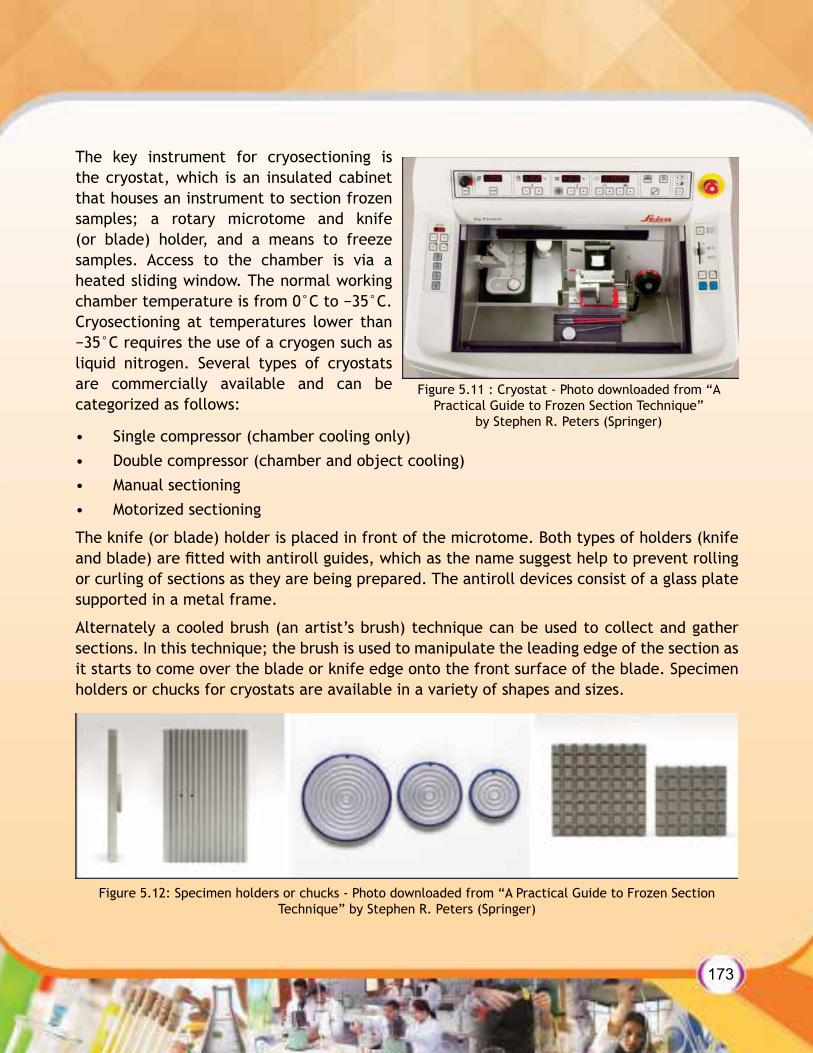
173
The key instrument for cryosectioning is the cryostat, which is an insulated cabinet that houses an instrument to section frozen samples; a rotary microtome and knife (or blade) holder, and a means to freeze samples. Access to the chamber is via a heated sliding window. The normal working chamber temperature is from 0°C to −35°C. Cryosectioning at temperatures lower than −35°C requires the use of a cryogen such as liquid nitrogen. Several types of cryostats are commercially available and can be categorized as follows:
• Single compressor (chamber cooling only)• Double compressor (chamber and object cooling)• Manual sectioning• Motorized sectioning
The knife (or blade) holder is placed in front of the microtome. Both types of holders (knife and blade) are fitted with antiroll guides, which as the name suggest help to prevent rolling or curling of sections as they are being prepared. The antiroll devices consist of a glass plate supported in a metal frame.
Alternately a cooled brush (an artist’s brush) technique can be used to collect and gather sections. In this technique; the brush is used to manipulate the leading edge of the section as it starts to come over the blade or knife edge onto the front surface of the blade. Specimen holders or chucks for cryostats are available in a variety of shapes and sizes.
Figure 5.11 : Cryostat - Photo downloaded from “A Practical Guide to Frozen Section Technique”
by Stephen R. Peters (Springer)
Figure 5.12: Specimen holders or chucks - Photo downloaded from “A Practical Guide to Frozen Section Technique” by Stephen R. Peters (Springer)
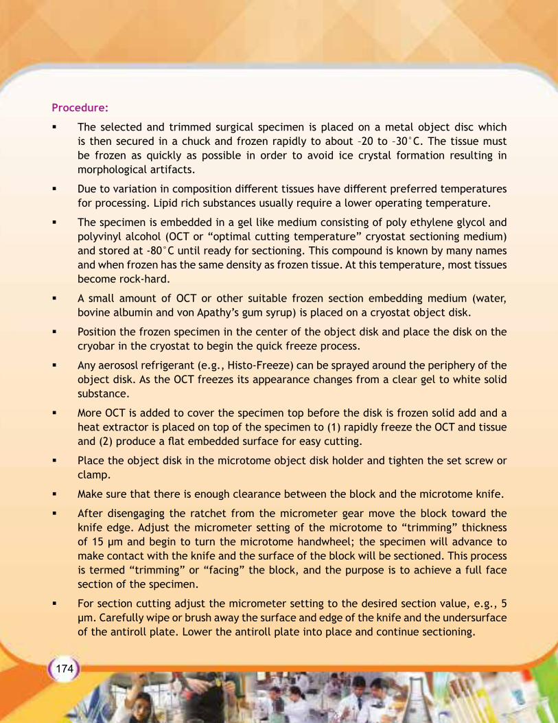
174
Procedure:
The selected and trimmed surgical specimen is placed on a metal object disc which is then secured in a chuck and frozen rapidly to about –20 to –30°C. The tissue must be frozen as quickly as possible in order to avoid ice crystal formation resulting in morphological artifacts.
Due to variation in composition different tissues have different preferred temperatures for processing. Lipid rich substances usually require a lower operating temperature.
The specimen is embedded in a gel like medium consisting of poly ethylene glycol and polyvinyl alcohol (OCT or “optimal cutting temperature” cryostat sectioning medium) and stored at -80°C until ready for sectioning. This compound is known by many names and when frozen has the same density as frozen tissue. At this temperature, most tissues become rock-hard.
A small amount of OCT or other suitable frozen section embedding medium (water, bovine albumin and von Apathy’s gum syrup) is placed on a cryostat object disk.
Position the frozen specimen in the center of the object disk and place the disk on the cryobar in the cryostat to begin the quick freeze process.
Any aerososl refrigerant (e.g., Histo-Freeze) can be sprayed around the periphery of the object disk. As the OCT freezes its appearance changes from a clear gel to white solid substance.
More OCT is added to cover the specimen top before the disk is frozen solid add and a heat extractor is placed on top of the specimen to (1) rapidly freeze the OCT and tissue and (2) produce a flat embedded surface for easy cutting.
Place the object disk in the microtome object disk holder and tighten the set screw or clamp.
Make sure that there is enough clearance between the block and the microtome knife.
After disengaging the ratchet from the micrometer gear move the block toward the knife edge. Adjust the micrometer setting of the microtome to “trimming” thickness of 15 μm and begin to turn the microtome handwheel; the specimen will advance to make contact with the knife and the surface of the block will be sectioned. This process is termed “trimming” or “facing” the block, and the purpose is to achieve a full face section of the specimen.
For section cutting adjust the micrometer setting to the desired section value, e.g., 5 μm. Carefully wipe or brush away the surface and edge of the knife and the undersurface of the antiroll plate. Lower the antiroll plate into place and continue sectioning.

175
Sections of fresh frozen tissue will adhere to plain glass slides due to the presence of free protein and lipid. Sections of fixed frozen tissue will need to be mounted on coated slides, e.g., poly-l-lysine.
Subsequently the section is picked up on a glass slide and stained with hematoxylin and eosin.
The sample preparation time is much more shorter as compared with traditional histology technique.
Electron microscopy
The energy source used in the electron microscope is a beam of electrons. Since the beam has an exceptionally short wavelength, it strikes most objects in its path and increases resolution of the microscope significantly.
Electron microscopes can be of the following types: Transmission electron microscope (TEM): It uses electrons passed through the sample
to build a picture of the sample’s internal structure. It is a more traditional form of electron microscope.
Scanning electron microscope (SEM): Here an electron beam is projected on the sample. The electrons do not go through the sample but bounce off. This way it is possible to visualize the surface structure of the specimen. The image which is thus formed appears three dimensional.
Transmission electron microscopy, a popular diagnostic adjunct in the 1970s and 1980s, has been largely replaced by immunohistochemical, cytogenetic and molecular techniques. However, EM continues to be used in the diagnosis of: Microbial diseases Lysosomal storage diseases Bullous skin disorders Peripheral neuropathies Soft-tissue tumors Glomerulopathies Evaluation of an unknown primary malignancy Differentiation of a mesothelioma from an adenocarcinoma

176
Advantages of EM over optical microscopes:
Modern electron microscopes can magnify up to two million times. Researchers use it to examine the minutest biological material, cells, molecules, and their physical/crystal-line structure, and characteristics of various metals and surfaces.
It is possible to view the three-dimensional external shape of an object with a scanning electron microscope.
Disadvantages:
It is extremely expensive and a high-maintenance equipment.
Requires elaborate sample preparation.
The prerequisite for a completely dry sample and also the high energy of the electron beam (converting into high radiation) makes it impossible for one to observe living or moving specimens with an electron microscope.
It is not possible to observe color as electrons do not possess a color.
Immunohistochemistry (IHC)
IHC is the method of localization of antigens in tissue by use of labeled antibodies through antigen–antibody interactions that are visualized by markers such as fluorescent dyes, enzymes, radioactive elements, or colloidal gold.IHC makes it possible to visualize distribution and localization of specific cellular components. Antibody-mediated antigen detection can be done by direct and indirect methods.
Most indirect methods employ inherent binding affinity of avidin to biotin to localize a reporter to target antigen and amplify signal that is detected. IHC target antigens are detected through either chromogenic or fluorescent means.
Chromogenic detection is based on activity of enzymes, most often horseradish peroxidase (HRP) or alkaline phosphatase (AP), which form colored (usually brown); insoluble precipitates upon addition of substrate, such as Diaminobenzidine tetra hydrochloride (DAB) and Aminoethylcarbazole (AEC).
Applications
IHC can be used for tumor diagnosis in case routine histology is not diagnostic.
Tumor markers can be used to differentiate between a benign or malignant tumor and identify the cell/site of origin of a metastasis.
Early or micrometastasis can be difficult to detect using conventional histology but can be diagnosed by IHC.

177
It can be used to prognosticate a cancer (localization of oncogenes, tumor suppressor genes, cell proliferation, or cell death/apoptosis markers) or to determine/predict the response of a tumor to therapy (localization of estrogen–progesterone receptors in carcinoma breast).
IHC tests are now available for diagnosis of infectious diseases, e.g., Herpes virus, Hepatitis B virus (HBV), Cytomegalovirus (CMV), Human Papilloma virus (HPV), and Leptospira.
Immunofluorescence (IF)
IF involves an antigen–antibody reaction wherein the antibodies are labeled with a fluorescent dye and the antigen–antibody complex is visualized using ultraviolet light (sourced from mercury vapor and xenon gas lamps) in a fluorescent microscope.
Dyes that absorb ultraviolet rays and emit visible light are called fluorochromes. Fluorochromes such as fluorescein isothiocyanate (FITC) or tetramethyl rhodamine isothiocyanate (TRITC) are chemically conjugated to antibodies that are allowed to bind (directly or indirectly) to the antigen of interest.
The antigen is thus demonstrated through fluorescence techniques. The emitted fluorescence can then be quantified using a flow cytometer, array scanner, or automated imaging instrument, or visualized using fluorescence or confocal microscopy.
There are two main methods of IF labeling - direct and indirect.
The direct immunofluorescence method, whereby the antibody against the molecule of interest is directly conjugated to the fluorescent dye or fluorochrome, is less commonly used.
In indirect immunofluorescence (sandwich technique), the antibody specific for the molecule of interest (called primary antibody) is unlabeled, and a second anti-immunoglobulin antibody directed toward constant portion of first antibody (called the secondary antibody) is tagged with the fluorescent dye.
Some substances fluoresce naturally and this fluorescence is called primary fluorescence or autofluorescence, e.g., chlorophyll.
Those substances that do not fluoresce spontaneously but require induction of fluorochromes are said to emit secondary fluorescence.
Applications:1. The test is routinely done on renal and skin biopsies for detection of deposits of
immunoglobulins, complement and fibrin in glomerulonephritis, and bullous disorders of skin.

178
2. It can be used for detection of autoantibodies in serum, e.g., antinuclear antibody (ANA) in SLE, anti smooth muscle antibody (SMA) in biliary cirrhosis, antimitochondrial antibody (AMA) in autoimmune hepatitis, autoantibodies to glutamic acid decarboxylase (GAD), the protein tyrosine phosphatase–like molecule, IA-2, and insulin in type 1 diabetes, thyroid autoantibodies in Hashimoto thyroiditis and Grave’s disease etc.
3. IF can be applied to analysis of antigens in fresh, frozen, or fixed tissues, and subcellular localization of antigens in tissue culture monolayers.
4. It can be used for detection and localization of the presence or absence of specific DNA sequences on chromosomes patterns of gene expression within cells/tissues.
Autopsy protocol
An autopsy is a medical procedure that consists of a thorough examination performed on a body after death, to evaluate disease or injury that may be present and to determine the cause and manner of death. There are three main types of autopsies1. Medico-Legal or Forensic autopsies: These are used to determine the cause and manner
of death and are generally performed, as prescribed by applicable law, in cases of violent, suspicious or sudden deaths.
2. Clinical or Pathological autopsies: These are performed to diagnose a particular disease or for research purposes.
3. Anatomical or Academic autopsies: These are performed by students of anatomy for study purpose only.
It is preferable that the autopsy examination be performed as soon after death as possible. Therefore, if delay is unavoidable, the body should be placed in the mortuary refrigerator (4°C) or arterial embalming should be carried out without delay provided that toxicological or microbiological examination are not indicated such embalming, well done, is most advantageous and given excellent preservation. Slices of organs taken at autopsy should be thin to facilitate fixation.
Principal Autopsy Techniques:
The four major autopsy techniques differ chiefly in the methods used in removal of the organs and the order in which they are opened.1. Technique of Virchow: Most widely used method in which the organs are removed one
by one. This approach is good when multiple organs are thought to be involved by the disease, for demonstrating pathological changes in individual organs. The disadvantage of this technique is that relationships between various organs may be hard to interpret. Several modifications are available.

179
2. Technique of Rokitansky: This procedure is characterized by in situ dissection, in part combined with en bloc removal.
3. En mass technique: Thoracic, cervical, abdominal, and pelvic organs are removed en masse and subsequently dissected into organ blocks. This is the best technique for demonstrating the vascular supply of organs and their relationship with one another. The major disadvantage is that it is difficult to handle the organ mass., and performing the autopsy is difficult without an assistant.
4. En Bloc Technique: Thoracic, cervical and abdominal organs, and the urogenital system are removed in functionally related blocks. This procedure is a compromise between the Virchow and en masse techniques, preserving anatomical relationships sufficiently for most cases while being simpler for one person to execute.
5.8 STORAGE AND ARCHIVING OF SPECIMENSMaintenance of histopathology records and surgical and autopsy specimens, is an extremely important aspect of pathology services. This includes filing and storing of paraffin blocks and stained tissue slides usually in order of accession number.
General guidelines for storage and archival in a pathology laboratory• It is advisable that surgical specimens are stored for a minimum of two weeks to
approximately six weeks after a report is issued so that if new clinical information becomes apparent that might require further investigation of the unprocessed tissue, it can be done.
• It is recommended that diagnostic histopathology slides and blocks should be kept for a minimum of ten years. Report duplicates/records/diagrams and copies of any representative images prepared should be kept for at least 20 years.
• All laboratories must retain cytology slides for a minimum of five years.• Currently, some patients with cancer survive for more than 10 years, and review of
previous pathology material may be required for comparison with recurrent tumor or for enrollment in clinical trials. Therefore it is recommended that once the regulated length of time for storage is met, institutions may continue to store pathology specimens based on the room they have for storage.
• The entire archive (paper records, glass slides, and tissue blocks) should be professionally stored in a climate-controlled environment, and its index should be available, preferable in digital format.
• Paper records should be stored as bound volumes. All documents, relevant radiographs, gross specimens and additional material available should be photographed and scanned to be digitally archived as well.

180
5.9 SAFETY IN THE LABORATORY
• Treat all biological materials used in the laboratory as potentially infectious and pathogenic to humans.
• Laboratory coats must be worn by laboratory personnel at all times.
• All open cuts on hands and other exposed skin surfaces must be covered by gloves.
• Long hair should be tied back neatly, away from the shoulders.
• The lab should be well-ventilated and should strictly follow the regulations governing the acceptable limits of formalin and hydrocarbons such as xylene and toluene. If solvents are used during practical sessions, the exhaust fan must be switched on.
• Whenever doing staining procedures ensure that protective gowns, gloves and safety glasses are worn. When ‘bringing sections to water’ or ‘dehydrating, clearing and mounting’ always ensure that the exhaust system is turned on.
• The wheel of each microtome must be in the locked position any time the microtome is not being used for cutting sections.
• Utmost care must be taken while handling embedding centers (as they contain paraffin wax at 61oC and above). When using, ensure that you do not burn your fingers.
• A safety data sheet should be maintained for every chemical compound used and it should specify the nature, toxicity, and safety precautions to be taken while handling the compound.
• Proper disposal of hazardous wastes is a must. Tissues that are collected should be stored in formalin and may be disposed by incineration or by putting them through a “tissue grinder” attached to a large sink (similar to a large garbage disposal unit).
• Sharps are to be placed in the sharps containers scalpel blades are to be disposed of in the blade removal system, which are disposed as per recommended guidelines.
• Every instrument used in the laboratory should meet electrical safety specifications and have written instructions regarding its use.
• It is advisable that flammable materials are stored with utmost care in appropriate storage cabinets that are designed for this purpose.
• Fire safety protocols are to be strictly adhered to. Safety equipment including first aid kits, fire extinguishers, fire blankets, and fire alarms should be within easy access. A shower and eyewash should be Specific hazards:
181
• Bouin’s solution contains picric acid, which is only sold in the aqueous state. This is because it is inherently explosive in the dry state.
• Sodium azide, used as a preservative in many kits is also explosive in nature. It should be flushed down the drain with lots of water, or there is a tendency for the azide to form metal azides in the plumbing.
• Benzidine, benzene, anthracene, and napthol containing compounds are carcinogens and should not be routinely used.
• Mercury-containing solutions (Zenker’s or B-5) should always be discarded into proper containers. Mercury, if poured down a drain, will form amalgams with the metal that build up and cannot be removed.
5.10 Self-Assessment
1. Define histopathology.
2. Enumerate the steps in specimen receiving and accessioning.
3. Enlist the list of equipment required for setting up of a histopathology laboratory.
4. What are the properties of an ideal fixative?
5. Classify fixatives. Which are the most common fixatives used in histopathology. Enumerate their specific uses and disadvantages.
6. Outline the steps in routine processing of histopathology specimens.
7. What is automated processing?
8. Enumerate the steps in specimen mounting for museum preparation.
9. Name the fixation induced pigments. Outline the steps in the methods used to remove them.
10. Enlist the factors affecting the rate of tissue processing.
11. Define dehydration. Enumerate the available dehydrating agents.
12. Define clearing. Enumerate the available clearing agents. Enumerate the criteria for choosing a suitable clearing agent.
13. Define embedding. Which is the most popular embedding medium for histology and why? Enumerate the alternatively available embedding agents.

182
14. Define microtomy. Enlist the different types of microtomes and knives used in histopathology specimen cutting.
15. Outline the steps in cutting of paraffin embedded tissue.
16. Name the fixatives preferred for processing of bone specimens.
17. Describe the principles of decalcification and enlist the various decalcifying agents used on bone processing. What are their advantages and disadvantages?
18. Enumerate and describe the methods used for checking the end point of decalcification. How does one test for the remaining calcium in decalcifying tissue.
19. Outline the principle of differential staining. Enumerate the steps in routine H&E staining.
20. Classify haematoxylins. Specify the uses of the different types.
21. Define:
a) Basophilic substances.
b) Acidophilic substances.
c) Vital staining.
d) Progressive staining.
e) Regressive staining.
f) Decolorization.
g) Mordants.
h) Bluing.
i) Metachromatic staining.
22. Enumerate the principle of PTAH staining and its applications.
23. Enlist the special stains used for demonstration of microorganisms and describe their principles.
24. Enlist the special stains used for demonstration of connective tissue and describe their principles.
25. Enlist the special stains used for demonstration of carbohydrates and describe their principles.

183
26. Enumerate the endogenous pigments. Name the special stains used for their demonstration and describe their principles.
27. What are the main indications for a frozen section? Outline the steps in its preparation.
28. What are the principles of electron microscopy? Enlist its types.
29. Enumerate the advantages and disadvantages of electron microscopy over light microscopy.
30. Write briefly on the applications of immunofluorescence techniques in histopathology.
31. Discuss the role of immunohistochemistry in histopathology.
32. What are the different types of autopsies? Write briefly on autopsy protocol.

184
OVERVIEW AND DESCRIPTION
OVERVIEW
This unit will provide the student information about the scope of cytopathology and the organizational structure of a cytopathology laboratory. It will help to understand the relevant terms. Procedures and working of equipments pertaining to cytopathology.
Organization of Cytopathology Laboratory:
The personnel needs of a laboratory depends on overall work laod and the different types of cytology materials to be processed.
The chief of the laboratory should be a cytopathologist / pathologist or a gynecologist / medical officer trained in cancer related cytology.
The chief of the laboratory should be a cytopathologist / pathologist or a gynecologist / medical officer trained in cancer related cytology.
The Cytotechnologist is a well trained and certified person who should have undergone one year cytology training from a recognized, accredited centre or should have passed National Examination for cytotechnologists conducted by Indian Academy of Cytologists (IAC), after graduation/post-graduation in any of the life science subjects. The duties of the cytotechnologist include preparation of stains and maintenance of its quality, processing of cytopathology material, screening of smears and formulation of preliminary diagnosis. They are also responsible for supervising the record keeping, analysis of data and slide filing system.
Cytotechnicians should have a diploma in medical laboratory technology from a recognized institution and must have undergone 6 months training course for cytotechnicians from an accredited laboratory or passed the National Examination for cytotechnicians conducted by IAC. They are responsible for specimen collection, cytology preparation and staining. Support Staff include clerical and secretarial workers in the laboratory. Physical Infrastructure of the laboratory must be well designed and conveniently located to enable the professional and support personnel to perform their duties effectively. It must contain four definitely separated areas:
UNIT - 6CYTOPAThOLOGY

185
• Reception• Specimen collection room• Processing and staining area• Reporting room
Receiving of specimens• Ensure that the specimen is properly labeled and submitted along with the specific
requisition form which has detailed patient history including the previous Cytology / Histopathology reports, if any.
• Verify the patient’s name, age, registration details, history and site of specimen collection. Mismatches, if any, are to be reported to the referring doctor.
• Give a unique accession number to the specimen.• The number of slides received from each site should be mentioned in the requisition
form.• Nature and method of sample collection are to be mentioned in the requisition form.
(cytobrush / spatula / swab / for gynaecalogical smears and plain / guided FNAC for aspiration smears).
• Check whether the fixation is proper (mention type of fixation: alcohol / spray fixative / prefixed / air dried).
• Enter relevant patient details (name, age, sex, address, brief clinical details, and name of referring hospital / doctor) in the register.
KNOWLEDGE AND SKILL OUTCOMESi) Understand the scope of cytopathology.ii) Know the organizational structure of a cytology laboratory.iii) Know the relevant terms, procedures and working of equipments pertaining to
cytopathology.
RESOURCE MATERIALS• George L. Wied, Catherine M. Keebler, Koss L.G, Regan JW, Compendium on Diagnostic
Cytology, Eighth Edition, Tutorials of Cytology. Chicago, Illinois, USA, 1997.• Svantle R. Orell, Gregory F. Sterrett, Max N-I. Walters: Manual and Atlas of Fine Needle
Aspiration Cytology. 4th ed. Churchill Livingstone; Elsevier; Churchill Livingstone, Edinburg, London, Madrid, Melbourne, New York and Tokyo 2005.
• Koss LG, Melamed MR. editors. Koss’ diagnostic cytology and its histopathologic basis. 5th ed Vol 2. Walnut street, Philadelphia: Lippincot 2006.

186
DURATION
LEARINING OUTCOMES AND OBJECTIVES
After completing this unit the students should be able to 1. Demonstrate knowledge, comprehension, and application of general techniques in the
areas of eytopathology including:
• Specimen accessioning• Fixation of specimen• Processing of specimen• Staining, cover slipping and labeling of slides• Storage and archiving of slides
2. Set-up, operate and maintain routine cytology equipments.3. Solve basic problems associated with reagents and methods relevant to general cytology
techniques.4. Apply principles of lab safety in completing all laboratory work.5. Ensure quality control while performing general cytology procedures.
6.1 INTRODUCTION TO CYTOPATHOLOGYDiagnostic cytopathology involves the interpretation of cells that spontaneously exfoliate or are removed from tissues by abrasion or fine needle aspiration, such as specimens from the cervix (Pap test), breast, thyroid, lymph node, liver, etc. Two broad categories of samples are received in the cytology laboratory:1. Exfoliative cytology: It is the study of cells that have been shed or removed from the
epithelial or mesothelial linings. Normal cells are cohesive in nature, but malignancy and infection increase exfoliation. Malignant cells show reduced intercellular adhesion due to defective desmosomes. These cells can be recovered either from natural secretions. Such as urine, sputum, vaginal, and prostaic fluids, or by artificial means, such as paracentesis or lavage of fluids like pleural, pericardial, cerebrospinal, synovial, ascetic, CSF, cyst fluid, bronchial washings etc.
2. Fine needle aspiration cytology includes aspiration done by the pathologist or the clinician as well as guided aspiration done by the radiologists and aspirations. It is a diagnostic procedure used to investigate pathological lesions in organs that do not shed cells spontaneously. In this technique, a thin, hollow needle is inserted into the lesion (usually a lump or a swelling) to obtain cells and tissue fragments, which, after being stained, are examined under a microscope.

187
Note: Consent form available in local language and in English should be signed by the patient prior to the procedure. Procedure details are explained to the patient by the consultant pathologist/radiologist.
6.2 EXFOLIATIVE CYTOLOGYCommon sites for exfoliative cytology1. Body Fluids
(a) Pleural
(b) Pericardial
(c) Peritoneal
(d) Synovial
(e) Cerebrospinal
2. Surface Epithelia
(a) Female genital tract
(b) Respiratory tract
(c) Nasopharynx
(d) Larynx
(e) Gastrointestinal tract
(f) Urinary tract
(g) Nipple discharge
3. Buccal Smear
Female Genital Tract (FGT)
The cytological specimens collected from FGT include cervical smear, vaginal smear, aspiration from posterior fornix of vagina (vaginal pool smear) and endometrial smear.
Cervical smear: Cancer of the uterine cervix is the commonest cancer in the FGT. Almost all invasive cancers of the cervix are preceded by a phase of preinvasive disease, which demonstrates microscopically a continuing spectrum of events progressing from cervical intraepithelial neoplasia (CIN) grade I to III including carcinoma in-situ before progressing to squamous cell carcinoma. This progressive course takes about 10 to 20 years. Early detection even at the preinvasive stage is possible by doing cervical smear (Pap Smear Test). This can identify patients who are likely to develop cancer and appropriate interventions may be carried out.

188
Advantages of Pap Smear:• It is painless and simple• Does not cause bleeding• Does not need anesthesia• Can detect cancer and precancer• Can identify non-specific and specific inflammations • Can be carried out as an outpatient procedure
Sampling Devices: the collection device may play an important role in sample adequacy. The shape, surface, texture and material of the device may determine how much of the scraped material is deposited on to the glass slide and is available for screening and analysis. Several methods of obtaining cytologic material from the uterine cervix are available. However, use of cotton swab for collection of cervical smear is to be discouraged, in view of the drying artifacts and loss of cells, which are caused by this method. Smears obtained with original Ayre’s spatula are often easier to screen. Wooden spatula is preferable to plastic spatula, because of its mildly rough surface that can collect more material. The disadvantages are that the method may occasionally be traumatic to the patient, and the tip of spatula that does not fit the external os may fail to remove some of the valuable material from the squamo-columnar junction.
Endo-cervical brush is a small bottlebrush like device with one end having fine bristles made up of nylons. This device is strictly for taking materials from endocervix. Gently insert the brush in endocervix and rotate one turn pressing in the upper and lower wall (Figure 3). The cytobrush is similar to that of endocervical brush except that the projected tip is without bristles. This can be used for obtaining cells from the whole cervix. Single sampling devices and methods have their limitations in obtaining adequate smears from the cervix. A combination of two devices, usually spatula and endocervical brush, give better results. Triple smear or the vaginal-cervical-endocervical (VCE) technique can provide the best results. However, feasibility and cost factor need to be taken into consideration.
In postmenopausal women. The squamo-columnar junction recedes making it difficult to obtain good amount of endocervical cells and cells from TZ.
Hence a combination of two devices, spatula plus endocervical brush is preferred.
Preparation of Smear: After smear collection, the cellular sample is evenly smeared on to the centre of the non-frosted area of the glass slide, by rotating both sides of the scrape end of the spatula in multiple clockwise swirls in contact with the slide and fixing it immediately. Excessively thin or thick smears can result in false-negative reports. The smear should be visually inspected after fixation. If it does not appear satisfactory, repeat it during the same examination and submit both slides for cytological examination. Some studies have shown

189
that two-slide cervical smears detect more abnormalities than a one-slide smear. Two smears do increase screening costs over a single-slide smear, but those costs are not double that of a single-slide examination. A two-instrument collection on a single slide increases screening time only minimally over a single instrument.
Vaginal smear: Introduce an unlubricated speculum, scrape the lateral vaginal wall at the level of cervix owith a spatula. The broad and flat end of Ayre’s spatula is used for this purpose. The cellular material is rapidly but gently smeared on a clean glass slide and the smears are fixed immediately. If no spatula is available a cotton swab dipped in normal saline can be used.
Vaginal pool Smear: The aspiration can be performed after the introduction of unlubricated speculum. The technique allows collection of cells under direct vision from posterior fornix pool. When a speculum is not employed the pipette is gently introduced in to the vagina until resistance is encountered. It is important to compress the suction bulb during the introduction of the pipette to avoid collecting the cellular material of the lower vaginal orgin. The cellular material is spread on a clean glass slide and fixed immediately.
Endometria aspiration smear: After preliminary visualization and cleaning of cervix a sterile cannula is introduced into the uterine cavity and aspiration is then carried out with a syringe. The specimen is squirted on a clean glass slide, gently spread and rapidly fixed.
Respiratory Tract
Respiratory tract malignancies can be detected mainly by sputum cytology or by bronchoscopic material.
Sputum Cytology: Sputum specimen can be obtained from the patient either spontaneously or by aerosol – induced method. Morning specimen resulting from overnight accumulation of secretion yields best results. Three to five consecutive days’ sputum samples should be examined to ensure maximum diagnostic accuracy. Fresh unfixed specimens are better than prefixed specimens in 70% ethyl alcohol or coating fixative such as carbowax or saccomano fixative. (Fixation of slides is discussed in a separate chapter). The sputum must be carefully inspected by pouring the specimen into a petri dish and examining on a dark background. Select any bloody, discolored or solid particles, if present, place a small portion of each particle on a micro slide, spread evenly and fix it immediately. Prefixed specimens should be smeared on albumen or polylysine coated slides.
Bronchoscopic Specimens: Specimens that are obtained by bronchoscopy are secretions (bronchio-alveolar lavage), direct needle aspirate from suspicious area and bronchial brushing and washings. Post bronchoscopic sputum is one of the most valuable specimens for the detection of pulmonary lesions.

190
Other Sites• Oral lesions: Scrape the lesion with a tongue depressor, spread material on a clean slide
and fix immediately.• Nasopharynx: Cotton tipped applicator is used to obtain material for cytological
examination.• Larynx: A cotton swab smear of larynx may be a useful adjunet to clinical diagnosis if
biopsy is not contemplated.
Fluid for cytology• On receiving, the sample is checked for labels and details to match it with the requisition.• Samples are processed immediately on receipt and must be refrigerated at 2oC to 4oC if
there is a delay.• Physical properties of the fluid are recorded on the requisition from i.e., volume, colour,
turbidity, presence of clot, etc.• All fluids except CSF receive in small quantity (<2ml) are processed on the regular
centrifuge.• If the fluid received is a large volume, the top portion is decanted into a second container
and the bottom portion is processed.• 5 to 10 ml of the fluid (depending on the volume received) is taken in a test tube and
centrifuged at 3000 rpm for 10 minutes.• The supernatant is emptied into the main container.• The sediment is removed with the aid of a plastic pipette, and a drop each is placed on
a minimum of three slides and a thin smear made. A drop of sediment is placed towards one end of the slide. A second glass slide is held at an angle of 45 degree to the first slide and with gentle pressure, the second slide is drawn out to make a tongue shaped smear.
• Smear is numbered using a diamound pencil.• At least one smear is immediately wet fixed in 95% alcohol. The remaining smears are
air dried.• The wet smears are stained with Pap stain and H&E, and the air dried smears are stained
with Giemsa. Special stains are put up if required. • Cell block is made form sediment left behind in all cases.
Procedure for hemorrhagic fluids- Frankly hemorrhagic fluids are centrifuged like all fluids and fish tailed smears made
from the sediment of the centrifuged deposit.

191
- Alternatively, if very hemorrhagic, smears can be air dried and then flooded with normal saline for 30 seconds. This causes layes of red cells, smears are than air dried or wet fixed and stained by pap and Giemsa stains respectively.
6.3 FNAC
Equipment required• Aspiration is done using disposable needles of 21 gauge (external diameter approximately
0.6-1.0mm) attached to a 20ml syringe.• The FNAC needles are available in a variety of lengths. Lenghts of a to 1/2 inches are
found to be adequate for most palpable measses.• The 31/2 inches 22 gauge disposable needle is used for deep seated soft-tissue masses.• Ultrasound or computerized tomography (CT) guidance can be utilized, whenever
indicated.• Multiple aspirates should be undertaken from different areas, in case of a large tumor,
to improve the yield and overcome the problem of tumor heterogeneity.
Technique• Taking all aseptic precautions, the lump is palpated and localized, and the site of
puncture determined.• The lump is then immobilized with the left hand in a position favorable for needle
aspiration and holding the syringe by the barrel in the right hand; the needle is pushed into predetermined site of the lump until needle tip penetrates the center of the lump.
• The plumger of the needle is then retracted backward to create a negative pressure inside the syringe and needle bore; and without withdrawing the needle through the skin, the syringe is rotated and moved in and out through the lump whilst negative pressure sucks cells into the lumen of the needle.
• In order to obtain sufficient material, particularly from fibrotic lesions, the needle is moved back and forth three or more times and directed into different areas of the tumor.
• Throughout this manipulation, negative pressure is maintained in the syringe by keeping the piston retracted.
• After ompletion of the aspiration, the pressure in the syringe is allowed to equalize before the needle is withdrawn from the lesion.
• This is achieved by releasing the piston of the syringe.

192
• After the needle had been withdrawn, the syringe is disconnected from the needle, filled with air and reconnected.
• The material in the needle is expelled onto a glass slide, care being taken to deposit it as a single drop at one end of the slide.
• The needle tip is then brought into light contact with the slide and the aspirate carefully expressed from it.
Preparation of smears• The aspirated material (tissue fragments, semisolid or fluid) is collected at the edge of
the slide and gently crushed by pressure with another slide/cover slip and spread.• Excess blood or tissue fluid in the aspirate is dealt separately with a centrifuge
preparation; if required. A “hematologic-type” smear is first prepared by touching the droplet with the edge of a cover-slip.
• A minimum of six smears should be prepared in all cases. Half the smears should be fixed in 95% ethanol and half air-dried.
• Air-dried smears are stained with May-Grunwald-Giemsa (MGG) stain.• Alcohol-fixed smears are stained with Haematoxylin and Eosin (H&E) and Papanicolaou
stain.• A cytology number is marked on all the slides received using a diamond pencil. • The smears are evaluated for detailed cytological features.• If fluid is aspirated, the physical details (volume, color and turbidity) are recorded and
the fluid is processed as described in section on fluids.• If solid particles are present, they are placed in 10% neutral buffered formalin and
processed as a small biopsy sample/cell blocks.• During the FNAC procedure, smears are checked, if required, by the concerned consultant
for adequacy by using the field stain.
Procedure the preparation of cell blocks• Cell blocks are made from all fluid aspirates received where sediment is present. • Fluid received is centrifuged at 3000 rpm for 10 minutes.• Smears are made and stained.• To the sediment, approximately double the volume of Bouin’s fluid is added followed by
one drop of egg albumin.• This is then centrifuged at 200 rpm for 10 minutes.
193
• Supernatant is poured off and the button is transferred with forceps to formalin for 4 to 6 hours, after which it is taken for processing.
6.4 Cytological Fixatives• It is critical to fix cytology specimens immediately after collection for proper preservation
of the cellular components. It is important that no air-drying occurs prior to fixation. If a smear is already air-dried it should not be put in an alcohol fixative. Please note on the requisition if the slide (s) being submitted are fixed or air-dried.
Properties of a good cytological fixative:• It should not excessively shrink or swell cells.• It should not distort or dissolve cellular components.• It should help preserve nuclear details.• It should improve optical differentiation and enhance staining properties of the tissues
and cell components.
Cytological fixatives are classified intoA. Rountine Fixatives
Freshly prepared smears can be immediately submerged in a liquid fixative. This is called wet fixation and is the ideal method for fixing all gynecological and non-gynecological smears. Any of the following alcohols can be used:1. 95% Ethyl Alcohol (Ethanol): The ideal fixative recommended in most of the laboratories
for cytological specimens is 95% ethanol alone. It produces optimal nuclear details but some amount of cell shrinkage. Absolute (100%) ethanol produces a similar effect on cells. But is much more expensive.
2. Ether alcohol mixture: This fixative was originally recommended by Papanicolaou. It consists of equal parts of ether and 95% ethyl alcohol. It is an excellent fixative, but ether is not used in most of the laboratories because of its safety hazards, odour and hygroscopic nature.
3. 100% Methanol: 100% methanol is an acceptable substitute for 95% ethanol. Methanol produces less shrinkage than ethanol, but it is more expensive than ethanol.
4. 80% Propanol and Isopropanol: Propanol and Isopropanol cause slightly more cell shrinkage than ether-ethanol or methanol. By using lower percentage of these alcohols the shrinkage is balanced by the swelling effect of water on cells.
5. Denatured alcohol: It is ethanol that has been changed by the addition of additives in order to render it unsuitable for human consumption. This can be used at a concentration

194
of 95% or 100%. One formula is 90 parts of 95% ethanol + 5 parts of 100% methanol +5 parts of 100% isopropanol.
Time of Fixation: Minimum 15 minutes fixation Fixation prior to staining is essential.B. Coating Fixatives• Coating fixatives are either aerosols applied by spraying or a liquid base, which is poured
onto the slide. They are composed of an alcohol base, which fixes the cells and wax like substance, which forms a thin protective coating over the cells e.g. Carbowax (Polyethylene Glycol) fixative. Diaphine fixative Spray coating fixative (Hairspray) with high alcohol content and a minimum of lanolin or oil is also an effective fixative.
• 10 to 12 inches is the optimum distance recommended for aerosol fixative.• Aerosol srays are not recommended for bloody smears, because they cause clumping of
erythrocytes.• Prior to staining, the slides have to be kept overnight in 95% alcohol for removal of the
coating fixative.C. Special Purpose Fixatives• Carnoy’s fixative: This is a special purpose fixative for haemorrhagic samples. The acetic
acid in the fixative haemolyses the red blood cells. It is an excellent nuclear fixative as well as preservative for glycogen but results in considerable shrinkage of cells. Carnoy’s fixative must be prepared fresh when needed and discarded after each use. It loses its effectiveness on long standing, and chloroform can react with acetic acid to form hydrochloric acid.
• AAF Fixative: This is the ideal fixative used for cellblock preparation of fluid specimens.• Saccomanno collection fluid: A green colored fixative of the collection of sputum.• Cytolyt solution: This is a clear water based buffered fixative for the collection of fluid
specimens. A 50:50 ratio of specimen to fixative is appropriate (if this unavailable use 50% alcohol).
• 50% Alcohol: This is a clear fixative for the collection of fluid specimens. A 50:50 ratio of specimen to fixative is considered appropriate.
6.5 Cytospin• Used for CSF, small volume of fluid samples and occasionally for larger volumes.• Two drops of fluid or of re-suspended semiment (for larger volume fluids after regular
centrifugation as above) are place in the special tubes and capped. • A miimum of four tubes are taken for each sample.

195
• Filter paper cards placed between the slide and tube and clipped.• Tubes balanced in the cytospin.• The basin is closed with the cover, the main lid shut and instrument programmed for
1000 rpm for 10 minutes.• A bleep sounded indicates the end of the procedure.• The main lid and the basin cover are opened and the tubes removed. • Two slides are immediately wet fixed in 95% alcohol (for Pap stain) and two are air dried.• Cytology numbers are assigned to the slides using a diamond pencil. • The fluid remaining after the processing is retained till the reporting is over.
Figure 13: Cytospin (courtesy Thermoshandon)
Procedure for disinfection of plastic reusable cytospin cuvettes• Cuvettes are immersed in 4% hypochlorite solution for 1 hour.• Latter washed with soap water.• After washing, dried and reused.
6.6 STAINING PROCEDURES IN CYTOLOGYPapanicolaus stain: Routine stain in cytology used for all cytology samples.
Solutions used:

196
a. Orange Gb. EA 50c. Harris’s Haematoxylin
Haematoxylin 5g
100% isopropyl alcohol 50ml
Ammonium/potassium alum 100g
Distilled water 1000ml
Mercuric oxide, red 2.5g
Dissolve the haematoxylin in the alcohol and the alum in the water with the aid of heat. Remove from heat and mix the two solutions. Bring to a boil as rapidly as possible (limit the heat to less than one minute and stir often). Remove from heat and add the mercuric oxide slowly. Reheat to a simmer until it becomes dark purple. Remoe from heat and plunge the vessels into a basin of cold water until it cools. Addition of 2-4 ml of glacial acetic acid per 100ml of solution increases the precision of the nuclear stain. Filter before use.
Procedure (manual):
Smears are fixed for a minimum of half an hour in 95% alcohol before staining.
Agitation is important during staining.
- 95% alcohol - 10 dips
- 80% alcohol - 10 dips
- 70% alcohol - 10 dips
- 50% alcohol - 10 dips
- Wash in running tap water - 1 min
- Harris’s Hematoxylin - 5 min
- Wash in running tap water - 2 min
- Decolorize in 1% acid alcohol
- Wash in running tap water - 2 min
- 1% Liquid ammonia - Few dips
- Wash in running tap water - 2 min
- 50% alcohol - 10 dips

197
- 70% alcohol - 10 dips
- 80% alcohol - 10 dips
- 95% alcohol - 10 dips
- OG – 6 Solution - 5 min
- 95% alcohol - 10 dips
- 95% alcohol - 10 dips
- EA – 50 solution - 5 min
- 95% alcohol - 10 dips
- 95% alcohol - 10 dips
- Xylene – I - 10 dips – 2 min
- Xylene – II - 10 dips – 2 min
- Slides mounted with DPX/Entellan and labeled.
Results:
Nuclei : Blue/Black
Cytoplasm
Non-keratinizing squamous cells : Blue/Green
Keratinizing cells : Pink/Orange
Solution changes:• 350 ml glass staining dishes are used.• All solutions changed every 7 days; change recorded in register.• Solutions can be topped up if level drops.
Haematoxylin and Eosin stain (manual):
Solutions: Refer to procedure in histopatholgy.
Procedure:• Fix the smears in 95% alcohol for 30 min• Wash in running tap water• Stain in haematoxylin for 5 min• Wash in running tap water

198
• Decolorize in acid alcohol • Wash in running tap water• Ammonia water – 1 dip• Wash in tap water• Stain in eosin for 2 min• Dip in 100% alcohol for 2 min• Dip in 100% alcohol for 2 min• Dip in 100% alcohol for 2 min• Dip in acetone for 2 min• Dehydrate and mount with DPX
Results:
Nuclei : Blue/Black
Cytoplasm : Varying shades of pink
Giemsa
Solution:• Readymade Giemsa – 500ml• Working solution (prepared fresh every day): 5 ml of Giemsa solution diluted with 45 ml
of distilled water.
Procedure:• This stain is performed on air dried smears.• The smears are appropriately assigned a cytology number using a diamond pencil.• Air – dried smears are fixed in methanol for 10 min.• Smears are placed on the staining rack and flooded with the working solution for 25 min.• Wash in running tap water• Allow to dry at room temperature
Results:
Nuclei : Blue
Cytoplasm : Pale blue/Grey; pink if metachromatic

199
6.7 MAINTENANCE OF STAINS AND SOLUTIONS AND GENERAL PRECAUTIONS• Stains keep longer if they are stored in dark colored, stoppered bottles.• All solutions and other stains should be filtered daily after use, to keep them free of
sediment.• Avoid contamination from one smear to another.• Keep stains and solutions covered when not in use.• All dishes should be washed daily.• OG and EA stains lose strength more rapidly than hematoxylin and should be replaced
wach week or as soon as the cells appear without crisp staining colors.• Bluing solution and HCI should be replaced at least once daily.• Water rinses should be changed after each use.• Alcohol used for the process of dehydration prior to the eytoplasmie stains may be
replaced weekly.• The absolute alcohols should be changed weekly and can be kept water free by adding
silica gel pellets.• Xylene should be changed as soon as it becomes tinted with any of the cytoplasmic
stains. Xylene becomes slightly milky if water is present in it and if so the clearing process may be disturbed.
6.8 STORAGE AND ARCHIVING OF SPECIMENS Maintenance of cytopathology records is an extremely important aspect of pathology services. This includes filing and storage of cytology slides usually in order of accession number.
General guidelines for storage and archival in a pathology laboratory• Report duplicates/records/diagrams and copies of any representative images prepared
should be kept for at least 20 years.• All laboratories must retain cytology slides for a minimum of five years.• Currently, some patients with cancer survive for more than 10 years, and review of
previous pathology material may be required for comparison with a recurrent tumor or for enrollment in clinical trials. Therefore it is recommended that once the regulated length of time for storage is met, institutions may continue to store cytology slides based on the room they have for storage.
• The entire archive should be professionally stored in a climate-controlled environment, and its index should be available, preferable in digital format.
• Paper records should be stored as bound volumes. All documents, relevant radiographs, and additional material available should be photographed and scanned to be digitally archived as well.
200
6.9 SAFETY IN THE LABORATORY• Treat all biological materials used in the laboratory as potentially infectious and
pathogenic to humans.• Laboratory coats must be worn by laboratory personnel at all times.• All open cuts on hands and other exposed skin surfaces must be covered by gloves.• Long hair should be tied back neatly, away from the shoulders.• The lab should be well-ventilated and should strictly follow the regulations governing
the acceptable limits of the reagents used. • If solvents are used during practical sessions, the exhaust fan must be switched on.• Whenever doing staining procedures ensure that protective gowns, gloves and safety
glasses are worn.• Inspect centrifuge tubes for cracks.• Never pipette samples with mouth.• A safety data sheet should be maintained for every chemical compound used and it
should specify the nature, toxicity, and safety precautions to be taken while handling the compound.
• Proper disposal of hazardous wastes is a must.• Every instrument used in the laboratory should meet electrical safety specifications and
have written instructions regarding its use.• It is advisable that flammable materials are stored with utmost care in appropriate
storage cabinets that are designed for this purpose.• Fire safety procedures are to be strictly adhered to. Safety equipment including first aid
kits, fire extinguishers, fire blankets, and fire alarms should be within easy access.
Self-Assessment1. Enumerate the broad categories of samples are received in the cytology laboratory.2. Enlist the indications of FNAC. Outline its methodology.3. Write briefly on
a) Cytological fixativesb) Exfoliative cytologyc) Fluid cytologyd) Cell block preparatione) Cytospin preparationf) Papanicolaou stainingg) Giemsa staining in cytology

Central Board of Secondary EducationShiksha Kendra, 2, Community Centre, Preet Vihar, Delhi-110301
Phone No. : 011-22527183, Fax : 011-22526580
E-mail : [email protected]
Website : www.cbsevocational.in