Thermodynamic Evidence That Ganglioside-Mediated Insertion ...
55
Brigham Young University Brigham Young University BYU ScholarsArchive BYU ScholarsArchive Theses and Dissertations 2007-12-14 Thermodynamic Evidence That Ganglioside-Mediated Insertion Of Thermodynamic Evidence That Ganglioside-Mediated Insertion Of Botulinum A Into The Cholinergic Nerve Ending May Precede Botulinum A Into The Cholinergic Nerve Ending May Precede Endocytosis And Acidification: A Langmuir Film Study Endocytosis And Acidification: A Langmuir Film Study Bradley Adam Strongin Brigham Young University - Provo Follow this and additional works at: https://scholarsarchive.byu.edu/etd Part of the Cell and Developmental Biology Commons, and the Physiology Commons BYU ScholarsArchive Citation BYU ScholarsArchive Citation Strongin, Bradley Adam, "Thermodynamic Evidence That Ganglioside-Mediated Insertion Of Botulinum A Into The Cholinergic Nerve Ending May Precede Endocytosis And Acidification: A Langmuir Film Study" (2007). Theses and Dissertations. 1273. https://scholarsarchive.byu.edu/etd/1273 This Thesis is brought to you for free and open access by BYU ScholarsArchive. It has been accepted for inclusion in Theses and Dissertations by an authorized administrator of BYU ScholarsArchive. For more information, please contact [email protected], [email protected].
Transcript of Thermodynamic Evidence That Ganglioside-Mediated Insertion ...

Brigham Young University Brigham Young University
BYU ScholarsArchive BYU ScholarsArchive
Theses and Dissertations
2007-12-14
Thermodynamic Evidence That Ganglioside-Mediated Insertion Of Thermodynamic Evidence That Ganglioside-Mediated Insertion Of
Botulinum A Into The Cholinergic Nerve Ending May Precede Botulinum A Into The Cholinergic Nerve Ending May Precede
Endocytosis And Acidification: A Langmuir Film Study Endocytosis And Acidification: A Langmuir Film Study
Bradley Adam Strongin Brigham Young University - Provo
Follow this and additional works at: https://scholarsarchive.byu.edu/etd
Part of the Cell and Developmental Biology Commons, and the Physiology Commons
BYU ScholarsArchive Citation BYU ScholarsArchive Citation Strongin, Bradley Adam, "Thermodynamic Evidence That Ganglioside-Mediated Insertion Of Botulinum A Into The Cholinergic Nerve Ending May Precede Endocytosis And Acidification: A Langmuir Film Study" (2007). Theses and Dissertations. 1273. https://scholarsarchive.byu.edu/etd/1273
This Thesis is brought to you for free and open access by BYU ScholarsArchive. It has been accepted for inclusion in Theses and Dissertations by an authorized administrator of BYU ScholarsArchive. For more information, please contact [email protected], [email protected].

THERMODYNAMIC EVIDENCE THAT GANGLIOSIDE-MEDIATED INSERTION
OF BOTULINUM A INTO THE CHOLINERGIC NERVE ENDING MAY PRECEDE
ENDOCYTOSIS AND ACIDIFICATION: A LANGMUIR FILM STUDY
by
Bradley Adam Strongin
A thesis submitted to the faculty of
Brigham Young University
in partial fulfillment of the requirements for the degree of
Master of Science
Department of Physiology and Developmental Biology
Brigham Young University
December 2007
BRIGHAM YOUNG UNIVERSITY
GRADUATE COMMITTEE APPROVAL
of a thesis submitted by
Bradley A. Strongin
This thesis has been read by each member of the following graduate committee and by
majority vote has been found to be satisfactory.
Date David D. Busath, Chair
Date Juliana Boerio-Goates
Date Dixon J. Woodbury

BRIGHAM YOUNG UNIVERSITY
As chair of the candidate's graduate committee, I have read the thesis of Bradley A.
Strongin in its final form and have found that (1) its format, citations, and bibliographical
style are consistent and acceptable and fulfill university and department style
requirements; (2) its illustrative materials including figures, tables, and charts are in
place; and (3) the final manuscript is satisfactory to the graduate committee and is ready
for submission to the university library.
Date David D. Busath
Chair, Graduate Committee
Accepted for the Department
James P. Porter
Chair, Department of Physiology
and Developmental Biology
Accepted for the College
_____________________________
Rodney J. Brown
Dean, College of Life Sciences

ABSTRACT
THERMODYNAMIC EVIDENCE THAT GANGLIOSIDE-MEDIATED INSERTION
OF BOTULINUM A INTO THE CHOLINERGIC NERVE ENDING MAY PRECEED
ENDOCYTOSIS AND ACIDIFICATION: A LANGMUIR FILM STUDY
Bradley A. Strongin
Department of Physiology and Developmental Biology
Master of Science
Botulinum Neurotoxin (BoNT) is one of the most potent toxins known to human
kind. The Atomic Force Microscope (AFM) was employed to investigate the conditions
under which BoNT type A heavy chain would bind and/or insert into mica supported
dipalmitoylphosphatidylcholine (DPPC) lipid bilayers. As an alternate technique,
DPPC/GT1b or total ganglioside extract (80:20) monolayers of a Langmuir Blodgett (LB)
Trough were adapted to be artificial membrane models for toxin insertion studies. We
conclude that LB monolayer studies are a promising candidate for BoNT/A membrane
insertion investigation. Botulinum neurotoxin serotype A insertions into the LB
monolayers in the presence of BoNT/A low affinity ganglioside receptor alone,
independent of pH. This thermodynamic evidence indicates that BoNT/A may begin its
heavy chain insertion into the cholinergic nerve ending before endocytosis and
acidification.

ACKNOWLEDGEMENTS
I would like to thank Dr. David Busath for his insights, accessibility, and undying
encouragement. Dr. Bal Ram Singh's supply of the mutated toxin made this work possible
and I am indebted to his generosity. I would also like to thank Dr. Juliana Boerio-Goates,
and Dr. Dixon J. Woodbury for their advice and patience with this project. Additional
thanks are expressed to Dr. Robert Davis, Diegu Xu and Travis Hughes for their help with
my development on the AFM. Genuine appreciation also must be extended to David
Bennett, Eli Harris and Brice Hatfield, undergraduates that put so much time and intellect
into this work. Finally a special thanks to my family, Kate and our two children Noah
and Amalie for their contributions of faith, love, and joy.

vii
TABLE OF CONTENTS
TITLE PAGE....................................................................................................................... i
GRADUATE COMMITTEE APPROVA L ....................................................................... ii
FINAL READING APPROVAL AND ACCEPTANCE .................................................. iii
ABSTRACT ...................................................................................................................... iv
ACKNOWLEDGEMENTS .............................................................................................. vi
TABLE OF CONTENTS ................................................................................................. vii
LIST OF FIGURES AND TABLES ............................................................................... viii
INTRODUCTION .............................................................................................................. 1
MATERIALS AND METHODS ........................................................................................ 7
RESULTS ......................................................................................................................... 24
DISCUSSION .................................................................................................................. 35
REFERENCES ................................................................................................................. 40
APPENDIX ...................................................................................................................... 45

viii
LIST OF FIGURES AND TABLES
Figure 1 .............................................................................................................................. 6
Figure 2 ............................................................................................................................ 12
Figure 3 ............................................................................................................................ 14
Figure 4 ............................................................................................................................ 15
Figure 5 ............................................................................................................................ 17
Figure 6 ............................................................................................................................ 19
Figure 7 ............................................................................................................................ 25
Figure 8 ............................................................................................................................ 26
Table 1 .............................................................................................................................. 27
Figure 9 ............................................................................................................................ 29
Figure 10 .......................................................................................................................... 31
Figure 11 .......................................................................................................................... 33
Figure 12 .......................................................................................................................... 34
Table 2 .............................................................................................................................. 45

1
Introduction
Botulinum Neurotoxin (BoNT) is generally considered the most poisonous of all
poisons (1,2). It is so potent that the minimum dosage needed for its toxic effects of
neuroparalysis in a vulnerable cell is yet to be measured but theory projects that only one
BoNT molecule may be sufficient to inactivate a nerve terminal. Nevertheless it follows
a complex multi-step pathway in order to intoxicate its target cell, the excitatory
peripheral cholinergic nerve ending(2).
BoNT is now being used with and considered for a wide variety of applications.
As an easily synthesized, potent and relatively stable toxin with a diverse etiology, being
able to enter and infect the body through oral ingestion, inhalation, injection and wound
contamination, this toxin poses as a viable candidate for bioterrorism. Kortepeter et al,
from the U.S. Army Medical Research Institute of Infectious Diseases lists botulism as
one of the top most dangerous bioterrorist threats (3). It is also being used to treat
diseases that involve excessive cholinergic nerve ending activity, like focal, foot and axial
dystonia. Therapeutically BoNT has also shown promise in treating many other ailments
including strabismus, bruxism, rhinitis, and tremor (4).
Unlike many toxins, BoNT moves through the body by exploiting existing
pathways, like transcytosis, that do not kill cells in the process. For this reason,
fragments of BoNT are being studied as potential drug carriers for new vaccines and
therapeutic drugs that can be administered orally or by inhalation (2).
Seven BoNT serotypes have been found: A,B,C,D,E,F and G. Clostridium
botulinum, Clostridium baratii, and Clostridium butyricum are the three bacteria known
to produce botulinum neurotoxins, with Clostridium botulinum being the most common.
2
All Botulinum neurotoxins (BoNTs) are translated as single polypeptide chains that must
later be nicked into two chains that consist of a ~100 kDa heavy chain and a ~50 kDa
light chain. The different strains of Clostridium vary in the proteolytic capability needed
to produce the di-chain form, which is the active form of the toxin. Some do not even
have the capability at all and therefore require exposure to exogenous proteases.
However, there is some disagreement as to which strains lack this capability (2,5).
All seven serotypes are synthesized as multimeric protein complexes containing
the 150 kDa toxin and other nontoxic proteins. The complex sizes are serotype specific
with types E and F forming the smallest at 300 kDa, and type A forming the largest at 900
kDa (5). There is little reason to believe that the full complex makes it intact to the target
cell, and experiments show that the complex dissociates from the 150 kDa toxin in pH
7.4 solution with physiologic ionic strength. It appears that the auxiliary nontoxic
proteins do play an important role in protecting the toxic portion from metabolic
processes, for example metabolism in the gut if the toxin has been ingested (2). Once the
toxin has entered the lumen of the gut or airway it must make its way out to reach its
target cell. To do this it must cross membrane barriers to enter the blood or lymph. From
there, it must exit again to reach the extracellular space in the vicinity of the target cell.
The peripheral cholinergic nerve ending is BoNT’s target cell. The nerve
terminus is believed to be recognized by the toxin through highly specific, high affinity
receptors found on the nerve ending’s outer cell membrane. There has been difficulty in
identifying these receptors, partially due to the challenges associated with the potency of
the toxin and that peripheral nerve endings contain so little tissue (2).
Several different labs have all indicated that a sialic acid containing molecule, is
3
involved in reception and binding of the molecule (6,7,8,9). It has been difficult to accept
a ganglioside as the specific BoNT receptor at the peripheral-neuron because it is so
broadly distributed in cell membranes. Additionally, the modest affinity for ganglioside
does not match well with the extreme potency of BoNTs. Finally, ganglioside as the
principle receptor does not coincide with the fact that there is serotype-specific binding
on the exterior of the nerve ending, as manifest by studies that have found little cross-
antagonism of binding in the presence of serotypes A and B (10). To obtain selectivity
and sensitivity, a two-step mechanism has been hypothesized. BoNT is assumed to bind
initially to a ganglioside on the neuronal membrane. This binding is weak but sufficient
to tether the toxin to the cell surface. The tethered toxin diffuses laterally until it is
exposed to a more serotype-specific binding site for which it has a greater affinity.
Work is continuing forward on this issue and it has been suggested that
synaptotagmin I and II are the high affinity binding receptors for BoNT serotype B
(BoNT/B) (11). Synaptotagmin is a conserved integral membrane protein of the synaptic
vesicles. Synaptic vesicles are neurotransmitter-containing lipid bilayer organelles found
in the nerve terminus. For BoNT serotype A (BoNT/A), the most common serotype, the
high affinity receptor remained illusive until recently. The synaptic vesicle protein SV2
has been identified as the missing high affinity BoNT/A receptor (12). Like
synaptotagmin, SV2 is also a conserved synaptic vesicle transmembrane protein. It has
been proposed (11) that once the synaptic vesicles release their neurotransmitters into the
synaptic cleft by fusing with the neuronal plasma membrane, the BoNT high affinity
receptors become exposed ephemerally to the toxin in the extracellular fluid. Once the
toxin binds to the receptors, synaptic vesicle recycling will occur. The membrane will
4
involute, then bud off into the nerve ending's cytosol with the toxin trapped inside the
endocytotic vesicle, as shown schematically in Figure 1b (12).
This mechanism of BoNT/A’s entrance into the cholinergic nerve ending and its
subsequent exit from the endosome (or endocytotic vesicle, or recycled synaptic vesicle)
into the nerve ending’s cytosol is highly debated in the field. This is a critical step, as its
elucidation could provide the knowledge upon which BoNT/A antidotes and the path way
for new highly specific cholinergic nerve pharmaceuticals could be based.
Electrophysiological experiments have shown conductance increases through a vertical
planar lipid bilayer once it becomes acidified. The observed current is believed to be a
result of BoNT heavy chain-formed channels (13). This acid-induced pore appears to be
the conduit through which the light chain leaves the vesicle and enters the cytosol.
Because the current tracings persist for so long, it is assumed that the heavy chain stays in
the membrane even after the light chain leaves.
A complex of three soluble N-ethylmaleimide-sensitive attachment receptor
(SNARE) proteins: SNAP 25, syntaxin, and VAMP (synaptobrevin), along with other
membrane and cytosolic constituents, are found in the nerve terminal. The SNARE
complex is necessary for vesicle fusion and ultimately exocytosis of neurotransmitters
(5). Once in the cytosol, BoNT light chain enzymatically cleaves one or more of these
three proteins at a precise location, which is contingent upon the toxin serotype. This
cleavage inhibits the binding and fusion of the neurotransmitter filled vesicles and thus
inhibits the release of neurotransmitters into the synaptic cleft, leading to eventual
complete neuroparalysis (Figure. 1b).
The pH induced translocation of the light chain endopeptidase from the endosome
5
is a critical step in BoNT neroparalytic activity. Multiple changes must occur in order for
the light chains’ successful final egress, and exposure to its substrate. The minimum
changes include: heavy chain insertion into the membrane, heavy chain conformation
appropriate for channel activity, light chain denaturation, exit through the heavy chain
pore, and finally, breaking of the disulfide bond connecting the two chains, releasing the
light chain into the nerve ending cytosol. The number of changes that require the acid
environment is unknown. In this study, we seek to address the pH dependence on the
initial event required for translocation, namely heavy chain insertion into the membrane.
The drive to uncover these features ranges from toxin antidotes to drug design, and to
furthering our understanding of neuroscience.
Furthering our repertoire of techniques will allow us to learn new aspects of
BoNT and further clarify current hypotheses. Atomic Force Microscopy and Langmuir
monolayers have been used in the past to successfully study peripheral proteins and even
Cholera toxin. This thesis explores the candidacy of these instruments for the study of
botulinum through a recently developed detoxified form of the neurotoxin.

6
Figure 1. A Cartoon representation of normal neurotransmission at the
neuromuscular junction. B Cartoon representation of Botulinum
Neurotoxin exposure at the neuromuscular junction. Here BoNT/A is
going through receptor mediated endocytosis into the peripheral
cholinergic nerve, translocation of BoNT light chain from the endosome
and finally enzymatic activity on the SNARE proteins according to
serotype. All BoNT light chain serotype activity inhibits
neurotransmition.

7
Materials And Methods
Atomic Force Microscopy
Both the Multimode Atomic Force Microscope (AFM) (Nanoscope IIIa Digital
Instruments, Santa Barbara, CA) and the Picoscan AFM (Molecular Imaging, Phoenix,
AZ) were employed. The supported lipid bilayer used for AFM investigation was
composed of dipalmitoylphosphatidylcholine (DPPC) (Avanti Polar Lipids). This was
deposited onto thin mica sheets (V-4 grade, Structure Probe, West Chester, PA) which are
atomically flat at typical AFM scan sizes of less than fifteen micrometers. This
minimizes background artifact and allows the user to better attribute the topology seen
with the AFM to features of the sample being investigated and not the mica substrate.
The lipid bilayer deposition was accomplished via the vesicle fusion method (14). This
method in brief entails: first that DPPC is solubilized in chloroform, then the solvent is
evaporated under a mild stream of nitrogen gas. After the solvent evaporates, a thin film
of lipid is left behind on the glass test tube. Ultra pure water is then added to the test
tube. When in lieu of pure lipid vesicles, toxin containing vesicles were desired, toxin
solution instead of pure water was added. Generally the proteoliposome solution
consisted of 1mg DPPC / 10mg BoNT/A heavy chain / 0.1 or 1.0 ml low ionic strength
sodium phosphate buffer. The sample is vortexed to agitate the lipid from the walls of the
test tube. The solution is then sonicated for about 30 seconds. The greater the time and
level of sonication, the greater the homogeneity of sizes for the single unilaminar vesicles
(SUV). The smaller the diameters of the SUVs, as roughly determined by the solution's

8
clarity (≤ 200nm diameter lipid spheres are too small to scatter light), the more complete
the deposition onto the mica (data not shown).
This vesicle solution was then added to a freshly cleaved mica sheet at room
temperature. A minimal amount of solution was added to completely wet the mica, and
allowed to sit covered for five minutes. A neutral-pH, high-ionic strength sodium
phosphate buffer (between 75mM and 150mM) droplet was added to the top of the
sample. The hydrophilic mica was previously bonded to an ultra-hydrophobic polyte-
trafluoroethylene (PTFE) disk using a cyanoacrylate adhesive (495 Superbonder, P/N
49504, Loctite, Rocky Hill, CT) so that a relatively large droplet of buffer could be
deposited to the substrate without running off (15). This was allowed to incubate for five
to thirty minutes at room temperature. During the incubation time, osmosis facilitated
vesicle adsorption to the mica surface.
The longer the incubation time, the more lipid was adsorbed to the mica surface.
Waiting too long resulted in regions with multiple layers of lipid bilayer. Non-adsorbed
free floating lipid vesicles interfered with the laser signal of the AFM. In order to remove
these from the buffer droplet, the droplet was exchanged five to ten times with a pipette.
After bilayer deposition, coverage was evaluated by initial AFM imaging. Near 100%
lipid coverage was sought after so as to minimize the exposure of the toxin directly to the
mica as it appeared to have a strong propensity to bind to the bare mica. The bilayer
could be distinguished from the mica surface by measuring the depth of defects in the
lipid bilayer. The bilayer was identified by a precise depth of 5nm +/- 0.5nm. The
thickness of a second bilayer on top of the first was distinctly greater (16). The bilayer
could also be recognized through the use of force-distance curves. This data is acquired

9
by oscillating the AFM tip vertically in the z direction starting from above the sample,
and ending with a strong, graded push against the stiff mica. The varying tip force is then
plotted on the y-axis, against the z position of the AFM tip on the x-axis. If a bilayer was
present, the force-distance curve would show a unique tracing when the tip pushed
against the lipid, resisted, and finally broke through. In the absence of lipid, pure mica
will not show any break through on the force-distance graph. The lipid bilayer was
robust enough to show no alterations even when a mild stream from a wash bottle was
used to rinse the sample surface (data not shown).
To maintain an aqueous environment, the buffer droplet remained on the sample
during AFM imaging. In order to optimize the electromagnetic interactions between the
AFM silicon nitride tip and the sample, a unique, sample-dependent imaging buffer was
used during imaging, ascertained by trial and error. The ionic strength of the buffer has an
inverse relationship with the Debye length of the tip/sample interactions. The ion
concentration was considered optimized when enough electrostatic attraction between the
AFM tip and the sample allowed sample contours to be followed accurately, but without
intolerable deformation of the sample. Within our spectrum of samples, the electrostatic
interaction was usually attractive, and the ions of the buffer would minimize that
interaction. If the salt concentration was too high, the AFM tip was difficult to keep near
the surface and would not follow the contour of the sample. If the buffer’s salt
concentration was too low the attractive force between the tip and sample became
excessive and the sample would often become deformed by the tip (17).
This aqueous environment allowed for an environment more physiological than
most other ultra high resolution microscopes, such as the scanning electron microscope
10
and the transmission electron microscope, both of which require high vacuum and often
an electron rich stain.
The maximum temperature attained by AFM heating within the imaging buffer
was 31.2 +/- 0.5 oC. as measured by a thin thermocouple (80µm diameter wires) inserted
directly into the imaging droplet during AFM scanning. The liquid phase of DPPC is
41oC and higher, therefore all AFM images were obtained when DPPC was in the gel
phase (18). Both the AFM’s Tapping and contact modes of imaging were employed. In
general, it was found that Tapping mode exerted less of a lateral pressure during scanning
resulting in less deformation of the soft biological samples. AFM imaging speeds ranged
from approximately 2 to 8µm/s, with line densities of 512 x 512, and typical scan areas of
0.5 to 12 µm2.
When a proteoliposome solution containing toxin was used, its results were
imaged, then a straight forward comparison to pure lipid controls was performed.
However when the sample preparation had two major stages, starting with images of pure
lipid vesicle fusion results and later incubating that sample with toxin, and finally
scanning the outcome, additional protocol was required. After the lipid bilayer coverage
was assessed, the buffer droplet was gently removed with a pipette without touching the
sample’s surface. An acidic buffer, of pH 4 to 5 was then added to the top of the sample.
This was removed and fresh acid buffer was added a total of five times, to insure the old
neutral pH buffer had been completely removed. The “flushing” process did not disturb
the lipid bilayer. BoNT/A heavy chain, was then added to the acid buffer droplet to a
concentration of approximately 0.2µM. This was allowed to incubate for about twenty
minutes at room temperature. After the incubation process, the acid buffer was again

11
exchanged for the neutral pH imaging buffer. The images were then compared to the pre-
toxin control images.
An alternate sample preparation to the vesicle fusion method was attempted to
create high-coverage, asymmetric supported bilayer films through the Langmuir-Blodgett
trough. The LB-trough is commonly used as a thin film deposition technique. This LB
deposition technique allows a substrate like mica, silicon or graphite to be slowly pulled
through the polar lipid monolayer resulting in deposition of a single monolayer per pass.
In the case of mica, which is negatively charged when in a neutral aqueous environment,
the lipid polar heads bind to the charged surface. The first pass starts in the aqueous
solution, passes through the lipid monolayer, and ends in air. As a result of the first pass,
the mica is coated with a single layer of lipid molecules with the polar lipid heads bound
to the charged mica and the lipid tails normal to the substrate. The second pass back
down into the solution deposits the second leaflet of the lipid bilayer. This happens as the
mica, with the hydrophobic lipid tails oriented out, is submerged into the water and
coated by a second layer of lipid. This time the lipid tails face the mica and the lipid
heads are out, allowing the hydrophobic tails a minimum exposure to the water, and the
polar heads a maximum exposure to the charged water-coated mica surface and the polar
water bath (Figure 2).
For the attempts at BoNT/A incorporation, the lipid used for the primary layer
was 1,2-dipalmitoylphospho-ethanolamine (DPPE). In order to incorporate the
gangliosides into the supported bilayer exclusively in the outer leaflet, the monolayer on
the LB-trough was changed between pass one and two. The second monolayer consisted
of DPPC and GT1b (mole ratio of 80:20). This method resulted in better lipid coverage
12
Figure 2. Photograph shows the LB-trough precision deposition arm loaded with a mica
substrate bound to its hydrophobic PTFE backing. The cartoon depicts the deposition of a
lipid bilayer on to the substrate. 1. Shows the lipid monolayer with the polar heads
touching the water and the lipid tails normal to the water/buffer surface. 2. After passing
the substrate up through the monolayer once, a single lipid monolayer is deposited onto
each side of the mica sheet. 3. The second and final pass back down through the
monolayer and into the subphase deposits the second leaflet of the bilayer on each side.
This second leaflet can be made of a different lipid than the first, simply by changing the
LB monolayer after the first pass.
13
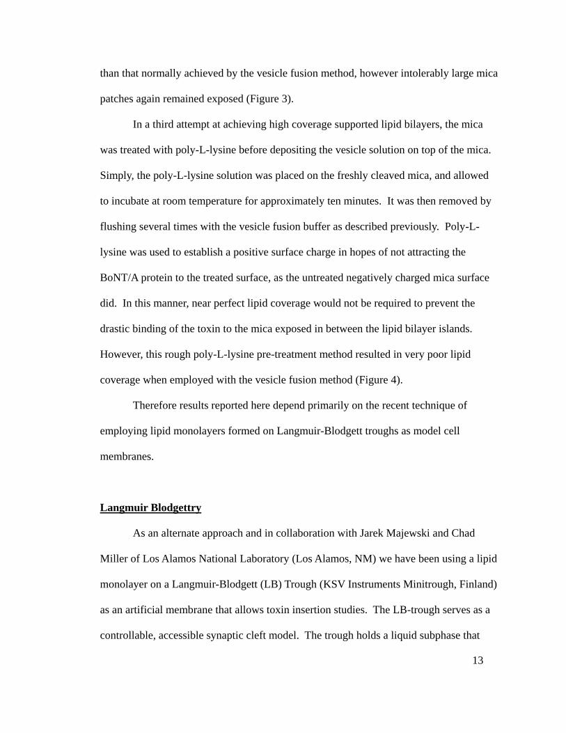
than that normally achieved by the vesicle fusion method, however intolerably large mica
patches again remained exposed (Figure 3).
In a third attempt at achieving high coverage supported lipid bilayers, the mica
was treated with poly-L-lysine before depositing the vesicle solution on top of the mica.
Simply, the poly-L-lysine solution was placed on the freshly cleaved mica, and allowed
to incubate at room temperature for approximately ten minutes. It was then removed by
flushing several times with the vesicle fusion buffer as described previously. Poly-L-
lysine was used to establish a positive surface charge in hopes of not attracting the
BoNT/A protein to the treated surface, as the untreated negatively charged mica surface
did. In this manner, near perfect lipid coverage would not be required to prevent the
drastic binding of the toxin to the mica exposed in between the lipid bilayer islands.
However, this rough poly-L-lysine pre-treatment method resulted in very poor lipid
coverage when employed with the vesicle fusion method (Figure 4).
Therefore results reported here depend primarily on the recent technique of
employing lipid monolayers formed on Langmuir-Blodgett troughs as model cell
membranes.
Langmuir Blodgettry
As an alternate approach and in collaboration with Jarek Majewski and Chad
Miller of Los Alamos National Laboratory (Los Alamos, NM) we have been using a lipid
monolayer on a Langmuir-Blodgett (LB) Trough (KSV Instruments Minitrough, Finland)
as an artificial membrane that allows toxin insertion studies. The LB-trough serves as a
controllable, accessible synaptic cleft model. The trough holds a liquid subphase that

14
Figure 3. 3μm x 3μm AFM image of an LB-trough-deposited lipid bilayer. The lowest
region (as indicated in the darkest color of the height image (left)) is exposed mica. The
highest regions, in white are vesicles adsorbed to the bilayer surface. The midgrade
brown color reveals an adsorbed lipid bilayer. The edges of these three levels can be more
clearly distinguished in the deflection image (right). The LB-Trough lipid bilayer
deposition allowed for thorough coverage, but the exposed mica surface, all though less
than the typical vesicle fusion method, was still too extensive for the purposes of BoNT/A
binding, insertion studies.
15
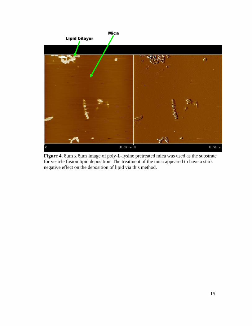
Lipid bilayer
Mica
Figure 4. 8μm x 8μm image of poly-L-lysine pretreated mica was used as the substrate
for vesicle fusion lipid deposition. The treatment of the mica appeared to have a stark
negative effect on the deposition of lipid via this method.

16
represents the extracellular fluid in the cleft of the neuromuscular junction. At the
interface between this liquid and the air, a monolayer of amphiphilic lipids is formed.
This monolayer models the outer leaflet of the nerve endplate membrane. The monolayer
will also represent the inner leaflet of the endosome upon receptor mediated endocytosis.
Although only half the membrane is represented by the monolayer system, the
membrane’s hydrophobic portion of its second half is electrostatically similar to the air
above the monolayer. The outerleaflet lipid headgroups and the water above them are
missing from our model which must be taken into account for interpretation.
We assumed that the surface pressure on the membrane would have a negligible
change due to toxin insertion, given the large membrane surface area to toxin ratio. To
mimic this characteristic, we held the monolayer’s surface pressure constant. This is
accomplished through the negative feedback loop response feature of our KSV
Instruments LB-trough. As a membrane associating protein inserts, the surface pressure
begins to increase, but is quickly relieved by a corresponding change in monolayer area.
This keeps the surface pressure constant (+/- 1 mN/m from the set point value, with a
strong mean at the set point surface pressure) (Figure 5) throughout the experiment. Thus,
the percentage of monolayer expansion versus time is the measure used to assess the
degree of toxin insertion to the membrane. The KSV Langmuir-Blodgett Minitrough's
surface pressure sensor is acutely sensitive to changes in level. The use of a vibration
dampening air table for the LB-trough ended up producing tilt related fluctuations in
surface pressure, which ultimately required us to run calibration experiments that
corrected for changes in level on surface pressure readings.
Due to the high cost of our toxin samples, an ultra small volume custom LB-
18
trough was constructed. We designed the LB-trough to not only hold a small volume, but
also be compatible with the remainder of our commercial KSV Instruments LB
Minitrough. The custom LB-trough was made out of milled PTFE stock. The final
dimensions were 65.1mm x 18.28mm x 3.25mm. Our custom LB-trough walls had
thicknesses of approximately 1mm. After milling, the final custom trough's surface
roughness was achieved with wet one-thousand-grit sand paper. When the custom LB-
trough was filled with buffer to a typical initial level, which always had a positive
meniscus, it held approximately 4.7ml (Figure 6). This is in contrast to the commercial
LB-trough which holds approximately 83ml, once the deposition well is covered.
Paper Wilhelmy plates (PWP) were used with the surface pressure sensor.
The PWPs were cut from Whatman filter paper. PWPs were calibrated before each run
against the standard of pure water surface pressure or a standard weight. Eventually the
PWP was replaced with a microroughened platinum Wilhelmy Rod (PWR) (KSV
Instruments, Finland) when our surface pressures were held above 20mN/m, as the PWP
was no longer reliable in that range (20).
Cleaning of all contact surfaces of the LB-trough was performed with a 2%
sodium octanoate detergent solution (octanoic acid, sodium salt, 99%, Aldrich Chemical
Company, Inc. Milwaukee, WI), then with a fresh, high-purity grade chloroform
(stabilized with a non-polar hydrocarbon) (Omnisolv). The LB-trough barrier was made
of Delrin and was rinsed with acetone, (because Delrin is solubilized by chloroform).
The LB-trough and barrier were then immediately rinsed in ultra pure water (Nanopure
Milli-Q filtering system) in order to minimize any trace amounts of non-volatile particles
the solvents would have otherwise left behind. In most cases the LB-trough was then

19
Figure 6. This photograph shows our custom LB-trough. Its dimensions are 65.1mm x
18.28mm x 3.25mm. With a typical initial positive meniscus, the custom trough holds
approximately 4.7ml.

20
filled with a freshly prepared pH 4.8 or pH 7.4 phosphate citrate buffer (Sodium
Phosphate, dibasic. Fisher, Lot No. 897108; Citric Acid, Monohydrate. Fisher, Lot
No. 922504). Laboratory quality sodium chloride was then added to bring the ionic
strength of the buffer up to the desired level.
The pH 4.8 buffer used for all monolayer experiments held at 20 (+/- 5)
mN/m had 239.2mM Na+, 140mM Cl
-, 49.6mM H2PO4
-, 25.2mM
HC3H7O(COO)32-
, resulting in an ionic strength of 243mM.
The pH 7.5 buffer used for all experiments held at 20 (+/- 5) mN/m had
314.4mM Na+, 140mM Cl
-, 87.2mM HPO4
2-, 6.5mM C3H5O(COO)3
3-,
resulting in an ionic strength of 430.9mM.
The pH 4.8 buffer used for all experiments held at 30 (+/- 1) mN/m had
158.4mM Na+, 118.4mM Cl
-, 20.0mM H2PO4
-, 10.3mM HC3H7O(COO)3
2-,
resulting in an ionic strength of 160mM.
The pH 7.7 buffer used for all experiments held at 30 (+/- 1) mN/m had
119.3mM Na+, 63.5mM Cl
-, 111.6mM HPO4
2-, 12.1mM C3H5O(COO)3
3-,
resulting in an ionic strength of 153mM.
Phosphate-citrate was chosen due to its large variety of pKa's. This allowed us to buffer
well at both of the pH values we were interested in, without altering the salt species.
Before lipid deposition, it is necessary to rid the subphase air-water interface
from trace amounts of surface active molecules, which contribute to the surface pressure.
This was accomplished by translation of the Delrin LB barrier to one end of the trough.
The barrier slowly skims along the subphase surface, pushing in front any surface active

21
molecules that may be present. If there are surface active molecules, an aspiration pump
fixed with a thin gel electrophoresis loading pipette tip was manually tapped along the
subphase surface, removing any contaminants. The LB barrier was then translated back
down the trough. If the surface pressure sensor did not change from its arbitrary initial
value after compression, the air-water interface was considered clean and the surface
pressure was then defined to be zero. This calibration is in accordance with the standard
of pure water, which is defined to have a surface pressure of zero. Cleaning and
calibration was done before every experiment.
The lipid monolayer was made of DPPC or 1,2-dipalmitoylphosphatidyl-
glycerol (DPPG) (Avanti Polar Lipids), mixed with 20% GT1b (Sigma Aldrich) or Total
Ganglioside Extract (porcine brain, ammonium salt. Avanti Polar Lipids. Lot #TGANG-
14) by mole fraction. This lipid mixture was suspended in a volatile solution not miscible
with water. Many lipids including DPPC and DPPG are very soluble in the volatile
solvent Chloroform, which is not miscible with water. However, gangliosides have a low
solubility in chloroform. The gangliosides were first solubilized in a minimal amount
(1.5% by total volume) of high purity methanol (Mallinckrodt Nanograde). Once they
were dissolved in the methanol they could be added to the lipid-containing chloroform
without falling out of solution.
This solution is placed drop wise using a glass, gas-tight syringe (Hamilton,
Reno, NV) onto the air/water interface of the LB subphase, such that the solution quickly
diffuses over the surface as its volatile solvent evaporates, leaving behind the amphiphilic
lipid and charged gangliosides. This forms a lipid monolayer on the subphase surface
with the lipid heads down to the water and the lipid tails up in the air. The gangliosides

17
Figure 5. Representational graph of typical surface pressure readings. The mean surface
pressure holds tightly around its set point value even as the monolayer makeup changes
due to BoNT/A insertion. The y-axis is surface pressure in mN/m, and the x-axis is time
in minutes x 103
.
22
are dispersed throughout the lipid molecules. As the LB barrier compresses the
monolayer it becomes highly ordered , like that of the inner leaflet of a membrane. We
compressed our monolayer until the surface pressure reached 20 (+/- 5) mN/m.
Once a baseline was obtained to check for drift, BoNT/A heavy chain, heavy
chain quarter (HCQ) which is the C-half of the C terminus of the heavy chain (27), or the
detoxified form of the holotoxin was added. All of these BoNT/A proteins were kindly
donated by Dr. Bal Ram Singh of the University of Massachusetts, Dartmouth. The
detoxified recombinant form of BoNT/A (DR BoNT/A) has been altered with
neutralizing mutations at two specific sites, E224-to-glutamine and E262-to-alanine,
within the active site of the light chain. According to ELISA activity assays (19), these
mutations eliminate light chain endopeptidase activities against the light chain substrate
SNAP 25. Additionally, bioassays were performed to study the effects of DR BoNT/A.
Groups of mice were injected with increasing doses of DR BoNT/A, their survival was
monitored. Mice receiving doses of DR BoNT/A as high as 0.0625 µg/g (131,000 times
the minimum lethal dose) demonstrated no symptoms of botulism. This indicates that
DR BoNT/A has a toxicity 100,000-times lower than native BoNT/A. Further, DR
BoNT/A was used to immunize mice, which provided complete protection against native
BoNT/A, showing that DR BoNT/A structure is similar to the native toxin because
antibodies raised against DR BoNT/A recognized both the mutated and active holotoxin.
Through far-UV circular dichroism spectroscopy, it has also been determined that the
secondary structure of DR BoNT/A is essentially visually identical to that of the native
neurotoxin (personal communication Bal Ram Singh).
The toxin was added to the subphase of the LB-trough by penetrating the
23
monolayer with the needle of a Hamilton microsyringe. The typical volume of toxin
solution injected was around 175µl, which resulted in an increase in surface pressure
reading. However the the syringe needle itself appeared to drop the surface pressure by
removing a small amount of the lipid molecules upon retraction. The net effect of these
two alterations was a mild drop in surface pressure reading that resulted in a small area
compression. This was done to yield a final toxin concentration of approximately 0.2µM.
To minimize microscopic monolayer perturbations, the LB-trough was placed on
a laboratory vibration dampening air table, although side effects were problematic, as
stated above. The shallow depth of the custom trough (3.25mm) also helped to minimize
the height of waves in the subphase surface. Further, no external pumps, nor stir bars
were used to speed up toxin diffusion in order to keep the monolayer as undisturbed as
possible. Stir bars and pumps caused visual disturbances in the monolayer, even when
the 0.5mm thick stir bar or the peristaltic pump were turned on to their lowest settings.
Instead the injection was done through several places in the lipid monolayer to maximize
the mixing of the toxin into the subphase. The chamber was then left alone to mix by
diffusion. Experiments with food coloring showed that this injection method mixed
thoroughly below the monolayer in approximately 15 minutes. Complete diffusion
throughout the custom LB-trough took nearly 150 minutes.
It was found that azolectin monolayers made from soy lipids were not ideal
because of their unstable surface pressures, given a fixed area. Wide LB-trough walls did
not allow for stable monolayers consistently. In order for the Delrin LB barrier to ride
true against the custom LB-trough walls, it was necessary for the walls to be narrow,
about 1mm wide (Figure 6).

24
RESULTS
ATOMIC FORCE MICROSCOPY
When toxin-incorporated vesicles were used, the AFM revealed that the vesicle
fusion method worked significantly less effectively than it did with pure lipid vesicles.
Measures were taken to compensate for this by sonicating longer, sonicating in warm
water, and incubating the sample over the mica longer. The result was more lipid on the
mica surface, however it was in excess. The mica was usually found completely covered
with a single lipid bilayer, with additional lipid micro domains on top. In order to see the
primary bilayer the AFM tip was used to purposely scan with an excessively high
pressure and tip velocity to remove the extra lipid. This would consistently leave only
the bottom lipid bilayer with near perfect coverage (Figure 7). On many occasions,
promising distinctions arose between images of pure supported planar DPPC bilayer
controls and samples with the addition of BoNT/A heavy chain (Figure 8). 14, 1-µm2
control images, and 7, 1-µm2 BoNT/A heavy chain images were searched, and raised
elements counted. Sample results are reported in Table 1. The search criterion was three-
fold. Typical amplitude images exhibit interference of a couple angstroms. The
minimum element height was therefore set at 4 angstroms. The maximum was 5
nanometers – chosen because small lipid layers are often as small as 5.5 nanometers.
Elements were grouped by height. A correction was made for the area of fused bi-layer
in the sample. For example, if an image was found to be 73.28% fused bi-layer by area,
the element-per-square-micron figure was obtained by dividing the number of observed
elements by 0.7328. The final row of Table 1 contains the probability that the difference

25
Figure 7. AFM image of excess lipid being removed revealing one lipid bilayer. All
three images are different renditions of the same topography of a pure DPPC sample on
mica in pH7.4 sodium phosphate buffer. The top left scan is the “height” image, which is
produced by the AFM piezoelectric movement. This scan has a z “grey scale” of 33.5
nanometers, where black is zero and white is 33.5 nm or larger. The top right scan is the
deflection image of the same topography, which is produced by the AFM cantilever
movement, or “deflection.” All three renditions show, after zooming out, the square
section of removed lipid by the AFM tip's high pressure/fast scanning. Bottom is a 3-
demensional rendition of the hieight image. The total scan size is 12μm x 12μm. 3-
demensional image created with WSxM v3.0 Beta 11.4 software, available through
Nanotec Electronica S. L.

26
Figure 8. A 0.824μm x 0.824μm height AFM image of the suspected BoNT/A Heavy
chain, inserted into a DPPC supported bilayer. B 2μm x 2μm AFM image depicting the
deflection of a DPPC supported on mica bilayer that had been incubated for 28 minutes
with BoNT/A Heavy chain. The red box describes where the zoomed in box B hight
image came from. C A cross section of box B. The cross section location is indicated
on the left, and its actual cross section is on the right. The height of the suspected
BoNT/A Heavy chain is measured as 0.8 nm.
C
B A

27
Element size (nm) [0.4,0.6) [0.6,1.0) [1.0,2.0) [2.0,3.0) [3.0,4.0) [4.0,5.0)
x (protein) 5.50 11.64 10.51 0.63 0.29 0
µ (control) 2.68 3.98 2.61 1.37 0.12 0.56
x-µ 2.82 7.66 7.90 -0.74 0.18 -0.56
σ 3.41 3.34 1.96 1.69 0.44 0.98
(x-µ)/σ 0.83 2.29 4.04 -0.44 0.41 -0.58
P(~H0)
71.96% 98.90% ≈100% N/A 65.89% N/A
Table 1. The average number of particles per μm2 of bilayer x, is reported for the
size range listed in the header for each column. 1mg DPPC / 10mg BoNT/A
heavy chain / 0.1 ml was the original subphase concentration the 250μl droplet.
The excess was rinsed away. The mean in protein free control samples is given in
the second row as μ (control). The difference, standard deviation for x, and ratio
are used to compare P(~Ho). The probability that the null hypothesis is false (i.e.
that there are more particles in the test case than control) from a single tailed T test
is listed in the bottom row.
28
between protein sample mean and control mean did not arise by random chance. Most
significant are the differences found in elements of height between 0.6 and 2.0
nanometers. This points strongly to these elements as candidates for BoNT/A heavy
chain proteins.
When we diluted the toxin incorporated vesicle solution from 1mg/10mg/0.1ml
(lipid/BoNT/A Heavy Chain / Solution volume) to 1mg/10mg/1.0ml we found that
through the vesicle fusion method it was easier to achieve good lipid single bilayer
coverage without excess. When these images were analyzed, significantly fewer
topographical particles were counted as compared to the images taken of undiluted
sample.
Upon close inspection it was noted that when the fast scanning, high pressure
AFM scan was used to remove excess lipid above the primary lipid bilayer, small lipid
topological particles (1 to 3 nanometers tall) were agitated loose and adhered to the
bilayer, giving the appearance of toxin particles, in the absence of toxin (Figure 9).
Therefore, we dismissed the results in Table 1 as likely artifact and abandoned (for
purposes of this thesis) the AFM line of experiments.

29
Figure 9. Demonstration that the AFM tip can produce small lipid particles (<1 to 3nm
tall). Figure 9-A shows a 2.1μm x 2.1μm scan of pure DPPC. Figure 9-B shows the
same sample, in the same location after a high pressure/fast small scan in the upper right
hand quadrant. This produced a defect in the bilayer (blue arrow), and small particles
(green arrows) of the order seen during past experiments in the presence of unnicked DR
BoNT/A.
A
B
30
Langmuir Blodgettry
The Langmuir-Blodgett custom 4.7ml PTFE trough demonstrated steady,
reproducible DPPC/Ganglioside (80:20) monolayer surface pressures after monolayer
compression to a fixed area.
When the surface pressure was held constant at about 20(+/- 5) mN/m or higher,
regardless of pH, ionic strength and buffering species in the subphase, a clear expansion
of the monolayer resulted after toxin injection. (Figure 10A). In Table 2 of the appendix,
a list of all LB monolayer experiments done with 0.1 - 0.2μM BoNT/A are presented.
Experiments 1-8 are BoNT/A injections at low surface pressure and 10-15 are BoNT/A
injections at cell membrane-like surface pressure. They nearly all show a clear expansion
(≥ 10%) of the monolayer. The monolayers were all made of DPPC and GT1b or a
mixture of gangliosides at 80:20 by mole fraction. In contrast, the controls (experiments
9, and 16 – 21) show little to no expansion of the monolayer. Conditions for experiment 9
were like those in experiments 1-8, but the DR BoNT/A sample was boiled for
approximately 12 minutes. The solution showed obvious DR BoNT/A denaturation as
manifest by the appearance of large white aggregates in the solution after heat exposure.
Injection of this sample with precipitate suspended gave a diminished monolayer
expansion about five-fold below the average. Experiments 17 – 22 are three different
controls. Experiments 17 and 18 have no ganglioside receptors in their DPPC monolayer.
Under these conditions no monolayer expansion was observed after DR BoNT/A
injection. (Figure 10B). Experiments 19 and 20 use the HCQ protein which is BoNT/A
heavy chain without the translocation domain (25). HCQ had a negligible monolayer
expansion of approximately 3%. Finally experiments 21 and 22 are with the control

31
Figure 10. A) DPPC/GT1b monolayer percent expansion (relative to initial area
immediately after injection) vs. experimental run time (minutes) due to DR BoNT/A
insertion. The tracing starts at the time of toxin injection into the subphase of the LB-
trough through the DPPC/GT1b or mixed ganglioside (80:20) monolayer. After a lag time
that averaged about 100 minutes the toxin began to insert into the monolayer causing the
monolayer to expand under a constant surface pressure. B) DPPC monolayer (without
ganglioside receptor) percent expansion vs. experimental run time (minutes) after 0.2 µM
DR BoNT/A injection. This graph is representative of the negative results acquired by
control experiments that consisted of receptor free monolayers, HCQ and Albumin. C)
Representational graphs demonstrating that the experiments were run long enough to see
a steady state. The conditions associated with the graphs found in box C, are located in
Table 2 of the appendix. From left to right the file names are: 080207.#00, 082907.#00,
and 081007.#00.
C
A B
32
plasma protein albumin. Albumin was tested at the standard 0.2 µM and also with a fifty
fold concentration increase at 10.0 µM. DPPC/Total ganglioside extract monolayers
revealed no monolayer expansion at either concentration with albumin (Figure 11).
When the ganglioside concentration is reduced from 20% to 10%, the expansion of the
monolayer in the presence of DR BoNT/A is reduced (Chad Miller and David Busath,
personal communication). In the absence of any gangliosides toxin-induced expansion is
eliminated. When the lipid monolayer is switched from the zwitterionic DPPC to the
negatively charged DPPG (identical ions in the bath, both lipids containing GT1b, and
the toxin concentration remaining at 0.2µM), DR BoNT/A inserts into the monolayer
more readily (tested at low pH only).
Figure 12 is a bar plot that summarizes the data of Table 2 by grouping the
experiments into relevant categories. The categories focus on BoNT/A version, and
subphase pH. Figure 12 must be recognized as preliminary results. These data have
shaped are hypothesis and will guide are future emphases, however more work is
required before this data can be considered compelling. Comparing groups within Figure
12, one to another through formal statistical methods is largely inappropriate. The N
values of each group are not large enough to possibly form normal data distribution
curves. The standard deviation, to which so many statistical tools are connected, is based
on the mean of the data. The mean of the data only represents center accurately when the
data distribution is normal. Unfortunately, these variations between experiments are too
large to allow analysis of how environmental variables (pH, ionic strength, surface
pressure, and temperature) affect the final percent expansion.

33
Figure 11. LB expansion graph of the control protein Albumin. The
eventual area compression is due to slow evaporation from the LB-
Trough. With this understanding it is clear that expansion due to
Albumin is negligible. This was done with a phosphate citrate buffer
at pH 4.8, and an ionic strength of 160mM.

34
Figure 12. Bar plot of various experimental groupings with essential parameters
listed under each. The details of each experiment and their variations within groupings
are contained in Table 2 of the appendix. The line above each bar represents one half of
the standard deviation. When combining the first four bars into one “insertable” group, a
mean of 35.1 % expansion with a standard error of the mean (SEM) at 9.4 results.
Further, a combination of the last three bars into a “non insertable” group results in a
mean percent expansion of 1, and an SEM of 0.6, revealing the two groups as statistically
different. The inset bar plot shows only those experiments that include all essential
features for monolayer insertion, grouped according to pH. The flag represents one SEM.
Neutral pH approaches but does not reach a statistically significant difference over the
acidic pH group. Appropriate formal application of statistical analysis will require
greater N values. As a whole, there is evidence that gangliosides in the lipid monolayer,
and the heavy chain with both translocation and ganglioside binding domains are
minimum requirements for toxin insertion into the monolayer in a pH independent
manner.
Mean Percent Expansion Due To BoNT/A Insertion Insertion

35
Discussion
Atomic Force Microscopy
The particles created in the control samples that had no toxin present by wiping
away the excess lipid with the AFM tip were within the size range of the particles
previously assumed to be BoNT/A Heavy chain. An explanation as to why the quantity
of so-called toxin particles found decreased when the toxin containing vesicle solution
was diluted is simple. The diluted BoNT/A heavy chain vesicle solution was easier to
adsorb to the mica. We were able to produce appropriate lipid coverage without the
excess. The result was less agitation required by the AFM tip to remove excess lipid. The
less this relatively large scale cleaning took place, the less the tiny particle confounders
were created. The distinction between toxin particles and tip induced lipid particles
proved unresolvable with the AFM and these sample preparations. At this point the
decision was made to set this project aside and shift to the recently discovered LB
monolayer technique, being used for neutron reflectometry studies by Dr. Busath’s
collaborators at Los Alamos National Lab for measuring toxin insertion.
In the future, a significant increase in toxin concentration may be used to
positively identify the BoNT particles with AFM, on an asymmetric supported bilayer
containing gangliosides. Additionally a minimally intrusive tag may also work to
positively identify BoNT/A protein in a supported bilayer. Avoiding a tag altogether until
after AFM imaging seems least intrusive of all, and an adaptation of an ELISA assay to
the imaged sample may perform this task well.
As noted earlier, the DPPC supported bilayer was in the gel phase. Cell

36
membranes are composed mainly by lipids in the liquid phase at body temperature. It
seems expedient therefore that the forthcoming supported bilayers used with the AFM
ought to be made up of lipids in the liquid phase. If the mobility of the inserted toxin into
a liquid phase bilayer is too great for high resolution AFM images, a lipid such as DPPC
could be used such that the temperature is raised to be in the liquid phase (>41o C) during
incubation, and finally cooled to room temperature during AFM imaging.
With the advantage of hind sight, this skepticism over the existence of BoNT/A
heavy chain in the supported DPPC bilayers is further bolstered by the LB data that
shows no monolayer expansion by the toxin in the absence of gangliosides, indicating
that the toxin does not insert into a pure DPPC membrane. Honing of the LB deposition
technique to produce asymmetric bilayers on mica, such that gangliosides and/or other
BoNT receptors can be incorporated into the outer leaflet may prove to be the best way to
prepare model membranes for future AFM studies.
Langmuir Blodgettry
BoNT/A does not expand a pure DPPC monolayer as our controls show.
However, in the presence of gangliosides it does expand the monolayer. This indicates
that gangliosides alone can facilitate BoNT/A insertion and potentially BoNT/A light
chain exit from the endosome, even in the absence of the high affinity binder SV2.
The DPPC/ganglioside monolayer did not expand with our control protein,
albumin. Albumin was chosen because it is an abundant plasma protein that, as such, is
considered unlikely to interact with cell membranes. If albumin molecules were to
interact by insertion into the monolayer, it would be manifest in monolayer expansion to
37
a similar degree to that of DR BoNT/A or BoNT/A heavy chain. The molecular weights
(bovine serum albumin (~66 kDa), DR BoNT/A (~150 kDa) and BoNT/A (~100 kDa))
are close enough to allow us to assume that the degree of monolayer area increase due to
insertion by one, is likely to be similar to the others. Even more conservatively we could
say that a control protein half the size of the sample protein is very unlikely to insert into
the monolayer unnoticed, while the sample insertion is clear. Our results refute the
possibility that our monolayer expansions, in the presence of DR BoNT/A, were actually
due to features of our monolayer and/or LB setup, independent of the protein makeup.
Our LB percent expansion results extended up to a maximum of 140 percent We
found no theoretical basis for disallowing this maximal expansion. Given an approximate
initial monolayer of 421 mm2, at 20% ganglioside by mole fraction, we would have
roughly 1.7 x 10¹⁴
ganglioside molecules if we assume the area of ganglioside in the
monolayer similar to DPPC (48.6 Å2). At 0.2µM there is more toxin than ganglioside. If
each ganglioside were to allow one DR BoNT/A (an approximate sphere of diameter
7.5nm) to bind and insert into the monolayer, the toxin would expand the monolayer
~9,600 mm2, or 2,300 % expansion. Thus, less than 7% of the toxin inserts.
Figure 12 demonstrates that, in nearly all experiments, BoNT/A expanded the
monolayer by 10% or more under conditions where the current BoNT/A hypothesis
would predict insertion. Namely, when the ganglioside binding domain and the
translocation domain are both a part of the toxin (this would include both the detoxified
holotoxin DR BoNT/A and the BoNT/A heavy chain), BoNT inserts into the model
membrane, so long as gangliosides are present. We therefore believe that LB monolayers
can be considered a promising model for future BoNT/A studies.
38
It is not known when BoNT/A heavy chain inserts into the nerve ending at the
neuromuscular junction, nor is the role of acidification understood in BoNT/A heavy
chain insertion. The addition to the current popular working hypothesis which is raised
by the data of this study, is that insertion of the BoNT/A heavy chain into the membrane
does not need to wait until acidification of the endosome, and may even occur before
BoNT receptor mediated endocytosis. At both neutral pH (extracellular pH) and acidic
pH (late endosome pH), our results show that when the complete heavy chain is exposed
to monolayers containing the ganglioside receptor, not just a peripheral binding, but an
interaction with the membrane, intimate enough to cause monolayer expansion, occurs. It
is our interpretation that this interaction is not a mere binding, therefore, but an insertion.
The evidence comes from two results. One, binding without inserting would cause little
to no noticeable monolayer expansion, and second, only when the BoNT/A insertion
domain is present do we see monolayer expansion.
We extrapolate this data to the native toxin, and believe that the role of
acidification in the endosome is not to enable BoNT/A heavy chain insertion into the late
endosome membrane, but, rather, it plays the critical role of final conversion of BoNT/A
heavy chain into the pore conformation, and/or it prepares the light chain's secondary
structure for its translocation.
When more DR BoNT/A and its truncates become available to our laboratory, all
of the experiments need to be further reproduced. Several possible confounders have
persisted throughout the project, namely:
unreliable condition of DR BoNT/A and its variants,
uncertainty in our surface pressure measurements due to air table leveling
39
inconsistencies, and
inherent deviations in our model system from nature such as the monolayer itself,
unphysiological concentrations of the toxin in the subphase, and an unnaturally
large ratio of gangliosides in the monolayer.
The last factor is most acceptable, and even arguably advantageous when looked at with a
minimalistic point of view. The monolayer, having air above the lipid tails, necessarily
stops the toxin from completely translocating, halting its progress at a stage that might
otherwise not have been testable. The high toxin concentrations used allow us to measure
readily what otherwise may have been too subtle an effect to notice.
Another major draw back to the monolayer model, is that when the monolayer is
made up of more than one type of molecule, the distribution may be erratic. It is possible
that, instead of a homogeneous mixture, there segregated domains can form. It is also
possible that the extent of this domain formation varies significantly from experiment to
experiment, depending on uncontrollable trace contaminants in the monolayer solution or
subphase. This unpredictable nature may contribute to the large interexperimental
variance we have observed.
The uncertainty in the surface pressure was discovered near the end of the project
and was found to be due to the tendency of the vibration table to re-level imprecisely
after a weight change to its surface. This was remedied for the high surface pressure
(30mN/m) experiments by disabling the air floating mechanism of the table. Calibration
tests demonstrated the surface pressure stayed within 1 mN/m of its target with the
vibration table disabled, whereas previously our surface pressures were dropped because
of table relabeling from the set point value by 5to15 mN/m; and, then the surface pressure
40
would hold to within 1 mN/m of that dropped value.
The initial experiments, therefore, intended to be run at 30mN/m, were actually
run at 20(+/- 5) mN/m. The accepted value for a physiologically relevant surface
pressure, in the field of monolayer studies, is 30 to 35mN/m. The expansion observed in
the higher pressure strongly suggests that the protein could insert into cell membranes as
well.
The final issue of whether the protein might have been partly degraded in some of
the experiments is the most difficult to assess. Because the toxin we use has point
mutations in the active site of the BoNT/A light chain, it has no enzymatic activity, so
enzyme activity assays cannot be used to test the integrity of the protein conformation.
Antibody binding assays could be performed but they would only indicate the condition
of one small motif, which may or may not pertain to the aspect of the protein of interest.
Therefore small, frequently prepared, freshly cloned nicked batches of the mutated toxin
should be used in the future, rather than the large infrequent batches we have worked
with in the past.
In conclusion, AFM studies of BoNT/A on supported ganglioside-incorporated
asymmetrical bilayers indicate potential as significantly contributing tools in this field of
toxin research. The LB-trough data demonstrates that this model for toxin studies is
promising. This study indicates that BoNT/A heavy chain can insert both at low and
neutral pH. We propose therefore that the required acidification step found elsewhere to
be necessary for BoNT/A toxicity, does not involve heavy chain membrane insertion.
Heavy chain insertion may occur as early as within the neutral extracellular fluid
of the synaptic cleft, immediately following receptor binding.

41
References
1. Arnon SS et al. Botulinum Toxin as a Biological Weapon. JAMA 285:1059-1070,
2001.
2. Simpson LL. Identification of the Major Steps in Botulinum Toxin Action. Annu
Rev Pharmacol Toxicol 44:167-193, 2004.
3. Kortepeter MG and Parker GW. Potential Biological Weapons Threats.
Emerging Infectious Diseases 5:523-527, 1999.
4. Jost WH. Other indications of botulinum toxin therapy. European Journal of
Neurology 13:65–69, 2006.
5. Silberstein S. Botulinum Neurotoxins: Origins and Basic Mechanisms of Action.
Pain Practice 4: S19-S26, 2004.
6. Kozaki S, Kamata Y, Watarai S, Nishiki T, Mochida S. Ganglioside GT1b as a
complementary receptor component for Clostridium botulinum neurotoxins.
Microb Pathog 25:91-99, 1998.
7. Kitamura M, Takamiya K, Aizawa S, Furukawa K, Furukawa K.
Gangliosides are the binding substances in neural cells for tetanus and botulinum
toxins in mice. Biochim Biophys Acta 1441:1-3, 1999.

42
8. Bullens RWM, O'Hanlon GM, Wagner E, Molenaar PC, Furukawa K et al.
Complex gangliosides at the neuromuscular junction are membrane receptors for
autoantibodies and botulinum neurotoxin but redundant for normal synaptic
function. J Neurosci 22:6876-6884, 2002.
9. Yowler BC, Kensinger RD, Schengrund CL. Botulinum neurotoxin A activity
is dependent upon the presence of specific gangliosides in neuroblastoma cells
expressing synaptotagmin I. J Biol Chem 227:32815-32819, 2002.
10. Black JD, Dolly JO. Interaction of 125-I labeled botulinum neurotoxins with
nerve terminals. I. Ultrastructural autoradiographic localization and quantiation of
distinct membrane acceptors for types A and B on motor nerves. J. Cell Biol.
103:521-534, 1986
11. Jahn R. A Neuronal Receptor for Botulinum Toxin. Science 312:540-541, 2006.
12. Dong M, Yeh F, Tepp WH, Dean C, Johnson EA, Janz R, and Chapman ER.
SV2 Is the Protein Receptor for Botulinum Neurotoxin A. Science 312: 592-596,
2006.
13. Koriazova LK and Montal M. Translocation of botulinum neurotoxin light
chain protease through the heavy chain channel. Nature Structural Biology 10:13-
18, 2002.

43
14. Brian AA and McConnell HM. Allogenic stimulation of cytotoxic T cells by
supported planar membranes. Proc Natl Acad Sci USA 81:6159–6163, 1984.
15. Schabert, F., and A. Engel. Reproducible acquisition of Escherichia coli porin
surface topographs by atomic force microscopy. Biophys J. 67:2394–2403. 1994.
16. Leidy, C, Kaasgaard T, Crowe J, Mouritsen O and Jorgensen K. Ripples and
the formation of anisotropic lipid domains: imaging twocomponent supported
double bilayers by atomic force microscopy. Biophys J. 83:2625–2633. 2002.
17. Muller DJ, Fotiadis D, Scheuring S, Muller SA, and Engel A. Electrostatically
Balanced Subnanometer Imaging of Biological Specimens by Atomic Force
Microscope. Biophys J. 76:1101–1111, 1999.
18. Hughes T, Strongin B, Gao FP, Vijayvergiya V, Busath DD, Davis RC. AFM
visualization of mobile influenza A M2 molecules in planar bilayers. Biophys J.
87(1):311-22, 2004.
19. Sharma SK and Singh BR. Enhancement of the Endopeptidase Activity of
Purified Botulinum Neurotoxins A and E by an Isolated Component of the Native
Neurotoxin Associated Proteins. Biochemistry 43:4791-4798, 2004.
20. Bigalke H, Muller H, and Dreyer F. Botulinum A neurotoxin unlike tetanus

44
toxin acts via a neuraminidase sensitive structure. Toxicon 24:1065-1074, 1986.
21. Sharma S, Zhou Y, and Singh BR. Cloning, expression, and purification of C-
terminal quarter of the heavy chain of botulinum neurotoxin type A. Protein
Expression and Purification 45: 288-295, 2006.

45
Appendix
Table 2. Raw listing of BoNT/A LB expansion parameters and results. Groupings found
in figure12 are as follows: Bar#1-(1,2), Bar#2-(4-8), Bar#3-(10-12), Bar#4-(14-16),
Bar#5-(17,18), Bar#6-(19,20), Bar#7-(21,22).
File Name
Surface Pressure (mN/m)
Ionic Strength (mM) pH
Lipid (80%)
Gang. Type (20%) Toxin Spec.
[Toxin] (μM)
% exp.
Initial Area (mm
2)
1 081007.#00 20 (+/- 5) 167 7.5 DPPC Mix DR BoNT/A 0.2 35 386
2 080207.#00 20 (+/- 5) 167 7.5 DPPC Mix DR BoNT/A 0.2 140 398
3 072007.#00 20 (+/- 5) 243 4.8 DPPC Mix DR BoNT/A 0.2 3 546
4 072307.#00 20 (+/- 5) 243 4.8 DPPC Mix DR BoNT/A 0.2 45 474
5 072507.#00 20 (+/- 5) 243 4.8 DPPC Mix DR BoNT/A 0.2 65 350
6 072607.#01 20 (+/- 5) 243 4.8 DPPC Mix DR BoNT/A 0.2 35 491
7 082007.#00 20 (+/- 5) 243 4.8 DPPC GT1b DR BoNT/A 0.2 53 526
8 092607.#01 20 (+/- 5) 160 4.8 DPPC Mix DR BoNT/A 0.2 10 469
9 082907.#00 20 (+/- 5) 243 4.8 DPPC Mix Boiled DR BoNT/A 0.2 9 531
10 110607.#00 30 (+/- 1) 153 7.7 DPPC Mix BoNT/A HC 0.12 92 460
11 110907.#00 30 (+/- 1) 153 7.7 DPPC Mix BoNT/A HC 0.12 12 492
12 111207.#01 30 (+/- 1) 153 7.7 DPPC Mix BoNT/A HC 0.12 37 510
13 090407.#01 20 (+/- 5) 243 4.8 DPPC Mix BoNT/A HC 0.12 20 446
14 100207.#00 30 (+/- 1) 160 4.8 DPPC Mix BoNT/A HC 0.12 20 518
15 111407.#00 30 (+/- 1) 160 4.8 DPPC Mix BoNT/A HC 0.12 16 555
16 111007.#00 30 (+/- 1) 160 4.8 DPPC Mix BoNT/A HC 0.12 14 436
17 PRE082007.#00 UNRECORDED 20 (+/- 5) 243 4.8 DPPC NONE DR BoNT/A 0.2 0
Undetermined
18 083007.#03 20 (+/- 5) 243 4.8 DPPC NONE DR BoNT/A 0.2 0 435
19 100907.#00 30 (+/- 1) 160 4.8 DPPC Mix HCQ 0.2 3 520
20 072307 UNRECORDED 20 (+/- 5) 243 4.8 DPPC Mix HCQ 0.2
~3
Undetermined
21 101607.#01 30 (+/- 1) 160 4.8 DPPC Mix ALBUMIN 0.2 0 495
22 30 (+/- 1) 160 4.8 DPPC Mix ALBUMIN 10 0 502

Bradley A. Strongin 2289 West 540 North Provo, UT 84601 (801)375-7268
EDUCATION
MS, Physiology and Developmental Biology, Brigham Young University, Provo UT, December 2007
BS, Applied Physics, Brigham Young University, Provo UT 2005 Mathematics Minor
SKILLS Scientific Research
Atomic Force Microscopy (AFM) Scanning Electron Microscopy
(SEM) Transmission Electron Microscopy
(TEM)
Langmuir-Blodgett Trough Thin Film Deposition
Langmuir Lipid Monolayer Artificial Membrane Studies
Bilingual: English/Spanish Microsoft Word, PowerPoint Scientific Writing Carpentry, End Mill, Wood and Metal Lathes
WORK / RELATED EXPERIENCE
Research Assistant, Department of Physics & Astronomy and Department of Physiology & Developmental Biology 2002-2007
Research Topics: “Botulinum Neurotoxin Serotype A,” 2005 - 2007 “The Influenza A M2 Proton Channel,” 2002 – 2005
Publication: Hughes, T., B. Strongin, F. Gao, V. Vijayvergiya, D. Busath, and R. Davis (2004), AFM
Visualization of Mobile Influenza A M2 Molecules in Planar Bilayers. Biophys. J. 87:311-322.
Poster Presentations, Biophysical Society Annual Meetings
Co-authored, Miller, C. et al. “Constant-Pressure Expansion of Lipid-Ganglioside Monolayer by Botulinum Neurotoxin Serotype A: Does pH or Dithiothreitol Have More Impact?” 2007
Co-authored, Swenson, R. et al. “Preliminary Structure, Function, and Modeling of the Botulinum Toxin Heavy Chain in Lipid Membranes.” 2006
Principal Investigator, Strongin, B. et al. “Atomic Force Microscope Imaging of Influenza M2 Proteins in Supported Planar Bilayers.” 2005
Poster, American Physical Society March Meeting Co-authored, Hughes, T. et al. “Bilayer Incorporated Influenza A M2 Single
Molecule Time- Dependent AFM Studies.” 2004
Teaching Assistant, Taught Principles of Physiology Lab, Brigham Young University, 2006 – 2007
15 to 20-Minute Introductory Lectures. 9 class sections with 11 labs each (99 lectures)
Collaborating Scientist, Los Alamos National Laboratory Neutron Reflectometry 5 Combined Days of Training and Beam Time, June 2006
3 Days of Beam Time, August 2007
Patient Care Technician (PCT), Cardiovascular Unit, Utah Valley Regional Medical Center, 1 year 2001 - 2002
Financial Clerk – Office Depot, 1 year 1998 - 1999 Performed Daily Internal Sales Audits
COMMUNITY SERVICE
- Volunteer Representative – The Church of Jesus Christ of Latter-day Saints, Guatemala, 2 years 1996 - 1998 - Eagle Scout, Boy Scouts of America 1995
Cub Scout Webelos Den Leader, 1 year 2006