Thermalresponseof the 70-kDa · celllineswereexposedto45Candtheirthermalsurvivalswere determined by...
5
Proc. Nati. Acad. Sci. USA Vol. 88, pp. 1681-1685, March 1991 Cell Biology Thermal response of rat fibroblasts stably transfected with the human 70-kDa heat shock protein-encoding gene (stable expression/ceil survival/thermal resistance/nudeus) GLORIA C. LI*tt, LIGENG LI*, YUN-KANG LIu*, JOHNSON Y. MAK*, LILI CHEN§, AND WILLIAM M. F. LEE§ *MCB-200, Radiation Oncology Research Laboratory, Department of Radiation Oncology, University of California, San Francisco, CA 94143; and §Hematology-Oncology Section and the Cancer Center, University of Pennsylvania, Philadelphia, PA 19104 Communicated by James C. Wang, November 29, 1990 ABSTRACT The major heat shock protein hsp7O is syn- thesized by cells of a wide variety of organisms in response to heat shock or other environmental stresses and is assumed to play an important-role in protecting cells from thermal stress. We have tested this hypothesis directly by transfecting a constitutively expressed recombinant human hsp7O-encoding gene into rat fibroblasts and exmining the relationship be- tween the levels of human hsp7O expressed and thermal resis- tance of the stably transfected rat cells. Successful transfection and expression of the gene for human hsp7O were characterized by RNA hybridization analysis, two-dimensional gel electro- phoresis, and immunoblot analysis. When individual cloned cell lines were exposed to 45C and their thermal survivals were determined by colony-formation assay, we found that the expression of human hsp7O conferred heat resistance to the rat cells. These results reinforce the hypothesis that hsp7O has a protective function against thermal stress. When exposed to nonlethal heat shock, a variety of orga- nisms and cell lines acquire transient resistance to subse- quent exposures to elevated temperatures (1, 2). This phe- nomenon has been termed thermotolerance (3, 4). The mech- anism for thermotolerance is not well understood, although several studies suggest that the heat shock proteins (hsps) may be involved in their development (5-7). Qualitative evidence exists for a causal relationship between hsp syn- thesis and thermotolerance (5-9): (i) heat shock induces transiently enhanced synthesis of hsps that correlates tem- porally with the development of thermotolerance; (ii) the persistance of thermotolerance correlates well with the sta- bility of hsps; (iii) agents known to induce hsps induce thermotolerance; (iv) conversely, agents known to induce thermotolerance induce hsps; and (v) stable heat-resistant variant cells express high levels of hsps constitutively. One notable exception to the correlation summarized above is that amino acid analogues have been shown to induce hsps but not thermotolerance; HA-1 cells treated with such compounds are more sensitive to elevated temperatures (10). This apparent lack of correlation can be attributed, however, to the dysfunction of analogue-substituted hsps: the nonfunctional, analogue-substituted hsps would not be expected to protect cells from thermal stress. In support of this notion, Li and Laszlo (10) found that the incorporation of amino acid analogues into cellular proteins inhibits the development of thermotolerance and that thermotolerant cells or permanently heat-resistant cells are more resistant to the thermal-sensitizing action of amino acid analogues. Quantitatively, of the many hsps preferentially synthesized after heat shock, the concentration of the 70-kDa heat shock proteins (hsp70s) appears to correlate best with heat resis- tance, either permanent or transient (6, 8, 9). Recently, good correlation between a 27-kDa heat shock protein (hsp27) and thermal resistance also has been reported (11). In mammalian cells, three types of experiments have been performed to vary the intracellular concentration of hsp70 and to correlate this change with thermal-stress response. (i) Microinjection of affinity-purified anti-hsp70 antibodies into rat cells appeared to prevent the nuclear and nucleolar accumulation of hsp70 after a test heat shock and greatly increased the lethality of a 450C, 30-min heat treatment (12). (ii) The 5'-control region of the hsp70-encoding gene was inserted into a plasmid containing the dihydrofolate reduc- tase gene; this recombinant plasmid was then introduced into a Chinese hamster ovary (CHO) cell line, and a 20,000-fold elevation in its copy number was achieved by selection of cells with methotrexate. These copies of the hsp70 regulatory region presumably competed with the endogenous hsp70- encoding gene(s) for factors that activate hsp70 expression to reduce heat-inducible expression from the intact endogenous gene for hsp70 by at least 90%. It was found that cells containing the amplified regulatory sequences display in- creased thermosensitivity (13). (iii) Human hsp70 microin- jected directly into CHO cells increased the resistance of cells to 450C heating (14). A technique likely to be superior to microinjection is the expression of a cloned mammalian gene for hsp70 in cells. Transient expression of heterologous hsp70s has been used previously to show that Drosophila hsp70 accelerates the recovery of monkey COS cell nucleoli after heat shock (15) and to dissect the domains of human hsp70 responsible for nucleolar localization and for ATP binding (16). In the present communication, we report the construction of rat cell lines stably expressing different levels of a cloned hsp70- encoding human gene. Studies of these lines provide addi- tional evidence that functional hsp70 improves the survival of mammalian cells during and after thermal stress. MATERIALS AND METHODS Construction of Plasmids Expressing Human hsp7O. The 2.3-kilobase (kb) BamHI-HindIII fragment of the human gene for hsp70 (17, 18) was excised from pHHsp7O, a plasmid containing the entire transcribed portion of the hsp70 locus plus 5' sequences required for heat-inducible expression (obtained from R. Morimoto, Northwestern University), by first cutting with HindIII, making the end blunt with mung bean nuclease, and then cutting with BamHI. This fragment was subcloned between the BamHI and Sma I sites in the polylinker region of pSVSP65 [a pSP65 (Promega) derivative Abbreviations: hsp, heat shock protein; hsp70, 70-kDa heat shock protein; hsp27, 27-kDa heat shock protein; IEF, isoelectric focusing; mAb, monoclonal antibody. tPresent address: Department of Medical Physics, Memorial Sloan- Kettering Cancer Center, New York, NY 10021. tTo whom reprint requests should be addressed. 1681 The publication costs of this article were defrayed in part by page charge payment. This article must therefore be hereby marked "advertisement" in accordance with 18 U.S.C. §1734 solely to indicate this fact.
Transcript of Thermalresponseof the 70-kDa · celllineswereexposedto45Candtheirthermalsurvivalswere determined by...

Proc. Nati. Acad. Sci. USAVol. 88, pp. 1681-1685, March 1991Cell Biology
Thermal response of rat fibroblasts stably transfected with thehuman 70-kDa heat shock protein-encoding gene
(stable expression/ceil survival/thermal resistance/nudeus)
GLORIA C. LI*tt, LIGENG LI*, YUN-KANG LIu*, JOHNSON Y. MAK*, LILI CHEN§, AND WILLIAM M. F. LEE§*MCB-200, Radiation Oncology Research Laboratory, Department of Radiation Oncology, University of California, San Francisco, CA 94143; and§Hematology-Oncology Section and the Cancer Center, University of Pennsylvania, Philadelphia, PA 19104
Communicated by James C. Wang, November 29, 1990
ABSTRACT The major heat shock protein hsp7O is syn-thesized by cells of a wide variety of organisms in response toheat shock or other environmental stresses and is assumed toplay an important-role in protecting cells from thermal stress.We have tested this hypothesis directly by transfecting aconstitutively expressed recombinant human hsp7O-encodinggene into rat fibroblasts and exmining the relationship be-tween the levels of human hsp7O expressed and thermal resis-tance of the stably transfected rat cells. Successful transfectionand expression ofthe gene for human hsp7O were characterizedby RNA hybridization analysis, two-dimensional gel electro-phoresis, and immunoblot analysis. When individual clonedcell lines were exposed to45C and their thermal survivals weredetermined by colony-formation assay, we found that theexpression of human hsp7O conferred heat resistance to the ratcells. These results reinforce the hypothesis that hsp7O has aprotective function against thermal stress.
When exposed to nonlethal heat shock, a variety of orga-nisms and cell lines acquire transient resistance to subse-quent exposures to elevated temperatures (1, 2). This phe-nomenon has been termed thermotolerance (3, 4). The mech-anism for thermotolerance is not well understood, althoughseveral studies suggest that the heat shock proteins (hsps)may be involved in their development (5-7). Qualitativeevidence exists for a causal relationship between hsp syn-thesis and thermotolerance (5-9): (i) heat shock inducestransiently enhanced synthesis of hsps that correlates tem-porally with the development of thermotolerance; (ii) thepersistance of thermotolerance correlates well with the sta-bility of hsps; (iii) agents known to induce hsps inducethermotolerance; (iv) conversely, agents known to inducethermotolerance induce hsps; and (v) stable heat-resistantvariant cells express high levels of hsps constitutively.One notable exception to the correlation summarized
above is that amino acid analogues have been shown toinduce hsps but not thermotolerance; HA-1 cells treated withsuch compounds are more sensitive to elevated temperatures(10). This apparent lack of correlation can be attributed,however, to the dysfunction of analogue-substituted hsps:the nonfunctional, analogue-substituted hsps would not beexpected to protect cells from thermal stress. In support ofthis notion, Li and Laszlo (10) found that the incorporationof amino acid analogues into cellular proteins inhibits thedevelopment of thermotolerance and that thermotolerantcells or permanently heat-resistant cells are more resistant tothe thermal-sensitizing action of amino acid analogues.
Quantitatively, ofthe many hsps preferentially synthesizedafter heat shock, the concentration of the 70-kDa heat shockproteins (hsp70s) appears to correlate best with heat resis-tance, either permanent or transient (6, 8, 9). Recently, good
correlation between a 27-kDa heat shock protein (hsp27) andthermal resistance also has been reported (11).
In mammalian cells, three types of experiments have beenperformed to vary the intracellular concentration of hsp70and to correlate this change with thermal-stress response. (i)Microinjection of affinity-purified anti-hsp70 antibodies intorat cells appeared to prevent the nuclear and nucleolaraccumulation of hsp70 after a test heat shock and greatlyincreased the lethality of a 450C, 30-min heat treatment (12).(ii) The 5'-control region of the hsp70-encoding gene wasinserted into a plasmid containing the dihydrofolate reduc-tase gene; this recombinant plasmid was then introduced intoa Chinese hamster ovary (CHO) cell line, and a 20,000-foldelevation in its copy number was achieved by selection ofcells with methotrexate. These copies ofthe hsp70 regulatoryregion presumably competed with the endogenous hsp70-encoding gene(s) for factors that activate hsp70 expression toreduce heat-inducible expression from the intact endogenousgene for hsp70 by at least 90%. It was found that cellscontaining the amplified regulatory sequences display in-creased thermosensitivity (13). (iii) Human hsp70 microin-jected directly into CHO cells increased the resistance ofcells to 450C heating (14).A technique likely to be superior to microinjection is the
expression of a cloned mammalian gene for hsp70 in cells.Transient expression of heterologous hsp70s has been usedpreviously to show that Drosophila hsp70 accelerates therecovery of monkey COS cell nucleoli after heat shock (15)and to dissect the domains of human hsp70 responsible fornucleolar localization and for ATP binding (16). In thepresent communication, we report the construction of rat celllines stably expressing different levels of a cloned hsp70-encoding human gene. Studies of these lines provide addi-tional evidence that functional hsp70 improves the survival ofmammalian cells during and after thermal stress.
MATERIALS AND METHODSConstruction of Plasmids Expressing Human hsp7O. The
2.3-kilobase (kb) BamHI-HindIII fragment of the humangene for hsp70 (17, 18) was excised from pHHsp7O, a plasmidcontaining the entire transcribed portion of the hsp70 locusplus 5' sequences required for heat-inducible expression(obtained from R. Morimoto, Northwestern University), byfirst cutting with HindIII, making the end blunt with mungbean nuclease, and then cutting with BamHI. This fragmentwas subcloned between the BamHI and Sma I sites in thepolylinker region of pSVSP65 [a pSP65 (Promega) derivative
Abbreviations: hsp, heat shock protein; hsp70, 70-kDa heat shockprotein; hsp27, 27-kDa heat shock protein; IEF, isoelectric focusing;mAb, monoclonal antibody.tPresent address: Department of Medical Physics, Memorial Sloan-Kettering Cancer Center, New York, NY 10021.tTo whom reprint requests should be addressed.
1681
The publication costs of this article were defrayed in part by page chargepayment. This article must therefore be hereby marked "advertisement"in accordance with 18 U.S.C. §1734 solely to indicate this fact.

Proc. Nati. Acad. Sci. USA 88 (1991)
containing the Pvu II-HindIII portion of simian virus 40 earlypromoter and the origin of replication]. In the resultingplasmid, pSV-hsp70, transcription of the human gene forhsp7o in eukaryotic cells is driven by the simian virus 40 earlypromoter and enhancer and ends with termination signals.inthe 3' region of the hsp7o locus.
Cell Cultures and DNA Transfection of Rodent Cells. Ratfibroblast, designated Rat-1, was used. Cultures were grownin Dulbecco's modified medium (DME-H21) supplementedwith 10% fetal bovine serum and appropriate antibiotics.Exponentially growing Rat-i cells were cotransfected withappropriate plasmids containing the human gene for hsp70and a gene conferring neomycin-resistance by the calciumphosphate precipitation method as described by Graham andvan der Eb (19). Neomycin-resistant cells were selected inmedium containing G418 (400 ug/ml), and colonies wereisolated, trypsinized, and grown to confluent monolayers forfurther characterization. In parallel experiments, about 100drug-resistant colonies were pooled, and each pool wassubjected to six cycles ofheating (twice at 450C for 60 min andfour times at 45TC for 75 min) over a period of 50 days; theseresultant pooled populations, after the repeated heat treat-ments, were named HR-lp, HR-2p, HR-3p, etc. Individualcolonies were then isolated from these pooled populations,trypsinized, grown to monolayers, and were designated HR-21, 23, 24, 25, and 26 (derived from HR-2p) or HR-33, 34, and35 (derived from HR-3p). All cell lines used in this study werederived from individual colonies.For control experiments, Rat-1 cells were transfected with
neomycin-resistance gene alone. Neomycin-resistant colo-nies were pooled and subjected to similar hyperthermictreatments. From six T-75 flasks of pooled populationstested, five had no surviving colonies. The sixth flask con-tained only a few colonies; they were trypsinized and grownto monolayers, designated as HR-0 cells, and used as con-trols.
All transfected cell lines were routinely maintained inDME-H21 medium supplemented with 10% fetal bovineserum, Gentamycin (25 ,ug/ml), and antibiotics G418 (200,g/ml). For thermal-survival studies, protein labeling, im-muno- and Northern (RNA) hybridization analysis, mono-layers of cells were plated on day 0 in medium containing noantibiotics G418, grown exponentially, and used on day 3. Allcell lines are stable. For example, after 6-mo culture, thelevels ofhuman hsp70 in HR-21, HR-24, and HR-35 remainedrelatively constant, and the degree of thermal resistance wasidentical to that of the early passages. Plating efficiencieswere 80-90%, 80-90%, 50-60%, 25-35%, and 45-65%, andthe doubling times were 12, 12, 20, 25, and 25 hr for Rat-1,HR-0, HR-21, HR-24, and HR-35, respectively.
Preparation of RNA and Northern Hybridization Analysis.RNA was prepared according to the procedure of Laski et al.(20). RNA (10-20 ,ug) was denatured with glyoxal (21),size-fractionated on 1% agarose gels, transferred to Hybond-N membrane (Amersham) in lOx SSC (ix SSC is 0.15 Msodium chloride/0.015 M sodium citrate, pH 7), and probedwith the 2.3-kb BamHI-HindIII fragment of the hsp70-encoding human gene labeled by the random primer method(22). After hybridization, the membranes were washed andautoradiographed with Kodak SB-5 x-ray film and LightningPlus intensifier screens at -75°C.
Protein Labeling and Gel Electrophoresis. Monolayers ofcells were labeled with [3H]leucine (specific activity 120Ci/mmol; 1 Ci = 37 GBq; Amersham) at concentrations of200-400 ,Ci/ml for two-dimensional gels. After labeling at370C for 8 hr, cells were washed twice with ice-cold phos-phate-buffered saline and lysed in isoelectric focusing (IEF)sample buffer. Two-dimensional gel electrophoresis wasdone as described (9, 23).
Antibodies, Preparation of Cell Lysates, Immunoblot, andImmunofluorescence. Monoclonal antibody (mAb) againsthsp70 used in these studies, C92F3A-5 and N27F3-4, wasobtained commercially (Amersham, StressGen, Sidney, BC,Canada). Second antibodies and color reagents (for anti-body-enzyme-coupled detection) were purchased from Vec-tor Laboratories. mAb C92F3A-5 reacts only with the induc-ible form of mammalian hsp70 and is of a similar reactivity tothe antibody reported by Welch and Suhan (24). Its reactivitywith normal, 370C, unstressed cells was only seen in humancell lines (24). mAb N27F3-4 is specific against both theconstitutive and inducible form of mammalian hsp70.Because boiling or reduction of samples tnight destroy
epitopes (25), we used a modification of the more gentleprocedure of Swok et al. (26). Briefly, cells were lysed inNonidet P-40 lysis buffer (50 mM Tris HCl, pH 6.8/150 mMNaCl/5 mM EDTA/0.5% Nonidet P-40/1 mM phenyl meth-ylsulfonyl fluoride) at 2 x 10⁶ cells per ml, incubated at 40Cfor 30 min, and centrifuged at 40C for 20 min. Cell lysateswere brought to a final concentration of 2% SDS/5% glyc-erol/0.001% bromophenol blue and incubated at room tem-perature for 15 min before loading onto gels. Immunoblottingwas done as described by Towbin et al. (27). Immunofluo-rescence studies were done with the protocols developed byWelch and coworkers (28, 29).Flow Cytometric Analysis of hsp7O. Fluorescein isothiocy-
anate-conjugated anti-hsp70 antibodies and flow cytometrywere used to quantify the level of hsp70. Cells were fixed in70% ethanol and stained with anti-hsp70 mAbs (1:500 dilu-tion). Fluorescein isothiocyanate-conjugated anti-mouseimmunoglobulin (Amersham, 1:100 dilution) was then addedto the cell samples, and immunofluorescence was analyzedby flow cytometry (Becton Dickinson, FACS 440).
Heating and Cell Survival. Monolayers of cells were heatedin hot water baths in specially designed incubators (6, 8).Thermal-survival studies were done as described (6, 8).Surviving fractions were always normalized by the platingefficiency. All experiments were done at least three times andyielded consistent results.
RESULTSHuman hsp7O Can Be Stably Expressed in Transfected Rat-i
Cells. Expression of human hsp70-encoding gene in trans-fected rat cells was first verified by Northern hybridizationanalysis. In Fig. 1, cytoplasmic RNA from Rat-1 cells, Rat-1
c cc M MO
cyc:s
FIG. 1. Northern hybridization analysis of cellular RNA fromRat-1 cells and Rat-1 cells transfected with the human gene for hsp7O.Northern blot ofcytoplasmic RNA was probed with the human hsp7Ogene. Successful expression of the human gene for hsp7O yieldsmature RNA of -2.3 kb. Rat-1: control wild type; HR-21, -23, -24,-25, -33, and -34: different lines derived from Rat-1 cells transfectedwith human hsp7O-encoding gene; HR-0: Rat-1 cells transfected withonly neomycin-resistance gene; 293: human cell line 293. The en-dogenous rat hsp7O message is indicated by an arrowhead. Thehuman hsp7O message (indicated by an arrow) is only present in cellstransfected with human hsp7O gene. Human cell line 293 is shown forcomparison.
1682 Cell Biology: Li et al.

Proc. Natl. Acad. Sci. USA 88 (1991) 1683
cells transfected with only the neomycin-resistance gene(HR-0), and HR clones derived from Rat-1 cells cotransfectedwith plasmids containing human hsp7O and neomycin-resistance gene were hybridized with a human hsp7O probe.Expression of the human hsp7O gene yields mature RNA of-2.3 kb. The human hsp7O mRNA is clearly separated fromthe endogenous rat hsp7O mRNA, and the human hsp7Omessage is only present in cells transfected with human hsp7Ogene.The identity and integrity of the human hsp7O expressed in
transfected Rat-1 cells were assessed by two-dimensional gelelectrophoresis of proteins extracted from transfected cells(e.g., HR-24 clone), human 293 cells, untransfected Rat-1cells, and Rat-1 cells transfected with only the neomycin-resistance gene (HR-0 cells).Our analysis of the pattern of hsp7O in Rat-1 cells using
two-dimensional gel electrophoresis reveals that the rathsp7O identified in Rat-i cells represents at least three majorcomponents of -73 kDa (rat hsp7O-a), -72 kDa (rat hsp7O-b),and -70 kDa (rat hsp7O-c). Rat hsp7O-b is found in cells undernormal growth condition at 370C, and its expression isenhanced after heat shock (compare Fig. 2 a and b). Rathsp7O-c is not detectable in Rat-i cells under normal growthcondition at 370C (Fig. 2a) and is induced upon heat shock(Fig. 2b). Rat hsp7O-a is probably not a heat shock-inducibleprotein (see Fig. 2 a and b).When the protein-synthesis profile of HR-24 cells was
examined, an additional protein with molecular mass -70kDa (70h) was found under normal growth condition at 37°C(Fig. 2c) or after 10-min heating at 45°C (Fig. 2d). This proteinis clearly separated from the constitutive rat hsp7O-b and theheat-inducible rat hsp7O-c in Rat-1 cells, and its electropho-retic mobility and isoelectric point are identical to those ofthehuman hsp7O in 293 cells (Fig. 3a).
All HR-cell lines express human hsp7O constitutively at37°C. The levels of human hsp7O in various clones aredifferent. Their expression is stable for at least 6 mo inculture. Introducing the human hsp7O gene into Rat-1 cells,as well as the heat-selection procedures employed, did not
a ....E?2E;..-:e;+v: -b;bu-e
+~~~~~
i .:t :.lj; h- t --t: tw~Mr.
.+,.*0gv ~~~~~~~~~~~~~~~~A'I'^
FIG. 2. Autoradiograms showing the successful expression ofhuman hsp7O in transfected rat HR-24 cells. Monolayers ofcells wereheated at 45°C for 10 min and then labeled with [3H]leucine for 8 hrat 37°C. Cellular proteins from control unheated or heat-shockedcells were analyzed by two-dimensional gel electrophoresis andautoradiography. (a) Rat-1 cells at 3rC. (b) Rat-1 cells at 45°C for 15min. (c) HR-24 cells at 37C. (d) HR-24 cells at 45SC for 15 min. Theendogenous rat hsp7Os are indicated by a downward arrowhead. Thehuman hsp7O expressed in HR-24 cells is indicated by an arrow (70h).Molecular size decreases from top to bottom; isoelectric pointincreases from left to right. A, actin; V, vimentin.
I)T L CV) Rr ulf: Ncc C) C' C)
N r I I = xI c
I.: b o& 4 ,cX = N x
V01r er QFL
N
_mmw __- -4
-' -
a
c
pH
IIFIG. 3. Immunoblot analysis of cellular proteins from Rat-1 cells
and Rat-1 cells transfected with human hsp7O gene. (a) Cells, grownat 37°C, were lysed in Nonidet P-40 lysis buffer, omitting boiling inthe presence of SDS and treatment of 2-mercaptoethanol. Equalamounts of cellular proteins were separated by gel electrophoresis,transferred to nitrocellulose membrane, and probed with mAbC92F3A-5. 293: human 293 line; Rat-1: wild type Rat-1 cells. HR-21,-23, -24, -25, -26, -33, -34, and -35: clonal lines derived from Rat-1cells transfected with human hsp7O gene. Human hsp7O (indicated as70h) is clearly expressed constitutively in the HR cells but not inwild-type Rat-1 cells. Human 293 line, which constitutively ex-presses human hsp7O, is shown for comparison. Note that mAbC92F3A-5 does not recognize the rat hsp7O in control unstressedRat-1 cells. (b and c) Cells grown at 37°C or after a 45°C heat-shocktreatment were lysed in IEF lysis buffer. Equal amounts of cellularproteins were separated by IEF slab gel, transferred to nitrocellulosemembrane, and probed with mAbs C92F3A-5 (b) and N27F34 (c).Nomenclature is identical to that in a. HR-24, C; 293, C; and Rat-1,C: cells grown at 37°C; Rat-1, H: Rat-1 cells heat shocked at 45°C for15 min and then incubated for 8 hr at 37°C. The human hsp7O geneproduct is indicated by an arrow. The heat-inducible rat hsp7O-c isindicated by a filled arrowhead, and the constitutive rat hsp7O-b isindicated by an open arrowhead. Human hsp7O is clearly expressedin the HR-24 cells but not in wild-type control Rat-1 or in heatshocked Rat-1 cells.
affect the synthesis profiles of other endogenous rat hsps,e.g., rat hsp27 or other high-molecular-size rat hsps (data notshown).
Expression of human hsp7O in transfected rat cells wasfurther confirmed by immunoblot analysis using anti-hsp7OmAb C92F3A-5 (specific against the inducible form) andN27F34 (specific against the constitutive and inducibleform). The immunoblot analysis of cellular proteins sepa-rated either on SDS/polyacrylamide gel (Fig. 3a) or on IEFgel (Fig. 3 b and c) demonstrated immunologically that theadditional 70-kDa polypeptide in Rat-i cells transfected withpSV-hsp7O is the human hsp7O.
Results depicted in Fig. 3a show that human hsp7O isexpressed constitutively only in the HR cells but not in thewild-type Rat-1 cells. The level of expression ofhuman hsp7Oin the various HR cell lines follows the order: HR-24 (highestamount) 2 HR-25, HR-26 > HR-35 > HR-23, HR-33, HR-34> HR-21 (lowest amount).The mAb C92F3A-5 recognized several isoforms of HeLa
hsp72 (the inducible form) and also reacts with the inducibleform of hsp7O in heat-shocked rat cells (24). Because humanand rat hsp7Os were located at almost the same place in aone-dimensional SDS/polyacrylamide gel, it was difficult toexclude the possibility that a rat hsp7O-encoding gene wasturned on by the process of transfection itself and/or by theheat-selection procedure. We therefore performed additionalimmunoblot analysis ofcellular proteins separated by slab gelIEF (Fig. 3 b and c). When the results of immunodetection of
Cell Biology: Li et al.
--o- 70h
Proc. Natl. Acad. Sci. USA 88 (1991)
human hsp70, inducible rat hsp70, and constitutive rat hsp70in HR-24, Rat-1, and human 293 cells were compared, again,our data clearly showed that the human hsp70 is expressedonly in HR cells but not in the control or in heat-shockedRat-1 cells.
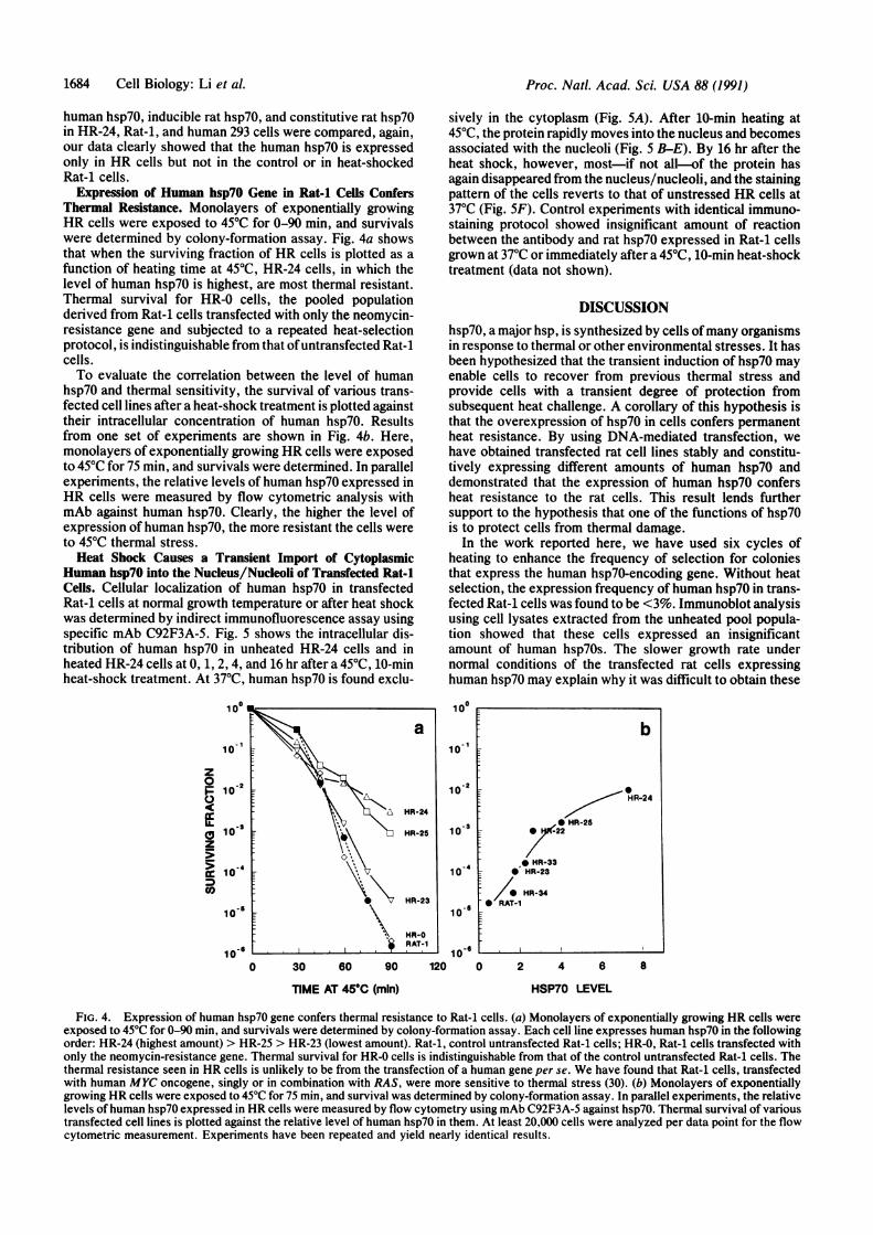
Expression of Human hsp7O Gene in Rat-i Cells ConfersThermal Resistance. Monolayers of exponentially growingHR cells were exposed to 450C for 0-90 min, and survivalswere determined by colony-formation assay. Fig. 4a showsthat when the surviving fraction of HR cells is plotted as afunction of heating time at 45TC, HR-24 cells, in which thelevel of human hsp70 is highest, are most thermal resistant.Thermal survival for HR-0 cells, the pooled populationderived from Rat-1 cells transfected with only the neomycin-resistance gene and subjected to a repeated heat-selectionprotocol, is indistinguishable from that ofuntransfected Rat-icells.To evaluate the correlation between the level of human
hsp70 and thermal sensitivity, the survival of various trans-fected cell lines after a heat-shock treatment is plotted againsttheir intracellular concentration of human hsp70. Resultsfrom one set of experiments are shown in Fig. 4b. Here,monolayers of exponentially growing HR cells were exposedto 450C for 75 min, and survivals were determined. In parallelexperiments, the relative levels of human hsp70 expressed inHR cells were measured by flow cytometric analysis withmAb against human hsp70. Clearly, the higher the level ofexpression ofhuman hsp70, the more resistant the cells wereto 450C thermal stress.Heat Shock Causes a Transient Import of Cytoplasmic
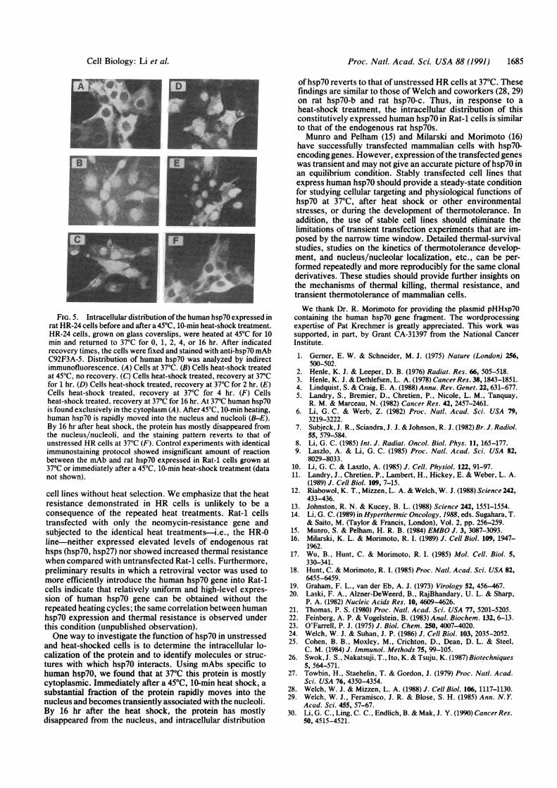
Human hsp7O into the Nucleus/Nucleoli of Transfected Rat-1Cells. Cellular localization of human hsp70 in transfectedRat-1 cells at normal growth temperature or after heat shockwas determined by indirect immunofluorescence assay usingspecific mAb C92F3A-5. Fig. 5 shows the intracellular dis-tribution of human hsp70 in unheated HR-24 cells and inheated HR-24 cells at 0, 1, 2, 4, and 16 hr after a 450C, 10-minheat-shock treatment. At 370C, human hsp70 is found exclu-
z
0
FL
CD
sively in the cytoplasm (Fig. 5A). After 10-min heating at45TC, the protein rapidly moves into the nucleus and becomesassociated with the nucleoli (Fig. 5 B-E). By 16 hr after theheat shock, however, most-if not all-of the protein hasagain disappeared from the nucleus/nucleoli, and the stainingpattern of the cells reverts to that of unstressed HR cells at370C (Fig. SF). Control experiments with identical immuno-staining protocol showed insignificant amount of reactionbetween the antibody and rat hsp70 expressed in Rat-1 cellsgrown at 370C or immediately after a 450C, 10-min heat-shocktreatment (data not shown).
DISCUSSIONhsp70, a major hsp, is synthesized by cells ofmany organismsin response to thermal or other environmental stresses. It hasbeen hypothesized that the transient induction of hsp70 mayenable cells to recover from previous thermal stress andprovide cells with a transient degree of protection fromsubsequent heat challenge. A corollary of this hypothesis isthat the overexpression of hsp70 in cells confers permanentheat resistance. By using DNA-mediated transfection, wehave obtained transfected rat cell lines stably and constitu-tively expressing different amounts of human hsp70 anddemonstrated that the expression of human hsp70 confersheat resistance to the rat cells. This result lends furthersupport to the hypothesis that one of the functions of hsp70is to protect cells from thermal damage.
In the work reported here, we have used six cycles ofheating to enhance the frequency of selection for coloniesthat express the human hsp70-encoding gene. Without heatselection, the expression frequency of human hsp70 in trans-fected Rat-1 cells was found to be <3%. Immunoblot analysisusing cell lysates extracted from the unheated pool popula-tion showed that these cells expressed an insignificantamount of human hsp70s. The slower growth rate undernormal conditions of the transfected rat cells expressinghuman hsp70 may explain why it was difficult to obtain these
100
101
10-2
103
1 0 - 4
10-
10 6
0 30 so 90 120 0 2 4 6 8
TIME AT 45°C (min) HSP70 LEVEL
FIG. 4. Expression of human hsp70 gene confers thermal resistance to Rat-1 cells. (a) Monolayers of exponentially growing HR cells wereexposed to 45°C for 0-90 min, and survivals were determined by colony-formation assay. Each cell line expresses human hsp70 in the followingorder: HR-24 (highest amount) > HR-25 > HR-23 (lowest amount). Rat-1, control untransfected Rat-1 cells; HR-0, Rat-i cells transfected withonly the neomycin-resistance gene. Thermal survival for HR-0 cells is indistinguishable from that of the control untransfected Rat-1 cells. Thethermal resistance seen in HR cells is unlikely to be from the transfection of a human gene per se. We have found that Rat-i cells, transfectedwith human MYC oncogene, singly or in combination with RAS, were more sensitive to thermal stress (30). (b) Monolayers of exponentiallygrowing HR cells were exposed to 45°C for 75 min, and survival was determined by colony-formation assay. In parallel experiments, the relativelevels of human hsp70 expressed in HR cells were measured by flow cytometry using mAb C92F3A-5 against hsp70. Thermal survival of varioustransfected cell lines is plotted against the relative level of human hsp70 in them. At least 20,000 cells were analyzed per data point for the flowcytometric measurement. Experiments have been repeated and yield nearly identical results.
b
HR-24
* HR-25.- 0 -22
* HR-330 HR-23
- HR-34-* RAT-1
1684 Cell Biology: Li et al.
Proc. Natl. Acad. Sci. USA 88 (1991) 1685
I
I
FIG. 5. Intracellular distribution of the human hsp70 expressed inrat HR-24 cells before and after a 45TC, 10-min heat-shock treatment.HR-24 cells, grown on glass coverslips, were heated at 450C for 10min and returned to 370C for 0, 1, 2, 4, or 16 hr. After indicatedrecovery times, the cells were fixed and stained with anti-hsp70 mAbC92F3A-5. Distribution of human hsp70 was analyzed by indirectimmunofluorescence. (A) Cells at 370C. (B) Cells heat-shock treatedat 450C, no recovery. (C) Cells heat-shock treated, recovery at 370Cfor 1 hr. (D) Cells heat-shock treated, recovery at 370C for 2 hr. (E)Cells heat-shock treated, recovery at 370C for 4 hr. (F) Cellsheat-shock treated, recovery at 370C for 16 hr. At 370C human hsp70is found exclusively in the cytoplasm (A). After 45TC, 10-min heating,human hsp70 is rapidly moved into the nucleus and nucleoli (B-E).By 16 hr after heat shock, the protein has mostly disappeared fromthe nucleus/nucleoli, and the staining pattern reverts to that ofunstressed HR cells at 370C (F). Control experiments with identicalimmunostaining protocol showed insignificant amount of reactionbetween the mAb and rat hsp70 expressed in Rat-1 cells grown at37°C or immediately after a 45°C, 10-min heat-shock treatment (datanot shown).
cell lines without heat selection. We emphasize that the heatresistance demonstrated in HR cells is unlikely to be a
consequence of the repeated heat treatments. Rat-1 cellstransfected with only the neomycin-resistance gene andsubjected to the identical heat treatments-i.e., the HR-0line-neither expressed elevated levels of endogenous rathsps (hsp7O, hsp27) nor showed increased thermal resistancewhen compared with untransfected Rat-1 cells. Furthermore,preliminary results in which a retroviral vector was used tomore efficiently introduce the human hsp7O gene into Rat-1cells indicate that relatively uniform and high-level expres-sion of human hsp7O gene can be obtained without therepeated heating cycles; the same correlation between humanhsp7O expression and thermal resistance is observed underthis condition (unpublished observation).One way to investigate the function of hsp7O in unstressed
and heat-shocked cells is to determine the intracellular lo-calization of the protein and to identify molecules or struc-tures with which hsp7O interacts. Using mAbs specific tohuman hsp7O, we found that at 37°C this protein is mostlycytoplasmic. Immediately after a 45°C, 10-min heat shock, asubstantial fraction of the protein rapidly moves into thenucleus and becomes transiently associated with the nucleoli.By 16 hr after the heat shock, the protein has mostlydisappeared from the nucleus, and intracellular distribution
ofhsp7O reverts to that of unstressed HR cells at 37TC. Thesefindings are similar to those of Welch and coworkers (28, 29)on rat hsp7O-b and rat hsp7O-c. Thus, in response to aheat-shock treatment, the intracellular distribution of thisconstitutively expressed human hsp7O in Rat-1 cells is similarto that of the endogenous rat hsp7Os.Munro and Pelham (15) and Milarski and Morimoto (16)
have successfully transfected mammalian cells with hsp7O-encoding genes. However, expression ofthe transfected geneswas transient and may not give an accurate picture ofhsp7O inan equilibrium condition. Stably transfected cell lines thatexpress human hsp7O should provide a steady-state conditionfor studying cellular targeting and physiological functions ofhsp7O at 37TC, after heat shock or other environmentalstresses, or during the development of thermotolerance. Inaddition, the use of stable cell lines should eliminate thelimitations of transient transfection experiments that are im-posed by the narrow time window. Detailed thermal-survivalstudies, studies on the kinetics of thermotolerance develop-ment, and nucleus/nucleolar localization, etc., can be per-formed repeatedly and more reproducibly for the same clonalderivatives. These studies should provide further insights onthe mechanisms of thermal killing, thermal resistance, andtransient thermotolerance of mammalian cells.We thank Dr. R. Morimoto for providing the plasmid pHHsp7O
containing the human hsp7o gene fragment. The wordprocessingexpertise of Pat Krechmer is greatly appreciated. This work wassupported, in part, by Grant CA-31397 from the National CancerInstitute.
1. Gerner, E. W. & Schneider, M. J. (1975) Nature (London) 256,500-502.
2. Henle, K. J. & Leeper, D. B. (1976) Radiat. Res. 66, 505-518.3. Henle, K. J. & Dethlefsen, L. A. (1978) CancerRes. 38, 1843-1851.4. Lindquist, S. & Craig, E. A. (1988) Annu. Rev. Genet. 22, 631-677.5. Landry, S., Bremier, D., Chretien, P., Nicole, L. M., Tanquay,
R. M. & Marceau, N. (1982) Cancer Res. 42, 2457-2461.6. Li, G. C. & Werb, Z. (1982) Proc. Nati. Acad. Sci. USA 79,
3219-3222.7. Subjeck, J. R., Sciandra, J. J. & Johnson, R. J. (1982) Br. J. Radiol.
55, 579-584.8. Li, G. C. (1985) Int. J. Radiat. Oncol. Biol. Phys. 11, 165-177.9. Laszlo, A. & Li, G. C. (1985) Proc. Natl. Acad. Sci. USA 82,
8029-8033.10. Li, G. C. & Laszlo, A. (1985) J. Cell. Physiol. 122, 91-97.11. Landry, J., Chretien, P., Lambert, H., Hickey, E. & Weber, L. A.
(1989) J. Cell Biol. 109, 7-15.12. Riabowol, K. T., Mizzen, L. A. & Welch, W. J. (1988) Science 242,
433-436.13. Johnston, R. N. & Kucey, B. L. (1988) Science 242, 1551-1554.14. Li, G. C. (1989) in Hyperthermic Oncology, 1988, eds. Sugahara, T.
& Saito, M. (Taylor & Francis, London), Vol. 2, pp. 256-259.15. Munro, S. & Pelham, H. R. B. (1984) EMBO J. 3, 3087-3093.16. Milarski, K. L. & Morimoto, R. I. (1989) J. Cell Biol. 109, 1947-
1962.17. Wu, B., Hunt, C. & Morimoto, R. I. (1985) Mol. Cell. Biol. 5,
330-341.18. Hunt, C. & Morimoto, R. I. (1985) Proc. Natl. Acad. Sci. USA 82,
6455-6459.19. Graham, F. L., van der Eb, A. J. (1973) Virology 52, 456-467.20. Laski, F. A., Alzner-DeWeerd, B., RajBhandary, U. L. & Sharp,
P. A. (1982) Nucleic Acids Res. 10, 4609-4626.21. Thomas, P. S. (1980) Proc. Natl. Acad. Sci. USA 77, 5201-5205.22. Feinberg, A. P. & Vogelstein, B. (1983) Anal. Biochem. 132, 6-13.23. O'Farrell, P. J. (1975) J. Biol. Chem. 250, 4007-4020.24. Welch, W. J. & Suhan, J. P. (1986) J. Cell Biol. 103, 2035-2052.25. Cohen, B. B., Moxley, M., Crichton, D., Dean, D. L. & Steel,
C. M. (1984) J. Immunol. Methods 75, 99-105.26. Swok, J. S., Nakatsuji, T., Ito, K. & Tsuju, K. (1987) Biotechniques
5, 564-571.27. Towbin, H., Staehelin, T. & Gordon, J. (1979) Proc. Natl. Acad.
Sci. USA 76, 4350-4354.28. Welch, W. J. & Mizzen, L. A. (1988) J. Cell Biol. 106, 1117-1130.29. Welch, W. J., Feramisco, J. R. & Blose, S. H. (1985) Ann. N.Y.
Acad. Sci. 455, 57-67.30. Li, G. C., Ling, C. C., Endlich, B. & Mak, J. Y. (1990) CancerRes.
50, 4515-4521.
Cell Biology: Li et al.