Therapy of AML · treatment of AML: supportive care only, standard ther-apy, and investigational...
20
Contents 1.1 Introduction .................... 1 1.2 Standard Therapy ................ 2 1.2.1 “3+7” ..................... 2 1.2.2 Outcome Following 3+7 ....... 2 1.2.3 Predictors of TRD ............ 2 1.2.4 Cytogenetics as the Principal Predictor of Resistance in AML . . . 3 1.2.5 Effect of Higher Doses of Ara-C .. 3 1.2.6 Beyond Cytogenetics .......... 4 1.2.7 Effect of Allogeneic Stem Cell Transplant (Allo SCT) .......... 5 1.2.8 Effect of Autologous Stem Cell Transplant (Auto SCT) ......... 6 1.2.9 Effect of Colony Stimulating Factors (CSFs) ............... 6 1.2.10 Candidates for “Standard” Therapy 7 1.2.10.1 Younger Patients (Age < 60) with CBF AML . 7 1.2.11 Candidates for Investigational Therapy ................... 8 1.2.11.1 Elderly Patients (Age ³ 60–65) ......... 8 1.2.11.2 Younger Patients (Age < 60) with Chromo- some Abnormalities Other Than inv (16) or t(8;21) .. 9 1.2.12 Candidates for Either Standard or Investigational Therapy ...... 10 1.2.12.1 Younger Patients with a Normal Karyotype ...... 10 1.2.13 When Should Therapy Start? .... 10 1.2.14 Patients for Whom Investigational Therapy is Unavailable ........ 11 1.2.15 Investigational Therapies ...... 11 1.2.15.1 Not Involving SCT ..... 11 1.2.15.2 RIC SCT ............ 13 1.2.15.3 SCT ............... 14 1.2.16 New Response Criteria ........ 14 1.2.17 Therapy for Relapsed or Refractory AML ........... 14 1.2.18 Treatment of Minimal Residual Disease (MRD) .............. 14 1.2.19 Clinical Issues .............. 15 1.2.20 New Approaches to Clinical Trials in AML [118] ............... 15 References ......................... 16 1.1 Introduction As with any disease, there are three general options for treatment of AML: supportive care only, standard ther- apy, and investigational therapy. Although, as discussed below, there are instances where the first option is pref- erable, the natural history of AML typically mitigates against it [1]. Since by definition there is much more in- formation available about standard than about investi- gational therapy, most patients prefer the former, pro- vided outcome with it is satisfactory. Hence, this review will begin by describing standard therapy, with empha- sis on the factors that predict success following its use. Subsequent discussion will focus on investigational op- tions of potential use for patients in whom results with standard therapy are poor. Therapy of AML Elihu Estey
Transcript of Therapy of AML · treatment of AML: supportive care only, standard ther-apy, and investigational...

Contents
1.1 Introduction . . . . . . . . . . . . . . . . . . . . 1
1.2 Standard Therapy . . . . . . . . . . . . . . . . 21.2.1 “3+7” . . . . . . . . . . . . . . . . . . . . . 21.2.2 Outcome Following 3+7 . . . . . . . 21.2.3 Predictors of TRD . . . . . . . . . . . . 21.2.4 Cytogenetics as the Principal
Predictor of Resistance in AML . . . 31.2.5 Effect of Higher Doses of Ara-C . . 31.2.6 Beyond Cytogenetics . . . . . . . . . . 41.2.7 Effect of Allogeneic Stem Cell
Transplant (Allo SCT) . . . . . . . . . . 51.2.8 Effect of Autologous Stem Cell
Transplant (Auto SCT) . . . . . . . . . 61.2.9 Effect of Colony Stimulating
Factors (CSFs) . . . . . . . . . . . . . . . 61.2.10 Candidates for “Standard” Therapy 7
1.2.10.1 Younger Patients(Age < 60) with CBF AML . 7
1.2.11 Candidates for InvestigationalTherapy . . . . . . . . . . . . . . . . . . . 81.2.11.1 Elderly Patients
(Age � 60–65) . . . . . . . . . 81.2.11.2 Younger Patients
(Age < 60) with Chromo-some Abnormalities OtherThan inv (16) or t(8;21) . . 9
1.2.12 Candidates for Either Standardor Investigational Therapy . . . . . . 101.2.12.1 Younger Patients with a
Normal Karyotype . . . . . . 101.2.13 When Should Therapy Start? . . . . 10
1.2.14 Patients for Whom InvestigationalTherapy is Unavailable . . . . . . . . 11
1.2.15 Investigational Therapies . . . . . . 111.2.15.1 Not Involving SCT . . . . . 111.2.15.2 RIC SCT . . . . . . . . . . . . 131.2.15.3 SCT . . . . . . . . . . . . . . . 14
1.2.16 New Response Criteria . . . . . . . . 141.2.17 Therapy for Relapsed
or Refractory AML . . . . . . . . . . . 141.2.18 Treatment of Minimal Residual
Disease (MRD) . . . . . . . . . . . . . . 141.2.19 Clinical Issues . . . . . . . . . . . . . . 151.2.20 New Approaches to Clinical Trials
in AML [118] . . . . . . . . . . . . . . . 15
References . . . . . . . . . . . . . . . . . . . . . . . . . 16
1.1 Introduction
As with any disease, there are three general options fortreatment of AML: supportive care only, standard ther-apy, and investigational therapy. Although, as discussedbelow, there are instances where the first option is pref-erable, the natural history of AML typically mitigatesagainst it [1]. Since by definition there is much more in-formation available about standard than about investi-gational therapy, most patients prefer the former, pro-vided outcome with it is satisfactory. Hence, this reviewwill begin by describing standard therapy, with empha-sis on the factors that predict success following its use.Subsequent discussion will focus on investigational op-tions of potential use for patients in whom results withstandard therapy are poor.
Therapy of AML
Elihu Estey

The criteria for a diagnosis of AML have changedwith publication of a report sponsored by the WHO[2]. Previous criteria were those of the FAB group andrequired a minimum of � 30% blasts [3]. The WHOhas lowered this to � 20% blasts, in the process eliminat-ing the myelodysplastic syndrome refractory anemiawith excess blasts in transformation (RAEB-t). Al-though data mentioned below suggest that blast countsbetween 10–100% are not themselves independent pre-dictors of outcome, we will adhere to the WHO criterionin the material that follows.
1.2 Standard Therapy
1.2.1 “3+7”
Standard therapy consists of “induction” and “postre-mission” phases. The intent of the former is to produce,and of the latter to prolong, a complete remission (CR)defined as a marrow with < 5% blasts and peripheralblood with > 1 000 neutrophils and > 100 000 platelets.The importance of CR relates to its ability to prolongsurvival. Thus, 40 years ago, Freireich et al. [1] docu-mented that patients who achieved CR lived longer thanthose who did not. The difference in survival time wasentirely due to the time spent in CR, suggesting that thisdifference resulted from achievement of CR rather thanfrom a superior natural history. The risk of relapse fromCR is constant for the first 2 years, but once patientshave been in CR for 3 years it declines precipitously(to < 10%), allowing such patients to be considered po-tentially cured [4].
For 30 years most patients with AML who have beentreated have received remission induction therapy withwhat is commonly called “3+7.” The “3” refers to the 3days on which patients receive an agent (most com-monly an anthracycline such as daunorubicin or idaru-bicin) that affects topoisomerase II and the “7” to the 7days of cytosine arabinoside (ara-C) that accompanyand follow the anthracycline. If blasts remain in a mar-row aspirated 14 days after beginning therapy (day 14), asecond course is often administered, with the number ofdays of anthracycline reduced to 2 and of ara-C to 5. Ifthe day-14 marrow contains very few blasts, the marrowis reaspirated weekly until response (CR or reappear-ance of blasts) becomes clear.
Upon documentation of CR, patients frequently re-ceive additional courses of anthracycline + ara-C, with
a reduction in the doses or in the number of doses.While some such therapy is almost certainly necessarytoday, the proper amount likely depends on the intensityof the first several courses. For example, a German AMLCooperative Group (AMLCG) trial randomly assigningpatients who had received 1 post-CR course to nofurther therapy or 3 years of maintenance found thatthe latter prolonged relapse-free survival time (RFS)from 7% to 30% [5]. However, a subsequent randomizedAMLCG trial found much less improvement in RFS(28% vs. 35% at 3 years) and no difference in survivalwhen patients in the no-further-therapy group receiveda more intense induction regimen and one intense post-remission course [6]. Regarding the specific number ofpostremission courses to administer following a firstcourse of 3+7, the British NCRI (formerly MRC) groupfound no difference between 4 and 7 courses [7].
1.2.2 Outcome Following 3+7
CR, survival, event-free survival, and relapse-free sur-vival rates are very variable after administration of3+7; substantial numbers of patients die within a fewweeks of beginning therapy and substantial numbersare potentially cured. Thus, speaking of an average out-come is not particularly informative. As with all anti-AML therapy, two general types of variables are asso-ciated with outcome: those that predict treatment-re-lated death before response to induction therapy canbe evaluated (“TRD”) and those that predict true resis-tance to therapy. The criterion for “early death” is some-what arbitrary. Very few patients achieve CR before day21. Thus, deaths before day 21 are true early deaths re-sulting from supportive care failure. However, half thepatients who will achieve CR have done so by day 35. Ac-cordingly, failure in patients who die between days 21and 35 and who have not achieved CR is due to both fail-ure of supportive care and resistance. Beyond day 35, re-sistance to therapy becomes increasingly responsible forfailure to enter CR. In CR, treatment-related mortality israre (5–10%) in contrast to relapse from CR (50–100%).
1.2.3 Predictors of TRD
The principal predictor of TRD is pretreatment perfor-mance status. Table 1.1 illustrates that the proportion ofpatients who are bed-ridden most (performance sta-
2 Chapter 1 · Therapy of AML
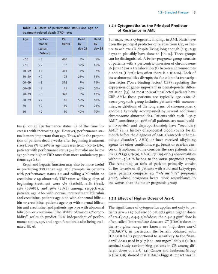
tus 3), or all (performance status 4) of the time in-creases with increasing age. However, performance sta-tus is more important than age. Thus, while the propor-tion of patients dead 5 weeks after beginning treatmentrises from 5% to 26% as age increases from < 50 to � 80,patients with performance status 3–4 but who are belowage 50 have higher TRD rates than more ambulatory pa-tients age � 80.
Renal and hepatic function may also be more usefulin predicting TRD than age. For example, in patientswith performance status < 2 and calling a bilirubin orcreatinine > 1.9 abnormal, TRD rates within 35 days ofbeginning treatment were 5% (43/808), 21% (7/34),13% (91/688), and 36% (21/58) among, respectively,patients age < 60 with normal pretreatment bilirubinand creatinine, patients age < 60 with abnormal biliru-bin or creatinine, patients age > 59 with normal biliru-bin and creatinine, and patients age > 59 with abnormalbilirubin or creatinine. The ability of various “comor-bidity” scales to predict TRD independent of perfor-mance status, age, and organ function is also being eval-uated [8, 9].
1.2.4 Cytogenetics as the Principal Predictorof Resistance in AML
For many years cytogenetic findings in AML blasts havebeen the principal predictor of relapse from CR, or fail-ure to achieve CR despite living long enough (e. g., > 35days) to plausibly have done so [10–12]. Three groupscan be distinguished. A better-prognosis group consistsof patients with a pericentric inversion of chromosome16 [inv 16] or a translocation (t) between chromosomes8 and 21 (t 8;21); less often there is a t(16;16). Each ofthese abnormalities disrupts the function of a transcrip-tion factor (“core binding factor,” CBF) regulating theexpression of genes important in hematopoietic differ-entiation [13]. At most 10% of unselected patients haveCBF AML; these patients are typically age < 60. Aworse-prognosis group includes patients with monoso-mies, or deletions of the long arms, of chromosomes 5and/or 7 typically accompanied by several additionalchromosome abnormalities. Patients with such “–5/–7AML” constitute 30–40% of all patients, are usually old-er (> 50–60), and disproportionately have “secondaryAML,” i.e., a history of abnormal blood counts for � 1month before the diagnosis of AML (“antecedent hema-tologic disorder”, AHD) or have received alkylatingagents for other conditions, e. g., breast or ovarian can-cer or lymphoma. Some consider the rare patients withinv (3)/t (3;3), t(6;9), t(6;11), t(11;19) or > 3 abnormalitieswithout –5/–7 to belong to the worse prognosis group.The remaining 50–60% of patients primarily consistof the 35–40% of all patients with a normal karyotype;these patients comprise an “intermediate” prognosisgroup, whose prognosis bears more resemblance tothe worse- than the better-prognosis group.
1.2.5 Effect of Higher Doses of Ara-C
The significance of cytogenetics applies not only to pa-tients given 3+7 but also to patients given higher dosesof ara-C, e. g., 0.4–3 g/m2/dose; the 0.4–1.5 g/m2 dose isoften called “intermediate-dose ara-C” (IDAC); doses inthe 2–3 g/m2 range are known as “high-dose ara-C(“HDAC)”); in particular, the benefit obtained withIDAC/HDAC is proportional to sensitivity to the “stan-dard” doses used in 3+7 (100–200 mg/m2 daily � 7). In aseminal study randomizing patients in CR among dif-ferent doses of ara-C [14], Cancer and Leukemia GroupB (CALGB) showed that HDAC’s biggest impact was in
a 1.2 · Standard Therapy 3
Table 1.1. Effect of performance status and age on
treatment-related death (TRD) rates
Age Perfor-
mance
status
(Zubrod)
Pa-
tients
Dead
by
day 21
Dead
by
day 35
< 50 < 3 490 3% 5%
< 50 > 2 37 32% 46%
50–59 < 3 361 4% 7%
50–59 > 2 28 25% 38%
60–69 < 3 372 7% 11%
60–69 > 2 45 43% 50%
70–79 < 3 328 8% 17%
70–79 > 2 46 52% 68%
80 < 2 60 16% 26%
80 > 2 10 40% 70%
CBF AML where it produced average cure rates in excessof 50%. In the normal karyotype group, IDAC andHDAC were equivalent, with each superior to standarddoses, i.e., those in 3+7. In the worse-prognosis groupany differences among HDAC, IDAC, and standarddoses were small relative to the poor outcome observedwith all three doses. NCRI data suggest that similar re-sults can be obtained in CBF AML with IDAC as withHDAC [10], leading to an NCRI trial randomizing be-tween these 2 doses.
1.2.6 Beyond Cytogenetics
Although cytogenetic findings remain the most impor-tant prognostic factor in AML, there is considerablevariability in outcome particularly within the inter-mediate and favorable groups. The presence of (a) sec-ondary AML, (b) “white blood cell index,” (c) “second-ary” chromosome abnormalities superimposed on theprimary abnormalities noted above, and (d) molecularabnormalities such as gene mutations and deregulatedgene expression are useful in unravelling this heteroge-neity. The poorer outcome in secondary rather than inde novo AML is well known and appears independentof the association between secondary AML and worse-prognosis cytogenetics [15]. Nguyen et al. for the FrenchAML Intergroup found that relapse-free survival in pa-tients with t (8;21) given IDAC (or an allogeneic trans-plant) varied as a function of a “white blood cell index”defined as [WBC � % marrow blasts]/100 [16]. Long-term RFS was > 75% with an index < 2.5, 60% with anindex 2.5–20, and 30% with an index > 20. In general,the presence of secondary chromosome abnormalitieshas little affect on prognosis. However, the GermanAML Intergroup and Cancer and Leukemia Group B(CALGB) have shown that trisomy 22 improves re-lapse-free survival in inv [16] AML [17, 18], while theGerman group has also shown that a missing Y chromo-some is associated with shorter survival t(8;21) [17]. Ofmore general interest, mutations in receptor tyrosine ki-nases (RTK), such as KIT, and in RAS genes have beenfound in 25% of cases of inv 16 AML and in 10% of casesof t(8;21) AML; KIT mutations appear associated with aninferior prognosis [19–22].
Given its frequency, the normal karyotype group isthe one in which prognostic heterogeneity is most prob-lematic. Such patients often have molecular abnormali-ties involving FLT3, NPM1, CEBPA, MLL, RAS, BAALC,
or EVI,1. Internal tandem duplications (ITD) withinthe juxtamembrane domain of the RTK FLT3 occur in28–34% of patients with normal karyotype AML andare consistently associated with a significantly inferioroutcome [23–27]. An additional 10–15% of these pa-tients have mutations within the activation loop of thesecond tyrosine kinase domain (TKD) [25, 26, 28, 29].A recent meta-analysis suggests that FLT3 TKD muta-tions also negatively affect RFS, although the BritishNCRI group has recently reported a favorable effect[30, 31]. The most common somatic gene alterations inAML are mutations in the nucleophosmin (NPM1)gene, resulting in cytoplasmic rather than nuclear lo-calization of the NPM1 protein. NPM1 mutations havebeen reported in 48–64% of normal karyotype AML[32–36]. Recent studies have found that overall surviv-al (OS) and relapse-free survival (RFS) are signifi-cantly better in NPM1+/FLT3 ITD– patients contrastedwith NPM1– and NPM1+/FLT3 ITD + patients [32–36].NRAS/KRAS mutations occur in approximately 18% ofnormal karyotype AML [37]. Although no consistentprognostic effect has yet been shown, there may besuch an effect after accounting for mutations in othergenes, such as dominant negative mutations in thetranscription factor CEBPA and partial tandem dupli-cations (PTD) in the MLL1 gene, which occur in 15–18% and 8–11% of normal karyotype cases, respec-tively. CEBPA mutations are associated with superiorOS and RFS [38–40], while MLL1 mutations predictfor inferior RFS without significant effect on OS [41–44]. A significant negative prognostic effect on thesetwo outcomes has also been reported in cases withaberrant overexpression of BAALC, a gene that is phy-siologically expressed in brain tissue and in hemato-poietic progenitor cells [45, 46].
Genome-wide gene expression profiling based onDNA microarrays has provided additional prognosticinformation [47–49]. In particular, hierarchical cluster-ing has identified two normal karyotype-predominantclasses that differed in OS, and a gene expression pre-dictor emerged as the strongest prognostic factor inmultivariate analysis. These findings have been vali-dated prospectively in an independent data set [50].
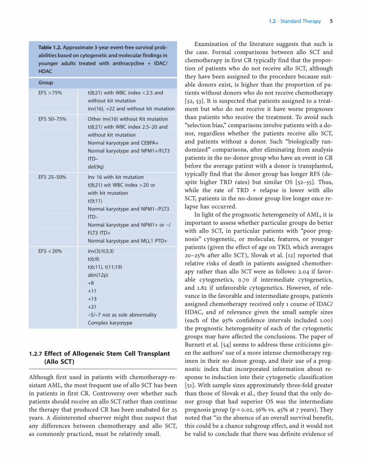
Table 1.2, based on outcome in younger adults givenanthracycline + IDAC/HDAC, provides a prognostic sys-tem combining genetic and cytogenetic information.The value of cytogenetics in predicting RFS can alsobe enhanced by incorporating information regardingresponse to induction therapy [51].
4 Chapter 1 · Therapy of AML
1.2.7 Effect of Allogeneic Stem Cell Transplant(Allo SCT)
Although first used in patients with chemotherapy-re-sistant AML, the most frequent use of allo SCT has beenin patients in first CR. Controversy over whether suchpatients should receive an allo SCT rather than continuethe therapy that produced CR has been unabated for 25years. A disinterested observer might thus suspect thatany differences between chemotherapy and allo SCT,as commonly practiced, must be relatively small.
Examination of the literature suggests that such isthe case. Formal comparisons between allo SCT andchemotherapy in first CR typically find that the propor-tion of patients who do not receive allo SCT, althoughthey have been assigned to the procedure because suit-able donors exist, is higher than the proportion of pa-tients without donors who do not receive chemotherapy[52, 53]. It is suspected that patients assigned to a treat-ment but who do not receive it have worse prognosesthan patients who receive the treatment. To avoid such“selection bias,” comparisons involve patients with a do-nor, regardless whether the patients receive allo SCT,and patients without a donor. Such “biologically ran-domized” comparisons, after eliminating from analysispatients in the no-donor group who have an event in CRbefore the average patient with a donor is transplanted,typically find that the donor group has longer RFS (de-spite higher TRD rates) but similar OS [52–55]. Thus,while the rate of TRD + relapse is lower with alloSCT, patients in the no-donor group live longer once re-lapse has occurred.
In light of the prognostic heterogeneity of AML, it isimportant to assess whether particular groups do betterwith allo SCT, in particular patients with “poor prog-nosis” cytogenetic, or molecular, features, or youngerpatients (given the effect of age on TRD, which averages20–25% after allo SCT), Slovak et al. [12] reported thatrelative risks of death in patients assigned chemother-apy rather than allo SCT were as follows: 2.04 if favor-able cytogenetics, 0.70 if intermediate cytogenetics,and 1.82 if unfavorable cytogenetics. However, of rele-vance in the favorable and intermediate groups, patientsassigned chemotherapy received only 1 course of IDAC/HDAC, and of relevance given the small sample sizes(each of the 95% confidence intervals included 1.00)the prognostic heterogeneity of each of the cytogeneticgroups may have affected the conclusions. The paper ofBurnett et al. [54] seems to address these criticisms giv-en the authors’ use of a more intense chemotherapy reg-imen in their no donor group, and their use of a prog-nostic index that incorporated information about re-sponse to induction into their cytogenetic classification[51]. With sample sizes approximately three-fold greaterthan those of Slovak et al., they found that the only do-nor group that had superior OS was the intermediateprognosis group (p = 0.02, 56% vs. 45% at 7 years). Theynoted that “in the absence of an overall survival benefit,this could be a chance subgroup effect, and it would notbe valid to conclude that there was definite evidence of
a 1.2 · Standard Therapy 5
Table 1.2. Approximate 3-year event-free survival prob-
abilities based on cytogenetic and molecular findings in
younger adults treated with anthracycline + IDAC/
HDAC
Group
EFS > 75% t(8;21) with WBC index < 2.5 and
without kit mutation
inv(16), +22 and without kit mutation
EFS 50–75% Other inv(16) without Kit mutation
t(8;21) with WBC index 2.5–20 and
without kit mutation
Normal karyotype and CEBPA+
Normal karyotype and NPM1+/FLT3
ITD–
del(9q)
EFS 25–50% Inv 16 with kit mutation
t(8;21) wit WBC index > 20 or
with kit mutation
t(9;11)
Normal karyotype and NPM1–/FLT3
ITD–
Normal karyotype and NPM1+ or –/
FLT3 ITD+
Normal karyotype and MLL1 PTD+
EFS < 20% inv(3)/t(3;3)
t(6;9)
t(6;11), t(11;19)
abn(12p)
+8
+11
+13
+21
–5/–7 not as sole abnormality
Complex karyotype

benefit.” A donor–no donor comparison by Jourdan etal. [55] also found that only the intermediate prognosisdonor group had superior OS (p = 0.02, 55% vs. 70% at 7years), although the same comment about subgroup ef-fect can be made, in addition to which only 79% in theno-donor group received � 1 course of IDAC/HDAC, andthe authors’ prognostic index incorporated WBC countand FAB category, neither commonly recognized as par-ticularly “prognostic,” in addition to cytogenetics andresponse to the first course of treatment. Two groupshave used a donor–no donor comparison to examinethe effect of allo SCT in patients with poor prognosismolecular abnormalities. The NCRI found equivalentOS and RFS in patients with a FLT3 ITD [56] (68 donor,114 no-donor patients), while examining patients whowere NPM1– or NPM1+/FLT3 ITD + the German-Aus-trian AML group reported longer RFS in the donorgroup (p = 0.001, e. g., 60% vs. 40% at 2 years) [33];OS was not reported.
All the patients in the Jourdan et al. [55] paper wereage < 45, while those in the papers of Slovak et al. [12]and Burnett et al. [54] were age < 56. Only the latter pa-per examined the effect of age, finding no difference inOS among patients aged 0–14, 15–34, or 35–56 accordingto whether they were in the donor or no-donor group.This finding, which contradicts prior belief that patientsless than 20–30 years old should receive an allo SCT infirst CR, resulted from improvements in chemotherapy(e. g., use of IDAC/HDAC). It is not implausible thatsimilar improvements may eventually affect SCT. For ex-ample, TRD rates may be reduced by use of peripheralblood as the stem cell source or by using reduced-doseconditioning regimens (discussed in the section on in-vestigational treatments), thus altering OS in favor ofallo SCT. However, there are currently no data indicat-ing that allo SCT prolongs OS in any patient in firstCR; furthermore, allo SCT typically has more long-termcomplications than chemotherapy. If conventional alloSCT is performed, there seems to be no advantage to ad-minister HDAC or other postremission therapy prior totransplantation.
While the donor–no donor comparison approachhas obvious value, it tends to mask the value of alloSCT in patients who are fit enough to undergo the pro-cedure. Thus, a transplant–no transplant comparisonmight also be of interest. Lending credence to perform-ing this type analysis is the NCRI’s report that patientswith a donor who were not transplanted had similaroutcomes as patients without a donor [54]. Of course,
if the proportion of patients with a donor who receiveallo SCT is sufficiently low, the transplant–no transplantcomparison becomes primarily of academic interest. Inthis regard, it has been shown that even in relativelyyoung patients the impact of allo SCT on the manage-ment of AML is quite small [57, 58].
1.2.8 Effect of Autologous Stem Cell Transplant(Auto SCT)
In several of the studies discussed above, patients in theno-donor group were randomized between chemother-apy and auto SCT in first CR. Comparisons of the lattertwo strategies may be confounded if, as in the NCRItrial, both groups get the same amount of chemother-apy, with the chemotherapy group then stopping treat-ment, but the auto SCT group then proceeding to autoSCT [59]. Typically studies report longer RFS with autoSCT, but have not found longer OS, either on average orin any subgroup [52, 53]. While there is little doubt thatTRD rates with auto SCT will decline thus possibly tilt-ing the OS balance in its favor vis a vis chemotherapy, itis also plausible that this balance might be shifted tofavor chemotherapy if new non-SCT, such as those dis-cussed below, reduce the risk of relapse.
1.2.9 Effect of Colony Stimulating Factors (CSFs)
As with auto SCT, the space devoted to a discussion ofCSFs in a current review of treatment of AML is muchless than would have been the case several years ago, re-flecting a decreased interest in such therapy. CSFs andspecifically G- and GM-CSF have neither decreasedTRD (or serious morbidity) when administered dur-ing/after anthracycline + ara-C [60, 61] nor decreasedresistance (e. g., by “priming” blasts to the effects ofthese drugs) when given before/during such therapy[62–64]. An exception was a trial [65] randomizing640 patients aged 18–60 to receive or not receive G-CSF in conjunction with 2 courses of standard doseara-C, the first with idarubicin and the second with am-sacrine, whose mechanism of action is presumed simi-lar to that of idarubicin. Idarubicin was given on days6–8, rather than days 1–3 as in the usual 3+7 regimen,and amsacrine on days 4–6, rather than 1–3. The authorshypothesized that delayed administration would preventinterference of the “cell-cycle-dependent synergy” be-
6 Chapter 1 · Therapy of AML
tween G-CSF and ara-C. G-CSF did not affect outcomein unfavorable prognosis patients (defined to includethose with secondary AML), but improved RFS in inter-mediate-prognosis patients (as defined by cytogeneticsand de novo AML) at 4 years from 33 +/3% to 45 +/3%. There currently appear to be few large trials at-tempting to confirm this result; until then there is noreason to routinely use G- or GM-CSF. Rather, a 3- to4-day trial of these CSFs might be tried in patientswho have developed serious infections while still neu-tropenic after chemotherapy.
1.2.10 Candidates for “Standard” Therapy
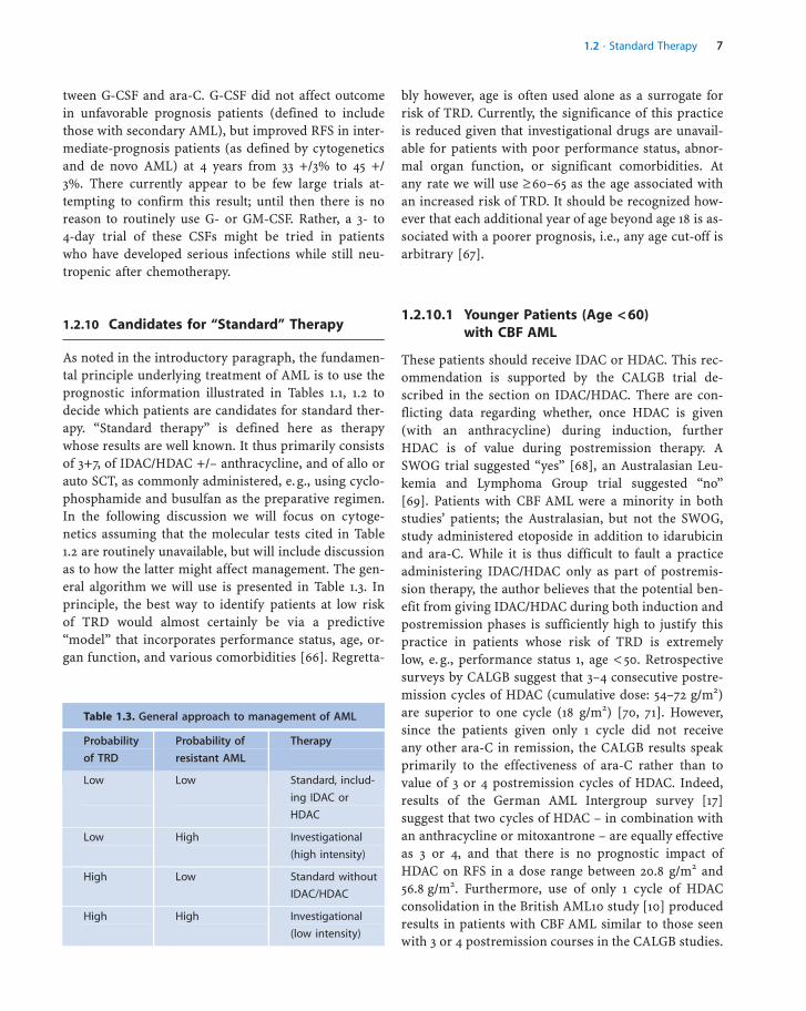
As noted in the introductory paragraph, the fundamen-tal principle underlying treatment of AML is to use theprognostic information illustrated in Tables 1.1, 1.2 todecide which patients are candidates for standard ther-apy. “Standard therapy” is defined here as therapywhose results are well known. It thus primarily consistsof 3+7, of IDAC/HDAC +/– anthracycline, and of allo orauto SCT, as commonly administered, e. g., using cyclo-phosphamide and busulfan as the preparative regimen.In the following discussion we will focus on cytoge-netics assuming that the molecular tests cited in Table1.2 are routinely unavailable, but will include discussionas to how the latter might affect management. The gen-eral algorithm we will use is presented in Table 1.3. Inprinciple, the best way to identify patients at low riskof TRD would almost certainly be via a predictive“model” that incorporates performance status, age, or-gan function, and various comorbidities [66]. Regretta-
bly however, age is often used alone as a surrogate forrisk of TRD. Currently, the significance of this practiceis reduced given that investigational drugs are unavail-able for patients with poor performance status, abnor-mal organ function, or significant comorbidities. Atany rate we will use � 60–65 as the age associated withan increased risk of TRD. It should be recognized how-ever that each additional year of age beyond age 18 is as-sociated with a poorer prognosis, i.e., any age cut-off isarbitrary [67].
1.2.10.1 Younger Patients (Age < 60)with CBF AML
These patients should receive IDAC or HDAC. This rec-ommendation is supported by the CALGB trial de-scribed in the section on IDAC/HDAC. There are con-flicting data regarding whether, once HDAC is given(with an anthracycline) during induction, furtherHDAC is of value during postremission therapy. ASWOG trial suggested “yes” [68], an Australasian Leu-kemia and Lymphoma Group trial suggested “no”[69]. Patients with CBF AML were a minority in bothstudies’ patients; the Australasian, but not the SWOG,study administered etoposide in addition to idarubicinand ara-C. While it is thus difficult to fault a practiceadministering IDAC/HDAC only as part of postremis-sion therapy, the author believes that the potential ben-efit from giving IDAC/HDAC during both induction andpostremission phases is sufficiently high to justify thispractice in patients whose risk of TRD is extremelylow, e. g., performance status 1, age < 50. Retrospectivesurveys by CALGB suggest that 3–4 consecutive postre-mission cycles of HDAC (cumulative dose: 54–72 g/m2)are superior to one cycle (18 g/m2) [70, 71]. However,since the patients given only 1 cycle did not receiveany other ara-C in remission, the CALGB results speakprimarily to the effectiveness of ara-C rather than tovalue of 3 or 4 postremission cycles of HDAC. Indeed,results of the German AML Intergroup survey [17]suggest that two cycles of HDAC – in combination withan anthracycline or mitoxantrone – are equally effectiveas 3 or 4, and that there is no prognostic impact ofHDAC on RFS in a dose range between 20.8 g/m2 and56.8 g/m2. Furthermore, use of only 1 cycle of HDACconsolidation in the British AML10 study [10] producedresults in patients with CBF AML similar to those seenwith 3 or 4 postremission courses in the CALGB studies.
a 1.2 · Standard Therapy 7
Table 1.3. General approach to management of AML
Probability
of TRD
Probability of
resistant AML
Therapy
Low Low Standard, includ-
ing IDAC or
HDAC
Low High Investigational
(high intensity)
High Low Standard without
IDAC/HDAC
High High Investigational
(low intensity)

Nonetheless, for reasons similar to those explaining hispreference for IDAC/HDAC during both induction andpostremission therapy, the author would favor adminis-tration of 3 or 4 postremission cycles. Neither allo SCTnor auto SCT should be used in the average CBF patientin first CR.
CBF patients with KIT mutations and t(8;21) patientswith WBC index > 20 have much less favorable out-comes than other patients with similar cytogenetics[19–22] (Table 1.2). Such “poor-prognosis” CBF patientsmight be candidates for investigational therapy pro-vided large elements of the standard therapy describedabove are retained in deference to the 25–50% successrate even in such poor-prognosis patients. For example,a tyrosine kinase inhibitor might be added in patientswith KIT mutations.
Other candidates for standard therapy are those age< 60 (low risk of TRD) with a normal karyotype whohave either a CEBPA mutation or have an NPM muta-tion and have wild-type FLT3 (50–75% success rate, Ta-ble 1.2). Given the low-risk of TRD, the author favors useof IDAC/HDAC with anthracycline during both induc-tion and postremission phases.
1.2.11 Candidates for Investigational Therapy
1.2.11.1 Elderly Patients (Age � 60–65)
Table 1.4 presents results of trials investigating standardtherapy in elderly patients. The depicted average medi-an survival times of 10 months make it difficult to rec-ommend standard therapy to many such patients. Thisis particularly so given the 15–20% risk of TRD occur-ring during the approximately 1–2 month remission in-duction period (Table 1.4), noting that some of the trialsshown in Table 1.4 limited eligibility to patients withbetter performance status, normal organ function, andno major infection. Reducing the doses/duration of an-thracycline and/or cytarabine has decreased both earlymortality and the anti-AML efficacy of treatment, re-sulting in no improvement in survival [76]. Additionof G-CSF or GM-CSF to an anthracycline + ara-C, sub-stitution of mitoxantrone for an anthracycline, or use ofsingle agent gemtuzumab ozogamycin [77] have alsofailed to lengthen survival (Table 1.4).
It is useful to attempt to identify groups of older pa-tients for whom standard therapy might be reasonable.Two such groups are patients CBF AML and patientswith a normal karyotype, who are age 60–69, have denovo AML, a Zubrod performance status 0–2, normal
8 Chapter 1 · Therapy of AML
Table 1.4. Outcomes in older patients given anthracycline + Ara-C
Study
[reference]
Patients Median
survival
Probability
survival at
2 years
CR
rate
Induction
death rate
Comment
ECOG [72] 234,
age � 56
7–8 months �20% 41% 19% Results same with dauno-
rubicin, idarubicin, or mito-
xantrone and +/– GM-CSF
priming
NCRI (for-
merly MRC),
(AML 11)
[7]
1314,
age � 56
�12 months �25% 62% 16% Results same with 1 or 4
courses post-CR and
+/– G-CSF starting 8 days
after end induction
SWOG [73] 161,
age � 56
9 months 19% 43% 15% Survival worse with mito-
xantrone + etoposide
HOVON
[74]
211,
age � 60
�10 months �25% 48% 15% Results same +/– PSC-833
M. D.
Anderson
[75]
31,
age � 65
� 12 months �20% 48% Not given Ara-C at 1.5 g/m2 daily � 3;
survival worse with single
agent gemtuzumab

values for bilirubin and creatinine, and no pretreatmentinfection. Both groups had a median survival of 18months following various cytarabine-containing regi-mens, as given at M.D. Anderson Hospital (MDA), withonly a 5–10% TRD rate (Fig. 1.1). However, the probabil-ities of being in CR 3 years after initial diagnosis, corre-sponding to the time when patients can be considered“potentially cured” ranged between 9% and 16%. Giventhese data, some patients might prefer standard, andother patients investigational, therapy.
The “favorable” elderly patients described aboveconstituted only 8% of the 968 patients age 60 andabove given induction therapy at MDA over the past de-cade. The others had a median survival, when givencytarabine- or anthracycline-containing therapy, of 6months (Fig. 1.1), with a 6% probability of being inCR at 3 years, and a 25% TRD rate. Thus, I recommendnew therapies for the great majority of older patientswith untreated AML. The National Comprehensive Can-cer Network, a consortium of prominent American can-cer centers explicitly cites “clinical trial” as the preferredoption in patients with untreated AML age 60 and above[78]. There is of course no assurance that results withinvestigational therapy will not be worse than thosewith 3+7. For example, a CALGB study in older patients
was stopped before accrual was completed because pa-tients randomized to the investigational arm (daunoru-bicin + cytarabine + etoposide + PSC-833) had a higherdeath rate than patients randomized to the same 3 drugswithout PSC-833 [79]. While investigational therapymay thus prove worse than standard, it seems fair toask: “how much worse than 3+7 can an investigationaltherapy be.” The weight given to the recommendationfor investigational therapy should increase as the num-ber of unfavorable prognostic factors (age > 69, adversecytogenetics, secondary AML, poor performance status,infection, abnormal organ function) increases (see alsoTable 1.2).
Any discussion of choice of therapy must refer toSekeres et al.’s observations [80] that 74% of older pa-tients estimated that their chances of cure with 3+7 wereat least 50%; in contrast, 85% of their physicians esti-mated this chance to be < 10%. While the most plausiblecause of this discrepancy is patients’ natural tendencyto believe what they want to believe, there may also begaps in communication between physicians and pa-tients.
1.2.11.2 Younger Patients (Age < 60)with Chromosome AbnormalitiesOther Than inv (16) or t(8;21)
Given their median age of 45, these patients would havea very considerable life expectancy if they did not haveAML. Hence potential cure is of more significance forthem than for older patients (age > 60), whose medianage is 70. Figure 1.2 (top) indicates that the probabilityof being in CR 3 years after starting initial IDAC/HDAC-containing treatment (corresponding to our criterionfor potential cure) in the most favorable subset ofyounger patients with –5/–7 (i.e., patients with de novoAML and a performance status < 3) is 10%. Theirmedian event-free survival is 39 weeks (Fig. 1.2, bot-tom). Hence the author believes that investigationaltreatment should be the preferred option in younger pa-tients with –5/–7, recalling that the weight of evidencesuggests that these patients are not materially benefitedby allo or auto SCT, as typically performed. Exceptionsmight be made for the rare patients who have –5/–7 as asingle abnormality [81].
Younger patients with performance status < 3, denovo AML and other cytogenetic abnormalities (exclud-ing inv 16 and t(8;21) have 3-year EFS that while statis-tically different than those in comparable -5/-7 patients
a 1.2 · Standard Therapy 9
Fig. 1.1. Survival probabilities in three groups of elderly patients.Black line denotes patients with inv(16) or t(8;21) (n = 22), gray linedenotes patients with a normal karyotype and de novo AML whowere age 60–69 with a good performance status, normal pretreat-ment organ function, and no pretreatment infection (n = 54), whiledashed line denotes other patients (n = 627). The differences be-tween the first two groups, on the one hand, and the third group, onthe other (p < 0.001), suggest that while standard therapy might beappropriate for the first two groups, it is not for the third group.

are not materially different (e. g., 17% EFS at 3 years; Fig.1.2 top). Hence, despite their median event-free survivalof 74 weeks (Fig. 1.2), I believe that the preferred optionin these patients is also investigational therapy. Ob-viously some would disagree with what is essentially asubjective view and believe that these patients are can-didates for standard as well as investigational treatment.It is noteworthy however that phase 2 studies of newdrugs are routinely performed in untreated patientswith pancreas, or metastatic lung cancer, diseases inwhich median event-free survival is not materially dif-ferent than the 74-week figure noted here.
1.2.12 Candidates for Either Standardor Investigational Therapy
1.2.12.1 Younger Patients witha Normal Karyotype
M. D. Anderson data (326 patients treated since 1990with IDAC/HDAC-containing therapy) indicate thatthese patients have a 3-year event-free survival probabil-ity of 26%. It is not implausible that some patients whengiven this information would choose standard therapy,
assuming correctly that investigational therapy couldbe worse. Certainly given the reasonable success rateobserved with standard therapy, investigational therapyin these patients should be based on standard therapy, aconstraint that is less applicable for the patients de-scribed above in whom investigational therapy is thepreferred option, particularly elderly patients and thoseyoung patients with –5/–7. In light of the relatively lowrisk of TRD, and the CALGB data suggesting the super-iority of IDAC to standard doses of ara-C [14], IDACshould be included in either induction or postremissionphases or both.
Young patients with a normal karyotype are a groupfor whom the molecular information outlined in Table1.2 will be particularly important. For example, the pres-ence of a FLT3 ITD would weigh the choice toward in-vestigational therapy, while an NPM or CEBA mutationwould favor the standard therapy option.
1.2.13 When Should Therapy Start?
At least several days are needed for results of cytoge-netic and molecular studies to become available. How-ever, patients with high (> 100 000) or rapidly risingWBC counts or a somewhat lower count (> 10 000–20 000) and symptoms of lung or brain involvement re-quire immediate institution of definitive therapy, whichshould not be delayed by leukapheresis or use of hydro-xyurea. However, in other patients delaying therapy toawait cytogenetic and molecular results may be feasible.Knowledge of such results prior to beginning inductiontherapy is less important in younger patients since therisk of TRD is low, and investigational therapy couldbe given when results of induction therapy, and of cyto-genetic/molecular tests, are known. However, initiationof investigational therapy only in CR is not optimalsince it is known that the type of therapy used for in-duction has a powerful influence on outcome in CR(see, for example, [82, 83]). Older patients have higherrates of TRD, and hence the desirability of delaying atherapy that is unlikely to be successful (e. g., if the pa-tient has –5/–7) is even greater. Older patients usuallydo not present with features demanding immediatetherapy, making it possible to delay therapy for cytoge-netic results. Among 197 patients presenting to MDAwith WBC < 50 000, multivariate analysis indicated thatage and unfavorable cytogenetics were independent pre-dictors of CR, but that days from MDA diagnosis to start
10 Chapter 1 · Therapy of AML
Fig. 1.2. Event-free survival probabilities following IDAC- or HDAC-containing regimens in patients age < 60 with performance status< 2 and with de novo AML (M. D. Anderson data 1991–2005). Despitethese “favorable” features, outcome (particularly EFS at 3 years) inpatients with “–5/–7” is such that investigational therapies are thepreferred option. Although outcome is somewhat better in patientswith other cytogenetic abnormalities (inv 16 and t(8;21) excluded)(p = 0.02 for both EFS and OS), there is sufficient qualitative similaritybetween the two groups that the investigational therapy is also thepreferred option in the other cytogenetic abnormality group.

of treatment (delay) was not [84]. A delay of > 9 days oc-curred in only 25% of patients. However, since 2000,survival is equivalent in the 49 patients age � 60 begin-ning treatment > 1 month from MDA diagnosis and inthe 560 similarly aged patients beginning treatmentsooner. Although selection bias may influence thesefindings, which are in contrast to those reported bythe ECOG [72], they suggest that the risk of waiting toobtain results that might influence choice of therapyis less than the risk of giving therapy that is unlikelyto be successful and may, as with 3+7 in older patients,be associated with significant rates of TRD. Further-more, both the ECOG and MDA studies undoubtedlyunderestimate the interval from diagnosis to treatmentsince neither account for the interval from diagnosis byreferring physicians to confirmation of diagnosis at thetertiary center.
1.2.14 Patients for Whom InvestigationalTherapy is Unavailable
It is important to recognize that, for logistical or finan-cial reasons, investigational therapies are unavailablefor many patients. The major group for whom abilityto access new drugs is problematic are patients at highrisk of TRD, e. g., older patients and patients with per-formance status 3–4. The options for these patientsare 3+7, low-dose ara-C (LDAC, e. g., 20 mg bid � 10–14days by subcutaneous injection every 4–6 weeks), or“supportive care.” An EORTC study randomly assignedpatients age > 65 to immediate 3+7 or to observation/supportive care, with use of chemotherapy (hydroxyur-ea or LDAC) if blood counts worsened or symptoms de-veloped [85]. Median survival was 21 weeks in the 3+7arm and 10 weeks in the observation arm, and the num-ber of days spent in hospital was essentially identical.However, 80% of the 60 patients enrolled had a perfor-mance status < 3, and all patients had relatively normalorgan function. Furthermore, 50% of the patients ran-domized to observation had to begin therapy within 1month, suggesting that they were on the verge of pro-gression when randomized. Thus, data from this trialcan be used to support immediate 3+7 only in patientswho are relatively fit and likely to progress. In itsAML14 trial in patients age > 60 not considered fit for3+7 by community physicians, the NCRI terminatedrandomization between LDAC and hydroxyurea becauseof longer survival (medians of 6 vs. 4 months) with
LDAC [86]; number of days in hospital were similar.However similar to the EORTC trial, only 12% of the pa-tients had a performance status of 3–4, with another18% having a performance status of 2; furthermore, itis unclear, although likely, that hydroxyurea and suppor-tive care are exchangeable. Finally, Tilly et al. foundequivalent survival in older patients randomized be-tween LDAC and 3+7, but with less time spent in hospi-tal in the LDAC group; patients with abnormal organfunction or poor performance status were excluded[87].
Although the differences in survival between LDAC,3+7, and supportive care are thus relatively modest, itappears reasonable to administer LDAC to older pa-tients who are unable to receive investigational therapybut have a performance status of Zubrod 0–2, and rela-tively preserved organ function. Because higher dosesof ara-C produce better outcomes in patients with a nor-mal karyotype, use of 3+7 rather than LDAC should bestrongly considered in such patients. In contrast, in pa-tients with poor performance status or age � 80, suppor-tive care should be the first option given the TRD rateslisted in Table 1.1 and the absence of data suggestingbenefit from LDAC or 3+7. Similarly, a low and stableWBC would add support to a choice of supportive care,since experience suggests that at least some such olderpatients can survive for 1–2 years without undue mor-bidity.
1.2.15 Investigational Therapies
1.2.15.1 Not Involving SCT
Table 1.5 [88–100] lists various investigational agents inclinical trial. I will divide these into “noncytotoxic”(NCT) and “cytotoxic” (CT). Examples of the formerare tipifarnib, flt3 inhibitors such as PKC412 andCEP701, hypomethylating agents such as decitabine,and agents such as oblimersen that act to acceleratethe apoptosis of AML blasts. Examples of CT agentsare clofarabine and cloretazine. Although NCT, butnot CT, agents are often viewed as “targeted,” it shouldbe kept in mind that CRs following use of standard CTagents result from the greater sensitivity of AML blaststhan normal cells, i.e., targeting. Clinically the principaldifferences between NCT and CT agents are the lowerpresumably TRD rates with NCT, consequent to lackof effect on organs such as the lung or gut. Table 1.5 il-lustrates that CR rates seem highest when NCT drugs
a 1.2 · Standard Therapy 11

are combined with standard CT agents, or when the newagent (e. g., clofarabine) bears some resemblance to tra-ditional CT. Nonetheless, patients at high risk of TRDmight still initially receive NCT given the risk of TRDwith CT (Table 1.3). In contrast, patients at lower riskof TRD might begin therapy with a combination of anNCT agent and standard CT or with an investigational
CT agent (Table 1.3). This is particularly the case sincemuch of the morbidity and mortality associated withAML results from bone marrow failure, which is mostrapidly reversed by producing a CR; indeed, as dis-cussed below, it appears that responses < CR are less ef-fective at prolonging survival than is CR.
12 Chapter 1 · Therapy of AML
Table 1.5. Examples of new drugs being tested in AML
Drug class Example Patients CR
rate
Response
< CR rate
Effect
on survival
Ref.
Farnesyl-
trans-
ferase
inhibitor
Tipifarnib 170 untreated,
median age 73
18% 16% Median
5.6 months
[88]
FLT3
inhibitor
PKC412 20 relapsed/refractory 5% 35–70% Not stated [89]
CEP701 14 relapsed 0% 36% Not stated [90]
24 untreated age > 65
& not considered fit for
more standard therapy
0% 32% Not stated [91]
Proteosome
inhibitor
Bortezomid (+ ida-
rubicin & cytarabine)
12 untreated (age > 60)
and relapsed
33% 42% Not stated [92]
Hypo-
methylat-
ing agent
Decitabine 36 with MDS 28% 59% Mortality rate
at 8 weeks 7%
vs 26% with
AML-type
therapy
[93]
Decitabine + all-
trans retinoic acid
(ATRA)
29 age > 60 and not
eligible for standard
induction for untreated
AML
14% 17% Median
7.5 months
[94]
Nucleoside
analog
Clofarabine 28 untreated age > 70,
or > 60 & not consid-
ered fit for more
standard therapy
59% 11% 19% induction
mortality rate
[95]
Alkylating
agent
Cloretazine 28 relapsed/refractory 4% Not given Median survival
9 weeks
[96]
Enhancer of
apoptosis
Bcl-2 antisense
(oblimeresen
sodium) (+ daunoru-
bicin & cytarabine)
29 untreated age > 60 48% 10% Not stated [97]
P glycopro-
tein inhibitor
Zosuquidar (+ dau-
norubicin & cytara-
bine)
16 untreated
and relapsed
69% 6% Median 18
months
[98]

Not all investigational therapies need involve newdrugs. For example, for many years it was thought thatno more than 60 mg/m2 of daunorubicin could be givendaily � 3 when combined with standard dose ara-C.However, perhaps reflecting improvements in suppor-tive care, data from CALGB indicate that the MTD is� 90 mg/m2 daily � 3 when given with standard doseara-C + etoposide. This result has prompted an ECOGtrial randomizing patients among the 60 and 90 mg/m2 daily � 3 schedules [101]. By analogy to ara-C thisapproach might be most useful in young patients withnormal karyotype or CBF AML.
There is little doubt of the existence of myeloid leu-kemia-specific antigens. These antigens underlie thegraft-versus-leukemia (GVL) effect. The most obviousproof of the existence of GVL is the reinduction of re-mission by donor lymphocyte infusions after failure ofSCT [102]. An example of a leukemia-associated antigenis PR1, an epitope of proteinase 3 (PRTN3), a serine pro-tease expressed in the azurophilic granules of myeloidleukemia cells (CML and AML) at two- to five-fold theamount found in normal myeloid cells. Molldrem etal. suggested that failure of myeloid leukemia patientsto spontaneously develop an immune response PR1 re-sulted from overexpression of PRTN3 leading to apopto-sis of PR1-specific high-avidity cytotoxic T-lympho-cytes (CTL) [103]. Vaccination with PR1 has been stud-ied under the hypothesis that it might increase PR1 im-munity if done when the number of AML blasts had
been reduced, e. g., following chemotherapy. Clinical re-sponses have followed PR1 vaccination of patients withAML [104] and have occurred only in patients in whomthere was at least a two-fold increase in the number ofPR1-specific CTL; in contrast there was no relation be-tween response and the number of CTL directed againstthe “control” antigen PP65. Immune response was in-deed more common in patients who had minimal resid-ual, rather than overt, disease when beginning vaccina-tion. A trial in which patients in CR will be randomizedto receive or not receive PR1 vaccine was initiated in late2006. Wilms tumor antigen-1 (WT-1) is also an AML-specific antigen; a vaccine against WT-1 has producedresponses in early phase trials [105, 106].
1.2.15.2 RIC SCT
As noted above, TRD is a major reason that survivalafter allo SCT is not superior to that seen with CT.The use of reduced intensity conditioning (RIC) is in-tended to address this problem. Although it was be-lieved that much of the efficacy of allo SCT stemmedfrom the high doses of chemotherapy/radiation madepossible by the transplant, current thinking assignsmore of a role to GVL. If GVL, not high dose chemo-therapy, is the principal mediator of the effectivenessof allo SCT, it becomes feasible to employ RIC SCT(“minitransplant”), thus reducing TRD. Tibes et al. de-scribed a systematic attempt to perform minitransplants
a 1.2 · Standard Therapy 13
Table 1.5 (continued)
Drug class Example Patients CR
rate
Response
< CR rate
Effect
on survival
Ref.
Anti CD33
antibody
attached to
toxin
Gemtuzumab ozo-
gamycin [GO]
(+ daunorubicin &
cytarabine or
+ fludarabine
& cytarabine
& idarubicin)
64 untreated
age < 60
84% – 80% alive with
median follow-
up (f/u) of
8 months
[99]
GO (+ dauno-
rubicin + cytarabine)
53 untreated
age < 60
83% – 68% alive with
median f/u 9
months
[100]
GO (+ cytarabine)
Gemtuzumab
ozogamycin
21 age > 60 43% – 48% alive with
median f/u 7
months

in first CR in all MDA patients age � 50 with an abnor-mal karyotype and a sibling or matched, unrelated do-nor [107]. Matching for known prognostic factors sug-gested that the 14 who were transplanted had better out-comes than the 83 who were not, but these 14 repre-sented only 5% of all treated patients age > 50 with ab-normal karyotypes. These results question the generalapplicability of minitransplant and suggest that it mightbe done before patients enter first CR so as to increasethe number of patients who might be candidates giventhe low CR rates in older patients (and high-risk young-er patients). For example, older patients might initiallyreceive an NCT agent. While waiting to see this agent’seffect, a donor search might be completed, as might var-ious logistical arrangements. Minitransplant would beperformed once the response to the NCT agent isknown.
1.2.15.3 SCT
An important development has been the introduction ofnew radioimmuno-conjugates as part of the pretrans-plant preparative regimen [108]. For example, Pagel etal. have shown that addition of 131I-anti CD45 antibodyto a busulfan/cyclophosphamide (Bu/Cy) conditioningregimen may improve outcome of allo SCT in first CRrelative to that seen with Bu/Cy [107].
1.2.16 New Response Criteria
For many years response to induction therapy for AMLwas classified as CR or no CR. As seen in Table 1.5, re-sponses less than CR have recently been recognized[109]. An example is CRp, i.e., CR with incompleteplatelet recovery. Attaining CRp suggests that a treat-ment is more “active” than if, despite survival time suf-ficiently long to observe CR or CRp, neither responseoccurred (“resistant”). However, it is also important toassess whether CRp conveys clinical benefit, i.e., length-ens survival relative to resistant. This appears to be thecase [110]; however, little information is available re-garding responses such as “hematologic improvement”or “marrow CR.”
1.2.17 Therapy for Relapsed or Refractory AML
Most patients with AML will require “salvage therapy”because of failure to enter CR after initial treatment(“primary refractory”) or, more typically, relapse aftera brief CR. As with therapy for untreated disease, thetype of salvage therapy administered should dependon expected outcome with standard salvage regimenssuch as high-dose ara-C or fludarabine + ara-C. Thefactors most predictive of response are the duration ofthe previous remission (zero in primary refractory pa-tients) and the number of previous salvage attempts[111, 112]. If the first CR lasted less than 6 months to 1year, standard regimens produce CR rates averaging10–20% when used as first salvage and < 5% in > firstsalvage. In contrast, first salvage CR rates in patientswith first CRs > 1-year average 40–50%. Accordingly,the first treatment option in patients with short firstCRs or who are receiving > first salvage is investiga-tional therapy. If such therapy is not available, an argu-ment can be made for a supportive care only approach.A stronger case for standard regimens can be made inpatients with longer first CRs. Allo SCT can also be usedfor salvage or in second CR. Its potential value is sug-gested by Wong et al.’s observation [113] that survivalin 135 patients who were either primary refractory orbeyond first salvage was similar to that reported byBreems et al. [114] in 507 first salvage, nonprimary re-fractory patients, who presumably had a better prog-nosis than Wong et al.’s patients, but only 20% of whomreceived an allo SCT. Outcome of allo SCT is clearly bet-ter in second CR than in first relapse, often prompting adesire to postpone the procedure until second CR.Nonetheless, allo SCT in first relapse may still be supe-rior to chemotherapy in first relapse (the real compar-ison of interest), even in the presence of circulatingblasts, which is associated with a much greater reduc-tion in the effectiveness of allo SCT than in the effective-ness of chemotherapy [113]. Of course, as with allo SCTin first CR, various selection biases likely affect theseconclusions.
1.2.18 Treatment of Minimal ResidualDisease (MRD)
Most patients ostensibly in remission have residualAML that eventually becomes apparent, leading to diag-nosis of “relapse.” Detection of MRD would in principle
14 Chapter 1 · Therapy of AML
permit treatment to be changed before relapse and to bediscontinued in patients with levels of MRD sufficientlylow that relapse is very unlikely. Since relapse can onlybe diagnosed when > 5% blasts are present in the mar-row, the sensitivity of morphologic examination of themarrow for detection of relapse is only 1 in 20. In con-trast, if 30 metaphases are examined, cytogenetic exam-ination has a sensitivity of 1 in 30, while fluorescent insitu hybridization typically has a sensitivity of 1 in 500.Polymerase chain reaction (PCR) techniques allow de-tection of transcripts of such fusion genes as RUNX1-CBFA2T1, CBFB-MYH11, or PML-RAR� (characteristicof acute promyelocytic leukemia) at a frequency of� 10(–4). Assays for NPM1 have been proposed as an-other means of MRD detection [114].
Although the molecular abnormalities describedabove are not detectable even at diagnosis in many pa-tients, all patients may have blasts characterized byaberrant surface marker expression; for example, thesame blast may display markers characteristic of bothan early and a later stage of differentiation. These pat-terns are quite specific for AML, as opposed to normalblasts. Once detected at diagnosis, such leukemia-asso-ciated immunophenotypes (LAIPs) can thus be used toserially assess MRD. Flow cytometric methods allow de-tection of 1 cell expressing an LAIP among 1 000–10 000normal marrow cells [115].
The detection of MRD does not necessarily mean re-lapse is inevitable. Hence, although more sensitivemethods of MRD detection will lead to more sensitivityfor diagnosis of subsequent relapse, specificity mustalso be assured before using a method of MRD detec-tion to guide clinical decisions. Currently, flow cytome-try to detect LAIP appears to be both reasonably sensi-tive and specific [116, 117]. Thus, Kern et al. reportedthat the change in the number of LAIP+ cells betweendiagnosis and the end of either induction or consolida-tion therapy predicted subsequent RFS independent ofcytogenetics and in both patients with intermediate orunfavorable cytogenetics [116].
1.2.19 Clinical Issues
Several practices which may have little effect on mortal-ity, but considerable effect on patients’ lives, should bementioned:1. Hospitalization. Given the more serious nature of
hospital-acquired than community-acquired infec-
tion, routine hospitalization during induction orpostremission therapy should be discouraged. In ad-dition to the possibility of infection being passedfrom one hospitalized patient to another by medicalpersonnel who fail to wash their hands, it has beenpointed out that hospital water distribution systemsmay serve as reservoirs of aspergillus and othermolds that are aerosolized and subsequently in-haled. The possibility of hospitalization in the typi-cal “reverse isolation” room has little effect on thisrecommendation.
2. Masks and avoidance of crowds. The advice to avoidcrowds flies in the face of observations that bacteriaand fungi, rather than viruses, are the typical causesof infection. The bacteria are invariably residents ofthe patients’ own skin (e. g., staph), mouth (pseudo-monas maltophilia), or intestines (gram-negativebacilli). The fungi are similarly found on the skin(candida) or are airborne (aspergillus). However, itis unlikely that masks will restrict entry of aspergil-lus into the nose.
3. Fresh fruits and vegetables. Because there is no evi-dence that avoidance of these foods lessens the riskof infection, we are conducting a randomized trial inwhich patients are either encouraged to eat freshfruits and vegetables or advised not to eat them.Similarly, there are no data suggesting that exposureto flowers or plants is detrimental.
4. Antidepressants. It is the author’s opinion thatsymptoms such as persistent nausea or lack of appe-tite after completion of chemotherapy are oftensymptoms of depression. Another perhaps underap-preciated cause is antibiotics. Megesterol acetate(“megace”) may also be useful to promote appetite.
1.2.20 New Approaches to Clinical Trialsin AML [118]
Randomized trials in patients with AML typically enroll100–200 patients per treatment arm. Such numbers areneeded to have 80% power (type 2 error 20%) to detectfrequently small absolute differences with a false posi-tive rate (type 1 error) < 5%. These rates are sensiblewhen studying a new drug in a disease where standardtreatment is reasonably good, and thus where the prin-cipal concern is to prevent use of a falsely promisingdrug that might usurp standard therapy. In contrast,AML is a disease for which there is often no satisfactory
a 1.2 · Standard Therapy 15

treatment. Thus, there may be more reason to protectagainst a false negative than a false positive result. Atthe least, it might be reasonable to specify type 1 andtype 2 errors of 20% each. Such a change together withan interest in detecting only larger, more clinicallymeaningful, differences would enable fewer patients tobe entered per treatment arm, thus allowing more treat-ments to be studied [119]. While such trials would benominally “underpowered,” this ignores the false nega-tive rate inherent in the selection of which, of many, in-vestigational regimens to study. For example, if thereare three potential regimens that could be investigatedand if preclinical rationale is an imperfect predictor ofclinical results, limiting investigation to one regimenpotentially entails a false negative rate of 67%. In partic-ular, the most egregious false negative results when atreatment is not studied at all [119]. This is problematicsince, although by no means exhaustive, Table 1.5 listsnine different classes of drugs undergoing investigation.Use of each class rests on a presumably sound preclini-cal rationale, and each class may contain many drugs.Furthermore, there may eventually be combinations ofdrugs across classes.
Patients with poor performance status or other fac-tors likely to increase TRD are often ineligible for clin-ical trials, although the development of new NCT agentsmight particularly benefit such patients. Even when eli-gible, patients may be excluded because it is believedthey will do poorly. Although the extent of this subtlerform of selection bias is unknown, papers reporting re-sults of clinical trials only infrequently note that consec-utive patients were entered and treated. Obviously thegreater the extent of such bias the less likely it becomesthat results will be reproducible in a more representativegroup of patients.
References
1. Freireich EJ, Gehan EA, Sulma D, et al. (1961) The effect of che-motherapy on acute leukemia in the human. J Chron Dis 14:593–608
2. Jaffe ES, Harris NL, Stein H, Vardiman JW (eds) (2001) World HealthOrganization Classification of Tumours. Pathology and Genetics ofTumours of Haematopoietic and Lymphoid Tissues. IARC Press,Lyon
3. Bennett JM, Catovsky D, Daniel MT, Flandrin G, Galton DA, GralnickHR (1985) Proposed revised criteria for the classification of acutemyeloid leukemia. A report of the French-American-British Coop-erative Group. Ann Intern Med 103:620–625
4. de Lima M, Strom S, Keating M, et al. (1997) Implications of poten-tial cure in acute myelogenous leukemia: Development of subse-quent cancer and return to work. Blood 90:4719–4724
5. Buchner T, Urbanitz D, Hiddemann W, et al. (1985) Intensified in-duction and consolidation with or without maintenance che-motherapy for acute myeloid leukemia (AML): Two multicenterstudies of the German AML Cooperative Group. J Clin Oncol3:1583–1589
6. Buchner T, Hiddemann W, Berdel B (2001) Requestioning the roleof prolonged maintenance chemotherapy in AML: A randomizedtrial by the German AML Cooperative Group. Blood 98:462a
7. Goldstone AH, Burnett AK, Wheatley K, Smith AG, Hutchinson RM,Clark RE (2001) Attempts to improve treatment outcomes in AMLin older patients: The results of the United Kingdom Medical Re-search Council AML11 trial. Blood 98:1302–1311
8. Charlson ME, Pompei P, Ales KL, et al. (1987) A new method ofclassifying prognostic comorbidity in longitudinal studies: Devel-opment and validation. J Chr Dis 40:373–383
9. Sorror MI, Maris MB, Storer B, et al. (2004) Comparing morbidityand mortality of HLA-matched unrelated donor hematopoieticcell transplantation after nonmyeloablative and myeloablativeconditioning: Influence of pretransplantation comorbidities.Blood 104:961–968
10. Grimwade D, Walker H, Oliver F, et al. (1998) The importance ofdiagnostic cytogenetics on outcome in AML: Analysis of 1,612 pa-tients entered into the MRC AML 10 trial. The Medical ResearchCouncil Adult and Children’s Leukemia Working Parties. Blood92:2322–2333
11. Grimwade D, Walker H, Harrison G, et al. (2001) The predictive va-lue of hierarchical cytogenetic classification in older adults withacute myeloid leukemia: Analysis of 1065 patients entered intothe United Kingdom Medical Research Council AML 11 trial. Blood98:1312–1320
12. Slovak ML, Kopecky KJ, Cassileth PA, et al. (2000) Karyotypic anal-ysis predicts outcome of preremission and postremission therapyin adult acute myeloid leukemia: A Southwest Oncology Group/Eastern Cooperative Oncology Group Study. Blood 96:4075–4083
13. Downing JR (2003) The core-binding factor leukemias: Lessonslearned from murine models. Curr Opin Genet Dev 13:48–54
14. Bloomfield CD, Lawrence D, Byrd JC, et al. (1998) Frequency ofprolonged remission duration after high-dose cytarabine intensi-fication in acute myeloid leukemia varies by cytogenetic subtype.Cancer Res 58:4173–4179
15. Goldstone AH, Burnett AK, Avivi I, et al. (2002) Secondary AML hasa worse outcome than de novo AML even taking into account cy-togenetics and age. AML 10,11,12 MRC trials. Blood 100:88a (abstr)
16. Nguyen S, Leblanc T, Fenaux P, et al. (2002) A white blood cell in-dex as the main prognostic factor in t(8;21) acute myeloid leuke-mia (AML): A survey of 161 cases from the French AML Intergroup.Blood 99:3517–3523
17. Schlenk RF, Benner A, Krauter J, et al. (2004) Individual patientdata-based meta-analysis of patients aged 16–60 years with corebinding factor acute myeloid leukemia: A survey of the GermanAcute Myeloid Leukemia Intergroup. J Clin Oncol 15:3741–3750
18. Marcucci G, Mrózek K, Ruppero AS, et al. (2005) Prognostic factorsand outcome of core binding factor acute myeloid leukemia pa-tients with t(8;21) differ from those of patients with inv(16): A Can-cer and Leukemia Group B Study. J Clin Oncol 23:5705–5717
16 Chapter 1 · Therapy of AML

19. Care RS, Valk PJ, Goodeve AC, et al. (2003) Incidence and prog-nosis of c-KIT and FLT3 mutations in core binding factor (CBF)acute myeloid leukaemias. Br J Haematol 121:775–777
20. Carioli R, Beghini A, Grillo G, et al. (2006) Prognostic impact of c-KIT mutations in core binding factor leukemias: An Italian retro-spective study. Blood 107:3463–3468
21. Schnittger S, Kohl TM, Haferlach T, et al. (2006) KIT-D816 muta-tions in AML1-ETO positive AML are associated with impairedevent-free and overall survival. Blood 107:1791–1799
22. Paschka P, Marcucci G, Ruppert A, et al. (2006) Mutations of KITtyrosine kinase (TK) gene predict relapse in adult patients (pts)with core binding factor acute myeloid leukemia (CBF AML): Acancer and leukemia group B (CALGB) study. Proc Am Soc Clin On-col 24:1 (abstr)
23. Yokota S, Kiyoi H, Nakao M, et al. (1997) Internal tandem duplica-tion of the FLT3 gene is preferentially seen in acute myeloid leu-kemia and myelodysplastic syndrome among various hematolo-gical malignancies: A study on a large series of patients and celllines. Leukemia 10:1605–1609
24. Kottaridis PD, Gale RE, Frew ME, et al. (2001) The presence of aFLT3 internal tandem duplication in patients with acute myeloidleukemia (AML) adds important prognostic information to cyto-genetic risk group and response to the first cycle of chemother-apy: Analysis of 854 patients from the United Kingdom MedicalResearch Council AML 10 and 12 trials. Blood 98:1752–1759
25. Fröhling S, Schlenk RF, Breitruck J, et al. (2002) Prognostic signifi-cance of activating FLT3 mutations in younger adults (16 to 60years) with acute myeloid leukemia and normal cytogenetics: Astudy of the AML Study Group Ulm. Blood 100:4372–4380
26. Thiede C, Steudel C, Mohr B, et al. (2002) Analysis of FLT3-activat-ing mutations in 979 patients with acute myelogenous leukemia:Association with FAB subtypes and identification of subgroupswith poor prognosis. Blood 99:4326–4335
27. Whitman SP, Archer KJ, Feng L, et al. (2001) Absence of the wild-type allele predicts poor prognosis in adult de novo acute myeloidleukemia with normal cytogenetics and the internal tandem du-plication of FLT3: A cancer and leukemia group B study. CancerRes 61:7233–7239
28. Yamamoto Y, Kiyoi H, Nakano Y, et al. (2001) Activating mutationof D835 within the activation loop of FLT3 in human hematologicmalignancies. Blood 97:2434–2439
29. Fröhling S, Scholl C, Gilliland DG, Levine RL (2005) Genetics ofmyeloid malignancies�Pathogenetic and clinical implications. JClin Oncol 23:6285–6295
30. Yanada M, Matsuo K, Suzuki T, Kiyoi H, Naoe T (2005) Prognosticsignificance of FLT3 internal tandem duplication and tyrosine ki-nase domain mutations for acute myeloid leukemia: A meta-ana-lysis. Leukemia 19:1345–1349
31. Mead A, Linch D, Hills R, et al. (2005) Favorable prognosis asso-ciated with FLT3 tyrosine kinase domain mutations in AML in con-trast to the adverse outcome associated with internal tandem du-plications. Blood 106:334 (abstr)
32. Falini B, Mecucci C, Tiacci E, et al. (2005) Gimema Acute LeukemiaWorking Party. Cytoplasmic nucleophosmin in acute myelogen-ous leukemia with a normal karyotype. N Engl J Med 352:254–266
33. Döhner K, Schlenk RF, Habdank M, et al. (2005) Mutant nucleo-phosmin (NPM1) predicts favorable prognosis in younger adults
with acute myeloid leukemia and normal cytogenetics – Interac-tion with other gene mutations. Blood 106:3740–3746
34. Verhaak RGW, Goudswaard CS, van Putten W, et al. (2005) Muta-tions in nucleophosmin NPM1 in acute myeloid leukemia (AML):Association with other genetic abnormalities and previously es-tablished gene expression signatures and their favorable prog-nostic significance. Blood 106:3747–3754
35. Schnittger S, Schoch C, Kern W, et al. (2005) Nucleophosmin genemutations are predictors of favorable prognosis in acute myelo-genous leukemia with a normal karyotype. Blood 106: 3733–3739
36. Suzuki T, Kiyoi H, Ozeki K, et al. (2005) Clinical characteristics andprognostic implications of NPM1 mutations in acute myeloid leu-kemia. Blood 106:2854–2861
37. Bowen DT, Frew ME, Hills R, et al. (2005) RAS mutation in acutemyeloid leukemia is associated with distinct cytogenetic sub-groups but does not influence outcome in patients < 60 yrs. Blood106:2113–2119
38. Preudhomme C, Sagot C, Boissel N, et al. (2002) Favorable prog-nostic significance of CEBPA mutations in patients with de novoacute myeloid leukemia: A study from the Acute Leukemia FrenchAssociation (ALFA). Blood 100:2717–2723
39. Barjesteh van Waalwijk van Doorn-Khosrovani S, Erpelinck C, Mei-jer J, et al. (2003) Biallelic mutations in the CEBPA gene and lowCEBPA expression levels as prognostic markers in intermediate-risk AML. Hematol J 4:31–40
40. Fröhling S, Schlenk RF, Stolze I, et al. (2004) CEBPA mutations inyounger adults with acute myeloid leukemia and normal cytoge-netics: Prognostic relevance and analysis of cooperating muta-tions. J Clin Oncol 22:624–633
41. Caligiuri MA, Schichman SA, Strout MP, et al. (1994) Molecular re-arrangement of the ALL-1 gene in acute myeloid leukemia with-out cytogenetic evidence of 11q23 chromosomal translocations.Cancer Res 54:370–373
42. Caligiuri MA, Strout MP, Lawrence D, et al. (1998) Rearrangementof ALL1 (MLL) in acute myeloid leukemia with normal cytoge-netics. Cancer Res 58:55–59
43. Döhner K, Tobis K, Ulrich R, et al. (2002) Prognostic significance ofpartial tandem duplications of the MLL gene in adult patients 16to 60 years old with acute myeloid leukemia and normal cytoge-netics: A study of the Acute Myeloid Leukemia Study Group Ulm. JClin Oncol 20:3254–3261
44. Steudel C, Wermke, Schaich M, et al. (2003) Comparative analysisof MLL partial tandem duplication and FLT3 internal tandemduplication mutations in 956 adult patients with acute myeloidleukemia. Genes Chromosomes Cancer 37:237–251
45. Baldus CD, Tanner SM, Ruppert AS, et al. (2003) BAALC expressionpredicts clinical outcome of de novo acute myeloid leukemia pa-tients with normal cytogenetics: A Cancer and Leukemia Group BStudy. Blood 102:1613–1618
46. Bienz M, Ludwig M, Mueller BU, et al. (2005) Risk assessment inpatients with acute myeloid leukemia and a normal karyotype.Clin Cancer Res 11:1416–1424
47. Bullinger L, Valk PJM (2005) Gene expression profiling in acutemyeloid leukemia. J Clin Oncol 23:6296–6305
48. Bullinger L, Döhner K, Bair E, et al. (2004) Use of gene expressionprofiling to identify prognostic subclasses in adult acute myeloidleukemia. N Engl J Med 350:1605–1616
a References 17
49. Valk PJ, Verhaak RG, Beijen MA, et al. (2004) Prognostically usefulgene-expression profiles in acute myeloid leukemia. N Engl J Med350:1617–1628
50. Marcucci G, Radmacher MD, Ruppet AS, et al. (2005) Independentvalidation of prognostic relevance of a previously reported gene-expression signature in acute myeloid leukemia with normal cy-togenetics: A Cancer and Leukemia Group B Study. Blood 106:755(abstr)
51. Wheatley K, Burnett AK, Goldstone AH, et al. (1999) A simple, ro-bust, validated and highly predictive index for the determinationof risk-directed therapy in acute myeloid leukaemia derived fromthe MRC AML 10 trial. United Kingdom Medical Research Council’sAdult and Childhood Leukaemia Working Parties. Br J Haematol107:69–79
52. Cassileth PA, Harrington DP, Appelbaum FR, et al. (1998) Che-motherapy compared with autologous or allogeneic bone mar-row transplantation in the management of acute myeloid leuke-mia in first remission. N Engl J Med 339:1649–1656
53. Zittoun RA, Mandelli F, Willemze R, et al. (1995) Autologous or al-logeneic bone marrow transplantation compared with intensivechemocherapy in AML. N Engl J Med 332:217–223
54. Burnett AK, Wheatley K, Goldstone AH, et al. (2002) The value ofallogeneic bone marrow transplant in patients with acute myeloidleukaemia at differing risk of relapse: Results of the UK MRC AML10 trial. Br J Haematol 118:385–400
55. Jourdan E, Boiron J, Dastugue N, et al. (2005) Early allogeneicstem-cell transplantation for young adults with acute myelo-blastic leukemia in first complete remission: An intent-to-treatlong-term analysis of the BGMT experience. J Clin Oncol 23:7676–7684
56. Gale R, Hills R, Kottaridis PD, et al. (2005) No evidence that FLT3status should be considered as an indicator for transplantation inacute myeloid leukemia (AML): An analysis of 1135 patients ex-cluding promyelocytic leukemia from the UK MRC AML10 and12 trials. Blood 106:3658–3665
57. Berman E, Little C, Gee T, et al. (1992) Reasons that patients withAML do not undergo allogeneic bone marrow transplantation. NEngl J Med 326:156–160
58. Proctor SJ, Taylor PRA, Stark A, et al. (1995) Evaluation of the im-pact of allogeneic transplant in first remission on an unselectedpopulation of patients with AML aged 15–55 years. Leukemia9:1246–1251
59. Burnett AK, Goldstone AH, Stevens RM, et al. (1998) Randomizedcomparison of addition of autologous bone-marrow transplanta-tion to intensive chemotherapy for acute myeloid leukaemia infirst remission: Results of MRC AML 10 trial. UK Medical ResearchCouncil Adult and Children’s Leukaemia Working Parties. Lancet351:700–708
60. Stone RM, Berg DT, George SL, et al. (1995) Granulocyte-macro-phage colony-stimulating factor after initial chemotherapy for el-derly patients with primary AML. N Engl J Med 332:1671–1677
61. Godwin JE, Kopecky KJ, Head DR, et al. (1998) A double blind pla-cebo controlled trial of G-CSF in elderly patients with previouslyuntreated AML: A Southwest Oncology Group Study 9031. Blood91:3607–3615
62. Zittoun R, Suciu S, Mandelli F, et al. (1996) Granulocyte-macro-phage colony-stimulating factor associated with induction treat-ment of AML: A randomized trial by the European Organization
for Research and Treatment of Cancer Leukemia CooperativeGroup. J Clin Oncol 14:2150–2159
63. Lowenberg B, Boogaerts MA, Daenen SMGJ, et al. (1997) The valueof different modalities of GM-CSF applied during or after induc-tion therapy of AML. J Clin Oncol 15:3496–3506
64. Lowenberg B, Suciu S, Archimbaud E, et al. (1997) Use of recom-binant GM-CSF during and after remission induction chemo-therapy in patients aged 61 years and older with acute myeloidleukemia: Final report of AML-21, a phase III randomised studyof the EORTC-LCG and HOVON groups. Blood 90:2952–2961
65. Lowenberg B, van Putten W, Theobald M, et al. (2003) Effect ofpriming with granulocyte colony-stimulating factor on the out-come of chemotherapy for acute myeloid leukemia. N Engl JMed 349:743–752
66. Estey E, Smith T, Keating M, et al. (1989) Prediction of survival dur-ing induction therapy in patients with newly diagnosed AML. Leu-kemia 3:257–263
67. Thall P, Estey E (2001) Graphical methods for evaluating covariateeffects in the Cox model. In: Crowley J (ed) Handbook of statisticsin clinical oncology. Marcel Dekker, New York, pp 411–433
68. Weick J, Kopecky K, Appelbaum F, et al. (1996) A randomised in-vestigation of high-dose versus standard-dose cytosine arabino-side with daunorubicin in patients with previously untreatedacute myeloid leukemia: A Southwest Oncology Group Study.Blood 88:2841–2851
69. Bradstock KF, Matthews JP, Lowenthal RM, et al. (2005) A ran-domised trial of high- versus conventional-dose cytarabine in con-solidation chemotherapy for adult de novo acute myeloid leuke-mia in first remission after induction therapy containing high-dose cytarabine. Blood 105:481–488
70. Byrd JC, Dodge RK, Carroll A, et al. (1999) Patients with t(8;21)(q22;q22) and acute myeloid leukemia have superior failure-free andoverall survival when repetitive cycles of high-dose cytarabineare administered. J Clin Oncol 17:3767–3775
71. Byrd JC, Ruppert AS, Mrózek K, et al. (2004) Repetitive cycles ofhigh-dose cytarabine benefit patients with acute myeloid leuke-mia and inv(16)(p13q22) or t(16;16)(p13; q22): Results from CALGB8461. J Clin Oncol 22:1087–1094
72. Rowe JM, Neuberg D, Friedenberg W, et al. (2004) A phase 3 studyand of priming with GM-CSF in older adults with AML: A trial bythe Eastern Cooperative Oncology Group. Blood 103:479–485
73. Anderson JE, Kopecky KJ, Willman CL, et al. (2002) Outcome afterinduction chemotherapy for older patients with AML is not im-proved with mitoxantrone and etoposide compared to daunoru-bicin and cytarabine: A Southwest Oncology Group Study. Blood100:3869–3876
74. Van der Holt B, Lowenberg B, Burnett AK, et al. (2005) The value ofthe MDR1 reversal agent PSC-833 in addition to daunorubicin andcytarabine in the treatment of elderly patients with previously un-treated AML in relation to MDR1 status at diagnosis. Blood106:2646–2654
75. Estey EH, Thall PF, Giles FG, et al. (2002) Gemtuzumab ozogamycinwith or without interleukin 11 in patients 65 years of age or olderwith untreated AML or high-risk MDS: Comparison with idarubicinplus high-dose continuous infusion cytosine arabinoside. Blood99:4343–4349
76. Kahn SB, Begg CB, Mazza JJ, Bennett JM, Bonner H, Glick JH (1984)Full dose vs. attenuated daunorubicin, cytosine arabinoside and
18 Chapter 1 · Therapy of AML

6-thioguanine in the treatment of acute non-lymphocytic leuke-mia in the elderly. J Clin Oncol 2:865–870
77. Amadori S, Suciu S, Stasi R, et al. (2005) Gemtuzumab ozogamycinas single-agent treatment for frail patients age 61 years of age andolder with acute myeloid leukemia: Final results of AML-15B, aphase 2 study of the European Organization for Research andTreatment of Cancer and Gruppo Italiano Malattie Ematologichedell’ Adulto Leukemia Groups. Leukemia 19: 1768–1773
78. National Comprehensive Cancer Network (NCCN) (2003) AML Pa-nel. AML clinical practice guidelines. Journal of the NCCN, vol 1,4:520–539
79. Baer MR, George SL, Dodge RK, et al. (2002) Phase 3 study of themultidrug resistance modulator PSC-833 in previously untreatedpatients 60 years of age and older with acute myeloid leukemia:Cancer and Leukemia Group B Study 9720. Blood 100:1224–1232
80. Sekeres MA, Stone RM, Zahrieh D, et al. (2004) Decision-makingand quality of life in older adults with AML or advanced MDS. Leu-kemia 18:809–816
81. Estey E, Pierce S, Keating M (2000) Identification of a group ofAML/MDS patients with a relatively favorable prognosis who havechromosome 5 and/or 7 abnormalities. Haematologica 85:246–249
82. Bishop JF, Lowenthal RM, Joshua D, et al., for the Australian Leu-kemia Study Group (1990) Etoposide in acute nonlymphocyticleukemia. Blood 75:27–32
83. Bishop J, Matthews J, Young G, et al. (1996) A randomised trial ofhigh-dose cytarabine in induction in acute myeloid leukemia.Blood 87:1710–1717
84. Estey E, Wang X-M, Thall P, et al. (2004) Plausibility of delaying in-duction therapy in untreated AML. Blood 104:879a (abstr)
85. Lowenberg B, Zittoun R, Kerkhofs H, et al. (1989) On the value ofintensive remission induction therapy in elderly patients of 65 +years with AML: A randomized phase III study of the EuropeanOrganization for Research and Treatment of Cancer Group. J ClinOncol 7:1268–1274
86. Burnett AK, Milligan D, Prentice AG, et al. (2004) Low dose ara-Cversus hydroxyurea with or without retinoid in older patients notconsidered fit for intensive chemotherapy: The UK NCRI AML14trial. Blood 104:872a (abstr)
87. Tilly H, Castaigne S, Bordessoule D, et al. (1990) Low dose cyta-rabine vs. intensive chemotherapy in the treatment of acute non-lymphocytic leukemia in the elderly. J Clin Oncol 8:272–279
88. Lancet J, Gotlib J, Gojo I, et al. (2004) Tipifarnib (ZARNESTRATM) inpreviously untreated poor-risk AML of the elderly: Updated resultsof a multicenter phase 2 trial. Blood 104:874a (abstr)
89. Stone R, DeAngelo D, Klimek V, et al. (2005) Patients with acutemyeloid leukemia and an activating mutation in FLT3 respondto a small-molecule FLT3 tyrosine kinase inhibitor, PKC412. Blood105:54–60
90. Smith D, Levis M, Beran M, et al. (2004) Single-agent CEP-701, anovel FLT3 inhibitor, shows biologic and clinical activity in patientswith relapsed or refractory acute myeloid leukemia. Blood103:3669–3676
91. Knapper S, Burnett A, Kell J, et al. (2004) A phase II trial of the FLT3inhibitor CEP701 as first line treatment for older patients withacute myeloid leukemia not considered fit for intensive che-motherapy. Blood 104:864a (abstr)
92. Attar E, DeAngelo D, Ballen K, et al. (2004) Phase I dose escalatingtrial of bortezomib (in combination with idarubicin and cyt-arabine in patients with acute myeloid leukemia. Blood 104:1799a (abstr)
93. Kantarjian H, Ravandi F, O’Brien S, et al. (2004) Decitabine low-dose schedule (100 mg/m2/course) in myelodysplastic syn-drome. Comparison of 3 different dose schedules. Blood 104:1437a (abstr)
94. Lubbert M, Ruter B, Schmid M, et al. (2005) Continued low-dosedecitabine is an active first-line treatment of older AML patients:First results of a multicenter phase II study. Blood 106:527a(abstr)
95. Burnett A, Russell N, Kell J, Milligan D, Culligan D (2004) A phase2 evaluation of single agent clofarabine as first line treatment forolder patients with AML who are not considered fit for intensivechemotherapy. Blood 104:869a (abstr)
96. Giles F, Thomas D, Garcia-Manero G, et al. (2004) A phase I andpharmacokinetic study of VNP40101M, a novel sulfonylhydra-zine alkylating agent, in patients with refractory leukemia. ClinCancer Res 10:2908–2917
97. Marcucci G, Stock W, Dai G, et al. (2005) Phase I study of oblimer-sen sodium, an antisense to Bcl-2, in untreated older patientswith acute myeloid leukemia: Pharmacokinetics, pharmacody-namics, and clinical activity. J Clin Oncol 23:3404–3411
98. Gerrard G, Payne E, Baker RJ, et al. (2004) Clinical effects and P-glycoprotein inhibition in patients with acute myeloid leukemiatreated with zosuquidar trihydrochloride, daunorubicin and cyt-arabine. Haematologica 89:782–790
99. Kell W, Burnett A, Chopra R, et al. (2003) A feasibility study ofsimultaneous administration of gemtuzumab ozogamicin withintensive chemotherapy in induction and consolidation inyounger patients with acute myeloid leukemia. Blood 102:4277–4283
100. De Angelo D, Stone RM, Durant S, et al. (2003) Gemtuzumab ozo-gamicin (Mylotarg) in combination with induction chemo-therapy for the treatment of patients with de novo acute mye-loid leukemia: Two age-specific phase 2 trials. Blood 102:100a(abstr)
101. Kolitz J, George S, Dodge R, et al. (2004) Dose escalation studiesof cytarabine, daunorubicin, and etoposide with and withoutmultidrug resistance modulation with PSC-833 in untreatedadults with acute myeloid leukemia younger than 60 years: Finalinduction results of cancer and leukemia group B study 9621. JClin Oncol 22:4290–4301
102. Kolb HJ, Schattenberg A, Goldman JM, et al. (1995) Graft-versus-leukemia effect of donor lymphocyte transfusions in marrowgrafted patients. European Group for Blood and Marrow Trans-plantation Working Party Chronic Leukemia Blood 86:2041–2050
103. Molldrem JJ (2006) Vaccination for leukemia. Biol Blood MarrowTransplant 12:13–18
104. Qazilbash M, Wieder E, Rios R, et al. (2004) Vaccination with thePR1 leukemia-associated antigen can induce complete remis-sion in patients with myeloid leukemia. Blood 104:259 (abstr)
105. Azuma T, Makita M, Ninomiya K, et al. (2002) Identification of anovel WT-1-derived peptide which induces human leukocyteantigen-A24-restricted anti-leukaemia cytotoxic T lymphocytes.Brit J Haematol 116:601–603
a References 19

106. Keilholz U, Scheibenbogen C, Letsch A, et al. (2005) WT1-peptide vaccination shows high immunogenicity and clinicalactivity in patients with acute myeloid leukemia. Blood106:122a (abstr)
107. Tibes R, de Lima M, Estey E, Shahjahan R, Champlin R (2005) Non-myeloablative HSCT in elderly AML/MDS patients in first CR. ProcAm Soc Clin Oncol ASCO abstract # 6655
108. Pagel J, Appelbaum F, Eary J (2006) 1311-anti-CD45 antibodyplus busulfan and cyclophosphamide before allogeneic hemato-poietic cell transplantation for treatment of acute myeloid leu-kemia in first remission. Blood 107:2184–2191
109. Sievers EL, Larson RA, Stadtmauer EA, et al. (2001) Efficacy andsafety of gemtuzumab ozogamicin in patients with CD33-posi-tive AML in first relapse. J Clin Oncol 19:3244–3254
110. Estey E, Garcia-Manero G, Giles F, Cortes J, O’Brien S, KantarajianH (2005) Clinical relevance of CRp in untreated AML. Blood 106(abstr)
111. Estey E (2000) Treatment of relapsed and refractory acute mye-loid leukemia. Leukemia 14:476–479
112. Breems D, Van Putten W, Huijgens P, et al. (2005) Prognostic in-dex for adult patients with acute myeloid leukemia in first re-lapse. J Clin Oncol 23:1969–1978
113. Wong R, Shajahan M, Wang X, et al. (2005) Prognostic factors foroutcomes of patients with refractory or relapsed acute myelo-
genous leukemia or myelodysplastic syndromes undergoing al-logeneic progenitor cell transplantation. Biol Blood MarrowTransplant 11:108–114
114. Gorello P, Cazzaniga G, Alberti F, et al. (2006) Quantitative assess-ment of minimal residual disease in acute myeloid leukemia car-rying nucleophosmin (NPM1) gene mutations. Leukemia Mar 16[Epub ahead of print]
115. Vidriales M, San Miguel J, Orfao A, et al. (2003) Minimal residualdisease monitoring by flow cytometry. Best Pract Res Clin Hae-matol 16(4):599–612
116. Kern W, Voskova D, Schoch C, et al. (2004) Determination of re-lapse risk based on assessment of minimal residual disease dur-ing complete remission by multiparameter flow cytometry in un-selected patients with acute myeloid leukemia. Blood 104:3078–3085
117. San Miguel J, Vidriales M, Lopez-Berges C, et al. (2001) Early im-munophenotypical evaluation of minimal residual disease inacute myeloid leukemia identifies different patient risk groupsand may contribute to postinduction treatment stratification.Blood 98:1746–1751
118. Estey E (2004) Clinical trials in AML of the elderly: Should wechange our methodology? Leukemia 18:1772–1774
119. Estey E, Thall P (2003) New designs for phase 2 clinical trials.Blood 102:442–448
20 Chapter 1 · Therapy of AML