THERAPEUTIC MANIPUTATION OF INFLAMMATORY ......mediators derived from leucocyte populations and...
198
í' ti" Ð ¿1otrrI 4 THERAPEUTIC MANIPUTATION OF INFLAMMATORY MEDIATORS A thesis submitted for the degree of DOCTOR OF PHILOSOPHY 1n The Department of Pathology The University of Adelaide, South Australia. by David R. Haynes, B.Sc.(Hons) ,\,¡.-,,'''q {;Tiir: |une 1993
Transcript of THERAPEUTIC MANIPUTATION OF INFLAMMATORY ......mediators derived from leucocyte populations and...

í'ti"Ð ¿1otrrI
4
THERAPEUTIC MANIPUTATIONOF
INFLAMMATORY MEDIATORS
A thesis submitted for the degree of
DOCTOR OF PHILOSOPHY
1n
The Department of PathologyThe University of Adelaide, South Australia.
by
David R. Haynes, B.Sc.(Hons)
,\,¡.-,,'''q {;Tiir:|une 1993

ll
Table of Contents
Title page
Table of contents
Abstract
Declaration
Acknowledgments
Chapter
1. Background: Mediators of inflammation
2.
3.
11
iv
vi
vii
1
Stimulation of cytokine-induced lymphocyte proliferation
in vitro and in vivo by inhibitors of cydooxygenase.
Introduction
Materials and Methods
Results
Discussion
The prostaglandin E1 analogue, Misoprostol, regulates
inflammatory rytokines and immune functions in vitro like
the natural E-prostaglandins (1,2 and 3).
Introduction
The effects of some anti-arthritic drugs, prostanoids, cyclic
nucleotides and cytokines on the shape and function of
rodent macrophases in vitro.
Introduction 32
Materials and Methods 33
Results g6
Discussion 42
17
18
27
26
4.
49

lu
Materials and Methods
Results
Discussion
Cyclosporin prevents experimental arthritis in rats by
regulation leucocyte subpopulations and inflammatory
mediators.
Introduction
Material and Methods
Results
Discussion
General conclusions and future directions
Bibliography
50
54
58
5
66
67
70
75
82
87
6

lv
Abstract
Inflammation normally fulfils an important protective role for the host. However,
under certain conditions, such as rheumatoid arthritis, the chronic inflammatory
responses can be detrimental. Central to the process of inflammation is the
complex interaction of different inflammatory cells. They communicate by
releasing mediators that target appropriate cells to induce changes in their
function. The manipulation of these mediators may provide a way of controlling
the progression and tissue damage of chronic inflammation.
The drugs most commonly used in the treatment of both chronic and acute
inflammation are the 'Aspirin like' nonsteroidal antiinflammatory drugs
(NSAIDs). It is generally accepted their mode of action is the inhibition of
prostaglandin (PG) production by inhibiting the enzyme arachidonate
cyclooxygenase. This thesis shows that the production and action of inflammatory
cytokines, such as interleukin (IL)-l, IL-2 and tumour necrosis factor (TNF), are
enhanced with NSAID treatment in vivo and in vitro by reducing PG's which
normally suppress IL-'L,, IL-z, interferon (IFN)y and TNF: Conversely, IL-6
production is enhanced by PG's.
Like PGE 2, the PGE's 1 and 3 regulate cytokines and other cell functions. In
addition, PGE analogues, such as Misoprostol, have similar effects. All these PG's
seem to bind to the same cell surface receptor(s) and effectively raise levels of
intracellular cyclic AMP. PGE's enhance IL-6 production by stimulating gene
transcription.
Cyclosporin A (CsA) is very effective in preventing the development of adjuvant
induced arthritis in rats. CsA inhibits production of the inflammatory cytokines
IL-'l',IL-2,IFNy and TNF. IL-6 production is not affected in vitro but enhanced ex

v
vivo. Assays with monoclonal antibodies indicate that these effects may be
mediated by selectively targeting T-helper fype 1 lymphocytes.
Overall, this study indicates that PGE's and CsA may have similar modes of
action. The findings suggest that therapies that selectively target subpopulations
of leucocytes, and manipulate the inflammatory mediators they produce, may be
effective in the treatment of chronic immuno-inflammatory diseases similar to
rheumatoid arthritis.

vi
DECLARATION
This work contains no material which has been accepted for the award of any
other degree or diploma in any university or other tertiary institution and, to the
best knowledge and belief, contains no material previously published or written
by another person, except where due reference had been made in the text.
I give consent to the copy of my thesis, when deposited in the University Library,
being available for photocopying and loan.
SIGNED DATE:. z+1< Jí^t l???

v11
ACKNOWLEDGMENTS
I would like to thank:
Professor Barrie Vernon-Roberts for his excellent supervision and critical
evaluation. Without his encouragement and his courtesy in allowing me to
undertake this study this thesis would not have been possible.
Dr. Michael W. Whitehouse for his advice and supervisory assistance. I am
especially grateful for his friendly discussions and enthusiastic encouragement.
Ms. Angela Stefanidis for her excellent technical assistance. I am greatly indebted
to her for her reliable and expert help with many of the tissue culture experiments.
Dr. Paul F. A. Wright for his encouragement and his collaboration with
experiments in Chapter L. Dr. Ravi Krishnan for his help with the molecular
biology in Chapter 4. Dr. Stephen f. Gadd for his expert help with the fluorescent
analysis of cells in Chapter 5.
My other colleagues, particularly in the Department of Pathologlr, University of
Adelaide, for their help and encouragement.
My parents for their support and encouragement throughout my education.
My wife, Penelope, and children, Cadence and Denham, for their understanding
and support throughout this study.

1
Chapter 1.
Background: Mediators of inflammation
Concerning inflammation in general
Inflammation is characterised by the movement of fluid and white cells from the
circulation into the extravascular tissues. Since classical Greek and Roman times
the clinical signs of inflammation have been characterised as rubor, calor, tumor,
and dolor (redness, heat, swelling and, pain respectively). These signs may also be
associated with loss of function of the affected organ or associated tissues.
Inflammation arises in response to a pathogenic insult and usually represents the
host's attempt to eliminate altered cells, foreign particles or microorganisms and
their antigens. Under normal conditions the pathogenic insult and any damaged
tissue is removed (or isolated). Repair and the return of normal function usually
follows. Sca¡ tissue forms when regeneration of specialized tissues is not possible.
Normally there is an orderly progression from the initial pathological insult
through the inflammatory response to repair. In these circumstances, the process
of inflammation fulfits an important protective role for the host. However, under
certain conditions this orderly progression to repair may be impaired. There may
be a¡r inability to dear the foreign agents or injured tissue. Immune responses may
be directed against the host's own tissue, now recognised as "foreign" due to
altered tissue components or aberrations of the host's immune responses. There
may also be a perturbation of the regulatory mechanisms which'are needed for the
resolution/orderly progression of the inflammatory process. Under these
circumstances inflammation can be harmful with continued tissue damage leading
to loss of function in the affected regions. A common example of this harmful
inflammatory response is the ch¡onic disease, rheumatoid arthritis.

2
Concerning rheum ato i d arthritis in p articular
Rheumatoid arthritis is a systemic inflammatory disease that involves the joints
which, while affecting all age groups, usually has its onset during the third or
fourth decade of life. It has varying effects on the individual patient, ranging from
transient and limited arthritis through to severe and disabling multi-system
disease with occasional life'threatening complications. It is a disease which has
severe detrimental social and economic effect on the community.
The pathology of rheumatoid arthritis is the result of complex and interactive
inflammatory and immune processes, many of the details of which still are
undetermined. Simplified, the arthritis is characterised by two interrelated, but
largely functionally separate processes. These are (i) ongoing chronic
inflammatory changes in the synovial tissues, and (ii) episodes of acute
inflammation dominantly affecting the synovial fluid.
Concetning the relationship between chronic and acute inflammation inrheumatoid arth¡itis
The chronic inflammation in rheumatoid a¡thritis is associated with accumulatioir
of macrophages, lymphocytes and plasma cells in the affected tissues with
invasive destruction of cartilage, bone and ligamentous structures. This is
followed, in some instances, by fibrous repair of damaged tissues. The episodes of
acute inflammation are dominated by an accumulation of neutrophil polymorphs
and fluid exudate within the synovial fluid (Vernon-Roberts 1983). Generally,
acute inflammation is a non-specific local process which can occur in response to
active foreign compounds (such as bacterial lipoplysaccharide or carrageenan),
activated components of the complement system or mediators ( eg histamine,
cytokines, platelet activating factor or arachidonate metabolites) released by
leucocytes and platelets. In rheumatoid a¡thritis the acute inflammation is thought

3
to be mediated by the underlying, immunologically sustained, chronic
inflammation occurring within the diseased joint.
Non-steroidal anti-inflammatory drugs (NSAIDs), successful in treating acute
inflammatory models, are the mainstay of attempts to suppress acute
inflammatory episodes in rheumatoid arthritis, but, practically, have little or no
effect on the intensity or progression of the underlying ch¡onic disease.
This thesis acknowledges that chronic inflammation underlies many aspects of the
expression of rheumatoid arthritis and therefore investigates some ways of
utilising or manipulating naturally-occurring (endogenous) control mechanisms to
optimise or replace exogenous drug therapy.
Concerning cell mediators of inflammation
Central to the process of inflammation is the complex interaction of different
inflammatory cells. These cells communicate by releasing mediators that target
appropriate cells to induce a change in their function. Plasma derived
inflammatory mediators are also important in directing inflammatory cell
functions. Complement components and kinins are examples of such mediators,
recognised as playing an essential role in inflammation due to their chemotactic
and vasodilatory effects.
This thesis will concenEate on cell-derived products, particularly those released
by activated macrophages and lymphocytes, involved in sustaining and
regulating chronic inflammation (see table L.1). These mediators are usually
discrete dremical molecules and can be divided into two classes, protein and non-
protein. The central theme of this thesis is the regulation of cytokines, protein
mediators derived from leucocyte populations and certain other cells. The other
major group of mediators studied in this thesis are the non-protein prostanoids.
M*y other mediators known to have an active role in inflammation, for example

Table 1.1
Comparison of inflammatory mediators produced by activatedmacronhages.
II,-1 TNF IL-6 PGEzBiological propertyEndogenous pyrogen (fever)Induction of acute phase proteinsInduction of B lymphocyteimmunoglobulin synthesisInduction of lymphocyteproliferationInduction of fibroblast proliferationInduction of IL-l productionInduction of TNF productionInduction of IL-6 productionInduction of PGE2 productionInduction of ÍL-2 productionChemotactic for neutrophilsActivation of endotheliumVasodilator (odema)Induction of cachexiaInduction of cartilage destructionInduction of bone resorption
++++0
+++
0
+++++
++
+
0
0
0
0
++0
0
++
++
+++++++++++++++++0
++++
+
++++
++++++
+++
++++++
0
0
0
0
0
+0
0
0
0
0
+/0
0
++
Information obtained from references described in text and data presented inthis thesis.++ = strong activity, + = moderate activiV, +/0 = conflicting reports,0 = roeffect, - = moderate inhibition and - - = strong inhibition.

4
the vasoactive amines (eg histamine and serotonin) and the lysosomal derived
enzymes (eg cathepsin). Reactive oxygen species, whose role in drug metabolism
is investigated in Chapter 2, may also be important local mediators.
Inflammatory cytokines
Inflammatory rytokines comprise a large group of low molecular weight proteins
(generally 10,000 - 30,000 kDaltons) that are produced by a wide variety of cell
types and usually act in a paracrine or autocrine fashion. Their production is often
transient and their release is under the control of complex mechanisms. Different
cytokines may share many of the same control mechanisms, and, consequently, a
number of different rytokines are usually produced at the same time following a
single stimulus. An individual cytokine can also simulate the production of
several other cytokines as well as itself, thus generating a network of interactions
involving many types of cell.
Over the past two decades, cytokine research has proved to be increasingly
relevant to the study of inflammation (Rees 7992). This has been largely due to the
advent of recombinant DNA technology which has allowed study of their
biological properties in vivo and in vitro with adequate quantities of highly pure
proteins. Until recently, their overlapping biological properties and the pleotropic
effects of individual cytokines has made it difficult to analyse the exact role of a
particular cytokine in the inflammatory process. In addition, their similar size and
chemical nature has made them difficult to isolate from inflamed tissues and
inflammatory fluids.
Interleukin-l (IL-l)
Two forms of IL-1 have been cloned, the more abundant form of IL-1p was
artificially cloned from human blood monocytes (Auron et al 1984), and the less
common and usually cell-associated ll--lcr, doned from a mouse macrophage cell
line (Lomedico et al, 19U).In macrophages, the biologically active cell associated

5
IL-l may be a particularly important in lymphoid tissue where lymphocytes form
rosettes around macrophages. Both forms are synthesised from 31 kDalton
precursors but share little amino acid homology,26Vo in the case of the two human
forms of IL-l (Dinarello 1989). There is more amino acid homology between the
IL-14 (or the [-tp) of different species, such as between human and mouse ll,-lcr,
than between the two forms of IL-L from the same species. This may indicate that
the two forms developed separately, either before or early, in the evolution of
mammals. Comparison of the sequences of the two forms suggests that they may
have been reÍo transformed (Dower 1992).
Despite the differences in amino acid sequence, both types of IL-l appear to bind
to the s¿une IL-L receptor(s) with similar affinities (Dower 1992, Bomsztyk et al
'I.,989, Chizzonite et al 7989). The only other naturally occurring polypeptide
known to bind to this receptor is the IL-L receptor-antagonist whose amino acid
sequence is very similar to IL-lp. Two types of receptor have been identified, type
1 (p80), the predominant type found on B lymphocytes, and type 2 (p60) the
predomina4t type found on T lymphocytes and mesenchymal cells (Dower 1992).
IL-1 receptors a¡e widespread on cells from all lineages tested in vitro.
The biological effects of IL-1 a¡e wide ranging both in vivo and in vitro (table t.t).Before adequate chemical analysis and cloning, the properties of IL-1 were
ascribed to many ( at least 11) different proteins based on the biological properties
of purified preparations. Some of the historic names describe properties of IL-lthat indicate its central role in the process of inflammation, and of rheumatoid
arthritis in particular. Some of these n¿unes (and functions) given to the protein
now recognised as IL-l are; catabolin (inducing cartilage destruition); endogenous
Pyrogen (inducing fever); osteodast-activating factor (inducing bone resorption);
lymphocyte-activating factor (stimulation of lymphocyte proliferation and
activation); fibroblast-activating factor (stimulating fibroblast proliferation); and,
hepatocyte-stimulating factor (induction of acute phase protein release). Most of

6
these properties were originally demonstrated in vitro. However, in vivo
administration has recently been shown to lead to a diverse array of physiological
responses that also reflect many of the actions previously recognised in vitro
(Dinarello L989, Dower 7992, Dinarello 1,992).
The potent activity of IL-1, and the co-existence of inhibitors of its activity, have
made it difficult to detect its activity in vivo. It can elicit responses at fetomolar
concentrations (f O-15 M or <1, pg/rnl) in biological assays. In the biological assays
described elsewhere in this thesis activity at concentrations as low as 10-13 M was
measured for standard commercial recombinant preparations. Generally, the
biological activities do not show species specificity. The presence of biological
inhibitors has made the ELISA the method of choice for detection in biological
fluids. Flowever, fluids with levels of IL-l below the limit of detection by ELISA
may still contain biologically active concentrations of IL-1. More sensitive
procedr.ues using antisense mRNA specific for IL-L, and PCR techniques have the
potential to detect the low levels of IL-l production in small amounts of tissue or
cells.
A wide variety of cells can be induced to produce detectable levels of IL-1 in its
precursor forms. Flowever, most cells accumulate IL-l in their cytoplasm and only
a f,ew, such as monorytes/macrophages, release active IL-l into the extracellular
environment. hrterestingly, the IL-14 precursor is active and the IL-lp precursor is
inactive (Mosley et al1987). Only cells of the myelo-monocytic lineage seem to
possess the requisite protease to produce the active form (Black et al 1989). In
rheumatoid arthritis high levels of IL-1 are found in the synovial fluid (Fontana et
al 1,982, Wood et al '1,983, Nouri et al 1984 ) and activated monocytes and
macrophages are thought to be the major source of this IL-l.
Tumor¡r necrosis factor (TNF)

7
TNF was originally defined by its anti-tumoral activity in vivo and in vitro
(Carswell et aL1.975, O'Malley et al1962, Old 1985). The same protein was also
called cachectin or cachectin/TNF due to its ability to induce weight loss by
inhibiting the enzyme lipoprotein lipase (Beutler et al 1.985, Mahony et al 1985). A
related protein, lymphotoxin was identified as having similar properties after
being released from activated lymphocytes (Granger & Williams 1.968, Ruddle &
Waksman 1968).I¡r recent years these two molecules have been given the names of
TNFc¡ and p respectively since their relationship to one another is similar to that
described for the two forms of IL-1. TNFo and P have only limited sequence
homology but seem to exert the same range of biological properties. The genes for
the TNF's are separated by only 1100 base pairs and reside within the major
histocompatability locus (Chromosome 6 in humans) (Nedwin et al 1985). The two
cytokines share the same cell surface receptor and bind to it with similar affinities
(Schall et al 1990). X-ray crystallography has revealed that TNFo forms trimers
which form the specific structures which binds to its receptor(s), (monomers seem
to be inactive (]ones et al L989)
It was originally assumed that TNFcr was solely a product of
monocytes/macrophages and that TNFB was derived from lymphocytes. With the
advent of more sensitive detection techniques (specific imunoassay and mRNA
analysis) it is clear that this is not correct. Lymphocytes exposed to particular
stimuli can produce TNFc¡ (Cuturi et al1987) and both forms are also produced
by a wide variety of other cells. It is, however, generally true that
monocytes/macrophages produce TNFa, lymphocytes produce predominantly
TNFP, and that these cells are the prime sources of TNF activity in inflammation.
The natural inhibitor of TNF activity is not a receptor antagonist as is the case for
[-1. It seems that inhibition is mediated by soluble receptor molecules which bind
to free TNF and prevent its binding to the receptors on the target cells (Grey et al
1990). At least two forms of TNF receptor exist (p60 and p80) (Hohman et al 1989,

8
Englemann et al 1990). Both have molecular weights of about 100 kDaltons and
both can be cleaved to release the soluble extracellular domain of about 30 kDalton
which can be isolated from serurn and urine (Englemann et al 1.990, Olsson et al
1,989, Seckinger et al 1989). These soluble forms of the receptor may be extremely
important in regulating TNF and provide a way of therapeutically controlling
TNF activity in a number of disorders.
Like other cytokines TNF is extremely potent and exhibits biological activity in
vitro in the sub picomolar range. Many biological activities for TNF have been
demonstrated which indicate that it has roles in both normal and abnormal
pathology other than tumour killing and cachexia. It shares many of the pro-
inflammatory functions of IL-l. including activation of neutrophils, macrophages,
lymphocytes and fibroblasts (Dinnarello 1.989, 1992'). TNFo (but not TNFB) has
been detected in the synovial fluids of patients with rheumatoid arthritis (Saxne T
et al 1988) and, together with IL-1, is considered to be largely responsible for the
tissue damage and inflammation of the joints.
In vitro monocyte/macrophages and tymphocytes release biologically active TNF
rapidly (peak levels in approximately 3 hours) following a strong stimulus (eg
bacterial endotoxin). Other mediators, such as IL-1., PGEZ and IL-6, may not reach
peak levels f.or '/..2 hours or more. Since TNF is a strong stimulator of these
mediators, it has been postulated that TNF may be a key factor which triggers the
overall inflammatory response. This may be a simplistic concept, taking into
account the complex interactions that occur amongst inflammatory mediators in
chronic inflammation, nevertheless the development of TNF antagonists may be of
great benefit for the treatment of arthritis.
Interleukin-6 0L-6)
IL-6 is another multifunctional cytokine acting on a wide variety of cells. Like IL-L,
its pleotropic nature has historically resulted in it being given many names based

9
on the biological function which each investigating group of workers was
interested. What is now recognised as the same 26 kDalton protein (IL-6) was
cloned almost at the same time by groups working in separate biological
disciplines (Hirano et al 198ó, Zilberstein et aI 7986, Haegeman et al 1986).IL-6 is
recognised to have a positive influence on the maturation and function of B
Iymphocytes, stimulates the release of acute phase proteins from liver cells, and
was once considered to have anti-viral activity. A single gene codes for IL-6 which
is a26 kDalton protein in humans, although variations in glycosylation have led to
different isoforms being identified. Like both IL-l and TNF, its activity is not
species specific; the only reported exception being that mouse IL-6 does not act on
human cells (Sugita et al 1990).
There is strong evidence that IL{ and granulocyte colony-stimulating factor have
evolved from a common ancestor gene, since their amino acid sequence, tertiary
structures and binding proteins are very similar (Nagata et al 1986, Yasukawa et
aL1987). The action of IL-6 on hepatocytes is well characterised. Interestingly, the
binding of IL{ to its specific receptor has no effect. It is the association of IL{ and
its receptor on the cells su¡face that triggers an association of the IL-6 receptor and
the protein gp130 (Kishimoto et al 1992). As a consequence, gp130 activates
tyrosine kinase activity which, in turn, leads to the activation of the transcription
factor NF-IL6 (Akira et aL 1992). Other cytokines may also activate the same
second messenger (NF-IL6) (Akira et aI1992). It seems that, while the expression
of receptors determines the cell's responses, the signal transduction and the
second messengers for endocellular signalling, are identical for many cytokines.
IL-6 release can be stimulated in a large number of cell types including
lymphocytes (T and B), monocytes, macrophages, and fibroblasts. While IL-6
production is seen in the absence of many stimuli in vitro.IL-l, TNF and PGE2 are
strong stimulators. The role of intracellular rydic AMP in this process is discussed
later.

10
The in vivo half life of lLó may be up to I hour (Castel et al 1988). Consequently,
serum levels can be detected, particularly in patients with inflammation or
undergoing graft rejection (Van Oers et al 1988). Its effects may not just be
localised to the site of inflammation, although high levels are found in synovial
fluid (Houssiau et al 1988, Swaak et al 1988), and its systemic effects account for
the acute phase protein production by the liver in such inflammatory diseases. IL-
6 seems to play a central role in host defence mechanisms by regulating immune
responses, haematopoiesis and acute phase reactions. Its role in inflammation is
not certain since it is a poor stimulator of the other inflammatory cytokines (IL-l.,
TNF) and its other actions could not be considered strongly proinflammatory.
Perhaps its role will be better understood when we further understand the role of
the acute phase proteins in inflammation.
Interleukin-2 (IL-2)
IL-2 was one of the first cytokines to be identified. It was originally defined as a T-
cell growth factor produced by lymphocytes, following antigen or mitogen
stimulation, that allowed long-term growth of human T lymphocytes (Morgan et
aI 1976).
The presence of IL-2 and proliferating lymphocytes in the joints of patients with
rheumatoid arthritis suggests a role for this cytokine in the progression of the
disease. The presence in the rheumatoid joint of inhibitors of its action (Smith et al
1988) may also be important.
Interferon gamma ûFN^¿
IFNy is a cytokine with a molecular weight of 17,000 kDalton, released by
lymphocytes of the T-helper type one suÞclass and is active in the form of a dimer
(Ealick et al 1991). Although there is considerable amino acid homology between
species,40% homology between human and mouse IFNy (Gray & Goeddel1.982),

11
its activity is species specific (unlike the cytokines discussed previously).
Originally identified because of its anti-viral activities, it is now recognised as
having a number of other regulant properties.
Its role in inflammation is unclear. Unlike the other cytokines discussed here, only
very low levels have been detected in the synovial fluid of patients with
rheumatoid arthritis (Firestein & Zvaifler '1,987).It also inhibits proliferation of a
variety of cells and can inhibit osteodast activation. Animal studies, in which IFNy
suppressed induction of animal models of arthritis, has led to treatment trials in
patients with rheumatoid arthritis (Cannon et al 1990). However, IFNI may play
an important role in initiating and maintaining chronic inflammation (see chapter
5) since it is a strong activator of macrophages (see Chapter 3) and cell-mediated
immune functions (Grey 1991).
Other Cytokines
There are many other protein mediators which are known to play important roles
in inflammation which were not investigated in the experimental studies reported
here. For example the ability of leucocytes to produce chemotactic cytokines has
been recognised for some time. Many of these (eg GMCSF) have also been found
in the synovial fluid of patients with rheumatoid a¡thritis and have been shown to
induce unidirectional leucocyte movement in various in vitro assay systems (Xu et
al'1,989, Bignold et al 1990). These cytokines, together with several non-protein
mediators, hâI be responsible for the rapid influx of polymorphonuclear cells to
an inflammatory site.
However, not all cytokines produced during the course of inflammation induce
pro-inflammatory responses. Several, such as IL-4, are known to down regulate
the production and action of some of the inflammatory cytokines described above
(Hart et al 1989). In addition,IL-1.0 is thought to suppress the production of these
and other cytokines that are important in chronic inflammation (Street and

12
Mosmann 1991, ). The regulation of these cytokines and their endogenous use in
the treatment of inflammation has been considered (Gautam et aL1992).
There is increasing interest in those cytokines which seem to have a role in the
repair phase of inflammation following tissue damage. As mentioned previously,
the inhibition of the repair phase of the inflammatory process and the
continuation of chronic inflammation with damaging acute inflammatory
episodes may an important site to target antiinflammatory therapy in the future.
Growth factors, such as platelet derived growth factor, transforming growth factor
(TGF) B, and insulin-like growth factor, can stimulate repair of damaged tissue.
Although these factors may stimulate directed migration of polymorphonuclear
cells and monocytes, they are also potent chemoattractants for fibroblasts and
smooth musde cells, stimulating activities in these cells (eg release of collagenase)
essential for tissue repair (Sporin and Roberts 1989). These growth factors are also
known to induce bone formation and the regeneration of extracellular matrix in
many tissues. These functions could be very important for the repair of an arthritic
joint as well as directing healing in a variety of other degenerative disorders.
TGFp, like IL-4 and IL-10, may also down regulate many immune response. This
raises the possibility that TGFp may not only induce repair but also play as
important role in the switch from tissue-damaging immune-mediated chronic
inflammation to tissue repair. TGFp is released as a biologically inactive preqrrsor
which, when cleaved, forms active dimmers (Roberts and Sporin 1990).
Prostanoids (PG's)
Among the many non-grtokine mediators, products of membrane phospholipids
and their component fatty acids are very important. These mediators are derived
from arachidonate released from membrane lipids by one of two mechanisms.
Firstly through the action of phospholipase A2 or secondly through the cleavage
of arachidonic acid from diacylglycerol, stimulated indirectly by phospholipase C.

13
Once free arachidonate (20:4) is available, it can be metabolised by one of rwo
pathways: cyclooxygenation to form the prostanoids and thromboxanes; and
lipoxygenation to form hyroxyeicosatetranoic acids and leukoffienes. In addition,
other biologically active chemicals such as platelet activating factor may be
formed. The prostanoid products of the enzyme cyclooxygenase (prostaglandin
G /H synthetase) are the main focus of this thesis.
Unstable endoperoxide derivatives of arachidonate are generated by specific
cyclooxygenase enzymes in a variety of inflammatory cells. These are rapidly
metabolised to more stable prostaglandins (PGs) such as PGIZ (prostacyclin),
PGF26¡, PGE2, PGDZ and thromboxane 42. Different cells may produce different
proportions of each metabolite. For example, macrophages produce a range of
prostanoids, whereas platelets produce mainly thromboxane 42. Not all of the
PG's have similar actions (see Chapter 4). PGIZ and PGE2 are potent vasodilators,
while thromboxane A2 is a potent vasoconstrictor. This is consistent with
thromboxane A2's role in platelet aggregation. The effects of prostanoids E2 and 12
are mediated via specific receptors on the cell surface. When these receptors are
bound, adenyl cyclase is activated which rapidly induces an increase in the levels
of intracellular cyclic adenosine phosphate (cAMP) which mediates their effects.
The role of cAMP in the regulation of action (and production) of other mediators
and cell functions is discussed in more detail in Chapter 4.
The second pathway by which arachcidonate is metabolised, lipoxygenation to
form hyroxyeicosotetranoic acids and leukotrienes (LT's), is also important in
inflammation. A principal product, leukotriene A4 (LTA4), can be furthermetabolised to other biologically LT's. LTB4, which is chemoattractive for, and an
activator of, polymorphonuclear cells and macrophages is present in the synovial
fluid of patients with rheumatoid arthritis (Davidson et al 1983, Rola-Pleszczynski
and Lemaire 1985). Another group of LT's, originally known as slow-reactive
substances of anaphylaxis (SRlAs), stimulate symptoms associated with allergic

l4
type reactions. Their ability to induce contraction of smooth muscle and enhance
vascular permeability has indicated that they are potential targets for therapy oÍ
diseases such as asthma.
Conventional antiinfl ammatory therapies that affect infl ammatory mediators
The most common drugs used in the treatment of both chronic and acute
inflammation are the 'Aspirin like' nonsteroidal antiinflammatory drugs
(NSAIDs). They are used as a first line drug in the treatment of arthritis.
Salicylates, present in the leaves and bark of certain types of trees, have been used
for the treatment of inflammation for centuries. Last century, a commercial
method for synthesising Aspirin was developed by Felix Hoffmann at the Bayer
Pharmaceutical Company. However, its mode of its actions has only been
investigated in recent decades. In The early 1.970's, Vane showed that Aspirin
exerted its anti-inflammatory effect by inhibiting the enzyme cyclooxygenase,
thereby inhibiting prostaglandin production (Vane 1971). Since then many other
drugs have been produced with similar activities.
One of the major side effects of NSAIDs is that they not only inhibit PG
production at the site of inflammation but also beneficial PG production in the
gastric mucosa. This is a important problem in long term NSAID therapy since
reduction of gastro-protective PGs promotes ulcer formation and gastric bleeding
(Rainsford 1988). The role of PGs as gastroprotective agents is demonstrated by
the fact that these adverse side effects of NSAIDs can be partly reversed by the co-
administration of PG analogues such as Misoprostol or 16,16-dimethyl PGEZ
(Nicholson 1990) (see figure 1.1 and chapter 4).
Corticosteroids are also known to inhibit PG production but by a different
mechanism. This type of drug induces an inhibitor (now designated lipocortin) of
phospholipase A2, and, therefore, the availability of free arachidonate (Flower'l'978, Davidson et al7987). Metabolites of both ryclooxygenase and lipoxygenase


PRECURSOR
Figure 1.1
EICOSANOIDS.
ÆEt
ANALOGUEoo
coot
cþo.rùaholc rcld(d.rlyed ftom Gl¡,€yonlrg prlnroæ oll)
ooolt
elco¡rþùænolc lcH(ùrc{l¡doîþ acld)
eþorpooùænoic acH(Flrh olle etc)
o
PG%
PGE3
cooH
ooot{
coot{
HROXrcAT(Feldile)
HO
o
cæH
ofl
dt
Misoproctol
o00t{
HO ott
OH
ot{
t{o
o
t{o
HC
oH lÈtß, DMPGE
Nonsteroidalantiinflammatory drugs
t{o
q o
oo6troaD
xooõ(,
or da

15
are reduced by this mechanism. Recently, it has been recognised that
corticosteroids can also regulate the production of cytokines directly (Alison and
Lee 1988) and inhibition of IL-l and TNF may be important component of the
beneficial antünflammatory action of corticosteroids.
Inhibitors of lipoxygenase are another category of drug currently being developed
(Marshall and Chang 1989). These may be particularly useful in the treatment of
allergic disorders such as asthma.
Other drugs used successfully in the treatment of arthritis may also rely on the
regulation of endogenous mediators for their action. Gold compounds, and other
second line antiarthritic drugs, are used with variable effects. Recent evidence
suggests that gold complexes may regulate the generation of immature leucocytes,
possibly by inhibiting the action of cytokines and other colony stimulating factors
essential for proliferation (Haynes et al 1.988b, Hamilton and Williams 1.986). Other
drugs, such as the anti-malarials, have also been reported to regulate
inflammatory cytokines (Salmeron and Lipsky 1983) and recently, cyclosporin has
been used to successfully treat autoimmune disorders because of its ability to
suppress cytokines involved in cellular immune responses (see Chapter 5).
Regulation of inflammatory mediators by dietary factors (essential fatty acids)
Recently dietary factors have been trialed for the treaünent of arthritis with some
success (Kremer et aI1987, Cleland et al 1988). The use of fish oils arose from the
observation that Eskimos had a reduced incidence of heart disease, arthritis and
other degenerative diseases conunon to Western societies (Kormann and Green
1980). The role of essential fatty acids in the inflammatory process is thought to be
due to their effects as precursor molecules for the eicosanoids, and via their effects
on the inflammatory cell membrane. Figure 1.1 shows how, in theory, the
availability of precursor molecules other than arachidonate may lead tô the
production of other PG's, particularly PGEI or PGE3. Similarly, different products

1,6
of the lipoxygenase pathway may be formed and affect inflammation. Although
the results of human studies have been disappointing in the short term (Hansen et
al 1983), beneficial effects have been seen in the longer term (>12 months) (Belch et
al 1988). Treatments using dietary augmentation with eicosapentanoic acid in
animal models of arthritis have been shown to both decrease (Leslie et al 1985)
and augment (Prickett et at 1984) disease. This indicates the complex nature of the
metabolism of essential fatty acids. Not only are the effects on the Wpes of PG's
and lipoxygenase products important to consider, but also the interactions of these
new products may result in unanticipated effects. In addition, essential fatty acids
are important in the structure of the cell membrane and any changes to these may
greatly affect cell functions (Mead and Mertinl978).
This chapter indicates that therapeutic manipulation of inflammatory mediators
covers a very large and complex field. The aim of this thesis is to investigate a
small part of this expanding area of research. Despite each of the following 4
chapters being presented in the form of discrete studies, having their own
discussion sections, there are common themes which link these components of the
thesis. Initially the studies addressed the actions of the most widely used
antiinflammatory drugs, the NSAIDs (Chapters 2 and 3). NSAID's mode of action
in vivo and in vitro was shown to depend largely upon their ability to inhibit the
production of PG's. Investigations into the means by which different types of PG's
regulate inflammatory mediators was then extensively studied (Chapter 4). Since
cyclosporin A (CsA) is very effective in preventing the development of adjuvant
induced arthritis in rats in vivo and in vitro studies into the effect of CsA on
several inflammatory mediators were carried out and revealed that CsA had
similar effects to PG's (Chapter 5). Finally the overall findings were addressed and
some general conclusions drawn (Chapter 5).

77
Chapter 2
Stimulation of cytokine-induced lynphocyte proliferation in vitro and in vivo
by inhibitors of cyclooxygenase.
INTRODUCTION
Mononuclear phagorytes (MNPs) can modulate many aspects of inflammation by
releasing various pro-inflammatory and anti-inflammatory agents. Two examples
are lymphoproliferative cytokines (LCs) and prostaglandins (PGs), which have
opposing effects on lymphocyte proliferation (Otterness et al 1988). The rytokines,interleukin (IL)-l and 6, enhance proliferation of activated lymphocytes by
stimulatinglL-z production and IL-2 receptor expression (Dinarello 1989, Helle et al
1989). By contrast, PGs (PGEtand PGEZ) are known to inhibit lymphocyte
proliferation (Baker et al L981., Lewis 1983, Goodwin 798Ø,). Although PG inhibition
of cytokine action and production has been established in vitro, the in vivo
stimulation of cytokine activity by PG inhibitors has not been clearly demonstrated.
Although the concept of a single mode of action of some non-steroidal anti-
inflammatory drugs (NSAIDs) has been challenged (Goodwin 19U, Abrahamson et
al 1983), it is widely accepted that their principal mode of action is the inhibition of
PG production by inhibiting the enzyme arachidonate cydooxygenase (Vane 1971).
Therefore, as well as inhibiting the proinflammatory effects of PGs, NSAIDs may
also stimulate certain aspects of the immune response such as lymphocyte
functions (Lewis and Barett 1986) that are normally subject to PG autoregulation.
The effect of Piroxicam and other drugs on LC production by MNP, and on the
action of LCs on lymphocytes was investigated. Th¡ee possible sites of action were
identified using an in vitro model (Fig 2.1). Human monocytes and rodent

Figure 2.1. Drugs may regulate lymphoproliferation during inflammation byaffecting either the production of LC's (lymphoproliferative cytokines) at siteL, the production of a LC inhibitor at site 2, or lymphocyte proliferation at site3.

LC producer
Sfite n
Flg. 2.1
3 Possible sites of action
LC
Inhibitor of LC action
Sfite 2
LC responder
Sfite 3
MACROPHAGE LYMPHOCYTE

18
macrophages were used to generate LC and mouse thymocytes (T-lymphorytes)
were used as the LC responder (LAF assay). Anti-inflammatory drugs might affect
(i) the quantity of LC produced (at site L), or (ii) simultaneous production by the
monocyte/macrophages of an antagonist to the LC (at site 2), or (üi) the response of
the lymphocytes to LC (at site 3). The in vitro effect of NSAIDs on the
lymphoproliferative action of LCs produced by MNP has been previously described
(Kunkel and Chensue 1.984, Hart et al 1989). Flere, this regulation is demonstrated to
involve the action and production of lymphoproliferative cytokines and similar
effects are shown to occur in vivo in an animal model of inflammation induced by
oleyl alcohol.
MATERIALS AND METHODS
Chemicals
Drugs and chemicals were obtained from the following sources; PGEz, Piroxicam,berrzoeLe-s , sahc-.1( atZ-S
Indomethacin^and Phenylbutazonãfrom Sigma Chemical Co. USA; Isoúcam from
Warner Lambert International; Azapropazone from A. H. Robins UK; Tenoxicam
from Roche Products Australia; Naproxen (R and S enantiomers) from Syntex USA;
Sulindac and its metabolites from Merck, Sharp and Dome USA; Clozic from ICI
Pharmaceuticals UK; CGP drugs from Ciba-Geigy Switzerland; Nimesulide (R-805)
from both Riker-3M USA and Boehringer-Biochima Robin Italy; N-
Dichlorophenylanthranilic acids were kindly donated by Dr. R. A. Scherrer (St.
Paul, Minnesota). A1\ d...,ds drssotved rn kPi\\ rned.ra th"o.,¿hot¡t {hus etud.1 .
Isolation and culture of peritoneal MNPs
Normal mice (C3H/HeJ or LACA Swiss) were sacrificed by cervical dislocation
and their peritoneal cavities lavaged with Hank's buffered saline (HBS). The
peritoneal cells were washed once by centrifugation. One x 106 cells suspended in
1ml of RPMI-1640 medium (supplemented with 107o foetal calf serum,50 lulmlpenicillin and 50 U/ml streptomycin throughout these experiments) were placed

19
in 16mm flat-bottomed wells of a24 well tray (Costar) with 13mm glass coverslips
at the bottom of each well. After t hou¡ incubation at 37oC in SVo CO2, the non-
adherent cells were removed by washing 3 times with fresh medium. The
remaining adherent cells were incubated in 0.5m1 of RPMI medium containing
concentrations of drug. In some experiments E. coli 0111:B4 lipopolysaccharide
(LPS) (Sigma Chemical Co.) was added to stimulate the MNPs. After 24 hours the
supernatant was harvested and stored at -70oC for subsequent LC assay. More
than 96Vo of the adherent cells stained positively for the presence of non-specific
esterase. ( Yarn eb al tfl t) "
Assessment of MNP spreading
After the supernatant was taken for LC measurement, the MNPs were
immediately fixed by immersing the coverslip ín2.5Vo glutaraldehyde in HBS for
1.0 minutes. The glutaraldehydefixed cells were then stained with Giemsa's stain
and the coverslips mounted onto glass slides. With the aid of a microscope
graticule at least 5 random fields were sampled (greater than 500 cells), to
determine the percentage of adherent cells that were elongated and/or star
shaped. A cell was considered to be spread (elongated and/or stellate) if the width
was at least twice that of a normal rounded cell.
Cytokine-dependent lymphoprolif eration (LAF) assay
LC activity rvas assessed in the LAF assay as previously described (Haynes et al
1988b). Mouse (C3H/HeJ) thymocytes were cultured in the presence of suboptimal
(1 uglml) concentrations of PHA (Flow Labs),5 x 10-5 M 2-mercaptoethanol, IL-l
preparations and drugs. After incubation for 68 hours 3U- thymidine was added
and 4 hours later the amount of 3H-thymidine incorporated measured. One unit /ml was determined to be the concentration of LC required to give 50Vo oÍ maximal
thymoryte proliferation. This assay will measure both an IL-1 (recombinant IL-1B
donated by Otsuka Pharmaceutical Co., Tokushima, fapan) and IL-6 (recombinant
material purchased from Genzyme Corp. Boston, USA) activities.

20
Interleukin 6 assay
To assay Il,-6, TlDl hybridoma cells were used, a gift from Dr f. van Snick (van
Snick et al L986). Briefly, these hybridoma cells were grown in RPMI-1640 media
described above supplemented with 2-mercaptoethanot (5 x 10⁻⁵M) and
1OOunits/ml rIL-6. These cells were then washed 3 times in HBS, counted and
diluted to a concentration of.2x 104 ce[s/ml. This cell suspension (100u1) was
then added to an equal volume of serially diluted test sample. 72 hours later,
proliferation was measured using the 3-(4,5-Dimethylthiazol-2-yl)-Z,5-
diphenyltetrazolium bromide Thiazolyl blue (MTT) dye reaction (Mossman 1983).
Serially diluted human recombinant IL-6 (Genzyme) was used as a standard in all
these assays. Polyclonal antibodies (Genzyme) directed against human
recombinant IL-6 inhibited 95-97Vo of the activify of both recombinant IL-6 and the
IL-6 activity present in the supernatants from human cells.
Interleukin 2 assay
IL-2 activity was measured using a IL-Z dependent cell line (CTLL) (Gillis et al
7978).
PGEZ assay
PGE2 concentrations were measured by competitive radioimmunoassay as
previously described (Kelly et aI 1,987). Polyclonal antibodies raised in goats
directed against methoximated PGE2 were a kind gift from Dr R Seamark, Dept of
Obstetrics and Gynecology, University of Adelaide. All supernatants were stored
at -70oC and methoximated before assay. PGEZ (Sigma Chem. Co.) methofmated
before assay was used to prepare a standard curve.
Exp erimental infl ammation
Inflammation was induced in healthy 8-10 week old C3H/Hef mice by injecting
0.05 ml oleyl alcohol (OA) in the base of the tail on day 0. Piroxicam (5 mg / kg) or

21,
saline was given intraperitoneally duty, the first injection being given 2 hours prior
to the OA injection. This dose of Piroxicam was significantly higher than that given
to patients on medication. However the half life of the drug is much shorter in
rodents and a previous study in these animals indicated that this dose is required to
produce an antiinflammatory effect (Whitehouse 1986). The mice were sacrificed on
day 3.
Isolation of peripheral blood, spleen and thymic lymphocytes
Mice were exsanguinated via cardiac puncture after ether anaesthesia, and their
peritoneal cavities washed out with HBS as described above. Spleens were
removed and homogenised in HBS with a teflon pestle and glass mortar. The
thymuses were squeezed through a fine wire mesh. Blood samples from each
group were pooled to obtain enough cells for assay. The blood, and individual
spleen and thymus cell suspensions, were underlaid with Ficoll and centrifuged
(a00g for 40 minutes). The buffy coat at the Ficoll-Paque layer interface was
washed 3 times with HBS and resuspended to a concentration of 2 x 10⁶ cetls / ml
in RPMI media.
Lymphocyte proliferation
Lymphocytes isolated from the peripheral blood, spleen and thymus were
immediately incubated at a concentation of L x 106/mt (1 x 10⁵ / microtitre well)
with 0.5 uCi 3H-thymidine. 4 hours later the uptake of 3H-th)rmidine was measured
as described previously.
Statistical analysis
All statistically significant differences between groups were calculated using an
unpaired, fwo tailed student's f - test (Snedecor and Cochran 1989).
RESUTTS

22
In vitro studies
a. the effect of NSAIDs on LAF activity produced at sites 1 and 2
The lymphoproliferative activity of supernatants from normal mouse MNP cultured
with Pirofcam and LPS for 24 hours was measured in the co-mitogenic LAF assay.
Figure 2.2 shows an apparent increase in the LAF activity from the Piroxicam-
treated cultures at higher concentrations of supernatant (< 1/64 ditution). This was
highly significant (p < 0.01) for Piroxicam concentrations greater than 0.luM.
Piroxicam had no direct effect upon the activity of. IL-1 in the LAF assay at
concentrations below 500 uM (data not shown). 20uM Piroxicam enhanced
lymphoproliferation at a'I.. / 4 dilution of supernatant by 10 fold.
Figure 2.3 shows the effect of Piroxicam on PGEZ production by these same cells.
The amount of PGEZ produced is inversely proportional to the activity of LC
produced (DPM) when measured at a 1/16 dilution of supernatant.
b. the effect of NSAIDs mediated by PGE2 on site 3
The effect of PGE2 on the LAF activity of 10 units/ml recombinant human IL-l8 inthe LAF assay was determined (Fig 2.a). PGEZ inhibited the proliferation of
lymphocytes induced by IL-l in a dose dependent manner. Since IL-2 production is
required for this proliferation, the IL-2 levels in the supernatants of these cultures
after 48 hours of the 72 hour LAF assay was also measured. It was found that PGE2
inhibited (Il-1)-induced IL-2 production at a similar concentrations to those which
inhibited overall LAF activity.
c. Comparison of different NSAIDs, salicylate metabolites and
immunosuppressants at sites 1,2 and 3
The ability of different NSAIDs (Table 2.7) to increase apparent LAF activity, as
demonstrated with Piroxicam in figure 2.2,was compared by deriving a stimulation
index (SI). This was the concentration of drug which increased the LAF activity totwice that of controls at 1/16 dilution of supernatant. Known cyclooxygenase

Figure 2.2. Píroxicam treatment stimulates LAF activity at low dilutions ofsupernatants from mouse MNPs. Resident C3H/Hef mouse peritoneal MNPswere adhered to plastic for L hour and stimulated with LPS and variousconcentrations of Piroxicam for 24 hours. Supernatant were removed and theIymphoproliferative activity measured in the LAF assay. Each point representsthe mean of at least 3 experiments. The standard error (not shown) was alwaysless than 10%.

=Èo
40000
30000
20000
1 0000
Figure 2.2
10 1001/Dllutlon of macrophage supernatant
01 000
............€_
+----f-**--....r#
8uM2uM0.5uM0.125uM0.031uM0.015uM0uM
IPiroxicaml

Figure 2.3. The stimulation of LAF activity by Piroxicam is proportional to itsinhibition of PGEZ production. Mouse MNPs were cultured with variousconcentrations of Piroxicam as described in Figure 1. The LAF activity of a1./1.6 dilution of supernatant (closed circles) was compared to the PGE2
concentration found in the same supernatant (open squares). Each pointrepresents the mean of at least 3 experiments. The standard error (not shown)was always less than 15%.

Figure 2.370000
60000
50000
40000
30000
20000
1 0000
0
EH
10 -6
lo-7
ro'8
ro - 9
-àôìfrlz¡È
È̂¡-'a
Frto -10
71 .6 1 0 10 ro-u450 10 10' 0
lPiroxicaml M

Figure 2.4. Endogenous PGEZ inhibits the lymphoproliferative activity ofrecombinant human IL-1p by its inhibition of IL-2. 10 units/ml ofrecombinant human IL-1p was incubated with various concentrations of PGE2
in the LAF assay with. After 48 hours the supernatants were sampled andtheir IL-2 activity measured in the IL-2/CTLL assay (closed squares). This wascompared to the effect of the same concentrations of PGEZ on LAF assay (open
squares). Activity in the absence of PGEZ was determined to be 'I..00Vo. Each
point represents the mean of at least 4 experiments +/- standard error.

Figure 2.4
tr.9+roJEoLo.NtJs
120
100
80
60
40
20
0)
120
100
80
60
40
20
0
E.91F(úl-o:=oLÈñ
lo t.8
1 10 10-07 69111
010'0 10
tPGE2l M

Table 2.1.
The effect of NSAID's on sites 2 and 3
DRUGSITE 2SI uMl
cyclooxygenaseinhibition2
SITE 3IC50 uM3
PlasmConc.uM4
PiroxicamIsoxicamIndomethacinPhenylbutazone
AspirinSalicylate
SulindacSulindac sulphidemetaboliteSulindac sulphonemetabolite
S-NaproxenR-Naproxen
S-IbuprofenR-Ibuprofen
cGP-28237Nimesulide
2^3-DichlslsPAÂ52,L-DichlslePfu{S
DidofenaccGP 47i20t'Jt6
0.251.00.1432
8.0100
0.550
++++++++
+++
++
++
++++
++
280>72864>128
110150
100380
403.5
2l80t4325
1389
15
110
510590
2222
22
22100
160.25
>728
1220
3928
48
130
4.5
+
120
0.53.6
0.5850
0.62L
8
Meclofenamate 1.5 ++
Clozic >1000
BW755c 0.1 ++
1 SI = the concentration of drug which increases LAF activity (by suppressing an inhibitor of LCproduced by rat peritoneal macrophage treated with 2Ûuglml LPS by two times (measured atL/16 dilution of supernatant).-see results section.
Literatu¡e reports (see text) of in vitro inhibition of prostaglandin synthesis.The concentration of drug which will inhibit the LAF activiÇ of 1 unit/ml LCby 50VoFrom Lomba¡dino 1985.
PAA = N-phenylanthranilate.2,4-Dicloroisomer of diclofenac.
2
3456

23
inhibitors (Lombardino 1985) had lower SI values than their less active analogues or
inactive congeners. For example, the active S(+) enantiomer of Naproxen
(Tomlinson et al 7972) had a SI value of 8uM, whereas the inactive RG) enantiomer
had a SI of 100uM. The active metabolite of Sulindac, the sulphide (SI = 0.25uM)
(Duggan 7987), was much more active than the sulphone metabolite (SI > 128uM) or
sulindac itself (SI = 28uM). 2,3 dicloroisomer anthranilic acid, an active NSAID
(Scherrer 1974), had a SI = 0.05uM whereas the inactle2,S dichchloroisomer had a
SI = 85. Clozic was notable, among the clinically active NSAIDs tested, in not
stimulating LAF activity. Resting MNP (not stimulated with LPS) produced very
little PGE2 (< 10nM in the supernatants detected in the PGE2 assay) in contrast to
stimulated MNP ( see fig 2.3). It was, therefore not feasible to quantify the effects of
NSAIDs on LAF activity produced from these unstimulated MNP.
Table 2.2 shows that the metabolites of Aspirin (some of which are shown in figure
2.5) and some related salicylates affected the 3 sites in different ways. None of the
compounds tested affected site 1 at concentrations less than 1000uM. Of these
compounds aspirin was found to be the most active at site 2, whilst gentisate had no
effect at concentrations less than 1000uM. Amongst the structurally related
salirylates only 2,S-diacetylgentisate (2,5-DAG) and S-aminosalicylate (5-AS) had
any effect at the concentrations tested, both were effective at low concentrations,
having IC5g's of 45 and 25 uM respectively. Of the compounds affecting site 2 all
(except Aspirin) could be described as "weak". This meant that they stimulated
activity by only slightly more than 2-fold, even at their highest concentrations. High
concentrations of Piroxicam and other effective NSAIDs, however, resulted in a
greater than 4-fold increase in the stimulation of activity (see figure 2.2). Both
gentisate (2,5 DHB) and homogentisate (2"5 DHB) affected IL-l lymphoproliferative
activity (site 3) at low concentrations (IC50's of 80 and 11 uM respectively) whilst
the other hydoxybenzoates tested had little or no effect at the concentrations tested.

Table 2.2.
The effect of Aspirin metabolites and related salicylates on sites 1.2 and 3(see fig 2.11.
DRUGSITE 1IC56 uM
SITE 2SI uM
SITE 3IC56 uM
MetabolitesSodium salicylateAspirin (acetyl salicylate)2,3DrlB(dihydrorybenzoate)2,5 DHB (gentisate)
Related salicvlates
-
2Æ DHBS(dihydroxybenzenesulphonate)2^3 DAB (diacetoxybenzoate)2,5 DAG ( diacetylgentisate)2,5 DHG (homogentisate)5 AS (S-amino salicylate)5 AAS (S-acetyl aminosalicylate)AD S (5,5'-azodisalicylate)
100 (very weak)22
290 (weak)
>1000
>1000
>100045 (weak)>100025 (weak)
>1000>1000
>1000
>1000
>1000
>1000
>1000
>1000
>1000
>1000
>1000
>1000
>1000
590
510
>1000
80
990
>1000
390tt>1000
>1000
>1000
1 ICso = the concentration of drug which inhibits the titre of LAF activity by50%.2 SI = the concentration of drug which increases LAF activity (by suppressingan inhibitor of LC produced by rat peritoneal macrophage treated with 2OuglmlLPS by two times (measured at 7 /76 dilution of supernatant). '
3 IC5g= the concentration of drug which will inhibit the LAF activity of 1
unit/ml LCby 50Vo

Figure 2.5. Some possible products of salicylate oxygenation.

Figure 2.5Metabolism of Aspirin
cooH
cooH
occH
oll 3
Aspirin
Sal icylate
T
OH
\ coot{
HO
cooH
2,5 DHB (gentisate)a I
OHOH
aOH
2,3 DHB
\ cooH /
a
OH
2,3,5 THB
OH
HO
Oxidised to a quinone

24
The action of steroidal and immunosupressive drugs at the 3 sites was compared to
the NSAIDs (table 2.3). The immunosuppressants had no effect on sites 1 and 2 at
the concentrations tested; however all were very effective at suppressing IL-1 action
(site 3). Cyclosporin was extremely effective (IC5g = 0.L nM) while Azathioprine
was the least effective (IC50 = 4'1,0 nM). The effect of the drugs on 3H thymidine
uptake during the first 3 hours of incubation was measured to determine if these
effects were due to the immediate cytotoxicity of the drugs. When drugs were
observed to have immediate todcity, the concentrations were approximately 300
times higher than that which inhibited IL-l action (site 3). All the steroidal drugs
tested inhibited the titre of LAF activity produced. Figure 2.6 shows how the LAF
titre is reduced by increasing concentrations of Prednisolone. Compare this to the
effects of Pirodcam (Fig. 2.2) where the activity is enhanced at a dilution of 1/76but
the overall titre is not greatly affected. An enhancement of LAF activity at a dilution
of 1, /1,6 was noted at concentrations of 0.003 uM of Prednisolone and less, indicating
this drug may have similar effects to the NSAIDs at site 2. However, because of its
effects at sites L and 3 at concentrations of 0.003 uM and above, the full potential of
its action at site 2 could not be determined.
In vivo / ex vivo studies
The following four groups of C3H/He] or LACA Swiss mice were used: 1) saline-
treated (Control), 2) Piroxicam-treated (Px), 3) saline-treated and oleyl alcohol-
inflamed (OA), and 4) Piroxicam-treated and oleyl alcohol inflamed (Pxl6¡¡animals. Experiments were carried out with groups of 3-4 animals. Lymphocytes
were isolated from 3 separate sites (peripheral blood, spleen and thymus) and their
DNA synthesis immediately measured by 3H-thymidine uptake over 4 hours in
vitro (Table 2.4). Increases in DNA synthesis were seen in the spleen cells and blood
lymphorytes isolated from Piroxicam-heated mice. This effect was observed in both
inflamed and non-inflamed C3H/HeJ mice, but only in inflamed LACA Swiss mice.
Piroxicam heaEnent had little effect on normal high levels of DNA synthesis by
thymocytes which was greatly reduced in inflamed C3H/He] mice.

Table 2.3
The effect of immunosuppressants and steroids on sites 1.2 and 3
DRUGSITE 1IC56 nMl
SITE 2SI5s nM2
SITE 3IC5o nM3
3 hour SITE 3IC56 nM4
ImmunosuDDressants
-
Cyclosporin'6-mercaptopurineAzothioprineMethotrexate
>100>1000
>1000>1000
>100
>1000>1000>1000
>10>10>10
0.1
3.0
410
5.2
3.0
2.0
15
320
1000
>1000
>1000
>1000
>1000
>L000
SteroidsDexamethosonePrednisoloneHydocortisone
1.0
2.0
4.0
1 tcso = the concentration of drug which inhibits the titre of LAF activity by50Vo.2 SI = the concentration of drug which increases LAF activity produced by ratperitoneal macrophage treated with 2)ug/ml LPS by two times (measured at
1, /1,6 dilution of supernatant).3 IC56 = the concentration of drug which will inhibit the LAF activity of 1
unit/ml LCby 50Vo4 IC5g = Inhibition of 3H-thymidine uptake by 50% during the first 3 hoursincubation with drug.* So.r.."* "{ U."gs ; Clc\o.1r.,.in A {*^ Sa,ntclro¿ AG S*,tz*er\a,nâ ¡
6.-rn¿r¿a¡t:gucirre, Aro$.,opr(rìe.. ¡ )**."6.-thöSô\ e-¡ Qn*l.\5,3.t u.,'e- , $3drc;ce^r-ìrSovr(
{".-r, Si(""a Lhr¿n-,,ca[ É'olnp.ul3, Si Louig i fì^ìetl^,o{çe-Éõ'te- {-* Lede.l¿ Lats USÊ.

Figure 2.6. The steroidal dru& Prednisolone, inhibits the LAF activity and titreof supernatants from mouse MNPs. Resident C3H/HeI mouse peritonealMNPs were stimulated with LPS and treated with various concentrations ofPrednisolone for 24 hours. Supernatants were removed and LC activitymeasured in the LAF assay. Each point represents the mean of at least 3experiments +/- standard error.

Figure 2.6
=èê
120000
1 00000
80000
60000
**#*
-lF#--rF
1 000supernatant
0uM
0.0010.003u0.01uM0.03uM0.1uM
40000
20000
0101/d llut lo n
100of
1 0000
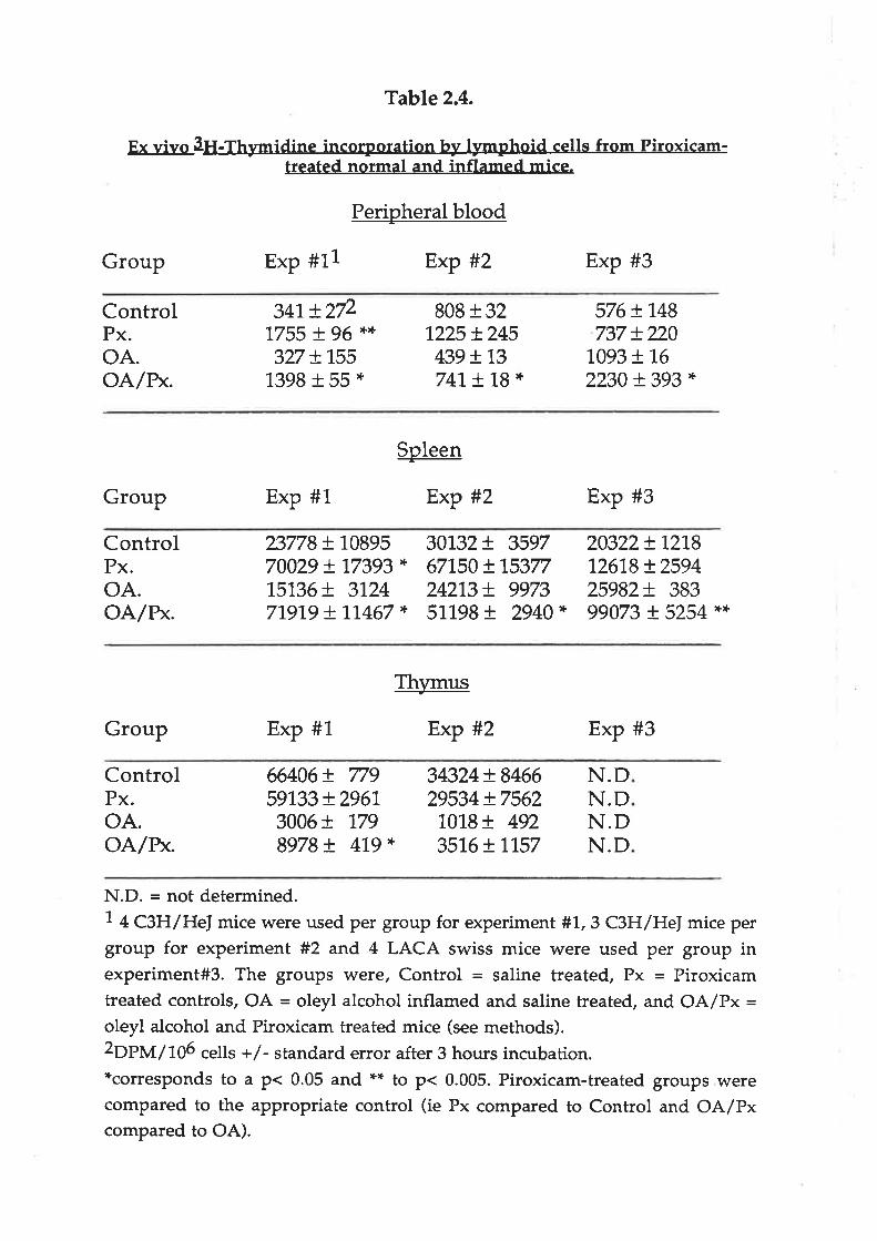
Table 2.4.
E'- vivo 3H-Thymidine incorporation-hy lymphoid cells from Piroxicam-treated normal and inflamed mice.
Perioheral blood
Group Exp #11 Exp #2 Exp #3
ControlPx.oA.OA/BK.
341, t2/21755 I )5 ',+',+
327 !1551398 + 55 *
808 r 321225 + 245
439 !t3741, ! "1,8 *
576 + 748737 + 220
1,093 + 1,6
2230 t 393 *
Soleen
-Group Exp #1 Exp #2 Exp #3
ControlPx.oA.oA/n*.
23778 + 1089570029 + 77393 *
15136+ 31247191,9 + t7467 *
30732+ 3597671,50 + 1,537724213!. 997351198+ 2940*
20322+ L27812618 + 259425982+ 38399073 + 5254 *$
Th',rmus+
Group Exp #1 Exp #2 Exp #3
ControlPx.oA.oA/r*.
66406 + 77959133 + 2961,3006 + 1798978 + 479 *
34324+ 846629534 + 75621018+ 492351,6+ 1,157
N.DN.DN.DN.D
N.D. = rìot determined.7 + CgH/He] mice were used per group for experiment #'!.,3 C3H/HeI mice pergroup for experiment #2 and 4 LACA swiss mice were used per group inexperiment#3. The groups were, Control = saline treated, Px = Piroxicamtreated controls, OA = oleyl alcohol inflamed and saline treated, and OA/Px =oleyl alcohol and Piroxicam treated mice (see methods).2Op}y'r/tO6 cells +/- standard error after 3 hou¡s incubation.*corresponds to a p< 0.05 and ** to p< 0.005. Piroxicam-treated groups werecompared to the appropriate control (ie Px compared to Control and OA/Pxcompared to OA).

25
Adherent cells were isolated from the peritoneal cavity of the same animals. No
significant differences in yields of cells were noted between the groups. Staining for
non-specific esterase indicated that greater than 96Vo of the adherent cells were
MNPs. No IL-2 activity was detected in the supernatants from cultures of these
MNPs. Unstimulated MNPs from Piroxicam-heated animals produced significantly
more LC activity than their corresponding controls (Table 2.5). Upon stimulation
with LPS, these MNPs produced more LC. Similarly, LPS stimulation enhanced LC
production in inflamed animals treated with Piroxicam. MNPs from inflamed
animals produced more LC (either unstimulated or when stimulated with LPS) than
corresponding cells from normal mice.
Figure 2.7 demonstrates that the LAF assay is sensitive to both IL-l and IL-6 .
Although the LAF assay was not very sensitive to recombinant human (rh) IL-6
alone (approximately 1,800 units /mlIL-6 = L u/rnl IL-l),low levels of rhIL-6 were
found to synergise strongly with rhll.-lp. For example, a mixture of 250 units of
rhIL-6 (as determined in the 7TD1 dependent cell assay) and 10-10 M rhll,-lÞ (
approximately 150 u/ml as determined on the LAF assay) had a LAF titre (u/ml) 3
fold greater than 10-10 M rhll.-lp alone. This slmergy was even greater with higher
concentrations of rNL-6 (a more than 10 fold increase in titre at 250,000 units /ml)but rhIL-6 had little effect on rhll,-lp activity at concentrations below 25 units /ml.Since IL-6 activities of greater than 1.0,000 u/ml are commonly found in stimulated
macrophage cultures (Table 2.6), data obtained using the LAF assay must be
interpreted with cautíon.
Even though IL-6 may have potent effects on the LAF assay it is probably not
contributing to the elevated LAF activity following Piroxicam treahnent. In contrast
to LAF activity, IL-6 activity produced by both peripheral blood monocytes and
peritoneal macrophages was reduced following in vivo Pirofcam treatment (Table
2.6). Significant reductions in IL-6 activity were observed in inflamed animals.

Table 2.5.
LAF activitJ¡ produced by peritoneal macrophages from Piroxicam-treated (Px)
normal and inflamed (OA) C3H/HeI mice.
Peritoneal macrophages incubated in the absence or presence of 10 ug/ml LPS
for 24 hou¡s and the supernatants assayed for LAF activity measured as units
of activity (means of 4 experiments * standard error).
LAF ACTIVITY.
units/ml
GROUP -LPS +LPS
Control
Px.
oA.OA./RK
1.13
32.00
29.70
2260.00
+ 0.08
+ 18.80
+ 8.41
+ 1520.00
387 t453 +
570 +2900 +
126,0
71..8
207.0
g02.0.
*P < 0.05. Piroxicam treated groups were compared to the appropriate control
(ie Px compared to Control and OA/Px compared to OA).

Figure 2.7.1L-6 enhances the activity of recombinant human IL-IP. A mixtureof recombinant human IL-IP (1 x 10⁻⁹ M, approximately 300 u/ml) andvarious concentrations of human recombinant IL-6 were serially diluted in theLAF assay and the activity of each dilution determined. Each point representsthe mean of at least 3 experiments +/- standard error.

=Èô
80000
60000
40000
1 0 100
1/dllutlon of
Figure 2.7
1000
lL-l and1 00000
mlxturc
20000
010000
tL-61 000000
units/mllL-6
2500 u/ml
250 u/ml25 ulml0 u/ml
'......g-**+

Table 2.6
IL-6 activity produced by peritoneal macrophagss and peripheral bloodmononuclear cells from Piroxicam-treated normal and inflamed C3H/Hef
mice.
Cells were incubated in the absence or presence of 10 ug/ml LPS for 24 hoursand the supernatants assayed for IL-6 activity using the 7TD1 cell assay
(measured as units of activity means of 3 experiments * standard error).
IL-6 ACTIVITY.units/ml x 103
GROUP. -LPS +LPS
Peritoneal macrophages
ControlPx.
oA.OA./Px
15.00
2.70
4.83
7.97
+ 2.00+ 1.15*+ 1.09+ 0,15*
+ 18.6+ 28.0+ 118+ 33.3
1,43
763513247
Peripheral blood mononuclear cells
ControlPx.
oA.OA./RK
0.03
0.08
1,.07
0.08
+ 0.01+ 0.02+ 0.29+ 0.02'Ê
2.573.1725.7
2.33
+ 0.73+ 0.49+ 7.U+ 0.33*
*P < 0.05. Piroxicam treated groups were compared to the appropriatecontrol (ie Px compared to Control and OA/Px compared to OA).

26
Differences were also noted in the morpholory of these MNPs after 24 hours
incubation. The numbers of spreading MNPs was greater in the cell population
isolated from animals treated with Piroxicam (Table 2.7). This enhancement of
spreading was most marked in MNPs cultured in the absence of LPS, as compared
to those cultured with LPS.
DISCUSSION
There have been many reports suggesting an immuno-regulant role for
prostaglandins (PGs) (Lewis '1,983, Lewis and Barett'1,986, Goodwin 1985). The
majority indicate that PGs, particularly PGET suppress immune functions such as
cytokine production, lymphocyte proliferation and antibody production. Since the
nonsteroidal antiinflammatory drugs (NSAIDs) are not only commonly used to
treat inflammatory diseases but are also potent inhibitors of PG production, they
may well enhance or restore immune functions suppressed by PGs (Goodwin et al
1,984, Goodwin 1985, Ceupens et al 1986). By contrast, only a few examples of
immune enhancement during NSAID treatment have been reported (Ceupens et al
1986, Koga et al 1983).
In vitro studies
a) NSAIDg
In vitro, PGE2 will inhibit inflammatory cytokine (Il-1)-induced proliferation of
lymphocytes in the LAF assay. In part, this inhibition is due to the suppression of
IL-2 production needed as a final stimulus for proliferation (Fig 2.4, Baker et al
1981).
MNPs produce a number of lymphoproliferative rytokines (LCs) in response to an
inflammatory stimulus. Of these IL;[,, II.-6 (Helle et al 1988), and possibly tumour
necrosis factor (TNF) (Ranges et al 1988), are detected in the LAF assay. The action
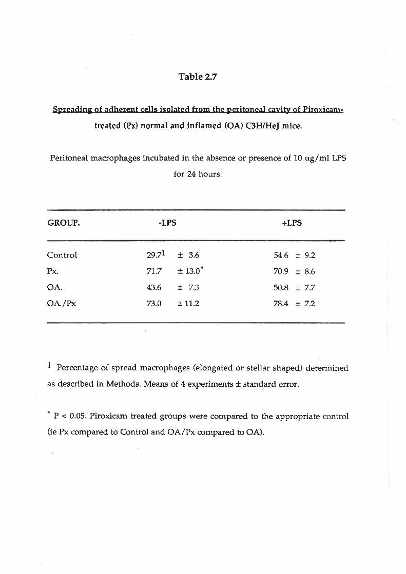
Table 2.7
Spreading of adherent cells isolated from the peritoneal cavity of Piroxicam-
treated (Pxl normal and inflamed (OAl C3FVHeI mice.
Peritoneal macrophages incubated in the absence or presence of 10 ug/ml LPS
for 24 hours.
GROUP. .LPS +LPS
Control
Px.
oA.oA'/r'*
29.77
71,.7
43.6
73.0
+ 3.6
+ 13.0*
+ 7.3
+'J,'t.2
54.6
70.9
50.8
78.4
+ 9.2
+ 8.6
+ 7.7
+ 7.2
1 Percentage of spread macrophages (elongated or stellar shaped) determined
as described in Methods. Means of 4 experiments * standard error.
* P < 0.05. Piroxicam treated groups were compared to the appropriate control
(ie Px compared to Control and OA/Px compared to OA).

27
of these LCs produced by MNP in vitro is also affected by simultaneous production
of PGs. The LAF activity of LCs in the supernatants of LPS-stimulated MNPs is
enhanced when Piroxicam is included during the 24 hour incubation. Increasing
concentrations of Piroxicam reduce the amount of PGE2 produced and enhance the
lymphoproliferative activity of the LCs present as measured in the LAF assay.
However this enhanced activity is only significant at higher concentrations of
supernatants from Piroxicam-treated cultures (<1/ 64 dilution). At lower
concentrations, where units of activity are normally determined, no significant
difference in activity is observed. This indicates that although the actual amount of
LC (detected in the LAF assay) produced is not greatly affected by Piroxicam, the
activity of the LCs produced is reduced by simultaneous PG production.
b) Aspirin metabolites
Many natural metabolites of Aspirin have been reported (reviewed Brooks et al
'1.986a, Rainsford 19U). For example, the metabolite 2,5 DHB (gentisate) has been
identified at concentrations of up to 15uM in the plasma and serum of patients
receiving salicylate therapy for rheumatoid arthritis (Cleland et al 1985b, Grootveld
and Halliwell 1986). Hydroxylation of salirylate to form not only 2,3 DF{B but also
2^5 DHB (and possibly 2,3,5 THB) occurs freely in the presence of hydroxy radicals
(see fig 2.5) in cell-free systems (Ledvina 1969, Grootveld and Halliwetl 1986). This
hydroxylation of salicylate and its metabolites may also reduce inflammation by
quenching tissue-damaging oxyradicals (Cleland et al 1985a). Studies in which
salicylate was incubated with neutrophils pre-activated to produce oxyradicals
indicated that 2þ DHB was the preferred product of hydroxylation by a ratio of
about 5:1 (Wright 1,989).2,5 DHB (gentisate) may therefore be an important active
metabolite of Aspirin therapy, not only because it may be produced preferentially at
an inflammatory site, but also because of its ability to inhibit IL-L induced
lymphoproliferation.

28
Therefore, the ability of Gentisate, but not 2,3 DHB, to inhibit the
lymphoproliferative activity of IL-1 (site 3) might represent a another important
mode of action of Aspirin. Two oxybenzoates related to 2,5 DHB, homogentisate
and, to a lesser extent, diacetylgentisate, also inhibited at site 3. This may indicate
that hydroxyl groups at both 2 and 5 positions on the benzene ring are a key
chemical structure for this effect.
Unfortunately, the other poly hydoxy metabolite of interest, 2,3,5T:Í18, could not be
tested since synthesis was difficult and, when obtained, (Wright 1989) it was very
unstable. All these hydoxy metabolites may undergo further oxidation to form a
quinone (Ledvina 1969).It is possible that these quinones may form in vitro (and in
vivo), and that it is the quinone, rather than the hydoxy metabolites, that inhibit the
action of IL-l at site 3. These observations challenge the long held view of a single
mode of action of NSAIDs and warant further investigation.
c) Immunosuppressants and steroidal drugs
Steroid drugs are recognised as potent antiinflammatory drugs. It has long been
thought that they reduce inflammation by inhibiting the release of arachidonic
acid and thus reduce the production of proinflammatory prostanoids and
leukotrienes (see chapter 3). If this were so, these drugs might be expected to
enhance LAF activity at site 2 like the NSAIDs. However, this is not the case.
While not stimulating LAF activity, steroid drugs inhibited its production (at site
1). This is consistent with recent findings that these drugs inhibit the production of
IL-l and other cytokines (Allison and Lee 1988). In addition, the steroidal drugs,
like the immunosuppressants, inhibited the action of IL-1 (site 3). This may be
mediated by inhibition of IL-2 production which is essential for proliferation
following IL-l stimulation in the LAF assay. Cyclosporin-A, for example, a potent
immunosuppressive drug, inhibits IL-l induced lymphoproliferation in this way(see Chapter 5). Whilst the steroid drugs may, like NSAIDs, still inhibit PGE
29
synthesis at site 2, their potent action at sites 1 and 3 will mask this effect and
make it difficult to detect in this assay system.
In vivo/ex vivo studies
d Lymphoproliferation
The effect of applying Piroxicam in vivo on ex vivo lymphoproliferation and LC
production was investigated using oleyl alcohol as an inflammatory stimulus. In
accord with the in vitro findings, lymphoproliferation ex vivo of peripheral blood
and spleen cells was enhanced by Piroxicam treatment of both inflamed and
noninflamed mice. Flowever, thymocyte proliferation was not affected by Piroxicam
treatment This may indicate that in vivo lymphocyte proliferation within the
thymus is not subject to PG suppression. The considerable reduction in lymphocyte
DNA synthesis in the thymus induced by inflammatory stress might be due to the
action of natural corticosteroids, so that Piroxicam treahnent only slightly increased
proliferation in the inflamed mice.
Surprisingly, in the absence of an inflammatory stimulus, Piroxicam treatment
considerably increased proliferation of peripheral blood and spleen lymphocytes.
This may indicate that, in these C3H/HeJ mice, either PG has a role in regulating
normal lymphocyte proliferation, or the mice may have had a low grade
inflammation even though they otherwise appeared healthy. Genetic factors may
also be involved since, when these experiments were repeated using another strain,
(LACA Swiss), the Piroxicam treatment of non-inflamed mice did not significantly
increase proliferation of peripheral blood and spleen cells. C3H/HeJ mice are
known to have some suppressed immune functions (Beutler et al 1986). These
results might indicate that this strain of mice is either more sensitive to the effects of
normal low levels of PG or they have higher basal levels of PG than other strains of
mice.
b) Lymphoproliferative c¡rtokines

30
Prior in vivo Piroxicam treaÍnent resr-¡Ited in a large increase in the LAF activity
(units/ml) produced ex vivo by peritoneal MNPs isolated from inflamed mice,
whilst IL-6 production by the same cells was reduced. Both basal and stimulated
LAF production was enhanced, and IL-6 production reduced, by this Piroxicam
treatmenÇ probably due to the inhibition of PG production in vivo by Piroxicam. A
recent report has indicated that fL-lP production by peripheral blood mononuclear
cells is also enhanced following in vivo treatment with the cyclooxygenase
inhibitor,Indomethacin (Endres et al 1989). Flowever, these authors did not observe
a significant increase in IL-lP production following Indomethacin treatment in
vitro.
The LAF assay is not specific for any one cytokine as IL-'l,,IL-6 (Fig2.7),IL-2 and
TNF are reported to act positively in this assay (Helle et al 1988, Ranges et al 1988).
Flowever, there are two reasons why the enhanced LAF activity is probably due to
increased IL-1 production. Firstly, no IL-2 or TNF was detected in the supernatants
tested (the CaH/He] strain of mice is deficient in producing TNF (Butler et al 86).
Secondly, although IL-6 can be detected in this assay (Fig. 2.7), it was reduced by
Piroxicam treatment (table 2.6). Therefore, if IL-ó is contributing significantly to the
LAF activity in these assays, the enhancement of IL-l activity following Piroxicam
treatment is underestimated.
There was no similar dramatic increase in the titre of LAF activity (site 1) produced
by perítoneal MNP treated with Piroxicam in vitro (Fig.2.2). This may indicate that
Piroxicam's inhibition of PG synthesis either i) does not directly affect IL-1
production by MNPs but inhibits the production of other cytokines produced by
lymphocytes (such as interferon gamma) required to maintain IL-1 production by
MNPs; or , (ii) Piroxicam treatment for more than 24 hours is needed to directly
affect IL-1 production. This aspect is discussed in detail in Chapter 4 and is in direct
contrast to the stimulation of IL-6 production by macrophages by Piroxicam in vitro(also Chapter 4).

31
c) Macrophage morphology
Pirodcam treatment also enhanced cell spreading, which is another marker of MNP
activation. This is probably mediated by the inhibition of PGEZ by Piroxicam since
PGEZ inhibits the spreading of MNPs in vitro (Cantarow 1978). This could indicate
that the effects of in vivo Piroxicam treahnent on MNPs (ie. inhibition of PGEZ
production) are still maintained ex vivo 24 hours after isolation and culture in
Piroxicam-free media. Further investigation into the effects of NSAIDs and PGE2 on
MNP morphology (Chapter 3) indicated these effects a¡e also seen after in vitro
Piroxicam treatment.
These findings indicate that PGs may indeed be potent regulators of immune
functions at concentrations comparable to those found in vitro. within inflammatory
sites (Bray and Gordon1,978, Robinson and Levine7974). Not only do PGs suppress
proliferation of lymphocytes directl)¡, but they may also suppress the production of
mitogenic cytokines which precede and trigger lymphoproliferation. Similar
regulation is observed in vitro with the more potent steroid and
immunosuppressive drugs. Consequently, an inhibitor of PG production
(Piroxicam) can stimulate some immune responses in mice with chronic
inflammation. NSAID therapy maf r therefore, enhance immune responses in
patients with chronic inflammation. This could be beneficial as a heightened
immune response might facilitate the removal of a persistent inflammagen.
However enhanced production of some inflammatory cytokines might prove
counter productive. For example,IL-1 not only stimulates the immune response but
also could induce detrimental effects such as fever and bone resorption. NSAID
therapy, via its effects on PG production, may enhance a variety of immune
functions during infl ammation.

32
Chapter 3
The effects of some anti-arthritic drugs, prostanoids, cyclic nucleotides and
cytokines on the shape and function of rodent macrophages in vitro.
INTRODUCTION
Many cell types undergo the active process of cell spreading, by which normally
round cells change shape and flatten onto a solid substrate. Cells undergoing this
process are generally adherent and this shape change is often associated with the
expression of motile structures, such as pseudopods. The spreading of a cell is an
essential prerequisite for movement of amoeboid cells on surfaces (Vasiliev,1982,
1985). The relationship between spreading, adhesion and locomotion is complex
since too much spreading may also be associated with excessive adhesion and
decreased motility (Keller et aL7979).
Cells of the macrophage/monoryte lineage play a central role in the immune and
inflammatory responses. They are directly involved in the removal of bacterial
and other foreign material as well as releasing many mediators that regulate the
immune and inflammatory responses (reviewed chapter 1). Macrophages are
adhesive and motile cells. Therefore, control of their spreading may play an
important role in determining the resolution or exacerbation of many immune and
inflammatory disorders. In addition to controlling macrophage movements,
spreading may also indicate their activation for a wide variety of functions
(Adams and Hamilton 1987).
Supernatants from endotoxin(Ll'S)-stimulated macrophages treated with NSAIDs
exhibited enhanced IL-l activity in yjlle, as measured in the LAF assay (Chapter
2). This was probably due to inhibition of prostaglandin(Pc)-E2 production. These
33
macrophages readily attached to, and spread over, plastic or glass coverslips. This
chapter describes the increased cell spreading of NSAID-treated macrophage
cultures when compared to untreated (no drug) cell cultures. Enhanced spreading
was also noted when the macrophages were treated with corticosteroids and some
of the immunosuppressant drugs used to treat chronic inflammation. This
enhanced spreading is one marker of macrophage activation. Macrophage
activating factor (MAF) or interferon gaûuna (IFNf will activate macrophages in a
variety of ways and increase their spreading (Schreiber et al. 1985).
Since the surface morphology of glass-adherent macrophages is well preserved by
critical point drying (Pollack & Gordon 1975), it is possible to study the changes in
surface structure (as revealed by scanning electron microscopy (SEM) induced by
various drug/cytokine treatments. This study confirms that PGE2 inhibits
macrophage spreading (Cantarow et aI7978) and presents evidence to show that
macrophages may autoregulate their own spreading by producing PGE2. This
increased spreading may indicate enhanced motility and/or cell-cell adherence.
MATERIALS ÁND METHODS
Chemicals
Drugs and chemicals were obtained from the following sources: PGE1, PGE2,
PGA2, PGFlcr,, PGF2cl, PGDZ bt2cAMP, btZcGMP and PiroxicÍun were purchased
from the Sigma Chem. Co. (St Louis Mo. USA); and PGEg from Cayman Chem.
Co. (Ann Arbor Mi. USA). Misoprostol was a gift from the G. D. Searle and Co.
(Skokie,Il. USA): Naproxen (R and S enantiomers) from Syntex USA; Ibuprofen (R
and S enantiomers) from Boots Drug Co UK.; Cortisol, Prednisolone,
Dexamethasone, 6-mercaptopurine and Indomethacin from the Sigma Chemical
Company St Louis; Methotrexate from Lederle Labs USA: Cyclosporin-A from
Sandoz AG Switzerland.

34
Cytokines
The following cytokines were used; recombinant human IL-1 P donated by Otsuka
Pharmaceutical Co., Tokushima,Iapan; recombinant human IL-l o, recombinant
human IL-6, recombinant murine and human IFNT and recombinant murine
TNFP from Genzyme, Boston; human recombinant TNFcr and P from British
Biotechnology; recombinant human IL-2 from Boerhringer Mannheim GmbH. Allrecombinant cytokines were produced in E. coli.
Isolation and culture of rodent peritoneal macrophages
Normal rats (Dark Agouti) or mice (C3H/HeJ. C57 /BL CBA or Balb/C) from the
central animal house (University of Adelaide) were sacrificed by cervical
dislocation and their peritoneal cavities lavaged with Hank's buffered saline
solution (HBS). The peritoneal cells were washed once by centrifugation. One x
1¡6 peritoneal cells suspended in 1ml of RPMI-1640 medium with 10% foetal calf
sertun were placed in 16mm flat-bottomed wells of a24 well tray (Costar) with 13
mm glass coverslips at the bottom of each well. After 1. hours incubation at 37oC inSVo C02 the non-adherent cells were removed by washing with HBS. The adherent
cells were then incubated in 0.5 ml of RPMI medium containing the appropriate
concentration of drug. In some experiments E. coli 0111:M lipopolysaccharide
(LPS) (Sigma Chemical Co.) at concentrations between 1-10 uglml was added to
stimulate the macrophages. After 24 hours the cells were fixed for 10 minutes in a
4Vo glutaraldehyde / HBS solution.
Histochemical procedures
Cell preparations were stained for myeloperoxidase by the Kaplow method
(Kaplow 1965). Briefly, cells fixed in gluteraldehyde (ZVo) were covered withfreshly prepared peroúdase stain for L minute as described (Kaplow L965) then
counterstained with neutral red stain before drying and mounting.

35
For non-specific esterase (NSE) staining the method described by Yam et al (797'I)
was used. Briefly, glutaraldehyde-fixed cells were covered with fresh NSE stain
for 30 minutes as described (Yam et al 1971). The cells were briefly counterstained
with methyl green before drying and mounting. Rodent macrophages stained
lightly with this NSE stain when compared to human monocytes.
Assessment of macrophage spreading by light microscopy
The glutaraldehyde-fixed cells were stained with Giemsa's stain and the coverslips
mounted onto glass slides. With the aid of a microscope at least 5 random fields
were sampled (greater than 500 cells) to determine the percentage of adherent cells
that were elongated and/or star-shaped. A cell was considered to be 'spread' (ie
elongated or stellate) if its width was at least twice that of a normal (rounded) cell.
There have been many previous reports, mainly in studies with
polymorphonuclear cells, using various techniques for estimating cell spreading
(reviewed Rogers and Bignold 1990). Most have relied on visual assessments of
spreading, whilst others used quantitative techniques such as using microscopic
graticules or computerised methods to determine cell area. This study has relied
on subjective visual assessment to quantitate spreading. However these
assessments were carried out "blind" and where possible independent observers
were used. Va¡iation found between and by the same observer(s) was small (less
than 87o and íVo respectively). An EDSO was determined as the concentration of
drug/cytokine which produced a two-fold increase in macrophage spreading
computed from a doseresponse plot.
Preparation of samples for scanning electron microscopy (SEM)
The glass coverslips with adherent cells were fixed as for light microscopy. The
coverslips were then rinsed twice. In HBS and dehydrated in a graded series of
aqueous/acetone solutions (up to 700Vo acetone) for 5 minutes at each step. After
two washes in 1.00% acetone the specimens were quickly transferred to a
precooled high pressure chamber of a critical point drying apparatus (Blazers

36
IJnion, Furstenum Liechtenstein) and dried using liquid N2. All samples were
then coated with gold/palladium and observed under a Phillips 505 SEM.
Photographs were taken on Kodak T-MAX 100 film with an accelerating voltage of
20kv at magnifications from 1-50 x 103 times. Ë <çe.,o.'e.w\s *ec* pqlocmeÀ rnr
kì¡ticu\e. and, appèarances; u)eF¿,¡ith\',-,'+heir:ool.c\ e,<pereC oltÌr SúN^ "
Actin staining
Cells were stained with rhodamine-labelled phalloidin, a cyclic peptide that
specifically binds to F-actin (Wutf et aL7979). Staining of macrophages was carried
out as described (Amato et al 1983). Briefly, 100u1 of 0.16 ug of rhodamine-
phalloidin/ml in PBS was applied to the coverslips after their 24 hour treatments
described above. After 20 min the coverslips were rinsed twice and viewed
immediately under a fluorescence microscope (Leitz).
PGE2 assay
PGE2 concentrations were measured by competitive radioimmunoassay as
described in chapter 2.
Statistical analysis
All statistically significant differences between groups were calculated using an
unpaired, two tailed student's t - test (Snedecor and Cochran 1989).
RESUTTS
Morphology and spreading induced by NSAIDs
Of the adherent cells isolated from the peritoneal cavity of healthy rats and mice,
more t}:.an 96Vo stained positively for non-specific esterase and less than 10% for
myeloperoxidase. This indicates the population consisted mainly of mature
macrophages since perofdase is lost as they mature whilst positive non-specific
esterase staining remains characteristic of this cell type. After 24 hours of culture
..^{. c>\ .r-.,Ot.\ our€-

37
macrophages isolated from C3H/HeJ mice appeared to be a heterogeneous
population of mostly round cells with some spread cells present (Fig 3.1a). The
percentage of peritoneal cells spreading varied from 20 to 50Vo depending upon (a)
the age (mice older than 12 weeks spread less) and strain of mouse (Fig 3.9) and
(b) the environmental conditions in which they resided before harvesting
(macrophages from infected mice or mice in poor health spread poorly). If a
NSAID (such as 20 uM Piroxicam) were included in the culture medium, there
was a significant increase in the proportion and numbers of cells spreading (Fig.
3.1b).
All the NSAIDs tested, that are known to inhibit PGEZ production, enhanced the
spreading of peritoneal macrophages (Table 3.1). For example the inactive R-
isomer of Naproxen was approximately 100 times less effective than the active Iisomer. The relative concentrations at which the drugs enhanced macrophage-
spreading correlated with the effective plasma concenffations of these drugs in
patients undergoing therapy with the drug, This in vitro effect of the NSAID in
enhancing macrophage-spreading was noted in both the presence and absence of
10 uglml lipopolysaccharide (LPS).
Spreading induced by corticosteroids/immunosuppressants
Dexamethasone was the most potent of the drugs tested, inducing a two fold
increase in macrophage spreading at concentrations as low as 1..9 x 10-9 M (Table
3.1). The other steroids tested had similar effects at higher concentrations. The
immunosuppressive drugs, Methotrexate and 6-mercaptopurine were more than
1000 fold less effective. The spreading induced by alt these drugs was stellar in
shape, similar to that induced by the \ISAIDs. Cyclosporin-A'had no effect upon
macrophage spreading and was rather surprisingly toúc at concentrations greater
than lp M.
PGEZ production and its effect on spreading

Figure 3.1. Peritoneal macrophages from C3H/HeJ mice cultured for 24
hours in the presence (a) and absence (b) of 20uM Piroxicam. This shows theenhanced spreading of macrophages treated with Piroxicam. Cells werefixed and stained with Giemsa's stain. Similar results were obtained usingmacrophages from different strains of mice (see Fig 3.9). Magnification -200x.
frÞ

ñ¡a
-l+v
aa
,0
-ò
oaf'
a
o.L tÓa
| ô'¡tt,o-
a rdtt o
1-
ar¡
,faa
ìa
t*J\
aa
a¡aoO
^ f!.¡ I
O¡
aa
o. 3't-eo
a
o
'
ì.e''l¡
-.e
a
OÒ
Jr,
fl tr\
aa
a
?
tq o
a
útt,tt\
t.o\
'¡
oa a
I\
+taì
lDi,i't'.t
J'
aÊata
aa
a
t\
I
at'
fFo
a
b
a
.}
o
a
\J
Tl)¡lq
a t*Tao
OO
e a,'
oa
flaa
o
a
a
Òl
åt
e.¡o
¡atI 'å.
o
hr
,o
¡
I
a a
o
I ìt,
.tÇ
aaoae
)
Q.¡
r¡tr
a
\ii,.l'
a
Io
a a, o
a aao
ö
a
a ,a a ootìr
a
(ì
aO
tc,
!!rt
ao rÐ
"a a.1 aÇ a
?t I
Ð
a
aO t fr
ìa
ta
f.a
oa,
I
O t,a
r|}
t
o
b.
.O
'fi
a)a20
ooa .D
a¡
a'
to
a -J.-rt
3
)À
'Tn, ''.¡ .-Ð,
¿,t
e\
¡a,tt.a
rtrOttt!r
C€
)
ì'
b¡
tî
I
a
IçI¡.c¡
* ç .tT, r* \ .-1{aeoe
,i.'o
o.t,¿
\,rt
\-
a
t!
oa
a
e
/
t),
t(o
.Ç
¡t
ts il.
i.\
-'*st,
o
* cl 4t
a
- t'.
þt.
¡\þ
a
\¡iI
'ol
a
.ttD
\
nrDo
t
t
a
¡ t ot| ;.\
a
.tf-"Í
?.4.r¡.t
ti^'. 'oo
Ia
ã I
a
"/Ita
- ti, a'
^.r Ot lf
l'
l.ot.l
. Ða- \.'
,xrYtru.'
,{ 1'tt
)¡Q
'J ts
'\ !Ùr¡
a I[.ùq
'a\
l
*.o
+a
ra".l ¡ tt
¡gt.o)
-t. o'l¡ tb
t.
q
.O n a
,û
t
a

Table 3.1
macroohase sDreadine.
A) Indomethacin
(S)Ibuprofen
(R)Ibuprofen
Piroxicam
(S)Naproxen
(R)Naproxen
Aspirin
5.7
9.9
2300
25
30
1700
12000
B) Dexamethasone
Prednisolone
Cortisol
6-mercaptopurine
Methotrexate
Cyclosporin-A
0.0019
0.0054
0.011
0.47
3.1
>1.0 **
* The concentration of drug which induced a two fold increased the number of
cells spreading (see Methods).** Toxic at 1pM.

38
The ability of Piroxicam to enhance the spreading of peritoneal macrophages
correlated closely with this drug's ability to inhibit the cells own production of
PGEZ (Fig. 3.2). It seemed likely therefore that endogenous PGE2, normally
produced by adherent macrophages, might inhibit the cells own spreading.
The effect of prostanoids and their analogues on spreading
Most of the prostanoids affected the morphology of mouse (C3H/HeI)
macrophages (Fig.3.3). They inhibited the spreading of glass-adherent
macrophages with ID5s's of 31nM for PGE1, 20nM for PGE2 610nM for PGE3 and
590 nM for Misoprostol. Of the non-E series prostanoids tested only PGA2
inhibited spreading at concentrations below 10,000 nM (ID50=222nN.{) (Fig. 3.a).
Ttre effect of cyclic nucleotides on spreading
Lipophilic (dibuçyl) analogues of cAMP (btZcAfvfp) and CGMP (btZcAtvtp) were
used to assess the effects of ryclic nucleotides on cell shape. Like the E-series
prostanoids, bt2cAMP also inhibited spreading (ID50 = 63 uM) whereas bt2cGMP
enhanced the already high levels of background spreading in the presence of 20
uM Piroxicam (Fig. 3.5).
Spreading induced by cytokines
Of the cytokines tested, namely IFN1 ÍL-'l.,IL-z, TNFcr, and TNFp (lymphotoxin),
only IL-l and IL-2 had no effect on normal peritoneal macrophage spreading. IFNy
of mouse origin only induced mouse macrophages to spread and did not affect rat
cells. Recombinant human IFNy did not alter the morpholory or spreading of rat
or mouse macrophages. Surprisingly rTNFc¡ (mouse and human) and rTNFp(human) increase the spreading of mouse macrophages (Table 3.2), which was
inhibited by adding PGEZ (data nor shown).
Scanning Electron Microscope (S.E.M.) studies of various cell populations
Nonnal cells (Fig. 3.6 a.b)

Figure 3.2. Peritoneal macrophages from C3H/HeJ mice were cultured in thepresence of l0ug/ml LPS and increasing concentrations of Piroxicam. After24 hours the supernatants were harvested to determine the PGEZ producedand the cells fixed and stained in Giemsa's stain to determine the percentageof cells spreading. Each point represents the mean of at least 3 experiments.

106
10 '7
10€
10€
10'10
Figure 3.2
M [PGE2]% Spreading
€ -710 10
M IPlroxlcam]
70
ôtI¡JoG
=
H
-Gt----*
m
50
40
m
Et
E.Uoo-c,
s
èræ
0 io€ 106 104 ioir

Figure 3.3. The effect of E-series prostaglandins on spreading of murineperitoneal macrophages cultured in the presence of 20uM Piroxicam and testcompounds for 24 hours. The cells were fixed, stained and the percentage ofcells spreading determined. Each point represents the mean of at least 3
experiments.

Figure 3.360
50
40
30
20
10
ols!6oo.lD
Ioo
*
\
.....ê
--{__l-
-#
E1
E2
E3
MPLA
0o 1 10 100 1000 10000100000
nM

Figure 3.4. The effect of some non-E-series prostaglandins on spreading ofmurine peritoneal macrophages cultured in the presence of 20uM Piroxicamand test compounds for 24 hours. The cells were fixed, stained and thepercentage of cells spreading determined. Each point represents the mean ofat least 3 experiments.

Figure 3.4
C').5!(Ec,Êodo(,
às
80
70
60
50
40
30
20
10
0
+{
'-È
-+*
PGA2
PGD2
PGFIa
PGF2a
o 1 10 '100 1000 10000100000nM

Figure 3.5. The effect of btcAMP and btcGMP on spreading of murineperitoneal macrophages cultured in the presence of 20uM Piroxicam and testcompounds for 24 hor.us. The cells were fixed, stained and the percentage ofcells spreading determined. Each point represents the mean of at least 3experiments.

Figure 3.5
Fct¡E!6oo.o
Iõ(,àe
80
70
60
50
40
30
20
10
0
**
o
CAMP
CGMP
100 '1000 'l000010
uM

Table 3.2
The effect of cytokines on C3ÉVHef mouse macrophage spreading
Cytokines EDso (Pvt)t
murine rIFN y
human rIFN 1
human rTNF o
human rTNF p
murine rTNF cr
human rll,-lphuman rIL-2
human rIL-6
86.5
>100,000
60.1
1280
87.0
1t¡z(>10,000)
(16)
(e60)
(23)
(>3.33 x to5)
(>1 x 10⁵)
(>1 x 10⁶)
>100,000
1 The concentration of drug which induced a two fold increase in the number
of cells spreading (see this chapter Methods).2 Values in parentheses are in units / ml.

39
After 24 hours, approximately half of the cells were still spherical in shape. Under
the S.E.M. the rounded (approx.lO um dia.) cells displayed irregular contou¡s with
ridge-like profiles and ruffled membranes. Very rarely (<2Vo) small round cells of
less than 0.6 um in diameter were seen with smoother membranes: these were
probably lymphocytes. Other cells (up to 507o in number) had begun to spread out
by extending cord-like processes which attached to the glass coverslips.
Occasionally a thin veil of rytoplasm extended from beneath the cell. When this
veil extended beyond the area covered by the rounded cell, the cells were flattened
leaving a raised dome-shaped nuclear pole. The ridge-like profile of the
membrane was considerably smoother on the spread out cells.
NSAID-treated cells (Fig. 3.6 c.d)
Cells treated for 24 hours with 20 uM Piroxicam were generally more spread out
and flattened than the untreated controls. A significant proportion (approx.20Vo)
of cells remained rounded, resembling those noted in the untreated population.
Flowever the majority of the cells (which did spread with Piroxicam treatment)
were considerably flatter than those cells showing spontaneous spreading in the
untreated population. With Piroxicam the cenhal nuclear pole was only slightly
raised and the ridge-like processes more flattened. These Piroxicam-responsive
cells were usually irregular in shape with radiating filopodia extending from two
or more poles. A prominent ruffling membrane was often noted at one or more
sides of the spread cells. Among this heterogeneous population we noted not only
an increase in the numbers of cells spreading but also in the degree of spreading.
Cytokine-treated cells (Fig. 3.7)
There were clear differences in the type of spreading induced by the various
cytokines and drugs. The spreading induced by TNF more closely resembled
spreading induced by NSAIDs and the corticosteroids/inununosupressants (Fig.
3.7 a,b). The spreading was polar with the cytoplasm extending in two, three or

Figure 3.6. Scanning E.M. was carried out on rnacrophages from C3H/HeJ mice cultured in media alone (hg3.6a,b)
or media supplemented with 20uM Piroxicam (fig 3.6c,d). Figures 3.6a and 3.6c were taken at a magnification of500x. Higher magnifications (2000x) are of a typical rounded cell from the untreated cultures (fig g.ób) and a typicalspread cell from the Piroxicam-treated cultures (fig 3.6d).


Figure 3.7. Scanning E.M. was carried out on macrophages from C3H/HeJ mice cultured in media supplementedwith 1000 units/ml murine recombinant TNFa (fig 3.7a,b) or media supplemented with 1000 units/ml murinerecombinant IFNI $ig3.7cd). Figures 5a and c were taken at a magnification of 500x. Higher magnifications (2000x)
a¡e of a typical spread cell from the TNF treated cultures (fig3.7b) and a typical spread cell from the IFNI treatedcultures (fig3.7d).

LE¡mAB.økU 5.1 3E3 EE36/BB
.a
I
a
tÊI
tr)t?a
lrt'-
4
16¡meø.BkU 3.1 3E3 EE39/88---
â
d
.F>'*
Ja--
<- 5-
.t
JI

40
more directions. A ruffling edge was usually seen at the end of each "cytoplasmic
arm". As seen with NSAID-induced spreading, cord-like processes attached the
cell to the glass coverslips. Unlike IFN^¡induced spreading, the cytoplasm was
often raised in places leaving a gap between the cell and glass coverslips. The cells
seemed to be only attached to the coverslip at the end of each spreading arm with
the centre of the cell being slightly raised.
By contrast, cells induced to spread by IFNI were usually circular with an even
distribution of cytoplasm extending around a slight dome shaped nuclear-pole
(Fig. 3.7 c,d). No gaps were seen between the edge of the cell and the glass
coverslip. Ruffling or rolled edges usually extended most of the way around the
cell. Many of the cells had raised cytoplasmic spheres.
The effect of treatments on the pattern of actin staining
Rhodamine-phalloidin was used to stain the macrophages after various treatments
as described above. Typically rounded cells found in the untreated populations(Fig.3.8a) showed diffuse staining throughout the cytoplasm with dense staining
around the circumference. At a high plane of focus, a diffuse cortical band of
fluorescence was seen, possibly associated with the nucleus. Elongated cells, such
as those seen following Piroxicam or TNF treatment (Fig. 3.8b and 3.8c) also
showed a cortical band of fluorescence at a high plane of focus. At a low plane of
focus linear fluorescence structures running throughout the cytoplasm were
observed. Strong staining was seen toward the ends of the polar rytoplasmic arms
and these extended thin 'spiky' fingers into the protruding cytoplasm. Strong
staining structures were seen throughout the cytoplasm; the more dense ones
seemed to be associated with the linear fluorescent fibres running throughout the
cell. In IFNy treated cells (Fig. 3.8d) the cortical band of fluorescence was observed
at a low plane of focus. The pattern of staining found at the ends of the
cytoplasmic arms (shown in Fig. 3.8b and c) was noted around the ci¡cumference
of the IFNy treated cells. The fluorescent structures throughout the cytoplasm

Figr.ue 3.8. Rhodamine-phalloidin staining was carried out on macrophages from C3H/HeJ mice cultured in mediaalone (fig 3.8a), media supplemented with 20uM Piroxicam (fig 3.Sb), media supplemented with 1000 units/mlrecombinant murine TNF o (fig 3.8c) or media supplemented with 1000 units/ml recombinant murine IFNy (fig3.8d).


41,
were not linear, as noted following NSAID and TNF treatment, but arranged in a
mesh or crosslinked structure. A few dense staining punctate structures were
occasionally seen randomly throughout the rytoplasm.
The effect of drugs and cytokines on macrophages from different strains of
mice
The percentage of macrophages spreading was assessed after 24 hours culture
with recombinant murine TNFcr (1000 units/ml), Piroxicam (20 uM),
Dexamethasone (0.1uM) and a mixture of TNF (1000 units/ml) and Piroxicam
(20uM) (Fig. 3.9). Peritoneal macrophages from 4 strains of mice were used
(C3H/HeJ, CBA, Balb/C and C57 / Bl) and experiments were carried out in the
presence and absence of 2ug/m\ LPS. In the absence of drugs and cytokine, the
numbers of C3H/He] macrophages spreading was significantly less than that seen
in cultures of macrophages from other strains. LPS treatment reduced the
numbers of cells spreading from all strains except C57 /BL In general, in all strains
of mice tested, Piroxicam and Dexamethasone treatment enhanced spreading;
both being more effective in the presence of LPS. Similarly TNF enhanced
spreading. However it was less effective in the presence of LPS. Simultaneous
treatment of macrophages with TNF and Piroxicam stimulated more spreading
than with either drug/cytokine alone. All mice were 8-12 weeks old, nominally
pathogen free and healthy. When older, possibly infected mice, were used as a
source of macrophages we noted greater variation in the effects of drugs and
cytokines. However similar trends were generally noted.
The effect of NSAIDs on macrophages from inflamed rats
Peritoneal macrophages were isolated from rats with adjuvant-induced arthritis
(14 days after injecting a mycobacterial adjuvant). These cells attached and spread
onto glass and plastic as described previously (Fig. 3.10a). Their spreading also
increased aÍter 24 hours treahnent with Piroxicam. However unlike macrophages
from normal animals, these adjuvant-activated macrophages often aggregated into

Figure 3.9. Peritoneal macrophages from C3H/HeJ, CBA, Balb/C andC57 /BI mice were cultured in media alone (Cont) or in the presence of 20 uMPiroxicam (Px). 1000 units/ml murine recombinant tumour necrosis factor(TNF) ø, 20uM Piroxicam and 1000 units/mt TNFcr (Px + TNF) and 10-8MDexamethasone (Dex). Experiments were carried out in the presence andabsence of 10 uglml LPS. After 24 hours the cells were fixed and stained andthe percentage of cells spreading determined. Each bar represents the mean+/- standa¡d error of at least 3 experiments.

100
90
80
70
50
50
40
30
20
Figure 3.9
Cont R TNF tu+TNF Dex Cont Br TNF B<+TNF Dex
Treatment
ÞoI
-cÉ()La(t)
èe
90
80
70
60
50
40
30
20
t0
C3H/HeJ
üÀ h
CBA
Balb/C

Figure 3.10. Peritoneal macrophages were isolated from male Dark Agoutirats with adjuvant arthritis (14 days after injection of mycobacterialadjuvant). These cells were cultured for 24 hours in the presence (a) andabsence (b) of 20uM Piroxicam. These cells not only spread more in thepresence of Pirodcam but also formed aggregates or clumps of cells. Cellswere fixed and stained with Giemsa's stain. Magnification -200x.

aa
a -.\.t\{
¡-a
It\
1
'
Dt
oo , ît' t rrl'
tt rt
aa .ii' .
.a .,I
4{+ t ?'rt .)
â
\
a
¡Ì t.)
aa
ata a
t- ¡a¡}f f]\
Ia
a
a¡
aì
aaa'(
¡oo o
Å,a
.tloo
tr
a
aa
arla¡
-a t
?
a
a.la
o
a
a
a
at
.t
o
a'
i
a-..aIP¡
a
a
-a a
ta
oo
¡
a
aaa
a
oJa
a
(
e
\
I
a
aaa
,a
a
o
a
aaaawt\..1.
'aa
{ .a
a¡t
O
0
attaa
eo
;.. aa
¡ì
-..taT
a
-Ja
;:
aa
a¡ t
t
t¡.
.r
H.tl'-
t¡.I ,-'l.
)
¡aat
a
t
'.o Yt rt '
fa
o
IL
aa
t!Ita
.t. e',a
\,
o
a
a
t
-â
lItl tt 4a
-a
a
o
¡!ì
'.I¿. .a e.aO.?
a
aaa
t
ì.Ò
a I
aaa
oa
+'oa
o a'Jj
a.la
a.
.f
r r å
//. : o
a
aaa {
a
¡r a
ary
T
'.
aaa
¡aa
aa .ooÞa
i.
a
Ò
atl
t
aIf
aa a
aa a Ifb'\ ¡¡¡\oa
Ia
ata
aa
ta
ra
O
C¡laaaa
aa
¡
J
T
a
o
I
to
I
tta
a
l
)
'tìI
¡.' :Ì
aaIt
Ja
t a\r'¡ t
..1
"'a
OIJo
t
a\ ,laì
aIalI
a
I
\l-
a
e
h
¡
l.'
a o a
I{
I-
tìr:a
aaa.,,1
o
a
a ì
t a a a a
aa
aa
aoa
. lr. {
a
taa)D
ta
o.¡ .la aa
¡a
'a ¡ô.aa¿
a>{
a
*aa
o
aa
Q^,'o

42
clumps of about L0 or more cells (Fig. 3.10b). The structure of these aggregates
changed little after 24 hours of culture. Cultu¡ing these cells for up to 96 hours
with various NSAIDs failed to reveal any fusing of cell membranes or the
formation of multinucleated giant cells. This aggregation of macrophages was
inhibited by adding PGEZ to the culture medium. We only occasionally noted this
spontaneous clumping with cells isolated from normal animals.
DISCUSSION
Drugs
NSAID treatment of macrophages attached to glass or plastic not only increases
the numbers of cells spreading but also the extent to which these cells spread.
Under normal circumsta¡rces macrophages produce PGEZ either in small amounts
upon attaching to a substrate or in larger amounts in response to an inflammatory
stimulus (eg LPS). NSAID treatment reduces the amount of PGEZ produced by
inhibiting arachidonate cyclo-oxygenase (Vane 1,971). Since PGEZ inhibits
macrophage spreading induced by a variety of treatments (NSAIDs, IFN1 and
TNF in mice) it is not surprising that NSAID treatsnent enhances the spreading of
both unstimulated and stimulated (LPS treated) cells.
All the prostaglandins of the E series as well as the PGE1 analogue Misoprostol
were very effective at inhibiting cell spreading. However some non-E series
prostanoids were much less effective, suggesting that receptor binding specificity
may regulate this and other functions such as cytokine production (see chapters 2
and 4). The implications of this conjecture, including the effects of diet and
prostanoid therapy, a¡e discussed in detail in Chapter 4.
The effects of cyclic nucleotides upon cell morphology indicate these second
messengers may mediate the actions of prostanoids, drugs and cytokines. The

43
lipophilic analogue of cAMP, bt2cAMP, inhibited cell spreading like the E-seriestho.r{h ñotr as pobevJi
prostanoids. Pros?anoids, patrticularly those of the E-series, raise the levels of'Àintracellular cAMP (Table 4.3, discussed in Chapter 4) that may be responsible for
changes in the cytoskeleton which cause cells to round up. Conversely bt2cGMP
(the lipophilic analogue of cGMP) enhanced spreading, indicating that
intracellular cGMP levels may be involved in the organisation of actin and/or
other structural proteins. Intracellular cGMP is reported to stimulate
pseudopodium formation and motility in the slime mold Dictyostelium discoideum
(Liu & Newell 1991).It is thought that CGMP exerts its effect in this organism by
regulating myosin association with the rytoskeleton, possibly through Ca++ and
calmodulin. This suggests there may also be complex interactions of cyclic
nucleotides and calcium-dependent processes taking place in the cytoplasm of
macrophages that regulate the functions described here and elsewhere in this
thesis.
Corticosteroids were also potent stimulators of macrophage spreading. The
relative potencies of the steroids tested was in accord with their published
glucocorticoid potencies (Brooks et al. 1986b). Corticosteroids may thus also affect
macrophage spreading by inhibiting PGE2 synthesis as a consequence of reducing
the availability of arachidonic acid by inhibiting phospholipase AZ(PLAZX Flower
7978. Kaplan etaI.1978). These steroids were considered to induce certain proteins
(lipocortin/macrocortin/calpactins) that inhibit PLA2 (Hirata et al 1980). However
it has been recently suggested that these particular proteins are not normally
PLA2 inhibitors (Allison & Lee 7987, Davidson et aL.1987) but relatively abundant
cytoskeletal-associated proteins. Since NSAIDs may also inhibit PLA2 ffigo 1987)
it would of interest to know if they can also induce these, or other, rytoskeletalproteins that might be involved in the changes in cell morphology induced by
both NSAID and corticosteroids.
Cytokines
M
IFNy stimulates a number of macrophage functions, including cell spreading in
vitro (Schultz 1980). Mouse IFNy (either recombinant or purified) selectively
stimulated the spreading of mouse peritoneal macrophages but not rat peritoneal
macrophages. Human IFNy did not affect mouse or rat macrophages. This would
indicate that unlike other cytokines (eg interleukin L and 2, TNF) IFNy is only
effective on autologous macrophages ie. of the species from which it is derived.
Both TNFcr and TNFP (lymphotodn) were effective stimulators of peritoneal
macrophage spreading. Unlike IFNT mouse and human TNF (a and Þ) were
equally effective on rat and mouse cells, indicating their effects are not species-
specific. Some previous reports indicated that TNF treatment of
polymorphonuclear leucorytes can increase their adherence to endothelial cells
(Gamble et al 1985). The effects of TNF (and IFN9 on the microfilament network
of endothelial cells (Stolpen et al 198ó) may have functions in common with the
effects of TNF on macrophages described here. It is possible that promotion of
macrophage spreading and endothelial cell adherence by TNF could involve
similar mechanisms of action. TNF may not only kill tumour cells directly but also
indirectly by stimulating the spreading of macrophages, thus increasing
macrophage/tumour cell contact and subsequent tumou¡ cell necrosis.
The spreading responses to TNF and a NSAID appeared very similar inmorphology. When mouse macrophages were treated with TNF and a NSAID
(Piroxicam) simultaneously in the presence of LIrS, the increase in spreading was
the sum of their individual effects. This might indicate that, although the types of
spreading induced are similar, the actual mechanisms by which they induce
spreading may differ. NSAIDs most probably stimulate spreading by inhibiting
the production of an inhibitor (endogenous PG) whereas TNF may directly induce
macrophage spreading. However these two mechanisms may not be unrelated
since NSAIDs enhances production of TNF (by suppressing PGEZ an inhibitor of
TNF production, see Chapter 4) which in turn c¿ìn promote spreading.

45
Genetic influences
The variation in the spreading of macrophages obtained from different strains of
mice suggests there may be a genetic component in this phenomenon. The
percentage of C3H /HeI macrophages spreading was certainly lower when
compared to the other strains tested. This is not unexpected since macrophages
from CgH/HeI mice are defective in their response to LPS (Beutler et al 1986) and
in other macrophage functions (Ruco & Meltzer 1978, Vogel & Rosenstreich 1979).
Drugs and TNF could restore the normally defective spreading of these cells. As a
consequence the stimulation of spreading by both drugs and TNF was more
marked in cells from this strain of mice.
Conversely macrophages from C57 /BL mice responded poorly to drugs and
cytokines in the presence of LPS. C57 /Bl was also the only strain in which LPS
treatment increased the percentage of cells spreadi^g. This may indicate that LPS
induces lower levels of PGE2 production by macrophages from C57 /Bl mice
compared with other strains.
Morphology
For a cell to demonstrate spreading it must, (i) adhere to the substrate and (ii) be
able to change its shape (Gustafson and Wolpert 7967). Cell adhesiveness may be
influenced by changes in the cell membrane, substrate or medium (Rabinovitch
and DeStefano 1973). Va¡ious treatments (eg cytokines and PGEZ) might modify
the expression on the cell surface of those adhesive components involved in the
attachment of the cell to its substrate. For example, IFNy increases the expression
of several membrane components including Fc receptors (Becker 1983) and Ia
antigens (Fertsch & Vogel 19U).IFNI treated macrophages weré particularly flat
with no gaps between the cytoplasm and the substrate being seen under S.E.M. By
contrast, cells treated with TNF or NSAIDs often had gaps between the cytoplasm
and substrate. This might indicate that IFNy increases the numbers of attachment

46
sites which are more evenly spread underneath the cell. Cells induced to spread
(by drug or TNF treatment) only attached to the substrate at the ends of the stellar
arms, indicating that the membrane components involved might not be
distributed so evenly under the cell. IFNy, but not TNF or the tested drugs might
therefore increase the numbers of adhesive components on the cell su¡face. It is
also possible that cytokines might enhance spreading by inhibiting PGE2 release
(Boraschi et al 1984).
If PGEZ inhibits spreading by reducing the numbers of cell-anchoring
components, one might expect cells to detach from the substrate when treated
with PGE2. However we found PGEZ treatment only sightly increased the
numbers of cells detaching after 24 hours culture. It is unlikely that PGEZ blocks
cell attachment by binding to the substrate or affecting anchorage components
present in the medium.
Previous SEM studies have also noted that the cell surface of macrophages became
smoother as the cell spread out (Pollack and Gordon 1,975, Rabinovitch and
DeStefano 1973'). Although the area taken up by spread cells may be L0 times
greater than that of the rounded forms, the actual surface area might not change.
The numerous folds and ridges of the rounded forms might represent a reserve of
membrane only used as the cell spreads to maximise its adherence (Follett and
Goodma¡r 1970, Rabinovitch & DeStefano 1973).
Previous studies with electron and immunofluorescence microscopy have
revealed that actin plays an important role in cell adhesion and spreading (Amato
et al 1983, Allison et al1971,). SEM studies indicate the spreading lamellipodia of
macrophages are largely made up of a dense foci, interconnected by radiating
filaments and filament bu¡dles, which may link actin to the substratum (Trotter
1981). This is consistent with observations showing strong actin staining at the
ends of the extended arms of the stellar spreading cell (Fig 3.8c), ie the same site at

47
which SEM revealed close attachment of the cell membrane to the glass coverslip.
Other studies have also shown that the fluorescent punctate structures described
here may act as actin-substratum attachment sites (Berlin and Oliver 7978, Painter
et al 1981). Following NSAID and TNF treatments, fully spread macrophages
showed clearly orientated actin fibres running, often longitudinally, throughout
the cell. Flowever, in contrast to these more stella¡ spread forms, these fibres were
absent in the rounder spread cells following IFNy treatment. It would seem that
the type of rreatment stimulating macrophage spreading can determine if these
fluorescent actin fibres form. This may explain why some (Berlin and Oliver'1.978,
Amato et al 1983) but not others (Phaire-Washington et al 1.980, Oropeza-Rendon
et aL1979) have observed fluorescent actin fibres in fully spread macrophages.
Function
Unless a cell can anchor itself to a fixed substrate it will be unable to control its
own movements. In the blood stream it will follow the flow of blood until itattaches to the endothelial wall. If the cell is to move through the extracellular
tissue it must become motile. Locomotion of cells, like other vehicles, requires
traction and this can only occur when good contact is made with the surface across
which it is moving. Cells which spread may then respond more readily to
chemoattractants facilitating their congregation within sites of inflammation.
NSAIDs may enhance this process in more than one respect. Firstly by inducing
rtrffling edges, an indication of motility. Secondly peritoneal macrophages from
inflamed rats treated with NSAIDs in vitro not only spread more readily but also
spontaneously aggregated into clumps. Presumably these NSAID-treated cells can
now resPond to a chemoattractant, move towards the site of its production(possibly another adherent cell) and adhere to one another. NSAIDs may also
promote cell-cell adherence and/or enhance production of chemoattractant.
This dumping of macrophages may represent the first stages of multinucleate
giant cell (MNGC) formation. Subsequent cell fusion, which we did not observe,

48
may require the presence of other cytokines such as those produced by T
lymphocytes and known to induce MNGC formation (Mdnnes and Rennick 1988,
Galindo et al 1974). PGEZ might therefore regulate the formation of MNGC
associated with chronic inflammations and the tissue damage they cause.
Motile macrophages would be attracted to the inflammatory site but upon arrival,
the high local concentrations of PGEZ therein could cause the cells to lose their
motility by reducing their capacity to spread. In this way PGE2 might act like a
trapping agent or a migration inhibiting factor at the site of inflammation. The
PGEZ may be produced by the macrophages themselves in response to either the
inflammatory stimulus or to various inflammatory cytokines (IFNy, TNF etc).
NSAID therapy would reduce the endogenous PGEZ thereby restoring motility to
these macrophages. Motile cells may be better able to resolve the inflammatory
processes, being free to move away from the centre of an inflammatory site
perhaps taking phagocytosed inflammagenic cell/tissue debris with them. Thus
the enhancement of macrophage spreading may be yet another facet of the anti-
inflammatory effects of NSAIDs which has not yet been given due consideration.

49
Chapter 4
The prostaglandin E1 analogue, Misoprostol, regulates inflammatory cytokines
and immune functions in viho like the natural E-prostaglandins (f2 and 3.).
INTRODUCTION.
The products of arachidonate (20:4cli6) oxygenation, in particular the monocyclic
prostanoids, are known to play a central role in inflammation. Furthermore a
variety of multifunctional rytokines, such as interleukin-l (IL-l), tumour necrosis
factor (TNF) and interleukin-6 (IL-6) are considered to be important in the
development and maintenance of inflammation (Dinarello 1989, Akira et al 1990).
It is, therefore, not surprising that antiinflammatory therapies are often directed
towa¡ds suppressing the production of prostanoids (PG's) and/or inflammatory
cytokines.
Therapy directed towards modifying the PG production largely depends on
nonsteroidal antiinflammatory drugs (NSAIDs). More recently, dietary
supplements have been studied to attain the same goal, with the possible
advantage that they might provide the same benefits as NSAIDs but have less
noxious side effects. Fish oil supplements, rich in eicosapentaenoic acid (20:5013)
and docosahexaenoic acid (22:6o3), have been under trial for the treatment of
iheumatoid arthritis (Kremer 1987, Cleland 1983). Diets enriched with gamma-
linolenic acid (22:3ro6) have also been suggested to be beneficial (Horrobin 1988).
These diets raise the levels of precursors of PGEI and PGE3, namely dihomo-
gamma-linolenic acid (20:3w6) and eicosapentaenoic acid (20:5w6) respectively,
which compete with arachidonate for the enzyme cydooxygenase (see figure 1.1).

50
The net effect would be to increase the production of PGEI and PGE3 possibly at
the expense of PGEz. Such changes in the relative levels of PG's may have
profound effects on the course of inflammation (Horrobin 1988).
The biologically related cytokines IL-1, TNF and IL-6 are also targets for drug
therapy. These cytokines have many activities in common, can antagonise the
action of one another, and further induce the production of each other and
themselves (Dinarello 1989). Cells of the monocyte/macrophage lineage not only
produce IL-1, TNFcr and IL-ó but also PG's (normally PGEZ) following an
inflammatory stimulus. It is important that the interrelationships between these
cytokines and PG's be clarified to understand how to maximise the beneficial
effects from both novel and traditional drug therapies.
Dietary supplements can affect IL-1 and TNFcr production (Endres et al 1989). We
have compared the effects of PGE's 1,2 and 3 on (i) the production by
mononudear phagocytes, and (ii) the action, of each of the three inflammatory
cytokines IL-1, TNF and IL-ó. We have also compared their effects with the
immunoregulatory/antiinflammatory action of the more stable methyl ester of a
PGE1 analogue, Misoprostol. The parent acid of this analogue has a much longer
half-life in vivo than natural E-prostaglandins (acids), approximately 20 minutes
compared to less than 1 minute for PGE1. Misoprostol is currently employed as an
anti-ulcer d*g to treat gastric injury caused by NSAIDs.
MATERIATS ÁND METHODS
Chemicals.
PGE1, PGE2, PGA2, PGF1a, PGF2a, PGDz, btZcRVp, bt2cGMP and Piroxicam
were purchased from the Sigma Chem. Co. (St Louis Mo. USA); and PGE3 from
Cayman Chem. Co. (Ann Arbor Mi. USA). Misoprostol was a gift from the G. D.
Searle and Co. (Skokie, n. USA)

51
Cytokines.
Human recombinant TNF and IL-6 were purchased from Genzyme Corp.
(Cambridge Ma. USA) and Boehringer Mannheim GmbH (Mannheim Germany).
Human recombinant IL-1 was a gift from Otsuka Pharmaceutical Co. Ltd.
(Tokushima Japan).
Isolation of cells.
Adherent peritoneal (AP) cells were isolated from Dark Agouti (DA) rats and
C3H/HeJ mice as previously described (Haynes et al 1988b, Chapters 2 and 3),
resuspended in RPMI-1640 medium (Flow Laboratories) supplemented with 10%
foetal calf serum (FCS), penicillín (50 lUlml) and streptomycin (50 u/ml) and
adjusted to a concentration of 1 x p6 /m1.100 ul of the cell suspension (1 x 10⁵
cells) was placed in wells of a flat-bottomed 96-well plastic culture tray . After
incubation at 37oC in íVo CO2 for 1. hour, cells not adhering to the bottom of the
plastic wells were removed by washing 3 times with HBS. As described in
Chapters 2 and 3, greater than 96Vo of these adherent cells stained positively for
non-specific esterase indicating that they were nearly all macrophages.
Human peripheral blood mononudear (PBM) cells from healthy volunteers were
isolated using Ficoll-Hypaque as previously described (Haynes et aI 1988a).
Rodent AP and human PBM cells were incubated in the presence of the test
compounds in a final volume of 250u1. In the assays for cell spreading and
cytokine production,2O uM Piroxicam was induded to inhibit endogenous PGE2
production by the MNP (Haynes et al 1988a) so only the effects of the added PG's
would be assessed. Sug/ml Lipopolysaccharide (E. co!! 0177:M, Sigma) was used
to stimulate cytokine production in the experiments measuring IL-l and TNF
production. After 24 hours the supernatants were collected and stored at -70oC
until assayed for cytokines.

52
Biological assays for cytokines.
The lymphocyte activating factor assay was carried out as described (Chapter 2).
A relatively specific (cytotoxic) IL-1 assay was conducted using the IL-l sensitive
A375 cell line as described (Nakai et al 1988).
TNF was assayed similarly using the TNF-sensitive cell line L929 as described
(Matthews and Neale 1,987). In our hands this cell line was sensitive to both
recombinant human TNFc¡ and p.
To assay [L-6,TID7 hybridoma cells (witn the kind permission of Dr. f. Van Snicþ
Ludwig Institute, Brussels, Belgium) were used as previously described (Bartold
and Haynes 1991).
In these cytokine bioassays 1 unit/ml of activity was determined to be that present
in a dilution of test sample which gave 50% of maximal activity. The appropriate
recombinant cytokine was included in every assay to monitor variations between
assays canied out at different times.
Other biological assays.
The assessment of macrophage spreading on glass cover slips was carried out as
previously described (Chapter 3).
Immunoassay for IFNI.
Human PBM cells were isolated as described above. 2 x 10⁵ of these cells were
incubated in a volume of 250u1 of RPMI with various concentrations of the PG's
and Sug/ml PHA. After 24 hours the supernatant was sampled and assayed for
human IFNT with a coûunercial enzyme immunoassay kit (Commonwealth Serum
Laboratories, Australia).

53
PGEz assay
The competitive immunoassay for PGE2 was carried out as described in Chapter 2.
Extraction of RNA following prostanoid stimulation.
Foreskin fibroblasts (FSF), a gift from the Deparbnent of Virology, IMVS, were
grown to confluence in DMEN media (Flow Laboratories) supplemented with 10%
foetal calf serum (FCS), penicillin (50 ru/d) and streptomycin (50 u/ml) in l.3mm
wells of a24 well tray (approx 5 x 10⁵ cells/well). These cells were washed and
incubated in media with various PG's in the presence of 20 uM Pirofcam. After 3
hours, total RNA was extracted using the acid-guanidinium thiocyanate-phenol-
chloroform method (Chomoczynski and Sacchi 1987).
Detection of IL-6 RNA.
The extracted RNA was adjusted to a concentration of 200 ug/ml (measured by
OD at 260nm) and 10ul loaded into wells on an agarose gel. The gel was run for 3
hours then the RNA transferred onto a nitrocellulose membrane (Northern
transfer) as described (Sambrook et at 1989). Hybridisation with a digoxigenin
labelled IL-6 probe (antisense) was carried out using the method described in the
Boehringer Mannheim product information bulletin (Nudeic Acid Detection Kit
cat. no. 1,775 047). The IL-6 probe was made from a cDNA for human IL-6 (Hirano
et al 1986), a gift from Prof. Toshio Hirano, Institute for Molecular and Cellular
Biology, Osaka University, ]apan. The IL-6 probe was labelled with digoxigenin-
UTP by in vitro transcription with T7 RNA polymerase using the method
described the Boehringer Mannheim product information (RNA Labelling Kit(SP6/T7) cat. no. 71750?5).
Radio labetled 35S-It-6 probe was produced with 3SS-UTP (uridine S'-[cr-
thioltriphosphate, [35S]- Bresatec Adelaide, South Australia, Cat no. SRU-2) in a
similar way to that described above, using the Message Maker in vitro

54
Transcription Kit (Bresatec Ltd., Adelaide, South AusEalia, cat. no. MMk-l). 2ul of
the above RNA extracts were blotted directly onto nitrocellulose membrane and
hybridised with the 35S IL-6 probe as described above. Each of the dot blots were
carefully cut out and the binding of probe determined by detection of 35S
associated with each blot using a scintillation counter.
Inhibition of 3H-PGE2 binding assay.
Human PBM and rat AP cells were isolated as described above. 10 x 106 cells were
incubated in wells with SnM 3H-pG¡z (Amersham, code TRK 431) and various
PGs at a concentration of 50 nM in 500 ul RPMI media. After 30 minutes
incubation atSToCinSVo CO2 the cells were washed 3 times in cold HBS. The cells
were then lysed in Opti Phase "Hi Safe 3" scintillation fluid (LKB, England) to
determine their 3H content in a scintillation counter. Background binding was
determined to be the 3H bound in the presence of 2000-fold excess of non-
radioactive PGE2 (10uM). Maximum binding (1,007o) was determined to be the 3H
bound in the absence of competing PG.
Induction of intracellular cAMP.
Induction and measurement of intracellutar cAMP was carried out as described
(Finney et al 1990) using a cAMP assay kit (Amersham, code TRK 432).
Statistics.
The level of significance was determined to be p < 0.05 as Çalculated using an
unpaired, two tail student T-test (Snedecor and Cochran 1989).
RESUTTS
The effect of PGE's and cyclic nucleotides on IL-l action
The effect of prostanoids on the lymphoproliferative action of human recombinant
IL-L was assessed using the LAF assay. PGE1,2,3 and Misoprostol were all

55
effective inhibitors of the IL-l induced proliferation of mouse thymocytes (FiB.
4.1). PGEf and 2 were the most effective, inhibiting 50Vo oÍ the activity (ID50) of 10
units/ml of IL-lb at 4.4nM and 8.9nM respectivety; PGEe and Misoprostol were
less effective with ID50's of 17 nM and 87nM respectively. Bt2cAMP also inhibited
IL-L activity (ID50= 1900nM) whereas bt2cGMP had no effect at the concentrations
tested (f OL1O6 nfr{).
The effect of Misoprostol on cytokine production
Figure 4.2 shows the effect of Misoprostol on the TNF, IL-l and IL{ activity and
the concentration of IFNy produced by i) human PBM cells, ii) murine AP cells
and iii) rat AP cells . Misoprostol greatly inhibited the production of TNF (Fig,
4.2a) by human PBM cells and rat AP cells . AP cells of the mouse strain used
(C3H/HeJ) produced no detectable TNF activity. Figure 4.2b shows the effect of
Misoprostol on IL-1 production. We consistently found a slight increase in IL-l
activity produced by rat and mouse macrophages (AP cells) with the lowest
concentrations of Misoprostol (1 -fO nM). This effect was also noted when the
other PGE's were added to rodent AP cells but not observed in experiments with
human PBM cells. Misoprostol stimulated IL-6 activity in the 3 cell types (Fig.
4.2c). Misoprostol was most effective at stimulating rat and mouse peritoneal
macrophages. Misoprostol also inhibited the levels of IFNy produced by human
PBM (Fig a.2d).
The effect of PGE's and cyclic nucleotides on cytokine production
Table 4.1 compares the inhibition (IDso) of the PGE's and btcAMP upon the
activity of TNF, IL-l and concentration of IFNy in the 3 cell types. The biological
activity of TNF produced by stimulating these cells with LPS was inhibited by
these 4 prostanoids and btZcAtW. IL-1 activity produced by human PBM cells
was not significantly affected by exogenous PG's. These.results were confirmed by
immunoassays (Genzyme kit) for IL-l (data not shown). However production of
IL-l by mouse, and to even a greater extent, by rat macrophages was reduced by
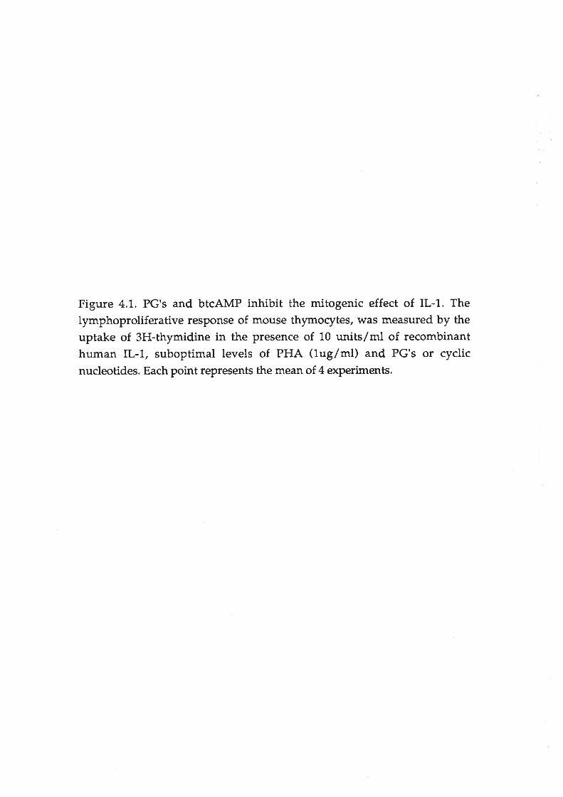
Figure 4.1.. PG's and btcAMP inhibit the mitogenic effect of IL-l. Thelymphoproliferative response of mouse thymorytes, was measured by theuptake of 3H-thyrnidine ín the presence of 10 units/ml of recombinanthuman IL-1., suboptimal levels of PHA (lug/ml) and PG's or cyclicnucleotides. Each point represents the mean of 4 experiments.

140
120
100
80
60
40
20
0
=.ìo6
s
Figure 4.1
010' 10 10 10 ro 3 10 4 10
nM
-È--'_--*-+
-+_-.ts
PGEl
PC€2
PGEs
MPL
bt cAMP
bt cGMP
2 2 5 6 710- 10 10

Figure 4.2. Misoprostol inhibits the production of a)TNF, b)IL-l (in rat and
mouse cells) and c) IFNT but stimulates IL-6 production. Fluman PB
mononuclear and rodent adherent peritoneal cells were incubated with 5ug/ml LPS (except for IL-6 assays) and Misoprostol at variousconcentrations. After 24 hours the supernatants were sampled and theircytokine content measured as described in the methods. Each pointrepresents the me¿ìn + the standard error for at least 4 human donors(squares) and at least 3 experiments for mice (circles) and rats (triangles).

.=
.za)6
*
140
120
100
80
60
40
20
120
100
80
60
40
20
1000
100
120
100
80
60
40
20
0
Figure 4.2
a) TNF
b) rL-1
c) lL-6
d) rFN T
.=
.za)o
àe
:.:C'o
àQ
a)6
èe
11 10 100nM
1 000 1 0000 1 00000

Table 4.1Inhibitory effect of E-prostaglandins on the productionl of. al TNF. bl IL-l and
cl IFNYby three twes of mononuclear phagocytes"
IDso lnsoleslD_2
Cells Human PBM Mouse AP Rat AP
a) TNFPGElPGEZPGE3
MPLbtZcAlvfP
43.1 t]6.1344.3+ 20.2
126.6 + 60.3
750.7 + 256.6
18"300 + 6,300
N.D.4N.D.N.D.N.D.N.D.
17.3!8.234.5 + 11.0
274.6+ 95,7
232.5+ 69.1,
77,900+ 9,670
b) rl,-lPGErPGEZPGE3
MPLbt2cAMn
>1,000
>1,000
>1,000
>10,000
692,001186,000
81rX261.953 fl48l,!23+3'1,37,100+1740456,000 x49,500
24.1+9,520.2+0.3t70 !28.9346+76.660,000 +2890
c) IFNTPGEtPGEZPGEg
MPLbt2cAlvIP
29.8 +11.3
33.1+10.239.3 +13.3
591 +168
29,700-5,070
_5
1 As measured in, a) Lg29 rytotoxicity assay, b) 4375 cytotoxicity assay and c)IFNy immuno assay.2 Concentration (nanomolar) which inhibited by 50% the activity of cytokineproduced after stimulation with Sug/ml LPS (for IL-l and TNF) or Sug/ml Con A(for IFNf. Values were derived from dose-response curves based on data derivedfrom the means of duplicate experiments, using at least 5 donors for human PBMcells and 3 separate experiments using animal AP cells. Endogenous PGproduction was suppressed with 20 uM Piroxicam (see Methods).3 Standard error.4 Not detectable following LPS stimulation of C3H/Het mouse Ap cells.5 Not tested.

56
these PGE's. Dibutyryl cAMP at concentrations greater than 670uM reduced IL-1
activity produced by human PBM cells by more than 50%,IFNyproduction could
only be measured in experiments with human cells since the immunoassay used
was specific for human IFN1. Like TNF production, IFNI production was
inhibited by all4 PGE's and btcAMP. Summarising the results given in table 4.7, a)
PGEI and z were just as effective at inhibiting TNF, IL-L and IFNy production by
the cell types tested; b) PGEg was slightly less effective; and c) Misoprostol was
always markedly (often 10-40 times) less effective than PGEr and PGE2.
In contrast to other cytokines, the (mitogenic) activity of IL-6 present in the
supernatants from all 3 cell types was enhanced after treating the cells with the
prostanoids or btZcAMP (Table 4.2). This stimulation of IL-6 production was
observed without LPS stimulation. The relative (stimulant) potencies of the PGE's
was very similar to that noted for their inhibitory effects on the production of
other cytokines (IL-l, TNF and IFNy).
The IL-6 response of hr¡man peripheral blood (PB) cells to PG's
Table 4.3 show the response of adherent PB cells from healthy donors to various
PG's and cyclic nucleotides. The numbers of adherent cells were calculated with
the aid of a graticule as described in chapter 3. Cells from 5 different donors all
responded to the three PG's at concentrations of 1. uM, and to Misoprostol at 10
uM, by increasing IL-6 production by about 2-3 f.old. 1mM cAMP strongly
stimulated IL-6 production while 1 mM cGMP only slightly stimulated IL-6
production. cAMP and cGMP together were less potent than cAMP alone
indicating that the effects of the two cydic nucleotides were not additive. The last
3 columns of table 4.4 shows the combined data for 3 types of PB cell populations.
The PG's and nucleotides had similar effects on adherent and non-adherent
populations; however the nonadherent population produced about 5 fold less IL-6
per cell than the adherent population.

Table 4.2
Stimulatory effect of E-prostaglandins on the production of IL-6 activityaby
three types of mononuclear phagocytes.
ED¿(nmoles/L)F
Cells Human PBM Mouse AP Rat AP
Treatment
PGEt
PGEZ
PGE3
MPL
btZcAMP
180 + 45.23
417 + 1.60
463 + 12'1,
966+225
437,000 + 18ó,000
23.0 +8.3
7.t+ 3.0
180 + 12.6
140 r 38.3
4J2,600 + 9,650
24.2+9.5
20.2+ 0.3
170 + 28.9
346 + 76.7
60,000 + 2,900
1 As measured in a mitogenic assay using TfD1. cell proliferation .
2 Concentration (nanomolar) which stimulated the activity of ÍL-6 produced two
fold (ED2) above control levels. Each value derived from the dose-response curves
based on means of duplicate experiments, using at least 5 donors for humans PBM
cells a¡rd 3 experiments for animal AP cells. Experiments were carried out in the
presence of 20 uM Piroxicam (see Methods).
3 Standard erïor.

Table 4.3
cells (PBMI.
Individual human PBM responses. Averaged PBM responses
D.H. S.G. D.R.H. L.B. S.R adherent non-adherent
total
277 + 641 53+7 243+28 297 !709 790+ 79 200!41. 7.6+ 0.9 237 + 37
Stimulus
control
1mM btcAVtP" btcGMP
lmM btcA + GMP
1uM PGEIluM PGEz
1uM PGE3
10uM Misoprostol
3767 + 338940 + 227
2733+ 584
1183 + 309
597 + 117
393+ 49663+ 42
1,433 + 240
370 !577367 !767
1033 r 120
400r3630 !7M
1505 r 597
454 t 129
11131,148
546 r 180
405+94444+ 111
615 + 130
36.5 + 73.6
75.2!4.915.9 r 4.5
72.5+ 0.4
25.6!7.577.9 + 3.0
21,.9 +2.6
1800 r 306
533 + 100
7060 !70
543+ 1,09
577 +242623+ 62
733+ 233
253 + 32
163 + 13
173 + 23
733 + 27
83+15138 t 28
1,07 + 24
7037 + 63
397 + 67
663 + 47
353 + 18
347 + 32
327 + 77
790 + 240
655 r 25
589 + 89
789 + 108
691, !95
407+3527 + 58
573 + 27
823 + 78
1 units/ 104 cells * standard error of at least 3 experiments for individuals and 3 donors for the pooled data.

57
Stimulation of lL-6by PG's and cAMP occurɡ at the level of nRNA ptoduction
Figure 4.3 shows that all three PG's at 1uM and Misoprostol at L0uM stimulated
IL-6 mRNA above basal levels in FSF. All these PG's stimulated IL-6 mRNA at
similar levels when detected either visually by non-radioactive digoxygenin or by
scintillation counting using 35S labeiled probes. White this radioactive probe
detected mRNA in control (untreated) FSF, the digoxygenin probe failed to detect
any mRNA by northern blot analysis. Both methods of detection indicated IL-l
was a strong stimulator of IL-6 mRNA in these cells (stronger than any of the PG's
tested).
Competition of PGE s with 3H-PGEz for cell binding
Table 4.4 shows that the binding of 3H-PGEz (5nM) to human PBM and rat AP
cells can be ma¡kedty inhibited by a 10 fold excess of cold PG's (50nM). PGE's r,z
and 3 alt inhibited by approximately 80% 3H-PGE2 binding to human PBM cells.
Misoprostol was significantly (P < 0.005) less effective, inhibitng 47% of 3H-PGEz
binding. Inhibition of 3H-pGE2 binding to rat cells was more affected by all the
PGE's tested. As noted with human cells Mísoprostol was significantly (P < 0.05)
less effective than the other PG's.
Stimulation of intracellula¡ cAMP by PGE's.
The effect of the PGE's on intracellular levels cAMP in human PBM and rat AP
cells is also shown in table 4.4. All PGE's markedly increased the levels of cAMP.
In human PBM cells PGE'S 1,2 and 3 increased the levels of cAMP approximately
lO-fotd whilst Misoprostol increased cAMP levels approximately S-fold. All the
PGE's had a similar effects on rat peritoneal cells; but the levels of cAMP detected
were approximately 100-fold less than those detected in a similar number of
human cells.
Preliminary experiments with 4 other PG's (PGA2, PGF1r, PGF2ø and PGDz)
showed that they were poor mimics of PGE's in regulating cytokines, as

Figure 4.3. PG's and IL-1 stimulate the production of IL-6 mRNA in humanforeskin fibroblasts. RNA was extracted from human foreskin fibroblastsincubated with PG's and human recombinant IL-lp for 3 hours and the levelsof IL{ mRNA detected using a digoxigenin labelled ribo-probe as describedin the methods. Each track is loaded as described below and the results of thedot blot analysis (see text) of the s¿une RNA extracts using a 35S labelled IL-6probe are given in the right hand column.
ackTr't.
2.
3.
4.
5.
6.
TreaEnentPGEr (luM)PGEz (1uM)PGEg (luM)Misoprostol (10uM)
nonefL-1p 11¡e sni¡s/ml)
DPM7,517
8,37084327,5493,679
10,801

r¿#lñs^"

Table 4.4
Inhibition of 3H-PGEA binding and stimulation of inhacellular cyclic AMP
%Inhibition 1 nM cAMP 2
Cells Human PBM Rat AP Fluman PBM Rat AP
PG
untreated
PGEr
PGEz
PGE3
MPL
0
79.9 + 12.3
85.6+9.4
88.5 + 7.6
47.2+ 6.9
0
86.4 + 5.4
88.0 + 3.5
90.4+ 6.4
73.6 + 8.8
1.43 + 0.35
13.8 + 5.12
14.6 + 2.08
14.5 + 3.11
7.00 + 0.72
0.01+ 0.00
0.19 + 0.05
0.12 + 0.11
0.15 + 0.08
0.08 + 0.07
1 Percent inhibition of binding of 3H-pGgzby a 10 fold excess (50nM) of PG.
Mean * standard error of 3 experiments.
2 Picomoles cAMP produced per 2 x 10⁵ cells. Mean * standard error of 3
experiments.

58
determined in the above assays. Only PGA2 had any significant effect at
concentrations below 1000nM (data not shown).
Stimulation of PGE production by cyclic nucleotides
Figure 4.4a shows that btcAMP at levels of 100 uM or greater strongly stimulated
PGE production by human PB monocytes and rat peritoneal macrophages. ,
Ffowever, concentrations of cAMP up to 10,000uM did not affect PGE production
by C3H/Hef mouse peritoneal macrophages. BtcGMP, while not as potent as
btcAMP, also stimulated PGE production in human PB monocytes and mouse
peritoneal macrophages. Dark Agouti rat peritoneal macrophages (AP cells) did
not respond to btcGMP and PGE production may have even been inhibited at the
highest concentrations (10,000uM). When this experiment was repeated withPorton rat peritoneal macrophages, cGMP stimulated PGE production in a similar
way to its effect on human PB monocytes. The normal levels of PGE produced per
104 cells were, 3,100pM for the Porton rat, 1,1.50pM for the Dark Agouti rat,
2,750pM for the C3H/HeI mouse and 1,700pM for human cells.
The effect of Misoprostol on PGE production
Since Misoprostol and the other PG's could stimulate intracellular cAMP (table
4.4), this in turn might stimulate PGE production. However, Misoprostol at
concentrations up to 100nM had no measurable effect on PGE production. Higher
concentrations could not be tested as Misoprostol itsetf, like PGE's 7,2 and 3 were
all detected with approximately equal efficiency in this immunoassay. Any
endogenous PGE produced would be swamped by an excess of exogenous PG's
added as stimulant.
DISCUSSION
Inhibition of cytokine action

Figure 4.4. B tzcAMP stimulates prostaglandin release by human PB
mononuclear cells, and rat AP cells but not C3H/HeJ mouse AP cells in vitro(a). BtzcGMP stimulates prostaglandin release by human PB mononuclearcells and C3H/HeJ mouse AP cells but inhibits its release by rat AP cells (b).

2000
1 000
800
600
400
200
++...........ts
'++"+
Mouse
Human
Rat
MouseHuman
Rat
Figure 4.4
1 10 100 100010000nM oAMP
a
l¡J(,È
õtroo
*
0
b
t¡¡c'ÀõEo(,
Illr
00
1 10nM cGMP
1 00 1 000 1 0000

59
PGE2 may inhibit lymphocyte proliferation induced by a variety of stimulants in
vitro and in vivo (Chapter 2). This is largely due to its inhibition of IL-2
production by lymphocytes (Chapter 2). Both PGEI and PGE3 (which may be
formed from dietary fatty acids) and the PGEI analogue Misoprostol, suppressed
lymphocyte proliferation in vitro. indicating their potential use as suppressants of
lymphoid functions in vivo.
Inhibition of cytokine production
Production of TNF,IL-1 and IL-6 was assessed by biological assays in this study.
The biological activity expressed represents the sum of the actions of a) the
cytokine produced and b) any inhibitors also present in the test sample. Therefore
we selected assays considered to be relatively specific and not likely to be affected
by extraneous PG's present in the test samples. However, we cannot rule out the
effects of other, as yet, unidentified factors that might also be regulated by any
PG's present in the test supernatants. It is, however, the total biological activity
produced (cytokine plus inhibitor) which will determine the final effects of PG's
on the rytokine mediated immune responses.
The inhibitory effect of PGE2 upon TNFp production has been well documented
(Endres et al 1989). This inhibition occurs predominantly, if not totally, at the level
of mRNA production (scales et aI 1989). PGE1, PGE3 and Misoprostol were also
inhibitors of the activity of TNF produced by human mononuclear cells and rat
peritoneal macrophages.
There has been some confusion in the literature concerning the ability of PGE2 to
regulate IL-l production. It was initially reported that IL-l production was
inhibited by PGE2 (Kunkel and Chensue 1985). Our experiments using human
PBM indicate that PGE's¡,2,3, and Misoprostol did not significantly affect IL-lproduction detected using the 4375 cell assay. Ffowever, when we used other,less
specific assays, to detect IL-l (eg LAF assay or the El-4-nobl / CTLL cell assay

60
(Ghering et al7987), the apparent IL-l activity was markedly reduced following
treatment with PGE's. This was probably due to the effects of the PG's on the
reporter assays used (Otterness et al 1988, fig4.2). Dialysis of the supernatants
only partially altered this inhibition (unpublished data) possibly indicating that
PGE's were only incompletely removed. Recent reports suggest that PGE2 does
not affect the levels of IL-1 mRNA produced by human monocytes (Knudsen et aI
1986) or mouse macrophages (Scales et al 1989).
In contrast to the results obtained with human cells, activity of IL-l produced by
rodent macrophages was reduced by PGE's as measured in our bioassay. However
the concentrations of prostanoids required were much higher than those affecting
the other cytokines (tables 4.1 and 4.2). Whilst high levels of TNF might also be
detected in this IL-l bioassay, it is unlikely that we measured only TNF
suppression. Firstly we noted this effect using peritoneal macrophages from
C3H/HeJ mice which did not produce detectable TNF (Table 4."1.,21). Secondly,
the levels of TNF found in the rat macrophage supernatants (<20 units/ml) did
not affect the action of IL-1 on 4375 cells. At very low concentrations of PGE's we
consistently observed a slight increase in IL-l activity indicating that PGE's may
possibly both stimulate (Kassis et al 1989) and suppress the formation of this
cytokine depending on its concentration. The effects of PGE's on IL-L production
may therefore vary depending upon the species and concentrations used.
Unlike the other cytokines, IFNy production was measured in an immuno-assay.
The inhibitory effect of PGE2 on IFN1 production has been described previously
and its effects on immune responses have been discussed (Cesario et al 1989).IFN1
has been extensively described as a stimulator of many monoryte/macrophage
functions, induding inflammatory cytokine production. Therefore, suppression of
IFNymay markedly affect progression of inflammatory diseases.
Stimulation of IL-6 production

67
All the prostanoids stimulated IL-6 production, in marked contrast to their effects
on the other cytokines investigated. This was noted at nanomolar concentrations
with both rodent AP and human PBM cells.It is consistent with reports that PGE1
and other stimulators of intracellular cAMP enhance IL-6 production in human
foreskin fibroblasts (Zanget al 1988).
PG receptors
Since PG81,2,3 and Misoprostol all had similar effects on the range of biological
activities investigated, they probably have the same mechanism of action and bind
to the same cell surface receptor(s). The relative abilities of the PGE's to compete
with radiolabelled PGEz for cell binding was similar to their biological activities.
This may indicate that observed differences in potency reflect their differentaffinities for such a receptor, ie PGEI=PGE2= or >PGE3>Misoprostol. Of the other
PG's tested only PGA2 had some effect (PGFta, PGF2cr and PGD2 had no effect)
probably indicating they bind to different receptor types as has been reported
(Kennedy et al 1982).
Regulation of intracellular cAMP
Cells generally respond to prostanoids by increasing the intracellular levels of
cAMP, a 'second messenger' involved in the transduction of signals from
membrane receptors to other sites within the cell (Berridge 1985). We found that
btZcAMP had the s¿une effects as these prostanoids on macrophages/monocytes.
Also, the PGE's were able to increase the intracellular levels of cAMP (in human
and rat cells) with similar relative potencies to their cell binding and their effects
on cytokines. This suggests that the effects of all 4 of the E-prostanoids are
mediated by their ability to increase intracellular cAMP.
The stimulation IL-6 production by PG's was preceded by increased levels of IL-6
mRNA (Fig a.Ð as has been reported for PGEI (Zau;rg et al 1988). This indicates
that stimulation by all the 3 PGE's (1,2 and 3) and Misoprostol was due to

62
transcriptional rather than post-transcriptional effects. Some rytoplasmic proteins
are known to be phosphorylated by cAMP-dependent enzymes (Hunt et al 1984).
Two of these in particula¡, CREB (cyclic AMP response element binding protein)
and AP-L, recognise and bind to specific sequences of DNA. If these proteins bind
to sites in the promoter region of genes, they can regulate the transcription and
eventually expression of these gene products. Sequences corresponding to CRE
sites have been identified in the promoter sequence oÍlL-6 (Tanabe et al 1988) and
many other genes. Therefore the 3 PGE's (1,2 and 3) and Misoprostol's may not only
induce CREB to promote IL-6 transcription but (at the same time) regulate
transcription of other cytokines. For example, the promoter region of TNF, which
is inhibited by PG's and cAMP, has sequences which closely resemble CRE and
AP-1 (Economou et al 1989).
Generally, we found that human PBM cells were less sensitive to the PGE's than
rodent peritoneal cells. Flowever, rather than reflecting a species difference, this
may indicate that mature peritoneal macrophages are more sensitive than
immature peripheral blood monorytes and lymphocytes. This is supported by the
observation that rat AP cells produce much less intracellular cAMP, either
normally or following PGE rreatrnent, than human PBM cells. More studies would
be needed to determine if this is due to increased sensitivity of the rodent to
intracellular cAMP, or to other factors, such as differences in the numbers of PGE
receptors.
Regulation of PGE production by cyclic nucleotides
BtcAMP was found to stimulate PGE synthesis in human PB monocytes and rat(Dark Agouti and Porton strains) peritoneal macrophages. Surpiisingly btcGMp
also stimulated PGE release but at higher concentrations than cAMP. The Dark
Agouti rat was different from the Porton strain in that cGMP inhibited PGE
release at the highest dilution. Most of these effects were noted at high

63
concenEations of the dibutylated cydic nucleotides and it is difficult to determine
if the levels of intracellular cyclic nucleotide attained are similar to those found
under physiological conditions. This stimulation of PGE synthesis by cyclic
nucleotides contrasts to a report that they inhibit prostanoid synthesis in platelets
(Alverez et al 1989).
Since Misoprostol and the other PGE's will stimulate higher intracellular cAMP
levels, if was of interest to see if they also can stimulate PGE release. However,
Misoprostol did not stimulate PGE release at concentrations up to 100nM.
Unfortunately higher levels could not be tested since the polyclonal antibody used
not only detected PGE in the immunoassay but also the exogenous Misoprostol.
This may indicate either, i) higher concentrations of Misoprostol are needed to
induce the required levels of cAMP for stimulation of PGE release, or ii) although
high enough levels of cAMP. are attained, binding to the PGE receptor induces
other signals which repress PGE release. If PGE's do stimulate their own release
this would be a potent mechanism by which PGE's can amplify acute
inflammation by a positive feedback effect.
PG regulation of cytokine mediated inflammation
The ability of E-prostaglandins to regulate the production of inflammatory
cytokines and other MNP functions described here, may be examples of natural
feedback mechanisms for controlling inflammation. Suppression of MNP
spreading, IL-l. action, IFNy, TNF and, occasionally, IL-1 production, whilststimulating the comparatively less acutely cytotoxic rytokine IL-6 , may lead to an
overall reduction of inflammation. IL-6 is also a potent stimulator of acute phase
protein production by the liver (reviewed Segal et al 1.989, Le and Vilcel 1989), and
a coÍunon function of these acute phase proteins may be to help restore the
homeostatic balance of the inflamed tissue (Koj 1935).

64
These regulatory effects may be particularly relevant during the acute phase of the
inflammatory process, such as active arthritis, where high levels of PGE2 have
been detected in synovial fluids (Robinson and Levine1974). These concentrations
(1-100nM) are similar to those at which we observed significant effects in vitro.
Since MNP's not only produce PGE's, but also are affected by them, PGE's effects
need only be short-ranged. Therefore, much higher effective concentrations in this
microenvironment may be attained. In addition, in this study we have often
assessed the PGE's suppression of responses to strong stimuli (10u/ml IL-l and
Sug/ml LPS), but these PGE's may be even more effective at suppressing
responses to weaker stimuli.
Such a regulatory mechanism has two major implications for current and
prospective therapies. Firstly. the commonly used NSAIDs which strongly inhibit
PG production, may be effective at relieving some symptoms of inflammation (eg
pain, vasodilatation etc) but might not reduce the long-term progression of the
disease sustained or mediated by inflammatory cytokines (eg IL-l, TNF etc).
Secondly, dietary control of inflammation by fish oils etc, may be more effective in
the long term if allowed to augment the natu¡al regulation by PGEz by producing
additional PGE1 or 3. Conversely, if dietary treatment were to result in lower
levels of total PGE (ie PGE2 + 1 or 3), as has been reported (Broughton 1991), the
suppressive effects of PGE's may be reduced. Flowever, suppression of cytokine
production by modiff^g the production of leukotriene 84 (Endres et aL1989, Rola-
Pleszczynski and Lemaire) or other products of lipoxygenase (Sirko et al 1991)
may balance/replace the loss of PGEz suppression.
Misoprostol was developed to reduce the damaging effects of NSAIDs and other
gastrotoxins on the stomach. However, recent findings suggest it may have other
beneficial effects such as prolonging the survival of transplants. This may reflect
the immunosuppressive (Wiederkher et al 1,990) and antiinflammatory(Whitehouse et al 1990a) effects of the drug. Although the data shown in this
65
report was generated with the ester form of Misoprostol, the acid form showed
identical activity in all the assays used (data not shown). This probably indicated
that cells used in these assays contain sufficient esterase activity to rapidly
generate the Misoprostol free acid. In all the experiments, Misoprostol closely
resembled the naturally occurring prostanoids, supporting the suggestion that
stable synthetic PG's might be useful in therapy where such immune regulation is
beneficial (Nicholson 1990). Its use in conjunction with other therapies reducing
PG production, eg NSAID or dietary controls as described above, may be of
particular value.
The results of this study indicate that PG's can modify the cytokine-dependent
immune responses. However the potential immunoregulant action of PGEI and
PGE3 indicate that diet enrichment with their precursor fatty acid might also affect
certain immunopathies by modifying the proportions of cyclooxygenase products
(PG's).

66
Chapter 5
Cyclosporin prevents experimental arth¡itis in rats by regulating leucocyte
subpopulations and infl ammatory mediators.
INTRODUCTION
The fungal cyclic peptide, cyclosporin (CsA), has been used extensively to
promote the survival of organ grafts (Morris 198'j,, Cohen et al 1984). CsA is very
effective in preventing allograft rejection and elicits less side effects in the graft
recipients than many other forms of immunosuppressive therapy (eg. Nitrogen
mustards, antimetabolites). Flowever, despite its wide usage, the exact mode of
action of CsA remains unclea¡.
Extensive in vitro studies have shown that CsA will selectively inhibit a number of
immune functions, and in most cases this is attributable to its suppression of Tlymphocyte activity. This inhibition may cause not only selective depletion of T-
helper cells (Clerici and Shea¡er 1990) but also suppress accessory cell (Varey et al
1986, Esa et al 1988) and B lymphoryte functions (Klaus 1983).These observations,
and the success of CsA in transplantation,led to the evaluation of CsA for treating
a variety of autoimmune disorders induding rheumatoid arthritis (Dougados et al
1988, Sany 1990).
CsA has been shown to be effective in the treaûnent of experimentally induced
arthritis in rats and mice (del-Pozo et al 1990, Cannon et al 1989, Rofe et al L990,
Rofe et aI1'992). Not only does CsA treatment reduce the severity of disease, buttreatment during the induction period also prevents development of adjuvantarthritis and renders the animals resistant to further challenge with the same
arthritogen (Whitehouse and Vernon-Roberts 1989). Surprisingly the effect of CsA

67
on lymph nodes draining the site of adjuvant injection and adjuvant disease
(paws) indicated that CsA modified, rather than suppressed, immune responses to
the arthritogenic adjuvant, probably by selective suppression of some T
lymphocyte rytokines.
MATERIATS AND METHODS
Adiuvant disease
Adjuvant arthritis was induced as previously described (Haynes et al 1988b).
Briefly, on Day 0, groups of male Dark Agouti (-2509) rats were injected
subdermally near the tail base with 50ul of finely ground, heat-killed, delipidated
mixed human strains of Mycobacterium tuberculosis dispersed in squalane at a
concentration of 1Omg/ml. Co-administration of CsA with the adjuvant was
achieved by grinding the CsA (100 mglml) in with the adjuvant. In some
experiments, CsA was injected subcutaneously into the hind flank of the animal.
On Day 14 after adjuvant injection, the polyarthritis was assessed by scoring each
paw for disease severity as previously described (Whitehouse et aI 1990b).
Detection of CsA in the blood
Blood levels of CsA were measu¡ed with an immunoassay ("Cyclotrac" SP RIA -
Incstar Co.p. Minnesota, USA) by Dr. Ray Morris at the Queen Elizabeth Hospital,
Woodville, S.A.
Preparation of rat paws for histology
Rat rear paws were amputated from animals sacrificed on days 3, 7 and 14 afterfçìnèin\{ìd
adjuvant injection. The skin was carefully removed and the^tissue fixåd in a 70Vo
formaldehyde solution for 1. week before hansfer to a solution of "Decal" reagent
(Omega Chemical Corp. New York, USA) for decalcification for 36 hou¡s at37oc.The paws were cut sagitally, embedded in paraffin blocks and sectioned when
required. Sections were taken from the central part of the paw.

68
Cell isolation
Peripheral blood mononuclear (PBM) cells were isolated as previously described
(Haynes et al 1988a). Popliteal lymph nodes (PLN) and inguinal lymph nodes
(ILN) were removed immediately after the rats were killed by cervical dislocation
and kept in cold Hank's buffered salt solution (HBS). The ly ph nodes were
passed through a fine wire mesh and the single cell suspensions washed in HBS.
Adherent peritoneal (AP) cells were isolated as described in previous chapters.
3H-thymidine uptake
Triplicate 100u1 cultures of the PBM, PLN and ILN cells in RPMI-1640 medium
containing 2 x 10⁵ cels were incubated with 50ul of 0.5 uci 3g-thymidine in 9G
well microtitre trays,3 hours later the cells were harvested on glass filters and 3H-
thymidine incorporation measured as described (Haynes et al 1988a).
Cytokine production (ex vivg)
2 x 10⁵ PBM, PLN and ILN cells were incubated in a final volume of 250u1RPMI
medium with 10% FCS. These cells were unstimulated or stimulated with either 5
uglml LPS or 5 uglml Con A. After 24 hours the supernatants were sampled añd
assayed for the various cytokines. The in vitro effect of CsA on PBM, PLN a¡rd AP
cells isolated from normal Dark Agouti rats was carried out in the same manner
with the addition of CsA at various concentrations. Samples of supernatants were
assayed immediately or stored at -70oC for up to one month before assay.
Assays for cytokines
The following assays were carried out as described in Chapter 4.IL-7 was assayed
using the IL-l sensitive A375 cell line (Nakai et al 1988). Although these cells are
also sensitive to high levels of TNF, the levels of TNF in the supernatants were
always too low to be detected at the dilutions used. TNF was assayed using the
TNF-sensitive cell line L929 (Matthews and Neale 1,987).7TD1, hybridoma cells
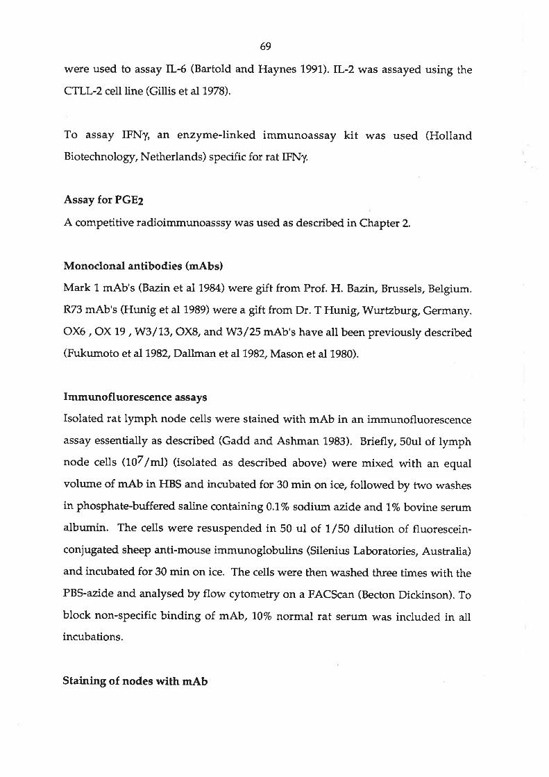
69
were used to assay IL-6 (Ba¡told and Haynes 1991).IL-2 was assayed using the
CTLL-? cell line (Gillis et aJ1978).
To assay IFNy, an enzyme-linked immunoassay kit was used (Holland
Biotechnolo gy, Netherlan ds) specific for rat IFNy.
Assay for PGE2
A competitive radioimmunoasssy was used as described in Chapter 2.
Monoclonal antibodies (mAbs)
Mark L mAb's (Bazin et al 1984) were gift from Prof. H. Bazin, Brussels, Belgium.
R73 mAb's (Hunig et al L989) were a gift from Dr. T Hunig, Wu¡tzburg, Germany.
OX6 , OX 19 ,W3/'1,3, OX8, andWS/25 mAb's have all been previously described
(Fukumoto et al 1982, Dallman et aI1982, Mason et al 1980).
Immunofluorescence assays
Isolated rat lymph node cells were stained with mAb in an immunofluorescence
assay essentially as described (Gadd and Ashman 1983). Briefly,50ul of lymph
node cetls (107lml) (isolated as described, above) were mixed with an equal
volume of mAb in HBS and incubated for 30 min on ice, followed by two washes
in phosphate-buffered saline containing 0.17o sodium azide and1,% bovine serum
albumin. The cells were resuspended in 50 ul of 7/50 dilution of fluorescein-
conjugated sheep anti-mouse immunoglobulins (Silenius Laboratories, Australia)
and incubated for 30 min on ice. The cells were then washed three times with the
PB$azide and analysed by flow cytometry on a FACScan (Becton Dickinson). To
block non-specific binding of mAb, 70Vo normal rat serum was included in all
incubations.
Staining of nodes with m.A,b

70
Lymph nodes required for staining with mAb were immediately embedded in
OTC compound (Miles Scientific, Il., USA) and stored at -70oC before 5 uM
sections were cut on a cryostat. The staining of sections was carried out within 24
hours on serial sections as described (Mayrhofer and Spargo 1,990).
Statistical analysis
Statistical significance values were calculated using a Student's t test (Snedecor
and Cochran 1989).
RESULTS
(summarised in table 5.7)
The effect of CsA on the expression of adjuvant-induced arthritis and associated
weight loss
Table 5.1 shows that both the weight loss and disease onset were prevented when
CsA was administered mixed with the arthritogenic adjuvant (given at day 0).
Subcutaneous administration of 10 mg/kg CsA every second day from day -L to
day 13 or from day -1 to day 7 was equally effective at preventing disease and
weight loss.
Histological examination (Fig 5.1) of the rear ankle joints and surrounding tissues
from untreated polyarthritic animals (Fig 5.1.a) showed there was a large influx ofpolymorphonuclear cells, lymphocytes and macrophages. occasionalmultinucleate giant cells (Fig 5.1.b) were present in the eroded pits of bone and
there was a large amount of cartilage destruction associated with an ingrowth of
thicker synovial membrane (Fig 5.1.c). Neither destruction of bone and tissue nor
the presence of multinucleate grant cells and polymorphonuclear cells were seen in
or around the ankle joint of CsA-treated animals (Fig 5.1.d), which appeared
identical with those from normal animals (no adjuvant treatrnent).

Table 5.1
The effect of CsA treatonents on the induction of arthritis by a mycobacterial
adiuvant (Adi) in male Dark Agouti rats.
Treatment Number of rats Severity of
arthritislWeight change
(gttt)2
None
Adj
Adj + CsA3
Adj + CsA s.c. (Days-ltofa
Adj + CsA s.c. (Days-1to13)a
20
31
32
5
9
0
7.85 + 0.70
0
0
0
+32.6 + 4.2
-21.5 + 3.0
+25.4+ 2.4
+32.2 ! 7 .8
+17.3+ 5.3
1 Mean * standard error scored 14 days after inoculating adjuvant in tail base.
The total score per rat was calculated by tallying the scores for each rear paw(maximum 4 each) and front paw (maximum 3 each).
2 The mean * standard error of the weight change over 14 days.3 +Omg/Kg CsA admixed with the Adjuvant injected in the tail (see methods).4 tOmg/Kg CsA injected subcutaneously every second day.

Figure5.l. The histology of the rear rat paw (ankte joint) shows the destruction of tissue and bone occurring during
expression of a¡thritic disease (day 14) in rats injected with adjuvant alone (5.1a). The soft tissue is crowded withinflammatory cells, mainly polymorphonuclear, but many mononuclear cells are Present. There is extensive erosion
of the bone and multinucleate giant cells are often seen in the eroded pits (5.1b). Loss of cartilage is associated withan ingrowth and thickening of the synovial membrane (5.1c). [r contrast none of these destructive and inflammatory
processes are seen at day 14 in rats injected with CsA admixed with the adjuvant (5.1d). The histology was identical
to that seen in normal healthy rats (not adjuvant injected or CsA treated).

II
,)
t
l¡å,t'
I

71,
Blood levels of CsA following administration of CsA admixed with the adjuvant
are shown in Figure 5.2. High levels (> O.g uM) were detected for the first 7 days
post injection, falling to less than 0.1 uM on day 1.4.
Analysis of lymphoid cell populations
Figure 5.3 shows the yields of lymphoid and blood cells isolated from Dark Agouti
rats injected in the tail base with either adjuvant alone or a mixture of adjuvant
and 40 mg/Kg CsA. 3H-tn)rrt idine incorporation by these cells is also shown.
Cells were isolated from 3 separate sites, namely mononuclear cells from the
peripheral blood (PBM), total cells from the inguinal lymph node 0LN) and total
cells from the popliteat lymph node (PLN). The yields of cells and the uptake of
3H-thymidine by PBM did not vary significantly throughout the 14 day period of
the experiment (Fig 5.3.a). Flowever, there was a slight reduction in 3H-thymidine
labelling on days 3 and 7 in the CsA-treated group. The yields of cells isolated
from the ILN increased markedly on days 3 to 1.4 in both groups of animals (Fig
5.3.b). 3H-Urymidine labelling rose dramatically only on day 3; however, the levels
were significantly lower (P< 0.05) in CsA-treated animals. The yields of PLN cells
(Fig 5.3.c) from both adjuvant and adjuvant/CsA-injected rats significantly
increased after day 7. 3H-Thymidine labelling was slightly greater on day 3 in bcith
groups but lower in cells from CsA-treated animals than those from untreated
animals. There was a ma¡ked increase in 3H-tn)rmidine uptake by PLN cells from
both groups of animals on day 14 .
PLN cells were isolated from normal, adjuvant-injected and adjuvant/CsA-
injected rats on day 74. Similar results were obtained when CsA was administered
subcutaneously. Subpopulations of cells were labelled with monoclonal antibodies
and compared by flow cytometric analysis (Table 5.2). There was no significant
difference between the three groups in the percentages of cells binding mAb
W3/13 (CD43, pan T cell ma¡ker) or OX8 (CD8+ subset of T cells). The proportion
of OX6 positive (MHC class II) cells was significantly higher (P<0.005) in all the

Figure 5.2. Levels of CsA detected by a immunoassay in the blood of ratsgiven a single dose co-administered with the adjuvant in the tail base. Eachpoint represents the mean * standard error of 3 experiments.

oo
=3
0.8
o.7
0.6
0.5
0.4
0.3
o.2
0.1
0.0
Figure 5.2
-10 1 2 3 4 s 6 7 8 9101112131415Day post adjuvant lnoculatlon

Figure 5.3. (i) The yield of and (ü¡ et thymidine uptake by, (a) mononuclearcells isolated from the peripheral blood, (b) total cells from the inguinallymph node and (c) total cells from the popliteal lymph node. Each pointrepresents the mean + standard error of 6 experiments.

Ptt (t
Dt rodscul uPI1.l8 (ll
DDrfiæq ¡[ânþ. tæd trq91
'1 €1 Z1 11 0r 6 3 ¿ t ¡ t Ê Z I 0 1-
¡cPou
Lþnrñæul ¡nü{pr ræd ¡(:O1l
sepou'q
uoFqncou! ¡u!^nlpr ¡¡od ¡(rO9t'1Ê121 ]t01 6 I ¿ 9 I
' € Z I 0 t-
Poolq
(m0l
IIcaga
taxo\qã$ oz
01
01 goz-xoE É"0'EÃoso09
0ür9 ÞI t*, àI (þos 1 5
0
0ool g3
oo0z ìÞa
ooos ga
(xþÞ
z
,9
goxQrt;ôL
Plcl^ (I
tþf¡r¡d¡ofl¡lUPI-Hs (ll
ptel (r
lþfitrdioor¡t Upl-He (tl
E'S ernoll
3
0l
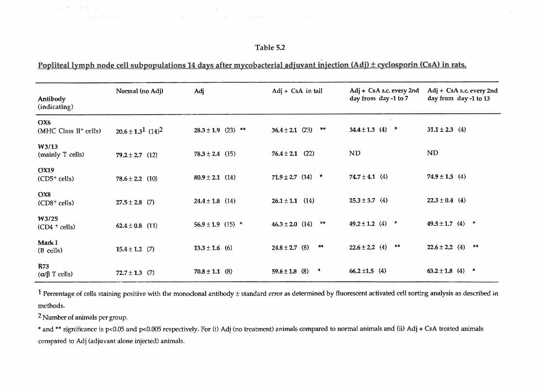
Table 5.2
Nonnal (no Adj) Adi Adi + CsA in tail
n.6t131 ioÐ2 28311.9 (23) ** 36.4!Lt Q3)
Adj + CsA s.c. every2ndday from day -1 to 7
Adj + CsA s.c every 2nddayfrom day-1to13Antibody
(indicating)
ox6MHC Class II+ cells)
w3/13(mainly T cells)
oxl9(CDS+ cells)
79.2tL7 (72) 78st2.4 (15)
80.91 2.1 (14)
76.4+ Lt (22',)
71.9tL7 (14)
34.411.3 (4)
ND
74.7 + 4.1 (4)
31.1r 2.3 (4)
ND
74.9t13 (4)
**
*
rl
78.6+ L2 (70)
ox8(CD8+ cells) 27.s!2.8 (7) 24.4!1.8 (74) 26.1+r.1 (14) 2s.3+ 3.7 (4) 223t0.4 (4)
w3125(CD4 + cells) 62.4 r 0.8 (11) 56.911.9 (15) * 46.3!\0 (14) r+¡+ 49.2t1.2 (4) 49.s!1.7 (4) *
MarkI(B cells) 15.4+1.2 (n 1331 1.6 (6) 24.8tL7 (8) r[¡+ T2.6!2.2 (41 ** 22.6t22 (4) Itt+
R73(o/p T cells) 72.7 + 1.3. (7) 70-8 r 1.1 (8) s9.6r L8 (8) ri 66.2 tl.s (4) ß.211.8 (4) ¡;
1 Percentage of cells staining positive with the monoclonal antibody + standard error as determined by fluorescent activated cell sorting analysis as described in
methods.2 Number of animals per group.* and ** significance is p<0.05 and p<0.005 respectively. For (i) Adj (no treatment) animals compared to normal animals and (ii) Adi+ CsA treated animals
compared to Adj(adiuvant alone injected) animals.

72
groups that had received the adjuvant compared with normal animals (no
adjuvant). The proportion of cells staining with Mark I mAb (B cells) was
unchanged in animals receiving adjuvant alone. Flowever, the proportion of cells
staining with OX6 and Mark I antibodies was significantly greater in rats receiving
adjuvant plus CsA. The proportion of cells labelled byWS/25 mAb (CD4+, T cells
and some macrophages) was reduced in all groups receiving adjuvant and lowest
in the groups which also received CsA (P<0.005 when compared to adjuvant
alone). Similarly, the proportion of cells staining with R73 antibodies (crlÞ T cells)
was also reduced significantly, particularly in those groups receiving adjuvant and
CsA. The proportion of cells staining with OX19 (CDs+ T cells) was only reduced
slightly by CsA treatment. CsA administered by subcutaneous routes (either on
days -1 to 7 or days -1 to 13 to adjuvant treated rats) gave similar results to CsA
admixed with the adjuvant.
Immunohistology was performed on PLN isolated from animals at day 1.4. Figure
5.4 illustrates that CsA treatment (CsA admixed with the adjuvant) had little effect
on the appearance of PLIrI following adjuvant injection. CsA treatment often
resulted in lager nodes, as represented in Figures 5.4b., d., f. and g. However, this
was not reflected in a significant increase in the yield of cells from these nodes (Fig
5.3). Lymph nodes isolated from both groups of animals had the features of
reactive lymph nodes, with expansion of both B cell areas (follicles) of Tdependent areas (paracortex). Staining with R73 antibodies indicated that large
numbers of a/þ positive T cells were evenly distributed throughout the
paracortical and cortical region in nodes from animals given adjuvant alone (Fig
5.4.c). Flowever, CsA treatment resulted in large circula¡ areas in the paracortical
region which were not a/Þ positive T cells (Fig s.a.d). A serial section of this node
stained with Mark 1 antibodies showed that these ¿ìreas were probably B cells and
indicated the presence of follicles in the paracortical region (Fig 5.a.0. This was not
noted in nodes isolated from animals injected with adjuvant alone (Fig 5.4.e).
Staining with OX6 mAbs indicated that the class II positive cells were mainly B

Figure 5.4. Immunohistology of PLN from rats injected with adjuvant (5.4a,c, e, g) and CsA admixed with adjuvant (5.4b, d, Í, h) rats was carried outwith mAb directed against various cells subset markers as described in thetext. Control antibody (an IgG class 1 mAb directed against a giardia cell wallantigen) did not stain PLN from either group of animal indicatingbackground staining is low (5.4a and b). Staining patterns obtained with themAb's R73 directed against the crlp T cell receptor (5.4c and d), Mark Idirected against cell surface immunoglobulin (5.4e and f) and OX6 directedagainst Class tr antigens (5.4g and h) are shown. Magnification -20x.

ba
\
PIt!
dc
te
hg

73
cells in nodes isolated from both groups of animals (Fig 5.a.g and h). The other T
cell mAbts (\,{l/13, OX79, OX8 and W3/25), like the R73 mAb, stained the
paracortical region of nodes from animals injected with adjuvant alone but large
circular areas, consistent with B cell follides, remained unstained in the paracortex
of nodes from animals treated with CsA.
Cytokine production by cells ex vivo after CsA treatment in vivo
Table 5.3 shows the effect of CsA treatment on the ability of PLN cells to produce a
number of cytokines. Although the results represented here were obtained with
animals injected with CsA admixed with the adjuvant, simila¡ results (for PLN,
PBM and AP cells) were obtained when the CsA was administered
subcutaneously. Cells were isolated from control animals and from animals during
the initiation (days 1 and 3) or the establishment phases of the arthritic disease.
Unstimulated cells produced levels of all cytokines (except IL-6) below the
detectable limits of our assays. Con A stimulated secretion of IFNT IL-2, TNF and
IL-6 but had little effect on IL-l secretion, while LPS induced secretion of IL-1.,
TNF and IL-6. The secretion of all cytokines, except TNF, was increased in animals
that had received the arthritic adjuvant. CsA treatment significantly suppressed
(p<0.05) IFNy(Days 1 and 3),lL-2 (day 1), and IL{ (day 1,4) production by Con A-
activated ly^ph node cells from adjuvant stimulated animals. TNF production
was also reduced by in vivo CsA treaunent (measured on days 1.,3 or 14) but this
was not significantly (p<0.05) different from animals given adjuvant alone. IL-l.
and IL-ó secretion was also significantly (p<0.05) suppressed by CsA in LPS-
stimulated lymph node cells on day 1,4.
CsA treahnent had similar effects on cytokine secretion by PBM cells (table 5.4) but
there was no effect on secretion of IL-6.
The effect of CsA treatment on cytokine secretion by adherent peritoneal (AP) cells
stimulated with Sug/ml LPS is shown in table 5.5. Although no IL-2 or IFNy could

Table 5.3
Group\C¡okine
Normal
Day11
Adi
Adi/CsA
Day 3
AiAdj/CsA
Day 14
Adj
Adj/CsA
24.3 + 5.0 <1
6.75 + 0.78* <1
IFNy+ConA
38.7 + 2.5
76.6+ 6.7*
5.58 r 1.39
3.60 + 0.20
86.0!1,6.6 <2
27.8+'1.0.4* <2
lL-2+ConA
55.0 t 14.4
75.5 !32.5
50.0 r 9.0
42.0+ 74.7
(CsAl.
IL-1+ConA
2.8 !7.22.6 !0.7
<2
4.53 + 0.68
TNF+ConA
57.0+ 76.7
27.0+7.3
IL{+ConA
156 r 85.6
253t35.2
37.4!22.0
2.98 r 1.38
3.81+1.132 <1 35.5 + 8.13 <2
+LPS +LPS +LIIS
4.8 +2.9 92.5 + 1.83 2.1, + t.4 0.59 10.104 0.63 + 0.22
+LPS +LIIS
<2
<2
<2
<2
.23
<2
<2
2.5!2.5<2
2.3!7.3<2
3.4 !2.07.8 r 1.0
<1
<1
<1
<1
13.6 r 0.86 10.9 r 2.86
3.59 + 7.23* 5.53 + 2.75
25.0 !7.5
8.5 rl.l3.8 r l_3
2.1 ! 0.7
<2
<2
15.9 t 13.3
42.8 !22.915.3t 8.5
4.ß !0.67
74.5+2.8
36.6 + 4.5*
6.69 + 0.77
37.8 + 7.27*
1 Post-injection with adjuvant.2 ng/7Os ce[s of IFNy measured in a immunoassay as described in methods. Mean + standard error of at least 4 animals.3 units/105 cells of cytokine measured in biological assays as described in methods. Mean + standard erïor of at least 4 animals.a units/103 ceils of cytokine measured in biological assays as described in methods. Mean + standard error of at least 4 animals.. p<0.05

Table 5.4
mycobacterial adiuvant (Adil t cyclosporin (CsAl.
Group\C¡okine
Normal
Day 11
Adi
Adj/CsA
Day 3
Adi
Adj/CsA
Day 14
Adi
Adj/CsA
18.8 r 11.5 <1
1.50 + 1.34* <1
<2
<2
<2
<2
<2
<2
<2
<2
<2
<2
0.73fl.ß2 <1 12.5 + 3.13 <2 a23 3.2 fl.6 20.0 + 7.73 95.0 + 3.5 0.81 10.214 6.7 + 0.42
IFNy+ConA
2-13+099
3.3812.1
0.99 t 0.16
0.74+ 0.1't
+LPS
tL-2+ConA
8.5 + 3.7
14.3 + 0.6
24.3 + 5.4
38.7 !23.9
325!6.418.0 r 3.3
+LIIS
IL-1
+C-onA
TNF+C-onA
35.4 + 31.4
36.8!73.2
IL.6+ConA
0.37 + 0.07
0.41 + 0.13
+LPS
74.3!2.411.8 + 2.0
3.5 + 1.7
7.8 + 0.9
5.9 r 0.8
2.9 + 0.6*
+LPS
455 + 25.7
62.3t?2.8
+LIIS
74.3 + 3.7
14.0 + 6.5
<2
<2
<1
<1
<1
<1
29.5 f8.9
9.30 13.90
95.0 r 32.0
119 !45.60.3810.05
0.22 r 0.05
12.8 r 8.8
72.6!6.7
25.0 r 11.0
28.5 r 11.9
63t76.790.3 r 3.6
6.70t7.79 76.0+2.76
1.01 + 0.34* 4.78 + 1.58*
1 Post-injection with adjuvant.2 ng/705 ceils of IFNy measured in a immunoassay as described in methods. Mean + standard error of at least 4 animals.3 units/105 cells of cytokine measured in biological assays as described in methods. Mean + standard error of at least 4 animals.a units/103 ceils of cytokine measured in biological assays as described in methods. Mean + standa¡d error of at least 4 animal*p<0.05

Table 5.5Cytokine production by lipopolysaccharide (Sudml) stimulated adherent
Peritoneal cavih' cells during the stages of disease initiation and expression.
Cells isolated from DA rats with/without mycobacterial adjuvant (Adi) tcyclosporin (CsA).
Group\CyOkine IL-1 TNF tL-6
Normal
Day 31
Adj
Adi/CsA
Day 7
Adi
Adj/ CsA
Day 14
Adi
Adi/CsA
75.2 +2.9
10.3 + 4.8
18.5 + 5.5
53.8 r 7.5
38.5 + 6.2*
25.2+ 5.2
73.8 + 2.9*
89.5 + 12.1
37.8 + 8.3
17.8 + 5.0*
745 + 32.4
32.4 + 1.4.7*
277 + 55.5
47.3 + 23.3+
34.4 + 4.8
255 + 111
295 + 103
502+ 747
82 + 28*
763 + 22
63+ 27'
1 Post-injection with adjuvant.2 units/105 cells of cytokine measured in biological assays as described inmethods. Mean * standard error of at least 4 animals.3 units/103 ce[s of cytokine measured in biotogical assays as described. inmethods. Mean * standard error of at least 4 animals.- p<0.05

74
be measured, substantial amounts of IL-l, TNF and IL-6 were produced by these
cells. During the early stages of disease initiation (Day 3) CsA treatment only
affected TNF production, significantly reducing its levels. During the later stages,
just before and during disease expression (Days 7 and "1.4),IL-1., TNF and IL-6
production was markedly reduced by CsA treatment. CsA tended to raise
production of these cytokines to the levels produced by normal (untreated and
non-adjuvant injected) animals.
Effects of CsA on secretion of rytokines and PGE2 by cells in vitro (table 5.6)
Cytokine production was also assessed following stimulation of normal popliteal
lymph node (PLN), PBM and AP cells !n vitro with either Con A or LPS in the
presence of graded concentrations of CsA. Table 4 shows the concentrations of
CsA calculated to inhibit cytokine productionby 50%.IFNy and IL-2 production
by Con A-stimulated PLN and PBM cells were particularly sensitive to CsA
treatment with ID5g's of about 10nM or less. Con A stimulated TNF production by
PLN and PBM cells and this was also inhibited by CsA in vitro (ID5g's < 100nM).
By contrast,IL-6 production was only inhibited by relatively high concentrations
of the drug (ID5g's > luM) . Production of IL-l by LPgstimulated PBM cells was
inhibited at moderate concentrations of CsA (ID5g's < 150nM), but TNF and IL-6
production were only affected by much higher concentrations of CsA (> luM).Cytokine production by AP cells was resistant to CsA (IDS9's > 1000nM). IL-6
production was unaffected by CsA in vitro (<10uM). CsA was toxic to the cell
types tested at concentrations greater than L0mM as assessed by trypan blue
exclusion.
, PGEZ production by PBM and AP cells was inhibited by CsA treahnent (Table 5.6).
Con A-stimulated PBM cells were more sensitive to the effects of CsA (IDSO of
9.9nM) than either LPgstimulated PBM cells or LP$stimulated AP cells (ID56's of
2.9 and >10mM respectively). However Con A-stimulated PBM cells

Table 5.6
The differential effects of CsA in vitro on the Foduction of IL-l.IL-2,IL-6.TNF and PGE¿by rat popliteal lymph node (PLN) cells.peripheral blood
mononuclear (PBMI cells and adherent peritoneal (APl cells stimulated withSug/ml Con A or LPS.
Cell type (Stimulus)
PLN (+ Con A) PBM (+Con A) PBM (+LPS) AP (+LPS)
Cytokinel
TL.2
IL-1
TL.6
TNF
IFNy
5.8 + 3.52
NM6,930 + 520
85+12
11..2+2.2
8.7 + 5.7
NM't.,590 + 530
19 + 7.8
5.9 r 1.1
9.9 + 4.1,
NM3
1,47 t577,130 t 133
41,1, + 1,77
NM
NM3,300 + 900
1,030 + 720
1,350 + 521,
NM
Prostanoid4
PGE2 NM 2,930 + 2,050 >10,000
1 Cytokine measured in supernatant after 24 hours culture of cells with CsA
measured in appropriate biological assay as described in text.2 The concentration (ID5g) of CsA which reduced the activity of cytokine or
PGEz present in supernatant after 24 hours by 507o. Each value represents the
mean * standard error from at least 4 experiments.3 NM = the level produced was too low to calculate an ID5g value accurately (ie
levels less then 2 units/ml activity for the cytokines and 3.lnglml for PGEz).4 PGEz measured in radioimmunoassay.
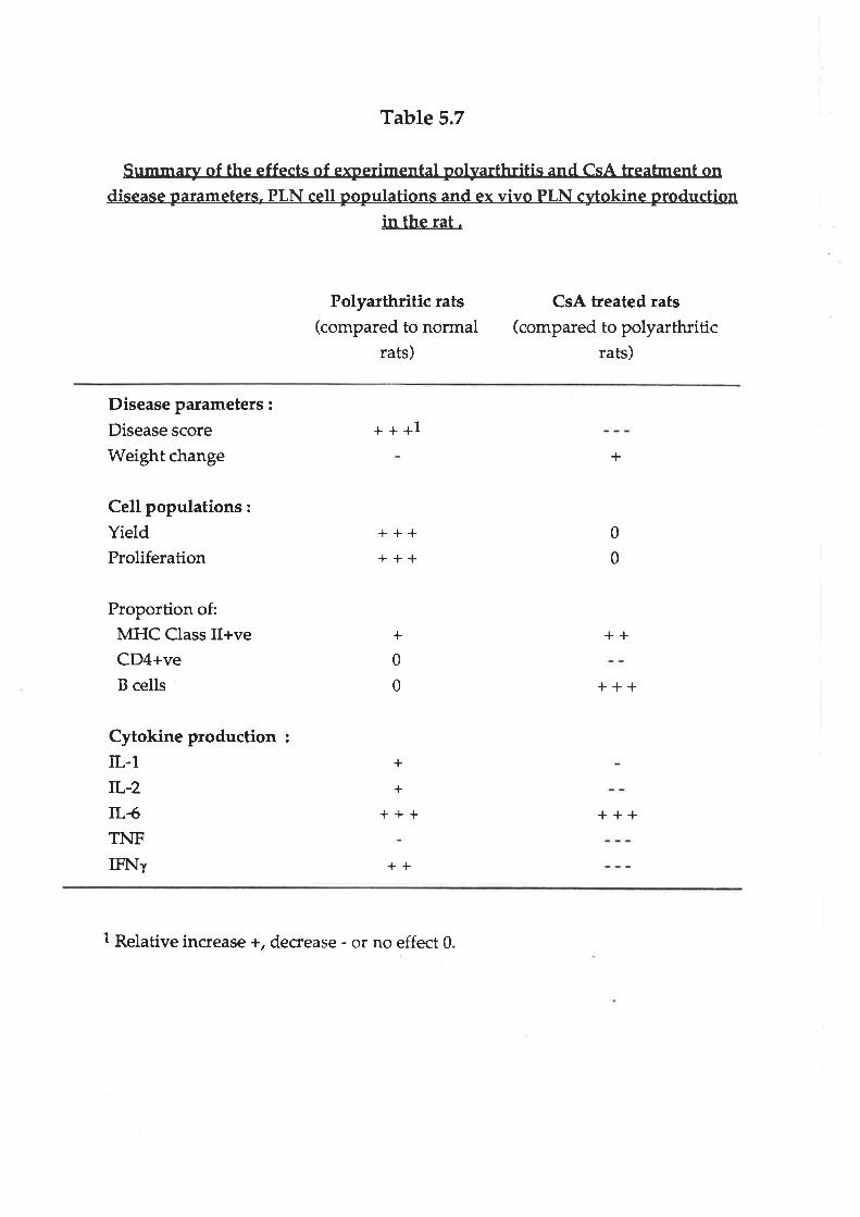
Table 5.7
Summary of the effects of experimental polyarthritis and CsA treabnent ondisease parameters. PLN cell populations and ex vÌvo PLN cytokine production
in the rat.
Polyarthritic rats(compared to normal
rats)
CsA treated rats(compared to polyarthritic
rats)
Disease parameters :
Disease scoreWeight change
Cell populations:YieldProliferation
Proportion of:MHC Class II+veCD4+veB cells
Cytokine production :
IL-1rL-2lL-6TNFIFNl
+++1
++++++
+
+++
++
+
0
0
+
0
0
++
+++
+++
+
1 Relative increase +, decrease - or no effect 0

75
approximately 4 fold less PGE2 than LPS stimulated PBM cells and 10 fold less
than LPS stimulated AP cells per cell.
DISCUSSION
CsA as an antiinflammatory drug
There is abundant evidence that CsA prevents graft rejection and expression of
some autoimmune disorders by affecting the activation of T lymphocytes.
Although the adjuvant-induced arthritis in rats and rheumatoid arthritis are not
identical, they share some clinical and pathological features. Both diseases are
examples of immunologically mediated chronic inflammation in which T
lymphocytes (Yoshino et aL1990), particularly Cn++ cells (Sany 1990, Larsson et al
1985), play a central role in both establishing and sustaining chronic inflammation.
The present study (results summarised in table 5.7) shows that appropriate CsA
treatment can completely prevent the expression of this experimental arthritis in
rats by suppressing some T lymphocyte activities. This anti-arthritic effect of CsA
may be due to its ability to selectively inhibit the secretion of several rytokines.
The levels of CsA achieved in the blood of rats receiving 40 mg/kg CsA admixed
with the arthritogenic adjuvant were similar (>100nM) to those reported inrheumatoid arthritis patients during continuous treatment (Madhok and Capell
1988). Similar levels (> 0.2-0.3 uM CsA) were also found to be required for about
60 days during the induction of tolerance to tissue allografts in. animals (reviewed,
Lim and White 1989). High levels of CsA were maintained following the single
injection of CsA, probably because it was released slowly from the lipid phase ofthe adjuvant, and because the hepatic extraction of CsA from the blood isapproximately 1.0 times higher in man than in rats (Kahan 1985, Wagner et al
1'987). CsA injected subcutaneously every second day from days -1 to7, or days -1
to 13, was as effective in preventing polyarthritis as CsA given in a single dose
mixed in with the adjuvant. Both these therapies also prevented the cachexia

76
which usually accompa.nies the adjuvant administration (Rofe et al 1990), and had
similar effects on lymph node enlargement and lymphocyte functions. This
indicates that CsA, to be effective, need not be given in dose juxtaposition to the
arthritogen, and its action is therefore unlikely to involve interaction and
modification of the mycobacterial antigen(s) (Cohen et al 1985) that may initiate
the disease. It is thus more likely to be affecting the initial response to the
arthritogen.
The effect of CsA therapy on lymphocyte proliferation
Cohen et al 1985 suggested that the pathogenesis of adjuvant arthritis primarily
involves T cell recognition of mycobacterial epitopes that are cross-reactive with
comPonents of slmovium. The suppressive effect of CsA on expression of the
disease might therefore be due to inhibition of T cell activation by the processed
mycobacterial antigen(s). However the enlargement of the regional lymph nodes
draining the hind feet of rats with active disease was also seen in rats treated with
CsA. Since the increase in cell recoveries was also associated with a marked
increase in 3H-thymidine uptake, it is likely that node cell proliferation, rather
than lymphocyte recruitment, mainly contributed to the node enlargement in both
CsA-treated and untreated rats. The failure of CsA to reduced tymph node cell
proliferation during the stages of disease expression (Day 14) was surprising since
previous reports have indicated CsA may inhibit certain events underlying
lymphocyte proliferation (Hess et al 1.982, Groeniwegen et al 1985). It is clear that
resPonses to antigens by these node cells were not entirely suppressed since node
enlargement was always evident, even in the absence of clinical or histological
arthritis, in the CsA-treated rats. This suggests that lymphocytes do responds to
the arthritogen in CsA-treated animals, but without generating the type of
response that induces a¡thritis.
CsA is reported to affect some T lymphocyte subpopulations more than others
(Clerici and Shearer'1.990, Klaus 1988). Therefore, it is of interest to compare the

nlymphocyte subpopulations in the affected ly*ph nodes of arthritic rats and rats
protected by CsA treatment. The results indicate small but significant changes in
lymphocyte subsets. The proportion of T lymphocytes (CD43+, CDs+) and B
lymphocytes (Ig+) were similar to that previously reported (Mason et at 1980).
Adjuvant treatment, although increasing cell yields dramatically, did not change
the proportion of cells detected by the series of mAb except for a modest (8%)
increase in the proportion of cells expressing MHC class II antigens. The
proportion of these cells was further increased by CsA treatment. This contrasts
with claims that CsA treatment reduces the expression of MHC class IIdeterminants in dog epithelial cells (Groeniwegen et al 1985).
The effect of CsA therapy on leucocyte subpopulations
CsA treatment almost doubled the proportion of cells expressing Ig, presumabty B
cells, while simultaneously decreasing the proportion of cells expressing the CD4
antigen by more than 10%. Macrophages, as well as T lymphocytes, can express
the CD4 antigen (Barclay 1981), and may account for at least part of the reduction
of CD4+ cells. The effective treatment of experimentat arthritis by anti-CD4 mAb
also indicates that this cell subpopulation is essential for expression of arthritis(Larsson et al 1985, Van den Broek et a11992).
Immunohistology of the PLN was consistent with the findings using
immunofluorescence carried out with the same mAb. While small differences(-10Vo) in the proportions of CD4+ cells and total T cells are not readily detectable
using immunohistology, the large increase in the proportion of B cells followingCsA treatment was noted. There was a increase in the numbers of B cell folticles
and these extended into the paracortical region, possibly at the expense of some T
cell populations normally present there. These B cells also expressed class IIantigens and may have accounted for the increase in the proportion of cells
detected with the ox6 mAb in the nodes of CsA treated animals.

78
The most striking effect of prior in vivo treatrnent with CsA was a reduction in the
secretion of IL-2, TNF and IFNT, in response to Con A, by lymph node cells
harvested during the early phase of arthritis induction. This suggests that the node
lymphocyte population in CsA-treated rats is enriched in T cells of the T helper
(T¡$ type L class (Mosman and Coffman 1989). Unfortunately, there is no
complementary data relating to the production of those cytokines (IL-4,IL-s, IL-
10) that characterise the CD4+ T cells (TH2) that promote humoral immunity.
Flowever, in vitro studies with human PBM cells have shown CsA will inhibit IL-4
and IL-S production (Andersson et aL 1992). The secretion of IL-6 (a B cell growth
factor) by lymph node cells harvested from CsA-treated rats, may explain the
increase in B cells observed in the lymph nodes of these animals, suggesting that
not all B cells responses are affected by CsA (Shindani et aI 1983). These findings
are also consistent with reports of enhanced B cell responses in CsA treated mice
(Kunkl and Klaus 1980) and elevated IgG levels in CsA-immunosuppressed
transplant recipients (White et al 1980).
The effect of CsA therapy on ex vivo cytokine production
The effects of CsA treatment on cytokine production by PLN and PBM cells and
during the early phase of disease initiation (days 1 to 3) were consistent with a
direct effect of CsA on these cells in vitro. The inhibition by CsA of IL-2, IFNyand
TNFa production by human peripheral blood mononuclear cells in vitro has also
been observed (Andersson et al 7992). That report also indicated that TNFo and
IL-6 production by LPS stimulated monocytes was unaffected by CsA in vitro, as
found in the experiments described here using rat PBM and AP celts.
Supplementing the previous findings of Anderson et al, the results presented here
show that Con-A stimulated IL-6 production by PLN cells was relatively
unaffected by CsA in vitro. Since these cells are mainly lymphocytes, and Con Apredominantly stimulates this tlpe of cell, it seems likely that the IL-6 is produced

79
by lymphocytes. This indicates that in contrast toIL-2, TNF and IFNyproduction,
IL-6 production by lymphocytes is relatively unaffected by CsA.
Of particular interest was the observation that the Con A stimulated production of
IL-6by PLN and PBM cells was relatively unaffected by CsA in vitro. Since these
cell populations are mainly lymphocytes, and Con A is predominantly a
stimulator of lymphocytes, it seems likely that IL-6 production by these node
populations originates from lymphocytes and is unaffected by CsA. IL-6 may be
one of the rare factors produced by lymphocytes which is not affected by CsA in
contrast to other cytokines (IL-z,I1--4, TI--5 and TNFcr) which are sensitive to CsA
in vitro (Andersson et al 1992).
The in vitro effect of CsA on cytokine production
The inhibition of in vitro TNF production by CsA was variable. The method used
for assaying TNF production will detect both TNFo (mainly produced by
macrophages and monocytes) and TNFP (mainly produced by lymphocytes). Con
A is primarily a stimulator of lymphocytes and therefore of TNFP production;
whereas LPS, a stimulator of monocytes and macrophages, should preferentially
stimulate TNFa production. Indeed the present study found that Con A-
stimulated TNF production was much more sensitive to CsA than the LPS-
stimulated TNF production. This probably indicates that CsA more effectively
inhibits TNFP production by lymphocytes than TNFa production by monocytes
and macrophages as has been reported in studies with human PBM cells in vitro(Andersson et al 1992)
While in vivo CsA treatment reduced the levels of IL-L, TNF and IL-6 produced by
macrophages (table 5.5), in vitro treatment with CsA had üttle effect on the
production of these cytokines by the s¿une cells (table 5.6). This may indicate that
CsA does not affect macrophage cytokine production directly but will inhibit their
ability to produce cytokines indirectly by affecting other types of cell which

80
maintain or stimulate cytokine production by macrophages. Since IFNy is a
stimulator of many macrophage functions, including IL-"!., TNF and IL-6
production (Hart et al 1989, Cheung et al 1990), inhibition of lymphoryte derived
IFNy by CsA (Reem et al 1983, table 5.6) may result in decreased responsiveness of
macrophages in vivo. This indirect effect is consistent with the in vivo findings
(table 5.5) which indicate that csA heatment did not affect the production of IL-land IL-6 by macrophages (AP cells) early (day 3) but markedly reduced
production later (days 7 and 14). The indirect inhibition of macrophage functions,
particularly by reducing lymphocytederived IFNy as observed on days 1 and 3,
may be a major disease-preventing effect of CsA.
CsA and prostanoids
Prostanoids, such as PGE2, can regulate cytokines in a similar way to CsA (ie.
inhibiting IL-L, lL-2, TNF and IFN1 whilst stimulating IL-6 production) (see
chapter 4).If CsA stimulates PGEZ release (Esa et al 1983) then the regulation of
cytokines and cells by CsA may involve PGE2 as a mediator. However, the present
data show that CsA can inhibit, rather than stimulate, PGE2 release as others have
reported (Churchill et al 1990). CsA was a strong inhibitor of PGEZ production by
Con A-stimulated PBM cells, but not LP$stimulated PBM or AP cells, indicating
that PGE2 production by monocytes or macrophages is, like TNF, relatively
unaffected by CsA. Prostaglandin-mimics, such as Misoprostol, are sometimes
used in conjunction with CsA for organ transplantation to reduce undesired effects
caused by CsA's inhibition of eicosanoid production. These prostaglandin-mimics
may not only prevent some of CsA's side effects (eg kidney damage) but also
enhance its regulation of immune functions (Iyengar et at 1991).
In conclusion, CsA prevents both the polyarthritis and the cachexia associated
with the immune response against arthritogenic adjuvant in rats, but this is not
due to complete suppression of the immune response to the initiatingarthritogen(s). CsA may modify the activation of certain subsets of lymphocytes,

81
essential for development of polyarthritis, by suppressing the production of some
T cell cytokines. The data suggest that the expression of polyarthritis may involve
the preferential activation of the Tg1 subset. These findings are consistent with
observations that T¡11. activation is associated with development of chronic
inflammation such as experimental allergic encephalomyelitis in mice (Merril et al
7992) and systemic inflammation in humans (Brod et al 1991). Therapy directed
against T¡11 cells (eg with CsA), perhaps combined with a prostanoid agonist that
regulates monokine and cytokine production and complement the action of CsA
on lymphocytes (chapter 4,Betz and Fox 1991), may be particularly helpful in the
management of rheumatoid arthritis.

82
Chapter 6General conclusions and futu¡e directions
While each chapter contains its own detailed discussion, there ¿ue some overall
general conclusions that can be drawn from the findings presented in this thesis.
Prostanoids as regulators of inflammatory mediators
Chapters 2,3 and 4 deal with NSAIDs and the role prostanoids (PG's) may play in
the regulation of inflammatory mediators. The results indicate they may have a
beneficial effect on chronic inflammation. PG's ability to regulate the production
and action of important inflammatory cytokines, such as TNF and IL-l, indicate
they may be important regulators of inflammation. The results also confirm the
long held view that PG's can suppress the production and action of the
lymphoproliferative cytokine ÍL-2.1L-2, produced by lymphocytes, is important
for maintaining and expanding some lymphoid populations. T lymphocyte
populations in particular depend upon IL-2 for proliferation, and these cells are
considered central to the cell mediated immune responses seen in auto-immune
diseases. The production of IFNI, a lymphoryte derived activator of macrophages,
is also suppressed. Taken together these results indicate that PG's may play an
active role in the resolution and suppression of chronic inflammatory diseases.
However PG's are not, as originally thought, just suppressors of immune
resPonses. They a¡e also stimulators of some immune functions. This study shows
that IL-6 production in greatly enhanced in several cell types. Others indicate that
additional cytokines such as GM-CSF (Quillet al 1989) and IL-S (Betz & Fox 1991)
may be stimulated by PGE2. A consistent pattern of response has emerged from
the recent literature, indicating that PG's (and other agents that elevate cAMP)inhibit the production of cytokines which characterise the T helper (TH) type 1

83
subset of lymphocytes. By contrast, cytokine production by Tyy-2 cells seems
relatively unaffected, or even enhanced. These effects, in particular stimulating IL-
6 production, may enhance various humoral immune functions. This is consistent
with the reported enhancement of IgGl and IgE production by PGE (Phipps et al
1ee7).
Taken together, these findings indicate that PG's may'switch' the immune system
from TH-l and cell mediated responses that generate immune-mediated chronic
inflammation to less inflammatory Tfl-Z and humoral responses. PGE's may,
therefore, be a natural mechanism for directing immune responses away from
generating chronic inflammation towa¡ds healing and repair. The episodes of
acute inflammation, dominated by PGE production, that commonly flare up
during rheumatoid arthritis may represent failed attempts by the body to regulate
the underlying chronic inflammation which sustains the disease.
Can PGE's be used for therapy?
Administration of PGE's systemically may not be effective because of their very
short half life in the circulation. Even analogues, such as Misoprostol, which have
a much longer half life in vivo. may not remain active for long enough to provide
the necessary benefit at an inflammatory site. Low doses of PGE's co-administered
with other drugs may, however, be effective (Whitehouse et al 1990a). Therapies
which raise levels of PGE's at inflammatory sites may be more potent. One way to
achieve this may be through dietary control through essential fatty acids derived
from fish and plant oils. Increasing the quantities of substrates available for
cyclooxygenase (see figure 1.1) may increase the levels of pGE's produced at an
inflammatory site where PGE synthesis is enhanced (probably stimulated by
cytokines like IL-l). The results of Chapter 4 indicate that precursors derived fromfish or plant oils may form PGE's which are as effective as pGE2.

uWhile therapies which elevate PGE at an inflammatory site may reduce chronic
inflammation, the additional levels of a PGE may stimulate aspects of acute
inflammation (such as vasodilatation, pain and fever) and so its benefit may be
questioned. Since the effect of PGE's is mediated by intracellular cAMP, other
agents having simila¡ effects may also be considered. While PGE's deleterious pro-
inflammatory role in acute inflammation is well understood, its more helpful role
as a suppressor of cytokines mediating chronic inflammation indicate it may be
overall a potent beneficial agent in the treatment of chronic inflammation.
Similarities between the action of PGE's and Cyclosporin A (CsA)
Following in vitro treatment both CsA and PGE's seem to down-regulate the Tg-1
sub class of lymphocytes as well as stimulating IL-6 production. However, unlike
PGE's, CsA's direct effects seem confined to lymphocytes. CsA did not affect the
production of cytokines by monocytes/macrophages or the morphology of
macrophages. CsA's affect on lymphocytes was not mediated by PGE since its
production was inhibited by CsA in these cells. The effectiveness of CsA inpreventing adjuvant arthritis indicates that the action of lymphocytes are essential
for the induction of the disease. Flowever, CsA was less effective in suppressing
pre-established disease (Whitehouse et al 1990b), and this may indicate that,
although Tft-l lymphocytes are essential for establishing the disease, other cells,
such as macrophages are important for maintaining the disease.
Taking into consideration the abiliry of both CsA and PGE's to suppress aspects of
chronic inflammation, a combination of both CsA and PGE's might be worth
considering for the treatment of arthritis. Misoprostol is currently used incombination with CsA to counteract the side effects of CsA treatment intransplantation recipients.
Other therapies directed against T lymphocytes

85
In recent years several new "CsA like" drugs have been developed which seem to
be effective at preventing graft rejection. These drugs (Lobenzarit, FK506 and
Rapamycin) may also be effective, like CsA, in the treatment of auto-immune
mediated inflammation. Some of these new drugs may have fewer side effects
than CsA, a major cause for withdrawing long term effective CsA treatment.
Further investigation of these drugs as suppressants of immune-mediated chronic
infl ammation is warranted.
The therapeutic potential of monoclonal antibodies directed against T lymphocyte
antigens has also been demonstrated (Chapter 5 discussion). Antibodies directed
against T lymphocyte subsets, such as CD4 cells, or against activation antigens,
such as IL-2 receptor, may suppress T cell functions in a similar way to CsA
therapy. Most monoclonal antibodies are derived from rodents, and are
recognised as foreign by the human immune system and can produce harmful
side effects. Recombinant cDNA techniques are being used to "humanise" the
monoclonal antibodies by replacing the Fc portion of the rodents monoclonal
antibody with a human Fc fragment.
Conditional phannacology
The concept that an antiinflammatory drug's effect is enhanced by inflammatory
mechanisms has been proposed OVhitehouse and Vernon-Roberts 1991,,Gadd et al
7992). The activity of aspirin and its metabolites, that may be preferentially formed
at an inflammatory site, indicate that aspirin may be a pro-drug; These metabolites
are shown to regulate the activity of some inflammatory rytokines (Chapter 2).
Drugs which become active at the inflammatory site may have reduced side
effects because actívity is targeted to where the drug will give most benefit.
New molecular therapies
A number of newer approaches, still at the experimental level, utilise molecular
biological techniques to produce active proteins. These proposed therapies often

86
involve antagonists of particular cytokines (eg soluble receptors or synthetic
peptides which block receptors) and trials are underway with purified material.
Monoclonal antibodies are also being developed which are directed against
particular cytokines or block their binding to receptors. These specific molecules
target a particular cytokine. Given the complex relationships of inflammatory
cytokines the effects of blocking a single cytokine, may have unexpected
consequences. Furthermore the overlapping activities of many inflammatory
cytokines (see table 1.1) may mean that blocking the activity of one cytokine leads
to another cytokine replacing its activity (Finkelman 7992).
A therapeutic strategy which regulates groups of cytokines may be more effective.
Cytokines, such as IL-4 and IFNy, hâI modify the overall direction of the
inflammatory response in similar ways to that described for PGE's and
"Cyclosporin-like" drugs. These natural proteins may have less side effects than
exogenous drug therapies. In addition, "designer drugs", specifically made to
block transcription or translation of particular genes and small molecules which
interfere with the signal transduction process are being considered (Gibbons 1992).
Therapies which mimic or regulate intracellular messengers (such as cAMP and
cGMP investigated in this thesis) may also have profound effects upon the
activities of inflammatory cells and the course of inflammation in general.
This thesis aimed to explore how inflammation may be influenced by the
regulation of inflammatory mediators. While it has explored only a small portion
of this broad and expanding field the findings have presented important insights
into the actions of endogenous and exogenous regulators of inflammatory
mediators. By better understanding the processes of chronic inflammation and
defining the mode of action of effective therapy this type of research may help
identify new approaches to treat chronic inflammatory diseases.

87
BIBLIOGRAPHY

88
ABRAHAMSON S., EDELSON H., KAPLAN H., GIVEN W. & WEISSMAN G.
(1983) Rheumatoid arthritis: its role and the inhibition of its activation by
nonsteroidal antiinflammatory drugs. Seminars Arth Rheun . l3(Suppl 1), 148.
ADAMS D.O. & HAMILTON T.A. (1987> Molecular mechanisms by which IFN
and other signals regulate macrophage development. Immunol. Rert.97.5.
AKIRA S., HIRANO T., TAGA T. & KISHIMOTO T. (1990) Biology of
multifunctional cytokines: IL 6 and related molecules (IL 1 and TNF). F.A.S.E.B.4,
2860.
AKIRA S., ISSHIKI H., NAKAIIMA T., KINOSHITA S., NISHIO Y., NATSUKA S.
& KISHIMOTO T. (1992) Regulation of the expression of the interleukin 6 gene:
structure and function of the transcription factor NF-IL6. in Polyfunctionøl
cytokines: lL-6 ønd LIF, Ciba Foundation Symposium (eds Bock GR & Widdows K),
p47,lohn Wiley & Sons Ltd, England.
ALLISON A.C. & LEE S.W. (1988) Pro-inflammatory and catabolic effects of
interleukin-l and their antagonism by glucocorticoids. Agents Actions. Suppl. 24,
207.
ALLISON 4.C., DAVIES P. & DEPETRI S. (1971) Role of contractilemicrofilaments in macrophage movement and endocytosis. Nature Na;o Biol.232,
153.
ALVEREZ R. (1989) Regulation of ryclic nucleotide metabolism in human platelets
by prostanoids in Høndbook of eícosanoids: Prostaglandíns and relatd lipids Volume IL
(ed. Willis A.L.), p373, CRC Press. Florida, USA.

89
AMATO P.4., UNANUE E.R. & TAYLOR D.L. (1983) Distribution of actin in
spreading macrophages: A comparative study on living and fixed cells. l. Cell Biol.
96,750.
ANDERSSON J., NAGY S., GROTH C.-G. & ANDERSSON U. (7992) Effects of FK
506 and cyclosporin A on rytokine production studied in vitro at a single-cell level.
lmmunol.75,'1,36.
AURON p.E., WEBB 4.C., ROSENWASSE& L.J., MUCCI S.F., RICH A., WOLFF
S.M. & DINARELLO C.A. (1984) Nucleotide sequence of the human monocyte
interleukin-1. precursor cDNA. Proc. Natl. Acai. Sci. USA. 81,7907.
BAKER P.E., FAHEY J.V. & MLTNCK A. (1981) Prostaglandin inhibition of T-cell
proliferation is mediated at two levels. Cellular lmmunol.6\,52.
BARCLAY A.N. (1981) The localisation of populations of lymphocytes defined by
monoclonal antibodies in rat lymphoid tissue s. lmmunol. 42, 593.
BARTOLD P.M. & HAYNES D.R. (1991) Interleukin-6 production by human
gingival fibroblasts. l.Pridonf. R¿s. 26, 339.
BAZN H., XFIURDEBISE L.M., BURTONBOY G., LEBACQ A.M., DE CLERCQ L.
& CORMONT F. (79U) Rat monoclonal antibodies. I. rapid purification from in
vitro cultu¡e supernatants. J. lmmunol. Methods 66,26'1..
BECKER S. (1983) Interferons as modulators of human monocytemacrophage
differentiation. I. Interferon-gamma increases HLA-DR expression and inhibits
phagocytosis of zymoza^. | . Immunol. 132, 7249.

90
BELCH I.l.F., ANISELL D., MADHOK R., O'DOWD A. & STURROCK R.D. (1988)
Effects of altering dietary essential fatty acids on requirements for non-steroidal
anti-inflammatory drugs in ¡íatients with rheumatoid arthritis: a double-blind
placebo controlled study. Ann. Rheum. Dis. 47,96.
BERLIN R.D. & OLIVER l.M. (1,978) Analogous ultrastructure and surface
properties during capping and phagocytosis in leukorytes. /. CellBiol.77,789.
BERRIDGE M.I. (1985) The molecular basis of communication within the cell. Sci
4m.253,124.
BETZ, M. & FOX B.S. (1.991) Prostaglandin EZ inhibits the production of Th1
lymphokines but not of Th2 lymphokines. l . lmmunol. 146, 1.08.
BEUTLER 8., MAHONEY 1., LE TRANG N., PEKALA p. & CERAMI A. (1985)
Purification of cachectin, a lipoprotein lipase-suppressing hormone secreted by
endotoxin-induced RAW 264.7 cells. I. Exp. Mel. 1.6f-,9%.
BEUTLER 8., TRACENKO U., MILSARK I., KROCHIN N. & CERAMI A. (1986)
Effect of f-interferon on cachectin expression by mononuclear phagocytes.
Reversal of the tpsd Gndotoxin Resistance) phenotyp e. I, Erp. Meil.164, t7gt.
BIGNOLD L.P., FERRANTE A. & HAYNES D.R. (1990) Studies of chemotactic,
chemotactic movement-inhibiting effects of interleukin-l alpha and beta, tumour
necrosis factors alpha and beta and interferon garruna on human neutrophils in
assays using'sparsepore'polycarbonate (Nucleopore) membranes in the Boyden
chamber. Int. Ar ch. Aller g. AWl. Immunol. 9f-,'I.,.
BLACK R.A., KRONHEIM S.R. & SLEATH P.R. (1989) Activation of IL-1p by a co-
induced protease. FEBS letts. 247, 3Í36.
97
BOMSZTYK K., SIMS I.E., STANTON T.H., SLACK J., MCMAHAN C.I.,
VALENTINE M.A. & DOWER S.K. (1989) Evidence for different IL-l receptors in
T and B cell lines. Proc. Natl. Acad. Sci. U9A.86,8034.
BORASCHI D., CENSINI S. & TAGLIABUE A. (19U) Interferon gamma reduces
macrophage-suppressive activity by inhibiting prostaglandin E2 release and
inducing interleukin L production. | . lmmunol. 133, 7 64.
BRAY M.A. & GORDON D. (1.978) Prostaglandin production by macrophages and
the effect of anti-inflammatory drugs. Br.l. Plurmac.63,635.
BROD S,4., BENJAMIN D. & HAFLER. D.A. (1991) Restricted T cell expression of
IL-27 ¡¡¡-gamma mRNA in human infl ammatory disea se. J . Immunol. 147, 81.0.
BROOKS P.M., KEAN W.F. & BUCI{ANAN W.W. (19f36a) Salirylates. jnThe clinicøl
pharmacology of anti-ínflammatory drugs. (eds Brooks P.M., Kean W.F, and Buchanan
W.W.) p46,Taylor & Francis Ltd, London
BROOKS P.M., KEANI W.F. & BUCHANAN W.W. (1986b) Corticosteroids. In: The
clinicøl pharmacology of anti-lnflammatory agents .(Eds P.M. Brooks. M.F. Kean &
W.W. Buchanan.),p148, Taylor and Francis Ltd, London.
BROUGHTON K.S.,WHELAN J., FIARDARDOTTIR I. & KINSELLA I.E. (1991)
Effect of increasing the dietary (n-3) to (n-6) polyunsaturated fatty acid ratio on
murine liver and peritoneal cell fatty acids and eicosanoid formation. /. Nurt. \21,155.
CANNON G.W., EMKEY R.D., DENES A., COHEA S.A., SAWY A., WOLFE F.,
JAFFER A.M., WEAVER A.L., COGEN L.,GULINELLO J., KENNEDY S.M. & I.D.

92
(1,990) Prospective two year followup of recombinant interferon-gamma in
rheumatoid arthriti s. l. Rheumatol. 17, 3M.
CANNON G.W., S. MCCALL, B.C. COLE, M.M. GRIFFTTHS, L.A. RADOV & I.R,
WARD. (7989) Effects of indomethacin, cyclosporin, cyclophosphamide, and
placebo on collagen-induced arthritis of mice. Agents- Actions. 29, 3"1.5.
CANTAROW W.D., CHENG H.T. & SUNDHARADAS G. (1,978) The effects of
prostaglandins on the spreading, adhesion and migration of mouse peritoneal
MNPs. Prostaglandins \6, 39.
CARSWELL E.A., OLD L.I., KASSEL R.L., GREEN S., FIORE N. & WLLIAMSONB. (1975) An endotoxin-induced serum factor that causes necrosis of tumors . Proc.
NatL Acad. Sci. U9A.72,3666.
CASTEL J.V., GEIGER T., GROSS V., ANDUS T., WALTER E., HIRANO T.,
KISHIMOTO T. & HEINRICH P.L. (1988) Plasma clearance, organ distributionand target cells of interleukin-6/hepatocyte-stimulating factor in the rat. Eur.l.
Biochem.177,357.
CESARIO T.C., YOUSELF S. & CARANDANG G. (19s9) The regulation of
interferon production by aspirin, and other inhibitors of the cyclooxygenase
pathway and agents influencing calcium flux. Bull.N.Y. Acad. Med.65,26.
CEUPENS J.L., ROBAEYS G., VERDICKT W ., VERTESSEN S., DECKMYN H. ANDDEQUEKER I. (1986) Immunomodulatory effects of treatment with Naproxen in
patients with rheumatic disease. Arthritis Rheum. Zg,3OS.

93
CHELING D.L., HART P.H., VTITI G.F., WHITTY G.A. & HAMILTON I.A. (1990)
Contrasting effects of interferon-yand interleukin-4 on the interleukin-6 activify of
s timulated human monocyte s. Immunol. 1990. 77, 7 0.
CHIZZONITE R., TRUITE T., KILIAN P.L., STERN A.S., NUNES P., PARKER
K.P., KAFFKA K.L., CHUA 4.o., LUGG D.K. & GURLER u. (1989) Two high
affinity interleukin-L receptors represent separate gene products. Proc. Natl. Acad.
Scí. USA .86,8029.
CHOMCZNSKI P. & SACCHI N. (1987) Single-step method of RNA isolarion by
a ci d guanidinium thiocyanate-phenol-chlorofrom extraction . An øly t. B io chem. 162,
1,56.
CHURCHILL, P.C., N.F. ROSSA, V.R. ELLIS, M.C. CHURCHILL & F.D. MC
DONALD. (1990) Cyclosporine a inhibits prostaglandin E2 release, and has no
effect on renin secretion, from rat renal cortical slices. Life-Sci.4z,ls79.
CLELAND L.G., FRENCH J.K., BETTS W.H., MURPI{Y G.A. & ELLIOT M. (1988)
Clinical and biochemical effects of dietary fish oil supplements in rheumatoid
arthritis. | .RheumatollS, 1,47 7.
CLELAND L.G., LOWTHAN p.I., IMHOFF D., BOCHNER F., BETTS W.H. &O'CALLAGHAN I. (1985a) Plasma and slmovial fluid gentisate in patients recieving
salicylate therapy. l. Rheumatol. 12, 736.
CLELAND L.G., WHITEHOUSE M.W. & BETTS W.H. (1985b) Gentisare an
salicylate metabolite with antioddant properties. Drugs ExVtl. Clín. Res.ñ,,zzg.
CLERICI M. & SHEARER G.M. (1990) Differential sensitivity of human T helper
cell pathways by in vitro exposure to cydosporin A. l. Immunol ].q4,24g0.

94
coHEN, D.J., LOERTSCHER R., RUBIN M., TILNEY N.L., CARPENTER C.B. &
STROM T.8.. (1984) Cyclosporin: A new immunosuppressive agent for organ
transplantation. Ann. lntqn. Md. 701, 667.
COHEN, I.R., HOLOSHITZ J., VAN EDEN & FRENKEL A. 1985. T lymphocyte
clones illuminate pathogenesis and affect therapy of experimental arthritis.
Arthrítis Rheum. 28, U'1..
DALLMAN , M.1., MASON D.W. & WEBB M. (1982) The roles of host and donor
cells in the rejection of skin allografts by T cell-deprived rats injected with
syngeneic T cells. Eur. l. lmmunol. 12, 5"1.'1.
DAVIDSON E.M., RAE S.A. & SMITH M.I. (1983) Leucotriene 84, a mediator of
inflammation present in synovial fluid in rheumatoid arthritis. Ann. Rheum. Dis.
43,677.
DAVIDSON F.F., DENNIS E.A., POWELL M. & GLENNEY IR J.R. (1937)
Inhibition of phospholipase AZ by "lipocortins" and calpactins. An effect of
binding to substrate phospholipids. |. Biol. Chem. 262,7698.
DEL-POZO 8., GRAEBER M., ELFORD P. & PAYNE T. (1990) Regression of bone
and cartilage loss in adjuvant arthritic rats after treatment with cyclosporin A.
Ar thritís-Nteum.. 33, 247 .
DINARELLO C.A. (1,992) Role of interleukin-l. and tumor'necrosis factor insystemic responses to infection and inflammation. in Inflamnutíon: Basic yincípøls
and clínical correløtes. (2nd edition) (Eds. Gallin [, Goldstein IM & Snyderman M.),
p21,1,, Raven Press. New York.

95
DINARELLO, C.A. (1989) Interleukin-l. and its biologicaly related rytokines. Ada.in
lmmunol. L4,'j,53.
DOUGADOS M., AWADA H. & AMOR B. (1988) Cyclosporin in rheumatoid
arthritis: a double blind controlled study in 52 patients. Ann. Rheum. Dis. 47,727.
DOWER S.K. (1992) Interleukin-l. in Humøn Cytokines (eds Aggarwal BB &
Gutterman IU), p46, Blackwell Scientific Publications USA.
DUGGAN D.E. (1981) Sulindac: theraputic implications of the pro-
drug / pharmacophore equilibri um. Dr ug Met ab. Reu s . 72, 325.
EALICK S.E., COOK W.I., VIJAY-KUMAR S., CARSON M., NAGABHUSHAN
T.L., TROTTA P.P. & BUGG C.E. (1991) The three dimentional structure of
recombinant human interferon -y. S cience 252, 698,
ECONOMOU J.S., RHOADES K., ESSNER R., MCBRIDE W.H., GASSON I.C. &
MORTOND.L. (1989) Genetic analysis of the human tumor necrosis factor
alpha/cachectin promotor region in a macrophage cell line. I.Erp.Med.l70,321,.
ENDRES S., GHORBANI R., KELLEY V.E., GEORGILIS K., LONNEMANN G.,
vAÀI DER MEER I.W.M., CANNON J.G., ROGERS T.S., KLEMPNER M.S.,
WEBER P.c., SCHAEFER E.J., woLFF S.M. & DINARELLO c.A. (1989) The effecr
of dietary supplementation with n-3 polyunsaturated fatty acids on the synthesis
of interleukin-l and tumor necrosis factor by mononuclear cells. Nat: Engl.I. Med.
320,265.
ENGLEMANN H., NOVACK D., & WALLACH D. (1990) Two tumor necrosis
factor-binding proteins purified from human urine. l. Biol. Chetn.26s,ls3't .

96
ESA 4.H., PA)CVÍAN D.G., NOGA S.I. & FüSS A.D. (1988) Sensitivity of monocyte
subpopulations to cyclosporin : arachidonate metabolism and in vitro antigen
presentati on. Trøntplant. P roc. 20, Suppl. 2, 80.
FERTSCH D. & VOGEL S.N. (19U) Recombinant interferons increase macrophage
Fc receptor capacity. l. Immunol. 132, 2436.
FINKELMAN F.D. (1992) Down, but not out for the count. Current Bíology 2,274.
FINNEY M., GUy R.G., MICHELL R.H., GORDON J., DUGAS 8., RIGLEY K.p. &
CALLARD R.E. (1990) Interleukin 4 activates human B lymphocytes via transient
inositol tipid hydrolysis and delayed cyclic adenosine monophosphate reneration.
Eur. J. Immunol. 20,"1.5'I...
FIRESTEIN G.S. & ZVAIFLER N.I. 0987) Peripheral blood and synovial fluidmonoryte activation in inflammatory arthritis. tr. Low levels of synovial fluid and
synovial tissue interferon suggest that yinterferon is not the primary macrophage
activating factor. Arthritis Rheum. 30, f364.
FLOWER R.J. 0978) Steroid anti-inflammatory drugs as inhibitors ofPhospholipase 42. In Adoønces in yostøglandin ønd Thromboxønes Research. (Eds. C.
Galli & G. Porcellati) p105, Raven Press New York.
FOLLETT E.A. & GOLDMAN R.D. (1970) The occurrence of microvilli duringspreading and growth of BHK21-C13 fibroblasts. E*p.Cell Res. s9,124.
FONTANIA A., HENGARTNER H., WEBER E., FEHR K., GOBO P.I. & COFTEN G.
(1982) Interleukin L activity in the synovial fluid of .patients with rheumatoid
arthritis. Nteumatol. lnt. 2, 49.

97
FUKUMOTO T., MCMASTER W.R & WILLIAMS A.F. (7982) Mouse monoclonal
antibodies against rat major histocompatability antigens. Two Ia antigens and
class I antigens in rat thymus. Eur.l.Immunol.12,237.
GADD S.J., WHITEHOUSE M.W. & VERNON-ROBERTS B. (1992) Conditional
pharmacology:Il. Ambivalent effects of aurocyanide, a putative active metabolite
of anti-arthritic gold drugs, on human and rat PMN leucocytes.Inflammophørm, l,305.
GADD S.I. & ASHMAN L.K. (1983) Binding of mouse monoclonal antibodies to
human leukemic cells via the Fc receptor: a possible source of ' false positive '
reactions in specificity screeníng. Clin. erp. lmmunol. 54, 81,1,.
GALINDO 8., LAZENS I. & CASTILLO R. (1974) Fusion of normal rabbit aveolar
macrophages induced by supernatant fluids from BCG-sensitized Iymph node
cells after elicitation by antigen . Infect. lmm. 9, 212.
GAMBLE J.R., HARLAN 1.M.. KLEBANOFF S.l. & VADAS M.A. (19Ss)
Stimulation of the adherence of neutrophils to umbilical vein endothelium by
human recombinant tumour necrosis factor. Proc. NatL Acad. Sci. 82,8667.
GAUTAM S., TEBO I.M. & HAMILTON T,A. (1,992) IL-4 suppresses cytokine
gene expression induced by lFN-gamma and/or IL-2 in murine peritoneal
macropha ges. I . lmmunol. 148,1725.
GHERING A.I.H., BIRD C.R., BRISTOW A. & THORPE R. (1987) A simple
sensitive bioassay for interleukin-L which is unresponsive to 103 U/ml ofinterleukin -2. l. Immunol. Methods 99, 7 .
GIBBONS A. (1992) Biotechs second generation. Scíence 256,766.

98
GILLIS S:, FERM M.M., OU W. & SMITH K.A. (1,978) T cell growth factor:
par¿uneters of production and a quantitative micro assay for activity.l. Immunol,
120,2027.
GOODWIN J.S. (1984) Mechanism of action of nonsteroidal anti-inflammatory
agents. Am. I. Med:.77 Suppl.7A,57.
GOODWIN I.S. (1985) Immunological effects of nonsteroidal anti-inflammatory
agents. Medical Clínics of North Amerícø. 69,793.
GOODWIN J.S., CEUPPENS I.L. & GUALDER N. (1984) Control of the immune
response in humans by prostaglandins. Ada. in Inflømmation Res.7,79.
GRANGER G.A. & WILIAMS T.W. (1968) Lymphocyte toxicity in vitro:
activation and release of a cytotoxic factor. Nature278,1253.
GREY P.W. & GOEDDEL D.V. (1982) Structure of the human immune interferon
gene. Nature 298,859.
GREY P.W. (1,991,) Interferon-y. in Human Cytokines (.eds Aggarwal B.B. &
Gutterman I.U.), p30, Blackwell Scientific Publications USA.
GREY p.W., BARRET K., CFIANTRY D., TURNER M. & FELDMAN M. (1990)
Cloning of human tumor necrosis factor (TNF) receptor cDNA and expression of
recombinant soluable TNF-binding protein. Proc. Natl. Acad.. Sci. llSA. 87,7380.
GROENIWEGEN, G., BUURMAN W.S., JEANHOMME G.M.A. & VAN DER
LINDEN C.I. (1985) Effect of cyclosporin on MHC class fI antigen expression on
arterial and venous endothelium in vitro. Transplantøtion. 40,21.

99
GROOTVELD M. & HALLIWELL B. (1986) Aromatic hydroxylation as a measure of
hydroxyl radical formation in vivo. Biochem.l. 234,499.
GUSTAFSON T. & WOLPERT L. (1,967) Cellular movement and contact in sea
urchin morphogenisis. Biol. Ras. 42, 42.
HAMILTON I.A. & WLLIAMS N. (1986) ln uitro inhibition of myelopoisis by gold
salts and D-penicillamine. l, Rheumatol. 12, 896,
HA\ISEN T.M., LERCHE A., KASSIS V., LORENZEN I. & SONDERGAARD J.
(1983) Treatment of rheumatoid arthritis with prostaglandin E1 precurssors cis-
linoleic acid and ylinolenic acid. Scønd. l, Nteumatol.12,85.
FIART, P.H., WHITTY G.4., PICCOLI D.S. & HAMILTON I.A. (1989) Control by
IFN-y and PGE2 of TNFcr and IL-1 production by human monocytes, Immunol. 66,
374.
HAYNES D.R., GARRETT I.R. & VERNON.ROBERTS B. (19S8a). Effect of gold
salt treatment on receptor binding activity of monocytes and macrophages
isolated from rats with adjuvant artfuitis. Rheumatol Int. S,'1.59.
HAYNES D.R., GARRETT I.R., WHITEHOUSE M.W. & VERNON-ROBERTS B.
(1988b). Do gold drugs inhibit interleukin-l? Evidence from an in vitrolymphocyte activating factor assay. | . Rheumatol. 15, 775.
FIAEGEMAN G., CONTENT I., VOLCKAERT G., DERYNICK R.; TAVENER J. &FRIERS W. (1986) Structural analysis of the sequence encoding for an inducible 26-
kDa protein in human fibriblasts. Ear. |. Biochetn.159,625.

100
HELLE M., BOEIII L. & AADEN L.A. (1988) Functional discrimination between
interleukin-6 and interleukin -"1,. Eur. l . Immunol. 18, 1535.
HESS, 4.D., TUTSCHKA P.I. & SANTOS G.W. (1982) Effect of cyclosporin A on
human lymphocye responses in vitro. III. CsA inhibits the production of T
lymphocyte growth factors in secondary mixed lymphocyte responses but does
not inhibit the response of primed lymphocytes to TCGF. l.Immunol.l28,355.
HIRANO T., YASUKAWA K., HARADA H., TAGA T., WATANABE Y.,
MATSUDA T., KASAIWAMURA S., NAKAIIMA K., KOYAMA R., IWAMATSU
4., TSUNASAWA S., SAKIYAMA F., MATSIJI A., TAKAHARA Y., JANIGUCHIT. & KISHIMOTO T. (1986) Complementary DNA for a novel human interleukin
(BSF-2) that induces B lymphocytes to produce immunoglobulin. Nature 324,73.
HIRATA F., SCHIFFMANN E., VENKATASUBRAMANIAN K., SOLOMON D. &A)GLROD I. (1980) A Phospholipase A2 inhibitory protein in rabbit neutrophils
induced by glucocorticoids. Proc. Natl. Acad. Sci. U9A.77,2533.
HOHMAN H., REMY R., BROCKHOUSE M. & VAN LOON P.G.M. (1989) TWO
different cell types have different major receptors for human tumor necrosis factor
(TNFcr). l. Bíol. Chetn. 2A, "1.4927.
HORROBIN D.F. (1988) Prostaglandin El: Physiological significance and clinical
use. Wímq klinísche Wochenschrift. L4,471,.
HOUSSIAU F.A., DEVOGELAER J.P., VAN DAMME T., NAGANT DE
DEUCHAISNES C.H. & VAN SNICK I. (1988) Interleukin-6 in synovial fluid and
serum of patients with rheumatoid arthritis and other rheumatoid arthitides.
Arthrítis Nmtm.3L,7U.

101
HUNIG T., WALLNY H-I., HARTLEY J.K., LAWETZKY A. &(1989) A monodonal antibody to a constant determinant of the rat T cell antigen
receptor that induces T cell activation: different reactivity with subsets of
immature and mature T lymphocytes. I. Erp. Mel. L69,73.
HUNT N.H., LIM L.K., EICHNER R.D., BUFFINTON G.D. & WEIDEMANN M.J.
(1984) Activation of cyclic AMP-dependent protein kinase in macrophages.
Biochem. Bíophys. Res. Comm. Llg, 1 082.
IYENGAR S., CONTRERAS P.C., MICK S.I., BREMER M.E. & MCKEARN J.p.
(1991) Immune modifying effects of misoprostol and natural prostaglandins. Br. /.
Rheumatol. 30,712.
JONES E.Y., SruART D.I. & WALKER (1989) Structure of tumor necrosis factor.
Nature 338,225.
KAHAN B.D. (1985) Individualization of Cyclosporin therapy using
pharmacokinetic and pharmacodynamic paramet ers. Tr ønsplantation, 40, 457 .
KAPLAN L., WEISS J. & EISBACH P. (7978) Low concentrations of indomethacin
inhibit phospholipase A2 of rabbit polymorphonuclear leucocytes. Proc. Natl,
Acad. 5ci.75,2955.
KAPLOW L.S. (1965) Simplified myeloperoxidase stain using benzidine
dihydrochloride. Blood. 26, 2'1.5,
KASSIS S., LEE I.C. & HANNA N. (1989) Effects of prostaglandins and cAMP
levels on monocyte IL-l production. Agents Actions27,274.

1,02
KELLER H.-tJ., BARADLIN S., KISTLER P. & PLOEM l.S. (7979) Locomotion and
adhesion of neutrophil granulocytes. Effects of albumin, fibrinogen and gamma
globulins studied by reflection contrast microscopy. Exp. Cell Res.122,35'1,.
KELLY R.W., DEAM S., CAMERON M.I. & SEAMARK R.F. (1987) Measurement by
radioimmunoassay of prostaglandins as their methyl oximes. Prostaglandíns
Leucotrienes Med. 24, 1..
KENNEDY I., COLMAN R.4., HUMPHREY p.p.A., LEVY G.p. & LUM p. (1982)
Studies on the characterisation of prostanoid receptors: A proposed classification.
Prostaglandins 24, 667.
KISHIMOTO T., HIBI M., MURAKAMIT M., NARAZAI{ M., SAITO M. & TAGA
T. (7992) The molecular biology of interleukin 6 and its receptor. inPolyfunctional
cytokines: IL-6 and LIF, Ciba Foundation Symposium. (eds Bock GR & Widdows K),
p5, John Wiley & Sons Ltd, England.
KLAUS G.G.B. (1988) Cyclosporin-sensitive and cyclosporin-insensitive modes of
B cell stimulation. Transpløntation. 44, 1,5.
KNUDSEN P.I., DINARELLO C. A. & STROM T. B. (1986) Prostaglandins
postranscriptionally inhibit monocyte expression of interleukin-l. activity by
increasing intracellular cyclic adenosine monophosphate. l. lmmunol.137,3189.
KOGA Y., TANIGUCHI K., KUBU C. & NOMOTO K. (1983) Thymus dependenr
increases in splenic T-cell population by indomethacin. Cellular Immunol.75,43.
KOI A. (1985) Biological functions of acute-phase proteins. in : The acute-phøse
responses to ínjury and infection. (ed. A.H. Gordon and A. Koj) p145 Elsevier
Science Publishers, New. York.

103
KORMANIN N. & GREEN A. (1980) Epidemiological studies in the Upernavik
districÇ Greenland. Acta Md. 9cand.208,401..
KREMER J.M., JUBIZW., MICHALEK A., RYNES R.L, BARTHOLOMEW L.E.,
BIGAOETTE I., TIMCHALK M., BEELER D. & LININGER L. (1987) Fish-oil fatty
acid supplements in active rheumatoid arthritis: a double blinded, controlled,
crossover study. Ann. Intern. Med. 106, 497.
KUNKEL S.L. & CHENSUE S.W. (1984) Prostaglandins and the regulation of
immune responses. Ada. in lnflømm. Res.7,93.
KUNKEL S.L. & CHENSUE S.W. (1985) Arachidonic acid metabolites regulate
interleukin-l. production. Biochem. Biophy s. Res. Comm. 128, 892.
KUNKL A. & KLAUS G.G.B. (1980) Selective effects of ryclosporin A on functional
B cell subsets. l. Immunol. 725, 2526.
LARSSON P., HOLMDAHL R., DENKER L. & KLARESKOG L. (1985) In vivo
treatment with W3l13 (anti-pan T) but not with OX8 (anti-suppressor/cytotoxic
T) monoclonal antibodies impedes the development of adjuvant arthritis in rats.
lmmunol.56,383.
LE I. & VILCEK I. (fg8g) Biology of disease. I¡rterleukin 6: A multifunctional
cytokine regulating immune reactions an the acute phase protien response. Løb.
Inaest.6l, 588.
LEDVINA M. (1969) The role of hydroxylation products in hypersensitivity to
s alicylates and aminopyrine. F EB S Symposium. 16, 227 .

l,M
LESLIE C.A., GONNERMANN W.4., ULLMAN M.D., HAYES K.C.,
FRANZBLAU C. & CATHCART E.S. (1985) Dietary Fish oil modulates
macrophage fatty acids and decreases arthritis suseptibility in mice. l. Erp. Med.
162,7336.
LEWIS G.P. (1983) Immunoregulatory activity of metabolites of arachidonic acid
and their role in inflammalon. Britísh Med.8u11.39,243.
LEWIS G.P. & BARETT M.L. (1986) Immunosuppressive actions of prostaglandins
and the possible increase in chronic inflammation after cyclo-oxygenase inhibitors.
Agents Actions.19,59.
LIM L.K., HUNT N.H., EICHNER R.D. & WEIDEMANN M.I. (rgSg) Cyclic AMP
and the regulation of prostaglandin production by macrophages. Biochem. Biophys.
Res. Comm.174,248.
LIM S.M.L. & WHITE D.J.G. (1989) Cyclosporin A and tolerance induction in
experimental animals in Cyclosporin: Mode of action and clinical implícations, (ed.
Thomson A.W), p82, Kluwer Academic Publishers, London.
Lru G. & NEWELL P.C. (1,991) Evidence that cyclic GMP may regulate the
association of myosin II heavy chain with the cytoskeleton by inhibiting itsphosphorylation. l. CelI Scí. 98, 483.
LOMBARDINO G.I. (1985) Medical chemistry of acidic nonsteroidal
antiinflammatory drugs. inNonsteroidal antünflammatory drugs. (Lombardino G.|.
ed.) p253, Wiley-interscience USA.

105
LOMEDICO P.T.,GUBLER U., HELLMAN M., DUKOVICH M., GIRI I.G., pAN y-
C.E., COLLIER K.,CHUA A.O. & }'dIZEL S.B. (1984) Cloning and expression of
murine interlukin-l cDNA in Escherichia coli. Nature.3t2,458.
MADHOK, R. & CAPELL H.A. (1988) Cyclosporin A in Rheumatoid Arthritis:
Results at 30 months. Transplant. Proc.20; suppl.4,248.
MAHONY J, BEUTLER B, LE TRANG N., PEKALA p. & CERAML A. (1985)
Purification of cachectin, a lipoprotein lipase-suppressing hormone secreted by
endotoxin-induced RAW 264.7 cells. I. Erp. Med. L61.,9U.
MANGAN F.R. RAGER I.P. & THOMSON M.I. (1985) The effect of adjuvant
arthritis on the subsequent local inflammatory response in rats. Br. I.Erp.Path.66,1,.
MARSHALL L.A. & CHANG J. (1989) Novel eicosanoid inhibitors. In Ner¡r
danelopements in antírheumatic thøapy. (eds Rainsford KD and Velo GP), p733, MTP
Press: Lancaster.
MASON, D.W., BRIDEAU R.J., MCMASTER W.R., WEBB M., WHITE R.A.H. &
WILLIAMS A.F. (1980) Monoclonal antibodies that define T-lymphocyte subsets
in the rat. In Monoclonal antibodíes. (eds R.H. Kennett, T.|. McKearn and K.B.
Bechtol), p251, Plenum Publishing Corporation, London, England.
MATTHEWS N. & NEALE M.L. (1987) Cytotodcity assays for tumour necrosis
factor and lymphotoxin. In Lymphokinæ and Interferons, a prøctical approach. (eds
Clements M.]., Morris A.G. and Ghea¡ing A.].H.),p22'l.,IRL Press, Oxford.

1.06
MAYRHOFER G., & SPARGO L.D.J. (1990) Distribution of class II major
histocompatability antigens in enterocytes of the rat jejunums and their
association with organelles of the endocytic parthway . Immunol. 70, 77 .
MCINNES A. & RENNICK D.M. (1988) Interleukin 4 induces cultured
monocytes/macrophages to form giant multinudeated cells. /. Erp. Med. 1G7,598.
MEAD C.C. & MERTIN I. 0978) Fatty acids and immurttty. Ada. Lipid Res.1:6,127
MERRIL J.E., KONO D.H., CLAYTON J., ANDO D.c., HINTON D.R. &HOFMAN F.M. (1992) Inflammatory leukocytes and cytokines in the peptide-
induced disease of experimental allergic encephalomyelitis in SIL and B10.PL
mice. Proc. Natl. Acad. Sci. ,89,574.
MORGAN D.4., RUSCETTI F.W. & GALLO R.G. (1926) Selective in virro growth
of Tlymphocytes from normal human bone marrows. Scíence 24G,249.
MORRIS PI. (1 981 ) Cyclospor in A. Tr ansplantation 32, 349.
MOSLEY 8., URDALD,L., PRICKETT K., LARSEN A., COSMAN D., CONLON
P.J., GILLIS S. & DOWER S.K. (1987) The IL-l receptor binds the human
interlenkin-Lø precursor but not the human interleukin-1p precurssr. /. Biot. Chern.
262,2941.
MOSSMAN T.R. (1983) Rapid colorimetric assay for cellular growth and survival:
Application to proliferation and cytotoxicity assays. I.Exp.Methods 6s,9679.
MOSMANN, T.R. & COFFMAN R.L. (1989) THl and TI{2: different pattterns oflymphokine secretion lead to different functional properties. Annu. Rea.Immunol.
7,'1,45.
1,07
NAGATA S., TSUCHIYA M., ASANO S., YAMAMATO O., HIß,T.*TA Y., KUBOTA
N., OHEDA M., NOMURA H & YAM AZ.AKI T. (1986) The chromosomal gene
structure and two mRNA's for human granulocyte colony-stimulating factor.
EMBO I. 5,575.
NAKAI S., MIZLINO K., KANETA M. & HIRAI Y. (1988) A simple, sensitive
bioassay for the detection of interleukin-l using human melanoma A375 cell line.
Bíochem. Biophys. Res. Comm. 7il, 1 189.
NEDWIN G.E., NAYLOR S.L., SAKAGUCHI E., SMITH D., JARRETT-NEDWIN
J., PENNICA D., GOEDDEL D.V. & GRAY P.W. (1985) Human lymphotoxin and
tumor necrosis factor genes: structure, homology and chromosomal localisation.
NucI. Acíds Res. 13, 6361..
NICHOLSON P.A. (1990) Recent advances in defining the role of Misoprostol in
rheumatology. l. Rheumatol. suppl. 20, 17, 50.
NOURI A.M.E., PANAYI G.S. & GOODMAN S.M. (1984) Cytokines and the
chronic inflammation of rheumatic disease I. The presence of interleukin 1 in
synovial fluids. Clin. ExV. Immunol. 55,295.
O'MALLEY W.E., ACHINSTEIN B. & SHEAR M.I. (o962) Action of bacterial
polysaccharide on tumors. II: damage of sarcoma37 by serum of mice treated withSerratia mÉìrcescens polysaccharide, and induced tolerance. l. Natl. Cancer lnst.29,1,1,69.
OLD LJ. (1985) Tumor Necrosis Factor (TNF).Scíen ce 230,630.

108
OLSSON I.., LANTZ M., NILSSON E., PEETRE C., THYSELL H., GRUBB A. &
ADOtf G. (1989) Isolation and characterisation of a tumor necrosis factor binding
protein from urine. Eur.l. Hae¡nato\.42,270.
OROPEZA-RENDON R.L., SPETH V., HILLER G., WEBER K. & FISI]ER H. (1979)
Prostaglandin E1 reversably induces morphological changes in macrophages and
inhibits phagocytosis. Erp. Cell Res. 179, 365.
OTTERNESS I.G., BLIVEN M.L., ESKRA J.D., REINKE M. & HANSON D.C. (1988)
Regulation of Interleukin-1 production: The role of prostaglandins. Cell. Immunol.
114,395.
PAINTER R.G., WHISENAND l. & MCINTOSH (1981) Effects of cytochalasin B on
actin and mysin associated with particle binding sites in mouse macrophages:
implications with regard to the mechanism of action of the cytochalasins. l. CelI
Biol.9t,373.
PHAIR-WASHINGTON L., SILVERSTEIN S.C. & WANG E. (1980) phorbal
myrisate acetate stimulates microtubule and 10-nm filament extension and
lysosome redistribution in mouse macrophages. /. Cell Bio. 8G,64'1,.
PHIPPS R.P., STEIN S.H. & ROPER R.L. (1991) A new view of prostaglandin E
regulation of the immune response Immunol. Today. 12,349.
POLLACK A. & GORDON S. (1975) Scanning electron miuoscopy of murine
macrophages surface characteristics during maturation, activation and
phagocyto sis. I-ab. Inoest. 33, M9.

1,09
PRICKETT I.D., TRENTHAM D.E. & ROBINSON D.R. (1984) Dietary fish oiI
augments the induction of arthritis in rats immunized with type II collagen. /.Immunol132,725.
QUILL H., GAUR A. & PHIPPS R.P. (1989) Prostaglandin E2-dependent induction
of granuloryte-macrophage colony-stimulating factor secretion by cloned murine
helper cells. l. Immunol.l42,813.
RABINOVITCH M. & DESTEFANO M.I. 0973) Macrophage spreading in vitro 1.
Inducers of spreading. Erp.Cell Res.77,323.
RAINSFORD K.D. (7984) Aspirin and the salicylates. (ed Rainsford K.D.),
Butterr,vorth and Co Ltd., London.
RAINSFORD K.D. (1988) Interplay between anti-inflammatory drugs and
eicosanoids in gastrointestinal damage. In Eicosønoids and the gøstrointestinal tract,
(ed Hiller K.), p111. MTP Press: Lancaster.
RANGES G.E., ZLOTNIK A., ESPEVIK T., DINARELLO C.A., CERAMI A. &PALLADtr\IO IR M.A. (1988) Tumou¡ necrosis crlcachectin is a growth factor for
thymocytes. Synergistic interactions with other cytokines. 1.lrp.Med.767,1.472.
REEM G.H., COOK L.A. & VILCEK I. (1983) Gamma interferon synthesis by
human thymocytes and T lymphocytes is inhibited by ryctosporin A. Scíence .22,
63.
REES R.C. (1992) Cytokines as biological response modifiers. /. Ctin. Pathol. 45,99.
CUTURI M.C., MURPFIY M., COSTA-GIOMI M.P., WEINMANN R., PERUSSIA
B. & TRICHIERI G. (1997) independent regulation of tumor necrosis factor and
110
lymphotoxin production by human peripheral blood lymphocytes. /. Exp, Med.
165,1581.
ROBERTS A.B. & SPORIN M.B. (1990) The transforming growth factors-p.In The
handbook of erperimental pharmacology, Vol 95 fl): Pqtide groutth factors ønd theír
receptors. (eds. Sporin M.B. & Roberts A.B.), p 419, Heidelberg: Springer-Verlag.
ROBINSON D.R. & LEVINE L. (1974) Prostaglandin concentrations in synovial
fluid in rheumatic diseases: Action of indomethacin and aspirin. In Prosúaglandin
synthetase ínhíbitors (ed H.¡. Robinson and ].R. Vane) , p223, Raven Press, New
York.
ROFE A.M., COYLE P., PHILCOX G., FIAYNES D.R. & WHITEHOUSE M.W.
(1992) Changes in metallothienein in adjuvant-induced arthritis: effects of
cyclosporin A. BioI. Trace Eliment Res. 43:143.
ROFE A.M., WHITEHOUSE M.W., BOURGEOIS C.S., HAYNES D.R. &VERNON-ROBERTS B. (1990) Prevention of adjuvant-induced cachexia by
cyclosporin A. Immunol. Cell Biol. 68,63.
ROGERS S.D. & BIGNOLD L.P. (1990) Which method for the assessment of
spreading of polymorphonudear leucorytes ? /. Immunol. Metho ds l3S, 2B1-..
ROLA-PLESZCZYNSKI M. & LEMAIRE I. (1985) Leukorrienes augment
interleukin L production by human monocytes. J. Immunol.l3S,39S8.
RUCO L.P. & MELTZER M.S. (1,978) Macrophage activation for tumourcytotoúcity: Tumouricidal activity by macrophages from C3H/Hef mice requires
at least two activation signals. Cell Immunol.41^,35.

111
RUDDLE N.H. & WAKSMAN B.H. (1968) Cytotoxicity mediated by soluble
antigens and lymphocytes in delayed hypersensitivity. ].hcp.Med.729,7271,.
SALMERON G. & LIPSKY P.E. (1983) The immunosuppressive potential of
antimalarials. Am. I. Med. 75, 79.
SAMBROOK J., FRITSCH E.F. & MANIATIS T. (1989) in Molecular cloning Book 3
second edition (Ed. Nolan C.) Cold Spring Harbor Laboratory Press, Cold Spring
Harbor, USA.
SANY I. (1990) Immunological treatment of rheumatoid arthritis. Clín. Exp.
RheumøtoL 8, suppl. 5, 81.
SAXNE T., PALLADINO M.A. IR., HEINEGARD D., TALAL N. & WOLLHEIM
F.A. (1988) Detection of tumor necrosis factor a but not tumor necrosis factor p in
rheumatoid synovial fluid and serum. Arthritis Rheum.31,'l..04't .
SCALES W.E., CHENSUE S. W., OTTERNESS I. & KUNKEL S. L. (1939)
Regulation of monokine gene expression: prostaglandin E2 suppresses tumor
necrosis factor but not interleukin-L alpha or beta-mRNA and cell-associated
bioactivity . l. kucocyte Biol. 45, 4'l.,6.
SCHALL T.J., LEWIS M., KOLLER K.J., LEE A., RICE G.C., WONG G.H.W.,
GATANAGA T., GRANGER G.A., LENTZ R., RAAB H., KOHR W.I. & GOEDDEL
D.V. (1990) Molecular cloning and expression of a receptor for tumor necrosis
factor. Cell. 61,361,.
SCHERRER R.A. (197Ð Aryt- and heteroarylcarboxylic acids. ln Anti-inflammatory
Agents. Chemístry and Phørmøcology. Vol. L (eds Scherrer R.A. and Whitehouse
M.W.), p 4ó, Academic Press London IlK.
712
SCHREIBER R.D., HICKS L.J.. CELADA A., BUCHMIEIR N.A. & cREy p.W.
(1985) Monoclonal antibodies to murine interferon which differentially modulate
macrophage activation and antiviral activity. l. Immunol.l34,7609.
SCHULTZ R.M. (1980) Macrophage activation by interferons. In Lymphokine
Reports. Vol. 1. (Ed. E. Pick.), p1.13, Academic Press. New York.
SECKINGER P., ISAAZ S. & DAYER J-M. (1939) Purification and biological
characterisation of a specific tumor necrosis factor cr inhibitor. l. BioI. Chem.264,
1,1966.
SEGAL 8.S., GRIENINGER G. & TOSATO G. (editors) (1989) Regutarion of the
acute phase and immune responses: Interleukin-6. Ann.N.Y. Acad. Sci.SS7,1,.
SHINDANT, 8., coLLE J.H., MorrA I. & TRUFFA-BACHI P. (1983) Effect of
cyclosporin A on the induction and activation of B cell memory cells by thymus-
independent antigens in mice. Eur.l. Immuno. 73, 356.
SIRKO S.P., SCHINDLER R., DOYLE M.I., WEISMAN S.M. & DINARELLO C.A.
(1991) Transcription, translation and secretion of interleukin L and tumor necrosis
factor: effects of tebufelone, a dual ryclooxygenase /S-liporygenase inhibitor. Ear,
J.lmmunol.2f-,243.
SMITH K.A. (1988) The interleukin-2 receptor. Ado.Immunol. 42,'r,6s.
SNEDECO& G.W. & COCHRAN W.G. (1989) in Statisticøl Methods, Edítíon 8 . (eds
G.W. Snedecor and W.G. Cochran), p84,Iowa State University Press, Ames,Iowa.

113
SPORIN M.B. & ROBERTS A.B. (1989) Transforming growth factor-p: multiple
actions and potential clinical applicati ons. I AMA; 262, 938.
STOPEN 4.H., GUINAN E.C., FIERS W. & POBER J.S. (1986) Recombinant tumor
necrosis factor and imune interferon act singly and in combination to reorganise
human vascula¡ endothelial cell monolayers. Am. l. Patol.123,'l..6.
STREET N.E. & MOSMANN T.R. (1991) Functional diversity of T lymphocytes
due to secretion of different rytokine patterns. FASEB 5,17'1..
SUGITA T., TOTSUKA M., SAITO M., YAMASAKI K., TAGA T., HIRANO T. &
KISHIMOTO T. (1990) Functional murine-Il-6 receptor with the intracisternal A-
particle gene product at its cytoplasmic domain: its possible role inplasmacytomagenesis. /. Erp. Med. 171,2001.-9.
SWAAK AIG, VAN ROOYEN A, NIEUWENHUIS E & ARDEN LA (1988).
Interleukin-6 (IL-6) in synovial fluid and serum of patients with rheumatic disease.
Scand, l . Rheumatol. 1,7, 46947 4.
TANABE O., AKIRA S., KAMIYA T., WONG G.G., HIRANO T. & KISHIMOTO
T. (1988). Genomic structure of the murine IL{ gene. High degree conservation of
potential regulatory sequences between mouse and human. l. Immunology.lll,
3874.
TOMLINSON R.V., RINGOLD H. T., QURESHI M.C. & FORCHEILLI E. (7972)
Relationship between inhibition of prostaglandin synthesis and drug efficency:
support for the current theory on the mode of action of aspirin-like drugs. Biochem.
Biophys. Res. Commun. 46, 552.

71,4
TROTTER J.A. (1981) The organisation of actin in spreading macrophages: the
actin-cytoskeleton of peritoneal macrophages is linked to the substratum via
transmembrane conection s. Erp. Cell Res. 132, 235.
VAN DEN BROEK, M.F., VAN DE LANGERIT L.G., VAN BRUGGEN M.C.,
BILLINGHAM M,E. & VAN DEN BERG W.B. (1992) Treatment of rats withmonoclonal anti-CD4 induces long term resistance to streptococcal cell wall-
induced arthritis. Eur, l. lmmunol. 22,57.
vAN OERS M.H.I., VAN DER HEYDEN A.A.P.A.M. & AARDEN L.A. (1988)
Interleukin 6 (IL-6) in serum and urine of renal transplant recipients. Clin. Exp.
Immunol.7l,3'1,4.
vAN SNICK J., CAYPHAS S., VINK A., UYTTENHOVE C., COITLIE p.G.,
RUBIRA M.R. & SIMPSON R.I. (1986) Purification and NH2-terminal amino acid
sequence of a T cell-derived lymphokine with growth factor activity for B-cell
hybridomas. Proc. Natl. Acad. Sci. USA 83,9679.
VANE I.R. (7971) Inhibition of prostaglandin synthesis as a mechanism of action for
aspirin-like drugs. Nøture Nats Biology 231,232.
VAREY A, CFIAMPION B.R. & COOKE A. (1936) Cyclosporin affects the function
of antigen-presenting cells. lmmunol. 57, 777.
VASILIEV J.M. (1982) Spreading and locomotion of tissue cells: factors controling
the distribution of pseudopodia. Phil.Trans. R. soc. Lond.B2gg,rsg.
VASILIEV I.M. (1985) Spreading of tranasformed and non-transformed cells.
Biochima Biophysicø Acta 780, 27 .

115
VERNON-ROBERTS B. (1983) Rheumatoid joint pathology revisited. Recent
Adaances in Nteumatol. 3, 73.
VIGO C. (7987) Phospholipases: Specificity. In CRC Handbook of eicosanoids:
Prostaglandins ønd relatd lipids, Vol L. Part A. (Ed, A.L. Willis) p1,45, CRC Press Inc.
VOGEL S.N. & ROSENSTREICH D.L. (1.979) Defective Fc receptor-mediated
phagocytosis by C3H/HeJ macrophages I. Correction by lymphokine.induced
stimulation. l. lmmunol. 723, 2U2.
WAGNER, O., SCHREIER E., HERTZ F. & MAURER G. (1987) Tissue distribution,
deposition, and metabolism of ryclosporin in rats. Drug Metab. Dispos.15,3T.
WHITE, D.J.D., PLUMB A. & CALNE R.Y. (1980) The immune status of
transplantation recipients immunosuppressed with ryclosporin A.Transplant. Proc.
13,'1.666.
WHITEHOUSE M.W. (1986) Oxicams: Relative safety and anti-injury effects in rats
Br. J. clin Pharmac.22:7715.
WHITEHOUSE M.w. & VERNON-ROBERTS B. (1,989) Prevention of ratpolyarthritis induced with mycobacterial or avridine adjuvants. XVIIth ILAR
Congress of Rheumatology ,1.3.
WHITEHOUSE M.W. & VERNON-ROBERTS B. (1991). Conditionatpharmacology: expression of anti-inflammatory activity may require pre-existent
inflammatory mediators. Inflammopharm. l, 61.

1,1,6
WHITEHOUSE M.W., HAYNES D.R. & VERNON-ROBERTS B. (1990a)
Misoprostol: Anti-arthritic activity in rats. Reoista Mexicana Rheumatologica 5,
Suppl.l (xth pANLAR Congress), 116.
WHITEHOUSE M.W., RAINSFORD K.D., TAYLOR R.M. & VERNON-ROBERTS
B. (1990b) Zinc monoglycerate: A slow release source of zinc with anti-arthritic
activity in rats. Agents Actíons. 31,47.
WIEDERKHER J.C., DUMBLE L., POLLAK R. & MORAN M. (1990)
Immunosuppressive effect of Misoprostol: A new synthetic prostaglandin E1
anologue. Aust. N. Z. l. Surg. 60, 721,.
WOOD D.D., IHRIE J., DINARELLO C.A. & COHEN P.L. (1983) Isolation of an
interleukin 1 like factor from the human joint effusions. Arthritis Rheum.26,975-
983.
WRIGHT P.F.A. (1988) Systemic oxidønt stress and its effects on hepatotoxicity. AThesis submitted for the degree of Doctor of Philosophy, University of Adelaide.
WULF E., DEBODEN A., BAUTS F.A., FALILSTICH H. & WEILAND T. (1979)
Fluorecent phallotoxin, a tool for the visualízation of cellular actin. Proc. Natl.
Acad. Sci. USA76, M98.
XU W.D., FIRESTEIN G.S., TAETLE R., KAVSHANSKY K. & ZVAIFLER N.I.(1989) Cytokines in chronic inflammatory arthritis. tr. Granulocyte-macrophage
colony stimulating factor in rheumatoid synovial effusions.l. Clín.Inaest. Bg,876.
YAM L.T., LI C.Y. & CROSBY W.H. (1,971,) Cytochemical identification ofmonocytes and granulocytes. Am.l. Clin. pathol. SS,2gg.


al ¿odq
Erratum
Page No/Figl Table613731,3
1518
61521.22215
Para/ Errorline
Para
231825
3015301
FeldeneeicosantrienoicdeletedichloroAverage Plasma ConcBurroughs Wellcome LIK.dictrloropotentcompared wÍthremove fulIstop3000xStoplenMHC receptors (Becker..)Fc receptors (Fertsch...)Z}:r^gVilcekproteinadd 1985latgerrespondmpbpoeisismorphogenesisCeramiphorbolmove to after Cohen et alElementtransformedYasukawa
Conection
fetomolar femtomolarprostanoids prostaglandinsarachcidonate arachidonatehydroxyeicosatetranoic hydroxyeicos a-tetr aenoicAlison Allisonreference to deleteAzapropazoneFeldineeícosatreinoic"with"dicloroPlasmaConcBW755c source ?
dicloroeffectivecompared to2nd last sentence2000xStopenFc receptors (Becker..)Ia antigens (Fertsch...)ZangVilcelprotienreference date missinglagerrespondsmyelopoisismorphogenisisCeramlphorbalref Cuturi et aIElimenttranasformedYaskawa
Fig 1.1
Fig2.4Table2.L
Fig.3.7M/L1345
61.63/1,0310364727699
10510870:9110774177
4
2323,242435
6
1
3rd
29
75818