Theelementsoflifeand...
56
rsta.royalsocietypublishing.org Discussion Cite this article: Chellan P, Sadler PJ. 2015 The elements of life and medicines. Phil. Trans. R. Soc. A 373: 20140182. http://dx.doi.org/10.1098/rsta.2014.0182 One contribution of 18 to a discussion meeting issue ‘The new chemistry of the elements’. Subject Areas: chemical biology Keywords: periodic table, essential elements, genetic codes, inorganic chemistry, coordination chemistry, metals in medicine Author for correspondence: Peter J. Sadler e-mail: [email protected] The elements of life and medicines Prinessa Chellan and Peter J. Sadler Department of Chemistry, University of Warwick, Coventry CV4 7AL, UK Which elements are essential for human life? Here we make an element-by-element journey through the periodic table and attempt to assess whether elements are essential or not, and if they are, whether there is a relevant code for them in the human genome. There are many difficulties such as the human biochemistry of several so-called essential elements is not well understood, and it is not clear how we should classify elements that are involved in the destruction of invading microorganisms, or elements which are essential for microorganisms with which we live in symbiosis. In general, genes do not code for the elements themselves, but for specific chemical species, i.e. for the element, its oxidation state, type and number of coordinated ligands, and the coordination geometry. Today, the biological periodic table is in a position somewhat similar to Mendeleev’s chemical periodic table of 1869: there are gaps and we need to do more research to fill them. The periodic table also offers potential for novel therapeutic and diagnostic agents, based on not only essential elements, but also non-essential elements, and on radionuclides. Although the potential for inorganic chemistry in medicine was realized more than 2000 years ago, this area of research is still in its infancy. Future advances in the design of inorganic drugs require more knowledge of their mechanism of action, including target sites and metabolism. Temporal speciation of elements in their biological environments at the atomic level is a major challenge, for which new methods are urgently needed. 1. Introduction The question ‘Which elements are essential for human life?’ is frequently asked and seems simple enough 2015 The Authors. Published by the Royal Society under the terms of the Creative Commons Attribution License http://creativecommons.org/licenses/ by/4.0/, which permits unrestricted use, provided the original author and source are credited. on May 13, 2018 http://rsta.royalsocietypublishing.org/ Downloaded from
Transcript of Theelementsoflifeand...
rsta.royalsocietypublishing.org
DiscussionCite this article: Chellan P, Sadler PJ. 2015The elements of life and medicines. Phil. Trans.R. Soc. A 373: 20140182.http://dx.doi.org/10.1098/rsta.2014.0182
One contribution of 18 to a discussion meetingissue ‘The new chemistry of the elements’.
Subject Areas:chemical biology
Keywords:periodic table, essential elements, geneticcodes, inorganic chemistry, coordinationchemistry, metals in medicine
Author for correspondence:Peter J. Sadlere-mail: [email protected]
The elements of life andmedicinesPrinessa Chellan and Peter J. Sadler
Department of Chemistry, University of Warwick,Coventry CV4 7AL, UK
Which elements are essential for human life? Herewe make an element-by-element journey throughthe periodic table and attempt to assess whetherelements are essential or not, and if they are, whetherthere is a relevant code for them in the humangenome. There are many difficulties such as thehuman biochemistry of several so-called essentialelements is not well understood, and it is not clearhow we should classify elements that are involvedin the destruction of invading microorganisms, orelements which are essential for microorganisms withwhich we live in symbiosis. In general, genes do notcode for the elements themselves, but for specificchemical species, i.e. for the element, its oxidationstate, type and number of coordinated ligands, andthe coordination geometry. Today, the biologicalperiodic table is in a position somewhat similar toMendeleev’s chemical periodic table of 1869: thereare gaps and we need to do more research to fillthem. The periodic table also offers potential for noveltherapeutic and diagnostic agents, based on not onlyessential elements, but also non-essential elements,and on radionuclides. Although the potential forinorganic chemistry in medicine was realized morethan 2000 years ago, this area of research is still in itsinfancy. Future advances in the design of inorganicdrugs require more knowledge of their mechanismof action, including target sites and metabolism.Temporal speciation of elements in their biologicalenvironments at the atomic level is a major challenge,for which new methods are urgently needed.
1. IntroductionThe question ‘Which elements are essential for humanlife?’ is frequently asked and seems simple enough
2015 The Authors. Published by the Royal Society under the terms of theCreative Commons Attribution License http://creativecommons.org/licenses/by/4.0/, which permits unrestricted use, provided the original author andsource are credited.
on May 13, 2018http://rsta.royalsocietypublishing.org/Downloaded from
2
rsta.royalsocietypublishing.orgPhil.Trans.R.Soc.A373:20140182
.........................................................
to answer. However, the longer you consider it, the more complicated the answer becomes. Weknow the sequence of the human genome, and surely all essential elements are coded for—butare they? And if so, how? Moreover, an element can be both good and bad for life. For example,there is approximately 80 mg of Cu in the body, and copper, an essential trace element, playsseveral roles in human physiology, including the development of connective tissue, bone andnerve coverings, but chronic copper toxicity, while rare, can lead to liver damage, and acutecopper intoxication can lead to severe gastrointestinal effects [1].
Our answer will also depend on how we define ‘human life’. We live in symbiosis withmicroorganisms. Approximately 500–1000 species of bacteria live in the human gut [2] and thereare more than 10 times as many bacterial cells as human cells in the body [3,4]. If these bacteriahave a different requirement for elements than our own cells, do we also call these elementsessential for human life?
The human body contains at least 60 detectable chemical elements, however, only about 25 ofthese elements are believed to participate in the healthy functioning of the human body [5]. Theessentiality of additional elements is still in contention, among them arsenic, chromium, boronand lithium, but most studies have been carried out on laboratory mammals (and may or maynot be applicable to humans) and the ultra-trace levels of some elements that are present in thebody make it difficult to establish their nutritional value [5,6].
Forty to 50 years ago was a period during which nutritionists were carrying out dietaryexperiments to determine the essentiality of elements, often using rodents. Such experimentswere expensive and time-consuming, with the need to purify all components of the diet and tomake sure the animals were living under defined conditions. Nutritional researchers includedSchwarz, who is not only credited with discovery of the essentiality of selenium [7–10] butalso investigated the essentiality of many other elements including Sn [11], Si [12] and Cd [13].Some of his studies showed that despite being considered non-essential, cadmium, for example,influenced the growth of rats. The growth of weaning rats fed on diets supplemented by Cd atlevels normally found in foods showed a small but consistent increase, e.g. 13% growth increasefor 0.2 ppm Cd2+ sulfate.
Nielsen has discussed the establishment of a recommended daily allowance (RDA) forelements to safeguard against deficiencies. These include not only proven essential elementsbut also elements thought to be beneficial but for which essentiality has not been proved[14–17]. It can also be argued that nutritional requirements should include consideration of thetotal health effects of nutrients, not just their roles in preventing deficiency pathology alone.Ultratrace elements with health benefits which were thought to merit specific RDAs includeI, Se, Mn, Mo, Cr and B, as well as Co as vitamin B12. Elements with ‘apparent beneficialintake’ included arsenic, fluorine, lithium, nickel, silicon and vanadium [17–20]. It has also beensuggested that a healthful diet should provide an appropriate intake of Al, Br, Cd, Ge, Pb, Rb andSn [18].
Nutritional research on the essentiality of trace elements does not appear to gain suchprominence today. Care has to be taken even when extrapolating the requirements of rodents tohumans because there are differences in their genome sequences, and therefore protein sequences.The levels of some low molecular weight metabolites (e.g. metal chelating agents such as citrate)may also change.
This paper is not intended to be a comprehensive review, but attempts to highlight currentknowledge of essential elements and elements useful in diagnosis and therapy and notablefeatures in their chemistry which relate to their biological activity. We shall find that any codefor an essential element that we can recognize is nearly always a code for a specific species of thatelement, often requiring, for a metal ion, recognition of a particular oxidation state, coordinationnumber, geometry and ligand set.
We travel through the periodic table group by group, starting with group 1 (hydrogen + alkalimetals), followed by groups 2, 3–12 (the transition metals, lanthanides and actinides), 13–17(p-block elements, mostly non-metals and metalloids) and finally group 18, the noble gases. Weconclude with a list of elements for which there is good evidence of essentiality for humans.
on May 13, 2018http://rsta.royalsocietypublishing.org/Downloaded from
3
rsta.royalsocietypublishing.orgPhil.Trans.R.Soc.A373:20140182
.........................................................
Perhaps surprisingly, the list is shorter than is believed. The uncertainties in the list will hopefullystimulate much-needed future research in this field.
2. Group 1: hydrogen and the alkali metalsThere is no doubt about the essentiality of hydrogen (Z = 1), the most abundant element in ouruniverse. Hydrogen can be placed in either group 1 or 17. The proton H+ stands alongside thealkali metal ions, but hydride H− is also important in the body, not as free ion, but as donated bythe reduced coenzyme nicotine adenine dinucleotide, NAD(P)H.
The control of pH is important in the body. In most body fluids and tissues, the pH is tightlyheld at pH 7.4 and it is a sign of distress if there is a significant deviation from this value. Thecontrol of pH is achieved by buffering, for example, by carbonic acid (H2CO3)/bicarbonate inblood and by proteins. In tumour tissues, the pH can drop to 6–7 [21], in lysosomes to 4–5 [22]and in endosomes the pH drops to 5.5 and plays a key role in, for example, the release of Fe3+from its transport protein transferrin [23]. Down the gastrointestinal tract, the pH drops from 6 to6.5 in the duodenum, to 3.5–7 in the large intestine, to 1–3 in the stomach. The consequences ofthis for the speciation of some elements are intriguing (e.g. see fluorine).
Although most of natural hydrogen is protium 1H (99.9885%), there is a small amount ofthe heavier isotope deuterium 2H (0.0115% abundant) in everything we eat and drink. Theconsequences of kinetic isotope effects for life and the slowing down of biochemical reactionsinvolving the heavier isotopes are interesting and well recognized in the geochemical world[24]. Organisms can incorporate lighter isotopes of transition metals preferentially, which mayhave consequences for evolution on a long time scale [25]. The pharmaceutical industry uses 2Hin multiple ways, including for kinetic isotope effects which can slow down drug metabolism(e.g. a C−D bond is cleaved 6–10× more slowly than a C−H bond) [26], for tracing by massspectrometry, and in medical imaging [26]. Radioactive tritium (3H, half-life 12.3 yr, β− emitter)is used as a radiotracer. As far as we know, hydrogen gas is not used by man. However, H2is an important reductant used by a wide range of bacteria. Hydrogenase enzymes are presentin photosynthetic bacteria, nitrogen fixers, cyanobacteria, strict anaerobes, and Salmonella andEscherichia coli species.
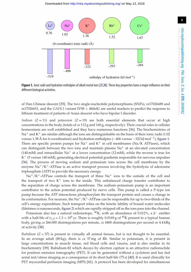
Lithium (Z = 3) is not thought to be an essential element, but is present, as Li+, in some naturalwaters, especially ‘spa’ waters (thermal baths) and in some commercial bottled mineral drinkingwater. There is ca 2.4 mg Li in the body.1 Perhaps, it has beneficial effects at these low levels. Inmedicine, lithium salts are widely used for treatment of bipolar disorders (BDs). Li+ is a verysmall ion (6-coordinate radius 0.76 Å, figure 1) with a high hydration enthalpy (−519 kJ mol−1).
The symptoms of lithium deficiency in humans are believed to manifest primarily asbehavioural abnormalities. A link between low lithium intake and altered behaviour andaggressiveness has been reported [29–31]. As a medicine, lithium is best recognized for its anti-manic properties [32]. It is often administered in the form of lithium carbonate, as a psychiatricdrug. More than 2 million American adults, or ca 1% of the population 18 years or older, sufferfrom BD [33]. A recent study was conducted on the influence of lithium on the peripheral bloodgene expression profiles of patients with BD [34]. For bipolar patients who responded to lithium,the genes which protect against cell death (including Bcl2 and IRS2) were upregulated, whilethose which promote cell death were downregulated, including the pro-apoptotic genes knownas BAD and BAK1 [34]. These results suggest that increased expression of BCL2 and related genesis necessary for the therapeutic effects of lithium.
Lithium is an inhibitor of the enzyme glycogen synthase kinase-3β (GSK-3β) which isresponsible for the hyper-phosphorylation of the tau protein in Alzheimer’s disease [33]. A linkbetween genetic variations in the gene encoding glutamate decarboxylase-like protein 1 (GADL1)and response to lithium maintenance treatment for bipolar I disorder has been found in patients
1In this article, we have used the data on the abundances of elements in humans given in WebElements:http://www.webelements.com/ (for a body weight of 80 kg).
on May 13, 2018http://rsta.royalsocietypublishing.org/Downloaded from
4
rsta.royalsocietypublishing.orgPhil.Trans.R.Soc.A373:20140182
.........................................................
0.76
–519
–406–322 –293 –264
Li+ Na+ K+ Rb+ Cs+
1.02
(6-coordinate) ionic radii (Å)
enthalpy of hydration (kJ mol–1)
1.38 1.52 1.67
Figure 1. Ionic radii and hydration enthalpies of alkali metal ions [27,28]. These key properties have a major influence on theirdifferent biological activities.
of Han Chinese descent [35]. The two single-nucleotide polymorphisms (SNPs), rs17026688 andrs17026651, and the GADL1 variant IVS8 + 48delG are useful markers to predict the response tolithium treatment of patients of Asian descent who have bipolar I disorder.
Sodium (Z = 11) and potassium (Z = 19) are both essential elements that occur at highconcentrations in the body (totals of ca 112 g and 160 g, respectively). Their crucial roles in cellularhomeostasis are well established and they have numerous functions [36]. The biochemistries ofNa+ and K+ are similar although the ions are distinguishable on the basis of their ionic radii (1.02versus 1.38 Å for 6-coordination) and hydration enthalpies (−406 versus −322 kJ mol−1), figure 1.There are specific protein pumps for Na+ and K+ in cell membranes (Na/K ATPases), whichcan distinguish between the two ions and maintain plasma Na+ at an elevated concentration(140 mM) and intracellular Na+ at a lower concentration (12 mM), while the reverse is true forK+ (5 versus 140 mM), generating electrical potential gradients responsible for nervous impulses[36]. The process of moving sodium and potassium ions across the cell membrane by theenzyme Na+/K+-ATPase is an active transport process involving the hydrolysis of adenosinetriphosphate (ATP) to provide the necessary energy.
Na+/K+-ATPase controls the transport of three Na+ ions to the outside of the cell andthe transport of two K+ ions to the inside. This unbalanced charge transfer contributes tothe separation of charge across the membrane. The sodium–potassium pump is an importantcontributor to the action potential produced by nerve cells. This pump is called a P-type ionpump because the ATP interactions phosphorylate the transport protein and causes a change inits conformation. For neurons, the Na+/K+-ATPase can be responsible for up to two-thirds of thecell’s energy expenditure. Such transport relies on the kinetic lability of bound water molecules(lifetimes ca nanoseconds, figure 2) which are rapidly stripped off as the ions pass into the channel.
Potassium also has a natural radioisotope, 40K, with an abundance of 0.012%, a β− emitterwith a half-life of t1/2 = 1.3 × 10⁹ yr. There is roughly 0.0169 g of 40K present in a typical humanbody, giving ca 266 000 disintegrations per minute, ca 4400 disintegrations per second or 4.4 kBqof activity [38].
Rubidium (Z = 37) is present in virtually all animal tissues, but is not thought to be essential.In an average adult (80 kg), there is ca 37 mg of Rb. Similar to potassium, it is present inlarge concentrations in muscle tissue, red blood cells and viscera, and is also similar in itsbiochemistry [39]. Rubidium-82 which decays by electron capture is an attractive radionuclidefor positron emission tomography (PET). It can be generated without a cyclotron and has fastserial rest/stress imaging as a consequence of its short half-life (75 s) [40]. It is used clinically forPET myocardial perfusion imaging (MPI) [41]. A protocol has been developed for simultaneous
on May 13, 2018http://rsta.royalsocietypublishing.org/Downloaded from
5
rsta.royalsocietypublishing.orgPhil.Trans.R.Soc.A373:20140182
.........................................................
Ir3+ Rh3+ Cr3+Ru3+ Al3+Fe3+ V3+Ga3+
Ti3+
Yb3+ Gd3+Tb3+Dy3+Ho3+Er3+
In3+Tm3+
Be2+ Mg2+
Li+Na+ K+ Rb+
Cs+
Ca2+Eu2+Ba2+
Sr2+
Ru2+
Pd2+
V2+ Ni2+ Co2+Fe2+
Mn2+ Cr2+ Cu2+
Zn2+ Cd2+ Hg2+Pt2+
10–10 10–8 10–6 10–4 10–2
10–1010–810–610–410–2
1
kH2O s–1
102 104 106 108 1010
1010 108 106 104 102 1
day nanosecondtH2O s–1
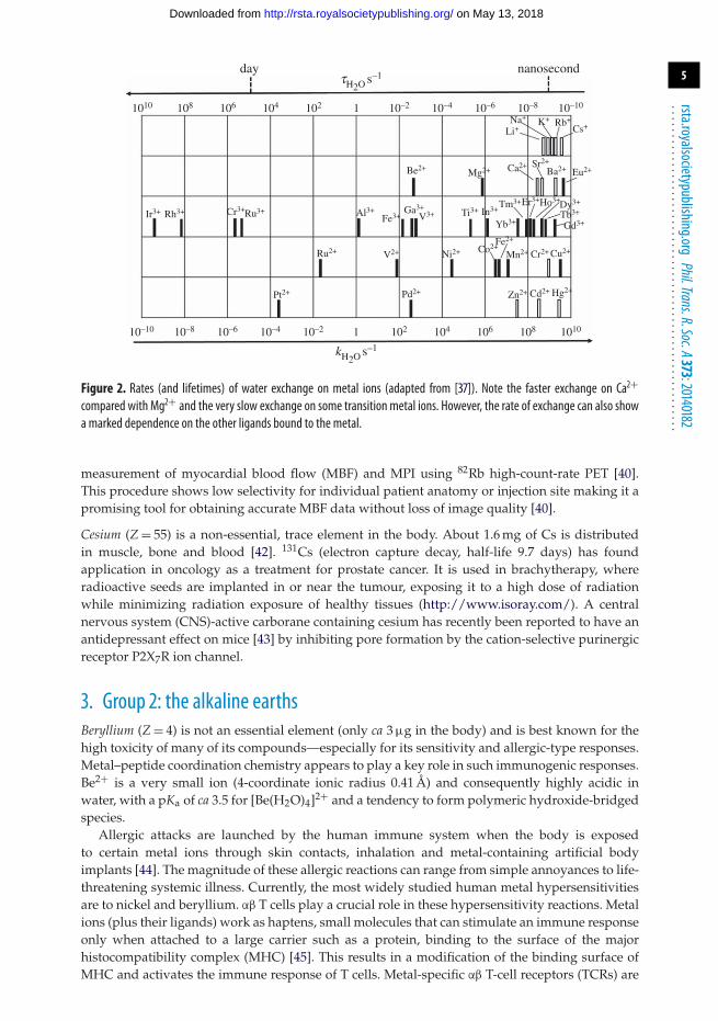
Figure 2. Rates (and lifetimes) of water exchange on metal ions (adapted from [37]). Note the faster exchange on Ca2+
compared with Mg2+ and the very slow exchange on some transition metal ions. However, the rate of exchange can also showa marked dependence on the other ligands bound to the metal.
measurement of myocardial blood flow (MBF) and MPI using 82Rb high-count-rate PET [40].This procedure shows low selectivity for individual patient anatomy or injection site making it apromising tool for obtaining accurate MBF data without loss of image quality [40].
Cesium (Z = 55) is a non-essential, trace element in the body. About 1.6 mg of Cs is distributedin muscle, bone and blood [42]. 131Cs (electron capture decay, half-life 9.7 days) has foundapplication in oncology as a treatment for prostate cancer. It is used in brachytherapy, whereradioactive seeds are implanted in or near the tumour, exposing it to a high dose of radiationwhile minimizing radiation exposure of healthy tissues (http://www.isoray.com/). A centralnervous system (CNS)-active carborane containing cesium has recently been reported to have anantidepressant effect on mice [43] by inhibiting pore formation by the cation-selective purinergicreceptor P2X7R ion channel.
3. Group 2: the alkaline earthsBeryllium (Z = 4) is not an essential element (only ca 3 μg in the body) and is best known for thehigh toxicity of many of its compounds—especially for its sensitivity and allergic-type responses.Metal–peptide coordination chemistry appears to play a key role in such immunogenic responses.Be2+ is a very small ion (4-coordinate ionic radius 0.41 Å) and consequently highly acidic inwater, with a pKa of ca 3.5 for [Be(H2O)4]2+ and a tendency to form polymeric hydroxide-bridgedspecies.
Allergic attacks are launched by the human immune system when the body is exposedto certain metal ions through skin contacts, inhalation and metal-containing artificial bodyimplants [44]. The magnitude of these allergic reactions can range from simple annoyances to life-threatening systemic illness. Currently, the most widely studied human metal hypersensitivitiesare to nickel and beryllium. αβ T cells play a crucial role in these hypersensitivity reactions. Metalions (plus their ligands) work as haptens, small molecules that can stimulate an immune responseonly when attached to a large carrier such as a protein, binding to the surface of the majorhistocompatibility complex (MHC) [45]. This results in a modification of the binding surface ofMHC and activates the immune response of T cells. Metal-specific αβ T-cell receptors (TCRs) are
on May 13, 2018http://rsta.royalsocietypublishing.org/Downloaded from
6
rsta.royalsocietypublishing.orgPhil.Trans.R.Soc.A373:20140182
.........................................................
bE26
bE69bY28
bE68p5Pp-2Arg
p6F
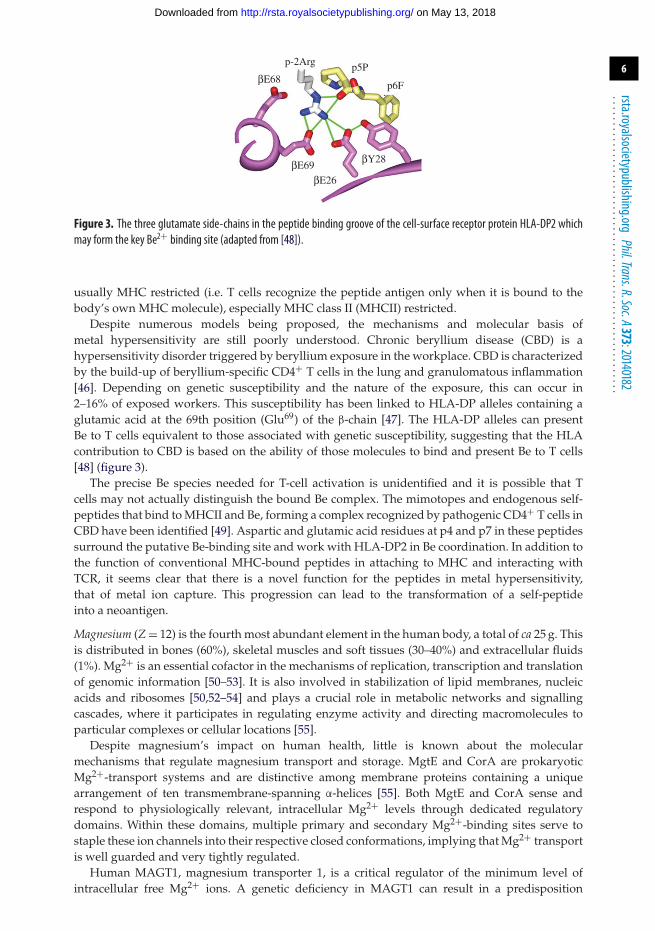
Figure 3. The three glutamate side-chains in the peptide binding groove of the cell-surface receptor protein HLA-DP2 whichmay form the key Be2+ binding site (adapted from [48]).
usually MHC restricted (i.e. T cells recognize the peptide antigen only when it is bound to thebody’s own MHC molecule), especially MHC class II (MHCII) restricted.
Despite numerous models being proposed, the mechanisms and molecular basis ofmetal hypersensitivity are still poorly understood. Chronic beryllium disease (CBD) is ahypersensitivity disorder triggered by beryllium exposure in the workplace. CBD is characterizedby the build-up of beryllium-specific CD4+ T cells in the lung and granulomatous inflammation[46]. Depending on genetic susceptibility and the nature of the exposure, this can occur in2–16% of exposed workers. This susceptibility has been linked to HLA-DP alleles containing aglutamic acid at the 69th position (Glu69) of the β-chain [47]. The HLA-DP alleles can presentBe to T cells equivalent to those associated with genetic susceptibility, suggesting that the HLAcontribution to CBD is based on the ability of those molecules to bind and present Be to T cells[48] (figure 3).
The precise Be species needed for T-cell activation is unidentified and it is possible that Tcells may not actually distinguish the bound Be complex. The mimotopes and endogenous self-peptides that bind to MHCII and Be, forming a complex recognized by pathogenic CD4+ T cells inCBD have been identified [49]. Aspartic and glutamic acid residues at p4 and p7 in these peptidessurround the putative Be-binding site and work with HLA-DP2 in Be coordination. In addition tothe function of conventional MHC-bound peptides in attaching to MHC and interacting withTCR, it seems clear that there is a novel function for the peptides in metal hypersensitivity,that of metal ion capture. This progression can lead to the transformation of a self-peptideinto a neoantigen.
Magnesium (Z = 12) is the fourth most abundant element in the human body, a total of ca 25 g. Thisis distributed in bones (60%), skeletal muscles and soft tissues (30–40%) and extracellular fluids(1%). Mg2+ is an essential cofactor in the mechanisms of replication, transcription and translationof genomic information [50–53]. It is also involved in stabilization of lipid membranes, nucleicacids and ribosomes [50,52–54] and plays a crucial role in metabolic networks and signallingcascades, where it participates in regulating enzyme activity and directing macromolecules toparticular complexes or cellular locations [55].
Despite magnesium’s impact on human health, little is known about the molecularmechanisms that regulate magnesium transport and storage. MgtE and CorA are prokaryoticMg2+-transport systems and are distinctive among membrane proteins containing a uniquearrangement of ten transmembrane-spanning α-helices [55]. Both MgtE and CorA sense andrespond to physiologically relevant, intracellular Mg2+ levels through dedicated regulatorydomains. Within these domains, multiple primary and secondary Mg2+-binding sites serve tostaple these ion channels into their respective closed conformations, implying that Mg2+ transportis well guarded and very tightly regulated.
Human MAGT1, magnesium transporter 1, is a critical regulator of the minimum level ofintracellular free Mg2+ ions. A genetic deficiency in MAGT1 can result in a predisposition
on May 13, 2018http://rsta.royalsocietypublishing.org/Downloaded from
7
rsta.royalsocietypublishing.orgPhil.Trans.R.Soc.A373:20140182
.........................................................
to lymphoma as well as elevated levels of Epstein–Barr virus (EBV) [56]. Decreased levels ofintracellular free Mg2+ have been linked to defective expression of the natural killer activatingreceptor NKG2D in both natural killer (NK) and CD8+ T cells as well as impairment of cytolyticresponses to EBV [57]. The intracellular Mg2+ and NKG2D levels are restored when MAGT1-deficient patients are given magnesium supplements, and a reduction in EBV-infected cells in vivois also observed [57]. These results show a link between NKG2D cytolytic activity and EBVantiviral immunity in humans.
The biochemistries of Ca2+ and Mg2+ are similar, although Mg2+ is the smaller ion (radius 0.72versus 1.00 Å for 6-coordination) and exchanges its bound water ligands more slowly (half-life10−5 versus 10−8 s), with Mg2+ dominating inside cells (15–20 mM) [6].
Despite the high level of calcium (Z = 20) in the body (ca 1.1 kg), and the high total concentrationin cells (1–10 mM), free Ca2+ at nanomolar and micromolar concentrations regulates skeletal andcardiac muscle contraction by inducing structural changes in the Ca2+-sensor protein troponin,a gene product [58]. Control over Ca2+ coordination by proteins and small ligands ensuresthat blood plasma can remain supersaturated in Ca2+ without continuously depositing calciumphosphate mineral in bone. Ca2+ ATPases in the plasma membrane export Ca2+ from alleukaryotic cells and in man are the products of four separate genes [59].
In bones and teeth, the creation and mineralization of extracellular collagen matrix iscontrolled by osteoblasts and odontoblasts. By contrast, osteoclasts remove bone mineral andbone matrix, making bone cells important for regulation of the formation and resorption of bone.This is a key step in regulating body Ca2+ as well as Mg2+ and phosphate. Osteoclasts canlower the pH to as low as 4.5 inside the bone-resorbing compartment due to the action of theirplasma membrane H+ pump, thus assisting bone mineral mobilization [60] while simultaneouslysecreting lysosomal enzymes such as acid phosphatase, cathepsin K, matrix metalloproteinase9, and gelatinase [61] that digest the organic matrix. Vast amounts of Ca2+ are absorbed bymature osteoclasts during resorption and their survival is ensured by Na+/Ca2+ exchangersthat prevent harmful Ca2+ overload in the cytoplasm by extruding Ca2+ ions into extracellularspace [62,63].
Although there is about 0.4 g of strontium (Z = 38) in the body, it is not thought to be essential.It is handled in the body in a similar manner to calcium. Some of its effects appear to bebeneficial. Strontium salts are well known toothpaste additives. Regular tooth brushing with astrontium-supplemented toothpaste increases the Sr content in the exposed enamel, which can bean advantage in the prevention of cariogenesis and perhaps strengthens the enamel [64].
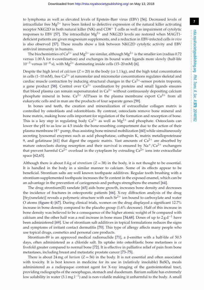
The drug strontium(II) ranelate [65] aids bone growth, increases bone density and decreasesthe incidence of fractures in osteoporotic patients [66]. X-ray diffraction analysis of the drug[Sr2(ranelate)] reveals a polymeric structure with each Sr2+ ion bound to carboxylate and waterO atoms (figure 4) [67]. During clinical trials, women on the drug displayed a significant 12.7%increase in bone density compared to the placebo group (1.6% decrease). Half of this increase inbone density was believed to be a consequence of the higher atomic weight of Sr compared withcalcium and the other half was a real increase in bone mass [34,68]. Doses of up to 2 g d−1 havebeen administered [69]. Use of strontium salt additives in topical formulations reduces the signsand symptoms of irritant contact dermatitis [70]. This type of allergy affects many people whouse topical drugs, cosmetics and personal care products.
Strontium-89 is an approved medical radionuclide [71], a β-emitter with a half-life of 50.5days, often administered as a chloride salt. Its uptake into osteoblastic bone metastases is cafivefold greater compared to normal bone [72]. It is effective in palliative relief of pain from bonemetastases, including breast and metastatic prostate cancer [73–75].
There is about 24 mg of barium (Z = 56) in the body. It is not essential and often associatedwith toxicity. It is best known in medicine for its use in (relatively insoluble) BaSO4 mealsadministered as a radiopaque contrast agent for X-ray imaging of the gastrointestinal tract,providing radiographs of the oesophagus, stomach and duodenum. Barium sulfate has extremelylow solubility in water (3.1 mg l−1) and is non-volatile making it unharmful to the body. A small
on May 13, 2018http://rsta.royalsocietypublishing.org/Downloaded from
8
rsta.royalsocietypublishing.orgPhil.Trans.R.Soc.A373:20140182
.........................................................
–O
O
S
CN
N
O–
O
O–
O
O
O–
Sr2+
OH2OH2
H2O
O
O
O
OSr2+
OO
O
H2O
OH2
H2O
Sr2+
Sr2+
Sr2+
Sr2+ Sr2+
Sr2+
n
3.79nH2O
Figure 4. The polymeric structure of strontium ranelate. Each monomer unit contains two Sr2+ ions.
amount of Ba2+ may be solubilized by gastric HCl. Barium in Brazil nuts is sometimes a concern.There is a report of a man consuming a single dose of 179 mg Ba from 92 g of Brazil nuts, withmore than 91% of the dose absorbed [61].
All isotopes of radium (Z = 88) are radioactive. There are ultra-trace amounts in our body withno beneficial role. Radium-223 dichloride is an α-emitting pharmaceutical (half-life 11.4 days)approved for treatment of symptomatic bone metastases and castration-resistant prostate cancer(http://www.xofigo-us.com/index.php (accessed 10 July 2014)). 223Ra2+ is a Ca2+-mimetic thatselectively targets bone metastases with short-range high-energy α particles which cause double-strand breaks in DNA as well as greatly localized cytotoxicity, with negligible myelosuppression[76–80]. The activity of 223Ra in vivo has prompted evaluation of its efficiency and safety in clinicaltrials of patients suffering from bone-metastatic prostate cancer [77,79,81–84]. It is an effectivecandidate for palliation of bone pain.
4. Groups 3–12: the transitions metals
(a) First transition seriesScandium (Z = 21) is not an essential element, but its radioisotopes have potential in PET andSPECT imaging, as well as therapy [85]. Both 44Sc (t1/2 = 3.93 h, 94% β+, Emax 1474 keV) and47Sc (t1/2 = 3.35 days, 100% β−, Emax 162 keV) display appropriate properties for diagnosis ortherapy, and, in combination, can be used for theranostic applications. 44Sc is a candidate for PETimaging using radiometalated peptides or other small targeting biomolecules [86]. Preclinicalevaluations of DOTA-functionalized biomolecules radiolabelled with 44Sc have demonstratedeffectiveness for PET imaging [87]. 44Sc could be a useful radioisotope for clinical nuclear imagingand pre-therapeutic dosimetry of cancer patients prior to treatment with a therapeutic 177Lu-labelled DOTA derivative. 47Sc being a β-emitter has potential as a therapeutic radionuclideand combined with 44Sc would allow use of matching radiopharmaceuticals with the samepharmacokinetics [87].
Titanium (Z = 22) is not thought to be essential and is best known in medicine as a light,strong metal in implants, and in anti-cancer complexes, two of which have been on clinicaltrials. Mice given TiIV oxalate supplements show positive weight gains and a reduction intumour development [88]. Beneficial effects on other animals have also been reported [89,90], andinterestingly titanium compounds, patented as fodder additives, are claimed to improve weight
on May 13, 2018http://rsta.royalsocietypublishing.org/Downloaded from

9
rsta.royalsocietypublishing.orgPhil.Trans.R.Soc.A373:20140182
.........................................................
Ti
Cl
Cl
Ti
Cl
Cl
O
O
O
OTi
O
O
OO
TiO
O
NN
O
O
Br
Br
titanocenedichloride
budotitane
titanocene Y titanium salan complex
Figure 5. Titanium complexes with anti-tumour properties. Titanocene dichloride and budotitane both underwent clinicaltrials but were abandoned because of instability in aqueous media.
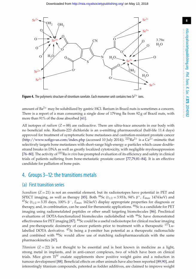
gain in domestic animals [91]. Titanium(IV) complexes were the first class of metal compoundsto enter clinical trials after platinum complexes for the treatment of cancer [92]. Budotitane andtitanocene dichloride (figure 5) exhibit anti-tumour activity and low toxicity in several cancer celllines [93]. Unfortunately, clinical trials of these compounds initiated in the 1980s were finallyabandoned due their instability in water [92,94]. Since then, other TiIV complexes have beendeveloped with the aim of overcoming this instability. Among these, titanocene derivativesand titanium salan complexes show promise [95,96]. The titanium complexes Ti-Salan andTi-Y display contrasting behaviour regarding their reactivities with DNA or albumin, theircellular uptake and intracellular distribution [97]. Ti-Salan shows relatively low binding tobiomolecules but increased serum-dependent cellular uptake while Ti-Y shows lower cellularaccumulation and high binding to albumin and DNA. Biodistribution data indicate that for Ti-Y transport into nuclei and DNA interactions are crucial whereas mitochondrial targeting isimportant for Ti-Salan.
Interest in the essentiality of vanadium (Z = 23) was aroused by the finding about 35 years ago thatvanadate present as an impurity in commercial horse skeletal muscle ATP inhibits the enzymeNa+/K+-ATPase [98]. Vanadium is thought to be essential for man, but its roles in the body (totalof ca 2.4 mg) are poorly understood. Vanadium complexes have been on clinical trials recently asantidiabetic agents.
on May 13, 2018http://rsta.royalsocietypublishing.org/Downloaded from
10
rsta.royalsocietypublishing.orgPhil.Trans.R.Soc.A373:20140182
.........................................................
bis(maltolato)oxovanadium(IV)
V
O OO
O
O
OO
bis(ethyl-maltolato)oxovanadium(IV)
O OO
OO O
OV
(b)(a)
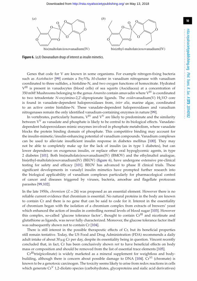
Figure 6. (a,b) Oxovanadium drugs of interest as insulin mimetics.
Genes that code for V are known in some organisms. For example nitrogen-fixing bacteriasuch as Azotobacter [99] contain a Fe7VS9 M-cluster in vanadium nitrogenase with vanadiumcoordinated to three sulfides, a histidine-N, and two oxygen functions of homocitrate. HydratedVIII is present in vanadocytes (blood cells) of sea squirts (Ascidiacea) at a concentration of350 mM! Mushrooms belonging to the genus Amanita contain amavadin where VIV is coordinatedto two tetradentate N-oxyimino-2,2′-dipropionate ligands. The oxidovanadium(V) H2VO coreis found in vanadate-dependent haloperoxidases from, inter alia, marine algae, coordinatedto an active centre histidine-N. These vanadate-dependent haloperoxidases and vanadiumnitrogenases remain the only identified vanadium-containing enzymes in nature [99].
In vertebrates, particularly humans, VIV and VV are likely to predominate and the similaritybetween VV as vanadate and phosphate is likely to be central to its biological effects. Vanadate-dependent haloperoxidases mimic enzymes involved in phosphate metabolism, where vanadateblocks the protein binding domain of phosphate. This competitive binding may account forthe insulin-mimetic/insulin-enhancing potential of vanadium compounds. Vanadium complexescan be used to alleviate insufficient insulin response in diabetes mellitus [100]. They maynot be able to completely make up for the lack of insulin (as in type 1 diabetes), but canlower dependence on exogenous insulin, or replace other oral hypoglycemic agents, in type2 diabetes [101]. Both bis(maltolato)oxovanadium(IV) (BMOV) and the ethylmaltol analogue,bis(ethyl-maltolato)oxovanadium(IV) (BEOV) (figure 6), have undergone extensive pre-clinicaltesting for safety and efficacy [101]. BEOV has advanced to phase II clinical trials. Thesesignificant developments in vanadyl insulin mimetics have prompted further research intothe biological applicability of vanadium complexes particularly for pharmacological controlof cancer and diseases triggered by viruses, bacteria, amoebae and flagellate protozoanparasites [99,102].
In the late 1950s, chromium (Z = 24) was proposed as an essential element. However there is noreliable current evidence that chromium is essential. No natural proteins in the body are knownto contain Cr and there is no gene that can be said to code for it. Interest in the essentialityof chromium began with the isolation of a chromium complex from extracts of brewers’ yeastwhich enhanced the action of insulin in controlling normal levels of blood sugar [103]. Howeverthis complex, so-called ‘glucose tolerance factor’, thought to contain CrIII and nicotinate andglutathione as ligands, was never fully characterized. Moreover, the glucose tolerance factor itselfwas subsequently shown not to contain Cr [104].
There is still interest in the possible therapeutic effects of Cr, but its beneficial propertiesstill remain tentative. Today, the US Food and Drug Administration (FDA) recommends a dailyadult intake of about 30 μg Cr per day, despite its essentiality being in question. Vincent recentlyconcluded that, in fact, Cr has been conclusively shown not to have beneficial effects on bodymass or composition and should be removed from the list of essential trace elements [105].
CrIIItris(picolinate) is widely marketed as a mineral supplement for weightloss and body-building, although there is concern about possible damage to DNA [104]. CrVI (chromate) isknown to be a genotoxic carcinogen. The toxicity seems likely to arise from redox reactions in cellswhich generate CrV 1,2-diolato species (carbohydrates, glycoproteins and sialic acid derivatives)
on May 13, 2018http://rsta.royalsocietypublishing.org/Downloaded from
11
rsta.royalsocietypublishing.orgPhil.Trans.R.Soc.A373:20140182
.........................................................
which can cause oxidative damage to DNA [29]. A recent study of CrV complexes with a varietyof monosaccharides and the model ligand cis-1,2-cyclopentanediol provided evidence of theirnuclease activity [29]. The CrV complexes can cause oxidative DNA damage without the presenceof added reductants or oxidants, supporting the participation of CrV 1,2-diolato complexes in thebiological activities of both CrVI and CrIII.
There is no doubt that manganese (Z = 25, total body Mn ca 16 mg) is essential, and that thereare genetic codes for a range of Mn enzymes with functions in metabolism, reproduction, theimmunological system, regulation of cellular energy and bone and connective tissue growth.Manganese as Mn2+ has chemistry similar to Mg2+, although unlike the latter it is redox-active,with the +3 state also being readily accessible, as in the enzyme mitochondrial manganesesuperoxide dismutase. The most abundant Mn-binding protein in the body is glutaminesynthetase which plays a prominent role in brain chemistry (in astrocytes).
However, excessively high levels of Mn in the body can be toxic [104]. Industrial Mntoxicity is well recognized, as manganism, a neurodegenerative disorder. Manganism results fromoverexposure and production of reactive oxygen species and toxic metabolites as well as alteredmitochondrial and ATP functions and exhaustion of cellular antioxidant defence mechanisms[106]. Mutations in the Mn exporter gene SLC30A10 are associated with motor impairment andParkinson’s disease-like symptoms.
An intriguing question is how does Mn cross the blood–brain barrier (BBB)? Despite severalstudies on the mechanism of transport of manganese across the BBB, the exact identity of thecarrier is still unclear. Mechanisms using active transport [107] or facilitated diffusion [108,109]have been suggested, as well the high affinity metal transporters of calcium and iron. Manganesemay also enter the brain via leak pathways in areas without an intact BBB [110]. Overall, it islikely that more than one transporter is responsible for transport of Mn across the BBB; several ofthem may work in a cooperative manner to maintain optimal Mn tissue concentrations.
There is about 4.8 g of iron (Z = 26) in the body and the human genome codes for over 500 Fe-containing proteins [111]. In eukaryotes and prokaryotes, the level of non-haem iron proteinsis relatively uniform and their relative number in the proteome decreases in passing fromarchaea (about 7%), to bacteria (about 4%), to eukaryotes (about 1%) [112]. Several genes areresponsible for haem synthesis, haem transport and insertion of haem groups into haem proteins[111,113,114]. Particularly intriguing from an inorganic chemistry standpoint, is the presenceof sulfide as a ligand in iron–sulfur proteins [115,116]. H2S itself is best known as a poison.Fe−S proteins are ubiquitous in cells and play many roles including electron transfer, catalysisand iron regulation (IRP proteins). Sulfide for Fe−S proteins is generated from cysteine by theenzyme cysteine desulfurase. Mutations in proteins involved in Fe−S cluster biogenesis areknown to cause at least five distinctive human diseases. Iron–sulfur clusters are also associatedwith enzymes involved in DNA processing.
The implications of overload or deficiency of iron have been discussed recently [114]. Thereis a need for new oral iron supplements—the most widely used supplement, ferrous sulfate,was introduced way back in 1832 as a treatment for anaemia. The bioavailability of Fe isaffected by plant foods, including phytate (inositol hexaphosphate), polyphenols and tannins[117]. Elevated Fe levels seem to play a role in neurodegeneration [118]. In therapy, the Fe(II)containing compound ferroquine (figure 7), a ferrocene derivative, is currently in phase II clinicaltrials [119] for treatment of malaria. It is believed to exert its antiplasmodial properties viatwo mechanisms: interaction with free haem and generation of reactive oxygen species [120].Ferrocifens, complexes containing ferrocene and tamoxifen fragments show promise as potentialdrugs for treatment of breast cancer [121]. Superparamagnetic iron oxides composed of nano-sized crystals are used as magnetic resonance imaging (MRI) contrast agents [122] and sodiumnitroprusside, Na2[FeII(CN)5NO], which slowly releases nitric oxide, has long been prescribed asa hypertensive agent [123]. Iron overload as in the inherited condition thalessaemia can be treatedwith iron chelating agents such as deferoxamine (desferrioxamine B, injectable) or more recentlydeferasirox (oral) (figure 7).
on May 13, 2018http://rsta.royalsocietypublishing.org/Downloaded from

12
rsta.royalsocietypublishing.orgPhil.Trans.R.Soc.A373:20140182
.........................................................
Cl
HN
N
Fe
N
CH3
CH3
HO
O
HO
OH
N
N N
H2N
deferoxamine
deferasirox ferroquine
OH
OHO
O
O O
OH
N
OHNN
N NH
Figure 7. The injectable iron chelator deferoxamine, oral chelator deferasirox and antimalarial drug ferroquine.
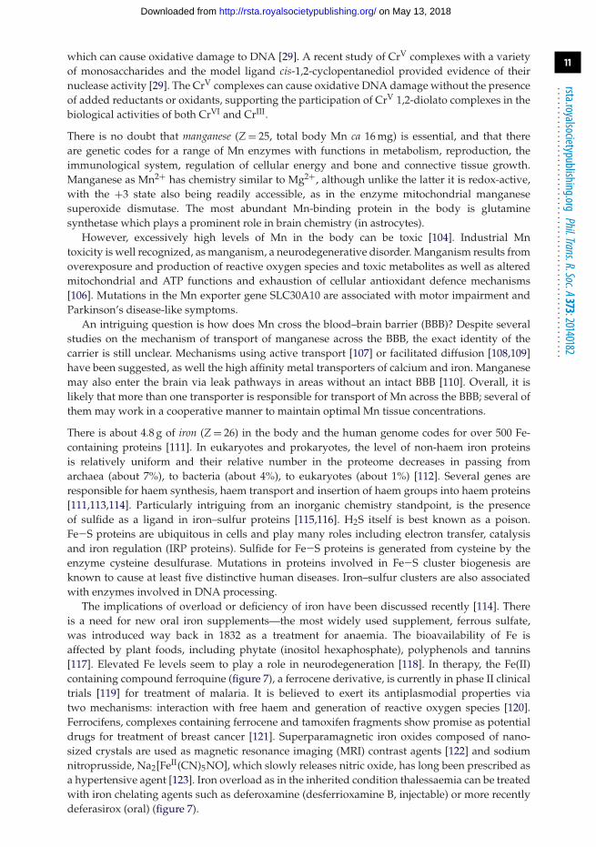
There is only ca 1.6 mg of cobalt (Z = 27) in the body, but it plays a vital role in the coenzymevitamin B12 (cobalamin), which has a recommended daily intake of 2–3 μg d−1. A deficiencyof this vitamin results in pernicious anaemia through inactivation of one of two humanenzymes for which B12 is the required coenzyme (methionine synthase and methylmalonyl-CoAmutase) [124]. Vitamin B12 can be synthesized only by microorganisms (bacteria and archaea). Itcontains cobalt tightly bound in a corrin ring and oxidation states +1, +2 and +3 are important inits biological activity. It was the first organometallic complex to be recognized in man and readilyforms Co−C bonds (to deoxyadenosyl and methyl groups).
Our genome codes for proteins in the digestive tract that selectively absorb B12 from the diet.Several gene products, including carrier proteins are involved in the absorption and distributionof vitamin B12 [125]. Altered cellular entry, transit or exit of the vitamin can result in deficiencyand eventually give rise to haematological and neurological disorders [125]. Under physiologicalconditions, vitamin B12 is bound to the gastric intrinsic factor (figure 8) and is internalized inthe ileum by a highly specific receptor complex composed of cubilin (Cubn) and amnionless(Amn) proteins [124,126]. Following exit of vitamin B12 from the ileum, general cellular uptakefrom the circulation requires the transcobalamin receptor CD320, whereas kidney reabsorption ofcobalamin depends on megalin.
Perhaps surprisingly, there are few cobalt-based drugs. Doxovir, also known as CTC-96, isa CoIII bis(2-methylimidazole) acacen derivative which has completed clinical phase I trials forophthalmic herpetic keratitis and adenoviral conjunctivitis and clinical phase II trials for herpeslabialis [127]. We can expect CoIII to be reduced to the more labile CoII inside cells.
There is ca 8 mg of nickel (Z = 28) in the body, but it is not clear whether nickel is an essentialelement for man or not. Nickel allergy is certainly well known—one of the most common allergiesin the world. Nickel is certainly an essential element for some bacteria.
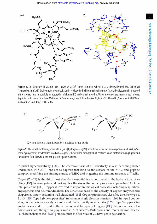
For vertebrates, no Ni-dependent enzymes are known [128]; however, in microorganismsseveral Ni-dependent enzymes have been well characterized. Owing to our symbiotic existencewith microbes (of which there are 10× more than human cells in our body), it is possible thatNi is essential for the survival of some of the microbes on which we depend for survival [129].However Ni can also make some microbes virulent. Nickel is a virulence determinant for thehuman gastric pathogen Helicobacter pylori which possesses two nickel enzymes that are crucialfor in vivo colonization, [NiFe] hydrogenase (figure 9) and urease, with urease containing 24 nickelions per active complex [130]. These two nickel trafficking partners in virulence are potentialnovel therapeutic targets for treatment of H. pylori infections which can prevent ulcers fromhealing [130].
As for beryllium (vide supra), metal hypersensitivity is also an immune disorder associatedwith Ni allergic hyper-reactivity [44]. Approximately 10% of the population suffers fromNi-contact dermatitis. CD4+ and CD8+ T cells have been identified as the major effector cells
on May 13, 2018http://rsta.royalsocietypublishing.org/Downloaded from
13
rsta.royalsocietypublishing.orgPhil.Trans.R.Soc.A373:20140182
.........................................................
Leu 377
Asp 204
Asp 153
Gln 252
Tyr 262
Ser 38
His 73
Ser 347
Tyr 115
Thr 70
Leu 350
Glu 360
Val 352
Phe 370
Val 381
Tvr 399
Cbl
CONH2
CONH2
CONH2
O
HO
HO
N
NH
O
O O
O–
N
P
H2NOC(a) (b)
H2NOC
H
N
N N
R N
Co+
H2NOC
O
Figure 8. (a) Structure of vitamin B12, shown as a CoIII corrin complex, where R= 5′-deoxyadenosyl Me, OH or CN(cyanocobalamin). (b) Environment around cobalamin (yellow) in the binding site of intrinsic factor, the glycoprotein producedin the stomach and responsible for absorption of vitamin B12 in the small intestine. Water molecules are shown as red spheres.Reprinted with permission fromMathews FS, Gordon MM, Chen Z, Rajashankar KR, Ealick SE, Alpers DH, Sukumar N. 2007 Proc.Natl Acad. Sci. USA 104, 17 311–17 316.
Cys
Cys
Cys
Cys
Cys Cys
Cys Cys
COCN
COCN
CN(SO) CN(SO)
X = non-protein ligand, possibly a sulfide or an oxide
Ni Fe Ni Fe
S
S
S
S
X(b)(a)
Figure 9. The nickel-containing active site in [NiFe] hydrogenases [128], a virulence factor for microorganisms such as H. pylori.These hydrogenases are classified into two categories, the oxidized form (a) which contains a non-protein bridging ligand andthe reduced form (b) where the non-protein ligand is absent.
in nickel hypersensitivity [131]. The chemical basis of Ni sensitivity is also becoming betterunderstood. Nickel(II) ions act as haptens that bind to the surface of the MHC and peptidecomplex, modifying the binding surface of MHC and triggering the immune response of T cells.
Copper (Z = 29) is the third most abundant essential transition metal in the body, a total of ca80 mg [132]. In eukaryotes and prokaryotes, the size of the copper proteome approaches 1% of thetotal proteome [133]. Copper is involved in important biological processes including respiration,angiogenesis and neuromodulation. The structural basis of the activity of copper enzymes andchaperones is now becoming well elucidated [134]. Copper proteins are classified as either type 1,2 or 3 [135]. Type 1 (blue copper sites) function in single electron transfers [136]. In type 2 coppersites, copper acts as a catalytic centre and binds directly to substrates [135]. Type 3 copper sitesare binuclear and involved in the activation and transport of oxygen [135]. Abnormalities in Cuhomeostasis are thought to play a role in Alzheimer’s, Parkinson’s and motor neuron disease[137], but Scheiber et al. [134] point out that the full roles of Cu have yet to be clarified.
on May 13, 2018http://rsta.royalsocietypublishing.org/Downloaded from
14
rsta.royalsocietypublishing.orgPhil.Trans.R.Soc.A373:20140182
.........................................................
lid
C
Zn
C412
C363
H473
C1210
C1213
C414
a22
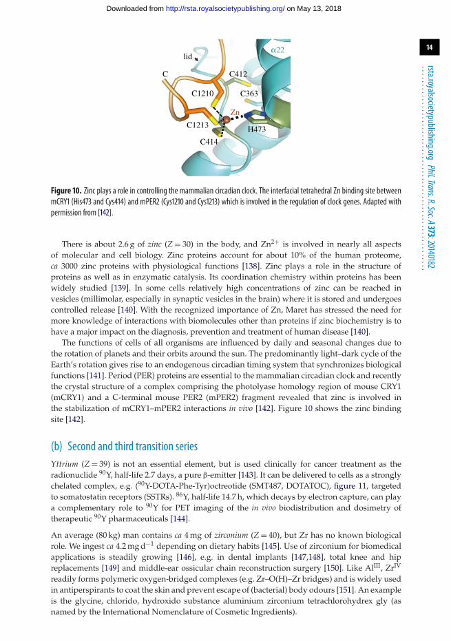
Figure 10. Zinc plays a role in controlling the mammalian circadian clock. The interfacial tetrahedral Zn binding site betweenmCRY1 (His473 and Cys414) and mPER2 (Cys1210 and Cys1213) which is involved in the regulation of clock genes. Adapted withpermission from [142].
There is about 2.6 g of zinc (Z = 30) in the body, and Zn2+ is involved in nearly all aspectsof molecular and cell biology. Zinc proteins account for about 10% of the human proteome,ca 3000 zinc proteins with physiological functions [138]. Zinc plays a role in the structure ofproteins as well as in enzymatic catalysis. Its coordination chemistry within proteins has beenwidely studied [139]. In some cells relatively high concentrations of zinc can be reached invesicles (millimolar, especially in synaptic vesicles in the brain) where it is stored and undergoescontrolled release [140]. With the recognized importance of Zn, Maret has stressed the need formore knowledge of interactions with biomolecules other than proteins if zinc biochemistry is tohave a major impact on the diagnosis, prevention and treatment of human disease [140].
The functions of cells of all organisms are influenced by daily and seasonal changes due tothe rotation of planets and their orbits around the sun. The predominantly light–dark cycle of theEarth’s rotation gives rise to an endogenous circadian timing system that synchronizes biologicalfunctions [141]. Period (PER) proteins are essential to the mammalian circadian clock and recentlythe crystal structure of a complex comprising the photolyase homology region of mouse CRY1(mCRY1) and a C-terminal mouse PER2 (mPER2) fragment revealed that zinc is involved inthe stabilization of mCRY1–mPER2 interactions in vivo [142]. Figure 10 shows the zinc bindingsite [142].
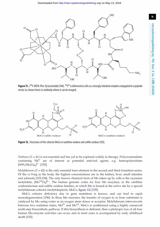
(b) Second and third transition seriesYttrium (Z = 39) is not an essential element, but is used clinically for cancer treatment as theradionuclide 90Y, half-life 2.7 days, a pure β-emitter [143]. It can be delivered to cells as a stronglychelated complex, e.g. (90Y-DOTA-Phe-Tyr)octreotide (SMT487, DOTATOC), figure 11, targetedto somatostatin receptors (SSTRs). 86Y, half-life 14.7 h, which decays by electron capture, can playa complementary role to 90Y for PET imaging of the in vivo biodistribution and dosimetry oftherapeutic 90Y pharmaceuticals [144].
An average (80 kg) man contains ca 4 mg of zirconium (Z = 40), but Zr has no known biologicalrole. We ingest ca 4.2 mg d−1 depending on dietary habits [145]. Use of zirconium for biomedicalapplications is steadily growing [146], e.g. in dental implants [147,148], total knee and hipreplacements [149] and middle-ear ossicular chain reconstruction surgery [150]. Like AlIII, ZrIV
readily forms polymeric oxygen-bridged complexes (e.g. Zr–O(H)–Zr bridges) and is widely usedin antiperspirants to coat the skin and prevent escape of (bacterial) body odours [151]. An exampleis the glycine, chlorido, hydroxido substance aluminium zirconium tetrachlorohydrex gly (asnamed by the International Nomenclature of Cosmetic Ingredients).
on May 13, 2018http://rsta.royalsocietypublishing.org/Downloaded from
15
rsta.royalsocietypublishing.orgPhil.Trans.R.Soc.A373:20140182
.........................................................
NN
NN
NH
NH
NH
HN
HN
HN
O
O
O
OO
HN
HN
O
O
O
HO
OO
O
OO O
NH
NH2
OH
OH
OH
S
S
90Y
Figure 11. (90Y-DOTA-Phe-Tyr)octreotide [144]. 90YIII is delivered to cells as a strongly chelated complex conjugated to a peptidevector (as shown here) or antibody where it can be imaged.
MoCo (sulfite oxidase) MoCo (xanthine oxidase)
O
OO
OP
O–
–O
O
O Mo
Cys
SS
SHN
NH
HN
NH2N
OH
OO
OP
O–
–O
O
O Mo
S
SS
HN
NH
HN
NH2N
Figure 12. Structures of the cofactor MoCo in xanthine oxidase and sulfite oxidase [153].
Niobium (Z = 41) is not essential and has yet to be explored widely in therapy. Polyoxometalatescontaining NbV are of interest as potential antiviral agents, e.g. heteropolyniobate[SiW9Nb3O40]7− [152].
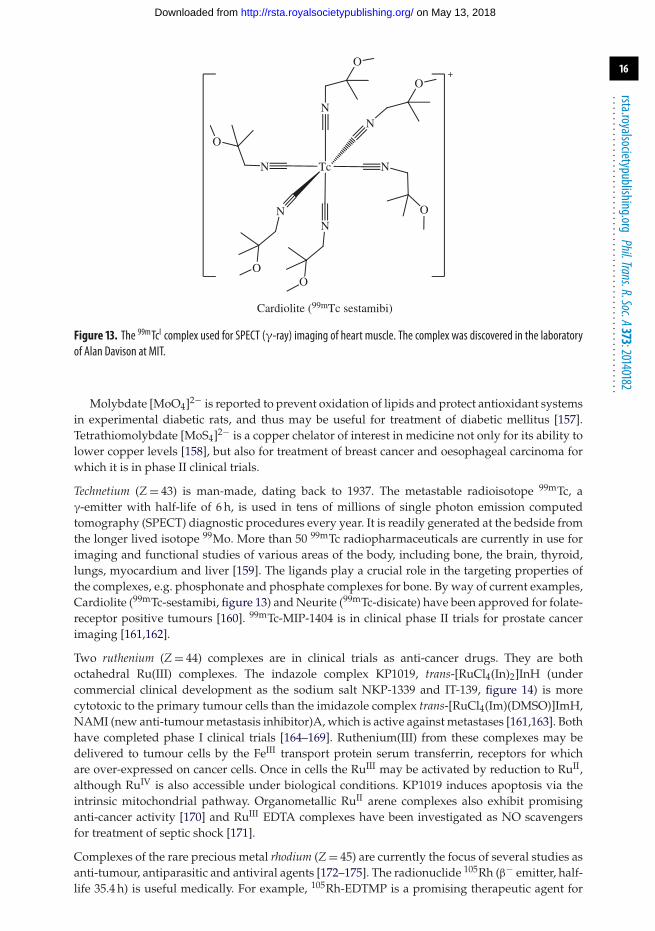
Molybdenum (Z = 42) is the only essential trace element in the second and third transition series.Of the ca 8 mg in the body, the highest concentrations are in the kidney, liver, small intestineand adrenals [153,154]. The only known chemical form of Mo taken up by cells is the oxyanionmolybdate, [MoVIO4]2−. The human genome codes for four Mo enzymes, in the xanthineoxidoreductase and sulfite oxidase families, in which Mo is bound at the active site by a specialmolybdenum cofactor (molybdopterin, MoCo, figure 12) [155].
MoCo cofactor deficiency due to gene mutations is known, and can lead to rapidneurodegeneration [156]. In these Mo enzymes, the transfer of oxygen to or from substrates iscatalysed by Mo using water as an oxygen atom donor or acceptor. Molybdenum interconvertsbetween two oxidation states, MoIV and MoVI. MoCo is synthesized using a highly conservedmulti-step biosynthetic pathway. If this biosynthesis is deficient, then a pleitropic loss of all fourhuman Mo-enzyme activities can occur, and in most cases is accompanied by early childhooddeath [153].
on May 13, 2018http://rsta.royalsocietypublishing.org/Downloaded from
16
rsta.royalsocietypublishing.orgPhil.Trans.R.Soc.A373:20140182
.........................................................
O
OO
O
O+
O
Cardiolite (99mTc sestamibi)
N
NN
N
NN
Tc
Figure 13. The 99mTcI complex used for SPECT (γ-ray) imaging of heart muscle. The complex was discovered in the laboratoryof Alan Davison at MIT.
Molybdate [MoO4]2− is reported to prevent oxidation of lipids and protect antioxidant systemsin experimental diabetic rats, and thus may be useful for treatment of diabetic mellitus [157].Tetrathiomolybdate [MoS4]2− is a copper chelator of interest in medicine not only for its ability tolower copper levels [158], but also for treatment of breast cancer and oesophageal carcinoma forwhich it is in phase II clinical trials.
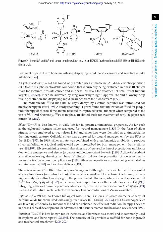
Technetium (Z = 43) is man-made, dating back to 1937. The metastable radioisotope 99mTc, aγ-emitter with half-life of 6 h, is used in tens of millions of single photon emission computedtomography (SPECT) diagnostic procedures every year. It is readily generated at the bedside fromthe longer lived isotope 99Mo. More than 50 99mTc radiopharmaceuticals are currently in use forimaging and functional studies of various areas of the body, including bone, the brain, thyroid,lungs, myocardium and liver [159]. The ligands play a crucial role in the targeting properties ofthe complexes, e.g. phosphonate and phosphate complexes for bone. By way of current examples,Cardiolite (99mTc-sestamibi, figure 13) and Neurite (99mTc-disicate) have been approved for folate-receptor positive tumours [160]. 99mTc-MIP-1404 is in clinical phase II trials for prostate cancerimaging [161,162].
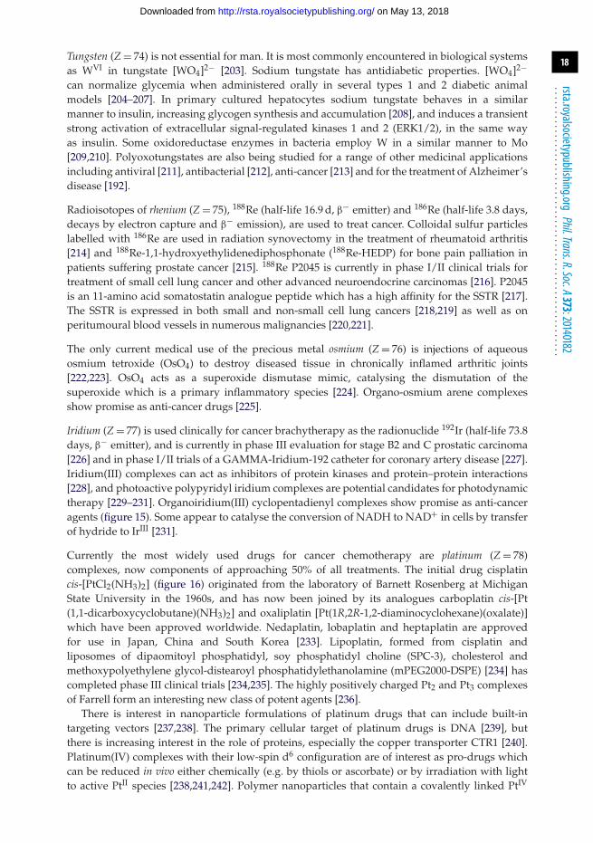
Two ruthenium (Z = 44) complexes are in clinical trials as anti-cancer drugs. They are bothoctahedral Ru(III) complexes. The indazole complex KP1019, trans-[RuCl4(In)2]InH (undercommercial clinical development as the sodium salt NKP-1339 and IT-139, figure 14) is morecytotoxic to the primary tumour cells than the imidazole complex trans-[RuCl4(Im)(DMSO)]ImH,NAMI (new anti-tumour metastasis inhibitor)A, which is active against metastases [161,163]. Bothhave completed phase I clinical trials [164–169]. Ruthenium(III) from these complexes may bedelivered to tumour cells by the FeIII transport protein serum transferrin, receptors for whichare over-expressed on cancer cells. Once in cells the RuIII may be activated by reduction to RuII,although RuIV is also accessible under biological conditions. KP1019 induces apoptosis via theintrinsic mitochondrial pathway. Organometallic RuII arene complexes also exhibit promisinganti-cancer activity [170] and RuIII EDTA complexes have been investigated as NO scavengersfor treatment of septic shock [171].
Complexes of the rare precious metal rhodium (Z = 45) are currently the focus of several studies asanti-tumour, antiparasitic and antiviral agents [172–175]. The radionuclide 105Rh (β− emitter, half-life 35.4 h) is useful medically. For example, 105Rh-EDTMP is a promising therapeutic agent for
on May 13, 2018http://rsta.royalsocietypublishing.org/Downloaded from
17
rsta.royalsocietypublishing.orgPhil.Trans.R.Soc.A373:20140182
.........................................................
RuCl
Cl
Cl
Cl
KP1019
N
N
NH
–
+
NH2
Ru Ru
NHNH
NHHN
Cl
Cl
Cl
+
H2N
N
P
N
N
RM175 RAPTA-CNAMI-A
Ru
S
N
–
+
CH3
CH3
HN
O
Cl
Cl Cl
ClHN
HN
Figure 14. Some RuIII and RuII anti-cancer complexes. Both NAMI-A and KP1019 (as the sodium salt NKP-1339 and IT-139) are inclinical trials.
treatment of pain due to bone metastases, displaying rapid blood clearance and selective uptakeinto bone [176].
As yet, palladium (Z = 46) has found only limited uses in medicine. A Pd-bacteriopheophorbide(TOOKAD) is a photoactivatable compound that is currently being evaluated in phase III clinicaltrials for localized prostate cancer and in phase I/II trials for treatment of small renal tumourtargets [177,178]. It can be activated by long wavelength light (approx. 763 nm) allowing deeptissue penetration and displaying rapid clearance from the bloodstream [177].
The radionuclide 103Pd (half-life 17 days, decays by electron capture) was introduced forbrachytherapy in 1989 [179]. A study spanning 11 years found that utilization of 103Pd for plaqueradiotherapy of choroidal melanoma resulted in improved visual function when compared to theuse of 125I [180]. Currently, 103Pd is in phase III clinical trials for treatment of early stage prostatecancer [181,182].
Silver (Z = 47) is best known in daily life for its potent antimicrobial properties. As far backas the eighteenth century silver was used for wound management [183]. In the form of silvernitrate, it was employed to treat ulcers [184] and silver ions were identified as antimicrobial inthe nineteenth century. Colloidal silver was approved for wound management by the FDA inthe 1920s [185]. In 1968, silver nitrate was combined with a sulfonamide antibiotic to producesilver sulfadiazine, a topical antibacterial agent prescribed for burn management that is still inuse [186,187]. Silver-containing wound dressings are often used in lieu of prescription antibioticsdue to the emergence and rise in (organic) antibiotic-resistant bacteria [188]. Acticoat absorbantis a silver-releasing dressing in phase IV clinical trial for the prevention of lower extremityrevascularization wound complications [189]. Silver nanoparticles are also being evaluated asantiviral agents [190] and for drug delivery [191].
There is cadmium (Z = 48) in the body (ca 56 mg) and although it is possible that it is essentialat very low doses (see Introduction), it is usually considered to be toxic. Cadmium(II) has ahigh affinity for sulfur ligands, e.g. in the protein metallothionein, where it can displace naturalZn2+ from Zn(Cys)4 sites [192], which may have implications for the cellular toxicity of Cd [193].Intriguingly, the cadmium-dependent carbonic anhydrase in the marine diatom T. weissflogii [194]uses Cd as its natural metal cofactor when only low concentrations of Zn are available.
Hafnium (Z = 49) has no known biological role. There is interest in 50 nm diameter spheres ofhafnium oxide functionalized with a negative surface (NBTXR3) [195,196]. NBTXR3 nanoparticlesare taken up efficiently by tumour cells and can enhance the effects of radiation therapy. They arein phase I clinical development for advanced soft tissue sarcomas and head and neck cancer [197].
Tantalum (Z = 73) is best known for its inertness and hardness as a metal and is commonly usedin implants and bone repair [198,199]. The porosity of Ta provides a scaffold for bone ingrowthand mechanical attachment [200–202].
on May 13, 2018http://rsta.royalsocietypublishing.org/Downloaded from
18
rsta.royalsocietypublishing.orgPhil.Trans.R.Soc.A373:20140182
.........................................................
Tungsten (Z = 74) is not essential for man. It is most commonly encountered in biological systemsas WVI in tungstate [WO4]2− [203]. Sodium tungstate has antidiabetic properties. [WO4]2−can normalize glycemia when administered orally in several types 1 and 2 diabetic animalmodels [204–207]. In primary cultured hepatocytes sodium tungstate behaves in a similarmanner to insulin, increasing glycogen synthesis and accumulation [208], and induces a transientstrong activation of extracellular signal-regulated kinases 1 and 2 (ERK1/2), in the same wayas insulin. Some oxidoreductase enzymes in bacteria employ W in a similar manner to Mo[209,210]. Polyoxotungstates are also being studied for a range of other medicinal applicationsincluding antiviral [211], antibacterial [212], anti-cancer [213] and for the treatment of Alzheimer’sdisease [192].
Radioisotopes of rhenium (Z = 75), 188Re (half-life 16.9 d, β− emitter) and 186Re (half-life 3.8 days,decays by electron capture and β− emission), are used to treat cancer. Colloidal sulfur particleslabelled with 186Re are used in radiation synovectomy in the treatment of rheumatoid arthritis[214] and 188Re-1,1-hydroxyethylidenediphosphonate (188Re-HEDP) for bone pain palliation inpatients suffering prostate cancer [215]. 188Re P2045 is currently in phase I/II clinical trials fortreatment of small cell lung cancer and other advanced neuroendocrine carcinomas [216]. P2045is an 11-amino acid somatostatin analogue peptide which has a high affinity for the SSTR [217].The SSTR is expressed in both small and non-small cell lung cancers [218,219] as well as onperitumoural blood vessels in numerous malignancies [220,221].
The only current medical use of the precious metal osmium (Z = 76) is injections of aqueousosmium tetroxide (OsO4) to destroy diseased tissue in chronically inflamed arthritic joints[222,223]. OsO4 acts as a superoxide dismutase mimic, catalysing the dismutation of thesuperoxide which is a primary inflammatory species [224]. Organo-osmium arene complexesshow promise as anti-cancer drugs [225].
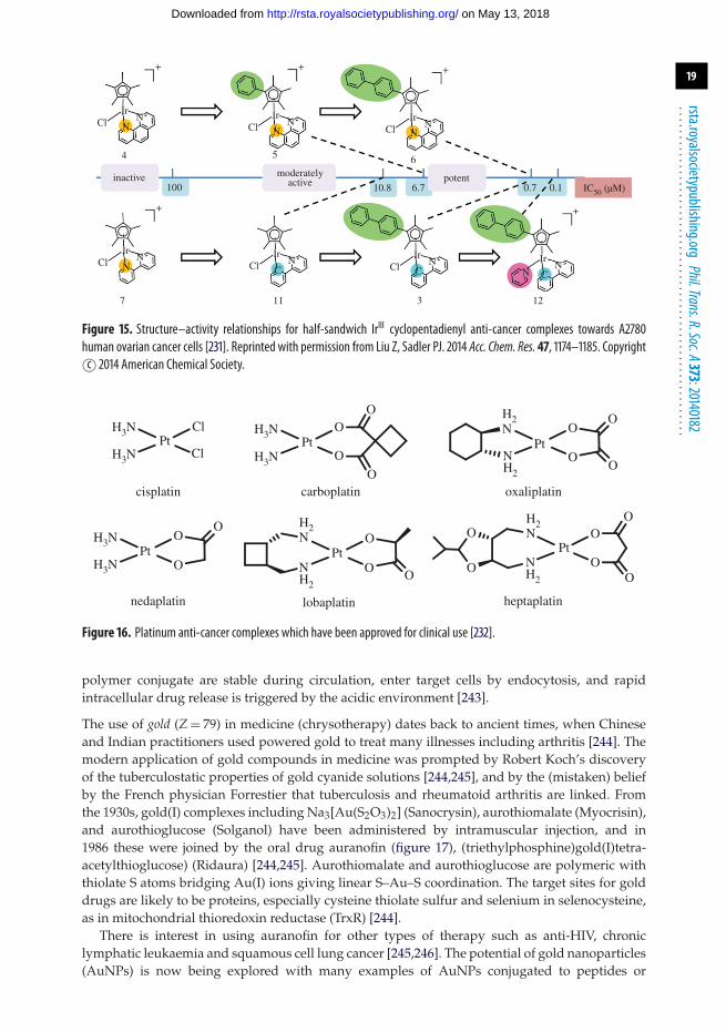
Iridium (Z = 77) is used clinically for cancer brachytherapy as the radionuclide 192Ir (half-life 73.8days, β− emitter), and is currently in phase III evaluation for stage B2 and C prostatic carcinoma[226] and in phase I/II trials of a GAMMA-Iridium-192 catheter for coronary artery disease [227].Iridium(III) complexes can act as inhibitors of protein kinases and protein–protein interactions[228], and photoactive polypyridyl iridium complexes are potential candidates for photodynamictherapy [229–231]. Organoiridium(III) cyclopentadienyl complexes show promise as anti-canceragents (figure 15). Some appear to catalyse the conversion of NADH to NAD+ in cells by transferof hydride to IrIII [231].
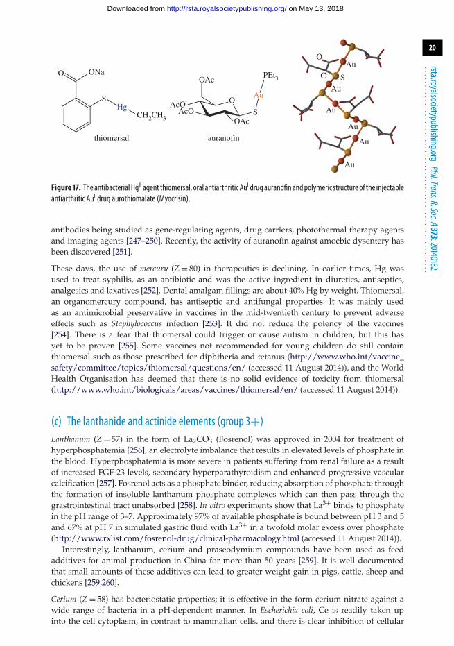
Currently the most widely used drugs for cancer chemotherapy are platinum (Z = 78)complexes, now components of approaching 50% of all treatments. The initial drug cisplatincis-[PtCl2(NH3)2] (figure 16) originated from the laboratory of Barnett Rosenberg at MichiganState University in the 1960s, and has now been joined by its analogues carboplatin cis-[Pt(1,1-dicarboxycyclobutane)(NH3)2] and oxaliplatin [Pt(1R,2R-1,2-diaminocyclohexane)(oxalate)]which have been approved worldwide. Nedaplatin, lobaplatin and heptaplatin are approvedfor use in Japan, China and South Korea [233]. Lipoplatin, formed from cisplatin andliposomes of dipaomitoyl phosphatidyl, soy phosphatidyl choline (SPC-3), cholesterol andmethoxypolyethylene glycol-distearoyl phosphatidylethanolamine (mPEG2000-DSPE) [234] hascompleted phase III clinical trials [234,235]. The highly positively charged Pt2 and Pt3 complexesof Farrell form an interesting new class of potent agents [236].
There is interest in nanoparticle formulations of platinum drugs that can include built-intargeting vectors [237,238]. The primary cellular target of platinum drugs is DNA [239], butthere is increasing interest in the role of proteins, especially the copper transporter CTR1 [240].Platinum(IV) complexes with their low-spin d6 configuration are of interest as pro-drugs whichcan be reduced in vivo either chemically (e.g. by thiols or ascorbate) or by irradiation with lightto active PtII species [238,241,242]. Polymer nanoparticles that contain a covalently linked PtIV
on May 13, 2018http://rsta.royalsocietypublishing.org/Downloaded from
19
rsta.royalsocietypublishing.orgPhil.Trans.R.Soc.A373:20140182
.........................................................
IC50 (µM)
Ir
NNCl
Ir
CNCl
Ir
CNCl
Ir
+
CN
N
7 11
Ir
NNCl
5
Ir
NNCl
6
Ir
NNCl
4
10.8 6.7 0.7 0.1
3 12
inactive moderatelyactive potent
100
+
+ + +
Figure 15. Structure–activity relationships for half-sandwich IrIII cyclopentadienyl anti-cancer complexes towards A2780human ovarian cancer cells [231]. Reprinted with permission from Liu Z, Sadler PJ. 2014 Acc. Chem. Res. 47, 1174–1185. Copyrightc© 2014 American Chemical Society.
H3N
H3N
Cl
ClPt
cisplatin carboplatin
lobaplatin heptaplatinnedaplatin
oxaliplatin
H3N
H3N
O
O
O
O
Pt
O
OO
NH2
H2N
Pt
OO
OOO
O
NH2
H2N
PtH3N
H3N
OO
OPt
OO
OON
H2
NPt
H2
Figure 16. Platinum anti-cancer complexes which have been approved for clinical use [232].
polymer conjugate are stable during circulation, enter target cells by endocytosis, and rapidintracellular drug release is triggered by the acidic environment [243].
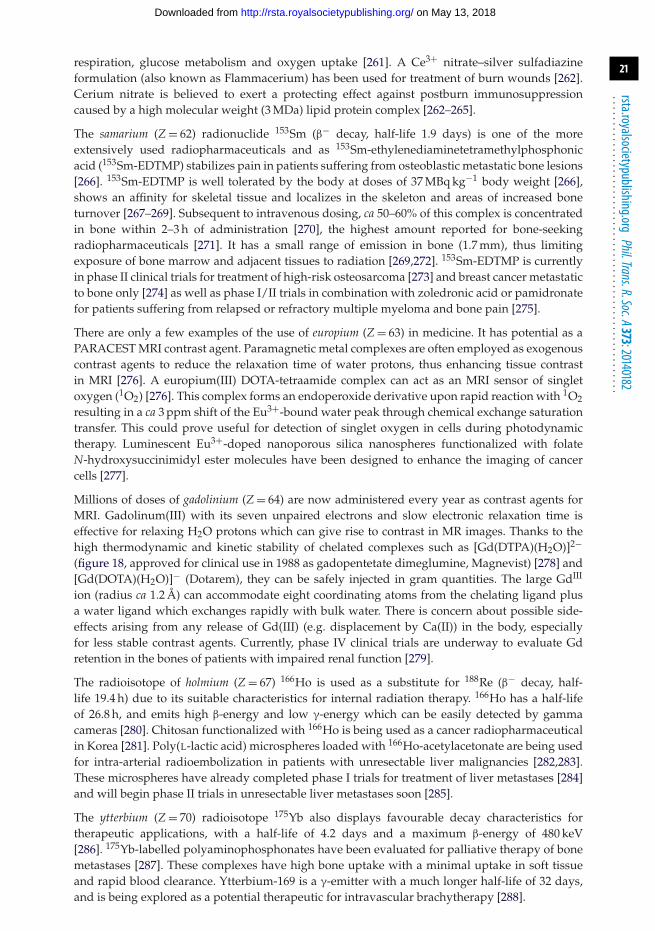
The use of gold (Z = 79) in medicine (chrysotherapy) dates back to ancient times, when Chineseand Indian practitioners used powered gold to treat many illnesses including arthritis [244]. Themodern application of gold compounds in medicine was prompted by Robert Koch’s discoveryof the tuberculostatic properties of gold cyanide solutions [244,245], and by the (mistaken) beliefby the French physician Forrestier that tuberculosis and rheumatoid arthritis are linked. Fromthe 1930s, gold(I) complexes including Na3[Au(S2O3)2] (Sanocrysin), aurothiomalate (Myocrisin),and aurothioglucose (Solganol) have been administered by intramuscular injection, and in1986 these were joined by the oral drug auranofin (figure 17), (triethylphosphine)gold(I)tetra-acetylthioglucose) (Ridaura) [244,245]. Aurothiomalate and aurothioglucose are polymeric withthiolate S atoms bridging Au(I) ions giving linear S–Au–S coordination. The target sites for golddrugs are likely to be proteins, especially cysteine thiolate sulfur and selenium in selenocysteine,as in mitochondrial thioredoxin reductase (TrxR) [244].
There is interest in using auranofin for other types of therapy such as anti-HIV, chroniclymphatic leukaemia and squamous cell lung cancer [245,246]. The potential of gold nanoparticles(AuNPs) is now being explored with many examples of AuNPs conjugated to peptides or
on May 13, 2018http://rsta.royalsocietypublishing.org/Downloaded from
20
rsta.royalsocietypublishing.orgPhil.Trans.R.Soc.A373:20140182
.........................................................
C
OAu
Au
Au
Au
Au
Au
SOAc
O
S
Au
PEt3
AcOAcO
OAc
auranofinthiomersal
ONa
CH2CH3
O
SHg
Figure 17. TheantibacterialHgII agent thiomersal, oral antiarthritic AuI drugauranofinandpolymeric structureof the injectableantiarthritic AuI drug aurothiomalate (Myocrisin).
antibodies being studied as gene-regulating agents, drug carriers, photothermal therapy agentsand imaging agents [247–250]. Recently, the activity of auranofin against amoebic dysentery hasbeen discovered [251].
These days, the use of mercury (Z = 80) in therapeutics is declining. In earlier times, Hg wasused to treat syphilis, as an antibiotic and was the active ingredient in diuretics, antiseptics,analgesics and laxatives [252]. Dental amalgam fillings are about 40% Hg by weight. Thiomersal,an organomercury compound, has antiseptic and antifungal properties. It was mainly usedas an antimicrobial preservative in vaccines in the mid-twentieth century to prevent adverseeffects such as Staphylococcus infection [253]. It did not reduce the potency of the vaccines[254]. There is a fear that thiomersal could trigger or cause autism in children, but this hasyet to be proven [255]. Some vaccines not recommended for young children do still containthiomersal such as those prescribed for diphtheria and tetanus (http://www.who.int/vaccine_safety/committee/topics/thiomersal/questions/en/ (accessed 11 August 2014)), and the WorldHealth Organisation has deemed that there is no solid evidence of toxicity from thiomersal(http://www.who.int/biologicals/areas/vaccines/thiomersal/en/ (accessed 11 August 2014)).
(c) The lanthanide and actinide elements (group 3+)Lanthanum (Z = 57) in the form of La2CO3 (Fosrenol) was approved in 2004 for treatment ofhyperphosphatemia [256], an electrolyte imbalance that results in elevated levels of phosphate inthe blood. Hyperphosphatemia is more severe in patients suffering from renal failure as a resultof increased FGF-23 levels, secondary hyperparathyroidism and enhanced progressive vascularcalcification [257]. Fosrenol acts as a phosphate binder, reducing absorption of phosphate throughthe formation of insoluble lanthanum phosphate complexes which can then pass through thegrastrointestinal tract unabsorbed [258]. In vitro experiments show that La3+ binds to phosphatein the pH range of 3–7. Approximately 97% of available phosphate is bound between pH 3 and 5and 67% at pH 7 in simulated gastric fluid with La3+ in a twofold molar excess over phosphate(http://www.rxlist.com/fosrenol-drug/clinical-pharmacology.html (accessed 11 August 2014)).
Interestingly, lanthanum, cerium and praseodymium compounds have been used as feedadditives for animal production in China for more than 50 years [259]. It is well documentedthat small amounts of these additives can lead to greater weight gain in pigs, cattle, sheep andchickens [259,260].
Cerium (Z = 58) has bacteriostatic properties; it is effective in the form cerium nitrate against awide range of bacteria in a pH-dependent manner. In Escherichia coli, Ce is readily taken upinto the cell cytoplasm, in contrast to mammalian cells, and there is clear inhibition of cellular
on May 13, 2018http://rsta.royalsocietypublishing.org/Downloaded from
21
rsta.royalsocietypublishing.orgPhil.Trans.R.Soc.A373:20140182
.........................................................
respiration, glucose metabolism and oxygen uptake [261]. A Ce3+ nitrate–silver sulfadiazineformulation (also known as Flammacerium) has been used for treatment of burn wounds [262].Cerium nitrate is believed to exert a protecting effect against postburn immunosuppressioncaused by a high molecular weight (3 MDa) lipid protein complex [262–265].
The samarium (Z = 62) radionuclide 153Sm (β− decay, half-life 1.9 days) is one of the moreextensively used radiopharmaceuticals and as 153Sm-ethylenediaminetetramethylphosphonicacid (153Sm-EDTMP) stabilizes pain in patients suffering from osteoblastic metastatic bone lesions[266]. 153Sm-EDTMP is well tolerated by the body at doses of 37 MBq kg−1 body weight [266],shows an affinity for skeletal tissue and localizes in the skeleton and areas of increased boneturnover [267–269]. Subsequent to intravenous dosing, ca 50–60% of this complex is concentratedin bone within 2–3 h of administration [270], the highest amount reported for bone-seekingradiopharmaceuticals [271]. It has a small range of emission in bone (1.7 mm), thus limitingexposure of bone marrow and adjacent tissues to radiation [269,272]. 153Sm-EDTMP is currentlyin phase II clinical trials for treatment of high-risk osteosarcoma [273] and breast cancer metastaticto bone only [274] as well as phase I/II trials in combination with zoledronic acid or pamidronatefor patients suffering from relapsed or refractory multiple myeloma and bone pain [275].
There are only a few examples of the use of europium (Z = 63) in medicine. It has potential as aPARACEST MRI contrast agent. Paramagnetic metal complexes are often employed as exogenouscontrast agents to reduce the relaxation time of water protons, thus enhancing tissue contrastin MRI [276]. A europium(III) DOTA-tetraamide complex can act as an MRI sensor of singletoxygen (1O2) [276]. This complex forms an endoperoxide derivative upon rapid reaction with 1O2resulting in a ca 3 ppm shift of the Eu3+-bound water peak through chemical exchange saturationtransfer. This could prove useful for detection of singlet oxygen in cells during photodynamictherapy. Luminescent Eu3+-doped nanoporous silica nanospheres functionalized with folateN-hydroxysuccinimidyl ester molecules have been designed to enhance the imaging of cancercells [277].
Millions of doses of gadolinium (Z = 64) are now administered every year as contrast agents forMRI. Gadolinum(III) with its seven unpaired electrons and slow electronic relaxation time iseffective for relaxing H2O protons which can give rise to contrast in MR images. Thanks to thehigh thermodynamic and kinetic stability of chelated complexes such as [Gd(DTPA)(H2O)]2−(figure 18, approved for clinical use in 1988 as gadopentetate dimeglumine, Magnevist) [278] and[Gd(DOTA)(H2O)]− (Dotarem), they can be safely injected in gram quantities. The large GdIII
ion (radius ca 1.2 Å) can accommodate eight coordinating atoms from the chelating ligand plusa water ligand which exchanges rapidly with bulk water. There is concern about possible side-effects arising from any release of Gd(III) (e.g. displacement by Ca(II)) in the body, especiallyfor less stable contrast agents. Currently, phase IV clinical trials are underway to evaluate Gdretention in the bones of patients with impaired renal function [279].
The radioisotope of holmium (Z = 67) 166Ho is used as a substitute for 188Re (β− decay, half-life 19.4 h) due to its suitable characteristics for internal radiation therapy. 166Ho has a half-lifeof 26.8 h, and emits high β-energy and low γ-energy which can be easily detected by gammacameras [280]. Chitosan functionalized with 166Ho is being used as a cancer radiopharmaceuticalin Korea [281]. Poly(L-lactic acid) microspheres loaded with 166Ho-acetylacetonate are being usedfor intra-arterial radioembolization in patients with unresectable liver malignancies [282,283].These microspheres have already completed phase I trials for treatment of liver metastases [284]and will begin phase II trials in unresectable liver metastases soon [285].
The ytterbium (Z = 70) radioisotope 175Yb also displays favourable decay characteristics fortherapeutic applications, with a half-life of 4.2 days and a maximum β-energy of 480 keV[286]. 175Yb-labelled polyaminophosphonates have been evaluated for palliative therapy of bonemetastases [287]. These complexes have high bone uptake with a minimal uptake in soft tissueand rapid blood clearance. Ytterbium-169 is a γ-emitter with a much longer half-life of 32 days,and is being explored as a potential therapeutic for intravascular brachytherapy [288].
on May 13, 2018http://rsta.royalsocietypublishing.org/Downloaded from
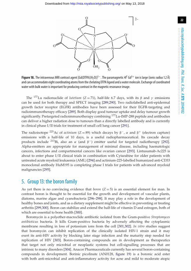
22
rsta.royalsocietypublishing.orgPhil.Trans.R.Soc.A373:20140182
.........................................................
2–
Gd
O
O
O
O–O–
O– O–
OH2OO
O
N
N N
Figure 18. The intravenous MRI contrast agent [Gd(DTPA)(H2O)]2−. The paramagnetic 4f7 Gd3+ ion is large (ionic radius 1.2 Å)and can accommodate eight coordinating atoms from the chelating DTPA ligand and awatermolecule. Exchange of coordinatedwater with bulk water is important for producing contrast in the magnetic resonance image.
The 177Lu radionuclide of lutetium (Z = 71), half-life 6.7 days, with its β and γ emissionscan be used for both therapy and SPECT imaging [289,290]. Two radiolabelled anti-epidermalgrowth factor receptor (EGFR) antibodies have been assessed for their EGFR-targeting andradioimmunotherapy efficacy [289]. Both display good tumour uptake and delay tumour growthsignificantly. Pretargeted radioimmunotherapy combining 177Lu-IMP-288 peptide and antibodiescan deliver a higher radiation dose to tumours than a directly labelled antibody and is currentlyin clinical phase I/II trials for treatment of small cell lung cancer [291].
The radioisotope 225Ac of actinium (Z = 89) which decays by β−, α and β+ (electron capture)emissions with a half-life of 10 days, is a useful radiopharmaceutical. Its cascade decayproducts include 213Bi, also an α (and β−) emitter useful for targeted radiotherapy [292].Alpha-emitters are appropriate for management of minimal disease, including haematologiccancers, infections and compartmental cancers like ovarian cancer [293]. Lintusumab-Ac225 isabout to enter phase I/II clinical trials in combination with Cytarabine for older patients withuntreated acute myeloid leukaemia (AML) [294] and actinium-225-labelled humanized anti-CD33monoclonal antibody HuM195 is completing phase I trials for patients with advanced myeloidmalignancies [295].
5. Group 13: the boron familyAs yet there is no convincing evidence that boron (Z = 5) is an essential element for man. Incontrast boron is thought to be essential for the growth and development of vascular plants,diatoms, marine algae and cyanobacteria [296–298]. It may play a role in the development ofhealthy bones and joints, and as a dietary supplement might be effective in preventing or treatingarthritis [299,300]. Boron can stabilize and extend the half-life of vitamin D and estrogen, both ofwhich are essential to bone health [300].
Boromycin is a polyether-macroclide antibiotic isolated from the Gram-positive Streptomycesantibioticus bacteria. It kills Gram-positive bacteria by adversely affecting the cytoplasmicmembrane resulting in loss of potassium ions from the cell [301,302]. In vitro studies suggestthat boromycin can inhibit replication of the clinically isolated HIV-1 strain and it mayexert its anti-HIV activity by blocking later stage infection and the maturity step needed forreplication of HIV [303]. Boron-containing compounds are in development as therapeuticsthat target not only microbial or neoplastic systems but cell-signalling processes that areintrinsic to many disorders [304]. Anacor Pharmaceuticals currently has several boron-containingcompounds in development. Borinic picolinate (AN0128, figure 19) is a boronic acid esterwith both anti-microbial and anti-inflammatory activity for acne and mild to moderate atopic
on May 13, 2018http://rsta.royalsocietypublishing.org/Downloaded from
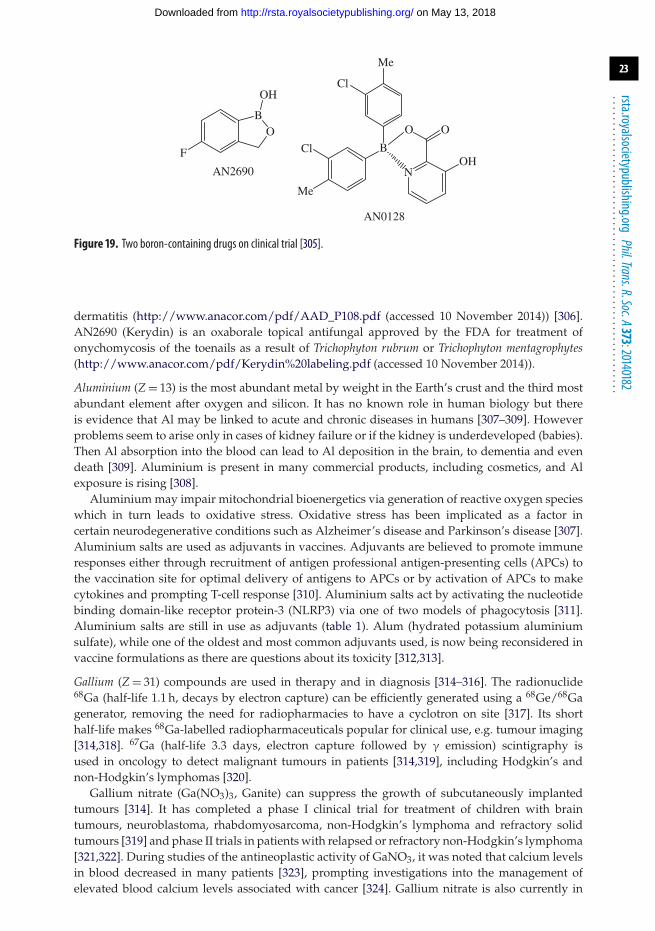
23
rsta.royalsocietypublishing.orgPhil.Trans.R.Soc.A373:20140182
.........................................................
Cl
ClF
Me
Me
AN2690
AN0128
O O OB
B
OH
OHN
Figure 19. Two boron-containing drugs on clinical trial [305].
dermatitis (http://www.anacor.com/pdf/AAD_P108.pdf (accessed 10 November 2014)) [306].AN2690 (Kerydin) is an oxaborale topical antifungal approved by the FDA for treatment ofonychomycosis of the toenails as a result of Trichophyton rubrum or Trichophyton mentagrophytes(http://www.anacor.com/pdf/Kerydin%20labeling.pdf (accessed 10 November 2014)).
Aluminium (Z = 13) is the most abundant metal by weight in the Earth’s crust and the third mostabundant element after oxygen and silicon. It has no known role in human biology but thereis evidence that Al may be linked to acute and chronic diseases in humans [307–309]. Howeverproblems seem to arise only in cases of kidney failure or if the kidney is underdeveloped (babies).Then Al absorption into the blood can lead to Al deposition in the brain, to dementia and evendeath [309]. Aluminium is present in many commercial products, including cosmetics, and Alexposure is rising [308].
Aluminium may impair mitochondrial bioenergetics via generation of reactive oxygen specieswhich in turn leads to oxidative stress. Oxidative stress has been implicated as a factor incertain neurodegenerative conditions such as Alzheimer’s disease and Parkinson’s disease [307].Aluminium salts are used as adjuvants in vaccines. Adjuvants are believed to promote immuneresponses either through recruitment of antigen professional antigen-presenting cells (APCs) tothe vaccination site for optimal delivery of antigens to APCs or by activation of APCs to makecytokines and prompting T-cell response [310]. Aluminium salts act by activating the nucleotidebinding domain-like receptor protein-3 (NLRP3) via one of two models of phagocytosis [311].Aluminium salts are still in use as adjuvants (table 1). Alum (hydrated potassium aluminiumsulfate), while one of the oldest and most common adjuvants used, is now being reconsidered invaccine formulations as there are questions about its toxicity [312,313].
Gallium (Z = 31) compounds are used in therapy and in diagnosis [314–316]. The radionuclide68Ga (half-life 1.1 h, decays by electron capture) can be efficiently generated using a 68Ge/68Gagenerator, removing the need for radiopharmacies to have a cyclotron on site [317]. Its shorthalf-life makes 68Ga-labelled radiopharmaceuticals popular for clinical use, e.g. tumour imaging[314,318]. 67Ga (half-life 3.3 days, electron capture followed by γ emission) scintigraphy isused in oncology to detect malignant tumours in patients [314,319], including Hodgkin’s andnon-Hodgkin’s lymphomas [320].
Gallium nitrate (Ga(NO3)3, Ganite) can suppress the growth of subcutaneously implantedtumours [314]. It has completed a phase I clinical trial for treatment of children with braintumours, neuroblastoma, rhabdomyosarcoma, non-Hodgkin’s lymphoma and refractory solidtumours [319] and phase II trials in patients with relapsed or refractory non-Hodgkin’s lymphoma[321,322]. During studies of the antineoplastic activity of GaNO3, it was noted that calcium levelsin blood decreased in many patients [323], prompting investigations into the management ofelevated blood calcium levels associated with cancer [324]. Gallium nitrate is also currently in
on May 13, 2018http://rsta.royalsocietypublishing.org/Downloaded from
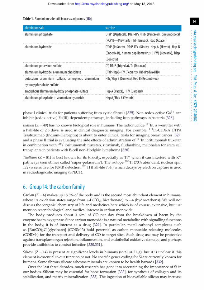
24
rsta.royalsocietypublishing.orgPhil.Trans.R.Soc.A373:20140182
.........................................................
Table 1. Aluminium salts still in use as adjuvants [310].
aluminium salt vaccine. . . . . . . . . . . . . . . . . . . . . . . . . . . . . . . . . . . . . . . . . . . . . . . . . . . . . . . . . . . . . . . . . . . . . . . . . . . . . . . . . . . . . . . . . . . . . . . . . . . . . . . . . . . . . . . . . . . . . . . . . . . . . . . . . . . . . . . . . . . . . . . . . . . . . . . . . . . . . . . . . . . . . . . . . . . . . . . . . . . . . . . . . . . . . . . . . . . . . . . . . .
aluminium phosphate DTaP (Daptacel), DTaP-IPV/Hib (Pentacel), pneumococcal
(PCV13—Prevnar13), Td (Tenivac), Tdap (Adacel). . . . . . . . . . . . . . . . . . . . . . . . . . . . . . . . . . . . . . . . . . . . . . . . . . . . . . . . . . . . . . . . . . . . . . . . . . . . . . . . . . . . . . . . . . . . . . . . . . . . . . . . . . . . . . . . . . . . . . . . . . . . . . . . . . . . . . . . . . . . . . . . . . . . . . . . . . . . . . . . . . . . . . . . . . . . . . . . . . . . . . . . . . . . . . . . . . . . . . . . . .
aluminium hydroxide DTaP (Infanrix), DTaP-IPV (Kinrix), Hep A (Havrix), Hep B
(Engerix-B), human papillomavirus (HPV) (Cervarix), Tdap
(Boostrix). . . . . . . . . . . . . . . . . . . . . . . . . . . . . . . . . . . . . . . . . . . . . . . . . . . . . . . . . . . . . . . . . . . . . . . . . . . . . . . . . . . . . . . . . . . . . . . . . . . . . . . . . . . . . . . . . . . . . . . . . . . . . . . . . . . . . . . . . . . . . . . . . . . . . . . . . . . . . . . . . . . . . . . . . . . . . . . . . . . . . . . . . . . . . . . . . . . . . . . . . .
aluminium potassium sulfate DT, DTaP (Tripedia), Td (Decavac). . . . . . . . . . . . . . . . . . . . . . . . . . . . . . . . . . . . . . . . . . . . . . . . . . . . . . . . . . . . . . . . . . . . . . . . . . . . . . . . . . . . . . . . . . . . . . . . . . . . . . . . . . . . . . . . . . . . . . . . . . . . . . . . . . . . . . . . . . . . . . . . . . . . . . . . . . . . . . . . . . . . . . . . . . . . . . . . . . . . . . . . . . . . . . . . . . . . . . . . . .
aluminium hydroxide, aluminium phosphate DTaP-HepB-IPV (Pediarix), Hib (PedvaxHIB). . . . . . . . . . . . . . . . . . . . . . . . . . . . . . . . . . . . . . . . . . . . . . . . . . . . . . . . . . . . . . . . . . . . . . . . . . . . . . . . . . . . . . . . . . . . . . . . . . . . . . . . . . . . . . . . . . . . . . . . . . . . . . . . . . . . . . . . . . . . . . . . . . . . . . . . . . . . . . . . . . . . . . . . . . . . . . . . . . . . . . . . . . . . . . . . . . . . . . . . . .
potassium aluminium sulfate, amorphous aluminium
hydroxy phosphate-sulfate
Hib/Hep B (Comvax), Hep B (Recombivax)
. . . . . . . . . . . . . . . . . . . . . . . . . . . . . . . . . . . . . . . . . . . . . . . . . . . . . . . . . . . . . . . . . . . . . . . . . . . . . . . . . . . . . . . . . . . . . . . . . . . . . . . . . . . . . . . . . . . . . . . . . . . . . . . . . . . . . . . . . . . . . . . . . . . . . . . . . . . . . . . . . . . . . . . . . . . . . . . . . . . . . . . . . . . . . . . . . . . . . . . . . .
amorphous aluminium hydroxy phosphate-sulfate Hep A (Vaqta), HPV (Gardasil). . . . . . . . . . . . . . . . . . . . . . . . . . . . . . . . . . . . . . . . . . . . . . . . . . . . . . . . . . . . . . . . . . . . . . . . . . . . . . . . . . . . . . . . . . . . . . . . . . . . . . . . . . . . . . . . . . . . . . . . . . . . . . . . . . . . . . . . . . . . . . . . . . . . . . . . . . . . . . . . . . . . . . . . . . . . . . . . . . . . . . . . . . . . . . . . . . . . . . . . . .
aluminium phosphate+ aluminium hydroxide Hep A/Hep B (Twinrix). . . . . . . . . . . . . . . . . . . . . . . . . . . . . . . . . . . . . . . . . . . . . . . . . . . . . . . . . . . . . . . . . . . . . . . . . . . . . . . . . . . . . . . . . . . . . . . . . . . . . . . . . . . . . . . . . . . . . . . . . . . . . . . . . . . . . . . . . . . . . . . . . . . . . . . . . . . . . . . . . . . . . . . . . . . . . . . . . . . . . . . . . . . . . . . . . . . . . . . . . .
phase I clinical trials for patients suffering from cystic fibrosis [325]. Non-redox-active Ga3+ caninhibit (redox-active) Fe(III)-dependent pathways, including iron pathways in bacteria [326].
Indium (Z = 49) has no known biological role in humans. The radionuclide 111In, a γ-emitter witha half-life of 2.8 days, is used in clinical diagnostic imaging. For example, 111In-CHX-A DTPATrastuzumab (Indium-Herceptin) is about to enter clinical trials for imaging breast cancer [327]and a phase II trial is evaluating the side effects of administration of 111In ibritumomab tiuxetanin combination with 90Y ibritumomab tiuxetan, rituximab, fludarabine, melphalan for stem celltransplants in patients with B-cell non-Hodgkin lymphoma [328].
Thallium (Z = 81) is best known for its toxicity, especially as Tl+ when it can interfere with K+pathways (sometimes called ‘super-potassium’). The isotope 205Tl (70% abundant, nuclear spin1/2) is sensitive for NMR detection. 201Tl (half-life 73 h) which decays by electron capture is usedin radiodiagnostic imaging (SPECT).
6. Group 14: the carbon familyCarbon (Z = 6) makes up 18.5% of the body and is the second most abundant element in humans,where its oxidation states range from +4 (CO2, bicarbonate) to −4 (hydrocarbons). We will notdiscuss the ‘organic’ chemistry of life and medicines here which is, of course, extensive, but justmention recent biological and medical interest in carbon monoxide.
The body produces about 3–6 ml of CO per day from the breakdown of haem by theenzyme haem oxygenase. Since carbon monoxide is a natural metabolite with signalling functionsin the body, it is of interest as a drug [329]. In particular, metal carbonyl complexes suchas [Ru(CO)3Cl(glycinate)] (CORM-3) hold potential as carbon monoxide releasing molecules(CORMs) for the transport and delivery of CO to target sites. Such drug use may be protectiveagainst transplant organ rejection, inflammation, and endothelial oxidative damage, and perhapsprovide antibiotics to combat infection [330,331].
Silicon (Z = 14) is present at significant levels in humans (total ca 21 g), but it is unclear if thiselement is essential to our function or not. No specific genes coding for Si are currently known forhumans. Some fibrous silicate asbestos minerals are known to be health hazards [332].
Over the last three decades, much research has gone into ascertaining the importance of Si inour bodies. Silicon may be essential for bone formation [333], for synthesis of collagen and itsstabilization, and matrix mineralization [333]. The ingestion of bioavailable silicon may increase
on May 13, 2018http://rsta.royalsocietypublishing.org/Downloaded from
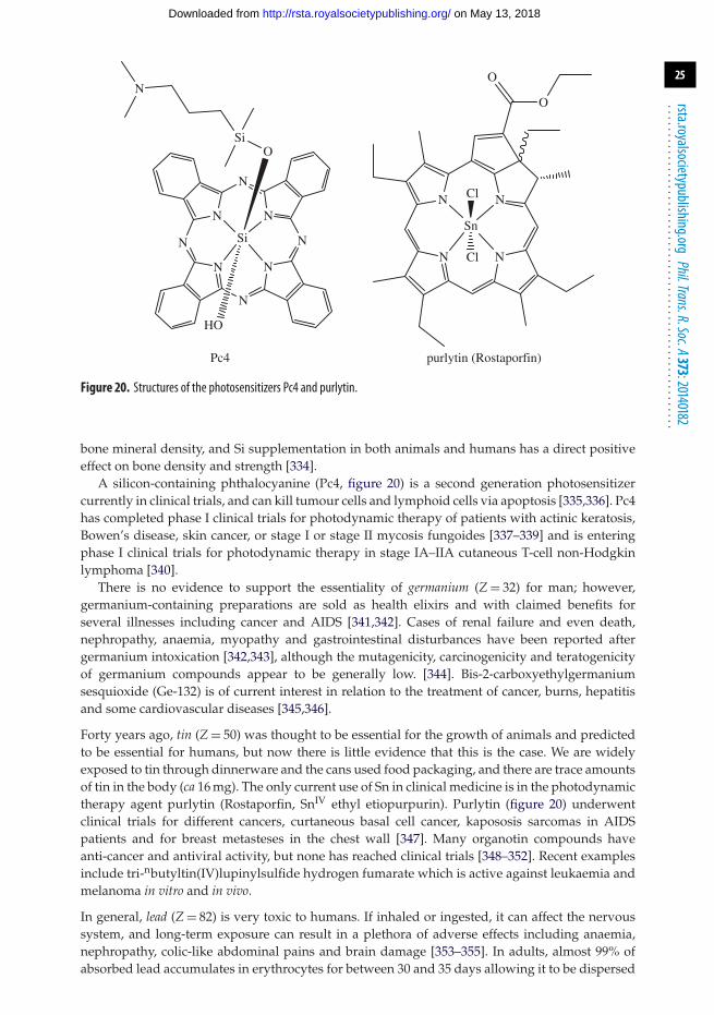
25
rsta.royalsocietypublishing.orgPhil.Trans.R.Soc.A373:20140182
.........................................................
Pc4 purlytin (Rostaporfin)
N
Si
Si
HO
N
N
N
N N
N
N
N
O
O
O
Cl
Cl
Sn
N
N N
N
Figure 20. Structures of the photosensitizers Pc4 and purlytin.
bone mineral density, and Si supplementation in both animals and humans has a direct positiveeffect on bone density and strength [334].
A silicon-containing phthalocyanine (Pc4, figure 20) is a second generation photosensitizercurrently in clinical trials, and can kill tumour cells and lymphoid cells via apoptosis [335,336]. Pc4has completed phase I clinical trials for photodynamic therapy of patients with actinic keratosis,Bowen’s disease, skin cancer, or stage I or stage II mycosis fungoides [337–339] and is enteringphase I clinical trials for photodynamic therapy in stage IA–IIA cutaneous T-cell non-Hodgkinlymphoma [340].
There is no evidence to support the essentiality of germanium (Z = 32) for man; however,germanium-containing preparations are sold as health elixirs and with claimed benefits forseveral illnesses including cancer and AIDS [341,342]. Cases of renal failure and even death,nephropathy, anaemia, myopathy and gastrointestinal disturbances have been reported aftergermanium intoxication [342,343], although the mutagenicity, carcinogenicity and teratogenicityof germanium compounds appear to be generally low. [344]. Bis-2-carboxyethylgermaniumsesquioxide (Ge-132) is of current interest in relation to the treatment of cancer, burns, hepatitisand some cardiovascular diseases [345,346].
Forty years ago, tin (Z = 50) was thought to be essential for the growth of animals and predictedto be essential for humans, but now there is little evidence that this is the case. We are widelyexposed to tin through dinnerware and the cans used food packaging, and there are trace amountsof tin in the body (ca 16 mg). The only current use of Sn in clinical medicine is in the photodynamictherapy agent purlytin (Rostaporfin, SnIV ethyl etiopurpurin). Purlytin (figure 20) underwentclinical trials for different cancers, curtaneous basal cell cancer, kapososis sarcomas in AIDSpatients and for breast metasteses in the chest wall [347]. Many organotin compounds haveanti-cancer and antiviral activity, but none has reached clinical trials [348–352]. Recent examplesinclude tri-nbutyltin(IV)lupinylsulfide hydrogen fumarate which is active against leukaemia andmelanoma in vitro and in vivo.
In general, lead (Z = 82) is very toxic to humans. If inhaled or ingested, it can affect the nervoussystem, and long-term exposure can result in a plethora of adverse effects including anaemia,nephropathy, colic-like abdominal pains and brain damage [353–355]. In adults, almost 99% ofabsorbed lead accumulates in erythrocytes for between 30 and 35 days allowing it to be dispersed
on May 13, 2018http://rsta.royalsocietypublishing.org/Downloaded from
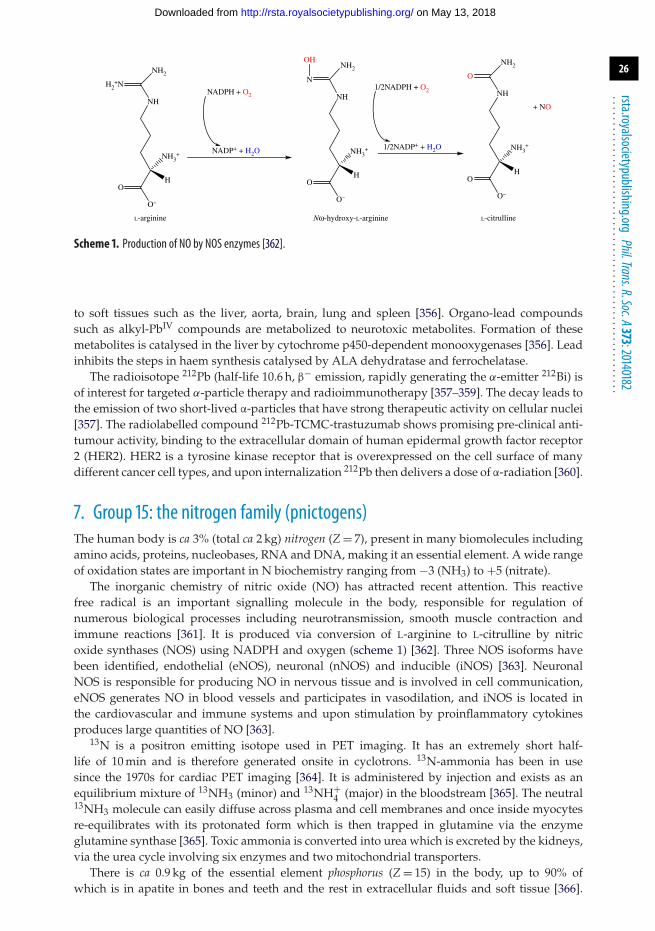
26
rsta.royalsocietypublishing.orgPhil.Trans.R.Soc.A373:20140182
.........................................................
O
O–
O O
O
O– O–
HH H
H2+N
NH2
OHNH2
1/2NADPH + O2
1/2NADP+ + H2O
NH2
NH
+ NONH
N
NH
NH3+ NH3
+ NH3+
L-arginine Nw-hydroxy-L-arginine L-citrulline
NADPH + O2
NADP+ + H2O
Scheme 1. Production of NO by NOS enzymes [362].
to soft tissues such as the liver, aorta, brain, lung and spleen [356]. Organo-lead compoundssuch as alkyl-PbIV compounds are metabolized to neurotoxic metabolites. Formation of thesemetabolites is catalysed in the liver by cytochrome p450-dependent monooxygenases [356]. Leadinhibits the steps in haem synthesis catalysed by ALA dehydratase and ferrochelatase.
The radioisotope 212Pb (half-life 10.6 h, β− emission, rapidly generating the α-emitter 212Bi) isof interest for targeted α-particle therapy and radioimmunotherapy [357–359]. The decay leads tothe emission of two short-lived α-particles that have strong therapeutic activity on cellular nuclei[357]. The radiolabelled compound 212Pb-TCMC-trastuzumab shows promising pre-clinical anti-tumour activity, binding to the extracellular domain of human epidermal growth factor receptor2 (HER2). HER2 is a tyrosine kinase receptor that is overexpressed on the cell surface of manydifferent cancer cell types, and upon internalization 212Pb then delivers a dose of α-radiation [360].
7. Group 15: the nitrogen family (pnictogens)The human body is ca 3% (total ca 2 kg) nitrogen (Z = 7), present in many biomolecules includingamino acids, proteins, nucleobases, RNA and DNA, making it an essential element. A wide rangeof oxidation states are important in N biochemistry ranging from −3 (NH3) to +5 (nitrate).
The inorganic chemistry of nitric oxide (NO) has attracted recent attention. This reactivefree radical is an important signalling molecule in the body, responsible for regulation ofnumerous biological processes including neurotransmission, smooth muscle contraction andimmune reactions [361]. It is produced via conversion of L-arginine to L-citrulline by nitricoxide synthases (NOS) using NADPH and oxygen (scheme 1) [362]. Three NOS isoforms havebeen identified, endothelial (eNOS), neuronal (nNOS) and inducible (iNOS) [363]. NeuronalNOS is responsible for producing NO in nervous tissue and is involved in cell communication,eNOS generates NO in blood vessels and participates in vasodilation, and iNOS is located inthe cardiovascular and immune systems and upon stimulation by proinflammatory cytokinesproduces large quantities of NO [363].
13N is a positron emitting isotope used in PET imaging. It has an extremely short half-life of 10 min and is therefore generated onsite in cyclotrons. 13N-ammonia has been in usesince the 1970s for cardiac PET imaging [364]. It is administered by injection and exists as anequilibrium mixture of 13NH3 (minor) and 13NH+
4 (major) in the bloodstream [365]. The neutral13NH3 molecule can easily diffuse across plasma and cell membranes and once inside myocytesre-equilibrates with its protonated form which is then trapped in glutamine via the enzymeglutamine synthase [365]. Toxic ammonia is converted into urea which is excreted by the kidneys,via the urea cycle involving six enzymes and two mitochondrial transporters.
There is ca 0.9 kg of the essential element phosphorus (Z = 15) in the body, up to 90% ofwhich is in apatite in bones and teeth and the rest in extracellular fluids and soft tissue [366].
on May 13, 2018http://rsta.royalsocietypublishing.org/Downloaded from
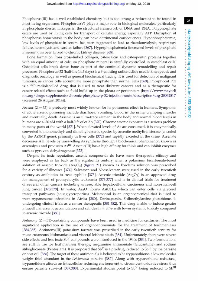
27
rsta.royalsocietypublishing.orgPhil.Trans.R.Soc.A373:20140182
.........................................................
Phosphorus(III) has a well-established chemistry but is too strong a reductant to be found inmost living organisms. Phosphorus(V) plays a major role in biological molecules, particularlyin phosphate diester linkages in the structural framework of DNA and RNA. Polyphosphateesters are used by living cells for transport of cellular energy, especially ATP. Disruption ofphosphorus homeostasis in the body can have detrimental consequences. Hypophosphatemia,low levels of phosphate in serum, has been suggested to lead to rhabdomyolysis, respiratoryfailure, haemolysis and cardiac failure [367]. Hyperphosphatemia (increased levels of phosphatein serum) has been linked to chronic kidney disease [368].
Bone formation from cross-linked collagen, osteocalcin and osteopontin proteins togetherwith an equal amount of calcium phosphate mineral is carefully controlled in osteoblast cells.Osteoblast cells break down bone as part of the continual dynamic remodelling and repairprocesses. Phosphorus-32 (half-life 14.3 days) is a β-emitting radionuclide used in therapeutic anddiagnostic oncology as well as general biochemical tracing. It is used for detection of malignanttumours, as cancer cells accumulate more phosphate than normal cells [369]. Phosphocol P32is a 32P radiolabelled drug that is used to treat different cancers and as a therapeutic forcancer-related effects such as fluid build-up in the pleura or peritoneum (http://www.mayoclinic.org/drugs-supplements/chromic-phosphate-p-32-injection-route/description/drg-20062817(accessed 26 August 2014)).
Arsenic (Z = 33) is probably most widely known for its poisonous effect in humans. Symptomsof acute arsenic poisoning include diarrhoea, vomiting, blood in the urine, cramping musclesand eventually, death. Arsenic is an ultra-trace element in the body and normal blood levels inhumans are 4–30 nM with a half-life of ca 2 h [370]. Chronic arsenic exposure is a serious problemin many parts of the world [371]. When elevated levels of As are consumed, it is enzymaticallyconverted to monomethyl- and dimethyl-arsenic species by arsenite methyltransferase (encodedby the As3MT gene), primarily in liver cells [372] and rapidly excreted in the urine. Arsenatedecreases ATP levels by unravelling its synthesis through a biochemical phenomenon known asarsenolysis and produces AsIII. Arsenic(III) has a high affinity for thiols and can inhibit enzymessuch as pyruvate dehydrogenase [373].
Despite its toxic reputation, arsenic compounds do have some therapeutic efficacy andwere employed as far back as the eighteenth century when a potassium bicarbonate-basedsolution of arsenic trioxide (As2O3) (figure 21) known as Fowler’s solution was prescribedfor a variety of illnesses [374]. Salvarsan and Neosalvarsan were used in the early twentiethcentury as antibiotics to treat syphilis [375]. Arsenic trioxide (As2O3) is an approved drugfor management of promyelocytic leukaemia [376,377] and is in clinical trials for treatmentof several other cancers including unresectable hepatocellular carcinoma and non-small-celllung cancer [378,379]. In water, As2O3 forms As(OH)3 which can enter cells via glyceroltransport pathways (aquaglyceroporins). Melarsoprol is an organoarsenical that is used totreat trypanosome infections in Africa [380]. Darinaparsin, S-dimethylarsino-glutathione, isundergoing clinical trials as a cancer therapeutic [381,382]. This drug is able to induce greaterintracellular arsenic accumulation and cell death in vitro with lower systemic toxicity comparedto arsenic trioxide [383].
Antimony (Z = 51)-containing compounds have been used in medicine for centuries. The mostsignificant application is the use of organoantimonials for the treatment of leishmaniases[384,385]. Antimony(III) potassium tartrate was prescribed in the early twentieth century formuco-cutaneous leishmaniasis and visceral leishmaniasis [384]. Unfortunately, there were severeside effects and less toxic SbV compounds were introduced in the 1940s [384]. Two formulationsare still in use for leishmaniasis therapy, meglumine antimoniate (Glucantime) and sodiumstibogluconate (Pentostam). It is proposed that SbV is a prodrug, reduced to SbIII by the parasiteor host cell [386]. The target of these antimonials is believed to be trypanothione, a low molecularweight thiol abundant in the Leishmania parasite [387]. Along with trypanothione reductase,trypanothione affords an intracellular reducing environment to circumvent oxidative stress thusensure parasite survival [387,388]. Experimental studies point to SbV being reduced to SbIII
on May 13, 2018http://rsta.royalsocietypublishing.org/Downloaded from
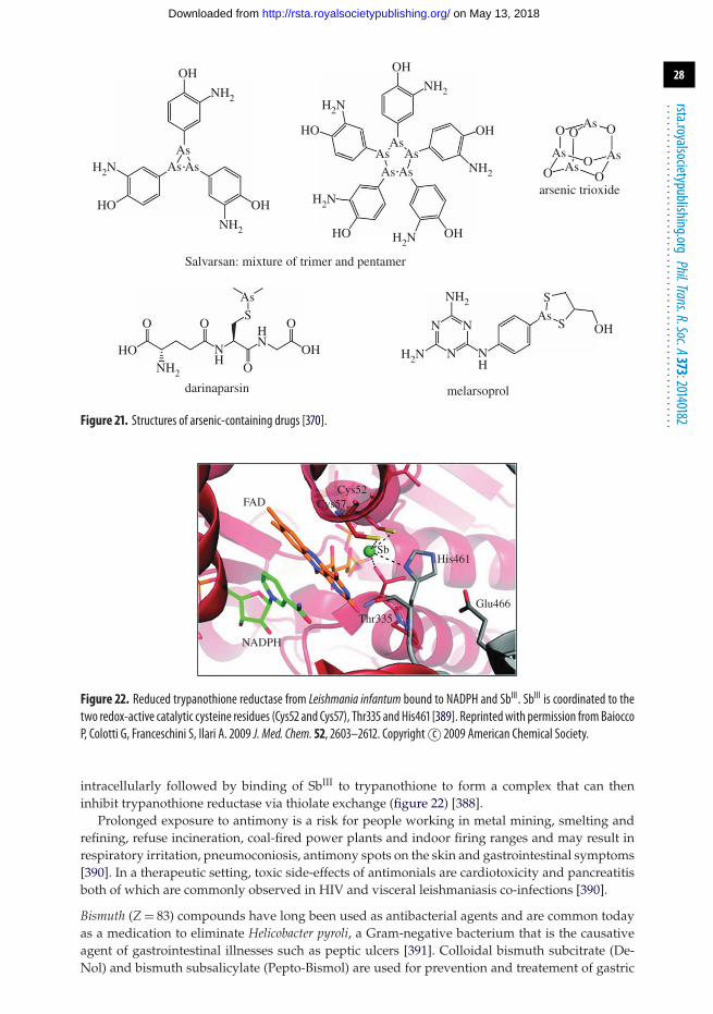
28
rsta.royalsocietypublishing.orgPhil.Trans.R.Soc.A373:20140182
.........................................................
arsenic trioxide
Salvarsan: mixture of trimer and pentamer
melarsoproldarinaparsin
OH
OH
OHHO
H2NAs As
As AsAs
AsAs As
As
As
As
S
S OH
OH
As
AsS
As
NH2
NH2
NH2
H2NHO
HO
O O O
O
H2N
HO OH
O OO
OO O
OH
H2N
NH2
NH2
NH2
H2N
N N
N NH
NN
H
H
Figure 21. Structures of arsenic-containing drugs [370].
FAD
NADPH
Thr335Glu466
His461Sb
Cys52Cys57
Figure 22. Reduced trypanothione reductase from Leishmania infantum bound to NADPH and SbIII. SbIII is coordinated to thetwo redox-active catalytic cysteine residues (Cys52 and Cys57), Thr335 and His461 [389]. Reprintedwith permission fromBaioccoP, Colotti G, Franceschini S, Ilari A. 2009 J. Med. Chem. 52, 2603–2612. Copyright c© 2009 American Chemical Society.
intracellularly followed by binding of SbIII to trypanothione to form a complex that can theninhibit trypanothione reductase via thiolate exchange (figure 22) [388].
Prolonged exposure to antimony is a risk for people working in metal mining, smelting andrefining, refuse incineration, coal-fired power plants and indoor firing ranges and may result inrespiratory irritation, pneumoconiosis, antimony spots on the skin and gastrointestinal symptoms[390]. In a therapeutic setting, toxic side-effects of antimonials are cardiotoxicity and pancreatitisboth of which are commonly observed in HIV and visceral leishmaniasis co-infections [390].
Bismuth (Z = 83) compounds have long been used as antibacterial agents and are common todayas a medication to eliminate Helicobacter pyroli, a Gram-negative bacterium that is the causativeagent of gastrointestinal illnesses such as peptic ulcers [391]. Colloidal bismuth subcitrate (De-Nol) and bismuth subsalicylate (Pepto-Bismol) are used for prevention and treatement of gastric
on May 13, 2018http://rsta.royalsocietypublishing.org/Downloaded from
29
rsta.royalsocietypublishing.orgPhil.Trans.R.Soc.A373:20140182
.........................................................
and duodenal ulcers [392,393]. Sulbogin is a topical ointment used for wound healing, andcontains bismuth subgallate as one of its main components [394]. Bismuth subgallate acts throughactivation of the Hageman factor (factor XII) in the clotting cascade to quicken coagulation [395].Bi3+ is a highly acidic metal ion (pKa of aqua complex ca 1.5) and often forms polymericcomplexes with hydroxide or oxide bridges, or citrate bridges in the case of bismuth subcitrate.
Interest in bismuth compounds as anti-cancer agents is growing due to the observation thatadministration of bismuth prior to chemotherapy can decrease the toxic side-effects associatedwith cisplatin [396,397]. Bismuth-213 is an α-emitter with a half-life of 45.6 min, prepared forclinical use from a 225Ac/213Bi generator. A phase I trial of 213Bi-lintuzumab demonstrated thatadministration of the radiopharmaceutical was safe up to 37 MBq kg−1 in patients with relapsedor refractory AML [398]. The phase I/II trial of sequential administration of cytarabine and 213Bi-lintuzumab for α-targeted radiotherapy in patients with AML found that 213Bi-lintuzumab gaverise to remissions in patients [399].
8. Group 16: the chalcogensOxygen (Z = 8), the third most abundant in the universe (after H and He) and most abundantelement in the Earth’s crust by weight, is vital as O2 to support cell respiration. We breathe airthat is 21% O2 by volume. Over half our body mass is oxygen (49/80 kg). Ground state O2 is atriplet (3O2) and relatively unreactive, even though a strong oxidant. Excited state singlet oxygen(1O2) in contrast is highly damaging and can kill cells (the basis of photodynamic therapy). Theoxidation states of oxygen in the body range from 0 (O2) to −2 (H2O).
Oxygen homeostasis is crucial for survival of all vertebrates. Our bodies have evolved toensure optimal oxygenation of all our cells, starting with entry through our lungs and thencirculation and delivery of oxygen within the body [400]. Oxygen is a critical electron acceptorin redox reactions that lead to the production of ATP, the direct source of energy needed forcell function [401]. Oxygen is also an important substrate for manufacture or decompositionof many cellular components, including signalling mediators [401]. Hypoxia (lack of oxygen)is an important factor in development of severe pathologies including myocardial and cerebralischaemia and cancer [400]. Hyperoxia (excess supply of O2 in tissues and organs) can also havedetrimental consequences such as production of high concentrations of reactive oxygen species(ROS: 1O2, O2
−, H2O2, OH.), disrupting the balance of oxidant and antioxidants and causingdamage to cells and tissues [402].
Understanding the role of ROS species for signalling in the body is an important area ofresearch [403]. Apart from its role as a destructive oxidant to ward off pathogens, H2O2 fulfils therole of ‘secondary messenger’: its enzymatic production and degradation as well as requirementfor the oxidation of thiols give rise to the conditions appropriate for signalling [403]. Thehypoxia-inducible factor (HIF) is a transcription factor that reacts to pathophysiologically lowlevels of oxygen via upregulation, setting off biological responses to hypoxia [404]. The HIFprolyl hydroxylase domain (PHD) enzymes selectively hydroxylate proline residues of HIF. Thedependence of HIF protein levels on the concentration of O2 present, mediated by the PHDenzymes, forms the basis for one of the most significant biological sensor systems of tissueoxygenation in response to ischemic and inflammatory events [404]. As a result, pharmacologicalinhibition of these enzymes, resulting in stabilization of HIF, has therapeutic potential for treatingconditions of tissue stress and injury [404].
Sulfur (Z = 16) is an essential element, involved in numerous biochemical processes in oxidationstates ranging from −2 (sulfide) to +6 (sulfate). It is the seventh most abundant element in thebody (ca 160 g).
Sulfur compounds act as both electron donors and electron acceptors in metabolic reactions.Methionine (Met), the thioether amino acid, manages major metabolic and catalytic activities,initiates protein synthesis and undergoes reversible redox reactions to protect protein integrity[405]. The abundant intracellular thiol tripeptide glutathione (GSH) is a defensive biomolecule
on May 13, 2018http://rsta.royalsocietypublishing.org/Downloaded from
30
rsta.royalsocietypublishing.orgPhil.Trans.R.Soc.A373:20140182
.........................................................
against toxic xenobiotics such as drugs, pollutants and carcinogens [406]. As an antioxidant,GSH participates in protection of the cell from oxidative stress either directly or as a cofactorof glutathione peroxidases [406]. Thioredoxins, with a dithiol-disulfide active site, are a class ofredox proteins that participate in redox signalling in the body and are coded by the TXN gene[407]. Cysteine (Cys) is biosynthesized in humans and participates in enzymatic reactions as anucleophile. Keratin’s disulfide bonds are mostly responsible for the strength of this protein thatis present in our skin, hair and nails.
Hydrogen sulfide (H2S), a highly toxic gas, is a mammalian messenger molecule. It is largelyformed from Cys or its derivatives by the enzymes cystathionine β-synthase and cystathionineγ-lyase (CSE) [408]. One mechanism of H2S signalling is the sulfhydration (persulfide RSSHformation) of reactive Cys residues in target proteins [408]. This sulfhydration is responsiblefor the regulation of inflammation and endoplasmic reticulum stress signalling as well asof vascular tension [409]. A number of additional pharmacological targets for H2S havebeen identified including K(ATP), the ATP-sensitive potassium channel, and transient receptorpotential channels, NF-κB nuclear translocation, extracellular signal-regulated kinase and Akt(protein kinase B) [410]. Atherosclerosis is a chronic disease that is considered to be theleading cause of cardiovascular morbidity and mortality [411]. Endogenous H2S productioncould be beneficial for prevention and treatment of atherosclerosis [412,413], since decreasedlevels of endogenous H2S induced by genetic deletion of CSE quickens the disease [412].Sulfide is ubiquitous in the iron sulfur redox proteins ferredoxins. The Fe/S clusters areassembled on a scaffold protein in mitochondria, transported out and then incorporated intoferredoxins.
There is only about 4 mg of selenium (Z = 34) in the body, but it is essential and its genetic codesare now well known. Selenium exerts its biological functions through selenoproteins.
Unlike the 20 standard amino acids, Sec is biosynthesized from serine on its tRNA. Theinsertion of Se donated by selenophosphate into the amino acid serine to give the 21st aminoacid selenocysteine is encoded by the termination codon UGA, which is one of three terminationcodons for mRNA translation in non-selenoprotein genes, resulting in 25 selenoproteins withredox and signalling functions [414]. Recognition of the UGA codon as a Sec insertion siteinstead of stop requires a Sec insertion sequence element in selenoprotein mRNAs and a uniqueselenocysteyl-tRNA, both of which are recognized by specialized protein factors.
Selenium has an antioxidant effect as selenocysteine in the enzyme glutathione peroxidase(GSH-Px) and is also present in other selenoproteins such as the type 1 iodothyronine 59-deiodinase, important in the activation and deactivation of thyroid hormones [414]. However,the functions of most selenoproteins and Se-containing biomolecules still remain unclear.Selenomethionine and Se-methyl-seleno-L-cysteine are in phase I trials for chemopreventitivetreatment of prostate cancer in healthy individuals [415]. Selenium sulfide is an antifungal agentthat is used as an additive to shampoos to treat dandruff and certain types of dermatitis [416].
Tellurium (Z = 52) has been regarded as a toxic, non-essential element but studies of thepharmacological properties of both organic and inorganic tellurium compounds have revealedsome potential applications in medicine [417]. One such drug is AS101, a non-toxic TeIV
compound that is in phase II clinical trials for treatment of external genital warts, as an additiveto the standard chemotherapy regimen of newly diagnosed elderly AML patients and AMLtransformed myelodysplastic syndrome patients and as a preventative agent of bone marrowtoxicity due to chemotherapy in cancer patients [418–420]. A study of the oral and intraperitonealadministration of AS101 revealed that the drug significantly reduced clinical manifestations ofinflammatory bowel diseases [421]. In a dextran sodium sulfate-induced colitis model, AS101exerted its anti-inflammatory and anti-apoptotic activities through the downregulation of coloniccytokine levels (IL-17 and IL-1β) and by the blockade of leucocyte (neutrophil and macrophage)migration into the colon [421].
on May 13, 2018http://rsta.royalsocietypublishing.org/Downloaded from
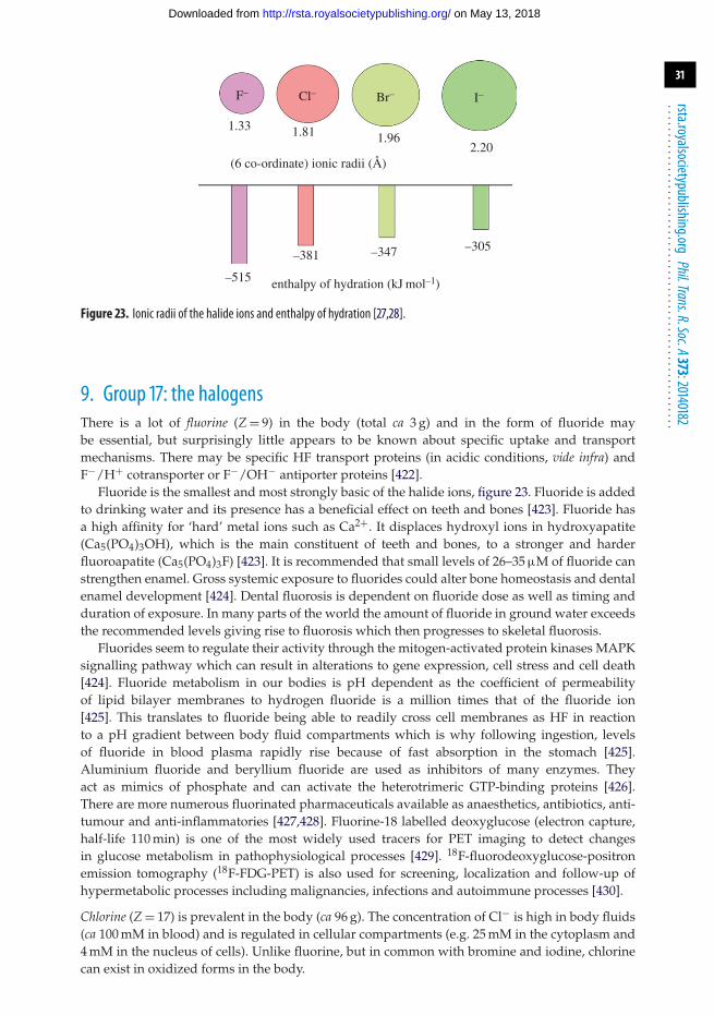
31
rsta.royalsocietypublishing.orgPhil.Trans.R.Soc.A373:20140182
.........................................................
(6 co-ordinate) ionic radii (Å)
F– Cl– Br– I–
1.33 1.81 1.962.20
enthalpy of hydration (kJ mol–1)–515
–381 –347 –305
Figure 23. Ionic radii of the halide ions and enthalpy of hydration [27,28].
9. Group 17: the halogensThere is a lot of fluorine (Z = 9) in the body (total ca 3 g) and in the form of fluoride maybe essential, but surprisingly little appears to be known about specific uptake and transportmechanisms. There may be specific HF transport proteins (in acidic conditions, vide infra) andF−/H+ cotransporter or F−/OH− antiporter proteins [422].
Fluoride is the smallest and most strongly basic of the halide ions, figure 23. Fluoride is addedto drinking water and its presence has a beneficial effect on teeth and bones [423]. Fluoride hasa high affinity for ‘hard’ metal ions such as Ca2+. It displaces hydroxyl ions in hydroxyapatite(Ca5(PO4)3OH), which is the main constituent of teeth and bones, to a stronger and harderfluoroapatite (Ca5(PO4)3F) [423]. It is recommended that small levels of 26–35 μM of fluoride canstrengthen enamel. Gross systemic exposure to fluorides could alter bone homeostasis and dentalenamel development [424]. Dental fluorosis is dependent on fluoride dose as well as timing andduration of exposure. In many parts of the world the amount of fluoride in ground water exceedsthe recommended levels giving rise to fluorosis which then progresses to skeletal fluorosis.
Fluorides seem to regulate their activity through the mitogen-activated protein kinases MAPKsignalling pathway which can result in alterations to gene expression, cell stress and cell death[424]. Fluoride metabolism in our bodies is pH dependent as the coefficient of permeabilityof lipid bilayer membranes to hydrogen fluoride is a million times that of the fluoride ion[425]. This translates to fluoride being able to readily cross cell membranes as HF in reactionto a pH gradient between body fluid compartments which is why following ingestion, levelsof fluoride in blood plasma rapidly rise because of fast absorption in the stomach [425].Aluminium fluoride and beryllium fluoride are used as inhibitors of many enzymes. Theyact as mimics of phosphate and can activate the heterotrimeric GTP-binding proteins [426].There are more numerous fluorinated pharmaceuticals available as anaesthetics, antibiotics, anti-tumour and anti-inflammatories [427,428]. Fluorine-18 labelled deoxyglucose (electron capture,half-life 110 min) is one of the most widely used tracers for PET imaging to detect changesin glucose metabolism in pathophysiological processes [429]. 18F-fluorodeoxyglucose-positronemission tomography (18F-FDG-PET) is also used for screening, localization and follow-up ofhypermetabolic processes including malignancies, infections and autoimmune processes [430].
Chlorine (Z = 17) is prevalent in the body (ca 96 g). The concentration of Cl− is high in body fluids(ca 100 mM in blood) and is regulated in cellular compartments (e.g. 25 mM in the cytoplasm and4 mM in the nucleus of cells). Unlike fluorine, but in common with bromine and iodine, chlorinecan exist in oxidized forms in the body.
on May 13, 2018http://rsta.royalsocietypublishing.org/Downloaded from
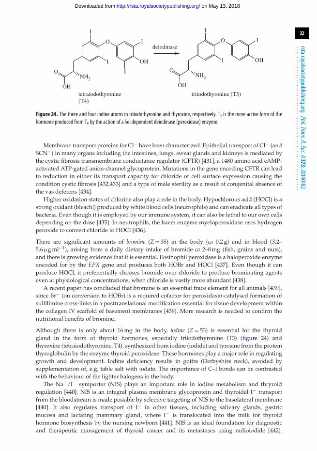
32
rsta.royalsocietypublishing.orgPhil.Trans.R.Soc.A373:20140182
.........................................................
OH
deiodinase
tetraiodothyronine(T4)
triiodothyronine (T3)OH
NH2
O
O
OH
NH2
O
I
I
I
I
OH
O
I
I
I
Figure 24. The three and four iodine atoms in triiodothyronine and thyroxine, respectively. T3 is the more active form of thehormone produced from T4 by the action of a Se-dependent deiodinase (peroxidase) enzyme.
Membrane transport proteins for Cl− have been characterized. Epithelial transport of Cl− (andSCN−) in many organs including the intestines, lungs, sweat glands and kidneys is mediated bythe cystic fibrosis transmembrane conductance regulator (CFTR) [431], a 1480 amino acid cAMP-activated ATP-gated anion-channel glycoprotein. Mutations in the gene encoding CFTR can leadto reduction in either its transport capacity for chloride or cell surface expression causing thecondition cystic fibrosis [432,433] and a type of male sterility as a result of congenital absence ofthe vas deferens [434].
Higher oxidation states of chlorine also play a role in the body. Hypochlorous acid (HOCl) is astrong oxidant (bleach!) produced by white blood cells (neutrophils) and can eradicate all types ofbacteria. Even though it is employed by our immune system, it can also be lethal to our own cellsdepending on the dose [435]. In neutrophils, the haem enzyme myeloperoxidase uses hydrogenperoxide to convert chloride to HOCl [436].
There are significant amounts of bromine (Z = 35) in the body (ca 0.2 g) and in blood (3.2–5.6 μg ml−1), arising from a daily dietary intake of bromide ca 2–8 mg (fish, grains and nuts),and there is growing evidence that it is essential. Eosinophil peroxidase is a haloperoxide enzymeencoded for by the EPX gene and produces both HOBr and HOCl [437]. Even though it canproduce HOCl, it preferentially chooses bromide over chloride to produce brominating agentseven at physiological concentrations, when chloride is vastly more abundant [438].
A recent paper has concluded that bromine is an essential trace element for all animals [439],since Br− (on conversion to HOBr) is a required cofactor for peroxidasin-catalysed formation ofsulfilimine cross-links in a posttranslational modification essential for tissue development withinthe collagen IV scaffold of basement membranes [439]. More research is needed to confirm thenutritional benefits of bromine.
Although there is only about 16 mg in the body, iodine (Z = 53) is essential for the thyroidgland in the form of thyroid hormones, especially triiodothyronine (T3) (figure 24) andthyroxine (tetraiodothyronine, T4), synthesized from iodine (iodide) and tyrosine from the proteinthyroglobulin by the enzyme thyroid peroxidase. These hormones play a major role in regulatinggrowth and development. Iodine deficiency results in goitre (Derbyshire neck), avoided bysupplementation of, e.g. table salt with iodate. The importance of C–I bonds can be contrastedwith the behaviour of the lighter halogens in the body.
The Na+/I− symporter (NIS) plays an important role in iodine metabolism and thryroidregulation [440]. NIS is an integral plasma membrane glycoprotein and thyroidal I− transportfrom the bloodstream is made possible by selective targeting of NIS to the basolateral membrane[440]. It also regulates transport of I− in other tissues, including salivary glands, gastricmucosa and lactating mammary gland, where I− is translocated into the milk for thyroidhormone biosynthesis by the nursing newborn [441]. NIS is an ideal foundation for diagnosticand therapeutic management of thyroid cancer and its metastases using radioiodide [442].
on May 13, 2018http://rsta.royalsocietypublishing.org/Downloaded from
33
rsta.royalsocietypublishing.orgPhil.Trans.R.Soc.A373:20140182
.........................................................
Transduction of NIS into various kinds of cancer cells to render them susceptible to destructionwith radioiodide has been reported [440,443]. Remarkably, more than 80% of human breast cancersamples express endogenous NIS, making the potential use of radioiodide for diagnosis andtreatment possible [444].
Astatine-211 is an α-emitter with a half-life of 7.2 h. α-Emitters have unique features that makethem ideal for elimination of focal pockets of tumour cells that are predominantly located innormal neutral tissue in the CNS [445]. For example, they can be used for targeted therapywithout risk of adjacent normal tissue toxicity that sometimes occurs with β-emitters; this featureis ideal for sensitive areas such the CNS [445]. A pilot clinical study has evaluated the feasibility,safety and efficacy of the chimeric antitenascin mAb 81 C6 labelled with 211At in patients withrecurrent malignant brain tumours [445]. The radiotherapeutic agent 211At-MX35 F(ab′)2 has alsocompleted phase I pharmacokinetic studies in women in complete clinical remission of recurrentovarian cancer [446].
10. Group 18: the noble gasesExploration of the noble gases in medicine has been limited, although helium (Z = 2) is beingconsidered for adjunct therapy for respiratory ailments such as asthma, croup and bronchiolitis[447]. It has also shown efficacy in protection of the myocardium from ischaemia, though themechanism of protection is unclear [411]. Employment of He in the operating theatre as areplacement for CO2 to insufflate the abdomens of patients undergoing laparoscopic abdominalprocedures is being evaluated [448]. Helium is a better alternative as it prevents respiratoryacidosis in patients with co-morbid conditions that cause CO2 retention [448]. Helium ionmicroscopy offers imaging of organs in very fine detail and hyperpolarized helium (HP 3He)for pulmonary MRI can be used for imaging of both normal and diseased lungs [447,449].
About 1% of the air we breathe is argon (Z = 18), mostly arising from the radioactive decay of40K in the Earth’s crust. Argon has found application in electrosurgery; gas discharges can inducemainly superficial thermal effects on tissue in a non-contact manner (argon plasma coagulation)[450]. It was first used in open surgery in the 1970s and then later adapted for endoscopy[451,452]. Examples of its use include repair of haemorrhages that can occur during surgery orulcers, injured blood vessels, varices and tumours, as well as devitalization and shrinkage oftumours and obstructing tissues [450].
Krypton (Z = 36) gas, as the quadrupolar isotope 83Kr (nuclear spin I = 9/2, natural abundance11.5%) can be used for imaging (MRI) of airways particularly to differentiate betweenhydrophobic and hydrophilic surfaces [453,454].
Xenon (Z = 54) is used as a general anaesthetic. It has a high affinity for the glycine site onthe N-methyl-D-aspartate (NMDA) receptor, and unlike most other clinical NMDA receptorantagonists, it is not neurotoxic [455,456]. Of particular note is the use of xenon as anadditive in the ventilation mix for a newborn baby to prevent brain injury (http://www.wales.nhs.uk/sitesplus/863/news/16104 (accessed 29 August 2014)). Xenon-133 (half-life 5.2 days, β−decay) is used in radioimaging of the lungs by SPECT [457], and measurement of blood flow[458]. Hyperpolarized 129Xe (nuclear spin I = 1/2, 26% abundant) is an MRI contrast agent usedfor imaging of the flow of gas in the lungs [459].
Radon (Z = 86) is a toxic radioactive (dense) gas produced from the breakdown of long-lived(billions of years) radioactive uranium (and thorium) via 226Ra in soils, rocks and water. Decayof radon yields radioactive solids (radon daughters) which are α-emitters and attach to dustparticles and are then inhaled. This inhalation has been linked to lung cancer [460,461]. Radonexposure may also lead to production of toxic reactive oxygen species. Dissolved radon in apregnant woman’s bloodstream can have detrimental consequences including retardation ofbrain development and even death [460]. Accumulation of radon in homes is a potential problem.
on May 13, 2018http://rsta.royalsocietypublishing.org/Downloaded from
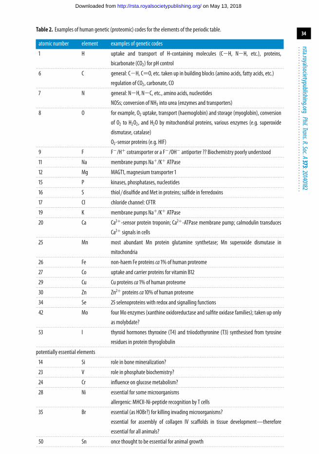
34
rsta.royalsocietypublishing.orgPhil.Trans.R.Soc.A373:20140182
.........................................................
Table 2. Examples of human genetic (proteomic) codes for the elements of the periodic table.
atomic number element examples of genetic codes. . . . . . . . . . . . . . . . . . . . . . . . . . . . . . . . . . . . . . . . . . . . . . . . . . . . . . . . . . . . . . . . . . . . . . . . . . . . . . . . . . . . . . . . . . . . . . . . . . . . . . . . . . . . . . . . . . . . . . . . . . . . . . . . . . . . . . . . . . . . . . . . . . . . . . . . . . . . . . . . . . . . . . . . . . . . . . . . . . . . . . . . . . . . . . . . . . . . . . . . . .
1 H uptake and transport of H-containing molecules (C−H, N−H, etc.), proteins,
bicarbonate (CO2) for pH control. . . . . . . . . . . . . . . . . . . . . . . . . . . . . . . . . . . . . . . . . . . . . . . . . . . . . . . . . . . . . . . . . . . . . . . . . . . . . . . . . . . . . . . . . . . . . . . . . . . . . . . . . . . . . . . . . . . . . . . . . . . . . . . . . . . . . . . . . . . . . . . . . . . . . . . . . . . . . . . . . . . . . . . . . . . . . . . . . . . . . . . . . . . . . . . . . . . . . . . . . .
6 C general: C−H, C=O, etc. taken up in building blocks (amino acids, fatty acids, etc.)
regulation of CO2, carbonate, CO. . . . . . . . . . . . . . . . . . . . . . . . . . . . . . . . . . . . . . . . . . . . . . . . . . . . . . . . . . . . . . . . . . . . . . . . . . . . . . . . . . . . . . . . . . . . . . . . . . . . . . . . . . . . . . . . . . . . . . . . . . . . . . . . . . . . . . . . . . . . . . . . . . . . . . . . . . . . . . . . . . . . . . . . . . . . . . . . . . . . . . . . . . . . . . . . . . . . . . . . . .
7 N general: N−H, N−C, etc., amino acids, nucleotides
NOSs; conversion of NH3 into urea (enzymes and transporters). . . . . . . . . . . . . . . . . . . . . . . . . . . . . . . . . . . . . . . . . . . . . . . . . . . . . . . . . . . . . . . . . . . . . . . . . . . . . . . . . . . . . . . . . . . . . . . . . . . . . . . . . . . . . . . . . . . . . . . . . . . . . . . . . . . . . . . . . . . . . . . . . . . . . . . . . . . . . . . . . . . . . . . . . . . . . . . . . . . . . . . . . . . . . . . . . . . . . . . . . .
8 O for example, O2 uptake, transport (haemoglobin) and storage (myoglobin), conversion
of O2 to H2O2, and H2O by mitochondrial proteins, various enzymes (e.g. superoxide
dismutase, catalase)
O2-sensor proteins (e.g. HIF). . . . . . . . . . . . . . . . . . . . . . . . . . . . . . . . . . . . . . . . . . . . . . . . . . . . . . . . . . . . . . . . . . . . . . . . . . . . . . . . . . . . . . . . . . . . . . . . . . . . . . . . . . . . . . . . . . . . . . . . . . . . . . . . . . . . . . . . . . . . . . . . . . . . . . . . . . . . . . . . . . . . . . . . . . . . . . . . . . . . . . . . . . . . . . . . . . . . . . . . . .
9 F F−/H+ cotransporter or a F−/OH− antiporter ?? Biochemistry poorly understood. . . . . . . . . . . . . . . . . . . . . . . . . . . . . . . . . . . . . . . . . . . . . . . . . . . . . . . . . . . . . . . . . . . . . . . . . . . . . . . . . . . . . . . . . . . . . . . . . . . . . . . . . . . . . . . . . . . . . . . . . . . . . . . . . . . . . . . . . . . . . . . . . . . . . . . . . . . . . . . . . . . . . . . . . . . . . . . . . . . . . . . . . . . . . . . . . . . . . . . . . .
11 Na membrane pumps Na+/K+ ATPase. . . . . . . . . . . . . . . . . . . . . . . . . . . . . . . . . . . . . . . . . . . . . . . . . . . . . . . . . . . . . . . . . . . . . . . . . . . . . . . . . . . . . . . . . . . . . . . . . . . . . . . . . . . . . . . . . . . . . . . . . . . . . . . . . . . . . . . . . . . . . . . . . . . . . . . . . . . . . . . . . . . . . . . . . . . . . . . . . . . . . . . . . . . . . . . . . . . . . . . . . .
12 Mg MAGT1, magnesium transporter 1. . . . . . . . . . . . . . . . . . . . . . . . . . . . . . . . . . . . . . . . . . . . . . . . . . . . . . . . . . . . . . . . . . . . . . . . . . . . . . . . . . . . . . . . . . . . . . . . . . . . . . . . . . . . . . . . . . . . . . . . . . . . . . . . . . . . . . . . . . . . . . . . . . . . . . . . . . . . . . . . . . . . . . . . . . . . . . . . . . . . . . . . . . . . . . . . . . . . . . . . . .
15 P kinases, phosphatases, nucleotides. . . . . . . . . . . . . . . . . . . . . . . . . . . . . . . . . . . . . . . . . . . . . . . . . . . . . . . . . . . . . . . . . . . . . . . . . . . . . . . . . . . . . . . . . . . . . . . . . . . . . . . . . . . . . . . . . . . . . . . . . . . . . . . . . . . . . . . . . . . . . . . . . . . . . . . . . . . . . . . . . . . . . . . . . . . . . . . . . . . . . . . . . . . . . . . . . . . . . . . . . .
16 S thiol/disulfide and Met in proteins; sulfide in ferredoxins. . . . . . . . . . . . . . . . . . . . . . . . . . . . . . . . . . . . . . . . . . . . . . . . . . . . . . . . . . . . . . . . . . . . . . . . . . . . . . . . . . . . . . . . . . . . . . . . . . . . . . . . . . . . . . . . . . . . . . . . . . . . . . . . . . . . . . . . . . . . . . . . . . . . . . . . . . . . . . . . . . . . . . . . . . . . . . . . . . . . . . . . . . . . . . . . . . . . . . . . . .
17 Cl chloride channel: CFTR. . . . . . . . . . . . . . . . . . . . . . . . . . . . . . . . . . . . . . . . . . . . . . . . . . . . . . . . . . . . . . . . . . . . . . . . . . . . . . . . . . . . . . . . . . . . . . . . . . . . . . . . . . . . . . . . . . . . . . . . . . . . . . . . . . . . . . . . . . . . . . . . . . . . . . . . . . . . . . . . . . . . . . . . . . . . . . . . . . . . . . . . . . . . . . . . . . . . . . . . . .
19 K membrane pumps Na+/K+ ATPase. . . . . . . . . . . . . . . . . . . . . . . . . . . . . . . . . . . . . . . . . . . . . . . . . . . . . . . . . . . . . . . . . . . . . . . . . . . . . . . . . . . . . . . . . . . . . . . . . . . . . . . . . . . . . . . . . . . . . . . . . . . . . . . . . . . . . . . . . . . . . . . . . . . . . . . . . . . . . . . . . . . . . . . . . . . . . . . . . . . . . . . . . . . . . . . . . . . . . . . . . .
20 Ca Ca2+-sensor protein troponin; Ca2+-ATPase membrane pump; calmodulin transducesCa2+ signals in cells
. . . . . . . . . . . . . . . . . . . . . . . . . . . . . . . . . . . . . . . . . . . . . . . . . . . . . . . . . . . . . . . . . . . . . . . . . . . . . . . . . . . . . . . . . . . . . . . . . . . . . . . . . . . . . . . . . . . . . . . . . . . . . . . . . . . . . . . . . . . . . . . . . . . . . . . . . . . . . . . . . . . . . . . . . . . . . . . . . . . . . . . . . . . . . . . . . . . . . . . . . .
25 Mn most abundant Mn protein glutamine synthetase; Mn superoxide dismutase in
mitochondria. . . . . . . . . . . . . . . . . . . . . . . . . . . . . . . . . . . . . . . . . . . . . . . . . . . . . . . . . . . . . . . . . . . . . . . . . . . . . . . . . . . . . . . . . . . . . . . . . . . . . . . . . . . . . . . . . . . . . . . . . . . . . . . . . . . . . . . . . . . . . . . . . . . . . . . . . . . . . . . . . . . . . . . . . . . . . . . . . . . . . . . . . . . . . . . . . . . . . . . . . .
26 Fe non-haem Fe proteins ca 1% of human proteome. . . . . . . . . . . . . . . . . . . . . . . . . . . . . . . . . . . . . . . . . . . . . . . . . . . . . . . . . . . . . . . . . . . . . . . . . . . . . . . . . . . . . . . . . . . . . . . . . . . . . . . . . . . . . . . . . . . . . . . . . . . . . . . . . . . . . . . . . . . . . . . . . . . . . . . . . . . . . . . . . . . . . . . . . . . . . . . . . . . . . . . . . . . . . . . . . . . . . . . . . .
27 Co uptake and carrier proteins for vitamin B12. . . . . . . . . . . . . . . . . . . . . . . . . . . . . . . . . . . . . . . . . . . . . . . . . . . . . . . . . . . . . . . . . . . . . . . . . . . . . . . . . . . . . . . . . . . . . . . . . . . . . . . . . . . . . . . . . . . . . . . . . . . . . . . . . . . . . . . . . . . . . . . . . . . . . . . . . . . . . . . . . . . . . . . . . . . . . . . . . . . . . . . . . . . . . . . . . . . . . . . . . .
29 Cu Cu proteins ca 1% of human proteome. . . . . . . . . . . . . . . . . . . . . . . . . . . . . . . . . . . . . . . . . . . . . . . . . . . . . . . . . . . . . . . . . . . . . . . . . . . . . . . . . . . . . . . . . . . . . . . . . . . . . . . . . . . . . . . . . . . . . . . . . . . . . . . . . . . . . . . . . . . . . . . . . . . . . . . . . . . . . . . . . . . . . . . . . . . . . . . . . . . . . . . . . . . . . . . . . . . . . . . . . .
30 Zn Zn2+ proteins ca 10% of human proteome. . . . . . . . . . . . . . . . . . . . . . . . . . . . . . . . . . . . . . . . . . . . . . . . . . . . . . . . . . . . . . . . . . . . . . . . . . . . . . . . . . . . . . . . . . . . . . . . . . . . . . . . . . . . . . . . . . . . . . . . . . . . . . . . . . . . . . . . . . . . . . . . . . . . . . . . . . . . . . . . . . . . . . . . . . . . . . . . . . . . . . . . . . . . . . . . . . . . . . . . . .
34 Se 25 selenoproteins with redox and signalling functions. . . . . . . . . . . . . . . . . . . . . . . . . . . . . . . . . . . . . . . . . . . . . . . . . . . . . . . . . . . . . . . . . . . . . . . . . . . . . . . . . . . . . . . . . . . . . . . . . . . . . . . . . . . . . . . . . . . . . . . . . . . . . . . . . . . . . . . . . . . . . . . . . . . . . . . . . . . . . . . . . . . . . . . . . . . . . . . . . . . . . . . . . . . . . . . . . . . . . . . . . .
42 Mo four Mo enzymes (xanthine oxidoreductase and sulfite oxidase families); taken up only
as molybdate?. . . . . . . . . . . . . . . . . . . . . . . . . . . . . . . . . . . . . . . . . . . . . . . . . . . . . . . . . . . . . . . . . . . . . . . . . . . . . . . . . . . . . . . . . . . . . . . . . . . . . . . . . . . . . . . . . . . . . . . . . . . . . . . . . . . . . . . . . . . . . . . . . . . . . . . . . . . . . . . . . . . . . . . . . . . . . . . . . . . . . . . . . . . . . . . . . . . . . . . . . .
53 I thyroid hormones thyroxine (T4) and triiodothyronine (T3) synthesised from tyrosine
residues in protein thyroglobulin. . . . . . . . . . . . . . . . . . . . . . . . . . . . . . . . . . . . . . . . . . . . . . . . . . . . . . . . . . . . . . . . . . . . . . . . . . . . . . . . . . . . . . . . . . . . . . . . . . . . . . . . . . . . . . . . . . . . . . . . . . . . . . . . . . . . . . . . . . . . . . . . . . . . . . . . . . . . . . . . . . . . . . . . . . . . . . . . . . . . . . . . . . . . . . . . . . . . . . . . . .
potentially essential elements. . . . . . . . . . . . . . . . . . . . . . . . . . . . . . . . . . . . . . . . . . . . . . . . . . . . . . . . . . . . . . . . . . . . . . . . . . . . . . . . . . . . . . . . . . . . . . . . . . . . . . . . . . . . . . . . . . . . . . . . . . . . . . . . . . . . . . . . . . . . . . . . . . . . . . . . . . . . . . . . . . . . . . . . . . . . . . . . . . . . . . . . . . . . . . . . . . . . . . . . . .
14 Si role in bone mineralization?. . . . . . . . . . . . . . . . . . . . . . . . . . . . . . . . . . . . . . . . . . . . . . . . . . . . . . . . . . . . . . . . . . . . . . . . . . . . . . . . . . . . . . . . . . . . . . . . . . . . . . . . . . . . . . . . . . . . . . . . . . . . . . . . . . . . . . . . . . . . . . . . . . . . . . . . . . . . . . . . . . . . . . . . . . . . . . . . . . . . . . . . . . . . . . . . . . . . . . . . . .
23 V role in phosphate biochemistry?. . . . . . . . . . . . . . . . . . . . . . . . . . . . . . . . . . . . . . . . . . . . . . . . . . . . . . . . . . . . . . . . . . . . . . . . . . . . . . . . . . . . . . . . . . . . . . . . . . . . . . . . . . . . . . . . . . . . . . . . . . . . . . . . . . . . . . . . . . . . . . . . . . . . . . . . . . . . . . . . . . . . . . . . . . . . . . . . . . . . . . . . . . . . . . . . . . . . . . . . . .
24 Cr influence on glucose metabolism?. . . . . . . . . . . . . . . . . . . . . . . . . . . . . . . . . . . . . . . . . . . . . . . . . . . . . . . . . . . . . . . . . . . . . . . . . . . . . . . . . . . . . . . . . . . . . . . . . . . . . . . . . . . . . . . . . . . . . . . . . . . . . . . . . . . . . . . . . . . . . . . . . . . . . . . . . . . . . . . . . . . . . . . . . . . . . . . . . . . . . . . . . . . . . . . . . . . . . . . . . .
28 Ni essential for some microorganisms
allergenic: MHCII-Ni-peptide recognition by T cells. . . . . . . . . . . . . . . . . . . . . . . . . . . . . . . . . . . . . . . . . . . . . . . . . . . . . . . . . . . . . . . . . . . . . . . . . . . . . . . . . . . . . . . . . . . . . . . . . . . . . . . . . . . . . . . . . . . . . . . . . . . . . . . . . . . . . . . . . . . . . . . . . . . . . . . . . . . . . . . . . . . . . . . . . . . . . . . . . . . . . . . . . . . . . . . . . . . . . . . . . .
35 Br essential (as HOBr?) for killing invading microorganisms?
essential for assembly of collagen IV scaffolds in tissue development—therefore
essential for all animals?. . . . . . . . . . . . . . . . . . . . . . . . . . . . . . . . . . . . . . . . . . . . . . . . . . . . . . . . . . . . . . . . . . . . . . . . . . . . . . . . . . . . . . . . . . . . . . . . . . . . . . . . . . . . . . . . . . . . . . . . . . . . . . . . . . . . . . . . . . . . . . . . . . . . . . . . . . . . . . . . . . . . . . . . . . . . . . . . . . . . . . . . . . . . . . . . . . . . . . . . . .
50 Sn once thought to be essential for animal growth. . . . . . . . . . . . . . . . . . . . . . . . . . . . . . . . . . . . . . . . . . . . . . . . . . . . . . . . . . . . . . . . . . . . . . . . . . . . . . . . . . . . . . . . . . . . . . . . . . . . . . . . . . . . . . . . . . . . . . . . . . . . . . . . . . . . . . . . . . . . . . . . . . . . . . . . . . . . . . . . . . . . . . . . . . . . . . . . . . . . . . . . . . . . . . . . . . . . . . . . . .
on May 13, 2018http://rsta.royalsocietypublishing.org/Downloaded from
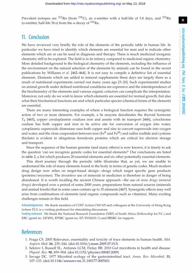
35
rsta.royalsocietypublishing.orgPhil.Trans.R.Soc.A373:20140182
.........................................................
Prevalent isotopes are 222Rn (from 238U), an α-emitter with a half-life of 3.8 days, and 220Rn(α-emitter, half-life 56 s) from the α-decay of 224Ra.
11. ConclusionWe have reviewed very briefly the role of the elements of the periodic table in human life. Inparticular we have tried to identify which elements are essential for man and to indicate otherelements which are or can be used in diagnosis and therapy. There is much medicinal inorganicchemistry still to be explored. The field is in its infancy compared to medicinal organic chemistry.More detailed background to the biological chemistry of the elements, including the influence ofthe environment on the natural selection of the elements by animals can be found in the recentpublications by Williams et al. [462–464]. It is not easy to compile a definitive list of essentialelements. Elements which are added to mineral supplements these days are largely there as aresult of nutritional experiments carried out many years ago [5–20]. Such experimental studieson animal growth under defined nutritional conditions are expensive and the interdependence ofthe biochemistry of the elements and various organic cofactors can complicate the interpretation.Moreover, not only do we wish to know which elements are essential for normal growth, but alsowhat their biochemical functions are and which particular species (chemical forms of the element)are essential.
There are many interesting examples of where a biological function requires the synergisticaction of two or more elements. For example, a Se enzyme deiodinates the thyroid hormoneT4 [465], copper ceruloplasmin oxidizes iron and assists with its transport [466], cytochromeoxidase has both copper and iron in its active site for conversion of oxygen into water,cytoplasmic superoxide dismutase uses both copper and zinc to convert superoxide into oxygenand water, and the close cooperation between iron (FeII and FeIII) and sulfur (sulfide and cysteinethiolate) is evident in ubiquitous ferredoxin proteins which are critical for electron storageand transport.
Since the sequence of the human genome (and many others) is now known, it is timely to askthe question ‘can we recognize genetic codes for essential elements?’ Our conclusions are listedin table 2, a list which produces 20 essential elements and six other potentially essential elements.
This short journey through the periodic table illustrates that, as yet, we are unable tounderstand the role of many elements found in the body in terms of genetic codes. Much moderndrug design now relies on target-based design—drugs which target specific gene products(proteins/enzymes). The inventive use of minerals in medicines is therefore in danger of beingabandoned. It is worth recalling the ancient Chinese approach—the use of stone drugs (mineraldrugs) developed over a period of some 2000 years, preparations from natural sources (mineralsand animal fossils) that in some cases contain up to 35 elements [467]. Synergistic effects may wellarise from combinations of elements (and organic compounds such as vitamins). Many excitingchallenges remain in this field.
Acknowledgements. We thank members of COST Action CM1105 and colleagues at the University of Hong Kong(where P.J.S. is a visiting professor) for stimulating discussions.Funding statement. We thank the National Research Foundation (NRF) of South Africa (Fellowship for P.C.) andERC (grant no. 247450), EPSRC (grant no. EP/F034210/1) and BBSRC for support.
References1. Fraga CS. 2005 Relevance, essentiality and toxicity of trace elements in human health. Mol.
Aspects Med. 26, 235–244. (doi:10.1016/j.mam.2005.07.013)2. Sekirov I, Russell SL, Antunes LCM, Finlay BB. 2010 Gut microbiota in health and disease.
Physiol. Rev. 90, 859–904. (doi:10.1152/physrev.00045.2009)3. Savage DC. 1977 Microbial ecology of the gastrointestinal tract. Annu. Rev. Microbiol. 31,
107–133. (doi:10.1146/annurev.mi.31.100177.000543)
on May 13, 2018http://rsta.royalsocietypublishing.org/Downloaded from
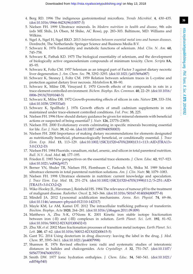
36
rsta.royalsocietypublishing.orgPhil.Trans.R.Soc.A373:20140182
.........................................................
4. Berg RD. 1996 The indigenous gastrointestinal microflora. Trends Microbiol. 4, 430–435.(doi:10.1016/0966-842X(96)10057-3)
5. Nielsen FH. 1999 Ultratrace minerals. In Modern nutrition in health and disease, 9th edn(eds ME Shils, JA Olsen, M Shike, AC Ross), pp. 283–303. Baltimore, MD: Williams andWilkins.
6. Sigel A, Sigel H, Sigel RKO. 2013 Interrelations between essential metal ions and human diseases.Dordrecht, The Netherlands: Springer Science and Business Media B.V.
7. Schwarz K. 1976 Essentiality and metabolic functions of selenium. Med. Clin. N. Am. 60,745–758.
8. Schwarz K, Pathak KD. 1975 The biological essentiality of selenium, and the developmentof biologically active organoselenium compounds of minimum toxicity. Chem. Scripta 8A,85–95.
9. Schwarz K, Foltz CM. 1957 Selenium as an integral part of Factor 3 against dietary necroticliver degeneration. J. Am. Chem. Soc. 79, 3292–3293. (doi:10.1021/ja01569a087)
10. Schwarz K, Stesney J, Foltz CM. 1959 Relation between selenium traces in L-cystine andprotection against dietary liver necrosis. Metabolism 8, 88–90.
11. Schwarz K, Milne DB, Vinuyard E. 1970 Growth effects of tin compounds in rats in atrace element-controlled environment. Bichem. Biophys. Res. Commun. 40, 22–29. (doi:10.1016/0006-291X(70)91040-5)
12. Schwarz K, Milne DB. 1972 Growth-promoting effects of silicon in rats. Nature 239, 333–334.(doi:10.1038/239333a0)
13. Schwarz K, Spallholz J. 1976 Growth effects of small cadmium supplements in ratsmaintained under trace-element controlled conditions. Fed. Proc. 35, 225.
14. Nielsen FH. 1996 How should dietary guidance be given for mineral elements with beneficialactions or suspected of being essential? J. Nutr. 126, 2377S–2385S.
15. Nielsen FH. 2000 Evolutionary events culminating in specific minerals becoming essentialfor life. Eur. J. Nutr. 39, 62–66. (doi:10.1007/s003940050003)
16. Nielsen FH. 2000 Importance of making dietary recommendations for elements designatedas nutritionally beneficial, pharmacologically beneficial, or conditionally essential. J. TraceElem. Exp. Med. 13, 113–129. (doi:10.1002/(SICI)1520-670X(2000)13:1<113::AID-JTRA13>3.0.CO;2-D)
17. Nielsen FH. 1984 Fluoride, vanadium, nickel, arsenic, and silicon in total parenteral nutrition.Bull. N.Y. Acad. Med. 60, 177–195.
18. Frieden E. 1985 New perspectives on the essential trace elements. J. Chem. Educ. 62, 917–923.(doi:10.1021/ed062p917)
19. Berner YN, Shuler TR, Nielsen FH, Flombaum C, Farkouh SA, Shiku M. 1989 Selectedultratrace elements in total parenteral nutrition solutions. Am. J. Clin. Nutr. 50, 1079–1083.
20. Nielsen FH. 1998 Ultratrace elements in nutrition: current knowledge and speculation.J. Trace Elem. Exp. Med. 11, 251–274. (doi:10.1002/(SICI)1520-670X(1998)11:2/3<251::AID-JTRA15>3.0.CO;2-Q)
21. Wike-Hooley JL, Haveman J, Reinhold HS. 1984 The relevance of tumour pH to the treatmentof malignant disease. Radiother. Oncol. 2, 343–366. (doi:10.1016/S0167-8140(84)80077-8)
22. Mindell JA. 2012 Lysosomal acidification mechanisms. Annu. Rev. Physiol. 74, 69–86.(doi:10.1146/annurev-physiol-012110-142317)
23. Mayle KM, Le AM, Kamei DT. 2012 The intracellular trafficking pathway of transferrin.Biochim. Biophys. Acta 1820, 264–281. (doi:10.1016/j.bbagen.2011.09.009)
24. Matthews A, Zhu X-K, O’Nions K. 2001 Kinetic iron stable isotope fractionationbetween iron (-II) and (-III) complexes in solution. Earth Planet. Sci. Lett. 192, 81–92.(doi:10.1016/S0012-821X(01)00432-0)
25. Zhu XK et al. 2002 Mass fractionation processes of transition metal isotopes. Earth Planet. Sci.Lett. 200, 47–62. (doi:10.1016/S0012-821X(02)00615-5)
26. Gant TG. 2014 Using deuterium in drug discovery: leaving the label in the drug. J. Med.Chem. 57, 3595–3611. (doi:10.1021/jm4007998)
27. Shannon R. 1976 Revised effective ionic radii and systematic studies of interatomicdistances in halides and chalcogenides. Acta Crystallogr. A 32, 751–767. (doi:10.1107/S0567739476001551)
28. Smith DW. 1977 Ionic hydration enthalpies. J. Chem. Educ. 54, 540–541. (doi:10.1021/ed054p540)
on May 13, 2018http://rsta.royalsocietypublishing.org/Downloaded from
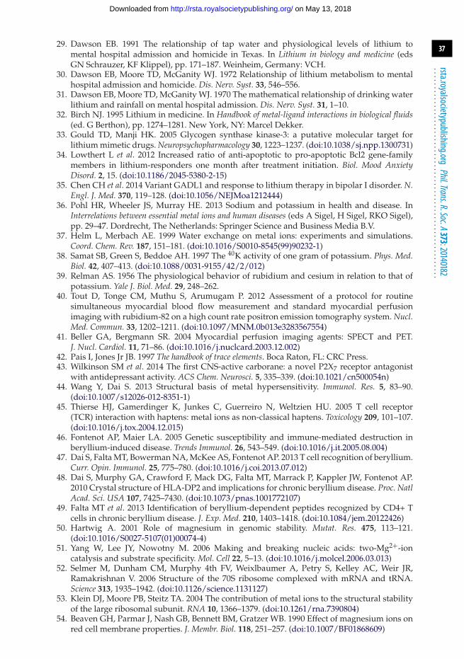
37
rsta.royalsocietypublishing.orgPhil.Trans.R.Soc.A373:20140182
.........................................................
29. Dawson EB. 1991 The relationship of tap water and physiological levels of lithium tomental hospital admission and homicide in Texas. In Lithium in biology and medicine (edsGN Schrauzer, KF Klippel), pp. 171–187. Weinheim, Germany: VCH.
30. Dawson EB, Moore TD, McGanity WJ. 1972 Relationship of lithium metabolism to mentalhospital admission and homicide. Dis. Nerv. Syst. 33, 546–556.
31. Dawson EB, Moore TD, McGanity WJ. 1970 The mathematical relationship of drinking waterlithium and rainfall on mental hospital admission. Dis. Nerv. Syst. 31, 1–10.
32. Birch NJ. 1995 Lithium in medicine. In Handbook of metal-ligand interactions in biological fluids(ed. G Berthon), pp. 1274–1281. New York, NY: Marcel Dekker.
33. Gould TD, Manji HK. 2005 Glycogen synthase kinase-3: a putative molecular target forlithium mimetic drugs. Neuropsychopharmacology 30, 1223–1237. (doi:10.1038/sj.npp.1300731)
34. Lowthert L et al. 2012 Increased ratio of anti-apoptotic to pro-apoptotic Bcl2 gene-familymembers in lithium-responders one month after treatment initiation. Biol. Mood AnxietyDisord. 2, 15. (doi:10.1186/2045-5380-2-15)
35. Chen CH et al. 2014 Variant GADL1 and response to lithium therapy in bipolar I disorder. N.Engl. J. Med. 370, 119–128. (doi:10.1056/NEJMoa1212444)
36. Pohl HR, Wheeler JS, Murray HE. 2013 Sodium and potassium in health and disease. InInterrelations between essential metal ions and human diseases (eds A Sigel, H Sigel, RKO Sigel),pp. 29–47. Dordrecht, The Netherlands: Springer Science and Business Media B.V.
37. Helm L, Merbach AE. 1999 Water exchange on metal ions: experiments and simulations.Coord. Chem. Rev. 187, 151–181. (doi:10.1016/S0010-8545(99)90232-1)
38. Samat SB, Green S, Beddoe AH. 1997 The 40K activity of one gram of potassium. Phys. Med.Biol. 42, 407–413. (doi:10.1088/0031-9155/42/2/012)
39. Relman AS. 1956 The physiological behavior of rubidium and cesium in relation to that ofpotassium. Yale J. Biol. Med. 29, 248–262.
40. Tout D, Tonge CM, Muthu S, Arumugam P. 2012 Assessment of a protocol for routinesimultaneous myocardial blood flow measurement and standard myocardial perfusionimaging with rubidium-82 on a high count rate positron emission tomography system. Nucl.Med. Commun. 33, 1202–1211. (doi:10.1097/MNM.0b013e3283567554)
41. Beller GA, Bergmann SR. 2004 Myocardial perfusion imaging agents: SPECT and PET.J. Nucl. Cardiol. 11, 71–86. (doi:10.1016/j.nuclcard.2003.12.002)
42. Pais I, Jones Jr JB. 1997 The handbook of trace elements. Boca Raton, FL: CRC Press.43. Wilkinson SM et al. 2014 The first CNS-active carborane: a novel P2X7 receptor antagonist
with antidepressant activity. ACS Chem. Neurosci. 5, 335–339. (doi:10.1021/cn500054n)44. Wang Y, Dai S. 2013 Structural basis of metal hypersensitivity. Immunol. Res. 5, 83–90.
(doi:10.1007/s12026-012-8351-1)45. Thierse HJ, Gamerdinger K, Junkes C, Guerreiro N, Weltzien HU. 2005 T cell receptor
(TCR) interaction with haptens: metal ions as non-classical haptens. Toxicology 209, 101–107.(doi:10.1016/j.tox.2004.12.015)
46. Fontenot AP, Maier LA. 2005 Genetic susceptibility and immune-mediated destruction inberyllium-induced disease. Trends Immunol. 26, 543–549. (doi:10.1016/j.it.2005.08.004)
47. Dai S, Falta MT, Bowerman NA, McKee AS, Fontenot AP. 2013 T cell recognition of beryllium.Curr. Opin. Immunol. 25, 775–780. (doi:10.1016/j.coi.2013.07.012)
48. Dai S, Murphy GA, Crawford F, Mack DG, Falta MT, Marrack P, Kappler JW, Fontenot AP.2010 Crystal structure of HLA-DP2 and implications for chronic beryllium disease. Proc. NatlAcad. Sci. USA 107, 7425–7430. (doi:10.1073/pnas.1001772107)
49. Falta MT et al. 2013 Identification of beryllium-dependent peptides recognized by CD4+ Tcells in chronic beryllium disease. J. Exp. Med. 210, 1403–1418. (doi:10.1084/jem.20122426)
50. Hartwig A. 2001 Role of magnesium in genomic stability. Mutat. Res. 475, 113–121.(doi:10.1016/S0027-5107(01)00074-4)
51. Yang W, Lee JY, Nowotny M. 2006 Making and breaking nucleic acids: two-Mg2+-ioncatalysis and substrate specificity. Mol. Cell 22, 5–13. (doi:10.1016/j.molcel.2006.03.013)
52. Selmer M, Dunham CM, Murphy 4th FV, Weixlbaumer A, Petry S, Kelley AC, Weir JR,Ramakrishnan V. 2006 Structure of the 70S ribosome complexed with mRNA and tRNA.Science 313, 1935–1942. (doi:10.1126/science.1131127)
53. Klein DJ, Moore PB, Steitz TA. 2004 The contribution of metal ions to the structural stabilityof the large ribosomal subunit. RNA 10, 1366–1379. (doi:10.1261/rna.7390804)
54. Beaven GH, Parmar J, Nash GB, Bennett BM, Gratzer WB. 1990 Effect of magnesium ions onred cell membrane properties. J. Membr. Biol. 118, 251–257. (doi:10.1007/BF01868609)
on May 13, 2018http://rsta.royalsocietypublishing.org/Downloaded from
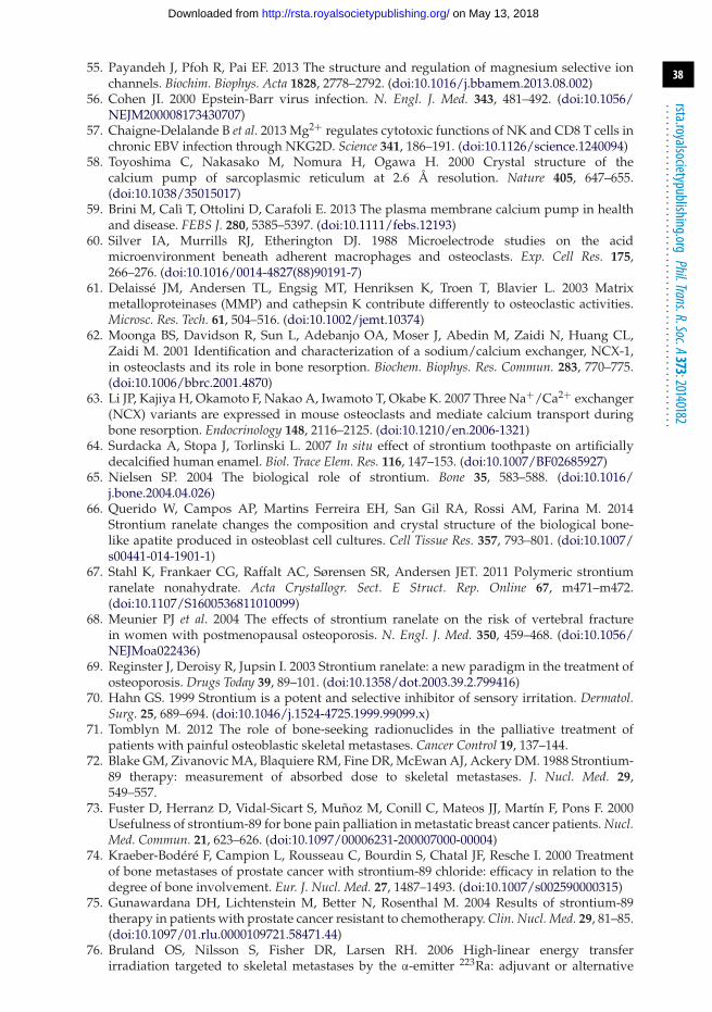
38
rsta.royalsocietypublishing.orgPhil.Trans.R.Soc.A373:20140182
.........................................................
55. Payandeh J, Pfoh R, Pai EF. 2013 The structure and regulation of magnesium selective ionchannels. Biochim. Biophys. Acta 1828, 2778–2792. (doi:10.1016/j.bbamem.2013.08.002)
56. Cohen JI. 2000 Epstein-Barr virus infection. N. Engl. J. Med. 343, 481–492. (doi:10.1056/NEJM200008173430707)
57. Chaigne-Delalande B et al. 2013 Mg2+ regulates cytotoxic functions of NK and CD8 T cells inchronic EBV infection through NKG2D. Science 341, 186–191. (doi:10.1126/science.1240094)
58. Toyoshima C, Nakasako M, Nomura H, Ogawa H. 2000 Crystal structure of thecalcium pump of sarcoplasmic reticulum at 2.6 Å resolution. Nature 405, 647–655.(doi:10.1038/35015017)
59. Brini M, Calì T, Ottolini D, Carafoli E. 2013 The plasma membrane calcium pump in healthand disease. FEBS J. 280, 5385–5397. (doi:10.1111/febs.12193)
60. Silver IA, Murrills RJ, Etherington DJ. 1988 Microelectrode studies on the acidmicroenvironment beneath adherent macrophages and osteoclasts. Exp. Cell Res. 175,266–276. (doi:10.1016/0014-4827(88)90191-7)
61. Delaissé JM, Andersen TL, Engsig MT, Henriksen K, Troen T, Blavier L. 2003 Matrixmetalloproteinases (MMP) and cathepsin K contribute differently to osteoclastic activities.Microsc. Res. Tech. 61, 504–516. (doi:10.1002/jemt.10374)
62. Moonga BS, Davidson R, Sun L, Adebanjo OA, Moser J, Abedin M, Zaidi N, Huang CL,Zaidi M. 2001 Identification and characterization of a sodium/calcium exchanger, NCX-1,in osteoclasts and its role in bone resorption. Biochem. Biophys. Res. Commun. 283, 770–775.(doi:10.1006/bbrc.2001.4870)
63. Li JP, Kajiya H, Okamoto F, Nakao A, Iwamoto T, Okabe K. 2007 Three Na+/Ca2+ exchanger(NCX) variants are expressed in mouse osteoclasts and mediate calcium transport duringbone resorption. Endocrinology 148, 2116–2125. (doi:10.1210/en.2006-1321)
64. Surdacka A, Stopa J, Torlinski L. 2007 In situ effect of strontium toothpaste on artificiallydecalcified human enamel. Biol. Trace Elem. Res. 116, 147–153. (doi:10.1007/BF02685927)
65. Nielsen SP. 2004 The biological role of strontium. Bone 35, 583–588. (doi:10.1016/j.bone.2004.04.026)
66. Querido W, Campos AP, Martins Ferreira EH, San Gil RA, Rossi AM, Farina M. 2014Strontium ranelate changes the composition and crystal structure of the biological bone-like apatite produced in osteoblast cell cultures. Cell Tissue Res. 357, 793–801. (doi:10.1007/s00441-014-1901-1)
67. Stahl K, Frankaer CG, Raffalt AC, Sørensen SR, Andersen JET. 2011 Polymeric strontiumranelate nonahydrate. Acta Crystallogr. Sect. E Struct. Rep. Online 67, m471–m472.(doi:10.1107/S1600536811010099)
68. Meunier PJ et al. 2004 The effects of strontium ranelate on the risk of vertebral fracturein women with postmenopausal osteoporosis. N. Engl. J. Med. 350, 459–468. (doi:10.1056/NEJMoa022436)
69. Reginster J, Deroisy R, Jupsin I. 2003 Strontium ranelate: a new paradigm in the treatment ofosteoporosis. Drugs Today 39, 89–101. (doi:10.1358/dot.2003.39.2.799416)
70. Hahn GS. 1999 Strontium is a potent and selective inhibitor of sensory irritation. Dermatol.Surg. 25, 689–694. (doi:10.1046/j.1524-4725.1999.99099.x)
71. Tomblyn M. 2012 The role of bone-seeking radionuclides in the palliative treatment ofpatients with painful osteoblastic skeletal metastases. Cancer Control 19, 137–144.
72. Blake GM, Zivanovic MA, Blaquiere RM, Fine DR, McEwan AJ, Ackery DM. 1988 Strontium-89 therapy: measurement of absorbed dose to skeletal metastases. J. Nucl. Med. 29,549–557.
73. Fuster D, Herranz D, Vidal-Sicart S, Muñoz M, Conill C, Mateos JJ, Martín F, Pons F. 2000Usefulness of strontium-89 for bone pain palliation in metastatic breast cancer patients. Nucl.Med. Commun. 21, 623–626. (doi:10.1097/00006231-200007000-00004)
74. Kraeber-Bodéré F, Campion L, Rousseau C, Bourdin S, Chatal JF, Resche I. 2000 Treatmentof bone metastases of prostate cancer with strontium-89 chloride: efficacy in relation to thedegree of bone involvement. Eur. J. Nucl. Med. 27, 1487–1493. (doi:10.1007/s002590000315)
75. Gunawardana DH, Lichtenstein M, Better N, Rosenthal M. 2004 Results of strontium-89therapy in patients with prostate cancer resistant to chemotherapy. Clin. Nucl. Med. 29, 81–85.(doi:10.1097/01.rlu.0000109721.58471.44)
76. Bruland OS, Nilsson S, Fisher DR, Larsen RH. 2006 High-linear energy transferirradiation targeted to skeletal metastases by the α-emitter 223Ra: adjuvant or alternative
on May 13, 2018http://rsta.royalsocietypublishing.org/Downloaded from
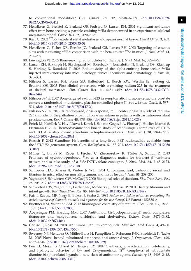
39
rsta.royalsocietypublishing.orgPhil.Trans.R.Soc.A373:20140182
.........................................................
to conventional modalities? Clin. Cancer Res. 12, 6250s–6257s. (doi:10.1158/1078-0432.CCR-06-0841)
77. Henriksen G, Breistol K, Bruland OS, Fodstad O, Larsen RH. 2002 Significant antitumoreffect from bone-seeking, α-particle-emitting 223Ra demonstrated in an experimental skeletalmetastases model. Cancer Res. 62, 3120–3125.
78. Kerr C. 2002 223Ra targets skeletal metastases and spares normal tissue. Lancet Oncol. 3, 453.(doi:10.1016/S1470-2045(02)00835-5)
79. Henriksen G, Fisher DR, Roeske JC, Bruland OS, Larsen RH. 2003 Targeting of osseoussites with α-emitting 223Ra: comparison with the beta-emitter 89Sr in mice. J. Nucl. Med. 44,252–259.
80. Lewington VJ. 2005 Bone-seeking radionuclides for therapy. J. Nucl. Med. 46, 38S–47S.81. Larsen RH, Saxtorph H, Skydsgaard M, Borrebaek J, Jonasdottir TJ, Bruland OS, Klastrup
S, Harling R, Ramdahl T. 2006 Radiotoxicity of the alpha-emitting bone-seeker 223Rainjected intravenously into mice: histology, clinical chemistry and hematology. In Vivo 20,325–331.
82. Nilsson S, Larsen RH, Fossa SD, Balteskard L, Borch KW, Westlin JE, Salberg G,Bruland OS. 2005 First clinical experience with α-emitting radium-223 in the treatmentof skeletal metastases. Clin. Cancer Res. 11, 4451–4459. (doi:10.1158/1078-0432.CCR-04-2244)
83. Nilsson S et al. 2007 Bone-targeted radium-223 in symptomatic, hormone-refractory prostatecancer: a randomised, multicentre, placebo-controlled phase II study. Lancet Oncol. 8, 587–594. (doi:10.1016/S1470-2045(07)70147-X)
84. Nilsson S et al. 2012 A randomized, dose-response, multicenter phase II study of radium-223 chloride for the palliation of painful bone metastases in patients with castration-resistantprostate cancer. Eur. J. Cancer 48, 678–686. (doi:10.1016/j.ejca.2011.12.023)
85. Pniok M, Kubíèek V, Havlíèková J, Kotek J, Sabatie-Gogová A, Plutnar J, Huclier-Markai S,Hermann P. 2014 Thermodynamic and kinetic study of scandium(III) complexes of DTPAand DOTA: a step toward scandium radiopharmaceuticals. Chem. Eur. J. 20, 7944–7955.(doi:10.1002/chem.201402041)
86. Roesch F. 2012 Scandium-44: benefits of a long-lived PET radionuclide available fromthe 44Ti/44Sc generator system. Curr. Radiopharm. 5, 187–201. (doi:10.2174/1874471011205030187)
87. Müller C, Bunka M, Reber J, Fischer C, Zhernosekov K, Türler A, Schibli R. 2013Promises of cyclotron-produced 44Sc as a diagnostic match for trivalent β−-emitters:in vitro and in vivo study of a 44Sc-DOTA-folate conjugate. J. Nucl. Med. 54, 2168–2174.(doi:10.2967/jnumed.113.123810)
88. Schroeder HA, Balassa JJ, Vinton Jr WH. 1964 Chromium, lead, cadmium, nickel andtitanium in mice: effect on mortality, tumors and tissue levels. J. Nutr. 83, 239–250.
89. Yaghoubi S, Schwietert CW, McCue JP. 2000 Biological roles of titanium. Biol. Trace Elem. Res.78, 205–217. (doi:10.1385/BTER:78:1-3:205)
90. Schwietert CW, Yaghoubi S, Gerber NC, McSharry JJ, McCue JP. 2001 Dietary titanium andinfant growth. Biol. Trace Elem. Res. 83, 149–167. (doi:10.1385/BTER:83:2:149)
91. Pais I, Ravasz MF, Nagy B, Bokori J, Szabo Z. 1984 Fodder and fodder additives promoting theweight increase of domestic animals and a process for the use thereof. US Patent 4482550 A.
92. Buettner KM, Valentine AM. 2012 Bioinorganic chemistry of titanium. Chem. Rev. 112, 1863–1881. (doi:10.1021/cr1002886)
93. Abeysinghe PM, Harding MM. 2007 Antitumour bis(cyclopentadienyl) metal complexes:titanocene and molybdocene dichloride and derivatives. Dalton Trans. 3474–3482.(doi:10.1039/b707440a)
94. Caruso F, Rossi M. 2004 Antitumor titanium compounds. Mini Rev. Med. Chem. 4, 49–60.(doi:10.2174/1389557043487565)
95. Sweeney NJ, Mendoza O, Müller-Bunz H, Pampillón C, Rehmann F-JK, Strohfeldt K, TackeM. 2005 Novel benzyl substituted titanocene anti-cancer drugs. J. Organomet. Chem. 690,4537–4544. (doi:10.1016/j.jorganchem.2005.06.039)
96. Peri D, Meker S, Shavit M, Tshuva EY. 2009 Synthesis, characterization, cytotoxicity,and hydrolytic behavior of C2- and C1-symmetrical TiIV complexes of tetradentatediamine bis(phenolato) ligands: a new class of antitumor agents. Chemistry 15, 2403–2415.(doi:10.1002/chem.200801310)
on May 13, 2018http://rsta.royalsocietypublishing.org/Downloaded from
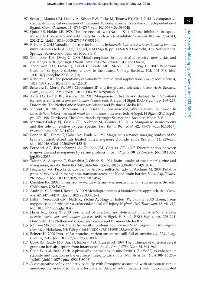
40
rsta.royalsocietypublishing.orgPhil.Trans.R.Soc.A373:20140182
.........................................................
97. Schur J, Manna CM, Deally A, Köster RW, Tacke M, Tshuva EY, Ott I. 2013 A comparativechemical-biological evaluation of titanium(IV) complexes with a salan or cyclopentadienylligand. Chem. Commun. 49, 4785–4787. (doi:10.1039/c3cc38604j)
98. Quist EE, Hokin LE. 1978 The presence of two (Na+ + K+)–ATPase inhibitors in equinemuscle ATP: vanadate and a dithioerythritol-dependent inhibitor. Biochim. Biophys. Acta 511,202–212. (doi:10.1016/0005-2736(78)90314-0)
99. Rehder D. 2013 Vanadium. Its role for humans. In Interrelations between essential metal ions andhuman diseases (eds A Sigel, H Sigel, RKO Sigel), pp. 139–169. Dordrecht, The Netherlands:Springer Science and Business Media B.V.
100. Thompson KH, Orvig C. 2006 Metal complexes in medicinal chemistry: new vistas andchallenges in drug design. Dalton Trans. 761–764. (doi:10.1039/b513476e)
101. Thompson KH, Lichter J, LeBel C, Scaife MC, McNeill JH, Orvig C. 2009 Vanadiumtreatment of type 2 diabetes: a view to the future. J. Inorg. Biochem. 103, 554–558. (doi:10.1016/j.jinorgbio.2008.12.003)
102. Rehder D. 2012 The potentiality of vanadium in medicinal applications. Future Med. Chem. 4,1823–1837. (doi:10.4155/fmc.12.103)
103. Schwarz K, Mertz W. 1959 Chromium(III) and the glucose tolerance factor. Arch. Biochem.Biophys. 85, 292–295. (doi:10.1016/0003-9861(59)90479-5)
104. Avila DS, Puntel RL, Aschner M. 2013 Manganese in health and disease. In Interrelationsbetween essential metal ions and human diseases (eds A Sigel, H Sigel, RKO Sigel), pp. 199–227.Dordrecht, The Netherlands: Springer Science and Business Media B.V.
105. Vincent JB. 2013 Chromium: is it essential, pharmacologically relevant, or toxic? InInterrelations between essential metal ions and human diseases (eds A Sigel, H Sigel, RKO Sigel),pp. 171–198. Dordrecht, The Netherlands: Springer Science and Business Media B.V.
106. Martinez-Finley EJ, Gavin CE, Aschner M, Gunter TE. 2013 Manganese neurotoxicityand the role of reactive oxygen species. Free Radic. Biol. Med. 62, 65–75. (doi:10.1016/j.freeradbiomed.2013.01.032)
107. London RE, Toney G, Gabel SA, Funk A. 1989 Magnetic resonance imaging studies of thebrains of anesthetized rats treated with manganese chloride. Brain Res. Bull. 23, 229–235.(doi:10.1016/0361-9230(89)90152-4)
108. Foradori AC, Bertinchamps A, Gulibon JM, Cotzias GC. 1967 Discrimination betweenmagnesium and manganese by serum proteins. J. Gen. Physiol. 50, 2255–2266. (doi:10.1085/jgp.50.9.2255)
109. Takeda A, Akiyama T, Sawashita J, Okada S. 1994 Brain uptake of trace metals, zinc andmanganese, in rats. Brain Res. 640, 341–344. (doi:10.1016/0006-8993(94)91891-0)
110. Fitsanakis VA, Piccola G, dos Santos AP, Marreilha A, Judy L, Aschner M. 2007 Putativeproteins involved in manganese transport across the blood-brain barrier. Hum. Exp. Toxicol.26, 295–302. (doi:10.1177/0960327107070496)
111. Crichton RR. 2009 Iron metabolism—from molecular mechanisms to clinical consequences, 3rd edn.Chichester, UK: Wiley.
112. Andreini C, Bertini I, Rosato A. 2009 Metalloproteomes: a bioinformatic approach. Acc. Chem.Res. 42, 1471–1479. (doi:10.1021/ar900015x)
113. Balla J, Vercellotti GM, Nath K, Yachie A, Nagy E, Eaton JW, Balla G. 2003 Haem, haemoxygenase and ferritin in vascular endothelial cell injury. Nephrol. Dial. Transplant. 18, v8–v12.(doi:10.1093/ndt/gfg1034)
114. Hider RC, Kong X. 2013 Iron: effect of overload and deficiency. In Interrelations betweenessential metal ions and human diseases (eds A Sigel, H Sigel, RKO Sigel), pp. 229–294.Dordrecht, The Netherlands: Springer Science and Business Media B.V.
115. Johnson MK, Smith AD. 2011 Iron–sulfur proteins. In Encyclopedia of inorganic and bioinorganicchemistry. Hoboken, NJ: Wiley. (doi:10.1002/9781119951438.eibc0109)
116. Beinert H. 2000 Iron–sulfur proteins: ancient structures, still full of surprises. J. Biol. Inorg.Chem. 5, 2–15. (doi:10.1007/s007750050002)
117. Cook JD, Reddy MB, Burri J, Juillerat MA, Hurrell RF. 1997 The influence of different cerealgrains on iron absorption from infant cereal foods. Am. J. Clin. Nutr. 65, 964–969.
118. Chen W et al. 2009 Abcb10 physically interacts with mitoferrin-1 (Slc25a37) to enhance itsstability and function in the erythroid mitochondria. Proc. Natl Acad. Sci. USA 106, 16 263–16 268. (doi:10.1073/pnas.0904519106)
119. A comparative safety and activity study with ferroquine associated with artesunate versusamodiaquine associated with artesunate in African adult patients with uncomplicated
on May 13, 2018http://rsta.royalsocietypublishing.org/Downloaded from
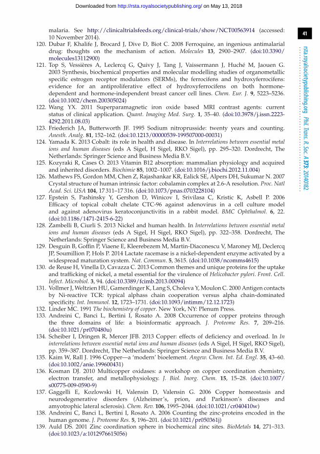
41
rsta.royalsocietypublishing.orgPhil.Trans.R.Soc.A373:20140182
.........................................................
malaria. See http://clinicaltrialsfeeds.org/clinical-trials/show/NCT00563914 (accessed:10 November 2014).
120. Dubar F, Khalife J, Brocard J, Dive D, Biot C. 2008 Ferroquine, an ingenious antimalarialdrug: thoughts on the mechanism of action. Molecules 13, 2900–2907. (doi:10.3390/molecules13112900)
121. Top S, Vessières A, Leclercq G, Quivy J, Tang J, Vaissermann J, Huché M, Jaouen G.2003 Synthesis, biochemical properties and molecular modelling studies of organometallicspecific estrogen receptor modulators (SERMs), the ferrocifens and hydroxyferrocifens:evidence for an antiproliferative effect of hydroxyferrocifens on both hormone-dependent and hormone-independent breast cancer cell lines. Chem. Eur. J. 9, 5223–5236.(doi:10.1002/chem.200305024)
122. Wang YX. 2011 Superparamagnetic iron oxide based MRI contrast agents: currentstatus of clinical application. Quant. Imaging Med. Surg. 1, 35–40. (doi:10.3978/j.issn.2223-4292.2011.08.03)
123. Friederich JA, Butterworth JF. 1995 Sodium nitroprusside: twenty years and counting.Anesth. Analg. 81, 152–162. (doi:10.1213/00000539-199507000-00031)
124. Yamada K. 2013 Cobalt: its role in health and disease. In Interrelations between essential metalions and human diseases (eds A Sigel, H Sigel, RKO Sigel), pp. 295–320. Dordrecht, TheNetherlands: Springer Science and Business Media B.V.
125. Kozyraki R, Cases O. 2013 Vitamin B12 absorption: mammalian physiology and acquiredand inherited disorders. Biochimie 85, 1002–1007. (doi:10.1016/j.biochi.2012.11.004)
126. Mathews FS, Gordon MM, Chen Z, Rajashankar KR, Ealick SE, Alpers DH, Sukumar N. 2007Crystal structure of human intrinsic factor: cobalamin complex at 2.6-A resolution. Proc. NatlAcad. Sci. USA 104, 17 311–17 316. (doi:10.1073/pnas.0703228104)
127. Epstein S, Pashinsky Y, Gershon D, Winicov I, Srivilasa C, Kristic K, Asbell P. 2006Efficacy of topical cobalt chelate CTC-96 against adenovirus in a cell culture modeland against adenovirus keratoconjunctivitis in a rabbit model. BMC Ophthalmol. 6, 22.(doi:10.1186/1471-2415-6-22)
128. Zambelli B, Ciurli S. 2013 Nickel and human health. In Interrelations between essential metalions and human diseases (eds A Sigel, H Sigel, RKO Sigel), pp. 322–358. Dordrecht, TheNetherlands: Springer Science and Business Media B.V.
129. Desguin B, Goffin P, Viaene E, Kleerebezem M, Martin-Diaconescu V, Maroney MJ, DeclercqJP, Soumillion P, Hols P. 2014 Lactate racemase is a nickel-dependent enzyme activated by awidespread maturation system. Nat. Commun. 5, 3615. (doi:10.1038/ncomms4615)
130. de Reuse H, Vinella D, Cavazza C. 2013 Common themes and unique proteins for the uptakeand trafficking of nickel, a metal essential for the virulence of Helicobacter pylori. Front. Cell.Infect. Microbiol. 3, 94. (doi:10.3389/fcimb.2013.00094)
131. Vollmer J, Weltzien HU, Gamerdinger K, Lang S, Choleva Y, Moulon C. 2000 Antigen contactsby Ni-reactive TCR: typical alphass chain cooperation versus alpha chain-dominatedspecificity. Int. Immunol. 12, 1723–1731. (doi:10.1093/intimm/12.12.1723)
132. Linder MC. 1991 The biochemistry of copper. New York, NY: Plenum Press.133. Andreini C, Banci L, Bertini I, Rosato A. 2008 Occurrence of copper proteins through
the three domains of life: a bioinformatic approach. J. Proteome Res. 7, 209–216.(doi:10.1021/pr070480u)
134. Scheiber I, Dringen R, Mercer JFB. 2013 Copper: effects of deficiency and overload. In Ininterrelations between essential metal ions and human diseases (eds A Sigel, H Sigel, RKO Sigel),pp. 359–387. Dordrecht, The Netherlands: Springer Science and Business Media B.V.
135. Kaim W, Rall J. 1996 Copper—a ‘modern’ bioelement. Angew. Chem. Int. Ed. Engl. 35, 43–60.(doi:10.1002/anie.199600431)
136. Kosman DJ. 2010 Multicopper oxidases: a workshop on copper coordination chemistry,electron transfer, and metallophysiology. J. Biol. Inorg. Chem. 15, 15–28. (doi:10.1007/s00775-009-0590-9)
137. Gaggelli E, Kozlowski H, Valensin D, Valensin G. 2006 Copper homeostasis andneurodegenerative disorders (Alzheimer’s, prion, and Parkinson’s diseases andamyotrophic lateral sclerosis). Chem. Rev. 106, 1995–2044. (doi:10.1021/cr040410w)
138. Andreini C, Banci L, Bertini I, Rosato A. 2006 Counting the zinc-proteins encoded in thehuman genome. J. Proteome Res. 5, 196–201. (doi:10.1021/pr050361j)
139. Auld DS. 2001 Zinc coordination sphere in biochemical zinc sites. BioMetals 14, 271–313.(doi:10.1023/a:1012976615056)
on May 13, 2018http://rsta.royalsocietypublishing.org/Downloaded from
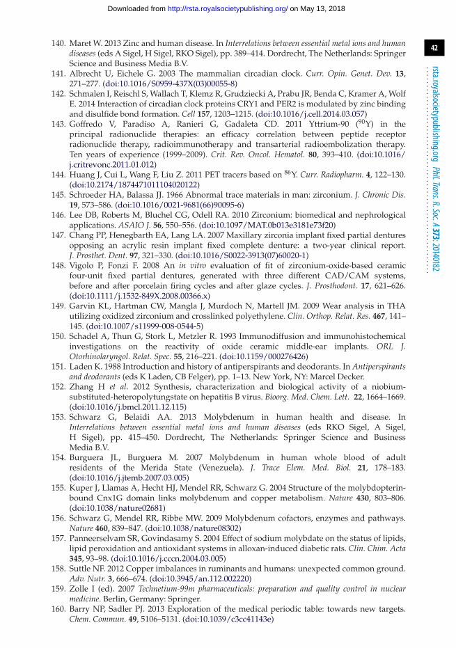
42
rsta.royalsocietypublishing.orgPhil.Trans.R.Soc.A373:20140182
.........................................................
140. Maret W. 2013 Zinc and human disease. In Interrelations between essential metal ions and humandiseases (eds A Sigel, H Sigel, RKO Sigel), pp. 389–414. Dordrecht, The Netherlands: SpringerScience and Business Media B.V.
141. Albrecht U, Eichele G. 2003 The mammalian circadian clock. Curr. Opin. Genet. Dev. 13,271–277. (doi:10.1016/S0959-437X(03)00055-8)
142. Schmalen I, Reischl S, Wallach T, Klemz R, Grudziecki A, Prabu JR, Benda C, Kramer A, WolfE. 2014 Interaction of circadian clock proteins CRY1 and PER2 is modulated by zinc bindingand disulfide bond formation. Cell 157, 1203–1215. (doi:10.1016/j.cell.2014.03.057)
143. Goffredo V, Paradiso A, Ranieri G, Gadaleta CD. 2011 Yttrium-90 (90Y) in theprincipal radionuclide therapies: an efficacy correlation between peptide receptorradionuclide therapy, radioimmunotherapy and transarterial radioembolization therapy.Ten years of experience (1999–2009). Crit. Rev. Oncol. Hematol. 80, 393–410. (doi:10.1016/j.critrevonc.2011.01.012)
144. Huang J, Cui L, Wang F, Liu Z. 2011 PET tracers based on 86Y. Curr. Radiopharm. 4, 122–130.(doi:10.2174/1874471011104020122)
145. Schroeder HA, Balassa JJ. 1966 Abnormal trace materials in man: zirconium. J. Chronic Dis.19, 573–586. (doi:10.1016/0021-9681(66)90095-6)
146. Lee DB, Roberts M, Bluchel CG, Odell RA. 2010 Zirconium: biomedical and nephrologicalapplications. ASAIO J. 56, 550–556. (doi:10.1097/MAT.0b013e3181e73f20)
147. Chang PP, Henegbarth EA, Lang LA. 2007 Maxillary zirconia implant fixed partial denturesopposing an acrylic resin implant fixed complete denture: a two-year clinical report.J. Prosthet. Dent. 97, 321–330. (doi:10.1016/S0022-3913(07)60020-1)
148. Vigolo P, Fonzi F. 2008 An in vitro evaluation of fit of zirconium-oxide-based ceramicfour-unit fixed partial dentures, generated with three different CAD/CAM systems,before and after porcelain firing cycles and after glaze cycles. J. Prosthodont. 17, 621–626.(doi:10.1111/j.1532-849X.2008.00366.x)
149. Garvin KL, Hartman CW, Mangla J, Murdoch N, Martell JM. 2009 Wear analysis in THAutilizing oxidized zirconium and crosslinked polyethylene. Clin. Orthop. Relat. Res. 467, 141–145. (doi:10.1007/s11999-008-0544-5)
150. Schadel A, Thun G, Stork L, Metzler R. 1993 Immunodiffusion and immunohistochemicalinvestigations on the reactivity of oxide ceramic middle-ear implants. ORL J.Otorhinolaryngol. Relat. Spec. 55, 216–221. (doi:10.1159/000276426)
151. Laden K. 1988 Introduction and history of antiperspirants and deodorants. In Antiperspirantsand deodorants (eds K Laden, CB Felger), pp. 1–13. New York, NY: Marcel Decker.
152. Zhang H et al. 2012 Synthesis, characterization and biological activity of a niobium-substituted-heteropolytungstate on hepatitis B virus. Bioorg. Med. Chem. Lett. 22, 1664–1669.(doi:10.1016/j.bmcl.2011.12.115)
153. Schwarz G, Belaidi AA. 2013 Molybdenum in human health and disease. InInterrelations between essential metal ions and human diseases (eds RKO Sigel, A Sigel,H Sigel), pp. 415–450. Dordrecht, The Netherlands: Springer Science and BusinessMedia B.V.
154. Burguera JL, Burguera M. 2007 Molybdenum in human whole blood of adultresidents of the Merida State (Venezuela). J. Trace Elem. Med. Biol. 21, 178–183.(doi:10.1016/j.jtemb.2007.03.005)
155. Kuper J, Llamas A, Hecht HJ, Mendel RR, Schwarz G. 2004 Structure of the molybdopterin-bound Cnx1G domain links molybdenum and copper metabolism. Nature 430, 803–806.(doi:10.1038/nature02681)
156. Schwarz G, Mendel RR, Ribbe MW. 2009 Molybdenum cofactors, enzymes and pathways.Nature 460, 839–847. (doi:10.1038/nature08302)
157. Panneerselvam SR, Govindasamy S. 2004 Effect of sodium molybdate on the status of lipids,lipid peroxidation and antioxidant systems in alloxan-induced diabetic rats. Clin. Chim. Acta345, 93–98. (doi:10.1016/j.cccn.2004.03.005)
158. Suttle NF. 2012 Copper imbalances in ruminants and humans: unexpected common ground.Adv. Nutr. 3, 666–674. (doi:10.3945/an.112.002220)
159. Zolle I (ed). 2007 Technetium-99m pharmaceuticals: preparation and quality control in nuclearmedicine. Berlin, Germany: Springer.
160. Barry NP, Sadler PJ. 2013 Exploration of the medical periodic table: towards new targets.Chem. Commun. 49, 5106–5131. (doi:10.1039/c3cc41143e)
on May 13, 2018http://rsta.royalsocietypublishing.org/Downloaded from
43
rsta.royalsocietypublishing.orgPhil.Trans.R.Soc.A373:20140182
.........................................................
161. Cocchietto M, Zorzet S, Sorc A, Sava G. 2003 Primary tumor, lung and kidney retention andantimetastasis effect of NAMI-A following different routes of administration. Invest. NewDrugs 21, 55–62. (doi:10.1023/A:1022916310694)
162. Hillier SM, Maresca KP, Lu G, Merkin RD, Marquis JC, Zimmerman CN, EckelmanWC, Joyal JL, Babich JW. 2013 99mTc-labeled small-molecule inhibitors of prostate-specificmembrane antigen for molecular imaging of prostate cancer. J. Nucl. Med. 54, 1369–1376.(doi:10.2967/jnumed.112.116624)
163. Sava G et al. 2003 Dual action of NAMI-A in inhibition of solid tumor metastasis: selectivetargeting of metastatic cells and binding to collagen. Clin. Cancer Res. 9, 1898–1905.
164. Bratsos I, Jedner S, Gianferrara T, Alessio E. 2007 Ruthenium anticancer compounds:challenges and expectations. Chimia 61, 692–697. (doi:10.2533/chimia.2007.692)
165. Bergamo A, Sava G. 2007 Ruthenium complexes can target determinants of tumourmalignancy. Dalton Trans. 1267–1272. (doi:10.1039/B617769G)
166. Sava G, Capozzi I, Clerici K, Gagliardi G, Alessio E, Mestroni G. 1998 Pharmacologicalcontrol of lung metastases of solid tumours by a novel ruthenium complex. Clin. Exp.Metastasis 16, 371–379. (doi:10.1023/A:1006521715400)
167. Bergamo A, Gaiddon C, Schellens JH, Beijnen JH, Sava G. 2012 Approaching tumour therapybeyond platinum drugs: status of the art and perspectives of ruthenium drug candidates. J.Inorg. Biochem. 106, 90–99. (doi:10.1016/j.jinorgbio.2011.09.030)
168. Hartinger CG, Jakupec MA, Zorbas-Seifried S, Groessl M, Egger A, Berger W, ZorbasH, Dyson PJ, Keppler BK. 2008 KP1019, a new redox-active anticancer agent—preclinicaldevelopment and results of a clinical phase I study in tumor patients. Chem. Biodivers. 5,2140–2155. (doi:10.1002/cbdv.200890195)
169. Lentz F et al. 2009 Pharmacokinetics of a novel anticancer ruthenium complex (KP1019,FFC14A) in a phase I dose-escalation study. Anticancer Drugs 20, 97–103. (doi:10.1097/CAD.0b013e328322fbc5)
170. Yan YK, Melchart M, Habtemariam A, Sadler PJ. 2005 Organometallic chemistry,biology and medicine: ruthenium arene anticancer complexes. Chem. Commun. 4764–4776.(doi:10.1039/B508531B)
171. Fricker SP. 1999 Nitric oxide scavengers as a therapeutic approach to nitric oxide mediateddisease. Expert Opin. Investig. Drugs 8, 1209–1222. (doi:10.1517/13543784.8.8.1209)
172. Chellan P, Land KM, Shokar A, Au A, An SH, Taylor D, Smith PJ, Chibale K, SmithGS. 2013 Di- and trinuclear ruthenium-, rhodium-, and iridium-functionalized pyridylaromatic ethers: a new class of antiparasitic agents. Organometallics 32, 4793–4804.(doi:10.1021/om400493k)
173. Simpson PV, Schmidt C, Ott I, Bruhn H, Schatzschneider U. 2013 Synthesis, cellular uptakeand biological activity against pathogenic microorganisms and cancer cells of rhodium andiridium N-heterocyclic carbene complexes bearing charged substituents. Eur. J. Inorg. Chem.5547–5554. (doi:10.1002/ejic.201300820)
174. Menon EL, Perera R, Navarro M, Kuhn RJ, Morrison H. 2004 Phototoxicity againsttumor cells and sindbis virus by an octahedral rhodium bisbipyridyl complex andevidence for the genome as a target in viral photoinactivation. Inorg. Chem. 43, 5373–5381.(doi:10.1021/ic0498586)
175. Geldmacher Y, Oleszak M, Sheldrick WS. 2012 Rhodium(III) and iridium(III) complexes asanticancer agents. Inorg. Chim. Acta 393, 84–102. (doi:10.1016/j.ica.2012.06.046)
176. Ando A, Ando I, Tonami N, Kinuya S, Okamoto N, Sugimoto M, Fukuda N, Matsumoto S.2000 Production of 105Rh–EDTMP and its bone accumulation. Appl. Radiat. Isot. 52, 211–215.(doi:10.1016/S0969-8043(99)00129-3)
177. Brun PM, Bucking M, DeGroot JL, Farahani M, Pottier RH. 2004 Neutron activation andliquid scintillation analysis of tissue samples containing palladium bacteriochlorophyllderivative, a potential photochemotherapeutic agent. Can. J. Anal. Sci. Spectrosc. 49, 55–63.(doi:10.1016/j.brachy.2013.04.001)
178. Vascular targeted photodynamic therapy T1a renal tumours (KCM201). Seehttp://clinicaltrials.gov/ct2/show/NCT01573156 (accessed 10 November 2014).
179. Taylor JM. 1989 Use of sources for brachytherapy. Fed. Regist. 54, 41 819–41 821.180. Finger PT, Berson A, Ng T, Szechter A. 2002 Palladium-103 plaque radiotherapy for
choroidal melanoma: an 11-year study. Int. J. Radiat. Oncol. Biol. Phys. 54, 1438–1445.(doi:10.1016/S0360-3016(02)03751-3)
on May 13, 2018http://rsta.royalsocietypublishing.org/Downloaded from
44
rsta.royalsocietypublishing.orgPhil.Trans.R.Soc.A373:20140182
.........................................................
181. Pd-103 dose de-escalation for early stage prostate cancer: a prospective randomized trial. Seehttp://clinicaltrials.gov/ct2/show/NCT00247312 (accessed 10 November 2014).
182. High versus low dose supplemental external radiation with Pd-103 for prostate cancer. Seehttp://clinicaltrials.gov/ct2/show/NCT00494546 (accessed 10 November 2014).
183. Chopra I. 2007 The increasing use of silver-based products as antimicrobial agents:a useful development or a cause for concern? J. Antimicrob. Chemother. 59, 587–590.(doi:10.1093/jac/dkm006)
184. Klasen HJ. 2000 Historical review of the use of silver in the treatment of burns. I. Early uses.Burns 26, 117–130. (doi:10.1016/S0305-4179(99)00108-4)
185. Leaper DJ. 2006 Silver dressings: their role in wound management. Int. Wound J. 3, 282–294.(doi:10.1111/j.1742-481X.2006.00265.x)
186. Fox Jr CL. 1968 Silver sulfadiazine—a new topical therapy for Pseudomonas in burns.Therapy of Pseudomonas infection in burns. Arch. Surg. 96, 184–188. (doi:10.1001/archsurg.1968.01330200022004)
187. George N, Faoagali J, Muller M. 1997 Silvazine (silver sulfadiazine and chlorhexidine)activity against 200 clinical isolates. Burns 23, 493–495. (doi:10.1016/S0305-4179(97)00047-8)
188. Gemmell CG, Edwards DI, Fraise AP, Gould FK, Ridgway GL, Warren RE. 2006 Guidelinesfor the prophylaxis and treatment of methicillin-resistant Staphylococcus aureus (MRSA)infections in the UK. J. Antimicrob. Chemother. 57, 589–608. (doi:10.1093/jac/dkl017)
189. Clinical trial of a Silver Eluting Dressing System (SILVER). See https://clinicaltrials.gov/ct2/show/NCT01229358 (accessed 10 November 2014).
190. Galdiero S, Falanga A, Vitiello M, Cantisani M, Marra V, Galdiero M. 2011 Silvernanoparticles as potential antiviral agents. Molecules 16, 8894–8918. (doi:10.3390/molecules16108894)
191. Soumya RS, Hela PG. 2013 Nano silver based targeted drug delivery for treatment of cancer.Der Pharmacia Lettre 5, 189–197.
192. Iqbal J, Barsukova-Stuckart M, Ibrahim M, Ali S, Khan A, Kortz U. 2013Polyoxometalates as potent inhibitors for acetyl and butyrylcholinesterases and aspotential drugs for the treatment of Alzheimer’s disease. Med. Chem. Res. 22, 1224–1228.(doi:10.1007/s00044-012-0125-8)
193. Suzuki CA, Ohta H, Albores A, Koropatnick J, Cherian MG. 1990 Induction ofmetallothionein synthesis by zinc in cadmium pretreated rats. Toxicology 63, 273–284.(doi:10.1016/0300-483X(90)90190-R)
194. Xu Y, Feng L, Jeffrey PD, Shi Y, Morel FM. 2008 Structure and metal exchange in the cadmiumcarbonic anhydrase of marine diatoms. Nature 452, 56–61. (doi:10.1038/nature06636)
195. Maggiorella L, Barouch G, Devaux C, Pottier A, Deutsch E, Bourhis J, Borghi E, Levy L.2012 Nanoscale radiotherapy with hafnium oxide nanoparticles. Future Oncol. 8, 1167–1181.(doi:10.2217/fon.12.96)
196. Marill J, Anesary NM, Zhang P, Vivet S, Borghi E, Levy L, Pottier A. 2014 Hafniumoxide nanoparticles: toward an in vitro predictive biological effect? Radiat. Oncol. 9, 150.(doi:10.1186/1748-717X-9-150)
197. Pottier A, Borghi E, Levy L. 2014 New use of metals as nanosized radioenhancers. AnticancerRes. 34, 443–453.
198. Bermudez MD, Carrion FJ, Martinez-Nicolas G, Lopez R. 2005 Erosion-corrosion of stainlesssteels, titanium, tantalum and zirconium. Wear 258, 693–700. (doi:10.1016/j.wear.2004.09.023)
199. Kato H, Nakamura T, Nishiguchi S, Matsusue Y, Kobayashi M, Miyazaki T, Kim H, KokuboT. 2000 Bonding of alkali- and heat-treated tantalum implants to bone. J. Biomed. Mater. Res.53, 28–35. (doi:10.1002/(SICI)1097-4636(2000)53:1<28::AID-JBM4>3.0.CO;2-F)
200. Zardiackas LD, Parsell DE, Dillon LD, Mitchell DW, Nunnery LA, Poggie R. 2001 Structure,metallurgy, and mechanical properties of a porous tantalum foam. J. Biomed. Mater. Res. Appl.Biomater. 58, 180–187. (doi:0.1002/1097-4636(2001)58:2<180::AID-JBM1005>3.0.CO;2-5)
201. Bobyn JD, Stackpool GJ, Hacking SA, Tanzer M, Krygier JJ. 1999 Characteristics of boneingrowth and interface mechanics of a new porous tantalum biomaterial. J. Bone Joint Surg.Br. 81, 907–914. (doi:10.1302/0301-620X.81B5.9283)
202. Hacking SA, Bobyn JD, Toh K, Tanzer M, Krygier JJ. 2000 Fibrous tissue ingrowth andattachment to porous tantalum. J. Biomed. Mater. Res. 52, 631–638. (doi:10.1002/1097-4636(20001215)52:4<631::AID-JBM7>3.0.CO;2-6)
203. Domingo JL. 2002 Vanadium and tungsten derivatives as antidiabetic agents: a review oftheir toxic effects. Biol. Trace Elem. Res. 88, 97–113. (doi:10.1385/BTER:88:2:097)
on May 13, 2018http://rsta.royalsocietypublishing.org/Downloaded from
45
rsta.royalsocietypublishing.orgPhil.Trans.R.Soc.A373:20140182
.........................................................
204. Muñoz MC, Barberà A, Domínguez J, Fernàndez-Alvarez J, Gomis R, Guinovart JJ. 2001Effects of tungstate, a new potential oral antidiabetic agent, in Zucker diabetic fatty rats.Diabetes 50, 131–138. (doi:10.2337/diabetes.50.1.131)
205. Barberà A, Fernàndez-Alvarez J, Truc A, Gomis R, Guinovart JJ. 1997 Effects of tungstatein neonatally streptozotocin-induced diabetic rats: mechanism leading to normalization ofglycaemia. Diabetologia 40, 143–149. (doi:10.1007/s001250050655)
206. Barberà A, Gomis RR, Prats N, Rodríguez-Gil JE, Domingo M, Gomis R, Guinovart JJ. 2001Tungstate is an effective antidiabetic agent in streptozotocin-induced diabetic rats: a long-term study. Diabetologia 44, 507–513. (doi:10.1007/s001250100479)
207. Barberà A, Rodríguez-Gil JE, Guinovart JJ. 1994 Insulin-like actions of tungstate in diabeticrats. Normalization of hepatic glucose metabolism. J. Biol. Chem. 269, 20 047–20 053.
208. Domínguez JE et al. 2003 The antidiabetic agent sodium tungstate activates glycogensynthesis through an insulin receptor-independent pathway. J. Biol. Chem. 278, 42 785–42 794.(doi:10.1074/jbc.M308334200)
209. Stiefel EI. 1998 Transition metal sulfur chemistry and its relevance to molybdenum andtungsten enzymes. Pure Appl. Chem. 70, 889–896. (doi:10.1351/pac199870040889)
210. Khangulov SV, Gladyshev VN, Dismukes GC, Stadtman TC. 1998 Selenium-containingformate dehydrogenase H from Escherichia coli: a molybdopterin enzyme that catalyzesformate oxidation without oxygen transfer. Biochemistry 37, 3518–3528. (doi:10.1021/bi972177k)
211. Fukuma M, Seto Y, Yamase T. 1991 In vitro antiviral activity of polyoxotungstate(PM-19) and other polyoxometalates against herpes simplex virus. Antiviral Res. 16, 327–339.(doi:10.1016/0166-3542(91)90047-U)
212. Inoue M, Segawa K, Matsunaga S, Matsumoto N, Oda M, Yamase T. 2005 Antibacterialactivity of highly negative charged polyoxotungstates, K27[KAs4W40O140] andK18[KSb9W21O86], and Keggin-structural polyoxotungstates against Helicobacter pylori.J. Inorg. Biochem. 99, 1023–1031. (doi:10.1016/j.jinorgbio.2005.01.010)
213. Wang L, Zhou B-B, Yu K, Su Z-H, Gao S, Chu L-L, Liu J-R, Yang G-Y. 2013 Novelantitumor agent, trilacunary Keggin-type tungstobismuthate, inhibits proliferation andinduces apoptosis in human gastric cancer SGC-7901 cells. Inorg. Chem. 52, 5119–5127.(doi:10.1021/ic400019r)
214. Deutsch E, Brodack JW, Deutsch KF. 1993 Radiation synovectomy revisited. Eur. J. Nucl. Med.20, 1113–1127. (doi:10.1007/BF00173494)
215. Lambert B, de Klerk JM. 2006 Clinical applications of 188Re-labelled radiopharmaceuticalsfor radionuclide therapy. Nucl. Med. Commun. 227, 223–229. (doi:10.1097/00006231-200603000-00004)
216. Phase I/II trial of rhenium 188-P2045 in small cell lung cancer and other advancedneuroendocrine carcinomas. See https://clinicaltrials.gov/ct2/show/NCT02030184.(accessed 10 November 2014).
217. Edelman MJ, Clamon G, Kahn D, Magram M, Lister-James J, Line BR. 2009 Targetedradiopharmaceutical therapy for advanced lung cancer: phase I trial of rheniumRe188 P2045, a somatostatin analog. J. Thorac. Oncol. 4, 1550–1554. (doi:10.1097/JTO.0b013e3181bf1070)
218. Fujita T, Yamaji Y, Sato M, Murao K, Takahara J. 1994 Gene expression of somatostatinreceptor subtypes, SSTR1 and SSTR2, in human lung cancer cell lines. Life Sci. 55, 1797–1806.(doi:10.1016/0024-3205(94)90090-6)
219. O’Byrne KJ, Schally AV, Thomas A, Carney DN, Steward WP. 2001 Somatostatin, its receptorand analogs in lung cancer. Chemotherapy 47, 78–108. (doi:10.1159/000049163)
220. Dizeyi N, Konrad L, Bjartell A, Wu H, Gadaleanu V, Hansson J, Helboe L,Abrahamsson PA. 2002 Localization and mRNA expression of somatostatin receptorsubtypes in human prostatic tissue and prostate cancer cell lines. Urol. Oncol. 7, 91–98.(doi:10.1016/S1078-1439(01)00173-9)
221. Kumar U, Grigorakis SI, Watt HL, Sasi R, Snell L, Watson P, Chaudhari S. 2005 Somatostatinreceptors in primary human breast cancer: quantitative analysis of mRNA for subtypes1–5 and correlation with receptor protein expression and tumor pathology. Breast Cancer Res.Treat. 92, 175–186. (doi:10.1007/s10549-005-2414-0)
222. Berglof FE. 1959 Osmic acid in arthritis therapy. Acta Rheumatol. Scand. 5, 70–74.(doi:10.3109/rhe1.1959.5.issue-1-4.11)
on May 13, 2018http://rsta.royalsocietypublishing.org/Downloaded from
46
rsta.royalsocietypublishing.orgPhil.Trans.R.Soc.A373:20140182
.........................................................
223. Bessant R, Steuer A, Rigby S, Gumpel M. 2003 Osmic acid revisited: factors that predict afavourable response. Rheumatology 42, 1036–1043. (doi:10.1093/rheumatology/keg283)
224. Goldstein S, Czapski G, Heller A. 2005 Osmium tetroxide, used in the treatment ofarthritic joints, is a fast mimic of superoxide dismutase. Free Radic. Biol. Med. 38, 839–845.(doi:10.1016/j.freeradbiomed.2004.10.027)
225. Noffke AL, Habtemariam A, Pizarro AM, Sadler PJ. 2012 Designing organometalliccompounds for catalysis and therapy. Chem. Commun. 48, 5219–5246. (doi:10.1039/c2cc30678f)
226. Radical radiotherapy by external beam radiation versus radical radiotherapy withtemporary iridium implant plus external beam radiation in carcinoma of the prostate. Seehttps://clinicaltrials.gov/ct2/show/NCT00548600.
227. A multicenter trial of localized radiation therapy to inhibit restenosis (GAMMA V).See https://clinicaltrials.gov/ct2/show/record/NCT00232778 (accessed 10 November2014).
228. Leung C-H, Zhong H-J, Chan DS-H, Ma D-L. 2013 Bioactive iridium and rhodium complexesas therapeutic agents. Coord. Chem. Rev. 257, 1764–1776. (doi:10.1016/j.ccr.2013.01.034)
229. Li SP-Y, Lau CT-S, Louie M-W, Lam Y-W, Cheng SH, Lo KK-W. 2013 Mitochondria-targetingcyclometalated iridium(III)–PEG complexes with tunable photodynamic activity. Biomaterials34, 7519–7532. (doi:10.1016/j.biomaterials.2013.06.028)
230. Flamigni L, Barbieri A, Sabatini C, Ventura B, Barigelletti F. 2007 Photochemistry andphotophysics of coordination compounds: iridium. In Photochemistry and photophysics ofcoordination compounds II (eds V Balzani, S Campagna), pp. 143–203. Berlin, Germany:Springer.
231. Liu Z, Sadler PJ. 2014 Organoiridium complexes: anticancer agents and catalysts. Acc. Chem.Res. 47, 1174–1185. (doi:10.1021/ar400266c)
232. Wheate NJ, Walker S, Craig GE, Oun R. 2010 The status of platinum anticancer drugs in theclinic and in clinical trials. Dalton Trans. 39, 8113–8127. (doi:10.1039/c0dt00292e)
233. Gabano E, Ravera M, Osella D. 2014 Pros and cons of bifunctional platinum(IV)antitumor prodrugs: two are (not always) better than one. Dalton Trans. 43, 9813–9820.(doi:10.1039/C4DT00911H)
234. Stathopoulos GP et al. 2010 Liposomal cisplatin combined with paclitaxel versus cisplatinand paclitaxel in non-small-cell lung cancer: a randomized phase III multicenter trial. Ann.Oncol. 21, 2227–2232. (doi:10.1093/annonc/mdq234)
235. Stathopoulos GP, Boulikas T. 2012 Lipoplatin formulation review article. J. Drug Deliv.581363. (doi:10.1155/2012/581363)
236. Farrel NP. 2004 Polynuclear platinum drugs. In Metal ions in biological systems: metal complexesin tumor diagnosis and as anticancer agents (ed. H Sigel), pp. 251–296. Dordrecht, TheNetherlands: Springer.
237. Butler JS, Sadler PJ. 2013 Targeted delivery of platinum-based anticancer complexes. Curr.Opin. Chem. Biol. 17, 175–188. (doi:10.1016/j.cbpa.2013.01.004)
238. Muhammad N, Guo Z. 2014 Metal-based anticancer chemotherapeutic agents. Curr. Opin.Chem. Biol. 19, 144–153. (doi:10.1016/j.cbpa.2014.02.003)
239. Todd RC, Lippard SJ. 2009 Inhibition of transcription by platinum antitumor compounds.Metallomics 1, 280–291. (doi:10.1039/b907567d)
240. Safaei R, Howell SB. 2005 Copper transporters regulate the cellular pharmacology andsensitivity to Pt drugs. Crit. Rev. Oncol. Hematol. 53, 13–23. (doi:10.1016/j.critrevonc.2004.09.007)
241. Pizarro AM, McQuitty RJ, Mackay FS, Zhao Y, Woods JA, Sadler PJ. 2014 Cellularaccumulation, lipophilicity and photocytotoxicity of diazido platinum(IV) anticancercomplexes. ChemMedChem 9, 1169–1175. (doi:10.1002/cmdc.201402066)
242. Farrer NJ, Sadler PJ. 2008 Photochemotherapy: targeted activation of metal anticancercomplexes. Aust. J. Chem. 61, 669–674. (doi:10.1071/CH08088)
243. Aryal S, Hu C-MJ, Zhang L. 2009 Polymer–cisplatin conjugate nanoparticles for acid-responsive drug delivery. ACS Nano 4, 251–258. (doi:10.1021/nn9014032)
244. Raubenheimer HG, Schmidbaur H. 2014 The late start and amazing upswing in goldchemistry. J. Chem. Educ. 91, 2024–2036. (doi:10.1021/ed400782p)
245. Bertini I, Gray HB, Lippard SJ, Valentine JS. 1994 Bioinorganic chemistry. Mill Valley, CA:University Science Books.
on May 13, 2018http://rsta.royalsocietypublishing.org/Downloaded from
47
rsta.royalsocietypublishing.orgPhil.Trans.R.Soc.A373:20140182
.........................................................
246. PKCi & mTOR inhibition with auranofin+sirolimus for squamous cell lung cancer. Seehttps://clinicaltrials.gov/ct2/show/NCT01737502 (accessed 10 November 2014).
247. Rosi NL, Giljohann DA, Thaxton CS, Lytton-Jean AKR, Han MS, Mirkin CA. 2006Oligonucleotide-modified gold nanoparticles for intracellular gene regulation. Science 312,1027–1030. (doi:10.1126/science.1125559)
248. Zhang D, Liu D, Zhang J, Fong C, Yang M. 2014 Gold nanoparticles stimulate differentiationand mineralization of primary osteoblasts through the ERK/MAPK signaling pathway.Mater. Sci. Eng. C 42, 70–77. (doi:10.1016/j.msec.2014.04.042)
249. Dhar S, Daniel WL, Giljohann DA, Mirkin CA, Lippard SJ. 2009 Polyvalent oligonucleotidegold nanoparticle conjugates as delivery vehicles for platinum(IV) warheads. J. Am. Chem.Soc. 131, 14 652–14 653. (doi:10.1021/ja9071282)
250. El-Sayed IH, Huang X, El-Sayed MA. 2005 Surface plasmon resonance scattering andabsorption of anti-EGFR antibody conjugated gold nanoparticles in cancer diagnostics:applications in oral cancer. Nano Lett. 5, 829–834. (doi:10.1021/nl050074e)
251. Debnath A et al. 2012 A high-throughput drug screen for Entamoeba histolytica identifies anew lead and target. Nat. Med. 18, 956–960. (doi:10.1038/nm.2758)
252. Masur LC. 2011 A review of the use of mercury in historic and current ritualistic and spiritualpractices. Altern. Med. Rev. 16, 314–320.
253. Ball LK, Ball R, Pratt RD. 2001 An assessment of thimerosal use in childhood vaccines.Pediatrics 107, 1147–1154. (doi:10.1542/peds.107.5.1147)
254. Baker JP. 2008 Mercury, vaccines, and autism one controversy, three histories. Am. J. PublicHealth 98, 244–253. (doi:10.2105/AJPH.2007.113159)
255. Parker SK, Schwartz B, Todd J, Pickering LK. 2004 Thimerosal-containing vaccines andautistic spectrum disorder: a critical review of published original data. Pediatrics 114,793–804. (doi:10.1542/peds.2004-0434)
256. Xu J et al. 2013 Lanthanum carbonate for the treatment of hyperphosphatemia in CKD 5D:multicenter, double blind, randomized, controlled trial in mainland China. BMC Nephrol. 14,29. (doi:10.1186/1471-2369-14-29)
257. Kalaitzidis RG, Elisaf MS. 2014 Hyperphosphatemia and phosphate binders:effectiveness and safety. Curr. Med. Res. Opin. 30, 109–112. (doi:10.1185/03007995.2013.841667)
258. Drüeke TB. 2007 Phosphorus metabolism and management in chronic kidney disease:lanthanum carbonate as a first-line phosphate binder: the ‘cons’. Semin. Dial. 20, 329–332.(doi:10.1111/j.1525-139X.2007.00299.x)
259. Rambeck WA, Wehr U. 2000 Rare earth elements as alternative growth promoters in pigproduction. Arch. Tierernahr. 53, 323–334. (doi:10.1080/17450390009381956)
260. Thacker P. 2013 Alternatives to antibiotics as growth promoters for use in swine production:a review. J. Anim. Sci. Biotechnol. 4, 35. (doi:10.1186/2049-1891-4-35)
261. Sobek JM, Talburt DE. 1968 Effects of the rare earth cerium on Escherichia coli. J. Bacteriol. 95,47–51.
262. Boeckx W, Blondeel PN, Vandersteen K, De Wolf-Peeters C, Schmitz A. 1992 Effect of ceriumnitrate-silver sulphadiazine on deep dermal burns: a histological hypothesis. Burns 18,456–462. (doi:10.1016/0305-4179(92)90177-V)
263. Monafo WW, Tandon SN, Ayvazian VH, Tuchschmidt J, Skinner AM, Deitz F. 1976 Ceriumnitrate: a new topical antiseptic for extensive burns. Surgery 80, 465–473.
264. Peterson VM, Hansbrough JF, Wang XW, Zapata-Sirvent R, Boswick Jr JA. 1985Topical cerium nitrate prevents postburn immunosuppression. J. Trauma 25, 1039–1044.(doi:10.1097/00005373-198505000-00007)
265. Garner JP, Heppell PSJ. 2005 Cerium nitrate in the management of burns. Burns 31, 539–547.(doi:10.1016/j.burns.2005.01.014)
266. Correa-González L, Arteaga de Murphy C, Pichardo-Romero P, Pedraza-López M, Moreno-García C, Correa-Hernández L. 2014 153Sm-EDTMP for pain relief of bone metastasesfrom prostate and breast cancer and other malignancies. Arch. Med. Res. 45, 301–308.(doi:10.1016/j.arcmed.2014.03.006)
267. Vigna L, Matheoud R, Ridone S, Arginelli D, Della Monica P, Rudoni M, IngleseE, Brambilla M. 2011 Characterization of the [153Sm]Sm-EDTMP pharmacokinetics andestimation of radiation absorbed dose on an individual basis. Phys. Med. 27, 144–152.(doi:10.1016/j.ejmp.2010.08.001)
on May 13, 2018http://rsta.royalsocietypublishing.org/Downloaded from

48
rsta.royalsocietypublishing.orgPhil.Trans.R.Soc.A373:20140182
.........................................................
268. Serafini AN. 2000 Samarium Sm-153 lexidronam for the palliation of bone painassociated with metastases. Cancer 88, 2934–2939. (doi:10.1002/1097-0142(20000615)88:12+<2934::AID-CNCR9>3.0.CO;2-S)
269. Lam MG, Dahmane A, Stevens WH, van Rijk PP, de Klerk JM, Zonnenberg BA. 2008Combined use of zoledronic acid and 153Sm-EDTMP in hormone-refractory prostatecancer patients with bone metastases. Eur. J. Nucl. Med. Mol. Imaging 35, 756–765.(doi:10.1007/s00259-007-0659-z)
270. Parlak Y, Gumuser G, Sayit E. In press. Samarium-153 therapy for prostate cancer: theevaluation of urine activity, staff exposure and dose rate from patients. Radiat. Prot.Dosimetry. (doi:10.1093/rpd/ncu237)
271. Bartlett ML, Webb M, Durrant S, Morton JA, Allison R, Macfarlane DJ. 2002 Dosimetryand toxicity of Quadramet for bone marrow ablation in multiple myeloma and otherhaematological malignancies. Eur. J. Nucl. Med. Mol. Imaging 29, 1470–1477. (doi:10.1007/s00259-002-0934-y)
272. Maini CL, Bergomi S, Romano L, Sciuto R. 2004 153Sm-EDTMP for bone painpalliation in skeletal metastases. Eur. J. Nucl. Med. Mol. Imaging 31, S171–S178.(doi:10.1007/s00259-004-1540-y)
273. Combination of external beam radiotherapy with 153Sm-EDTMP to treat high riskosteosarcoma. See http://clinicaltrials.gov/ct2/show/NCT01886105 (accessed 10November 2014).
274. Sm-EDTMP and autologous peripheral blood stem cell transplantation for breast cancerpatients with bone only metastases. See http://clinicaltrials.gov/ct2/show/NCT00429507(accessed 10 November 2014).
275. Samarium Sm 153 lexidronam pentasodium combined with zoledronic acid or pamidronatein treating patients with relapsed or refractory multiple myeloma and bone pain. Seehttp://clinicaltrials.gov/ct2/show/NCT00482378 (accessed 10 November 2014).
276. Song B, Wu Y, Yu M, Zhao P, Zhou C, Kiefer GE, Sherry AD. 2013 A europium(III)-based PARACEST agent for sensing singlet oxygen by MRI. Dalton Trans. 42, 8066–8069.(doi:10.1039/C3DT50194A)
277. Tagaya M, Ikoma T, Xu Z, Tanaka J. 2014 Synthesis of luminescent nanoporous silicaspheres functionalized with folic acid for targeting to cancer cells. Inorg. Chem. 53, 6817–6827.(doi:10.1021/ic500609g)
278. Caravan P, Ellison JJ, McMurry TJ, Lauffer RB. 1999 Gadolinium(III) chelates asMRI contrast agents: structure, dynamics, and applications. Chem. Rev. 99, 2293–2352.(doi:10.1021/cr980440x)
279. Long-term retention of gadolinium in bone. See https://clinicaltrials.gov/ct2/show/NCT01853163 (accessed 10 November 2014).
280. Chakraborty S, Unni PR, Banerjee S, Samuel G, Das T, Sarma HD, Ramamoorthy N,Pillai MRA. 2001 Potential 166Ho radiopharmaceuticals for intravascular radiation therapy(IVRT)-I: [166Ho] holmium labeled ethylene dicysteine. Nucl. Med. Biol. 28, 309–317.(doi:10.1016/S0969-8051(00)00197-9)
281. Suzuki YS, Momose Y, Higashi N, Shigematsu A, Park K-B, Kim YM, Kim JR, Ryu JM.1998 Biodistribution and kinetics of holmium-166-chitosan complex (DW-166HC) in rats andmice. J. Nucl. Med. 39, 2161–2166.
282. Vente MAD et al. 2009 Neutron activation of holmium poly(L-lactic acid) microspheresfor hepatic arterial radioembolization: a validation study. Biomed. Microdevices 11, 763–772.(doi:10.1007/s10544-009-9291-y)
283. Zielhuis SW, Nijsen JFW, de Roos R, Krijger GC, van Rijk PP, Hennink WE, van het Schip AD.2006 Production of GMP-grade radioactive holmium loaded poly(l-lactic acid) microspheresfor clinical application. Int. J. Pharm. 311, 69–74. (doi:10.1016/j.ijpharm.2005.12.034)
284. Radioactive holmium microspheres for the treatment of liver metastases (HEPAR). Seehttps://clinicaltrials.gov/ct2/show/NCT01031784 (accessed 10 November 2014).
285. Radioactive holmium microspheres for the treatment of unresectable liver metastases(HEPAR 2). See https://clinicaltrials.gov/ct2/show/NCT01612325 (accessed 10 November2014).
286. Chakraborty S, Unni PR, Venkatesh M, Pillai MRA. 2002 Feasibility study forproduction of 175Yb: a promising therapeutic radionuclide. Appl. Radiat. Isot. 57, 295–301.(doi:10.1016/S0969-8043(02)00100-8)
on May 13, 2018http://rsta.royalsocietypublishing.org/Downloaded from
49
rsta.royalsocietypublishing.orgPhil.Trans.R.Soc.A373:20140182
.........................................................
287. Mathew B, Chakraborty S, Das T, Sarma HD, Banerjee S, Samuel G, Venkatesh M, Pillai MRA.2004 175Yb labeled polyaminophosphonates as potential agents for bone pain palliation.Appl. Radiat. Isot. 60, 635–642. (doi:10.1016/j.apradiso.2003.09.016)
288. Patel NS, Fan P, Chiu-Tsao S-T, Ravi K, Sherman W, Quon H, Pisch J, Tsao H-S, HarrisonLB. 2001 Ytterbium-169: a promising new radionuclide for intravascular brachytherapy.Cardiovasc. Radiat. Med. 2, 173–180. (doi:10.1016/S1522-1865(01)00085-3)
289. Liu Z et al. 2014 177Lu-labeled antibodies for EGFR-targeted SPECT/CT imaging andradioimmunotherapy in a preclinical head and neck carcinoma model. Mol. Pharm. 11,800–807. (doi:10.1021/mp4005047)
290. Shi W, Ogbomo SM, Wagh NK, Zhou Z, Jia Y, Brusnahan SK, Garrison JC. 2014The influence of linker length on the properties of cathepsin S cleavable 177Lu-labeled HPMA copolymers for pancreatic cancer imaging. Biomaterials 35, 5760–5770.(doi:10.1016/j.biomaterials.2014.03.056)
291. TF2-small cell lung cancer radio immunotherapy. See https://clinicaltrials.gov/ct2/show/NCT01221675 (accessed 10 November 2014).
292. Borchardt PE, Yuan RR, Miederer M, McDevitt MR, Scheinberg DA. 2003 Targeted actinium-225 in vivo generators for therapy of ovarian cancer. Cancer Res. 63, 5084–5090.
293. Baidoo KE, Yong K, Brechbiel MW. 2013 Molecular pathways: targeted α-particle radiationtherapy. Clin. Cancer Res. 19, 530–537. (doi:10.1158/1078-0432.ccr-12-0298)
294. Low dose cytarabine and lintuzumab-Ac225 in older patients. See https://clinicaltrials.gov/ct2/show/NCT01756677 (accessed 10 November 2014).
295. Targeted atomic nano-generators (actinium-225-labeled humanized anti-CD33monoclonal antibody HuM195) in patients with advanced myeloid malignancies. Seehttps://clinicaltrials.gov/ct2/show/NCT00672165 (accessed 10 November 2014).
296. Dembitsky VM, Smoum R, Al-Quntar AAA, Ali HA, Pergament I, Srebnik M. 2002 Naturaloccurrence of boroncontaining compounds in plants, algae and microorganisms. Plant Sci.163, 931–942. (doi:10.1016/S0168-9452(02)00174-7)
297. Loomis WD, Durst RW. 1992 Chemistry and biology of boron. BioFactors 3, 229–239.298. Dinca L, Scorei R. 2013 Boron in human nutrition and its regulations use. J. Nutr. Ther. 2,
22–29.299. Newnham RE. 1994 Essentiality of boron for healthy bones and joints. Environ. Health
Perspect. 102, 83–85. (doi:10.1289/ehp.94102s783)300. Price CT, Langford JR, Liporace FA. 2012 Essential nutrients for bone health and a review
of their availability in the average North American diet. Open Orthop. J. 6, 143–149.(doi:10.2174/1874325001206010143)
301. Pache W, Zähner H. 1969 Metabolic products of microorganisms. 77. Studies on themechanism of action of boromycin. Arch. Mikrobiol. 67, 156–165. (doi:10.1007/BF00409681)
302. Baker SJ, Tomsho JW, Benkovic SJ. 2011 Boron-containing inhibitors of synthetases. Chem.Soc. Rev. 40, 4279–4285. (doi:10.1039/C0CS00131G)
303. Kohno J et al. 1996 Boromycin, an anti-HIV antibiotic. Biosci. Biotechnol. Biochem. 60, 1036–1037. (doi:10.1271/bbb.60.1036)
304. Soriano-Ursúa MA, Das BC, Trujillo-Ferrara JG. 2014 Boron-containing compounds:chemico-biological properties and expanding medicinal potential in prevention, diagnosisand therapy. Expert Opin. Ther. Pat. 24, 485–500. (doi:10.1517/13543776.2014.881472)
305. Sene S et al. 2014 A combined experimental-computational study of benzoxaborole crystalstructures. CrystEngComm 16, 4999–5011. (doi:10.1039/c4ce00313f)
306. Ciaravino V, Plattner J, Chanda S. 2013 An assessment of the genetic toxicology of novelboron-containing therapeutic agents. Environ. Mol. Mutagen. 54, 338–346. (doi:10.1002/em.21779)
307. Kumar V, Gill KD. 2014 Oxidative stress and mitochondrial dysfunction inaluminium neurotoxicity and its amelioration: a review. NeuroToxicology 41, 154–166.(doi:10.1016/j.neuro.2014.02.004)
308. Pineau A, Fauconneau B, Sappino A-P, Deloncle R, Guillard O. 2014 If exposure toaluminium in antiperspirants presents health risks, its content should be reduced. J. TraceElem. Med. Biol. 28, 147–150. (doi:10.1016/j.jtemb.2013.12.002)
309. Wills MR, Savory J. 1983 Aluminium poisoning: dialysis encephalopathy, osteomalacia, andanaemia. Lancet 322, 29–34. (doi:10.1016/S0140-6736(83)90014-4)
310. García A, De Sanctis JB. 2014 An overview of adjuvant formulations and delivery systems.APMIS 122, 257–267. (doi:10.1111/apm.12143)
on May 13, 2018http://rsta.royalsocietypublishing.org/Downloaded from
50
rsta.royalsocietypublishing.orgPhil.Trans.R.Soc.A373:20140182
.........................................................
311. Hornung V, Bauernfeind F, Halle A, Samstad EO, Kono H, Rock KL, Fitzgerald KA, LatzE. 2008 Silica crystals and aluminum salts activate the NALP3 inflammasome throughphagosomal destabilization. Nat. Immunol. 9, 847–856. (doi:10.1038/ni.1631)
312. Tomljenovic L, Shaw CA. 2011 Aluminum vaccine adjuvants: are they safe? Curr. Med. Chem.18, 2630–2637. (doi:10.2174/092986711795933740)
313. Malakoff D. 2000 Aluminum is put on trial as a vaccine booster. Science 288, 1323–1324.(doi:10.1126/science.288.5470.1323)
314. Chitambar CR. 2010 Medical applications and toxicities of gallium compounds. Int. J.Environ. Res. Public Health 7, 2337–2361. (doi:10.3390/ijerph7052337)
315. Lessa JA, Parrilha GL, Beraldo H. 2012 Gallium complexes as new promising metallodrugcandidates. Inorg. Chim. Acta 393, 53–63. (doi:10.1016/j.ica.2012.06.003)
316. Verron E, Bouler JM, Scimeca JC. 2012 Gallium as a potential candidate for treatment ofosteoporosis. Drug Discov. Today 17, 1127–1132. (doi:10.1016/j.drudis.2012.06.007)
317. Banerjee SR, Pomper MG. 2013 Clinical applications of gallium-68. Appl. Radiat. Isot. 76, 2–13.(doi:10.1016/j.apradiso.2013.01.039)
318. Khan MU, Khan S, El-Refaie S, Win Z, Rubello D, Al-Nahhas A. 2009 Clinical indicationsfor gallium-68 positron emission tomography imaging. Eur. J. Surg. Oncol. 35, 561–567.(doi:10.1016/j.ejso.2009.01.007)
319. Gallium nitrate in treating children with brain tumor, neuroblastoma, rhabdomyosarcoma,non-Hodgkin’s lymphoma, or refractory solid tumors. See https://clinicaltrials.gov/ct2/show/NCT00002543 (accessed 10 November 2014).
320. Nishiyama Y, Yamamoto Y, Toyama Y, Satoh K, Nagai M, Ohkawa M. 2003 Usefulness of67Ga scintigraphy in extranodal malignant lymphoma patients. Ann. Nucl. Med. 17, 657–662.(doi:10.1007/BF02984971)
321. Gallium nitrate in treating patients with AIDS-related non-Hodgkin’s lymphoma. Seehttps://clinicaltrials.gov/ct2/show/NCT00002578 (accessed 10 November 2014).
322. Phase II gallium nitrate in relapsed or refractory non-Hodgkin’s lymphoma. Seehttps://clinicaltrials.gov/ct2/show/NCT00054808 (accessed 10 November 2014).
323. Warrell Jr RP, Coonley CJ, Straus DJ, Young CW. 1983 Treatment of patientswith advanced malignant lymphoma using gallium nitrate administered as a seven-day continuous infusion. Cancer 51, 1982–1987. (doi:10.1002/1097-0142(19830601)51:11<1982::AID-CNCR2820511104>3.0.CO;2-L)
324. Leyland-Jones B. 2004 Treating cancer-related hypercalcemia with gallium nitrate. J. Support.Oncol. 2, 509–516.
325. Radiation therapy and gadolinium texaphyrin in treating patients with supratentorialglioblastoma multiforme. See https://clinicaltrials.gov/ct2/show/NCT00004262 (accessed10 November 2014).
326. Hoffman LR, Ramsey BW. 2013 Cystic fibrosis therapeutics: the road ahead. Chest 143,207–213. (doi:10.1378/chest.12-1639)
327. 111 indium CHX-A DTPA trastuzumab (indium-herceptin) for imaging breast cancer. Seehttps://clinicaltrials.gov/ct2/show/NCT01445054 (accessed 10 November 2014).
328. Yttrium Y 90 ibritumomab tiuxetan, rituximab, indium In-111 ibritumomab tiuxetan,fludarabine, melphalan, and donor stem cell transplant in treating patients with B-cellnon-Hodgkin lymphoma. See https://clinicaltrials.gov/ct2/show/study/NCT00577278(accessed 10 November 2014).
329. Romao CC, Blattler WA, Seixas JD, Bernardes GJL. 2012 Developing drug molecules fortherapy with carbon monoxide. Chem. Soc. Rev. 41, 3571–3583. (doi:10.1039/C2CS15317C)
330. Johnson TR, Mann BE, Clark JE, Foresti R, Green CJ, Motterlini R. 2003 Metalcarbonyls: a new class of pharmaceuticals? Angew. Chem. Int. Ed. Engl. 42, 3722–3729.(doi:10.1002/anie.200301634)
331. Mann BE, Motterlini R. 2007 CO and NO in medicine. Chem. Commun. 4197–4208.(doi:10.1039/B704873D)
332. Martin KR. 2013 Silicon: the health benefits of a metalloid. In Interrelations between essentialmetal ions and human diseases (eds RKO Sigel, A Sigel, H Sigel), pp. 451–473. Dordrecht, TheNetherlands: Springer Science and Business Media B.V.
333. Jugdaohsingh R. 2007 Silicon and bone health. J. Nutr. Health Aging 11, 99–110.334. Price CT, Koval KJ, Langford JR. 2013 Silicon: a review of its potential role in the
prevention and treatment of postmenopausal osteoporosis. Int. J. Endocrinol. 2013, 316783.(doi:10.1155/2013/316783)
on May 13, 2018http://rsta.royalsocietypublishing.org/Downloaded from
51
rsta.royalsocietypublishing.orgPhil.Trans.R.Soc.A373:20140182
.........................................................
335. Ahmad N, Gupta S, Feyes DK, Muktar H. 2000 Involvement of Fas (APO-1/CD-95) duringphotodynamic-therapy-mediated apoptosis in human epidermoid carcinoma A431 cells. J.Invest. Dermatol. 115, 1041–1046. (doi:10.1046/j.1523-1747.2000.00147.x)
336. Ke MS et al. 2008 Apoptosis mechanisms related to the increased sensitivity of Jurkat T-cellsvs A431 epidermoid cells to photodynamic therapy with the phthalocyanine Pc 4. Photochem.Photobiol. 84, 407–414. (doi:10.1111/j.1751-1097.2007.00278.x)
337. Baron ED et al. 2010 Silicon phthalocyanine (pc 4) photodynamic therapy is a safe modalityfor cutaneous neoplasms: results of a phase 1 clinical trial. Lasers Surg. Med. 42, 888–895.(doi:10.1002/lsm.20984)
338. Lee TK, Baron ED, Foster TH. 2008 Monitoring Pc 4 photodynamic therapy in clinical trialsof cutaneous T-cell lymphoma using noninvasive spectroscopy. J. Biomed. Opt. 13, 030507.(doi:10.1117/1.2939068)
339. Photodynamic therapy using silicon phthalocyanine 4 in treating patients with actinickeratosis, Bowen’s disease, skin cancer, or stage I or stage II mycosis fungoides. Seehttps://clinicaltrials.gov/ct2/show/NCT00103246 (accessed 10 November 2014).
340. Silicon phthalocyanine 4 and photodynamic therapy in stage IA-IIA cutaneous T-cell non-Hodgkin lymphoma. See https://clinicaltrials.gov/ct2/show/NCT01800838 (accessed 10November 2014).
341. Sabbioni E, Fortaner S, Bosisio S, Farina M, Del Torchio R, Edel J, Fischbach M. 2010 Metabolicfate of ultratrace levels of GeCl4 in the rat and in vitro studies on its basal cytotoxicityand carcinogenic potential in Balb/3T3 and HaCaT cell lines. J. Appl. Toxicol. 30, 34–41.(doi:10.1002/jat.1469)
342. Tao SH, Bolger PM. 1997 Hazard assessment of germanium supplements. Regul. Toxicol.Pharmacol. 25, 211–219. (doi:10.1006/rtph.1997.1098)
343. Schauss AG. 1991 Nephrotoxicity in humans by the ultratrace element germanium. Ren. Fail.13, 1–4. (doi:10.3109/08860229109022139)
344. Gerber GB, Léonard A. 1997 Mutagenicity, carcinogenicity and teratogenicity of germaniumcompounds. Mutat. Res. 387, 141–146. (doi:10.1016/S1383-5742(97)00034-3)
345. Gerik S, Maypole J. 2008 Overview of biologically based therapies in rehabilitation. InComplementary therapies for physical therapy (eds JE Deutsch, EZ Anderson), pp. 156–175. SaintLouis, MO: W.B. Saunders.
346. Menchikov LG, Ignatenko MA. 2013 Biological activity of organogermanium compounds (areview). Pharm. Chem. J. 46, 635–638. (doi:10.1007/s11094-013-0860-2)
347. Allison RR, Sibata CH. 2010 Oncologic photodynamic therapy photosensitizers: a clinicalreview. Photodiagnosis Photodyn. Ther. 7, 61–75. (doi:10.1016/j.pdpdt.2010.02.001)
348. Alama A, Tasso B, Novelli F, Sparatore F. 2009 Organometallic compounds in oncology:implications of novel organotins as antitumor agents. Drug Discov. Today 14, 500–508.(doi:10.1016/j.drudis.2009.02.002)
349. Kovala-Demertzi D. 2006 Recent advances on non-steroidal anti-inflammatory drugs,NSAIDs: organotin complexes of NSAIDs. J. Organomet. Chem. 691, 1767–1774.(doi:10.1016/j.jorganchem.2005.11.058)
350. Hadjikakou SK, Hadjiliadis N. 2009 Antiproliferative and anti-tumor activity of organotincompounds. Coord. Chem. Rev. 253, 235–249. (doi:10.1016/j.ccr.2007.12.026)
351. Basu B, Tushar S. 2008 Antimicrobial activity of organotin(IV) compounds: a review. Appl.Organomet. Chem. 22, 195–204. (doi:10.1002/aoc.1378)
352. Nath M. 2008 Toxicity and the cardiovascular activity of organotin compounds: a review.Appl. Organomet. Chem. 22, 598–612. (doi:10.1002/aoc.1436)
353. Tiwari S, Tripathi IP, Tiwari HL. 2013 Lead poisoning—a review. Res. J. Chem. Sci. 3,86–88.
354. Goyer RA. 1993 Lead toxicity: current concerns. Environ. Health Perspect. 100, 177–187.(doi:10.1289/ehp.93100177)
355. Flora G, Gupta D, Tiwari A. 2012 Toxicity of lead: a review with recent updates. Interdiscip.Toxicol. 5, 47–58. (doi:10.2478/v10102-012-0009-2)
356. Patrick L. 2006 Lead toxicity, a review of the literature. Part 1: exposure, evaluation, andtreatment. Altern. Med. Rev. 11, 2–22.
357. Meredith RF et al. 2014 Pharmacokinetics and imaging of 212Pb-TCMC-trastuzumab afterintraperitoneal administration in ovarian cancer patients. Cancer Biother. Radiopharm. 29,12–17. (doi:10.1089/cbr.2013.1531)
on May 13, 2018http://rsta.royalsocietypublishing.org/Downloaded from
52
rsta.royalsocietypublishing.orgPhil.Trans.R.Soc.A373:20140182
.........................................................
358. Yong KJ, Milenic DE, Baidoo KE, Brechbiel MW. 2013 212Pb-radioimmunotherapy potentiatespaclitaxel-induced cell killing efficacy by perturbing the mitotic spindle checkpoint. Br. J.Cancer 108, 2013–2020. (doi:10.1038/bjc.2013.189)
359. Tan Z, Chen P, Schneider N, Glover S, Cui L, Torgue J, Rixe O, Spitz HB, Dong Z. 2012Significant systemic therapeutic effects of high-LET immunoradiation by 212Pb-trastuzumabagainst prostatic tumors of androgen-independent human prostate cancer in mice. Int. J.Oncol. 40, 1881–1888. (doi:10.3892/ijo.2012.1357)
360. Milenic DE, Garmestani K, Brady ED, Baidoo KE, Albert PS, Wong KJ, Flynn J, BrechbielMW. 2008 Multimodality therapy: potentiation of high linear energy transfer radiation withpaclitaxel for the treatment of disseminated peritoneal disease. Clin. Cancer Res. 14, 5108–5115. (doi:10.1158/1078-0432.CCR-08-0256)
361. Farrugia G, Szurszewski JH. 2014 Carbon monoxide, hydrogen sulfide, and nitricoxide as signaling molecules in the gastrointestinal tract. Gastroenterology 147, 303–313.(doi:10.1053/j.gastro.2014.04.041)
362. Röszer T. 2012 The biology of subcellular nitric oxide, pp. 3–16. Dordrecht, The Netherlands:Springer.
363. Knowles RG, Moncada S. 1994 Nitric oxide synthases in mammals. Biochem. J. 298, 249–258.364. Harper PV et al. 1973 Clinical myocardial imaging with nitrogen-13 ammonia. Radiology 108,
613–617. (doi:10.1148/108.3.613)365. Machac J. 2007 Radiopharmaceuticals for clinical cardiac PET imaging. In Cardiac PET and
PET/CT imaging (eds MF Di Carli, MJ Lipton), pp. 73–82. New York, NY: Springer.366. Baker SB, Worthley LI. 2002 The essentials of calcium, magnesium and phosphate
metabolism: part I. Physiology. Crit. Care Resusc. 4, 301–306.367. Amanzadeh J, Reilly RF. 2006 Hypophosphatemia: an evidence-based approach to
its clinical consequences and management. Nat. Clin. Pract. Nephrol. 2, 136–148.(doi:10.1038/ncpneph0124)
368. Hruska KA, Mathew S, Lund R, Qiu P, Pratt R. 2008 Hyperphosphatemia of chronic kidneydisease. Kidney Int. 74, 148–157. (doi:10.1038/ki.2008.130)
369. Woolfenden JM, Barber HB. 1989 Radiation detector probes for tumor localizationusing tumor-seeking radioactive tracers. Am. J. Roentgenol. 153, 35–39. (doi:10.2214/ajr.153.1.35)
370. Wilcox DE. 2013 Arsenic. Can this toxic metalloid sustain life? In Interrelations betweenessential metal ions and human diseases (eds A Sigel, H Sigel, RKO Sigel), pp. 475–498.Dordrecht, The Netherlands: Springer.
371. Naujokas MF, Anderson B, Ahsan H, Aposhian HV, Graziano JH, Thompson C, Suk WA.2013 The broad scope of health effects from chronic arsenic exposure: update on a worldwidepublic health problem. Environ. Health Perspect. 121, 295–302. (doi:10.1289/ehp.1205875)
372. Thomas DJ, Li J, Waters SB, Xing W, Adair BM, Drobna Z, Devesa V, Styblo M. 2007 Arsenic(+3 oxidation state) methyltransferase and the methylation of arsenicals. Exp. Biol. Med. 232,3–13.
373. Samikkannu T, Chen C-H, Yih L-H, Wang ASS, Lin S-Y, Chen T-C, Jan K-Y. 2003 Reactiveoxygen species are involved in arsenic trioxide inhibition of pyruvate dehydrogenaseactivity. Chem. Res. Toxicol. 16, 409–414. (doi:10.1021/tx025615j)
374. Antman KH. 2001 Introduction: the history of arsenic trioxide in cancer therapy. Oncologist6, 1–2. (doi:10.1634/theoncologist.6-suppl_2-1)
375. Jaouen G, Beck W, McGlinchey MJ. 2006 A novel field of research: bioorganometallicchemistry, origins, and founding principles. In Bioorganometallics: biomolecules, labeling,medicine (ed. G Jaouen), pp. 1–37. Weinheim, Germany: Wiley-VCH.
376. Blower PJ. 2004 30 Inorganic pharmaceuticals. Annu. Rep. Prog. Chem. Sect. A 100, 633–658.(doi:10.1039/B312109G)
377. Fang J, Chen S-J, Tong J-H, Wang Z-G, Chen G-Q, Chen Z. 2002 Treatment of acutepromyelocytic leukemia with ATRA and As2O3. Cancer Biol. Ther. 1, 614–620. (doi:10.4161/cbt.308)
378. Icotinib and arsenic trioxide in treating non-small-cell lung cancer patients with resistance toEGFR-TKI. See https://clinicaltrials.gov/ct2/show/NCT02066870 (accessed 10 November2014).
379. Arsenic trioxide TACE and intravenous administration in unresectable hepatocellularcarcinoma (ACTION). See https://clinicaltrials.gov/ct2/show/NCT01861912 (accessed 10November 2014).
on May 13, 2018http://rsta.royalsocietypublishing.org/Downloaded from
53
rsta.royalsocietypublishing.orgPhil.Trans.R.Soc.A373:20140182
.........................................................
380. Bisser S, N’Siesi F-X, Lejon V, Preux P-M, Van Nieuwenhove S, Miaka Mia Bilenge C, BüscherP. 2007 Equivalence trial of melarsoprol and nifurtimox monotherapy and combinationtherapy for the treatment of second-stage Trypanosoma brucei gambiense sleeping sickness.J. Infect. Dis. 195, 322–329. (doi:10.1086/510534)
381. Tsimberidou AM et al. 2009 A phase I clinical trial of darinaparsin in patients with refractorysolid tumors. Clin. Cancer Res. 15, 4769–4776. (doi:10.1158/1078-0432.CCR-08-2984)
382. Wu J et al. 2010 Phase II study of darinaparsin in patients with advanced hepatocellularcarcinoma. Invest. New Drugs 28, 670–676. (doi:10.1007/s10637-009-9286-9)
383. Garnier N et al. 2014 The novel arsenical darinaparsin is transported by cystine importingsystems. Mol. Pharmacol. 85, 576–585. (doi:10.1124/mol.113.089433)
384. Frézard F, Demicheli C, Ribeiro RR. 2009 Pentavalent antimonials: new perspectives for olddrugs. Molecules 14, 2317–2336. (doi:10.3390/molecules14072317)
385. Haldar AK, Sen P, Roy S. 2011 Use of antimony in the treatment of leishmaniasis: currentstatus and future directions. Mol. Biol. Int. 2011, 571242. (doi:10.4061/2011/571242)
386. Nühs A et al. 2013 A novel marker, ARM58, confers antimony resistance to Leishmania spp.Int. J. Parasitol. Drugs Drug Resist. 4, 37–47. (doi:10.1016/j.ijpddr.2013.11.004)
387. Yan S, Wong ILK, Chow LMC, Sun H. 2003 Rapid reduction of pentavalent antimonyby trypanothione: potential relevance to antimonial activation. Chem. Commun. 266–267.(doi:10.1039/B210240D)
388. Yan S, Li F, Ding K, Sun H. 2003 Reduction of pentavalent antimony by trypanothione andformation of a binary and ternary complex of antimony(III) and trypanothione. J. Biol. Inorg.Chem. 8, 689–697. (doi:10.1007/s00775-003-0468-1)
389. Baiocco P, Colotti G, Franceschini S, Ilari A. 2009 Molecular basis of antimony treatment inleishmaniasis. J. Med. Chem. 52, 2603–2612. (doi:10.1021/jm900185q)
390. Sundar S, Chakravarty J. 2010 Antimony toxicity. Int. J. Environ. Res. Public Health 7,4267–4277. (doi:10.3390/ijerph7124267)
391. Thomas F, Bialek B, Hensel R. 2011 Medical use of bismuth: the two sides of the coin. J. Clin.Toxicol. S3:004. (doi:10.4172/2161-0495.S3-004)
392. Sun H, Li H, Sadler PJ. 1997 The biological and medicinal chemistry of bismuth. Chem. Ber.130, 669–681. (doi:10.1002/cber.19971300602)
393. Sadler PJ, Li H, Sun H. 1999 Coordination chemistry of metals in medicine: target sites forbismuth. Coord. Chem. Rev. 185–186, 689–709. (doi:10.1016/S0010-8545(99)00018-1)
394. Mai L-M, Lin C-Y, Chen C-Y, Tsai Y-C. 2003 Synergistic effect of bismuth subgallate andborneol, the major components of Sulbogin, on the healing of skin wound. Biomaterials 24,3005–3012. (doi:10.1016/S0142-9612(03)00126-1)
395. Thorisdottir H, Ratnoff OD, Maniglia AJ. 1988 Activation of Hageman factor (factor XII) bybismuth subgallate, a hemostatic agent. J. Lab. Clin. Med. 112, 481–486.
396. Yang N, Sun H. 2007 Biocoordination chemistry of bismuth: recent advances. Coord. Chem.Rev. 251, 2354–2366. (doi:10.1016/j.ccr.2007.03.003)
397. Tiekink ERT. 2002 Antimony and bismuth compounds in oncology. Crit. Rev. Oncol. Hematol.42, 217–224. (doi:10.1016/S1040-8428(01)00217-7)
398. Jurcic JG et al. 2002 Targeted alpha particle immunotherapy for myeloid leukemia. Blood 100,1233–1239.
399. Rosenblat TL et al. 2010 Sequential cytarabine and alpha-particle immunotherapy withbismuth-213-lintuzumab (HuM195) for acute myeloid leukemia. Clin. Cancer Res. 16, 5303–5311. (doi:10.1158/1078-0432.CCR-10-0382)
400. Michiels C. 2004 Physiological and pathological responses to hypoxia. Am. J. Pathol. 164,1875–1882. (doi:10.1016/S0002-9440(10)63747-9)
401. Ward JPT. 2008 Oxygen sensors in context. Biochim. Biophys. Acta 1777, 1–14. (doi:10.1016/j.bbabio.2007.10.010)
402. Mach WJ, Thimmesch AR, Pierce JT, Pierce JD. 2011 Consequences of hyperoxia and thetoxicity of oxygen in the lung. Nurs. Res. Pract. 2011, 260482. (doi:10.1155/2011/260482)
403. Forman HJ, Maiorino M, Ursini F. 2010 Signaling functions of reactive oxygen species.Biochemistry 49, 835–842. (doi:10.1021/bi9020378)
404. Rabinowitz MH. 2013 Inhibition of hypoxia-inducible factor prolyl hydroxylase domainoxygen sensors: tricking the body into mounting orchestrated survival and repair responses.J. Med. Chem. 56, 9369–9402. (doi:10.1021/jm400386j)
405. Ingenbleek Y, Kimura H. 2013 Nutritional essentiality of sulfur in health and disease. Nutr.Rev. 71, 413–432. (doi:10.1111/nure.12050)
on May 13, 2018http://rsta.royalsocietypublishing.org/Downloaded from
54
rsta.royalsocietypublishing.orgPhil.Trans.R.Soc.A373:20140182
.........................................................
406. Pompella A, Visvikis A, Paolicchi A, Tata VD, Casini AF. 2003 The changing faces ofglutathione, a cellular protagonist. Biochem. Pharmacol. 66, 1499–1503. (doi:10.1016/S0006-2952(03)00504-5)
407. Wollman EE et al. 1988 Cloning and expression of a cDNA for human thioredoxin. J. Biol.Chem. 263, 15 506–15 512.
408. Paul BD, Snyder SH. 2012 H2S signalling through protein sulfhydration and beyond. Nat.Rev. Mol. Cell Biol. 13, 499–507. (doi:10.1038/nrm3391)
409. Mustafa AK et al. 2009 H2S signals through protein S-sulfhydration. Sci. Signal. 2, ra72.(doi:10.1126/scisignal.2000464)
410. Li L, Rose P, Moore PK. 2011 Hydrogen sulfide and cell signaling. Annu. Rev. Pharmacol.Toxicol. 51, 169–187. (doi:10.1146/annurev-pharmtox-010510-100505)
411. Hausenloy DJ, Yellon DM. 2009 Preconditioning and postconditioning: underlyingmechanisms and clinical application. Atherosclerosis 204, 334–341. (doi:10.1016/j.atherosclerosis.2008.10.029)
412. Mani S et al. 2013 Decreased endogenous production of hydrogen sulfide acceleratesatherosclerosis. Circulation 127, 2523–2534. (doi:10.1161/CIRCULATIONAHA.113.002208)
413. Xu S, Liu Z, Liu P. 2014 Targeting hydrogen sulfide as a promising therapeutic strategy foratherosclerosis. Int. J. Cardiol. 172, 313–317. (doi:10.1016/j.ijcard.2014.01.068)
414. Kurokawa S, Berry MJ. 2013 Selenium. Role of the essential metalloid in health. InInterrelations between essential metal ions and human diseases (eds A Sigel, H Sigel, RKO Sigel),pp. 499–534. Dordrecht, The Netherlands: Springer Science and Business Media B.V.
415. Se-Methyl-seleno-L-cysteine or selenomethionine in preventing prostate cancer in healthyparticipants. See https://clinicaltrials.gov/ct2/show/NCT01497431.
416. Parish L, Parish J, Routh H. 2008 Selenium sulfide in the 21st century. J. Am. Acad. Dermatol.58, AB72. (doi:10.1016/j.jaad.2007.10.319)
417. Cunha RLOR, Gouvea IE, Juliano L. 2009 A glimpse on biological activities of telluriumcompounds. An. Acad. Bras. Cienc. 81, 393–407. (doi:10.1590/S0001-37652009000300006)
418. Sredni B, Albeck M, Tichler T, Shani A, Shapira J, Bruderman I, Catane R, Kaufman B,Kalechman Y. 1995 Bone marrow-sparing and prevention of alopecia by AS101 in non-small-cell lung cancer patients treated with carboplatin and etoposide. J. Clin. Oncol. 13,2342–2353.
419. Patients, safety and efficacy study of AS101 to treat elderly acute myeloid leukemia(AML) and myelodysplastic syndrome (MDS). See https://clinicaltrials.gov/ct2/show/NCT01010373.
420. Safety and efficacy double blind vehicle controlled study of 15% AS101 gel to treat externalgenital warts. See https://clinicaltrials.gov/ct2/show/NCT01943630.
421. Halpert G, Eitan T, Voronov E, Apte RN, Rath-Wolfson L, Albeck M, Kalechman Y, SredniB. 2014 Multifunctional activity of a small tellurium redox immunomodulator compound,AS101, on dextran sodium sulfate-induced murine colitis. J. Biol. Chem. 289, 17 215–17 227.(doi:10.1074/jbc.M113.536664)
422. Rocha RA, Devesa V, Vélez D. 2013 In vitro study of intestinal transport of fluoride using theCaco-2 cell line. Food Chem. Toxicol. 55, 146–163. (doi:10.1016/j.fct.2012.12.037)
423. Mohapatra M, Anand S, Mishra BK, Giles DE, Singh P. 2009 Review of fluoride removal fromdrinking water. J. Environ. Manage. 91, 67–77. (doi:10.1016/j.jenvman.2009.08.015)
424. Everett ET. 2011 Fluoride’s effects on the formation of teeth and bones, and the influence ofgenetics. J. Dent. Res. 90, 552–560. (doi:10.1177/0022034510384626)
425. Buzalaf MAR, Whitford GM. 2011 Fluoride metabolism. In Fluoride and the oral environment(ed. MAR Buzalaf), pp. 20–36. Basel, Switzerland: Karger.
426. Li L. 2003 The biochemistry and physiology of metallic fluoride: action, mechanism, andimplications. Crit. Rev. Oral Biol. Med. 14, 100–114. (doi:10.1177/154411130301400204)
427. Strunecká A, Patoèka J, Connett P. 2004 Fluorine in medicine. J. Appl. Biomed. 2, 141–150.428. Filler R, Saha R. 2009 Fluorine in medicinal chemistry: a century of progress and a 60-year
retrospective of selected highlights. Future Med. Chem. 1, 777–791. (doi:10.4155/fmc.09.65)429. Bhatnagar A, Hustinx R, Alavi A. 2000 Nuclear imaging methods for non-invasive drug
monitoring. Adv. Drug Deliv. Rev. 41, 41–54. (doi:10.1016/S0169-409X(99)00055-1)430. Schirmer M, Calamia KT, Wenger M, Klauser A, Salvarani C, Moncayo R. 2003 18F-
Fluorodeoxyglucose-positron emission tomography: a new explorative perspective. Exp.Gerontol. 38, 463–470. (doi:10.1016/S0531-5565(02)00267-X)
on May 13, 2018http://rsta.royalsocietypublishing.org/Downloaded from
55
rsta.royalsocietypublishing.orgPhil.Trans.R.Soc.A373:20140182
.........................................................
431. Akabas MH. 2000 Cystic fibrosis transmembrane conductance regulator: structure andfunction of an epithelial chloride channel. J. Biol. Chem. 275, 3729–3732. (doi:10.1074/jbc.275.6.3729)
432. Wine JJ. 1999 The genesis of cystic fibrosis lung disease. J. Clin. Invest. 103, 309–312.(doi:10.1172/JCI6222)
433. Pilewski JM, Frizzell RA. 1999 Role of CFTR in airway disease. Physiol. Rev. 79, S215–S255.434. De Braekeleer M, Férec C. 1996 Mutations in the cystic fibrosis gene in men with
congenital bilateral absence of the vas deferens. Mol. Hum. Reprod. 2, 669–677. (doi:10.1093/molehr/2.9.669)
435. Kettle AJ, Albrett AM, Chapman AL, Dickerhof N, Forbes LV, Khalilova I, Turner R. 2014Measuring chlorine bleach in biology and medicine. Biochim. Biophys. Acta 1840, 781–793.(doi:10.1016/j.bbagen.2013.07.004)
436. Kettle AJ, Anderson RF, Hampton MB, Winterbourn CC. 2007 Reactions of superoxide withmyeloperoxidase. Biochemistry 46, 4888–4897. (doi:10.1021/bi602587k)
437. Aldridge RE, Chan T, van Dalen CJ, Senthilmohan R, Winn M, Venge P, Town GI, Kettle AJ.2002 Eosinophil peroxidase produces hypobromous acid in the airways of stable asthmatics.Free Radic. Biol. Med. 33, 847–856. (doi:10.1016/S0891-5849(02)00976-0)
438. Mayeno AN, Curran AJ, Roberts RL, Foote CS. 1989 Eosinophils preferentially use bromideto generate halogenating agents. J. Biol. Chem. 264, 5660–5668.
439. McCall AS, Cummings CF, Bhave G, Vanacore R, Page-McCaw A, Hudson BG. 2014 Bromineis an essential trace element for assembly of collagen IV scaffolds in tissue development andarchitecture. Cell 157, 1380–1392. (doi:10.1016/j.cell.2014.05.009)
440. Dohán O, De la Vieja A, Paroder V, Riedel C, Artani M, Reed M, Ginter CS, Carrasco N. 2003The sodium/iodide symporter (NIS): characterization, regulation, and medical significance.Endocr. Rev. 24, 48–77. (doi:10.1210/er.2001-0029)
441. Carrasco N. 1993 Iodide transport in the thyroid gland. Biochim. Biophys. Acta 1154, 65–82.(doi:10.1016/0304-4157(93)90017-I)
442. Mazzaferri EL. 2000 Carcinoma of the follicular epithelium. In The thyroid: a fundamental andclinical text, 8th edn (eds LE Braverman, R Utiger), pp. 904–930. Philadelphia, PA: Lippincott.
443. Cho JY, Xing S, Liu X, Buckwalter TL, Hwa L, Sferra TJ, Chiu IM, Jhiang SM. 2000 Expressionand activity of human Na/I symporter in human glioma cells by adenovirus-mediated genedelivery. Gene Ther. 7, 740–749. (doi:10.1038/sj.gt.3301170)
444. Tazebay UH et al. 2000 The mammary gland iodide transporter is expressed during lactationand in breast cancer. Nat. Med. 6, 871–878. (doi:10.1038/78630)
445. Zalutsky MR, Reardon DA, Akabani G, Coleman RE, Friedman AH, Friedman HS,McLendon RE, Wong TZ, Bigner DD. 2008 Clinical experience with α-particle-emitting211At: treatment of recurrent brain tumor patients with 211At-labeled chimeric antitenascinmonoclonal antibody 81C6. J. Nucl. Med. 49, 30–38. (doi:10.2967/jnumed.107.046938)
446. Andersson H et al. 2009 Intraperitoneal α-particle radioimmunotherapy of ovarian cancerpatients: pharmacokinetics and dosimetry of 211At-MX35 F(ab′)2—a phase I study. J. Nucl.Med. 50, 1153–1160. (doi:10.2967/jnumed.109.062604)
447. Berganza CJ, Zhang JH. 2013 The role of helium gas in medicine. Med. Gas Res. 3, 18.(doi:10.1186/2045-9912-3-18)
448. Makarov DV, Daraspreet K, Link RE, Kavoussi LR. 2007 Physiologic changes during heliuminsufflation in high-risk patients during laparoscopic renal procedures. Urology 70, 35–37.(doi:10.1016/j.urology.2007.03.010)
449. Rice WL, Van Hoek AN, Pãunescu TG, Huynh C, Goetze B, Singh B, Scipioni L, Stern LA,Brown D. 2013 High resolution helium ion scanning microscopy of the rat kidney. PLoS One8, e57051. (doi:10.1371/journal.pone.0057051)
450. Raiser J, Zenker M. 2006 Argon plasma coagulation for open surgical and endoscopicapplications: state of the art. J. Phys. D Appl. Phys. 39, 3520–3523. (doi:10.1088/0022-3727/39/16/S10)
451. Farin G, Grund KE. 1994 Technology of argon plasma coagulation with particular regard toendoscopic applications. Endosc. Surg. Allied Technol. 2, 71–77.
452. Grund KE, Storek D, Farin G. 1994 Endoscopic argon plasma coagulation (APC): first clinicalexperiences in flexible endoscopy. Endosc. Surg. Allied Technol. 2, 42–46.
453. Pavlovskaya GE, Cleveland ZI, Stupic KF, Basaraba RJ, Meersmann T. 2005 Hyperpolarizedkrypton-83 as a contrast agent for magnetic resonance imaging. Proc. Natl Acad. Sci. USA 102,18 275–18 279. (doi:10.1073/pnas.0509419102)
on May 13, 2018http://rsta.royalsocietypublishing.org/Downloaded from
56
rsta.royalsocietypublishing.orgPhil.Trans.R.Soc.A373:20140182
.........................................................
454. Cleveland ZI, Pavlovskaya GE, Elkins ND, Stupic KF, Repine JE, Meersmann T.2008 Hyperpolarized 83Kr MRI of lungs. J. Magn. Reson. 195, 232–237. (doi:10.1016/j.jmr.2008.09.020)
455. Harris K, Armstrong SP, Campos-Pires R, Kiru L, Franks NP, Dickinson R. 2013Neuroprotection against traumatic brain injury by xenon, but not argon, is mediated byinhibition at the N-methyl-d-aspartate receptor glycine site. Anesthesiology 119, 1137–1148.(doi:10.1097/ALN.0b013e3182a2a265)
456. Esencan E, Yuksel S, Tosun YB, Robinot A, Solaroglu I, Zhang JH. 2013 Xenon inmedical area: emphasis on neuroprotection in hypoxia and anesthesia. Med. Gas Res. 3, 4.(doi:10.1186/2045-9912-3-4)
457. Suga K. 2002 Technical and analytical advances in pulmonary ventilation SPECT with xenon-133 gas and Tc-99m-Technegas. Ann. Nucl. Med. 16, 303–310. (doi:10.1007/bf02988614)
458. Henriksen OM, Kruuse C, Olesen J, Jensen LT, Larsson HB, Birk S, Hansen JM, WieneckeT, Rostrup E. 2013 Sources of variability of resting cerebral blood flow in healthysubjects: a study using 133Xe SPECT measurements. J. Cereb. Blood Flow Metab. 33, 787–792.(doi:10.1038/jcbfm.2013.17)
459. Albert MS, Balamore D. 1998 Development of hyperpolarized noble gas MRI. Nucl. Instrum.Methods Phys. Res. A 402, 441–453. (doi:10.1016/S0168-9002(97)00888-7)
460. Ojo TJ, Ajayi IR. 2014 A review on environmental radon and its potential health risk onhumans. J. Environ. Sci. Toxicol. Food Technol. 8, 1–8. (doi:10.9790/2402-08140108)
461. Truta LA, Hofmann W, Cosma C. 2014 Lung cancer risk due to residential radon exposures:estimation and prevention. Radiat. Prot. Dosimetry 160, 112–116. (doi:10.1093/rpd/ncu062)
462. Williams RJ. 1997 The natural selection of the chemical elements. Cell. Mol. Life Sci. 53,816–829. (doi:10.1007/s000180050102)
463. Williams RJP, Fraústo da Silva JJR. 1997 The natural selection of the chemical elements, theenvironment and life’s chemistry. Oxford, UK: Clarendon Press.
464. Williams RJP, Rickaby R. 2012 Evolution’s destiny: co-evolving chemistry of the environment andlife. Cambridge, UK: Royal Society of Chemistry.
465. Bianco AC, Kim BW. 2006 Deiodinases: implications of the local control of thyroid hormoneaction. J. Clin. Invest. 116, 2571–2579. (doi:10.1172/JCI29812)
466. Takahashi N, Ortel TL, Putnam FW. 1984 Single-chain structure of human ceruloplasmin: thecomplete amino acid sequence of the whole molecule. Proc. Natl Acad. Sci. USA 81, 390–394.(doi:10.1073/pnas.81.2.390)
467. Yu W, Foster HD, Zhang T. 1995 Discovering Chinese mineral drugs. J. Orthomol. Med. 10,31–58.
on May 13, 2018http://rsta.royalsocietypublishing.org/Downloaded from