The WaveOne reciprocating endodontic...
11
titanium file fracture is around 5%, with 70% of the fractures caused by flexural fatigue fracture and the remaining 30% by torsional fatigue. 3 M-Wire is a new nickel titanium alloy that is prepared by a special thermal process that is claimed to increase flexibility and resistance to cyclic fatigue. 4,5 It is reported that instruments made from M-Wire with a Profile instrument (Dentsply/Maillefer) design exhibit nearly 400% more resistance to cyclic fatique than super elastic wire instruments of the same size. 6 As mentioned, the instruments are single-use and this provides the clinician with the following advantages: • Elimination of repeated usage of instruments can reduce the instrument fatigue with a general decrease in instrument fracture. 7 • Elimination of possible cross contamination that is associated by the inability to adequately clean and sterilize previously used instruments. 1,7 • No need for disinfecting, cleaning, sterilizing and organizing because the instruments are disposed after each use. The WaveOne reciprocating endodontic system Peet van der Vyver 1 Clinical The WaveOne NiTi File System (Dentsply/Maillefer) was first introduced to the dental market in 2010. It is a pre- packaged, pre-sterilised, single-use system that is indicated to shape root canal systems to a continuously tapering preparation. 1,2 In the majority of cases a single-file can be used to complete root canal preparation in single or multiple root canal systems. Instead of a rotary motion, the files work in a reverse “balanced force” cutting motion 1 and is driven by a pre-programmed motor (X-Smart Plus motor fitted with 6:1 reducing hand piece)(Dentsply/Maillefer) that is capable of moving the files in a back and forth “reciprocating” motion. The counter clockwise (CCW) movement of 150 degrees is capable of advancing the instrument apically by engaging and cutting the dentine on the root canal wall, followed by a 30 degrees clockwise (CW) movement ensuring that the instrument disengages before excessive torsional stress is transferred onto the metal alloy and before the instrument can bind (taper lock) into the root canal. Three continuous reciprocating cycles will complete one complete reverse rotation and this will allow the instrument to advance apically into the root canal. 1 In addition, the WaveOne instruments are also manufactured using M-Wire technology to improve the fracture resistance of the instruments. Paraschos, Gordon and Messer (2004) reported that the incidence of nickel 6 INTERNATIONAL DENTISTRY – AFRICAN EDITION VOL. 3, NO. 5 1 Professor Peet van der Vyver, BChD, Extraordinary Professor, School of Dentistry, University of Pretoria, South Africa. Private practice, Sandton, South Africa Email: [email protected] Website: www.studio4endo.com Figure 1: WaveOne instruments: Small 21/06 (yellow ring); Primary 25/08 (red ring); Large 40/08 (black ring).
Transcript of The WaveOne reciprocating endodontic...
titanium file fracture is around 5%, with 70% of the
fractures caused by flexural fatigue fracture and the
remaining 30% by torsional fatigue.3 M-Wire is a new nickel
titanium alloy that is prepared by a special thermal process
that is claimed to increase flexibility and resistance to cyclic
fatigue.4,5 It is reported that instruments made from M-Wire
with a Profile instrument (Dentsply/Maillefer) design exhibit
nearly 400% more resistance to cyclic fatique than super
elastic wire instruments of the same size.6
As mentioned, the instruments are single-use and this
provides the clinician with the following advantages:
• Elimination of repeated usage of instruments can reduce
the instrument fatigue with a general decrease in
instrument fracture.7
• Elimination of possible cross contamination that is
associated by the inability to adequately clean and sterilize
previously used instruments.1,7
• No need for disinfecting, cleaning, sterilizing and
organizing because the instruments are disposed after
each use.
The WaveOne reciprocatingendodontic system
Peet van der Vyver1
Clinical
The WaveOne NiTi File System (Dentsply/Maillefer) was first
introduced to the dental market in 2010. It is a pre-
packaged, pre-sterilised, single-use system that is indicated
to shape root canal systems to a continuously tapering
preparation.1,2
In the majority of cases a single-file can be used to
complete root canal preparation in single or multiple root
canal systems. Instead of a rotary motion, the files work in a
reverse “balanced force” cutting motion1 and is driven by a
pre-programmed motor (X-Smart Plus motor fitted with 6:1
reducing hand piece)(Dentsply/Maillefer) that is capable of
moving the files in a back and forth “reciprocating” motion.
The counter clockwise (CCW) movement of 150 degrees is
capable of advancing the instrument apically by engaging
and cutting the dentine on the root canal wall, followed by
a 30 degrees clockwise (CW) movement ensuring that the
instrument disengages before excessive torsional stress is
transferred onto the metal alloy and before the instrument
can bind (taper lock) into the root canal. Three continuous
reciprocating cycles will complete one complete reverse
rotation and this will allow the instrument to advance
apically into the root canal.1
In addition, the WaveOne instruments are also
manufactured using M-Wire technology to improve the
fracture resistance of the instruments. Paraschos, Gordon
and Messer (2004) reported that the incidence of nickel
6 INTERNATIONAL DENTISTRY – AFRICAN EDITION VOL. 3, NO. 5
1 Professor Peet van der Vyver, BChD, Extraordinary Professor, Schoolof Dentistry, University of Pretoria, South Africa.Private practice, Sandton, South AfricaEmail: [email protected]: www.studio4endo.com
Figure 1: WaveOne instruments: Small 21/06 (yellow ring); Primary25/08 (red ring); Large 40/08 (black ring).

Guideline 2: Negotiate canals to patency and create areproducible glide path The author prefers to negotiate each root canal with a size
08 or 10 K-File until apical patency is established (Figure 3).
According to Ruddle (2012) one of the greatest challenges
of endodontic treatment is the ability to find, follow and
predictably secure any given canal to its terminus.11 Apical
patency is the ability to pass small K-Files 0.5 -1mm passively
through the apical constriction, beyond the minor diameter
without widening it.12
After working length determination and radiographic
confirmation (Figure 4), a reproducible glide path should be
established. According to West (2008) a glide path is a
smooth passage that extends from canal orifice in the pulp
chamber to its opening at the root apex.13 Most authors
recommend that the glide path should be the same size as,
or ideally a size bigger that the first rotary instrument that
will be introduced into the root canal system. 9,14,15
It is recommended to use stainless steel K-files in a
vertical in and out motion with an amplitude of 1mm and
gradually increasing the amplitude as the dentine wall wears
away and the file advances apically.13 West recommends a
minimum of a “super loose” size 10 K-file as the minimum
requirement.13,14 To confirm that a reproducible glide path
is present, a size 10 file is taken to full working length. The
file is then withdrawn 1mm and should be able to slide back
to working length by using light finger pressure. Thereafter,
Clinical
INTERNATIONAL DENTISTRY – AFRICAN EDITION VOL. 3, NO. 5 7
The WaveOne single-file reciprocating system (Figure 1) is
available in three different file sizes in lengths of 21, 25, and
31mm:
1.WaveOne Small File – The tip of the file is ISO 21 with a
continuous fixed taper of 6%.
2.WaveOne Primary File – The tip of the file is ISO 25 and
has a continuously decreasing taper from its tip to its shaft
(0.8, 0.65, 0.6, 0.55)
3.WaveOne Large File – The tip of the file is ISO 40 and has
a continuously decreasing taper from the tip to the shaft
(0.8, 0.65, 0.6, 0.55)
The three files are also characterized by different cross
sectional designs over the entire length of the working part
of the instruments. In the tip region the cross section
presents radial lands while in the middle part and near the
shaft the cross sectional diameter changes from a modified
triangular convex cross–section with radial lands to a neutral
rake angle with a triangular convex cross-section.8
Alternating (reciprocation) versus continuous rotation of
nickel titanium instruments has the following advantages:
• Binding of the instruments into the root canal dentine
walls will be reduced with a reduction in torsional stress.9
• By reducing the number of cycles within the root canal
during preparation the flexural stress on the instrument
will be lower.10
• Decreased risk of instrument fracture.7,9
Clinical guidelines for the use of WaveOneinstruments
Guideline 1: Create straight line access It is important to prepare an adequate access cavity that will
ensure straight-line access into each root canal system after
removal of all the pulp chamber contents. Ultrasonic
instruments eg. Start-X ultrasonic instruments
(Dentsply/Maillefer) are very useful instruments to remove any
pulp calcification and to refine the access cavity walls to
improve straight-line access. Radicular access can be
improved by using an SX instrument form the ProTaper
Universal system (Dentsply/Maillefer) to remove any internal
dentine triangles (Figure 2). The recommended method of
use is to introduce the file into the coronal portion of the root
canal ensuring that the file is able to rotate freely. Restrictive
dentine is then removed using a backstroke, outwards
brushing motion. This step will also relocate the canal orifices
more mesially (away from furcal danger in the case of lower
molars) and pre-flare the coronal third of the root canal.
Figure 2: (a) Internal dentine triangle (arrow) prevents straight lineaccess into mesio-buccal and mesio-lingual root canals; (b) ProTaperSX instrument is used in a backstroke outwards brushing motion toremove the restrictive dentine to ensure straight line access into theroot canals.
2a 2b

van der Vyver
to cyclic fatigue, ensures flexibility and improves cutting
efficiency. The tip angle is 50 degrees and is non-cutting,
which reduces the risk of ledge formation.
PathFile No.1 (purple) has an ISO 13 tip size, PathFile No.2
(white) has an ISO 16 tip size and PathFile No.3 (yellow) has
an ISO 19 tip size. The gradual increase in tip size facilitates
progression of the files. The manufacturer suggests using
the PathFile No.1 only after a size 10 K-file has been used to
explore the root canal to working length.15
The advantages of using NiTi rotary instruments for glide
path preparation are: reduced canal preparation time,15
reduced canal aberrations (ledges, zips and apical
transportation)15,18 with improved maintenance of original
anatomy,15,18 reduced apical extrusion of debris19 and post-
operative pain,20 less operator and hand fatigue.
the file is withdrawn 2mm and should be able to slide back
to working length, using the same protocol. When the file
can be withdrawn 3mm to 5mm and slide back to working
length (Figure 5) a reproducible glide path is confirmed.16
Guideline 3: Enlarge the glide pathIt is recommended to enlarge the glide path to either a size
15 K-File by hand or by using rotary PathFiles
(Dentsply/Maillefer) to enlarge the glide path to apical size
of ISO 1917 (Figure 6).
PathFile NiTi rotary files were introduced to the market in
2009 specifically for the purpose of glide path preparation.
The system consists of three instruments which are available
in 21mm, 25mm, and 31mm lengths. They have a square
cross section and a 2% taper, which makes them resistant
8 INTERNATIONAL DENTISTRY – AFRICAN EDITION VOL. 3, NO. 5
Figure 3: Canals are negotiated with either a size 08 or 10 K-File untilapical patency (arrow) is established.
Figure 4: Radiograhic confirmation of length determination.
3 4
Figure 5: Clinical confirmation of reproducible glide path: A super“loose” 10 K-File must able to travel 4-5 mm to and from workinglength without any obstruction.
Figure 6: PathFiles no. 1, 2 and 3 are taken in rotary motion to full working length in order to enlarge the glide path.

van der Vyver
Guideline 5 – Preparation is done with a progressiveinward (light apical directed force) and outwardcircumferential brushing motion with the WaveOneinstrument of choice in 3 mm cycles (root canal mustbe filled with irrigation solution of choice)A controlled and disciplined way to ensure a cutting cycle of
3mm at a time, is to insert the instrument into the root canal
(after glide path enlargement) and record the initial depth
of file penetration by adjusting the rubber stop to that
reference point on the cusp tip of the tooth (Figure 7a).
Remove the instrument from the root canal and record the
length. Move the rubber stop to a working length of 3mm
longer than the initial recorded length (Figure 7b). The
objective with the first cutting cycle will be to only cut with
the instrument until the rubber stop reaches the cusp
reference point (Figure 7c), thereby ensuring that a
maximum of 3mm of cutting is achieved before the file is
removed to clean the debris from the cutting flutes and from
the root canal.
Guideline 6 – Clean the cutting flutes of the instrumentafter each cutting cycleThe flutes of the instruments collect cutting debris very
quickly because most of the work is done with a single
instrument. Failure to clean the flutes of the instrument and
the cutting debris from the root canal regularly will result in
a decrease in cutting efficiency, resulting in the operator
Guideline 4 – Select the correct WaveOne fileThe following guidelines can be used for WaveOne file
selection after a reproducible glide path of size ISO 15-19
(hand or PathFiles) has been established:
a.WaveOne Small File (21/06, yellow ring) (Figure 1)• Canals with severe curvatures in the apical parts of the
root canal system.
• Very long root canals.
• Very narrow and complex mesio-palatal canals on upper
molars.
b.WaveOne Primary File (25/08, red ring) (Figure 1)• Majority of root canals (average length, moderate
curvatures in midroot and apical parts).
c. If the first instrument to working length is a size 25or larger it is recommended to use the WaveOneLarge File (40/08, black ring) (Figure 1). This file ismainly indicated for larger diameter and relatively straight
root canals.
In most cases, if the file selection is correct it will be possible
to prepare a root canal with only one instrument. If the
Primary 25/08 instrument fails to reach working length it
might be necessary to first prepare the canal to length with
the 21/06, followed by the 25/08 to create the final shape
and taper if necessary. In cases where the Primary 25/08 file
proceeds to length without any significant canal preparation
it is advisable to follow it up with the 40/08 file.
10 INTERNATIONAL DENTISTRY – AFRICAN EDITION VOL. 3, NO. 5
Figure 7: (a) WaveOne file is inserted into the root canal and the initial depth of file penetration is recorded byadjusting the rubber stop to a reference point on the cusp tip of the tooth; (b) The rubber stop is adjusted to aworking length of 3mm longer than the initial recorded length; (c) The reciprocating file is then activated andallowed to prepare the root canal until the rubber stop reaches the cusp reference point.
7a 7b 7c

van der Vyver
Guideline 7 – Irrigate and recapitulate the root canalsystem after each cutting cycleBefore the next cutting cycle, the debris from the root canal
system must be removed and the clinician must ensure that
the glide path is still reproducible and the canal is patent.2
This is achieved by placing irrigation solution (sodium
hypochlorite) into the root canal (Figure 9a) followed by
inserting a 08 or 10 K-File to full working length, using a
watch-winding motion (recapitulation) (Figure 9b), followed
by a final irrigation step (Figure 9c). The objective of
exerting more apical pressure on the instrument with a higher
risk of possible file fracture.
The easiest way to clean the cutting flutes is by using an
Endofoam (ADM, Australia) (Figure 8a). This the latest in file
cleaning technology and involves a patented foam
construction. It is triple-layered, consisting of a thin low-
density foam layer, an extended 4 mm thick scourer layer and
25 mm thick density foam. After each preparation cycle the
file is pushed into the foam to remove the cutting debris
(Figure 8b).
12 INTERNATIONAL DENTISTRY – AFRICAN EDITION VOL. 3, NO. 5
Figure 8: (a) EndoFoam (AMD) cleaning sponges; (b) After each preparation cycle the file is pushed intothe foam to remove the cutting debris.
8a 8b
Figure 9 (a): Irrigation solution (sodium hypochlorite) is dispensed into the root canal; (b) A 08 or 10 K-File istaken to full working length, using a watch-wind motion (recapitulation) in order to loosen up cutting debrisinside the root canal, (c) Final irrigation step is to flush out dislodged debris.
9a 9b 9c
van der Vyver
most important objectives of endodontic treatment. 21,22
According to Dunavant et al (2006), sodium hypochlorite
along with the use of ethylenediamine-tetraacetic acid
(EDTA) is able to achieve the goal of chemical debridement.23
Because of the reduced preparation time the tissue in un-
instrumented parts of the root canal system have generally
been exposed for a shorter period of time to the irrigation
solution of choice. Data from the manufacturer’s suggest
that global shaping time is reduced by 40% compared to
techniques where 3 to 4 rotary nickel titanium instruments
are used.24
There is increasing evidence to support that the activation
of fluid in well-shaped root canals can play a strategic role
recapitulation is to loosen up any compacted debris and
move it back into the irrigation solution before it is flushed
out of the canal. The root canal and the instrument are now
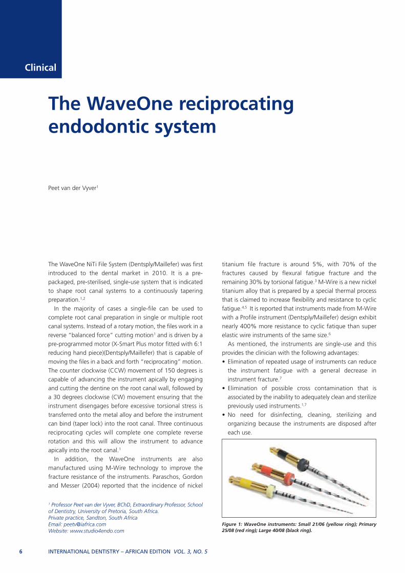
ready for the next cutting cycle.
The rubber stop is again adjusted to a further 3mm longer
or up to the point of the predetermined working length
(Figure 10a). Again, the objective of the second cutting cycle
will be to only cut with the instrument until the rubber stop
reaches the cusp reference point (Figure 10b).
The same protocol is followed after each cutting cycle as
outlined above until the pre-determined working length of
the root canal is reached.
When multiple canals are prepared in the same tooth a
slightly different protocol is followed. For example, a root
canal preparation on a lower molar where 3 root canals are
prepared the sequence will be as follows: Measure the depth
of the selected file penetration into the three root canals.
Record the shortest length, move the rubber stop 3mm and
complete a cutting cycle in the mesio-buccal canal. Remove
the instrument and clean the flutes. Now proceed with a
cutting cycle in the mesio-lingual canal, clean the flutes and
then complete a cutting cycle in the distal root canal. This
sequence is followed with irrigation and recapitulation of all
three root canals before moving onto the next cutting cycle.
Guideline 8 – Faster preparation time necessitateslonger irrigation times preferably with activation ofirrigation solutionChemo-mechanical debridement that allows elimination of
pulpal tissue, microbiota and their by-products, and organic
and inorganic debris removal by using mechanical
instruments and intracanal irrigation solutions is one of the
Figure 10: (a) The rubber stop is again adjusted to a further 3mmlength or up to the point of the predetermined working length; (b)The file is again activated and allowed to prepare the root canal untilthe rubber stop reaches the reference point.
10a 10b
Figure 11: (a) The EndoActivator (Dentsply/Maillefer); (b) to sonically activate irrigation solutions in thepreparared root canal systems.
11b11a
INTERNATIONAL DENTISTRY – AFRICAN EDITION VOL. 3, NO. 5 13

van der Vyver
phenomenon.26,27 According to Caron (2007) this activation
technique is capable to clean debris from lateral canals,
remove the smear layer, and dislodge clumps of simulated
biofilm within curved canals of molar teeth.28
Guideline 9 – Guidelines for obturationSome articles recommend that the final shape can be
confirmed when the apical flutes of the instrument are
loaded with dentine debris.11 However, in the author’s clinical
experience this is not always a reliable method although it
can be used as a guideline.
Gauging the apical foramen with a corresponding NiTi
hand file is another alternative. For example, if the final canal
preparation was done with a Primary WaveOne 25/08
instrument a size 25/02 NiTi hand file is fitted into the
prepared canal (Figure 12a). If the tip of the file is snug at
length the final shape is confirmed and a matching
WaveOne Primary Gutta-percha Point (Dentsply/Maillefer) is
used for obturation (Figure 12b).
If the 25/02 NiTi hand file is loose at length, or can be
pushed past working length it means that the apical
foramen is larger than 0.25mm (Figure 13a). In these cases
it is recommended to gauge the foramen with a size 30/02
NiTi hand file (Figure 13b). If this instruments is snug at
working length the shape is confirmed to an ISO size 30. It
is also possible to use a ProTaper Universal F3
(Dentsply/Maillefer) or ProTaper Next X3 (Dentsply/Maillefer)
instrument to working length in order to transform the final
apical shape to a size 30. Thereafter, a matching gutta-
percha point can be used for obturation (Figure 13c).
in the debridement and disinfection into all aspects of root
canal systems, including dentinal tubules, lateral canals, fins,
webs and anastomoses.25 The author would suggest to use
the EndoActivator (Dentsply/Maillefer) to activate irrigation
solutions after root canal preparation with the WaveOne
reciprocating instruments.
The EndoActivator (Dentsply/Maillefer) (Figure 11a) is a
sonically driven root canal irrigation activation device. It
consists of a battery operated portable hand piece and
different sizes of disposable, strong, flexible polymer tips.
Sonically vibrating the polymer tip, in combination with
moving the tip up and down in short vertical strokes (Figure
11b), synergistically produces a powerful hydrodynamic
14 INTERNATIONAL DENTISTRY – AFRICAN EDITION VOL. 3, NO. 5
Figure 12: (a) Size 25 /02 NiTi hand file (Dentsply/Maillefer) is fitted intothe prepared canal to gauge the apical foramen. If the tip of the file issnug at length the final shape is confirmed; (b) A matching WaveOnePrimary Gutta-Percha Point (Dentsply/Maillefer) is selected for obturation.
12a 12b
Figure 13: (a) If the 25/02 NiTi hand file is loose at length, or can be pushed past working length it indicates that the apicalforamen is larger than 0.25mm; (b) Apical foramen is gauged with a size 30/02 NiTi hand file. If this instrument is snug atworking length the shape is confirmed to an ISO size 30; (c) ProTaper F3 or a ProTaper Next X3 gutta-percha point(Dentsply/Maillefer) can be used for obturation.
13a 13b 13c

van der Vyver
primary or large) that can be used for single cone obturation
or for the continuous wave of condensation using the Calamus
Dual (Dentsply/Maillefer) or System B (Sybron Endo) units.
The second option is to use the matching WaveOne
obturators (Dentsply/Maillefer) heated up in the Thermaprep
Plus oven (Dentsply/Maillefer). The last option will be to
use GuttaCore cross linked gutta-percha obturators
(Dentsply/Maillefer). With this technique the canals has to be
verified again in order to select the size of obturator for each
prepared root canal. A NiTi verifier (available in size 20, 25, 30,
35, 40 ,45 and 50) is used to verify the canal shape (Figure 15a).
The verifier must be fitted into a wet canal, fit snug up to the
working length, and feel loose in the canal. If the verifier is too
tight, the GuttaCore obturator will not be able to fill the root
canal up to the prepared canal length (Figure 15b). Figure 15c
illustrates the final result after obturation with size 025 (mesial
canals) and size 040 (distal canal) GuttaCore Obturators.
If the size 30/02 NiTi hand file is loose at length or can be
pushed past working length (Figure 14a) the clinician could
proceed to use a WaveOne Large (40/08) instrument (Figure
14b) to complete shape in the apical portion of the root
canal. The terminal size is again gauged with a 40/02
instrument (Figure 14c) using the same protocol as described
above to confirm the final shape and a matching gutta-
percha coned is selected for obturation (Figure 14d).
If the 40/02 NiTi hand file is still loose at length it is
recommended to proceed to a ProTaper Universal F5 (50/06)
or ProTaper Next X5 (50/06). The terminal size is again
gauged with a 50/02 instrument using the same protocol as
described above to confirm the final shape. If the 50/02 NiTi
hand file is still loose at length it is recommended to pack
MTA in order to obtain an adequate apical seal.
Obturation can be done in several ways. The first option is
to use matching WaVeOne gutta-percha cones (sizes small,
16 INTERNATIONAL DENTISTRY – AFRICAN EDITION VOL. 3, NO. 5
Figure 14: (a) Size 30/02 NiTi hand file is loose at length or can be pushed past working length, (b) WaveOne Large (40/08)instrument is used to complete shape in the apical portion of the root canal, (c) The terminal size is again gauged with a40/02 instrument using the same protocol as described above to confirm the final shape, (d) A WaveOne Large gutta-perchapoint is selected for obturation.
14a 14b 14c 14d
Figure 15: (a) GuttaCore NiTi verifiers (Dentsply/Maillefer) are used to verify the final canal shape; (b) GuttaCorecrosslinked gutta-pecha obturators used for root canal obturation); (c) Final radiographic result after root canalobturation.
15a 15b 15c
van der Vyver
instrument can be pushed past working length it is
recommended to continue the shaping with the primary
25/08 instrument.
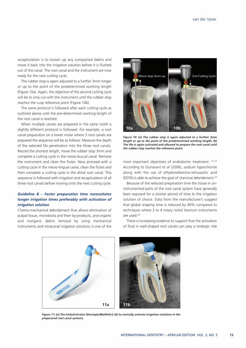
Case Report 1The patient, a 40 year old female presented with a non-vital
maxillary left central incisor (Figure 16a). Root canal
preparation was done with a WaveOne Large 40/08
instrument. Figure 16b shows the final result after obturation
with a size 40 GuttaCore obturator.
Case Report 2The patient, a 45 year old male presented with pain on his
maxillary right second premolar. The pre-operative
radiograph revealed a very long root, with a possible root
canal bifurcation towards apical third of the root (Figure
17a). Root canal preparation was done with a WaveOne
Small 21/06 instrument and obturated with two size 20
GuttaCore obturators (Figure 17b).
Case Report 3The patient, a 32 year old female presented with history of
poorly done root canal treatment on her mandibular right
second premolar (Figure 18a). After removal of the previous
gutta-percha, the root canal preparation was done with a
WaveOne Primary 25/08 instrument. Figure 18b
demonstrates the final result after the canal was obturated
with a Primary WaveOne gutta-percha cone using the
Calamus Dual Obturation unit. A core build-up was done
using a size red X-Post (Dentsply/Maillfer) and Core-X Flow
(Dentsply/Maillefer) core build-up material.
It must be noted that the 21/06 Small WaveOne
instrument should be used in root canals that are very long,
root canals that have severe curvatures in the apical third
and any challenging root canal system eg. complex second
mesio-buccal canals on maxillary molars or S-Shaped canal
curvatures in maxillary premolars. According to Ruddle
(2012) the 21/06 instrument can also be used as a “bridge
file” in the above mentioned cases before the Primary 25/08
file is used for final shape.11 However, in some cases the
clinician might decide to accept the shape created by the
21/06 instrument. The apical diameter will then be gauged
with a 20/02 K-File and if the file is snug at length the canal
can be obturated with a matching WaveOne Small gutta-
percha cone or size 20 GuttaCore obturator. If the 20/02
18 INTERNATIONAL DENTISTRY – AFRICAN EDITION VOL. 3, NO. 5
Figure 16: (a) Pre-operative radiograph of non-vital maxillary leftcentral incisor; (b) Post-operative radiograph after root canalpreparation was done with a WaveOne Large 40/08 instrument andthe canal obturated with a size 40 GuttaCore obturator.
16a 16b
Figure 17: (a) Pre-operative radiograph of maxillary right secondpremolar. Note the length of the root canal (25mm) and disappearanceof the main root canal towards apical third of the root indicating apossible root canal bifurcation; (b) Post-operative radiograph showingthe result after root canal preparation was done using a WaveOne Small21/06 instrument and obturated with size 20 GuttaCore obturators.
17a 17b
Figure 18: (a) Pre-operative radiograph of mandibular right secondpremolar with a history of previous poorly done root canal treatment;(b)Post-operative radiograph after root canal preparation was redonewith a WaveOne Primary 25/08 instrument. The canal was obturatedwith Primary WaveOne Gutta-Percha Cone (Dentsply/Maillefer) usingthe Calamus Dual Obturation Unit (Dentsply/Maillefer). A core build-up was done using a size red X-Post (Dentsply/Maillefer) and Core-XFlow (Dentsply/Maillefer) core build-up material.
18a 18b

van der Vyver
instruments. Reducing the number of instruments during
root canal preparation can also reduce or eliminate
procedural errors.1
5.Root canal preparation becomes more cost effective since
most cases can be prepared with a single WaveOne
instrument.
6.The “reciprocating” root canal instrumentation technique
requires a very short learning curve for new users in order
to master this technology.
References1. Webber J, Machtou P, Pertot W, Kuttler S, Ruddle C,
West J. The WaveOne single-file reciprocating system. Roots
2011; 1: 28-33.
2. Van der Vyver PJ. WaveOne Instruments: Clinical
application guidelines. Endodontic Practice Nov 2011: 45-54.
3. Parashos P, Gordon I, Messer HH. Factors influencing
defects of rotary nickel-titanium endodontic instruments
after clinical use. J Endod 2004; 30: 722-72.
4. Gambarini G, Grande NM, Plotino G, Somma F, Garala
M, De Luca M, Testarelli M. Fatigue resistance of engine-
driven rotary nickel-titanium instruments produced by new
manufacturing methods. J Endod 2008; 34: 1406-9.
5. Shen Y, Zhou MH, Zheng YF, Peng B, Haapasalo M.
Current challenges and concepts of the thermomechanical
treatment of nickel-titanium instruments. J Endod 2013; 39:
163-72.
6. Johnson E, Lloyd A, Kuttler S, Namerow K. Compasrison
between a novel nickel-titanium alloy and 508 nitinol on the
cyclic fatique life of profile 25/.04 rotary instruments. J
Endod 2008; 34: 1406-9.
7. Yared G. Canal preparation using only one Ni-Ti rotary
instrument: preliminary observations. Int Endod J 2008; 41:
339-44.
8. Bűrklein S, Hinschitza K, Dammaschke T, Schäfer E.
Shaping ability and cleaning effectiveness of two single-file
systems in severely curved root canals of extracted teeth:
Reciproc and WaveOne versus Mtwo and ProTaper. Int Endod
Journal 2012; 45: 449-61.
9. Varela-Patiňo P, Martin Biedma B, Rodriguez N,
Cantatore G, Malentaca A, Ruiz-Pinon M. Fracture rate of
nickel-titanium instruments using continuous versus
alternating rotation. Endodontic Practice Today 2008; 2:
193-7.
10. Sattapan B, Palmara JE, Messer HH. Torque during
canal instrumentation using rotary nickel-titanium files. J
Endod 2000; 26: 156-60.
11. Ruddle CJ. Endodontic canal preparation: WaveOne
single-file technique. Dentistry Today 2012: 124-29.
12. Buchanan LS. Management of the curved root canal.
Case Report 4The patient, a 51 year old female presented with non-vital
first and second maxillary right premolars (Figure 19a). Root
canal preparations on both teeth were done with one
WaveOne Primary 25/08 instrument. Figure 19b
demonstrates the final result after the canals were obturated
with Primary WaveOne gutta-percha cones using the
Calamus Dual Obturation Unit.
Conclusions1.Reciprocating root canal instruments offer the clinician a
few distinct advantages. There is less binding of the
instruments into the root canal wall with a reduction in
torsional stress. Because the number of cycles of rotation
is reduced during canal preparation, there is also less
flexural (bending) stress on the instrument. Both these
advantages provide the operator with a decreased risk of
instrument fracture.
2. In most cases only one reciprocating instrument of the
WaveOne system is required to prepare single or multiple
canal systems in a tooth. In general the total preparation
time for shaping a root canal system in a tooth is
decreased.
3.Faster mechanical root canal preparation with the
WaveOne system allows the clinician to spend more time
on chemical debridement. Activation of the irrigation
solutions with the EndoActivator is recommended.
4.The WaveOne instruments are manufactured by using M-
Wire technology that provide the clinician with more
safety against instrument fracture. However, it must be
taken into consideration that a single WaveOne file does
the work of three or more rotary nickel titanium
Figure 19: (a) Pre-operative radiograph of non-vital first and secondmaxillary right premolars; (b) Post-operative radiograph after the rootcanal preparations on both teeth were done with one WaveOnePrimary 25/08 instrument and obturated with Primary WaveOneGutta-Percha Cones (Dentsply/Maillefer) using the Calamus DualObturation Unit (Dentsply/Maillefer).
19a 19b
INTERNATIONAL DENTISTRY – AFRICAN EDITION VOL. 3, NO. 5 19
van der Vyver
after manual and mechanical glide path. A randomized
clinical trial. J Endod 2012; 38: 32-6.
21. Clegg MS, Verucci FJ, Walker C, Belanger M, Britto LR.
The effect of exposure to irrigant solutions on apical dentin
biofilms in vitro. J Endod 2006; 32: 434-37.
22. Desai P, Himel V. Comparative safety of various
intracanal irrigation systems. J Endod 2009; 35: 545-49.
23. Dunavant TR, Regan JD, Glickman GN, Solomon ES,
Honeyman AL. Comparative evaluation of endodontic
irrigants against Enterococcus faecalis biofilms. J Endod
2006; 32: 527-31.
24. WaveOne – single file technique with reciprocation.
www.tulsadentalspecialities.com/default/endodontics_brand
s/WaveOne _systems.aspx.
25. Yamada RS, Armas A, Goldman M, Lin PS. A scanning
electron microscopic comparison of a high volume final flush
with several irrigating solutions: Part 3. J Endod 1983; 9:
137-42.
26. Ruddle C. Endodontic disinfection: tsunami irrigation.
Endod Practice 2008: 7-15.
27. Gu L, Kim JR, Ling J, Choi KK, Pashley D, Tay F. Review
of contemporary irrigant agitation techniques and devices. J
Endod 2009; 35: 791-804.
28. Caron G. Cleaning efficiency of the apical millimetres
of curved canals using three different modalities of irrigant
activation: an SEM study. Paris VII University, Paris France:
Masters thesis 2007.
J Calif Dent Assoc 1989; 17: 18-27.
13. West JD. The endodontic glidepath: secret to rotary
safety. Dentistry Today 2008; 29: 7-15.
14. Berutti E, Negro AR, Lendini M, Pasqualini D. Influence
of manual preflaring on torque on the failure rate of
ProTaper rotary instruments. J Endod 2004; 30: 228-30.
15. Berutti E, Cantatore G, Castellucci A, Chiandussi G,
Pera F, Migliaretti D, Pasqualini D. Use of nickel-titanium
rotary Pathfile to create a glide path: comparison with
manual preflaring in simulated root canals. J Endod 2009;
35: 408-12.
16. Van der Vyver PJ. Creating a glide path for rotary NiTi
instruments: Part one. Endod Practice 2011: 40-3.
17. Van der Vyver PJ. Creating a glide path for rotary NiTi
instruments: part two. Endod Practice 2011: 46-53.
18. Pasqualini D, Bianchi CC, Paolino DS, Mancini L,
Cemenasco A, Cantatore G, Castellucci A, Berutti E.
Computed micro-tomogrpahic evaluationof glide path with
nickel-titanium Pathfile in maxillary first molar curved canals.
J Endod 2012; 38: 389-93.
19. Greco K, Carmignani E, Cantatore G. A comparative
study between manual and mechanic preflaring techniques.
Papers presented to the Fithteen Biennial Congress of the
European Society of endodontology; 2011 Sept 14-17;
Rome Italy.
20. Pasqualini D, Mollo L, Scotti N, Cantatore G,
Castellucci A, Migliaretti G, Berutti E. Postoperative pain
20 INTERNATIONAL DENTISTRY – AFRICAN EDITION VOL. 3, NO. 5