Hepatobiliary Imaging by Multidetector Computed Tomography ...

Page 1 of 17
The value of dual-energy multidetector computedtomography as an adjunct to conventional investigations inpregnancy losses: Initial findings
Poster No.: R-0172
Congress: 2014 CSM
Type: Scientific Exhibit
Authors: S. Constantine, L. Moore, S. Brown; NORTH ADELAIDE/AU
Keywords: Foetal imaging, Forensic / Necropsy studies, CT, Experimental,Computer Applications-Virtual imaging, Experimentalinvestigations, Congenital, Fetus, Forensics
DOI: 10.1594/ranzcr2014/R-0172
Any information contained in this pdf file is automatically generated from digital materialsubmitted to EPOS by third parties in the form of scientific presentations. Referencesto any names, marks, products, or services of third parties or hypertext links to third-party sites or information are provided solely as a convenience to you and do notin any way constitute or imply RANZCR/AIR/ACPSEM's endorsement, sponsorshipor recommendation of the third party, information, product or service. RANZCR/AIR/ACPSEM is not responsible for the content of these pages and does not make anyrepresentations regarding the content or accuracy of material in this file.As per copyright regulations, any unauthorised use of the material or parts thereof aswell as commercial reproduction or multiple distribution by any traditional or electronicallybased reproduction/publication method ist strictly prohibited.You agree to defend, indemnify, and hold RANZCR/AIR/ACPSEM harmless from andagainst any and all claims, damages, costs, and expenses, including attorneys' fees,arising from or related to your use of these pages.Please note: Links to movies, .ppt slideshows, .doc documents and any other multimediafiles are not available in the pdf version of presentations.

Page 2 of 17
Aim
The use of computed tomography (CT) scanning as part of the post-mortem examinationof adults is now well established [1]. The coronial departments in many industrializedcountries, including Australia, regularly utilize both CT and MRI (magnetic resonanceimaging) as part of their death investigations [2]. MRI is useful in the investigation of fetalabnormalities, particularly in the examination of the neural axis and soft tissues, but hasseveral limitations. The availability of MRI is limited in Australia as a result of Medicarelicensing restrictions. A comprehensive examination with MRI is also time consuming. AsMRI signal relies upon the movement of free hydrogen atoms, signal quality is reducedin the post mortem patient due to cooling and/or refrigeration.
A recent article by O'Donoghue et al [3] used multidetector computed tomography
(MDCT) as an adjunct to autopsy in the investigation of 3rd trimester stillbirths, findinggood correlation between measurements obtained in both settings. CT scanning in fetaldemise can avoid several MRI limiting factors. All but the most remote communities haveaccess to CT scanners, and the relative speed with which a scan can be performedallows scanning quickly after delivery. Body temperature is also not important, allowingCT scanning to be performed at any stage after fetal demise.
We aim to investigate the value of dual-energy MDCT as part of the investigation into
fetal abnormalities and pregnancy losses in the 2nd and 3rd trimesters, and early neonatalperiod. We present our initial findings, after the evaluation of six cases.
Methods and materials
The study was approved by the Women's and Children's Hospital Network HumanResearch Ethics Committee. Patients were recruited while giving consent forconventional autopsy. Fetuses were scanned as soon as possible after delivery. Thefetuses were scanned as they arrived in the Department of Surgical Pathology, wrappedin surgical drapes, and were not repositioned for the scan. A dual-energy CT techniquewas utilized, to maximise contrast resolution.
Each fetus was imaged twice using a single source Spectral Dual Energy CT ScanningSystem.

Page 3 of 17
Firstly, a high definition helical scan was completed, permitting the maximum spatialresolution possible for multiplanar bone and 3D volume rendered skeletal imaging (3Dskeleton). This was followed by a dual energy acquisition. Both high and low energy(kVp) data sets are acquired simultaneously for near perfect anatomical registration. Thedual energy image set was evaluated for the optimal image contrast and image noise.The optimal monochromatic energy level (keV) was concluded and a new image set wascreated for multiplanar evaluation of soft tissue organs. Radiation dose was, of course,not a significant consideration.
A conventional autopsy was performed on each fetus after CT scanning by anexperienced paediatric/neonatal pathologist. The CT scans were analysed by a perinatalradiologist (first author) together with a paediatric/neonatal pathologist (second author).The clinical details (except gestational age) were withheld. The fetal biometry as wellas findings relating to an external examination and internal organs were measured andrecorded. The CT findings were then compared with the autopsy result, which wasconsidered the "gold standard".
Results
Six fetuses were initially scanned. Three fetuses had been delivered after death in utero,and three were from pregnancies terminated due to fetal abnormalities ( Table 1 on page5 ).
Fetal Measurements and External Examination.
In all but one fetus, the crown-rump and crown-heel length measurements determinedon CT were within 6% of the autopsy measurements. There was quite poor correlationbetween CT and autopsy measurements of the fetal foot length and head measurements,which we believe is due to fetal positioning: the foot can be more easily "straightened"during a direct examination.
The genitalia were correctly identified on CT in three fetuses. The genitals were notassessed in two fetuses due to fetal position/umbilical clip position; in fetus 4 the genitaliawas thought to be male on CT but were ambiguous at autopsy examination. The earswere clearly seen in four fetuses on CT ( Fig. 1 on page 6 ), and were mentionedas present in five fetal autopsies. The eyes were identified in all fetuses, and the lenseswere seen in five cases on CT ( Fig. 2 on page 6 ). The eyes were seen at five of theautopsy examinations. The hard palate was correctly identified as intact in all fetuses (

Page 4 of 17
Fig. 3 on page 7 ), the lip was seen to be intact in five fetuses ( Fig. 1 on page 6) and was not able to be assessed in fetus 6.
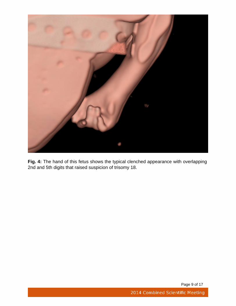
Five digits were correctly identified on every limb of each fetus. The hands were identifiedas being clenched in fetus 3 on CT scanning ( Fig. 4 on page 8 ), which was confirmedat autopsy. The foot position was thought to be normal in all fetuses on both CT scanningand at autopsy.
Fetal Skull and Skeleton.
Specific examination of the skeleton was not carried out at autopsy of these fetusesbecause there was no suspicion of a skeletal dysplasia.
CT showed twelve pairs of ribs in every fetus, and there were no fractures. The symphysismenti was distracted in fetus 6, and a hemi-vertebra was identified in fetus 4 ( Fig. 5 onpage 9 ). The skull was collapsed with overlapping bones in fetuses 2 and 6 ( Fig.6 on page 10 ), this was not assessable on autopsy in fetus 2 and was confirmed infetus 6.
Intracranial Structures.
A normal midline was identified on the CT scans in all fetuses. Ventriculomegaly wascorrectly diagnosed in fetus 3 ( Fig. 7 on page 11 ), and was not assessable in twofetuses on CT scanning or autopsy due to autolysis/maceration. Extra-axial haemorrhagewas correctly identified in fetus 3.
Cardiorespiratory System.
Evaluation of the heart was poor on CT, as was expected in a non-contrast post-mortemstudy. The heart was correctly identified as normal in fetus 5, but much of the heartcould not be commented upon, and even the great vessels were difficult to confidentlyidentify. Significant cardiac abnormalities were identified in fetuses 2 and 3 at autopsy.The diaphragm could be seen to be intact in four of the fetuses at CT scanning ( Fig. 8on page 12 ), and was intact in all fetuses at autopsy.
Abdominal Organs.

Page 5 of 17
The liver and spleen were identified in the correct position in four fetuses, and were notable to be assessed in two fetuses on CT scanning. The kidneys proved very difficult toidentify due to the absence of intra-abdominal fat. In the three fetuses they were identifiedon CT scanning ( Fig. 9 on page 13 ), the kidneys were normal at autopsy. Thekidneys were not identified in fetus 4 on scanning, and at autopsy the renal parenchymawas multicystic dysplastic in nature and horseshoe in morphology. The gallbladder andpancreas were not reliably seen in any fetus on CT scanning.
Diagnosis in Each Fetus.
After "CT autopsy" in fetuses 1, 2, 5 and 6, no anatomical abnormality was detected.This correlated with the autopsy findings for fetuses 1, 2 and 6. At autopsy, fetus 5 wasshown to have grey matter heterotopia and schizencephaly, as suspected on antenatalultrasound and MRI scanning. This was not visible on post mortem CT scanning.
Fetus 3 was suspected to have trisomy 18, which was confirmed on cytogenetic testing.The abnormalities detected on CT scanning were the clenched hands and dilatedcerebral ventricles. The cardiac abnormalities were not identified.
Fetus 4 was suspected to have the "VACTERL" sequence, due to the hemivertebra andinability to visualize the kidneys on CT scanning. The autopsy also found an imperforateanus.
Comments were made in fetuses 2 and 6 with regards to oedema, gas in the soft tissuesand lack of intracranial detail, which was interpreted as maceration/autolysis ( Fig. 10 onpage 14 ). This was confirmed at autopsy.
Images for this section:

Page 6 of 17
Table 1: Demographic features of the six fetuses scanned.
Fig. 1: Surface reconstruction in a 38 week fetus. The ear is well seen on the right. Thelip is intact.

Page 7 of 17
Fig. 2: Oblique reconstructed image in a 22 week fetus. The globe is normally positionedin the orbit and the lens in clearly seen (arrow).

Page 8 of 17
Fig. 3: The open mouth of this fetus allows the intact palate to be seen.
Page 9 of 17
Fig. 4: The hand of this fetus shows the typical clenched appearance with overlapping2nd and 5th digits that raised suspicion of trisomy 18.

Page 10 of 17
Fig. 5: 3D bony reconstruction of fetus 4. The spine in unbalanced due to a hemivertebra(arrow).

Page 11 of 17
Fig. 6: Axial image of the skull of fetus 6. There is significant collapse with overlappingof the calvarial bones.

Page 12 of 17
Fig. 7: Axial CT image of fetus 3. There is dilatation of the right lateral ventricle (arrow).

Page 13 of 17
Fig. 8: Coronal CT reconstruction of the fetal torso. The diaphragms are clearly seen(arrows).

Page 14 of 17
Fig. 9: Coronal CT reconstruction of the abdomen in fetus 5. The arrows show thesuperior and inferior poles of each kidney.

Page 15 of 17
Fig. 10: Axial CT image of the brain of fetus 6. There are almost no recognisableintracranial structures.

Page 16 of 17
Conclusion
Unexplained stillbirths account for more than 25% of all fetal deaths, and have multiplecauses [3]. Autopsy has been shown to provide important information about thepregnancy that was not available antenatally in at least a third of cases [4]. There isreluctance on the part of the treating doctors to ask for consent for autopsy, and somegrieving parents are unable to consent for variety of reasons. This leaves many parentsat a clear disadvantage when being counselled for future pregnancies, which previouslycould not be avoided.
A recent study by Cannie et al [4] found that MR virtual autopsy was almost universallyaccepted by parents compared with an acceptance rate of around 2/3 for conventionalautopsy. Although the specific reasons for acceptance were not examined, there was acorrelation with the mother's religious background, with Muslim parents far less likely toconsent to conventional autopsy.
While our preliminary scans did not detect all fetal abnormalities, some valuableinformation was obtained, and there are some features such as bony abnormalities,which can be difficult to elucidate at autopsy. We suggest that fetal CT scanning canbe a useful adjunct to conventional autopsy, and in cases where fetal autopsy is notappropriate, fetal CT scanning can still provide useful additional information to assist withparent counselling.
Scanning is best performed as soon as possible after delivery to maximise tissuecontrast, and does not significantly delay funeral/burial preparations. The potential use ofintravenous contrast may also assist in providing further diagnostic information. It is likelythat dual-energy MDCT will be useful in pregnancy losses where conventional autopsyis not appropriate.
Personal information
Dr Sarah Constantine MBBS, FRANZCR
Perinatal Radiologist
Women's and Children's Hospital, South Australia
Associate Professor Lynette Moore BMBS, FRCPA

Page 17 of 17
Paediatric Pathologist
Women's and Children's Hospital, South Australia
Mr Scott D Brown Grad Dip Med Rad, MIR
Head Radiographer, Computed Tomography
Women's and Children's Hospital, South Australia
References
1. Post-mortem imaging as an alternative to autopsy in the diagnosis of adultdeaths: a validation study. Roberts IS, Benamore RE, Benbow EW et al.Lancet 2012 Jan 14; 379(9811): 136 - 42.
2. Advances of dual source, dual-energy imaging in postmortem CT. PerssonA, Jackowski C, Engström E, Zachrisson H. Eur J Radiol 2008 Dec; 68(3):446 - 55.
3. O'Donoghue K, O'Regan KN, Sheridan CP et al. Investigation of the role ofcomputed tomography as an adjunct to autopsy in the evaluation of stillbirth.Eur J Radiol 2012 Jul; 81(7): 1667 - 75.
4. Acceptance, reliability and confidence of diagnosis of fetal and neonatalvirtuopsy compared with conventional autopsy: a prospective study. CannieM, Votino C, Moerman P et al. Ultrasound Obstet Gynecol 2012 Jun; 39(6):659 - 65.