The use of photographic color information for high ...
13
2019 40 7 ar rt. t 40 (7 (7) ) — — Pu Pu P bl b ishe ed d on o 1 16 6 6 6 Se Se Se Septem embe ber 20 20 019 19 19 ww www.cry ypto oga amie.co c m m/algo gol lo l l gie Algologie cryptogamie Will H. RYAN, Sabrina HEISER, Michelle D. CURTIS, Charles D. AMSLER, Till BAYER, Guido BONTHOND, Gaoge WANG, Florian WEINBERGER & Stacy A. KRUEGER-HADFIELD The use of photographic color information for high-throughput phenotyping of pigment composition in Agarophyton vermiculophyllum (Ohmi) Gurgel, J.N.Norris & Fredericq
Transcript of The use of photographic color information for high ...

2019 40 7
arrt.t 40 (7(7) ) —— PuPuP blb isheedd ono 116 66 6 SeSeSeSeptemembeber 20200191919wwwww.cryyptoogaamie.coc mm/algogolloll gie
Algologiecryptogamie
Will H. RYAN, Sabrina HEISER, Michelle D. CURTIS, Charles D. AMSLER, Till BAYER, Guido BONTHOND, Gaoge WANG, Florian WEINBERGER &
Stacy A. KRUEGER-HADFIELD
The use of photographic color information for high-throughput phenotyping
of pigment composition in Agarophyton vermiculophyllum
(Ohmi) Gurgel, J.N.Norris & Fredericq

Cryptogamie, AlgologieCryptogamie, Algologie is a fast track journal published by the Museum Science Press, Paris
The Museum Science Press also publishes: Adansonia Geodiversitas Zoosystema Anthropozoologica European Journal of Taxonomy Naturae Cryptogamie sous-sections Bryologie, Mycologie.
print electronic
DIRECTEUR DE LA PUBLICATION
RÉDACTEUR EN CHEF / EDITOR-IN-CHIEF : LINE LE GALL
ASSISTANT DE RÉDACTION / ASSISTANT EDITOR
MISE EN PAGE PAGE LAYOUT
RÉDACTEURS ASSOCIÉS / ASSOCIATE EDITORS
Ecoevolutionary dynamics of algae in a changing worldAnna GOHDE
Stacy KRUEGER-HADFIELD
Jana KULICHOVA
Cecilia TOTTI
Phylogenetic systematics, species delimitation & genetics of speciationSylvain FAUGERON
Marie-Laure GUILLEMIN
Diana SARNO
Comparative evolutionary genomics of algaeNicolas BLOUIN
Heroen VERBRUGGEN
Algal physiology & photosynthesisMario GIORDANO
Janet KÜBLER
Prokaryotic algaeNico SALMASO
Vitor VASCONCELOS
Cryptogamie, Algologie Algologie is indexed in:
Cryptogamie, Algologie Algologie is distributed electronically by:®

73
The use of photographic color information for high-throughput phenotyping of pigment composition in Agarophyton vermiculophyllum (Ohmi) Gurgel, J.N.Norris & Fredericq
Will H. RYAN Sabrina HEISER
Michelle D. CURTIS Charles D. AMSLER
Till BAYER Guido BONTHOND
Gaoge WANG
and
Florian WEINBERGER
Stacy A. KRUEGER-HADFIELD
Agarophyton vermiculophyllum . Crypto gamie, Algologie
ABSTRACTPigment variation within and among algal species may have important ecological consequences be-cause small changes in the concentration and composition of pigments can influence the photosyn-thetic efficiency and rate as well as the spectra of light utilized. Toward the goal of developing a rapid method for comparing pigment composition among algal thalli, we characterized the relationship between visual color information taken from photographs (e.g., red, green, and blue color values) and photopigment composition in the non-native red alga Agarophyton vermiculophyllum (Ohmi) Gurgel, J.N.Norris & Fredericq. We used a set of 19 thalli, collected from across the known native and non-native range in the Northern Hemisphere, which exhibited substantial color variation at the time of field collection, and sustained this variation after being maintained in a common garden. We identified a set of ecologically interesting pigment traits that are readily predicted by color information, including chlorophyll a and phycobilin concentration. Finally, we demonstrated the repeatability of estimating color phenotypes from photographs of thalli taken under a range of light conditions in order to evaluate the utility of this approach for field studies. We suggest this method could be useful for the rapid, high-throughput phenotyping of photopigments in other red algae as well.
KEY WORDSintraspecific variation,
ecology,photopigments,
phycobilin proteins,Rhodophyta.

conditions, pigment variation could be a key trait for predicting responses under climate change or during range expansion. Thus, there is an impetus for these data to be gathered more routinely during ecological studies.
Photopigment composition can also be highly dynamic within individuals. Thalli can respond to changes in irradiance, spectral quality, and other environmental variables by rapidly modulating the concentration and composition of accessory pigments (e.g., Rosenberg & Ramus 1982; Beach & Smith 1996; Grossman 2003; Kim et al. 2015). Such rapid responses suggest that changes in pigments may serve an acclimatory function over the course of a day, or across seasons for algae living in highly dynamic light environments, such as the intertidal zone (López-Figueroa 1992; Beach & Smith 1996; reviewed by Falkowski & Chen 2003). Measuring pigment variation through time is essential to our understanding of the role that plasticity plays in local acclimation, yet tracking changes in individual thalli is difficult without non-destructive protocols for characterizing pigments.
In order to incorporate pigment data into ecological and biogeographic studies, however, these data may need to be gathered from hundreds of sampled specimens (i.e., high throughput phenotyping), or under field conditions. Pigment extractions require destructive sampling, which may limit the use of tissues for characterizing additional traits (e.g., protein content) or evaluating changes at the individual level through time. Moreover, extractions are time consuming and require laboratory equipment (e.g., a spectrophotometer and liquid nitrogen), making it difficult to generate these data in the field, or for a large number of samples in a short time period. Nevertheless, intraspecific variation in phenotypic traits, including variation in pigment composition, may be
INTRODUCTION
Light-sensitive pigments, also known as photopigments, play a key role in defining the ecological niche of photosynthetic organisms (Falkowski & Raven 2007). The composition of photopigments determines the wavelengths of light an organ-ism can use for photosynthesis (Lemasson et al. 1973; Gantt 1981). Additional accessory pigments may also protect sen-sitive photosynthetic machinery against damage from high intensity visible or UV radiation (Siefermann-Harms 1985; Ben-Amotz et al. 1989; Bailey & Grossman 2008). Thus, variation in pigment composition might act as a key predictor of performance under differing environmental conditions.
Visible color variation has been linked to heritable differences in the concentration and relative composition of phycobilin pigments in color mutants found in both natural and lab-reared populations (e.g., Porphyra yezeoensis Ueda: Niwa et al. 2009; Chondrus crispus Stackhouse: van der Meer 1981; Cornish et al. 2013; Gracilaria tikvahiae McLachlan: Kursar et al. 1983; Gracilaria birdiae E.M.Plastino & E.C.Oliveira: Plastino et al. 2003; Costa & Plastino 2011). In some cases, pigment mutations have been linked to changes in growth rates under laboratory conditions (Plastino et al. 2003; Yokoya et al. 2007; Kim et al. 2015; but see also Ramus & van der Meer 1983), supporting the potential for pigment traits to be shaped by natural selection. Indeed, variation in pigment composition has long been recognized as an important driver of algal niche diversity at higher taxonomic levels, offering a potential mechanistic explanation of light resource niche partitioning (Stomp et al. 2007; Stockenreiter et al. 2013). Because heritable variation in pigment composition may be an important target for selection under changing environmental
74
Ryan et al.
RÉSUMÉUtilisation d’informations photographiques sur les couleurs pour le phénotypage à haut débit de la com-position de pigments chez Agarophyton vermiculophyllum (Ohmi) Gurgel, J.N.Norris & Fredericq.La variation intra et interspécifique des pigments au sein des espèces d’algues et entre celles-ci peut avoir des conséquences écologiques importantes, car de faibles variations dans la concentration et la composition des pigments peuvent influer sur l’efficacité et la vitesse de la photosynthèse, ainsi que sur le spectre de la lumière utilisée. Afin de développer une méthode rapide de comparaison de la composition en pigment parmi les thalles d’algues, nous avons caractérisé la relation entre les informations de couleur visuelles tirées de photographies (valeurs des couleurs rouge, verte et bleue) et la composition de photopigment dans l’algue rouge non native Agarophyton vermiculophyllum (Ohmi) Gurgel, J.N.Norris & Fredericq. Nous avons utilisé un ensemble de 19 individus, provenant de l’aire de répartition connue des autochtones et des non-autochtones de l’hémisphère Nord, qui présentait une variation de couleur substantielle au moment de la récolte sur le terrain et a maintenu cette variation après avoir été placé dans un jardin commun. Nous avons identifié un ensemble de caractéristiques pigmentaires intéressantes sur le plan écologique qui sont facilement prédites par les informations sur les couleurs, notamment la concentration de chlorophylle a et de phycobiline. Enfin, nous avons démontré la répétabilité de l’estimation des phénotypes de couleur à partir de photogra-phies d’algues prises dans diverses conditions d’éclairage afin d’évaluer l’utilité de cette approche pour des études sur le terrain. Nous suggérons que cette méthode pourrait être utile pour le phénotypage rapide et à haut débit de photopigments dans d’autres algues rouges.
MOTS CLÉSvariation intraspécifique,
écologie,photopigments,
protéines de phycobiline,Rhodophyta.
75
Photographic color information for high-throughput phenotyping
(see also Sotka et al. 2018). Thalli were kept in individual 50 ml conical polyethylene vials filled with 45 ml of filtered, natural seawater collected from Charleston Harbor, South Carolina, USA (salinity: 30 ppt) at a temperature of 15°C, and an irradiance of approximately 60 μmol photons ·m-2·s-1 with a 12:12 light:dark cycle. Fouling was minimal and the water was changed two times per week.
SELECTION OF THALLI FOR EXPERIMENT
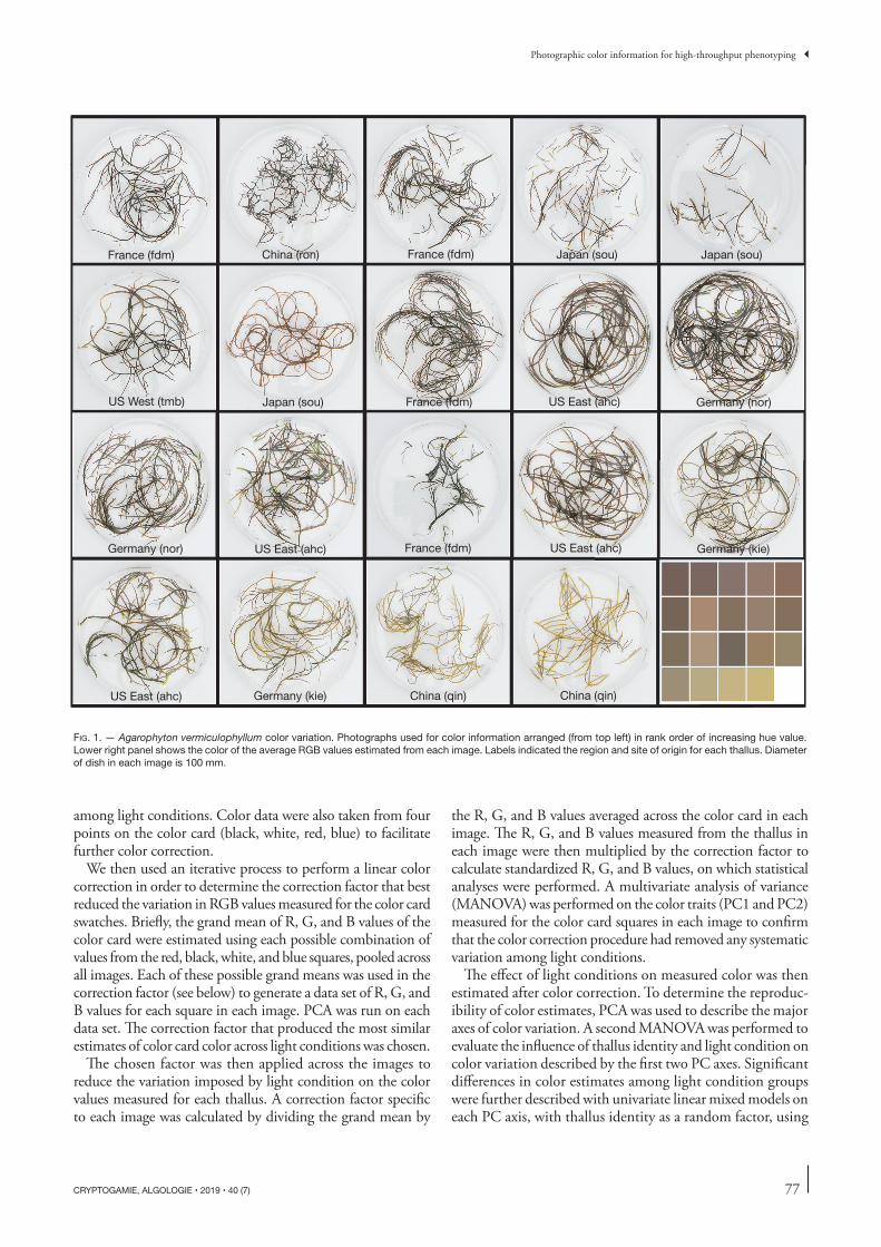
Approximately 50 algal cultures were available at the time of the experiment. In order to maximize the range of color morphs used, available samples were roughly clustered by visual similarity into four color groups (Red, Green, Yellow, and Black). Five thalli from each group were selected without taking site or origin (i.e., native vs non-native) into considera-tion. The concept of visual color groups was dropped after sample selection in favor of analyzing color as a continuous variable to facilitate a regression approach. One sample was removed prior to analyses because there was too little tissue to accurately measure phycobilin protein composition, leav-ing 19 thalli in total (Fig. 1).
PHOTOGRAPHING THALLI AND RETRIEVING COLOR DATA
To standardize the light conditions among samples, thalli were submerged in a Petri dish (100 × 15 mm) filled with seawater and photographed with a mounted camera (Canon SX710 HS; Canon Inc., Tokyo, Japan) inside a tent of lightweight, white muslin, which diffused light and minimized glare. Aperture (f 3.2), exposure time (⅛ second), and white balance (manually adjusted using the white background before the first picture) were identical for all photographs. To allow for better visual discrimination of thallus color, the brightness was increased slightly and uniformly for all images in Photoshop ver. CC 2015 (Adobe Systems Inc., San Jose, CA, United States). We did not digitally color correct using a color card as the images were all taken under the same light conditions.
To quantify the visual color of each thallus, RGB values were recorded for ten randomly chosen points, on the thal-lus in each photograph using the Color Picker Tool add-on
essential to understanding local adaptation and performance variation in natural populations of algae.
Here, we describe the wide range of color variation we observed in a subset of thalli from across the extant range of Agarophyton vermiculophyllum (Ohmi) Gurgel et al. (2018). We demonstrate that color variation is strongly predictive of some facets of photosynthetic pigment composition, and propose a rapid, non-destructive, and inexpensive method of predicting pigment composition from photographic color data to facilitate the high-throughput phenotyping of algae and to encourage the collection of these data in ecological studies.
MATERIAL AND METHODS
SAMPLE COLLECTION AND IDENTIFICATION
Whole algal thalli were collected from eight sites across the Northern Hemisphere range of A. vermiculophyllum in 2017 (Table 1). The wet mass of each alga varied depending on the genotype, but an algal thallus was divided into equal parts and shipped to the Weinberger lab at GEOMAR in Kiel, Germany or the Krueger-Hadfield lab at UAB in Birmingham, AL, USA. Three sites were from the native range, and sampled along the coastlines of China (qin, ron) and Japan (sou). Five sites were sampled from the non-native range, including one site from the west coast of the United States (tmb), one site from east coast of the United States (ahc), and three sites from Europe (fdm, kie, nor; see also Krueger-Hadfield et al. 2017). At all sites, thalli were collected with at least one meter sepa-rating each putative genet (i.e., unique genotype) to minimize the chance of sampling the same genet twice (Guillemin et al. 2008; Krueger-Hadfield et al. 2013, 2016).
MAINTENANCE OF SAMPLES PRIOR TO PHENOTYPING
Prior to phenotypic analyses conducted at the University of Alabama at Birmingham, all thalli were maintained in an environmental control chamber (I-36LL, Percival, Perry, IA, USA) for approximately two months to homogenize envi-ronmental influences on color and pigment composition
TABLE 1 et al.
Code Site name Country Region Latitude Longitude Sampled by Date
76
Ryan et al.
in ImageJ (Schneider et al. 2012). A unique grid of 100 randomly numbered points was overlaid onto each image of the thallus. Points were checked sequentially and the first ten points identified that met the following quality control criteria were used to collect color information. Points were rejected if they: i) occurred near the thallus edge; ii) occurred where tissue overlapped; or iii) occurred where obvious glare or shadow obscured the color.
PIGMENT EXTRACTION AND ANALYSIS
We quantified the concentration of chlorophyll a (chl a) in all thalli following the protocol of Torres et al. (2014). Briefly, approximately 50 mg of fresh tissue was frozen with liquid nitrogen, and ground with a pestle in a 2 ml tube. Pigments were extracted in 1.5 ml of methanol for 9 minutes, at which point tissues had lost their color, followed by centrifugation at 13 500 x g for 5 minutes. Extraction time was standardized across samples to minimize error while processing samples sequentially. The supernatant was transferred to a cuvette and absorbance was read at 665 and 750 nm on a spectrophotometer (Thermo Fisher Scientific, Waltham, MA, USA). Background absorbance at 750 nm was subtracted from values to account for variation in light scatter from debris or other impurities. The concentration of chl a was calculated as:
[chl a] = 12.61 · A665 (1)We extracted and quantified two classes of phycobilin proteins
following the protocol of Rosenberg (1981). Approximately 0.25 g of fresh tissue was frozen in liquid nitrogen, and ground to a fine powder with a mortar and pestle. The powder was transferred to a 2 ml tube with 750 ml of a 0.5% solution of Triton-X in a 0.1M phosphate buffer (6.8 ph). Samples were frozen and defrosted twice before centrifugation at 13 500 x g for 5 minutes. We, then, transferred the supernatant to a cuvette and read absorbance values at 565, 615, 650 and 750 nm. Background absorbance measured at 750 nm in each sample was subtracted from all absorbance measurements to account for variation in light scatter from debris or other impu-rities. Then, values were used to calculate the concentrations of phycoerythrin (PE) +and phycocyanin (PC) in microgram per gram of tissue wet weight using the following formulas:
[PE] = 123.5 · A565 − 73.5 · A615 + 16.3 · A650 (2)[PC] = 163.2 · A615 − 117.1 · A650 (3)We summed the concentrations of the two phycobilin types
in order to obtain total phycobilin protein concentration. To describe the relative composition of the two phycobilin pro-teins, we calculated the percent contribution of each phycobi-lin protein relative to the total concentration of phycobilins.
We also calculated the ratio of total phycobilins to chl a concentration in order to assess how changes in the relative composition of these pigments influenced thallus color. We added the chl a concentration to the total phycobilin value for a measure of total pigment concentration for each thallus.
ANALYSES OF PIGMENT AND COLOR VARIATION
To describe the extent of color and pigment variation among the thalli, we calculated the median and range of each color trait [red (R), green (G), and blue (B) values], as well as each
pigment trait (chl a, PE, and PC concentrations). To estimate the major axes of variation in color and pigment across traits and among thalli, we performed a Principal Components Analysis (PCA) on the scaled and natural log transformed R, G, and B values using the prcomp function in R (R Core Team 2017). To understand the relative contribution of each variable to overall variation, PCA loadings were derived from the prcomp function, and were plotted onto the scatterplot of PC1 versus PC2. The same routine was used separately to describe variation among pigment components, including the concentrations of chl a, PE, and PC. All variables were log transformed to meet the assumptions for normality for PCA.
To understand the relationship between variation in color traits and pigment composition, we evaluated a set of linear models using the first and second principle components axes (color PCA), and their interaction, as predictors of the first and second principle component axes estimated for pigment variables (pigment PCA). Step-wise model selection using the Akaike Information Criterion (AIC) was performed to identify the most informative combination of predictor variables for each pigment PCA axis in R. The model with the lowest AIC value was selected. When two models had similar AIC values (ΔAIC < 2), the model with the fewest parameters was chosen (Burnham & Anderson 2004).
VARIATION IN PHOTO-BASED COLOR ESTIMATES UNDER DIFFERENT LIGHT CONDITIONS
To estimate the variation in color information caused by dif-fering light conditions, we used five individual thalli (three originating from nor and two from kie; Table 1) which had been maintained in the laboratory for ten weeks. Thalli were cultivated in aerated artificial seawater enriched with Prov-asoli’s Enrichment at 15°C. All five thalli maintained color differences exhibited at the time of collection during these ten weeks. Each thallus was placed on a white cloth background with a color card (DGK Digital Kolor Kard, Digital Image Flow, Boston MA, USA). Thalli were then photographed under five different light conditions, while maintaining the same orientation of each thallus in each image. The five light conditions were: Direct midday sunlight (direct), indirect sunlight through a window (indirect), cool white light from a 680 nm wavelength neon light (white), interior incandescent light (yellow), and a 50:50 mix of cool white and blue LED light (blue; Fig. 1). Light conditions were chosen to create an extreme range of conditions on which to test the ability of digital color correction techniques to standardize extracted thallus color data.
To digitally standardize the light environment, images cap-tured under different light conditions were white balanced using the custom white balance tool in Photoshop Lightroom v. 5. 7.1 (Adobe Systems Incorporated, San Jose, CA, United States). The 80% grey square on the color card was used as a reference standard in each image.
A randomized grid of points was then laid over each image and RGB data were collected using the same technique as described above. The same grid was used for all images of the same thallus to standardize the points measured on the thallus
77
Photographic color information for high-throughput phenotyping
among light conditions. Color data were also taken from four points on the color card (black, white, red, blue) to facilitate further color correction.
We then used an iterative process to perform a linear color correction in order to determine the correction factor that best reduced the variation in RGB values measured for the color card swatches. Briefly, the grand mean of R, G, and B values of the color card were estimated using each possible combination of values from the red, black, white, and blue squares, pooled across all images. Each of these possible grand means was used in the correction factor (see below) to generate a data set of R, G, and B values for each square in each image. PCA was run on each data set. The correction factor that produced the most similar estimates of color card color across light conditions was chosen.
The chosen factor was then applied across the images to reduce the variation imposed by light condition on the color values measured for each thallus. A correction factor specific to each image was calculated by dividing the grand mean by
the R, G, and B values averaged across the color card in each image. The R, G, and B values measured from the thallus in each image were then multiplied by the correction factor to calculate standardized R, G, and B values, on which statistical analyses were performed. A multivariate analysis of variance (MANOVA) was performed on the color traits (PC1 and PC2) measured for the color card squares in each image to confirm that the color correction procedure had removed any systematic variation among light conditions.
The effect of light conditions on measured color was then estimated after color correction. To determine the reproduc-ibility of color estimates, PCA was used to describe the major axes of color variation. A second MANOVA was performed to evaluate the influence of thallus identity and light condition on color variation described by the first two PC axes. Significant differences in color estimates among light condition groups were further described with univariate linear mixed models on each PC axis, with thallus identity as a random factor, using
FIG. 1 Agarophyton vermiculophyllum

78
Ryan et al.
the lmer function in the lme4 package (Bates et al. 2015) in R. Significance values were compared to a Bonferroni adjusted alpha to account for multiple tests.
RESULTS
COLOR VARIATION
Approximately 99.2% of variation in R, G, and B values among 19 thalli was described by the first two principal components axes (95.2 and 4%, respectively; Fig. 2A). PC1 was primarily associated with levels of R and G. Increasing PC1 values were associated with a shift from dark to light yellowish-green thalli. PC2 was primarily associated with B value, shifting from reddish to brownish as PC2 increases. Untransformed R values ranged from 100 to 197, with a median value of 137, G values from 73 to 197, with a median value of 101, and B values ranged from 61 to 105, with a median value of 78.
PIGMENT VARIATION Approximately 96.5% of the variation in absolute pigment concentrations (chl a, PE, and PC) was explained by the first two axes (81.8 and 14.7%, respectively; Fig. 2B). PC1 was positively correlated with an increase in the concentra-tion of both phycobilin pigments, whereas PC2 reflected increasing chl a. The chl a concentration ranged from 77 to 558 μg/g wet weight, with a median value of 261 μg/g. The total concentration of phycobilin pigments ranged from 38 to 445 μg/g, with a median value of 145 μg/g. PE concentration ranged from 27 to 295 μg/g, with a median value of 100 μg/g. PC concentration ranged from 11 to 150 μg/g, with a median value of 45 μg/g. Additionally, the ratio of total phycobilin protein concentration to chl a con-centration ranged from 0.48 to 2.66, with a median value of 0.98. The proportional contribution of PE and PC to total phycobilin composition ranged from 0.60 to 0.86 and 0.14 to 0.40, respectively, with median values of 0.67 and 0.33. While there was ample variation in the relative composi-
FIG. 2 A BC
D
RG
B
3
A
PEPC
ChlA
B
R
3
B
R
D

79
Photographic color information for high-throughput phenotyping
tion of photopigments, composition did not appear to vary systematically with changes in total pigment concentration (R2 = 0.06, -0.05, -0.05, for total phycobilin/chl a, PE/total phycobilin, and PC/total phycobilin, respectively).
RELATIONSHIP OF THALLUS COLOR TO PIGMENT TRAITS
There was a strong association between thallus color and pigment composition. The major axis of pigment variation (pigment PC1) was best predicted by a combination of the color axes (color PC1 and PC2) (R2 = 0.74, p < 0.001, Fig. 2C). In general, darker colored thalli had higher pig-ment concentrations. Pigment PC2 was best predicted by color PC2 (R2 = 0.30, p = 0.009, Fig. 2D). Color PC1 was not included as a predictor in the best-fit model for pigment PC2. See model selection details in Table 2.
REPRODUCIBILITY OF THALLUS COLOR ESTIMATES UNDER DIFFERENT LIGHT CONDITIONS
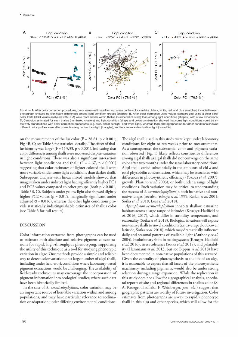
Photos taken under differing light conditions showed a large amount of variation in the average color of the image (Fig. 3). However, a simple white balance procedure greatly improved the similarity of the images (Fig. 3). The additional correction factor that most reduced variation among color card measure-ments took into account the average R, G, and B values of the red, white, and black, but not blue, color card standards. Measurements of the blue color standard were highly vari-able among images, even after color correction, highlighting
the disproportionate sensitivity of different parts of the color spectrum to ambient light conditions (Fig. 4A). After white balancing, there was no significant difference among the color of the color card standards, as described by the first two PC axes, in images taken under different light conditions (Pillai approximation = 0.09, F4,95 = 1.09, p = 0.37; MANOVA), indicating that images taken under very different light condi-tions can be standardized effectively.
Despite effectively standardizing the average color across images, there was a significant effect of light conditions detected
TABLE 2
Model AIC
Pigment PC1 ~ color PC1 + color PC2 50.19
Pigment PC2 ~ color PC2 35.48
TABLE 3A
B C
A: MANOVA
Factor DFPillai estimate
Approximate F P value
B: Color PC1
Random factor
Variance
Fixed factor Estimate DF T value P value
Indirect 1.86 241 11.37 < 0.001 *
C: Color PC2
Random factor
Variance
Fixed factor Estimate DF T value P value
Indirect 0.72 241 8.78 < 0.001 *
Yellow 0.20 241 2.46 0.015 *
FIG. 3
80
Ryan et al.
on the measurements of thallus color (F = 28.81, p < 0.001; Fig 4B, C; see Table 3 for statistical details). The effect of thal-lus identity was larger (F = 113.33, p < 0.001), indicating that color differences among thalli were recovered despite variation in light conditions. There was also a significant interaction between light conditions and thalli (F = 4.67, p < 0.001) suggesting that color estimates of lighter colored thalli were more variable under some light conditions than darker thalli. Subsequent analysis with linear mixed models showed that images taken under indirect light had significantly higher PC1 and PC2 values compared to other groups (both p < 0.001, Table 3B, C). Subjects under yellow light also showed slightly higher PC2 values (p = 0.015, marginally significant under adjusted = 0.016), whereas the other light conditions pro-vide statistically indistinguishable estimates of thallus color (see Table 3 for full results).
DISCUSSION
Color information extracted from photographs can be used to estimate both absolute and relative pigment concentra-tions for rapid, high-throughput phenotyping, supporting the utility of this technique as a tool for studying phenotypic variation in algae. Our methods provide a simple and reliable way to detect color variation on a large number of algal thalli, including under field-work conditions where laboratory-based pigment extractions would be challenging. The availability of field-ready techniques may encourage the incorporation of pigment information into ecological studies, where such data have been historically limited.
In the case of A. vermiculophyllum, color variation may be an important source of heritable variation within and among populations, and may have particular relevance to acclima-tion or adaptation under differing environmental conditions.
The algal thalli used in this study were kept under laboratory conditions for eight to ten weeks prior to measurements. As a consequence, the substantial color and pigment varia-tion observed (Fig. 1) likely reflects constitutive differences among algal thalli as algal thalli did not converge on the same color after two months under the same laboratory conditions. Algal thalli varied substantially in the amounts of chl a and total phycobilin concentration, which may be associated with differences in photosynthetic efficiency (Yokoya et al. 2007), growth (Plastino et al. 2003), or both under a range of light conditions. Such variation may be critical to understanding the success of A. vermiculophyllum in both its native and non-native ranges (see also: Yokoya et al. 1999; Raikar et al. 2001; Sotka et al. 2018, Lees et al. 2018).
Agarophyton vermiculophyllum inhabits shallow, estuarine habitats across a large range of latitudes (Krueger-Hadfield et al. 2016, 2017), which differ in turbidity, temperature, and seasonality (Sotka et al. 2018). Biological invasions will expose non-native thalli to novel conditions (i.e., average cloud cover, latitude, Sotka et al. 2018), which may dramatically influence daily and seasonal patterns of available light (Anthony et al. 2004). Evolutionary shifts in mating system (Krueger-Hadfield et al. 2016), stress tolerance (Sotka et al. 2018), and palatabil-ity (Hammann et al. 2013; but see Bippus et al. 2018) have been documented in non-native populations of this seaweed. Given the centrality of photosynthesis to the life of an alga, it is reasonable to expect that all facets of the photosynthetic machinery, including pigments, would also be under strong selection during a range expansion. While the replication in this study does not allow for a geographical analysis, anecdo-tal reports of site and regional differences in thallus color (S. A. Krueger-Hadfield, F. Weinberger, pers. obs.) suggest that geographic patterns are worthy of future investigation. Color estimates from photographs are a way to rapidly phenotype thalli in this alga and other species, which will allow for the
FIG. 4 AB
C

81
Photographic color information for high-throughput phenotyping
development of more detailed hypotheses based on observed variation.
Our results suggest that color estimates taken from photos are robust to a wide range of light environments, as long as a few simple precautions are taken. First, when images are taken at different times, or under differing conditions, a standard-ized color card must be included in every image to allow for nuanced color corrections above and beyond simple white balancing. Because different parts of the color spectrum are more or less sensitive to ambient light conditions, the choice colors to include as a reference standard need to be well-suited to the spectrum of colors to be estimated from the samples. Second, specimens must be evenly lit and relatively free of shadows and highlights. For example, the indirect sunlight treatment may have produced aberrant color estimates relative to other light environments because there was a subtle gradient in a light intensity across the specimen and photographed area. Such artifacts cannot be rectified by color correction algorithms and could lead to significant errors in estimating color prop-erties. Constructing or purchasing a lightbox to diffuse light and minimize shadows and glare is a simple way to standardize photos to be analyzed for color, regardless of whether images are taken in the field or in the laboratory. As high-quality camera technology has improved and become more affordable, color data derived from digital imagery have become increasingly common in ecological research (e.g., Endler & Mielke 2005; Siebeck et al. 2006; Winters et al. 2009; Kobayashi & Fujita 2014). As such, literature is available to assist the development of accurate and repeatable work flows using imaging technol-ogy (e.g., Endler 1990, 2012; Akkaynak et al. 2014), which could aid in the design of algal sampling projects.
Given the potential value of photopigment variation data for our understanding of algal ecology and evolution, we advocate for the collection of pigment data to become a routine part of describing phenotypic variation in algae. Our method offers a way for these data to be rapidly and non-destructively col-lected from large amounts of material direct from the field or after laboratory cultivation. Estimating photopigment proper-ties using comparative color data has been effective for study-ing coral reef health (e.g., Siebeck et al. 2006; Winters et al. 2009). Such techniques have formed the basis of large citizen science monitoring programs (e.g., coralwatch.org). Collecting similar data from field studies of algae would be valuable for generating initial hypotheses about the role of photopigment variation in ecological processes. In many cases, these data could help identify targets for subsequent sampling efforts with conventional pigment extraction techniques.
AcknowledgementsWe thank M. Yant for help in maintaining algal cultures, and the S. Mukhtar and J. B. McClintock labs at UAB for supplies. We also acknowledge the other participants of the experimental phycology class at UAB: E. Keister, L. MacMillan and J. Jackson. The project was funded in part by the University of Alabama at Birmingham through startup funds to S. A. Krueger-Hadfield and the DFG (WE 2700/5-1) to F. Weinberger.
Author contributionsWHR and SAKH framed hypotheses and experimental design; TB, GB, GW, FW, and SAKH performed field work; WHR, SH, MDC, FW and SAKH performed laboratory work; WHR, SH, MDC, CDA, and SAKH performed data analysis and interpretation; WHR and SAKH prepared the manuscript with help from all authors.
REFERENCES
AKKAYNAK D., TREIBITZ T., XIAO B., GÜRKAN U. A., ALLEN J. J., DEMIRCI U. & HANLON R. T. 2014. — Use of commercial off-the-shelf digital cameras for scientific data acquisition and scene-specific color calibration. Journal of the Optical Society of America A 31: 312-321. https://doi.org/10.1364/JOSAA.31.000312
ANTHONY K. R. N., RIDD P. V., ORPIN A. R., LARCOMBE P. & LOUGH J. 2004. — Temporal variation of light availability in coastal benthic habitats: effects of clouds, turbidity, and tides. Limnol-ogy and Oceanography 49: 2201-2211. https://doi.org/10.4319/lo.2004.49.6.2201
BAILEY S. & GROSSMAN A. 2008. — Photoprotection in cyanobacteria: regulation of light harvesting. Photochemistry and Photobioly 84: 1410-1420. https://doi.org/10.1111/j.1751-1097.2008.00453.x
BATES D., MÄCHLER M., BOLKER B. & WALKER S. 2015. — Fit-ting linear mixed-effects models using lme4. Journal of Statistical Software 67: 1-48. https://doi.org/10.18637/jss.v067.i01
BEACH K. S. & SMITH C. M. 1996. — Ecophysiology of tropical Rhodophytes. I. microscale acclimation in pigmentation. Jour-nal of Phycology 32: 701-710. https://doi.org/10.1111/j.0022-3646.1996.00701.x
BEN-AMOTZ A., SHAISH A. & AVRON M. 1989. — Mode of action of the massively accumulated beta-carotene of Dunaliella bardawil in protecting the alga against damage by excess irradiation. Plant Physiology 91: 1040-1043. https://doi.org/10.1104/pp.91.3.1040
BIPPUS P. M., KRUEGER-HADFIELD S. A. & SOTKA E. E. 2018. — Palatability of an introduced seaweed does not differ between native and non-native populations. Marine Biology 165: 781-793. https://doi.org/10.1007/s00227-018-3291-5
BURNHAM K. P. & ANDERSON D. R. 2004. — Multimodel inference: understanding AIC and BIC in model selection. Sociological Methods & Research 33: 261-304. https://doi.org/10.1177/0049124104268644
CORNISH M. L., O’LEARY S. J. B. & GARBARY D. J. 2013. — Phycobilisome composition in Chondrus crispus (Gigartinales, Rhodophyta) from a wild type strain and its vegetatively derived green mutant. Algae 28: 121-129. https://doi.org/10.4490/algae.2013.28.1.121
COSTA V. L. & PLASTINO E. M. 2011. — Color inheritance and pigment characterization of red (wild-type), greenish-brown, and green strains of Gracilaria birdiae (Gracilariales, Rhodophyta). Journal of Applied Phycology 23: 599-605. https://doi.org/10.1007/s10811-010-9642-3
ENDLER J. A. 1990. — On the measurement and classification of colour in studies of animal colour patterns. Biological Journal of the Linnean Society 41: 315-352. https://doi.org/10.1111/j.1095-8312.1990.tb00839.x
ENDLER J. A. 2012. — A framework for analysing colour pattern geometry: adjacent colours. Biological Journal of the Linnean Society 107: 233-253. https://doi.org/10.1111/j.1095-8312.2012.01937.x
ENDLER J. A. & MIELKE P. W. 2005. — Comparing entire colour patterns as birds see them. Biological Journal of the Linnean Society 86: 405-431. https://doi.org/10.1111/j.1095-8312.2005.00540.x
FALKOWSKI P. G. & CHEN Y. B. 2003. — Photoacclimation of light harvesting systems in eukaryotic algae, in GREEN B. R., PARSON W. W. (eds), Light Harvesting Antennas in Photosynthesis. Kluwer Academic Publishers, Dordrecht: 423-447.

82
Ryan et al.
FALKOWSKI P. G. & RAVEN J. A. 2007. — An introduction to photo-synthesis in aquatic systems, in Aquatic Photosynthesis. Princeton University Press: 1-44.
GANTT E. 1981. — Phycobilisomes. Annual Review of Plant Physiology 32: 327-347. https://doi.org/10.1146/annurev.pp.32.060181.001551
GROSSMAN A. 2003. — A molecular understanding of complemen-tary chromatic adaptation. Photosynthesis Research 76: 207-215. https://doi.org/10.1023/A:1024907330878
GUILLEMIN M.-L., FAUGERON S., DESTOMBE C., VIARD F., COR-REA J. A. & VALERO M. 2008. — Genetic variation in wild and cultivated populations of the haploid- diploid red alga Graci-laria chilensis: How farming practices favor asexual reproduc-tion and heterozygosity. Evolution 62: 1500-1519. https://doi.org/10.1111/j.1558-5646.2008.00373.x
GURGEL C. F. D., NORRIS J. N., SCHMIDT W. E., LEE H. N. & FREDERICQ S. 2018. — Systematics of the Gracilariales (Rhodo-phyta) including new subfamilies, tribes, and two new subgenera, Agarophyton gen. nov. and Crassa gen. nov. Phytotaxa 374: 001-023. https://doi.org/10.11646/phytotaxa.374.1.1
HAMMANN M., WANG G., RICKERT E., BOO S. & WEINBERGER F. 2013. — Invasion success of the seaweed Gracilaria vermiculo-phylla correlates with low palatibility. Marine Ecology Progress Series 486: 93-103. https://doi.org/10.3354/meps10361
KIM J. K., MAO Y., KRAEMER G. & YARISH C. 2015. — Growth and pigment content of Gracilaria tikvahiae McLachlan under fluorescent and LED lighting. Aquaculture 436: 52-57. https://doi.org/10.1016/j.aquaculture.2014.10.037
KOBAYASHI M. & FUJITA D. 2014. — Can thallus color of red algae be used as an environmental indicator in shallow waters? Journal of Applied Phycology 26: 1123-1131. https://doi.org/10.1007/s10811-013-0165-6
KRUEGER-HADFIELD S. A., KOLLARS N. M., BYERS J. E., GREIG T. W., HAMMANN M., MURRAY D. C., MURREN C. J., STRAND A. E., TERADA R., WEINBERGER F. & SOTKA E. E. 2016. — Invasion of novel habitats uncouples haplo-diplontic life cycles. Molecular Ecology 25: 3801-3816. https://doi.org/10.1111/mec.13718
KRUEGER-HADFIELD S. A., KOLLARS N. M., STRAND A. E., BYERS J. E., SHAINKER S. J., TERADA R., GREIG T. W., HAMMANN M., MURRAY D. C., WEINBERGER F. & SOTKA E. E. 2017. — Genetic identification of source and likely vector of a widespread marine invader. Ecology and Evolution 7: 4432-4447. https://doi.org/10.1002/ece3.3001
KRUEGER-HADFIELD S. A., ROZE D., MAUGER S. & VALERO M. 2013. — Intergametophytic selfing and microgeographic genetic structure shape populations of the intertidal red seaweed Chon-drus crispus. Molecular Ecology 22: 3242-3260. https://doi.org/10.1111/mec.12191
KURSAR T. A., VAN DER MEER J. & ALBERTE R. S. 1983. — Light-harvesting system of the red alga Gracilaria tikvahiae: II. Phy-cobilisome characteristics of pigment mutants. Plant Physiology 73: 361-369. https://doi.org/10.1104/pp.73.2.361
LEES L. E., KRUEGER-HADFIELD S. A., CLARK A. J., DUERMIT E. A., SOTKA E. E. & MURREN C. J. 2018. — Nonnative Gracilaria vermiculophylla tetrasporophytes are more difficult to debranch and are less nutritious than gametophytes. Journal of Phycology 54: 471-482. https://doi.org/10.1111/jpy.12746
LEMASSON C., MARSAC N. T. & COHEN-BAZIRE G. 1973. — Role of allophycocyanin as light-harvesting pigment in cyanobacteria. Proceedings of the National Academy of Sciences of the United States of America 70: 3130-3. https://doi.org/10.1073/pnas.70.11.3130
LÓPEZ-FIGUEROA F. 1992. — Diurnal variation in pigment content in Porphyra laciniata and Chondrus crispus and its relation to the diurnal changes of underwater light quality and quantity. Marine Ecology 13: 285-305. https://doi.org/10.1111/j.1439-0485.1992.tb00356.x
VAN DER MEER J. P. 1981. — The inheritance of spontaneous pig-ment mutations in Chondrus crispus Stackh. (Rhodophyceae).
Proceedings of the Nova Scotian Institute of Science 31: 187-192.NIWA K., HAYASHI Y., ABE T. & ARUGA Y. 2009. — Induction
and isolation of pigmentation mutants of Porphyra yezoensis (Bangiales, Rhodophyta) by heavy-ion beam irradiation. Phy-cological Research 57: 194-202. https://doi.org/10.1111/j.1440-1835.2009.00539.x
PLASTINO E. M., URSI S. & FUJII M. T. 2003. — Color inheritance, pigment characterization, and growth of a rare light green strain of Gracilaria birdiae (Gracilariales, Rhodophyta). Phycological Research 52: 45-52.
R CORE TEAM 2017. — R: A language and environment for sta-tistical computing. R Foundation for Statistical Computing, Vienna, Austria.
RAIKAR S. V., LIMA M., FUJITA Y. 2001. — Effect of temperature, salinity and light intensity on the growth of Gracilaria spp. (Gracilariales, Rhodophyta) from Japan, Malaysia and India. Indian Journal of Marine Sciences 30: 98-104.
RAMUS J. & VAN DER MEER J. 1983. — A physiological test of the theory of complementary chromatic adaptation. II. Brown, green and red seaweeds. Journal of Phycology 19: 173-178. https://doi.org/10.1111/j.0022-3646.1983.00173.x
ROSENBERG G. 1981. — Ecological growth strategies in the seaweeds Gracilaria foliifera (Rhodophyceae) and Ulva sp. (Chlorophyceae). PhD Thesis. Yale University, 302 p.
ROSENBERG G. & RAMUS J. 1982. — Ecological growth strate-gies in the seaweeds Gracilaria foliifera (Rhodophyceae) and Ulva Sp (Chlorophyceae) – the rate and timing of growth. Botanica Marina 66: 251-259. https://doi.org/10.1515/botm.1981.24.11.583
SCHNEIDER C. A., RASBAND W. S. & ELICEIRI K. W. 2012. — NIH Image to ImageJ: 25 years of image analysis. Nature Methods 9: 671-675. https://doi.org/10.1038/nmeth.2089
SIEBECK U. E., MARSHALL N. J., KLÜTER A. & HOEGH-GULDBERG O. 2006. — Monitoring coral bleaching using a colour refer-ence card. Coral Reefs. 25: 453-460. https://doi.org/10.1007/S00338-006-0123-8
SIEFERMANN-HARMS D. 1985. — Carotenoids in photosynthesis. I. Location in photosynthetic membranes and light-harvesting function. Biochimica Biophysica Acta 811: 325-355. https://doi.org/10.1016/0304-4173(85)90006-0
SOTKA E. E., BAUMGARDNER A. W., BIPPUS P. M., DESTOMBE C., DUERMIT E. A., ENDO H., FLANAGAN B. A., KAMIYA M., LEES L. E., MURREN C. J., NAKAOKA M., SHAINKER S. J., STRAND A. E., TERADA R., VALERO M., WEINBERGER F. & KRUEGER-HADFIELD S. A. 2018. — Combining niche-shift and population genetic analyses predicts rapid phenotypic evolution during invasion. Evolutionary Applications 11: 781-793. https://doi.org/10.1111/eva.12592
STOCKENREITER M., HAUPT F., GRABER A. K., SEPPÄLÄ J., SPILL-ING K., TAMMINEN T. & STIBOR H. 2013. — Functional group richness: implications of biodiversity for light use and lipid yield in microalgae. Journal of Phycology 49: 838-847. https://doi.org/10.1111/jpy.12092
STOMP M., HUISMAN J., STAL L. J. & MATTHIJS H. C. 2007. — Colorful niches of phototrophic microorganisms shaped by vibra-tions of the water molecule. The ISME Journal 159: 271-282. https://doi.org/10.1038/ismej.2007.59
TORRES P. B., CHOW F., FURLAN C. M., MANDELLI F., MERCADANTE A. & DOS SANTOS D. Y. A. C. 2014. — Standardization of a protocol to extract and analyze chlorophyll a and carotenoids in Gracilaria tenuistipitata Var. Liui. Zhang and Xia (Rhodo-phyta). Brazilian Journal of Oceanography 62: 57-63. https://doi.org/10.1590/s1679-87592014068106201
WINTERS G., HOLZMAN R., BLEKHMAN A., BEER S. & LOYA Y. 2009. — Photographic assessment of coral chlorophyll contents: Implications for ecophysiological studies and coral monitoring. Journal of Experimental Marine Biology and Ecology 380: 25-35. https://doi.org/10.1016/j.jembe.2009.09.004

83
Photographic color information for high-throughput phenotyping
YOKOYA N. S., KAKITA H., OBIKA H., KITAMURA T. 1999. — Effects of environmental factors and plant growth regulators on growth of the red alga Gracilaria vermiculophylla from Shikoku Island, Japan. Hydrobiologia 398/399: 339-347. https://doi.org/10.1023/A:1017072508583
YOKOYA N. S., NECCHI O., MARTINS A. P., GONZALEZ S. F. & PLASTINO E. M. 2007. — Growth responses and photosynthetic characteristics of wild and phycoerythrin-deficient strains of Hypnea musciformis (Rhodophyta). Journal of Applied Phycol-ogy 19: 197-205. https://doi.org/10.1007/s10811-006-9124-9
Submitted on 10 December 2018 accepted on 14 January 2019;
published on 16 September 2019.