The Use of Natural Polymers in Tissue Engineering: A - MDPI.com
32
Polymers 2010, 2, 522-553; doi:10.3390/polym2040522 polymers ISSN 2073-4360 www.mdpi.com/J./polymers Review The Use of Natural Polymers in Tissue Engineering: A Focus on Electrospun Extracellular Matrix Analogues Scott A. Sell 1,2 , Patricia S. Wolfe 1 , Koyal Garg 1 , Jennifer M. McCool 1 , Isaac A. Rodriguez 1 and Gary L. Bowlin 1, * 1 Department of Biomedical Engineering, Virginia Commonwealth University, 401 W. Main St, Richmond, VA 23284, USA; E-Mails: [email protected] (S.A.S.); [email protected] (P.S.W.); [email protected] (K.G.); [email protected] (J.M.M.); [email protected] (I.A.R.) 2 Physical Medicine & Rehabilitation Service, Hunter Holmes McGuire VA Medical Center, 1201 Broad Rock Blvd., Richmond, VA 23249, USA * Author to whom correspondence should be addressed; E-Mail: [email protected]; Tel.: 804-828-2592; Fax: 804-828-4454. Received: 1 September 2010; in revised form: 29 September 2010 / Accepted: 4 November 2010 / Published: 9 November 2010 Abstract: Natural polymers such as collagens, elastin, and fibrinogen make up much of the body’s native extracellular matrix (ECM). This ECM provides structure and mechanical integrity to tissues, as well as communicating with the cellular components it supports to help facilitate and regulate daily cellular processes and wound healing. An ideal tissue engineering scaffold would not only replicate the structure of this ECM, but would also replicate the many functions that the ECM performs. In the past decade, the process of electrospinning has proven effective in creating non-woven ECM analogue scaffolds of micro to nanoscale diameter fibers from an array of synthetic and natural polymers. The ability of this fabrication technique to utilize the aforementioned natural polymers to create tissue engineering scaffolds has yielded promising results, both in vitro and in vivo, due in part to the enhanced bioactivity afforded by materials normally found within the human body. This review will present the process of electrospinning and describe the use of natural polymers in the creation of bioactive ECM analogues in tissue engineering. Keywords: electrospinning; tissue engineering; natural polymers; collagen; elastin; fibrinogen; silk fibroin; extracellular matrix OPEN ACCESS
Transcript of The Use of Natural Polymers in Tissue Engineering: A - MDPI.com

Polymers 2010, 2, 522-553; doi:10.3390/polym2040522
polymersISSN 2073-4360
www.mdpi.com/J./polymers
Review
The Use of Natural Polymers in Tissue Engineering: A Focus on
Electrospun Extracellular Matrix Analogues
Scott A. Sell 1,2
, Patricia S. Wolfe 1, Koyal Garg
1, Jennifer M. McCool
1, Isaac A. Rodriguez
1
and Gary L. Bowlin 1,*
1 Department of Biomedical Engineering, Virginia Commonwealth University, 401 W. Main St,
Richmond, VA 23284, USA; E-Mails: [email protected] (S.A.S.); [email protected] (P.S.W.);
[email protected] (K.G.); [email protected] (J.M.M.); [email protected] (I.A.R.) 2 Physical Medicine & Rehabilitation Service, Hunter Holmes McGuire VA Medical Center, 1201
Broad Rock Blvd., Richmond, VA 23249, USA
* Author to whom correspondence should be addressed; E-Mail: [email protected];
Tel.: 804-828-2592; Fax: 804-828-4454.
Received: 1 September 2010; in revised form: 29 September 2010 / Accepted: 4 November 2010 /
Published: 9 November 2010
Abstract: Natural polymers such as collagens, elastin, and fibrinogen make up much of the
body’s native extracellular matrix (ECM). This ECM provides structure and mechanical
integrity to tissues, as well as communicating with the cellular components it supports to
help facilitate and regulate daily cellular processes and wound healing. An ideal tissue
engineering scaffold would not only replicate the structure of this ECM, but would also
replicate the many functions that the ECM performs. In the past decade, the process of
electrospinning has proven effective in creating non-woven ECM analogue scaffolds of
micro to nanoscale diameter fibers from an array of synthetic and natural polymers. The
ability of this fabrication technique to utilize the aforementioned natural polymers to create
tissue engineering scaffolds has yielded promising results, both in vitro and in vivo, due in
part to the enhanced bioactivity afforded by materials normally found within the human
body. This review will present the process of electrospinning and describe the use of
natural polymers in the creation of bioactive ECM analogues in tissue engineering.
Keywords: electrospinning; tissue engineering; natural polymers; collagen; elastin;
fibrinogen; silk fibroin; extracellular matrix
OPEN ACCESS

Polymers 2010, 2
523
1. Introduction
While the process of electrospinning has been used commercially since the early 20th
century,
originally devised as a textile fabrication process, the rather recent ability to electrospin naturally
occurring polymers has helped to reinvigorate the process and expand its potential applications.
Electrospinning is lauded for its ability to economically and efficiently create non-woven meshes of
sub-micron diameter fibers, particularly in the field of tissue engineering, where these fibrous scaffolds
can replicate both the form and function of native extracellular matrix (ECM). While a synthetic
polymer scaffold can mimic the structure of the native ECM, the ability to electrospin a naturally
occurring protein such as collagen can provide cells with a recognizable, physiologically relevant
platform from which to proliferate and ultimately remodel. With collagen constituting the majority of
the human body’s structural ECM, electrospun collagen has been used in a number of tissue
engineering applications; everything from cardiovascular tissues, musculoskeletal tissues, skin, and
nerve have used electrospun collagen fibers as the basis of their ECM analogues. Other natural
polymers such as fibrinogen, often considered to be nature’s provisional matrix due to its importance
in the early stages of hemostasis and wound repair, have been electrospun and used for their excellent
bioactive properties. This review will present the process of electrospinning, and describe the use of
the natural polymers collagen, gelatin, elastin, fibrinogen, and silk fibroin (SF) in the creation of
bioactive ECM analogues in tissue engineering.
1.1. The Native Extracellular Matrix
The tissues of the human body are composed of two main constituents: cells, which regulate body
processes and perform normal maintenance and wound healing, and the ECM, a collection of
macromolecules (Table 1) which serves as the major structural component of the body. This ECM
makes up a large majority of tissue volume, and also provides much of a tissue’s unique geometric
shape, consisting primarily of fibers with diameters between 50 and 500 nm [1]. Composed of a
dynamic, three-dimensional arrangement of polysaccharides and natural polymers (collagen, elastin,
fibrinogen, etc.), ECM components are constantly being synthesized, secreted, oriented, and modified
by the cellular components that they support. Historically, the function of native ECM was only
believed to be as a structural framework for tissues. However it is now understood that the ECM,
through interactions with receptors on the surfaces of cells, plays a large part in both day-to-day
cellular activity and in wound healing. During development, cell-ECM interaction is responsible for
pattern formation, morphogenesis, and phenotype acquisition and maintenance. During clot formation,
wound healing, inflammation, formation of granulation tissue, and remodeling are all mediated by
cell-ECM interaction. Cell adhesion, migration, growth, differentiation, and apoptosis are all
controlled, in part, by the transmission of signals between the cell nucleus and the ECM. Additionally,
growth factors and signaling molecules can be stored within the ECM, protecting them from premature
degradation, or they can attach to the surface of the ECM to present themselves more efficiently to cell
receptors [1-4].

Polymers 2010, 2
524
Table 1. Major components of the native ECM, their locations, and functions in the body.
Adapted from [3].
Component Location Function
Collagen Widely distributed Key component of tissue architecture, provide tensile
strength, cell-matrix interaction, matrix-matrix interaction
Elastin Highly elastic tissues
(lung, blood vessel, skin) Key component of tissue architecture, provide elasticity
Proteoglycans Widely distributed Cell-matrix interaction, matrix-matrix interaction, cell
proliferation, cell migration
Hyaluronan Widely distributed Cell-matrix interaction, matrix-matrix interaction, cell
proliferation, cell migration
Laminin Basement membranes Basement membrane component, cell migration
Fibronectin Widely distributed Component of tissue architecture, cell-matrix interaction,
matrix-matrix interaction, cell proliferation, cell migration
Fibrinogen Blood, sites of wound
healing Cell proliferation, cell migration, hemostasis
Various Adhesion
Molecules Widely distributed
Mediate cell adhesion to matrix, mediate transmembrane
signals
The multi-faceted cell-ECM communication takes place through both integrin and non-integrin
membrane-bound receptors. This communication is complex and dynamic, and plays critical roles
during development, wound healing, and environmental maintenance (Figure 1). Initial attraction and
adhesion of cells to the ECM is induced by multiple, low affinity charge and hydrophobic interactions.
Once cells have begun attaching, the spreading phase of adhesion is induced by heterodimeric
transmembrane proteins, known as integrins, on the cell surface which bind to specific small peptide
fragment sequences on the ECM. This connection allows cells to bind to the ECM, through focal
adhesions, and promote direct communication between the two. Integrin binding is both specific and
reversible and allows cells to differentiate, secrete and absorb matrix, and transmit signals [4]. The
ECM has the ability to communicate with cells through ―outside-in‖ signaling by sending signals
across the cell membrane to soluble molecules in the cytoplasm and through direct connections with
the cytoskeleton and into the cell nucleus. This direct contact allows for stronger, more specific
signaling than through the release of diffusible signaling molecules. ―Inside-out‖ cell-ECM
interactions are also possible, and occur when changes within the cell result in a feedback which alters
the activity of surface receptors, ultimately creating changes in the focal adhesions [4]. While the ECM
has the ability to alter cellular function, so too do cells have the ability to alter the ECM. In what is
known as dynamic reciprocity, the cellular response to the ECM can often alter the state of the ECM.
For example, cells may release matrix metalloproteases to break down an overly dense ECM to allow
for their migration or proliferation [2,3]. Creating an ECM analogue that can replicate these
physiologic cell-ECM interactions is extremely challenging, yet may be possible through the
manipulation of the natural polymers which constitute the native ECM, as these polymers may present
the normal signaling capabilities required by cells [1].

Polymers 2010, 2
525
Figure 1. Diagram of cell-ECM interaction depicting integrin binding sites.
1.2. Electrospinning
Ideally, a tissue engineering scaffold should mimic both the form and functionality of the native
ECM. However, due to its complexity and diversity, it is virtually impossible to manufacture an exact
replica that can encompass all of the roles of the native ECM. To be successful as a tissue engineering
scaffold, structures must: 1) have appropriate porosities to allow for cellular migration and penetration;
2) have sufficient surface area and the proper surface chemistry to promote cell adhesion, growth,
migration, and differentiation; 3) a rate of degradation that closely matches the rate of native tissue
regeneration to promote proper tissue ingrowth. In the past decade the process of electrospinning has
become a widely used technique for the creation of ECM analogue structures for tissue engineering
applications, in part because of its capacity to meet the three previously mentioned criteria. With its
ability to rapidly create structures composed of nanoscale fibers closely resembling the architecture of
the native ECM, this process has experienced a renaissance in recent years and continues to find new
niches in the field of tissue engineering. This relatively simple process has proven to be highly
adaptable, with the capacity for use with a number of natural and synthetic polymers, cost
effectiveness, easy scale-up for large volume production, and the ability to provide users control over a
number of processing parameters which can be modified to elicit fine control over the end-product’s
physical features [1,5-11].
In its simplest form the process of electrospinning requires little specialized equipment; all that is
needed is a high voltage power supply, a grounded target, and a small diameter conductive capillary to
emit a polymer solution in a controlled fashion (Figure 2). Polymers are dissolved in a solvent,
everything from water to organic solvents such as hexafluoroisopropanol (HFP) depending upon the
polymer, at a concentration high enough for polymer chain entanglement to occur. As solution
viscosity increases with polymer concentration, the resulting fiber diameter increases linearly [1,12].
The polymer solution is fed through a capillary (typically a needle with a blunt end) at a constant flow

Polymers 2010, 2
526
rate, with the polymer solution within the capillary charged to a high voltage potential opposite a
grounded target to create a static electric field. When the electric field produces a force strong enough
to overcome the surface tension of the polymer solution collected at the capillary tip, a Taylor cone
forms. This Taylor cone is then stretched until a jet of polymer is drawn from the tip of the Taylor
cone and attracted to the grounded collecting target. As the jet travels through the air towards the
collecting target the solvent gradually evaporates, resulting in the collection of nano to micron scale
fibers on the target. The charge from the fibers dissipates into the surrounding environment, and a non-
woven fiber mat consisting of fibers 50 nm to 10 µm in diameter is formed [1,5-11,13,14].
Figure 2. Schematic representation of a traditional electrospinning setup with a horizontal
orientation (Left), and the simultaneous microintegration of cells and electrospun polymer
using an airgap electrospinning setup (Right).
The ability for electrospinning to create tissue specific scaffolds arises from the adaptability of the
process and the system control offered by a number of tunable processing parameters: solution
properties (viscosity, elasticity, conductivity, and surface tension), processing conditions (voltage,
capillary diameter, distance from capillary orifice to grounded target), and environmental conditions
(temperature, humidity and static electricity) [6,9,10,15]. Each of the aforementioned parameters,
separately or in conjunction with one another, can affect fiber deposition, fiber diameter, and scaffold
porosity. By altering the orientation of the collected fibers (parallel alignment versus random fiber
orientation), and/or the type of polymer(s) used (natural, synthetic, or blended) it is possible to
significantly alter the mechanical properties of the scaffolds. In addition to altering the orientation of
the fibers, variations in the micro- and macro-structures of the scaffolds themselves can be obtained
for specific applications. For example, multilayering electrospinning results in a hierarchically ordered
structure composed of different types of polymers, while multicomponent electrospinning, where
multiple polymers are simultaneously electrospun together, forms a mixed fiber mesh [16]. Also, the

Polymers 2010, 2
527
way the fibers are collected on the grounded target can influence the fiber orientation as well as
scaffold fabrication. Some of the collection schemes currently used include a single ground, rotating
single ground, dual bar, dual ring, single horizontal ring, electrospinning in vitro onto cells [15,17],
electrospinning cells simultaneously with polymer (microintegration) [18], or using a rotating
electrode airgap collection method [19]. The microintegration process developed by Stankus et al. [18]
used the principle of electrospraying, a historical precursor to electrospinning, to disperse small
charged droplets of cell suspension onto a target simultaneously collecting electrospun fibers. This
process had no adverse effect on the cells, and proved to be a viable method for creating a cellularized
structure primed for regeneration.
2. Electrospun Natural Polymers
2.1. Collagen
Collagen fibrils and their networks form the ECM for most hard and soft tissues (bone, cartilage,
tendon, cornea, blood vessels, nerve, and skin) in the human body. Collagen plays a key role in
maintaining the biologic and structural integrity of the ECM architecture, and presents different
morphologies in different tissues. Additionally, it is highly dynamic, undergoing constant remodeling
for proper physiological functions [1,2].
To date, 28 collagen types have been identified; with types I, II, III, V and XI involved in forming
fibrillar structures. Collagen types I, II, III and V are the primary types that constitute the essential part
of collagen in bone, cartilage, tendon, skin and muscle. While each collagen type is somewhat unique,
all collagen molecules share a triple helical structure, and the presence of 4-hydroxyproline provides a
distinctive marker for these molecules [3,4]. Collagen type I consists of two α1 chains and one α2
chain forming fibrils of 50 nm in diameter, and is present in bone, skin, dentin, cornea, blood vessels,
fibrocartilage and tendon. Type II collagen is composed of three identical α1(II) chains forming fibrils
less than 80 nm in diameter and is found exclusively in cartilaginous tissues and is associated with
water, proteoglycans, glycoproteins and noncollagenous proteins. Type III collagen is present in our
skin as well as in ligaments, blood vessels and internal organs, and is composed of three α1(III) chains
resulting in fibrils ranging from 30 to 130 nm in diameter [5,6,9]. Type IV collagen, which is
composed of αI(IV) and α2(IV) chains, is the major skeletal macromolecule of basement membrane in
various tissues. The molecular organization of type V collagen, in terms of αI(V), α2(V), and α3(V)
chain compositions, still remains to be clearly identified. Type V collagen has been identified in many
tissues including blood vessel wall, synovium, corneal stoma, tendon, lung, bone, cartilage and skeletal
muscle [20].
Aside from its widespread presence natively in the body, collagen has several properties that make
it an attractive biomaterial for tissue engineering applications: low antigenicity, low inflammatory and
cytotoxic responses, high water affinity, good cell compatibility, availability of various methods of
isolation from a variety of sources, and biodegradability [4,8,11].
Polymers 2010, 2
528
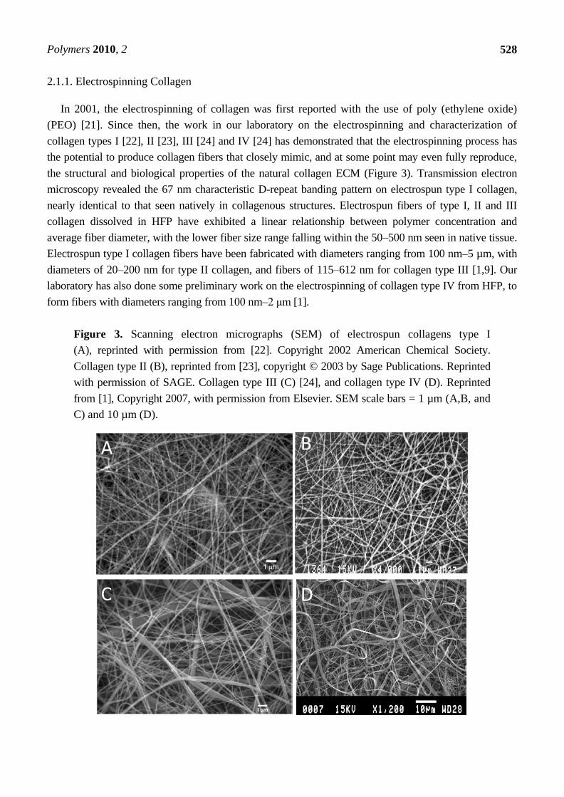
2.1.1. Electrospinning Collagen
In 2001, the electrospinning of collagen was first reported with the use of poly (ethylene oxide)
(PEO) [21]. Since then, the work in our laboratory on the electrospinning and characterization of
collagen types I [22], II [23], III [24] and IV [24] has demonstrated that the electrospinning process has
the potential to produce collagen fibers that closely mimic, and at some point may even fully reproduce,
the structural and biological properties of the natural collagen ECM (Figure 3). Transmission electron
microscopy revealed the 67 nm characteristic D-repeat banding pattern on electrospun type I collagen,
nearly identical to that seen natively in collagenous structures. Electrospun fibers of type I, II and III
collagen dissolved in HFP have exhibited a linear relationship between polymer concentration and
average fiber diameter, with the lower fiber size range falling within the 50–500 nm seen in native tissue.
Electrospun type I collagen fibers have been fabricated with diameters ranging from 100 nm–5 µm, with
diameters of 20–200 nm for type II collagen, and fibers of 115–612 nm for collagen type III [1,9]. Our
laboratory has also done some preliminary work on the electrospinning of collagen type IV from HFP, to
form fibers with diameters ranging from 100 nm–2 μm [1].
Figure 3. Scanning electron micrographs (SEM) of electrospun collagens type I
(A), reprinted with permission from [22]. Copyright 2002 American Chemical Society.
Collagen type II (B), reprinted from [23], copyright © 2003 by Sage Publications. Reprinted
with permission of SAGE. Collagen type III (C) [24], and collagen type IV (D). Reprinted
from [1], Copyright 2007, with permission from Elsevier. SEM scale bars = 1 µm (A,B, and
C) and 10 µm (D).
A B
C D

Polymers 2010, 2
529
As with other collagenous scaffolds, fabricated as gels or sponges, electrospun collagen lacks
mechanical and structural stability upon hydration. In order to increase the strength of electrospun
collagen, cross-linking with glutaraldehyde vapors, formaldehyde and epoxy compounds has been
evaluated. Glutaraldehyde cross-linked collagen type I scaffold showed tensile strength close to
several commercially available wound care products such as Beschitin®
and Resolute®
LT [5]. Lower
porosity and reduced numbers of keratinocytes and fibroblasts (FB) were observed on the cross-linked
scaffold nanofibers as compared to matrices treated with collagen type I.
This approach however, leads to an enhanced risk of cytotoxicity and calcification when used in
vivo. In 2007, our laboratory published a technique for the cross-linking of electrospun collagen, while
preventing any cytotoxic effects, imparting desirable mechanical properties, and maintaining the
nanofibrous structure, using 1-ethyl-3-(3-dimethylaminopropyl) carbodiimide hydrochloride (EDC) in
ethanol [25]. Shih et al. also studied the morphology, growth, adhesion, motility, and osteogenic
differentiation of human bone marrow-derived mesenchymal stem cells on EDC cross-linked
electrospun collagen fibrous mats [14]. Electrospinning of collagen type I using buffer/ethanol
solutions has been successfully accomplished by Dong et al. The scaffolds were cross-linked using
EDC, and the authors demonstrated that the triple helical structure of collagen was preserved and that
aligned fibers could be obtained by using this method [26].
2.1.2. Electrospinning Collagen and Natural Polymer Blends
Blends of collagen and glycosaminoglycans (GAG) have been utilized extensively for dermal
regeneration. Chondroitin sulfate has been added to collagen type I for dermal regeneration templates
and aggrecan (chondroitin sulfate/dermatan sulfate/keratin sulfate) to collagen type II for articular
cartilage tissue engineering [27]. Zhong et al. developed blended scaffolds of collagen and GAG,
cross-linked with glutaraldehyde. These scaffolds allowed for increased proliferation of rabbit
conjunctiva FBs [28]. Our group has electrospun collagen-GAG blends by adding the GAG directly to
the electrospinning solution or by dissolving the GAG at higher concentrations in distilled water prior
to blending with the electrospinning solution. Preliminary results indicated that the dimensions of the
fibers decreased relative to pure collagen scaffold [1].
Nanofibrous biocomposite scaffolds of type I collagen and nanohydroxyapatite (nanoHA) of varying
compositions (wt%) were prepared by Thomas et al. for bone tissue engineering applications [29]. The
surface roughness, fiber diameter and the tensile strength of the scaffold was increased with the
presence of nanoHA. Chemical cross-linking with glutaraldehyde further enhanced the mechanical
properties of the scaffold.
Chen et al. fabricated nanocomposite scaffolds of collagen, chitosan and PEO for wound dressing
applications. The constructs showed no cytotoxicity toward growth of 3T3 FBs and had good in vitro
biocompatibility [30]. In another study, collagen blended with chitosan showed both endothelial cell
(EC) and smooth muscle cell (SMC) proliferation on or within the nanofibers [31]. Yeo et al. used
collagen and SF to create two types of electrospun scaffolds, a blend scaffold and a hybrid scaffold.
The blended scaffold was created by mixing the two polymer solutions prior to electrospinning, while
the hybrid scaffold was electrospun simultaneously from two syringe needles [32]. They concluded
that the hybrid matrix was better for wound dressing applications as cell attachment and the spreading

Polymers 2010, 2
530
of normal human epidermal keratinocytes was better on the hybrid matrix as compared to the
blend matrix.
2.1.3. Electrospinning Collagen and Synthetic Polymer Blends
Blending collagen with other natural and/or synthetic polymers has enabled tissue engineers to fine
tune the desired properties of the electrospun scaffolds. Collagen acts as an adhesion protein in the
native ECM, enhancing cell attachment and proliferation through specific interactions between
domains in collagen molecules and integrin receptors in the cell membrane (i.e., RGD (Arg-Gly-Asp)).
Thus, the presence of collagen in a synthetic polymer scaffold is intended to impart biocompatibility
and bioactivity to the scaffolds, with the synthetic polymer providing mechanical integrity to the
structure. Other approaches for introducing proteins into nanofibrous structures, such as coating or
grafting, lead to slow mass transfer of the proteins into the three-dimensional porous materials.
Electrospinning of a blended collagen mixture is not only simpler, but also avoids the slow mass
transfer process and uses lesser amounts of chemical reagents. Moreover, the existence of collagen on
the surface and inside the structure provides sustained cell recognition signals with polymer
degradation, which is crucial for cell function and development [33].
Scaffolds composed of collagen and synthetic polymers have been widely used for cardiovascular
tissue engineering applications. In a study done by He et al. [33], it was demonstrated that collagen
type I blended poly (L-lactic acid)-co-poly(ε-caprolactone) [(PLLA-CL), 70:30] biohybrid scaffolds
could enhance the viability, spreading and attachment of human coronary artery endothelial cells
(HCAECs) and preserve their phenotype. A similar approach was used by Kwon and Matsuda [34] to
combine PLLA-CL with collagen type I. Scaffolds blended with collagen were found to increase the
attachment, spreading and proliferation of human umbilical vein endothelial cells (HUVECs).
Stitzel et al. fabricated a prototypic vascular graft using biodegradable poly (lactic acid) (PLA)
fibrous meshes and collagen type I fibers. Collagen fibers were wound using a novel winding
apparatus with variable pitch to mimic the ECM structure in an artery. PLA matrix was spun around
this fiber for SMC seeding and to hold the fibers in place during tissue regeneration. Confluent layers
of human aortic SMCs were observed in the luminal and external surfaces of the vascular graft. The
alignment of the SMCs along the direction of the collagen fibers was also observed [35].
Kidoki et al. created a tri-layered electrospun mesh composed of type I collagen, styrenated gelatin
(ST-gelatin), and segmented polyurethane (SPU) in which individual fiber meshes were deposited
layer by layer by sequential electrospinning. Confocal laser scanning micrographs of the scaffolds
revealed the SPU, ST-gelatin, and collagen layers to be segregated and hierarchically positioned in the
order of the vertical direction of the meshes. The authors speculated that such hierarchically designed
artificial grafts may provide compliance matching that of native arteries, cell and tissue ingrowth, and
transient antithrombogenicity in the early phase of implantation [16].
Polydioxanone (PDO) is a highly crystalline polymer, commonly used as a commercially available
suture that has been shown to exhibit excellent mechanical properties, elasticity, shape memory, a low
inflammatory response, and a slower rate of degradation than other resorbable suture materials [36].
Our laboratory has created electrospun blended scaffolds of PDO and collagen types I and III in ratios
of 100:0, 90:10, 80:20, 70:30 and 50:50 (PDO:collagen by volume). The addition of collagen to PDO

Polymers 2010, 2
531
resulted in average fiber diameter measurements comparable to those seen in the native ECM
fibers (210–340 nm), surprisingly with no significant difference in fiber diameters with the addition of
increasing amounts of collagen. The basic mechanical properties of the hydrated electrospun blends of
PDO and collagen were on the same order as those of clinically used autologous and synthetic vascular
grafts; peak stress values in the range of 4.6–6.7 MPa were achieved, with an average modulus
of 7.6–18.0 MPa, and an average strain at break of 56.6–186.4%. Preliminary in vitro cell culture with
human dermal FBs demonstrated favorable cellular interactions on all constructs containing collagen,
with prominent cell migration into the thickness of the scaffolds compared to simple surface spreading
with no penetration on pure PDO scaffolds [24].
Polycaprolactone (PCL) is an inexpensive, bioresorbable polymer with excellent mechanical
properties, lack of toxicity and slow degradation time [37,38]. Lee et al. developed PCL/collagen type
I composite scaffolds with the ability to resist high degrees of pressurized flow over long durations,
while still providing a favorable environment for the growth of vascular cells [37]. The burst pressure
of the composite scaffolds (4912 ± 155 mmHg) was much larger than that of the pure PCL scaffolds
(914 ± 130 mmHg), and it also exceeded the burst strength of native vessels. The scaffolds were also
found to be conducive to bovine EC and SMC attachment and proliferation. These PCL/collagen
electrospun scaffolds were also investigated for their in vivo stability in a rabbit aortoiliac bypass
model. It was observed that endothelialized grafts resisted adherence of platelets when exposed to
blood. Also, it was demonstrated by ultrasonography that these scaffolds were able to retain their
structural integrity over 1 month of implantation. Moreover, these scaffolds continued to maintain
biomechanical strength at retrieval that was comparable to native artery [38].
Blended nanofibers of PCL and collagen types I and III were fabricated with average fiber
diameters of 210–225 nm, tensile modulus of 18 MPa and a tensile strength of 7.79 MPa in a study by
Venugopal et al. [39]. These scaffolds supported growth, proliferation and migration of SMCs inside
the matrices. Cell proliferation was significantly increased up to 63%, 73% and 82% in PCL/collagen
nanofiber scaffolds compared to PCL nanofiber scaffolds after 2, 4 and 6 days, respectively.
Unidirectionally oriented electrospun PCL/collagen nanofibers were designed for use as a scaffold
system for implantable engineered muscle. In vitro studies revealed that the aligned nanofibers
significantly induced human skeletal muscle cell alignment and myotube formation as compared to
randomly oriented nanofibers [40]. Aligned PCL/collagen scaffold was also fabricated by
Schnell et al. for use as artificial nerve implants. It was found that Schwann cell migration, neurite
orientation, and process formation of Schwann cells, FBs and olfactory ensheathing cells was
improved on PCL/collagen nanofibers as compared to pure PCL scaffolds. Analysis of isolated sensory
neurons showed significantly better axonal guidance by the PCL/collagen scaffold [41].
2.2. Gelatin
Gelatin, a structurally similar derivative of collagen, is acquired by denaturing the triple-helix
structure of collagen [1,42]. Gelatin, which is typically derived from bovine or porcine skin, is an
attractive polymer for tissue engineering because of its biological origin and similarity to collagen.
The average fiber diameters of electrospun gelatin, as well as scaffold mechanical properties, have
been found to be similar to those of electrospun collagen fibers. For both polymers, fibers can be

Polymers 2010, 2
532
scaled down to 200–500 nm in diameter, and tensile strengths are around 8–12 MPa, with ultimate
elongations of 0.08–0.1 mm/mm [43]. However, gelatin fibers exhibit higher tensile moduli than
collagen fibers. Gelatin also exhibits excellent biodegradability, non-antigenicity, and cost efficiency;
similar characteristics to electrospun collagen [44-46]. One of the major drawbacks of gelatin is that it
dissolves as a colloidal sol at temperatures at or above 37 °C, and gels near room temperature. As
such, electrospun gelatin is often cross-linked or combined with synthetic polymers in order to
maintain a fibrous structure. Several studies have demonstrated the use of different cross-linkers to
reduce the limitations mentioned above, including 1,6-diisocyanatohexane (HMDI), glutaraldehyde,
genipin, and glyceraldehydes [47-50]. Various studies have also looked at electrospinning gelatin
nanofibers from different solutions, including the organic solvents HFP and trifluoroethanol (TFE), as
well as formic acid, acetic acid, ethyl acetate and water. Due to its similarity to collagen and its
bioactivity, electrospinning of gelatin and gelatin/synthetic polymer blended scaffolds have been gaining
interest for tissue engineering applications, and several studies have detailed their efficacy [43-46,51-58].
Electrospun gelatin scaffolds have been used for various applications such as wound healing [59,60],
nerve [52,61,62], dental [49], bone [48,50,57,63], dermal tissue engineering applications [64,65] and
vascular grafts [66]. Much of the work published on electrospun gelatin scaffolds has been directed
towards its use in cardiac tissue engineering applications. Li et al. [67] electrospun gelatin with a
conductive polymer, polyanaline. Pure gelatin scaffolds (8% w/v) were fabricated with fiber diameters
of 800 nm, tensile strength of 5.7 MPa, modulus of 499 MPa, and an elongation of 96%. The addition
of polyanaline (3% w/v at ratios of 15:85, 30:70, 45:55, and 60:40 polyanaline:gelatin) at increasing
concentrations caused a decrease in the fiber diameter and an increase in the mechanical integrity of
the scaffolds. Cell culture analysis done with H9c2 rat cardiac myoblasts resulted in attachment,
spreading, migration and proliferation to confluence on the scaffolds. In a similar study, PLGA,
gelatin, and elastin were co-electrospun at 10%, 8%, and 20% (w/v) respectively into scaffolds and
exhibited increased mechanical properties, with moduli of 122–254 MPa. These scaffolds also
facilitated excellent cell proliferation, morphology and penetration of H9c2 rat cardiac myoblasts and
neonatal rat bone marrow stromal cells demonstrating their ability to be used in cardiovascular tissue
engineering applications [68].
Gelatin has also been electrospun with PCL in different combinations to create scaffolds that avoid
the need for post-fabrication cross-linking [44]. In a study done by Heydarkhan-Hagvall et al.,
electrospun gelatin (5, 7, and 10% w/v) and PCL (1, 5, 7, and 10% w/v) scaffolds exhibited fibers
ranging from 640–880 nm in diameter depending on the amount of gelatin and PCL, where an increase
in the percentage of gelatin and PCL resulted in larger fiber diameters. Tensile strength was measured
to be 6.14–11.17 MPa, with a Young's modulus of 57–138 MPa. In the same study, PCL was also
blended with collagen, elastin, and gelatin biopolymers. It was observed that with increasing protein
and polymer concentrations, fiber size increased proportionally but pore size decreased. In terms of
mechanical properties, electrospun gelatin/PCL scaffolds displayed a higher tensile strength when
compared to collagen/elastin/PCL scaffolds. All hybrid scaffolds were seeded with adipose-derived
stem cells. SEM and nuclei staining of cell-seeded scaffolds showed complete cell attachment to the
surfaces of both gelatin/PCL and collagen/elastin/PCL hybrid scaffolds, but cell penetration into the
scaffold was primarily seen in the gelatin/PCL hybrid scaffold [46].

Polymers 2010, 2
533
Recently, interesting alternatives to bovine and porcine derived gelatin, which may prove to be
beneficial as a scaffold for tissue engineering, have been published. Gelatin extracted from Nile tilapia
was electrospun from an acetic acid solvent or a formic acid solvent and resulted in fiber diameters
ranging from 161–761 nm for acetic acid and 109–302 nm for formic acid. Mechanical properties for
the pure gelatin fiber mats included a tensile strength of 2.4–4.2 MPa, Young's modulus in the range
of 118–194 MPa, and the elongation of 3.3–37.6%. Gelatin scaffolds were cross-linked, which caused
the mats to become stiffer, with tensile strength increasing to 4.9–10.6 MPa, Young's modulus
being 366–570 MPa, and the elongation ranging from 3.3–25.7% [53]. Gelatin has also recently been
extracted from Channel catfish skin and electrospun from a formic acid solvent. The electrospun
gelatin scaffolds exhibited fiber diameters ranging from 184–485 nm. Mechanical properties were
similar to those of the gelatin extracted from Nile tilapia, with a tensile strength of 2.32 MPa, Young’s
modulus of 127.4 MPa, and elongation of 10.6% [69].
2.3. Elastin
Elastin is a key structural protein found in the native ECM of connective tissues where elasticity
and recoil are critical parameters. Physiologically, elastin constitutes the walls of arteries and veins,
ligaments, lung parenchyma, skin, and intestines [1,70]. A chemically inert, highly insoluble polymer,
elastin is composed of several covalently cross-linked molecules of its precursor, tropoelastin,
a 67-kDa soluble, non-glycosylated, highly hydrophobic protein [70,71]. Elastogenesis, the formation
of native elastin, starts with the synthesis of tropoelastin inside the cell, after which elastin binding
protein (EBP) transports these tropoelastin molecules to specific sites at the cell-surface, and prevents
its premature intracellular aggregation and proteolytic degradation. Once tropoelastin reaches the cell
surface, galactosugars on the microfibrils bind to the lectin-binding site of the EBP, excreting
tropoelastin into the extracellular space. Once the N-terminal part of the microfibrillar-associated
glycoprotein is aligned with the C-terminal end of tropoelastin, the Cu+2
-dependent enzyme lysyl
oxidase deaminates and oxidizes the lysyl residues to allysine [11,71,72]. Cross-links are then formed
by the reactions of these allysines with themselves or with an unmodified lysine, which leads to the
polymerization of soluble tropoelastin to insoluble elastin.
Elastin has become increasingly popular as a biomaterial for various tissue engineering
applications. As elastin synthesis naturally occurs in FBs, vascular SMC, ECs, and chondrocytes, it
also has been shown to modulate the cellular physiology, influencing signaling, chemotaxis,
proliferation, and protease release of these same cells, as well as monocytes, macrophages,
neutrophils, and lymphocytes [72]. The incorporation of elastin as a biomaterial has been used in
several forms, including insoluble elastin in autografts, allografts, xenografts, decellularized ECM, and
in purified elastin forms where the insoluble elastin has been hydrolyzed to a soluble form. Repeated
elastin-like sequences have been produced by synthetic or recombinant means [73], and even
recombinant tropoelastin or tropoelastin fragments have been used as biomaterials [74,75]. The use of
soluble elastin is advantageous over its insoluble form due to the straightforwardness in the handling
and analysis of the material. When used as a scaffold in vivo, soluble elastin exhibits no signs of
calcification, a problem with insoluble elastin scaffolds. Soluble elastin scaffolds have also been

Polymers 2010, 2
534
shown to exert positive biological effects on a variety of cell types, including increasing angiogenesis
and elastic fiber synthesis [75].
2.3.1. Electrospinning Elastin
As mentioned previously, elastin naturally constitutes the walls of many tissues, so to be able to
mimic this fibrous structure in tissue engineering applications is particularly favorable. Being one of
the major constituents of the vessel wall, much of the work on electrospun elastin is for vascular graft
applications, however, other studies have used elastin for applications such as skin [76], heart
valves [77], and elastic cartilage [78].
Alpha-elastin and tropoelastin have previously been electrospun by Li et al. [43]. The proteins were
electrospun from HFP, forming fibers that appeared flattened with ―quasi-elastic‖ wave-like patterns
with diameters in the nano- and micron scale range, depending on the polymer delivery rate. Like the
majority of all electrospun polymers, there was a linear correlation between the concentration of the
electrospinning solution and the fiber diameter. In addition to fiber diameter, mechanical properties
were determined with microtensile testing, and biocompatibility was analyzed by seeding scaffolds
with human embryonic palatal mesenchymal (HEPM) cells. Tropoelastin proved to be more elastic
than solubilized elastin, and both scaffolds supported cell attachment, migration and proliferation.
After 6 days, there were significantly more cells on both alpha-elastin and tropoelastin scaffolds than
tissue culture plastic controls. More recently, Nivison-Smith et al. electrospun tropoelastin from HFP
and cross-linked the scaffolds to form synthetic elastin microfibrous constructs [79]. The fibers
demonstrated a ribbon-like appearance, with diameters ranging from 0.9–2.7 µm. The biocompatibility
of the cross-linked and non-cross-linked structures was analyzed by characterizing cell growth,
attachment, and morphology of human dermal FBs, HUVECs and human coronary artery smooth
muscle cells (HCASMC) that were seeded on the scaffolds. Cells were found to attach and grow on the
seeded surface of the cross-linked scaffolds, and no effects from the different cross-linking methods
were seen on cell morphology and proliferation (Figure 4). The study demonstrated that human
tropoelastin retained its secondary structure after electrospinning and its functional ability to associate
by coacervation.
Huang et al. were able to electrospin a synthetically based elastin-mimetic. Based on the repeated
elastomeric peptide sequence of natural elastin, the synthetic protein was electrospun out of
ultrafiltered grade, distilled, deionized water, and was able to produce fibers that were 300–400 nm in
diameter [73]. Uniaxial stress-strain properties of the uncross-linked scaffolds were also characterized
and ultimate tensile strength was found to be 35 MPa, and modulus was found to be 1.8 GPa.

Polymers 2010, 2
535
Figure 4. Fluorescence microscopy images of DAPI and rhodamine phalloidin staining of
(A and B) HUVECS and (C and D) HCASMCs on HMDI cross-linked synthetic elastin
microfibers (A and C) and glutaraldehyde cross-linked microfibers (B and D)
after 24 hours. Reprinted from [79], Copyright 2010, with permission from Elsevier.
2.3.2. Electrospinning Elastin and Synthetic Polymer Blends
Similar to other electrospun natural polymers, pure electrospun soluble elastin exhibits poor
mechanical properties, and has been shown to dissolve in water instantaneously if not cross-linked.
One solution to this problem is to blend it with synthetic polymers, which not only help to maintain the
structural integrity of the scaffold, but also retains the elastin within the scaffold [80]. McClure et al.
found that elastin blended with PDO dissolves over time, but with increasing amounts of synthetic
polymer, the time to dissolve elastin from the scaffolds increases. In addition, scaffolds with a large
percentage of elastin experienced instantaneous weight loss, whereas scaffolds with increasing
amounts of PDO lost weight at a much slower rate. As part of that study, the elastin and PDO scaffolds
were cross-linked with either EDC or genipin, both which allowed the elastin scaffolds to retain their
original weight throughout the experiment, and only slightly altered their mechanical properties [80].
Other cross-linking agents that have been used involving soluble elastin include glutaraldehyde [81],
EDC [80,82], or HMDI [43,79]. In a different study performed by the authors of this manuscript,
compliance was determined for combinations of electrospun PDO and elastin grafts. Under dynamic
conditions which mimic physiological parameters, it was determined that certain PDO:elastin blends,
specifically 60:40 and 50:50 v/v, matched closely the compliance of the native artery (Figure 5) [83].

Polymers 2010, 2
536
Figure 5. Compliance determined for 120/80 mmHg pressure level for native artery and
vein, current synthetic vascular grafts, and electrospun PDO:Elastin blends. Reprinted with
permission from IOP Publishing Ltd. [83].
In a study by Smith et al. dual-layered PDO:elastin tubes were wound with either zero, one or
two 6-0 sutures between the layers [84]. Burst strength and compliance of these grafts was analyzed,
and displayed compliance similar to that of the native artery. This study also demonstrated the ability
to tailor the mechanical properties of the electrospun PDO:elastin scaffold, a major benefit for various
tissue engineering applications.
Thomas et al. have created an electrospun nanofibrous trilayered tubular conduit of PDO, gelatin
and elastin containing spatially designed layers of elastin:gelatin, PDO:elastin:gelatin, and
PDO:gelatin. The trilayered construct was designed to represent the intima, media and adventitia of an
artery. Incorporating more elastin to PDO and gelatin solutions caused the fiber diameter to decrease
from 850–900 nm to 300–450 nm [85]. In agreement with the results of various other studies on
natural and synthetic polymer blended scaffolds, with the addition of more natural proteins, the
mechanical integrity and degradation time of the scaffold decreased. A different trilayered electrospun
construct was composed of elastin:gelatin, Maxon:elastin:gelatin, and Maxon:gelatin [86]. Similar to
the trilayered structure with PDO, mechanical testing and degradation time decreased with increasing
natural protein components. In both cases, slightly changing the parameters in one layer subsequently
changed the overall material properties of the grafts.
2.3.3. Electrospinning Collagen and Elastin
Collagen and elastin are often electrospun together to produce tissue engineering scaffolds for vascular
graft applications, as they are the two main constituents of native blood vessel. Boland et al. [81], in an
attempt to create a biomimicking layered vascular structure, electrospun an 80/20 collagen type
I/elastin tube onto a 4 mm diameter mandrel. This tube was subsequently seeded with both FBs and
SMCs. Another tubular scaffold of 30/70 collagen type I/elastin was electrospun onto a 2 mm diameter
mandrel. This electrospun scaffold was then inserted into the 4 mm diameter scaffold and the lumen
was filled with a SMC suspension. After 3 days in culture, a suspension of HUVECs was injected into
the lumen, and the entire construct was cultured for 2 more days. Histological examination revealed

Polymers 2010, 2
537
complete cellular infiltration into the three-layered construct after 21 days. The artificial intima was
covered by morphologically mature ECs, and SMCs were present throughout the media and had begun
to align circumferentially around the axis of the scaffold. The FBs and SMCs in the adventitia created
a dense population throughout the outermost wall; Figure 6 demonstrates the degree of SMC
penetration and scaffold remodeling seen in the 500 µm thick collagen type I/elastin structure
after 21 days in culture.
Figure 6. Electrospun collagen/elastin scaffold showing extensive SMC infiltration, and a
highly dense distribution of cells across the entire cross-section of the scaffold. Reprinted
with permission from Frontiers in Bioscience [81].
To avoid the use of organic solvents, Buttafuco et al. electrospun collagen type I and soluble elastin
from aqueous acidic solutions [82]. With the addition of PEO and sodium chloride (NaCl) to ensure
homogenous fibers, electrospun scaffolds exhibited fibers with diameters ranging from 220–600 nm.
To stabilize the scaffolds, meshes were cross-linked in EDC in the presence of N-hydroxysuccinimide
(NHS) in ethanol/water (70% v/v), and did not appear to dissolve after 14 days. As expected, these
scaffolds proved to be biocompatible, as histological evaluation confirmed a confluent multi-layer of
SMCs on the surface after 14 days.
Synthetic polymers have also been used in combination with collagen and elastin to create scaffolds
with desirable biological and mechanical properties. Stitzel et al. [87] created nanofibrous scaffolds
using a mixture of collagen type I, elastin and PLGA. Scaffolds were found to be conducive to SMC
and EC growth. In vivo implantation studies in mice revealed no systemic or neurological toxicity. In a
similar study, collagen and elastin were mixed with PLGA, PLLA, PCL and PLCL to create a tubular
scaffold [88]. To evaluate dimensional stability, each tubular construct was incubated in culture
medium, and the inner diameter of each scaffold was measured over a 2 month time period. The
collagen:elastin scaffold without the addition of a synthetic polymer collapsed in culture after
only 4 days of incubation, while collagen:elastin:PLGA and collagen:elastin:PLCL scaffolds had only
reduced to 39.8% and 25.8% of their initial diameters after 2 months, respectively.
Collagen:elastin:PLLA and collagen:elastin:PCL scaffolds maintained their original diameters over
the 2 months without contraction or swelling, and had only reduced to 90.1% and 87.4% of their initial
diameters, respectively. El-Kurdi et al. electrospun a poly(ester urethane) urea, collagen and elastin

Polymers 2010, 2
538
blend from HFP onto freshly excised porcine internal jugular vein segments to further enhance the
structural integrity of the vein [89]. Histological and SEM images verified a strong attachment of the
polymer wrap to the adventitial surface of the vein. The polymer wrap proved to be efficient in adding
structural support, as electrospun porcine internal jugular veins appeared to be less stiff than sham
controls when exposed to arterial conditions for 24 hours (pulsatile pressure of 120/80 mmHg and a
mean perfusate flow of 100 mL/min). In addition, as confirmed by a live/dead assay, the
electrospinning process had no deleterious effects on tissue viability [89].
2.4. Fibrinogen
Fibrinogen is a 340 kDa glycoprotein comprised of a pair of three polypeptide chains: 2Aα, 2Bβ,
and 2γ. Fibrinogen is synthesized by the liver, is freely circulating in the bloodstream, and plays a
major role in hemostasis. During blood coagulation, fibrinogen is cleaved by thrombin converting it to
fibrin. Fibrin spontaneously aggregates to form fibrin protofibrils which in turn aggregate laterally into
larger fibers that branch to form a three-dimensional network. This results in the formation of a loosely
assembled clot, subsequently stabilized by covalent cross-links created by plasma transglutaminase
(factor XIIIa). This stable clot not only plays a haemostatic role, but also serves as an initial scaffold
for tissue regeneration, serving as a platform for cell migration and proliferation [90-96].
As an important protein to the coagulation cascade, fibrinogen is commonly thought of as a wound
dressing or haemostatic agent and not as a scaffold for tissue engineering. Fibrinogen, as a tissue
engineering scaffold, has the potential to go beyond its primary role in clotting as it is a protein with
the capacity to bind a wide variety of molecules. Fibrinogen contains integrin binding sites (i.e., RGD)
which commonly bind FBs and ECs, and has also been shown to bind with high affinity to fibroblast
growth factor (FGF), vascular endothelial growth factor (VEGF), and several other cytokines [94,97-99].
The presence of these functional growth factors could play a large role in promoting angiogenesis, a
critical aspect in providing nutrients to an engineered scaffold for regeneration, as well as enhancing
cell chemotaxis and mitogenesis at an implant site.
2.4.1. Electrospinning Fibrinogen
The electrospinning of fibrinogen to develop nanofibrous tissue engineering scaffolds, wound
dressings, and hemostatic products was first published by Wnek et al. [100]. Since then, electrospun
fibrinogen scaffolds have been demonstrated as tissue engineering scaffolds with great potential.
Preliminary in vitro cell culture with neonatal rat cardiac FBs proved electrospun fibrinogen scaffolds
to be extremely bioactive, with the FBs readily migrating through the scaffolds and depositing native
collagen [101]. In a similar study with human bladder smooth muscle cells (hBSMs), electrospun
fibrinogen scaffolds demonstrated the ability to be degraded and remodeled over a short time
course [102]. The rate of scaffold remodeling was so rapid, that seeded scaffolds were cultured in
media containing aprotinin, a proteolytic enzyme inhibitor, at varying concentrations (0, 100,
and 1,000 KIU/mL) to reduce the rate of scaffold degradation. As such, it was demonstrated that
scaffold remodeling and collagen matrix production were inversely related to the aprotinin
concentration, with nearly complete remodeling taking place after 14 days in even the highest
aprotinin concentrations (Figure 7).

Polymers 2010, 2
539
Figure 7. Optical micrographs of trichrome histology demonstrated the remodeling of
fibrinogen and the production of new collagen matrix in electrospun fibrinogen scaffolds
seeded with hBSMs. Scaffolds contained increasing amounts of aprotinin from left to right.
Images at 20×. Reproduced with permission from IOP Publishing Ltd. [102].
The mechanical properties of individual, electrospun fibrinogen fibers have been evaluated [103]
and compared to the mechanical properties of wet, non-woven mats made from randomly oriented
electrospun fibrinogen fibers [104]. Similarities were observed in the extensibilities of the individual
fibers and the mats. However, both the peak stress and the modulus of elasticity of the mats were
approximately an order of magnitude lower than the single fibers, suggesting that amongst other
factors, the mechanical properties of the mats are a complex combination of the mechanical properties
of the individual fibers making up the mats [103].
Similar to other natural polymers, electrospun fibrinogen lacks the mechanical integrity to serve as
a tissue engineering scaffold on its own for long periods of time [104]. Using a novel 2-1 nozzle setup,
solutions of fibrinogen and PDO were combined only at the Taylor cone to create a blended
structure of the two polymers (Figure 8) [105]. These fibrinogen-based blends contained 0, 10, 20,
40, and 50% PDO by volume, and scaffolds were uniaxially tested to failure. The modulus of

Polymers 2010, 2
540
elasticity (0.37–4.74 MPa) and peak stress (0.47–1.90 MPa) data demonstrated a linear increase
from 0–50% PDO concentration. When seeded with hBSMs scaffold bioactivity was not adversely
affected over the range of PDO concentrations, as collagen matrix production values were not different
after 7 days in culture.
Figure 8. Electrospinning setup of fibrinogen and PDO scaffolds using a novel 2-1
nozzle (Left) and SEM of a 50:50 PDO:fibrinogen blended scaffold. Image at 2000×,
scale bar = 5 µm.
Cross-linking of fibrinogen scaffolds has also been used to increase their mechanical strength and
slow their rate of degradation. The authors of this review [106] have used both EDC and genipin to
successfully alter the properties of electrospun fibrinogen, with cross-linked scaffolds remaining
mechanically viable after 14 days in culture compared to complete loss of mechanical integrity
after 7 days for non-cross-linked scaffolds. The degree to which these scaffolds were cross-linked
decreased the rate of scaffold remodeling, with little new collagen matrix visible in Masson’s
trichrome histological stains after 21 days in culture with FBs. However, this study did demonstrate
that through cross-linking it is possible to tailor the mechanical properties and rates of degradation of a
highly bioactive natural polymer scaffold; the higher the degree of cross-linking the longer the scaffold
will remain mechanically viable.
2.5. Silk Fibroin
Silk is a versatile biomaterial with significant crystallinity, high elasticity, strength and toughness,
and resistance to failure in compression (even compared to Kevlar). SF has been used in textiles for
centuries, and more recently has been used for medical and tissue engineering due to its high tensile
strength and biocompatibility. The natural silk fiber consists of two cores of fibroin covered with a
layer of sericin [107]. In nature sericin acts like an adhesive that is meant to help maintain the structure
of the cocoon [107], however, in terms of using silk as a biomaterial, sericin can cause an adverse
immune response if implanted in the body [108]. The gummy sericin protein is easily removed by
boiling silkworm cocoons in water with salts.

Polymers 2010, 2
541
SF consists of a heavy and light chain (350 kDa and 25 kDa), that are linked together by a disulfide
bond [109]. Hydrophobic interactions cause the protein’s random coil configuration to change to a
β-sheet formation, which is responsible for the protein’s tensile strength [110]. The combination of the
β-sheet crystals, the interphase between the crystals, the semi-crystalline regions and the shear
alignment of the molecular chains are the foundation for silk’s unique mechanical properties. While
the highly organized β-sheet regions of the protein provide the tensile integrity, the semi-crystalline
regions are the basis for the protein’s elasticity [111]. The β-sheet structure affects the tensile
properties, degradation rate and elasticity of the scaffold, so the tailoring of these properties can be
done in part with the cross-linking process. Methanol treatment is a widely used process to induce
β-sheet formation although it does not transform all molecular regions. Ethanol, EDC, glutaraldehyde,
and genipin cause the transition from random coils to β-sheet configurations of the scaffolds and is
dependant on the length of time exposed to the solvent as well as the solvent concentration [112-116].
Studies have shown that regenerated silk fibers can hold their initial tensile integrity for 21 days under
immune deficient in vitro culture conditions [117]. Moreover, the solvent used for electrospinning can
affect the β-sheet formation of the scaffold’s secondary structure, which in turn can alter the
mechanical properties. Formic acid, HFP and water have been used to electrospin silk scaffolds, and of
those, water and formic acid seem to enhance the mechanical properties of the scaffolds [118,119].
SF is an important natural polymer for tissue engineering because of the protein’s natural strength,
biocompatibility, slow degradation rate, good water vapor and oxygen permeability, minimal
inflammatory response and ability to be used in several forms [110]. Moreover, the resulting silk fibers
are thermally stable up to 245 °C which ensures its stability at body temperature [120]. SF can be used
as an electrospun scaffold, hydrogel, or film. Silk-based biomaterials can be tailored for vascular, bone
or ligament applications.
2.5.1. Electrospinning Silk Fibroin
SF was first electrospun from HFP by Zarkoob et al. in 1998 and patented in 2000 [121,122]. Soon
after, Sukigara et al. reported on the effects of the various electrospinning parameters on the
morphology and fiber diameter of silk scaffolds. It was found that the SF concentration played a key
role in producing uniform fibers [123,124]. In 2002, Jin et al. successfully electrospun SF from an
aqueous solution by adding PEO to the silk solution in order to increase the viscosity [125]. Further
studies, however, suggested that residual PEO in the SF scaffolds inhibited cell attachment and
proliferation as well as adversely affecting the mechanical properties of the scaffold [126].
Regenerated SF electrospins as a random coil structure, but, as mentioned previously, β-sheet
formations can be achieved by treating the scaffold with methanol or other cross-linking
agent [127].
A rather unique property of electrospun SF is the ability to tailor its rate of degradation. While silk
is considered to be non-degradable by most due to its retention of mechanical properties for periods
longer than 60 days, it does in fact degrade. Silk will lose most of its tensile strength within a year in
vivo, and will be unrecognizable at the implantation site within 2 years. Silk is considered
biodegradable due to its vulnerability to bacterial and enzymatic degradation. Studies show that
proteases will cleave the protein at the less-crystalline regions after which the resulting peptides can be

Polymers 2010, 2
542
phagocytosed by cells [111,116]. Post electrospinning treatment of scaffolds with methanol can
significantly decrease the scaffold rate of degradation [116,128]. Wang et al. showed that using an
aqueous solution instead of an organic solvent like HFP can increase the degradation rate while
promoting cell proliferation and penetration. SF scaffolds electrospun from an aqueous solution
degraded between 2 and 6 months while those electrospun out of HFP lasted over a year in vivo in
nude and Lewis rat subcutaneous implantation models. There is a wide variety of biocompatible
polymers used for tissue applications besides SF. However, the degradation rates of other polymers
cannot be tailored within such a high range as that of silk. Collagen degrades between 1 to 4 weeks
and sometimes longer depending on the cross-linking process [129], whereas. PCL can last within the
body for more than 2 years [130]. The synthetic polymer, PLGA (85:15) usually degrades
within 26 weeks, but PLGA (50:50) will degrade between 6 and 8 weeks in vitro [131-133]. Silk
scaffolds, however, can be modified to have similar degradation rates by changing the solvent for
electrospinning [128].
SF scaffolds have proven a feasible option for vascular grafts with their unique mechanical
properties and flexibility. SF produces tubular grafts that are porous and exhibit a high tensile strength
which would be suitable for vascular applications [134]. Furthermore, tubular silk scaffolds
electrospun out of formic acid can resist up to 575 mmHg in burst strength tests, which is more than
four times the upper physiological pressure of 120 mmHg and twice that of pathological upper
pressures of 180–220 mmHg [135]. Studies have shown that electrospun aqueous SF scaffolds
promote aortic EC and arterial SMC growth and proliferation while withstanding vascular pulsating
pressures [136,137].
The ability to electrospin SF out of water provides a way to introduce growth factors or other
components into the scaffold. In this way, SF scaffolds are a potential polymer for bone tissue engineering.
When bone morphogenetic protein-2 (BMP-2) or nanoHA are incorporated into the electrospinning
solution, the in vitro bone formation from mesenchymal stem cells greatly increased [138]. Such proteins
and nanocrystals may not withstand the electrospinning process if the scaffolds were electrospun from
organic solvents. As previously described, PEO may be added to the electrospinning aqueous solution
to increase viscosity and aid in the electrospinning process. Studies have shown that while electrospun
SF scaffolds support bone marrow stromal cell attachment and proliferation, residual PEO may
initially inhibit cell attachment. However, after a few days in growth media, PEO will dissolve out
from the scaffolds and cell attachment and growth will commence [126].
Ligament tissue engineering can also benefit from using silk-based scaffolds. An ideal scaffold for
ligament tissue engineering must be biodegradable, porous, mechanically strong, and promote the
formation of ligament tissue. Silk-based scaffolds meet all of these requirements. Studies show that
anterior cruciate ligament (ACL) FBs and mesenchymal stem cells grow well on silk scaffolds as well as
combined knitted silk scaffolds and silk sponges for ligament tissue engineering [108,139,140]. While
silk’s versatility as a biomaterial and its controllable mechanical properties make it a strong candidate for
ligament and tendon tissue engineering, much of its usage in this area has been in the form of woven or
knitted constructs, with little literature on creating an electrospun SF ligament scaffold.
The authors of this review have used the airgap electrospinning process to create highly porous
structures of SF and SF blended with a number of synthetic polymers for ligament engineering [19].
These constructs exhibited fiber alignment, increased porosities, and improved cellular penetration
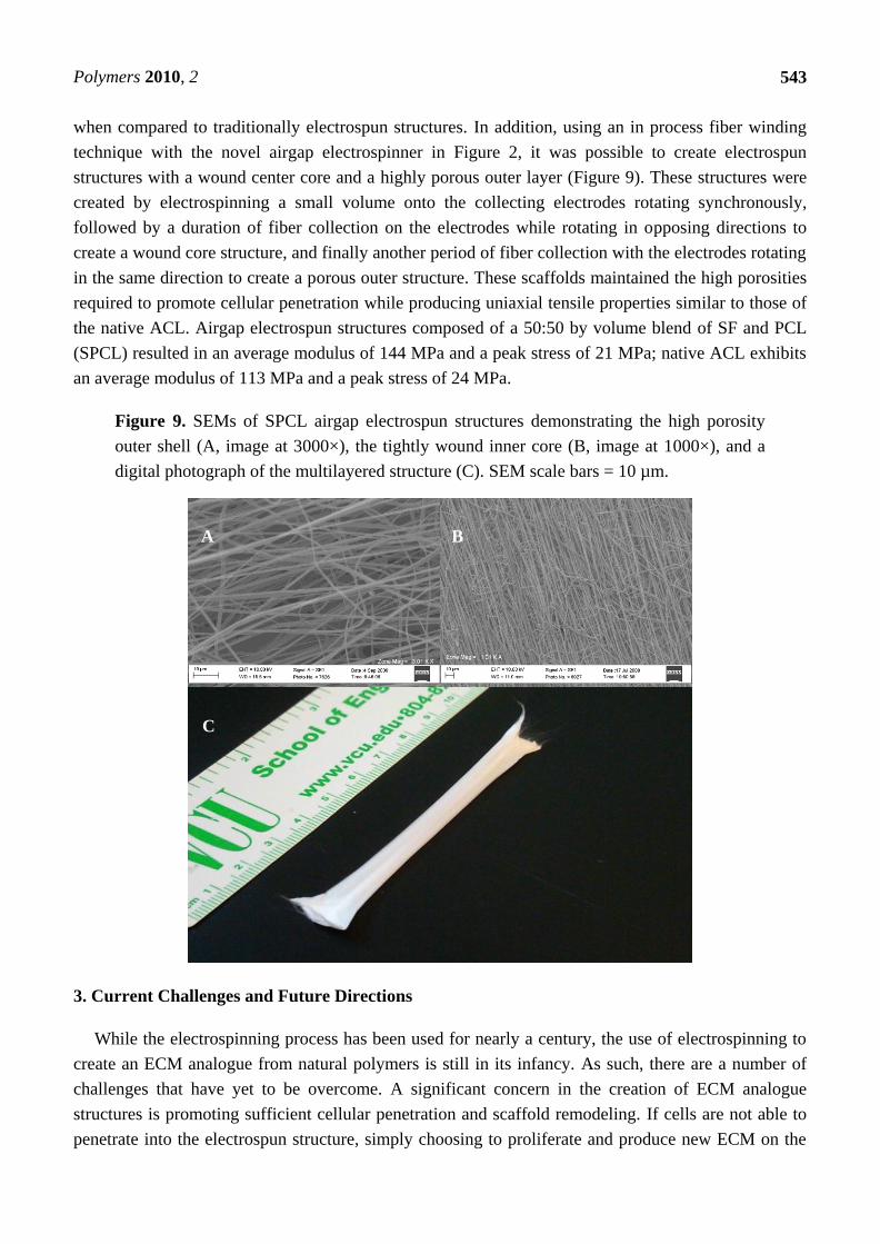
Polymers 2010, 2
543
when compared to traditionally electrospun structures. In addition, using an in process fiber winding
technique with the novel airgap electrospinner in Figure 2, it was possible to create electrospun
structures with a wound center core and a highly porous outer layer (Figure 9). These structures were
created by electrospinning a small volume onto the collecting electrodes rotating synchronously,
followed by a duration of fiber collection on the electrodes while rotating in opposing directions to
create a wound core structure, and finally another period of fiber collection with the electrodes rotating
in the same direction to create a porous outer structure. These scaffolds maintained the high porosities
required to promote cellular penetration while producing uniaxial tensile properties similar to those of
the native ACL. Airgap electrospun structures composed of a 50:50 by volume blend of SF and PCL
(SPCL) resulted in an average modulus of 144 MPa and a peak stress of 21 MPa; native ACL exhibits
an average modulus of 113 MPa and a peak stress of 24 MPa.
Figure 9. SEMs of SPCL airgap electrospun structures demonstrating the high porosity
outer shell (A, image at 3000×), the tightly wound inner core (B, image at 1000×), and a
digital photograph of the multilayered structure (C). SEM scale bars = 10 µm.
3. Current Challenges and Future Directions
While the electrospinning process has been used for nearly a century, the use of electrospinning to
create an ECM analogue from natural polymers is still in its infancy. As such, there are a number of
challenges that have yet to be overcome. A significant concern in the creation of ECM analogue
structures is promoting sufficient cellular penetration and scaffold remodeling. If cells are not able to
penetrate into the electrospun structure, simply choosing to proliferate and produce new ECM on the
A
C
B

Polymers 2010, 2
544
surface, then the electrospun structure will be of little use as a tissue engineering scaffold. Ideally, the
scaffold should become uniformly cellularized to promote adequate remodeling and eventual
replacement with native matrix. While scaffolds made from naturally occurring polymers tend to fare
better than their synthetic polymer counterparts when it comes to cellular penetration, they are still not
perfect. While the incorporation of recombinant human growth factors to induce cell chemotaxis into an
electrospun scaffold or the microintegration technique described previously can significantly enhance
scaffold cellularity, both require further optimization and research to become common practice.
Another challenge in the use of electrospinning to create ECM analogues is the ability for the
process to create truly three-dimensional structures that can replicate macroscopically the architecture
of specific native tissues. While the microscopic architecture of an electrospun scaffold can be made to
closely resemble the fibrous nature of the native ECM, the process has traditionally been limited to the
creation of thin sheets and tubes. On the other hand, a process such as 3-D printing can create intricate
three-dimensional structures, but lacks the precision to create fine nanofibers mimicking those of the
native ECM. Future work in the field of electrospinning for tissue engineering will have to address and
overcome this issue to be successful. The authors believe that the flexibility of the electrospinning
process may be its greatest asset. As the previously described airgap electrospinning process, and
previous research in the same vein have demonstrated, simply altering the way that fibers are collected
can have a profound impact on the macroscopic architecture of an electrospun scaffold. Again, more
research in this direction is needed, but the preliminary results have been promising.
As the electrospinning of natural polymers for tissue engineering applications continues to evolve,
so too will our ability to control the process and tailor its output to our tissue specific needs. Having
already harnessed the ability to create nanofibers nearly identical to those seen natively in the ECM,
from the same molecules as the native ECM, it is only a matter of time before truly biomimicking
scaffolds are created from electrospun natural polymers.
4. Conclusions
While many obstacles remain in the creation of the ideal ECM analogue structure, the capability to
electrospin the natural polymers found so prevalently in the native architecture has yielded promising
results. The inherent bioactivity of these proteins, combined with the nanoscale fiber producing
potential of the electrospinning process, has proven conducive to cellular adhesion, proliferation,
migration, and differentiation. The mechanical properties of these electrospun biomimicking scaffolds,
at times supplemented by synthetic polymers, have demonstrated mechanical properties capable of
replicating those of an array of native tissues. Additionally, preliminary in vivo studies with
electrospun natural polymer structures have proven the scaffolds to be adequately remodeled and
integrated with native tissue. With the diversity of the native ECM, it is highly unlikely that a single
processing technique or material will come to the forefront as the tissue engineering scaffold of choice,
however the adaptability of the electrospinning process and its ability to create nanoscale fibers from
the natural polymers found so prevalently in the native architecture holds great potential.

Polymers 2010, 2
545
References
1. Barnes, C.P.; Sell, S.A.; Boland, E.D.; Simpson, D.G.; Bowlin, G.L. Nanofiber technology:
designing the next generation of tissue engineering scaffolds. Adv. Drug Delivery Rev. 2007, 59,
1413-1433.
2. Martins-Green, M. The dynamics of Cell-ECM interactions with implications for tissue
engineering. In Principles of Tissue Engineering; Lanza, R. Langer, R, Chick, W, Eds.; R.G.
Landes Company: Austin, TX, USA, 1997; pp. 23-46.
3. Palsson, B.O.; Bhatia, S.N. Tissue Engineering; Pearson Prentice Hall: Upper Sadle River, NJ,
USA, 2004.
4. Farach-Carson, M.C.; Wagner, R.C.; Kiick, K.L. Extracellular matrix: Structure, function, and
applications to tissue engineering. In Tissue Engineering; Fisher, J.P., Mikos, A.G., Bronzino,
J.D., Eds.; CRC Press: Boca Raton, FL, USA, 2007; pp. 3-1-3-22.
5. Boland, E.D.; Espy, P.G.; Bowlin, G.L. Tissue engineering scaffolds. In Encyclopedia of
BioMaterials and Biomedical Engineering; Informa Healthcare: London, UK, 2004; pp. 1-7.
6. Chew, S.Y.; Wen, Y.; Dzenis, Y.; Leong, K.W. The role of electrospinning in the emerging field
of nanomedicine. Curr. Pharm. Des. 2006, 12, 4751-4570.
7. Lannutti, J.; Reneker, D.; Ma, T.; Tomasko, D.; Farson, D. Electrospinning for tissue engineering
scaffolds. Mater. Sci. Eng. C Bio. Supramol. Syst. 2007, 27, 504-509.
8. Sell, S.; Barnes, C.; Smith, M.; McClure, M.; Madurantakam, P.; Grant, J.; McManus, M.;
Bowlin, G. Extracellular matrix regenerated: tissue engineering via electrospun biomimetic
nanofibers. Polym. Int. 2007, 56, 1349-1360.
9. Kumbar, S.G.; James, R.; Nukavarapu, S.P.; Laurencin, C.T. Electrospun nanofiber scaffolds:
engineering soft tissues. Biomed. Mater. 2008, 3, 034002.
10. Sill, T.J.; von Recum, H.A. Electrospinning: Applications in drug delivery and tissue
engineering. Biomaterials 2008, 29, 1989-2006.
11. Sell, S.A.; McClure, M.J.; Garg, K.; Wolfe, P.S.; Bowlin, G.L. Electrospinning of
collagen/biopolymers for regenerative medicine and cardiovascular tissue engineering. Adv.
Drug Delivery Rev. 2009, 61, 1007-1019.
12. Jayaraman, K.; Kotaki, M.; Zhang, Y.; Mo, X.; Ramakrishna, S. Recent advances in polymer
nanofibers. J. NanoSci. Nanotechnol. 2004, 4, 52-65.
13. Engel, E.; Michiardi, A.; Navarro, M.; Lacroix, D.; Planell, J.A. Nanotechnology in regenerative
medicine: the Mater. side. Trends Biotechn. 2007, 26, 39-47.
14. Mano, J.F.; Silva, G.A.; Azevedo, H.S.; Malafaya, P.B.; Sousa, R.A.; Silva, S.S.; Boesel, L.F.;
Oliveira, J.; Santos, T.C.; Marques, A.P.; Neves, N.M.; Reis, R.L. Natural origin biodegradable
systems in tissue engineering and regenerative medicine: present status and some moving trends.
J. Royal Soc. Interface 2007, 4, 999-1030.
15. Ramakrishna, S.; Fujihara, K.; Teo, W.E.; Lim, T.C.; Ma, Z. Introduction to Electrospinning and
Nanofibers; World Scientific Publishing Company, Incorporated: Singapore, 2005.
16. Kidoaki, S.; Kwon, I.K.; Matsuda, T. Mesoscopic spatial designs of nano- and microfiber meshes
for tissue-engineering matrix and scaffold based on newly devised multilayering and mixing
electrospinning techniques. Biomaterials 2005, 26, 37-46.

Polymers 2010, 2
546
17. Hutmacher, D.; Woodfield, T.; Dalton, P.; Lewis, J. Scaffold design and fabrication. In Tissue
Engineering; Blitterswijk, C.V., Ed.; Elsevier Inc.: San Diego, CA, USA, 2008; pp. 403-454.
18. Stankus, J.J.; Soletti, L.; Fujimoto, K.; Hong, Y.; Vorp, D.A.; Wagner, W.R. Fabrication of cell
microintegrated blood vessel constructs through electrohydrodynamic atomization. Biomaterials
2007, 28, 2738-2746.
19. Sell, S.A.; McClure, M.J.; Ayres, C.E.; Simpson, D.G.; Bowlin, G.L. Preliminary investigation
of airgap electrospun silk fibroin-based structures for ligament analogue engineering. J.
Biomater. Sci. Polym. Ed. 2010, in press.
20. Konomi, H.; Hayashi, T.; Nakayasu, K.; Arima, M. Localization of type V collagen and type IV
collagen in human cornea, lung, and skin. Immunohistochemical evidence by anti-collagen
antibodies characterized by immunoelectroblotting. Am. of Pathol. 1984, 116, 417-426.
21. Huang, L.; Nagapudi, K.; Apkarian, R.P.; Chaikof, E.L. Engineered collagen-PEO nanofibers
and fabrics. J. Biomater. Sci. Polym. Ed. 2001, 12, 979-93.
22. Matthews, J.A.; Wnek, G.E.; Simpson, D.G.; Bowlin, G.L. Electrospinning of collagen
nanofibers. Biomacromolecules 2002, 3, 232-238.
23. Matthews, J.A.; Boland, E.D.; Wnek, G.E.; Simpson, D.G.; Bowlin, G.L. Electrospinning of
Collagen Type II: A Feasibility Study. J. Bioact. Compat. Polym. 2003, 18, 125-134.
24. Barnes, C.P.; Sell, S.A.; Knapp, D.C.; Walpoth, B.H.; Brand, D.D.; Bowlin, G.L. Preliminary
investigation of electrospun collagen and polydioxanone for vascular tissue engineering
applications. Int. J. Electrospun Nanofiber. Appl. 2007, 1, 73-87.
25. Barnes, C.P.; Pemble, C.W.; Brand, D.D.; Simpson, D.G.; Bowlin, G.L. Cross-linking
electrospun type II collagen tissue engineering scaffolds with carbodiimide in ethanol. Tissue
Eng. 2007, 13, 1593-1605.
26. Dong, B.; Arnoult, O.; Smith, M.E.; Wnek, G.E. Electrospinning of Collagen Nanofiber
Scaffolds from Benign Solvents. Macromol. Rapid Commun. 2009, 30, 539-542.
27. Choi, J.S.; Lee, S.W.; Jeong, L.; Bae, S.H.; Min, B.C.; Youk, J.H.; Park, W.H. Effect of
organosoluble salts on the nanofibrous structure of electrospun poly(3-hydroxybutyrate-co-3-
hydroxyvalerate). Int. J. Biol. Macromol. 2004, 34, 249-256.
28. Zhong, S.; Teo, W.E.; Zhu, X.; Beuerman, R.; Ramakrishna, S.; Yung, L.Y. Formation of
collagen-glycosaminoglycan blended nanofibrous scaffolds and their biological properties.
Biomacromolecules 2005, 6, 2998-3004.
29. Thomas, V.; Dean, D.R.; Jose, M.V.; Mathew, B.; Chowdhury, S.; Vohra, Y.K. Nanostructured
biocomposite scaffolds based on collagen coelectrospun with nanohydroxyapatite.
Biomacromolecules 2007, 8, 631-637.
30. Chen, P.J.; Chang, G.Y.; Chen, J.K. Electrospun collagen/chitosan nanofibrous membrane as
wound dressing. Colloids Surface. A 2008, 313-314, 183-188.
31. Chen, Z.G.; Wang, P.W.; Wei, B.; Mo, X.M.; Cui, F.Z. Electrospun collagen-chitosan nanofiber:
A biomimetic extracellular matrix for endothelial cell and smooth muscle cell. Acta Biomater.
2009, 6, 372-382.
32. Yeo, I.S.; Oh, J.E.; Jeong, L.; Lee, T.S.; Lee, S.J.; Park, W.H.; Min, B.M. Collagen-based
biomimetic nanofibrous scaffolds: preparation and characterization of collagen/silk fibroin
bicomponent nanofibrous structures. Biomacromolecules 2008, 9, 1106-1116.

Polymers 2010, 2
547
33. He, W.; Yong, T.; Teo, W.E.; Ma, Z.; Ramakrishna, S. Fabrication and endothelialization of
collagen-blended biodegradable polymer nanofibers: potential vascular graft for blood vessel
tissue engineering. Tissue Eng. 2005, 11, 1574-1588.
34. Kwon, I.K.; Matsuda, T. Co-electrospun nanofiber fabrics of poly(L-lactide-co-epsilon-
caprolactone) with type I collagen or heparin. Biomacromolecules 2005, 6, 2096-105.
35. Stitzel, J.D.; Pawlowski, K.J.; Wnek, G.E.; Simpson, D.G.; Bowlin, G.L. Arterial smooth muscle
cell proliferation on a novel biomimicking, biodegradable vascular graft scaffold. J. Biomater.
Appl. 2001, 16, 22-33.
36. Boland, E.D.; Coleman, B.D.; Barnes, C.P.; Simpson, D.G.; Wnek, G.E.; Bowlin, G.L.
Electrospinning polydioxanone for biomedical applications. Acta Biomater. 2005, 1, 115-123.
37. Lee, S.J.; Liu, J.; Oh, S.H.; Soker, S.; Atala, A.; Yoo, J.J. Development of a composite vascular
scaffolding system that withstands physiological vascular conditions. Biomaterials 2008, 29,
2891-2898.
38. Tillman, B.W.; Yazdani, S.K.; Lee, S.J.; Geary, R.L.; Atala, A.; Yoo, J.J. The in vivo stability of
electrospun polycaprolactone-collagen scaffolds in vascular reconstruction. Biomaterials 2009,
30, 583-8.
39. Venugopal, J.; Zhang, Y.Z.; Ramakrishna, S. Fabrication of modified and functionalized
polycaprolactone nanofibre scaffolds for vascular tissue engineering. Nanotechnology 2005, 16,
2138-2142.
40. Choi, J.S.; Lee, S.J.; Christ, G.J.; Atala, A.; Yoo, J.J. The influence of electrospun aligned
poly(epsilon-caprolactone)/collagen nanofiber meshes on the formation of self-aligned skeletal
muscle myotubes. Biomaterials 2008, 29, 2899-2906.
41. Schnell, E.; Klinkhammer, K.; Balzer, S.; Brook, G.; Klee, D.; Dalton, P.; Mey, J. Guidance of
glial cell migration and axonal growth on electrospun nanofibers of poly-epsilon-caprolactone
and a collagen/poly-epsilon-caprolactone blend. Biomaterials 2007, 28, 3012-3025.
42. Yoon, D.M.; Fisher, J.P. Polymeric Scaffolds for Tissue Engineering Applications. In Tissue
Engineering; Fisher, J.P., Mikos, A.G., Bronzino, J.D., Eds.; Taylor and Francis Group, LLC:
Boca Raton, FL, USA, 2007; p. 18.
43. Li, M.; Mondrinos, M.J.; Gandhi, M.R.; Ko, F.K.; Weiss, A.S.; Lelkes, P.I. Electrospun protein
fibers as matrices for tissue engineering. Biomaterials 2005, 26, 5999-6008.
44. Zhang, Y.; Ouyang, H.; Lim, C.T.; Ramakrishna, S.; Huang, Z.-M. Electrospinning of Gelatin
Fibers and Gelatin/PCL Composite Fibrous Scaffolds. J. Biomed. Mater. Res. B Appl. BioMater.
2005, 72B, 156-165.
45. Zhang, S.; Huang, Y.; Yang, X.; Mei, F.; Ma, Q.; Chen, G.; Ryu, S.; Deng, X. Gelatin
nanofibrous membrane fabricated by electrospinning of aqueous gelatin solution for guided
tissue regeneration. J. Biomed. Mater. Res. 2009, doi: 10.1002/jbm.a.32136.
46. Heydarkhan-Hagvall, S.; Schenke-Layland, K.; Dhanasopon, A.P.; Rofail, F.; Smith, H.; Wu,
B.M.; Shemin, R.; Beygui, R.E.; MacLellan, W.R. Three-dimensional electrospun ECM-based
hybrid scaffolds for cardiovascular tissue engineering. Biomaterials 2008, 29, 2907-2914.
47. Sisson, K.; Zhang, C.; Farach-Carson, M.C.; Chase, D.B.; Rabolt, J.F. Evaluation of
cross-linking methods for electrospun gelatin on cell growth and viability. BioMacromolecules
2009, 10, 1675-1680.

Polymers 2010, 2
548
48. Francis, L.; Venugopal, J.; Prabhakaran, M.P.; Thavasi, V.; Marsano, E.; Ramakrishna, S.
Simultaneous electrospin-electrosprayed biocomposite nanofibrous scaffolds for bone tissue
regeneration. Acta Biomater. 2010, 6, 4100-4109
49. Ohkawa, K.; Hayashi, S.; Kameyama, N.; Yamamoto, H.; Yamaguchi, M.; Kimoto, S.; Kurata,
S.; Shinji, H. Synthesis of collagen-like sequential polypeptides containing O-Phospho-L-
hydroxyproline and preparation of electrospun composite fibers for possible dental application.
Macromol. BioSci. 2009, 9, 79-92.
50. Sisson, K.; Zhang, C.; Farach-Carson, M.C.; Chase, D.B.; Rabolt, J.F. Fiber diameters control
osteoblastic cell migration and differentiation in electrospun gelatin. J. Biomed. Mater. Res. A
2010, 94A, 1312-1320.
51. Gauthaman, K.; Venugopal, J.R.; Yee, F.C.; Peh, G.S.L.; Ramakrishna, S.; Bongso, A.
Nanofibrous substrates support colony formation and maintain stemness of human embryonic
stem cells. J. Cell. Mol. Med. 2009, 13, 3475-3484.
52. Gupta, D.; Venugopal, J.; Prabhakaran, M.P.; Dev, V.R.G.; Low, S.; Choon, A.T.; Ramakrishna,
S. Aligned and random nanofibrous substrate for the in vitro culture of Schwann cells for neural
tissue engineering. Acta Biomater. 2009, 5, 2560-2569.
53. Songchotikunpan, P.; Tattiyakul, J.; Supaphol, P. Extraction and electrospinning of gelatin from
fish skin. Int. J. Biol. Macromol. 2008, 42, 247-255.
54. Song, J.-H.; Kim, H.-E.; Kim, H.-W. Production of electrospun gelatin nanofiber by water-based
co-solvent approach. J. Mater. Sci. Mater. Med. 2008, 19, 95-102.
55. Li, J.; He, A.; Zheng, J.; Han, C.C. Gelatin and gelatin-hyaluronic acid nanofibrous membranes
produced by electrospinning of their aqueous solutions. Biomacromolecules 2006, 7, 2243-2247.
56. Jiankang, H.; Dichen, L.; Yaxiong, L.; Bo, Y.; Hanxiang, Z.; Qin, L.; Bingheng, L.; Yi, L.
Preparation of chitosan-gelatin hybrid scaffolds with well-organized microstructures for hepatic
tissue engineering. Acta Biomater. 2008, in Press.
57. Gupta, D.; Venugopal, J.; Mitra, S.; Dev, V.R.G.; Ramakrishna, S. Nanostructured biocomposite
substrates by electrospinning and electrspraying for the mineralization of osteoblasts.
Biomaterials 2009, 30, 2085-2094.
58. Gui-Bo, Y.; You-Zhu, Z.; Shu-Dong, W.; De-Bing, S.; Zhi-Hui, D.; Wei-Guo, F. Study of the
electrospun PLA/silk fibroin-gelatin composite nanofibrous scaffold for tissue engineering. J.
Biomed. Mater. Res. 2010, 93A, 158-163.
59. Chong, E.J.; Phan, T.T.; Lim, I.J.; Zhang, Y.Z.; Bay, B.H.; Ramakrishna, S.; Lim, C.T.
Evaluation of electrospun PCL/gelatin nanofibrous scaffold for wound healing and layered
dermal reconstitution. Acta Biomater. 2007, 3, 321-330.
60. Kim, S.E.; Heo, D.N.; Lee, J.B.; Kim, J.R.; Park, S.H.; Jeon, S.H.; Kwon, I.K. Electrospun
gelatin/polyurethane blended nanofibers for wound healing. Biomed. Mater. 2009, 4, 1-11.
61. Ghasemi-Mobarakeh, L.; Prabhakaran, M.P.; Morshed, M.; Nasr-Esfahani, M.H.; Ramakrishna,
S. Electrical Stimulation of nerve cells using conductive nanofibrous scaffolds for nerve tissue
engineering. Tissue Eng. A 2009, 15, 3605-3619.
62. Ghasemi-Mobarakeh, L.; Prabhakaran, M.P.; Morshed, M.; Nasr-Esfahani, M.H.; Ramakrishna,
S. Electrospun poly(epsilon-caprolactone)/gelatin nanofibrous scaffolds for nerve tissue
engineering. BioMater. 2008, 29, 4532-4539.

Polymers 2010, 2
549
63. Kim, H.-W.; Yu, H.-S.; Lee, H.-H. Nanofibrous matrices of poly(lactic acid) and gelatin
polymeric blends for the improvement of cellular responses. J. Biomed. Mater. Res. 2008, 87A,
25-32.
64. Dhandayuthapani, B.; Krishnan, U.M.; Sethuraman, S. Fabrication and characterization of
chitosan-gelatin blend nanofibers for skin tissue engineering. J. Biomed. Mater. Res. B Appl.
BioMater. 2010, 94B, 264-272.
65. Powell, H.M.; Boyce, S.T. Fiber density of electrospun gelatin scaffolds regulates
morphogenesis of dermal-epidermal skin substitutes. J. Biomed. Mater. Res. 2008, 84A,
1078-1086.
66. Wang, S.; Zhang, Y.; Wang, H.; Yin, G.; Dong, Z. Fabrication and Properties of the Electrospun
Polylactide/Silk Fibroin-Gelatin Composite Tubular Scaffold. Biomacromolecules 2009, 10,
2240-2244.
67. Li, M.; Guo, Y.; Wei, Y.; MacDiarmid, A.G.; Lelkes, P.I. Electrospinning polyaniline-contained
gelatin nanofibers for tissue engineering applications. Biomaterials 2006, 27, 2705-2715.
68. Li, M.; Mondrinos, M.J.; Chen, X.; Gandhi, M.R.; Ko, F.K.; Lelkes, P.I. Co-electrospun
poly(lactide-co-glycolide), gelatin, and elastin blends for tissue engineering scaffolds. J. Biomed.
Mater. Res. 2006, 79A, 963-973.
69. Kejing, A.; Haiying, L.; Shidong, G.; Kumar, D.N.T.; Qingqing, W. Preparation of fish gelatin
and fish gelatin/poly(L-lactide) nanofibers by electrospinning. Int. J. Biol. Macromol. 2010, 47,
380-388.
70. Rodgers, U.R.; Weiss, A.S. Cellular interactions with elastin. Pathol. Biol. 2005, 53, 390-398.
71. Debelle, L.; Tamburro, A.M. Elastin: molecular description and function. Int. J. Biochem. Cell
Biol. 1999, 31, 261-272.
72. Duca, L.; Floquet, N.; Aliz, A.J.P.; Haye, B.; Debelle, L. Elastin as a matrikine. Crit. Rev. Oncol.
Hematol. 2004, 49, 235-244.
73. Huang, L.; McMillan, R.A.; Apkarian, R.P.; Pourdeyhimi, B.; Conticello, V.P.; Chaikof, E.L.
Generation of synthetic elastin-mimetic small diameter fibers and fiber networks.
Macromolecules 2000, 33, 2989-2997.
74. Partridge, S.M.; Davis, H.F. The Chemistry of Connective Tissues 3. Composition of the soluble
proteins derived from elastin. Biochem. J. 1955, 61, 21-30.
75. Daamen, W.F.; Veerkamp, J.H.; van Hest, J.C.M.; van Kuppevelt, T.H. Elastin as a biomaterial
for tissue engineering. Biomaterials 2007, 28, 4378-4398.
76. Lamme, E.N.; van Leeuwen, R.T.; Jonker, A.; van Marle, J.; Middelkoop, E. Living skin
substitutes: Survival and function of fibroblasts seeded in a dermal substitute in experimental
wounds. J. Invest. Dermatol. 1998, 111, 989-995.
77. Neuenschwander, S.; Hoerstrup, S.P. Heart valve tissue engineering. Transplant Immunol. 2004,
12, 359-365.
78. Xu, J.W.; Johnson, T.S.; Motarjem, P.M.; Peretti, G.M.; Randolph, M.A.; Yaremchuk, M.J.
Tissue-engineered flexible ear-shaped cartilage. Plast. Reconstr. Sur. 2005, 115, 1633-1641.
79. Nivison-Smith, L.; Rnjak, J.; Weiss, A.S. Synthetic human elastin microfibers: Stable
cross-linked tropoelastin and cell interactive constructs for tissue engineering applications. Acta
Biomater. 2010, 6, 354-359.

Polymers 2010, 2
550
80. McClure, M.; Sell, S.; Barnes, C.P.; Bowen, W.C.; Bowlin, G.L. Cross-linking Electrospun
Polydioxanone-Soluble Elastin Blends: Material Characterization. J. Eng. Fiber. Fabr. 2008, 3,
1-10.
81. Boland, E.D.; Matthews, J.A.; Pawlowski, K.J.; Simpson, D.G.; Wnek, G.E.; Bowlin, G.L.
Electrospinning collagen and elastin: Preliminary vascular tissue engineering. Front. BioSci.
2004, 9, 1422-1432.
82. Buttafoco, L.; Kolkman, N.G.; Engbers-Buijtenhuijs, P.; Poot, A.A.; Dijkstra, P.J.; Vermes, I.;
Feijen, J. Electrospinning of collagen and elastin for tissue engineering applications.
Biomaterials 2006, 27, 724-734.
83. Sell, S.A.; McClure, M.J.; Barnes, C.P.; Knapp, D.C.; Walpoth, B.H.; Simpson, D.G.; Bowlin,
G.L. Electrospun polydioxanone-elastin blends: potential for bioresorbable vascular grafts.
Biomed. Mater. 2006, 1, 72-80.
84. Smith, M.J.; McClure, M.J.; Sell, S.A.; Barnes, C.P.; Walpoth, B.H.; Simpson, D.G.; Bowlin,
G.L. Suture-reinforced electrospun polydioxanone-elastin small-diameter tubes for use in
vascular tissue engineering: A feasibility study. Acta Biomater. 2008, 4, 58-66.
85. Thomas, V.; Zhang, X.; Vohra, Y.K. A Biomimetic tubular scaffold with spatially designed
nanofibers of Protein/PDS bio-blends. Biotechnol. Bioeng. 2009, 104, 1025-1033.
86. Thomas, V.; Zhang, X.; Catledge, S.A.; Vohra, Y.K. Functionally graded electrospun scaffolds
with tunable mechanical properties for vascular tissue regeneration. Biomed. Mater. 2007, 2,
224-232.
87. Stitzel, J.; Liu, J.; Lee, S.J.; Komura, M.; Berry, J.; Soker, S.; Lim, G.; Van Dyke, M.; Czerw, R.;
Yoo, J.J.; Atala, A. Controlled fabrication of a biological vascular substitute. Biomaterials 2006,
27, 1088-1094.
88. Lee, S.J.; Yoo, J.J.; Lim, G.J.; Atala, A.; Stitzel, J. In vitro evaluation of electrospun nanofiber
scaffolds for vascular graft application. J. Biomed. Mater. Res. A 2007, 83, 999-1008.
89. El-Kurdi, M.S.; Hong, Y.; Stankus, J.J.; Soletti, L.; Wagner, W.R.; Vorp, D.A. Transient elastic
support for vein grafts using a constricting microfibrillar polymer wrap. Biomaterials 2008, 29,
3213-3220.
90. Doolittle, R.F. Fibrinogen and fibrin. Annu. Rev. Biochem. 1984, 53, 195-229.
91. Fuss, C.; Palmaz, J.C.; Sprague, E.A. Fibrinogen: structure, function, and surface interactions. J.
Vasc. Interv. Radiol. 2001, 12, 677-82.
92. Clark, R.A. Fibrin is a many splendored thing. J. Invest. Dermatol. 2003, 121, xxi-xxii.
93. Hanson, S. Blood coagulation and blood-materials interactions. In BioMaterials Science: An
Introduction to Materials in Medicine; Elsevier Academic Press: San Diego, CA, USA, 2004;
pp. 332-338.
94. Mosesson, M.W.; Siebenlist, K.R.; Meh, D.A. The structure and biological features of fibrinogen
and fibrin. Ann. N. Y. Acad. Sci. 2001, 936, 11-30.
95. Kollman, J.M.; Pandi, L.; Sawaya, M.R.; Riley, M.; Doolittle, R.F. Crystal structure of human
fibrinogen. Biochemistry 2009, 48, 3877-3886.
96. Kim, J.; Song, H.; Park, I.; Carlisle, C.R.; Bonin, K.; Guthold, M. Denaturing of single
electrospun fibrinogen fibers studied by deep ultraviolet fluorescence microscopy. Microsc. Res.
Tech. 2010, doi: 10.1002/jemt.20896.

Polymers 2010, 2
551
97. Rybarczyk, B.J.; Lawrence, S.O.; Simpson-Haidaris, P.J. Matrix-fibrinogen enhances wound
closure by increasing both cell proliferation and migration. Blood 2003, 102, 4035-4043.
98. Drew, A.F.; Liu, H.; Davidson, J.M.; Daugherty, C.C.; Degen, J.L. Wound-healing defects in
mice lacking fibrinogen. Blood 2001, 97, 3691-3698.
99. Sahni, A.; Francis, C.W. Vascular endothelial growth factor binds to fibrinogen and fibrin and
stimulates endothelial cell proliferation. Blood 2000, 96, 3772-3778.
100. Wnek, G.E.; Carr, M.; Simpson, D.G.; Bowlin, G.L. Electrospinning of nanofiber fibrinogen
structures. Nano Lett. 2003, 3, 213-216.
101. McManus, M.C.; Boland, E.D.; Simpson, D.G.; Barnes, C.P.; Bowlin, G.L. Electrospun
fibrinogen: Feasibility as a tissue engineering scaffold in a rat cell culture model. J. Biomed.
Mater. Res. A 2007, 81, 299-309.
102. McManus, M.; Boland, E.; Sell, S.; Bowen, W.; Koo, H.; Simpson, D.; Bowlin, G. Electrospun
nanofibre fibrinogen for urinary tract tissue reconstruction. Biomed. Mater. 2007, 2, 257-262.
103. Carlisle, C.R.; Coulais, C.; Namboothiry, M.; Carroll, D.L.; Hantgan, R.R.; Guthold, M. The
mechanical properties of individual, electrospun fibrinogen fibers. Biomaterials 2009, 30,
1205-1213.
104. McManus, M.C.; Boland, E.D.; Koo, H.P.; Barnes, C.P.; Pawlowski, K.J.; Wnek, G.E.; Simpson,
D.G.; Bowlin, G.L. Mechanical properties of electrospun fibrinogen structures. Acta Biomater.
2006, 2, 19-28.
105. McManus, M.C.; Sell, S.A.; Bowen, W.C.; Koo, H.P.; Simpson, D.G.; Bowlin, G.L. Electrospun
fibrinogen-polydioxanone composite matrix: Potential for in situ urologic tissue engineering. J.
Eng. Fiber. Fabr. 2008, 3, 12-21.
106. Sell, S.A.; Francis, M.P.; Garg, K.; McClure, M.J.; Simpson, D.G.; Bowlin, G.L. Cross-linking
methods of electrospun fibrinogen scaffolds for tissue engineering applications. Biomed. Mater.
2008, 3, Epub.
107. Perez-Rigueiro, J.; Elices, M.; Llorca, J.; Viney, C. Tensile properties of silkworm silk obtained
by forced silking. J. Appl. Polym. Sci. 2001, 82, 1928-1935.
108. Liu, H.; Ge, Z.; Wang, Y.; Toh, S.L.; Sutthikhum, V.; Goh, J.C.H. Modification of sericin-free
silk fibers for ligament tissue engineering applications. J. Biomed. Mater. Res. B 2007, 82B,
129-138.
109. Nagarkar, S.; Patil, A.; Lele, A.; Bhat, S.; Bellare, J.; Mashelkar, R.A. Some mechanistic insights
into the gelation of regenerated silk fibroin sol. Ind. Eng. Chem. Res. 2009, 48, 8014-8023.
110. Zhang, Q.; Yan, S.; Li, M. Silk fibroin based porous materials Materials 2009, 2, 2276-2295.
111. Altman, G.H.; Diaz, F.; Jakuba, C.; Calabro, T.; Horan, R.L.; Chen, J.; Lu, H.; Richmond, J.;
Kaplan, D.L. Silk-based biomaterials. Biomaterials 2003, 24, 401-416.
112. Alessandrino, A.; Marelli, B.; Arosio, C.; Fare, S.; Tanzi, M.C.; Freddi, G. Electrospun silk
fibroin mats for tissue engineering. Eng. Life Sci. 2008, 8, 219-225.
113. Bayraktar, O.; Malay, O.; Ozgarip, Y.; Batigun, A. Silk fibroin as a novel coating material for
controlled release of theophylline. Eur. J. Pharm. Biopharm. 2005, 60, 373-381.
114. Liu, L.; Liu, J.; Wang, M.; Min, S.; Cai, Y.; Zhu, L.; Yao, J. Preparation and characterization of
nano-hydroxyapatite/silk fibroin porous scaffolds. J. BioMater. Sci. Polym. Ed. 2008, 19,
325-338.

Polymers 2010, 2
552
115. Silva, S.S.; Maniglio, D.; Motta, A.; Mano, J.F.; Reis, R.L.; Migliaresi, C. Genipin-modified
silk-fibroin nanometric nets. Macromol. BioSci. 2008, 8, 766-774.
116. Kearns, V.; Macintosh, A.C.; Crawford, A.; Hatton, P.V. Silk-based biomaterials for tissue
engineering. In Topics in tissue engineering; Ashammakhi, N., Chiellini, R.R.F., Eds.;
Biomaterials and Tissue Engineering Group: Finland, 2008; Volume 4, pp. 1-19.
117. Altman, G.H.; Horan, R.L.; Lu, H.H.; Moreau, J.; Martin, I.; Richmond, J.C.; Kaplan, D.L. Silk
matrix for tissue engineered anterior cruciate ligaments. Biomaterials 2002, 23, 4131-4141.
118. Wang, M.; Jin, H.-J.; Kaplan, D.L.; Rutledge, G.C. Mechanical properties of electrospun silk
fibers. Macromolecules 2004, 37, 6856-6864.
119. Jeong, L.; Lee, K.Y.; Park, W.H. Effect of solvent on the characteristics of electrospun
regenerated silk fibroin nanofibers. Key Eng. Mater. 2007, 342, 813-816.
120. Zarkoob, S.; Eby, R.K.; Reneker, D.H.; Hudson, S.D.; Ertley, D.; Adams, W.W. Structure and
morphology of electrospun silk nanofibers. Polymers 2004, 45, 3973-3977.
121. Zarkoob, S. Structure and morphology of regenerated silk nano-fibers produced by
electrospinning. Ph.D. Thesis, The University of Akron: Akron, OH, USA, 1998.
122. Zarkoob, S.; Reneker, D.H.; Ertley, D.; Eby, R.K.; Hudson, S.D. Synthetically spun silk
nanofibers and a process for making the same. U.S. Patent 6110590, August 2000.
123. Sukigara, S.; Gandhi, M.; Ayutsede, J.; Micklus, M.; Ko, F. Regeneration of Bombyx mori silk
by electrospinning—Part 1: Processing parameters and geometric properties. Polymers 2003, 44,
5721-5727.
124. Sukigara, S.; Gandhi, M.; Ayutesede, J.; Micklus, M.; Ko, F. Regeneration of Bombyx mori silk
by electrospinning. Part 2. Process optimization and empirical modeling using response surface
methodology. Polymers 2004, 45, 3701-3708.
125. Jin, H.-J.; Fridrikh, S.V.; Rutledge, G.C.; Kaplan, D.L. Electrospinning Bombyx mori silk with
poly(ethylene oxide). Biomacromolecules 2002, 3, 1233-1239.
126. Jin, H.J.; Chen, J.; Karageorgiou, V.; Altman, G.H.; Kaplan, D.L. Human bone marrow stromal
cell responses on electrospun silk fibroin mats. Biomaterials 2004, 25, 1039-1047.
127. Vepari, C.; Kaplan, D.L. Silk as a biomaterial. Progr. Polym. Sci. 2007, 32, 991-1007.
128. Wang, Y.; Rudym, D.D.; Walsh, A.; Abrahamsen, L.; Kim, H.-H.; Kim, H.S.; Kirker-Head, C.;
Kaplan, D.L. In vivo degradation of three-dimensional silk fibroin scaffolds. Biomaterials 2008,
29, 3415-3428.
129. Ueda, H.; Hong, L.; Yamamoto, M.; Shigeno, K.; Inoue, M.; Toba, T. Use of collagen sponge
incorporating transforming growth factor-beta 1 to promote bone repair in skull defects in
rabbits. Biomaterials 2002, 23, 1003-1010.
130. Sun, H.; Mei, L.; Song, C.; Cui, X.; Wang, P. The in vivo degradation, absorption and excretion
of PCL-based implant. Biomaterials 2006, 27, 1735-1740.
131. Ramchandani, M.; Pankaskie, M.; Robinson, D. The influence of manufacturing procedure on
the degradation of poly(lactide-co-glycolide) 85:15 and 50:50 implants. J. Control. Release
1997, 43, 161-173.
132. Kister, G.; Cassanas, G.; Vert, M. Structure and morphology of solid lactide-gly-colide
copolymers from C-13 nmr, infra-red and Raman Spectroscopy. Polymers 1998, 39, 3335-3340.

Polymers 2010, 2
553
133. Liao, S.; Watari, F.; Zhu, Y.; Uo, M.; Akasaka, T.; Wang, W. The degradation of the three
layered nano-carbonated hydroxyapatite/collagen/PLGA composite membrane in vitro. Dent.
Mater. 2007, 23, 1120-1128.
134. Zhou, J.; Cao, C.; Ma, X. A novel three-dimensional tubular scaffold prepared from silk fibroin
by electrospinning. Int. J. Biol. Macromol. 2009, 45, 504-510.
135. Marelli, B.; Alessandrino, A.; Fare, S.; Freddi, G.; Mantovani, D. Compliant electrospun silk
fibroin tubes for small vessel bypass grafting. Acta Biomater. 2010, in Press.
136. Soffer, L.; Wang, X.; Zhang, X.; Kluge, J.; Dorfmann, L.; Kaplan, D.L.; Leisk, G. Silk-based
electrospun tubular scaffolds for tissue-engineered vascular grafts. J. BioMater. Sci. Polym. Ed.
2008, 19, 653-664.
137. Zhang, X.; Baughman, C.B.; Kaplan, D.L. In vitro evaluation of electrospun silk fibroin
scaffolds for vascular cell growth. Biomaterials 2008, 29, 2217-2227.
138. Li, C.; Vepari, C.; Jin, H.J.; Kim, H.J.; Kaplan, D.L. Electrospun silk-BMP-2 scaffolds for bone
tissue engineering. Biomaterials 2006, 27, 3115-24.
139. Seo, Y.-K.; Choi, G.-M.; Kwon, S.-Y.; Lee, H.-S.; Park, Y.-S.; Song, K.-Y.; Kim, Y.-J.; Park,
J.-K. The biocompatibility of silk scaffold for tissue engineered ligaments. Key Eng. Mater.
2007, 342, 73-76.
140. Liu, H.; Fan, H.; Wang, Y.; Toh, S.L.; Goh, J.C.H. The interaction between a combined knitted
silk scaffold and microporous silk sponge with human mesenchymal stem cells for ligament
tissue engineering. Biomaterials 2008, 29, 662-674.
© 2010 by the authors; licensee MDPI, Basel, Switzerland. This article is an open access article
distributed under the terms and conditions of the Creative Commons Attribution license
(http://creativecommons.org/licenses/by/3.0/).