
The Use of Dupilumab in Atopic Dermatitis During ...
9
Open Access Maced J Med Sci. 2020 Nov 05; 8(T1):399-407. 399 The Use of Dupilumab in Atopic Dermatitis During Coronavirus Disease-19 Era – A Review Laura Pauline Kosasih* Department of Dermatology, Cardiff University, Cardiff, Wales, United Kingdom Abstract The global pandemic of coronavirus (CoV) d0isease 2019 (COVID-19), caused by severe acute respiratory syndrome CoV (SARS-CoV 2), has been a challenging event for every individual. It is known that COVID- 19 may exhibit a vast range of symptoms ranging from mild to severe. Acute respiratory distress syndrome (ARDS) and multiple organ failure are the most common causes of death in COVID-19 cases [3]. Accumulating evidence shows that T-helper type (Th-1) inflammation cascade plays a major role in COVID-19 pathogenesis. It is proposed that aberrant immune reaction, or known as cytokine storm, is one of the main causals of ARDS in COVID-19 case, while dupilumab, the first Food and Drug Administration-approved immunomodulatory treatment for atopic dermatitis, is known for its effectiveness in suppressing the Th-2 inflammation pathway. It is postulated that both types of inflammation can cross-regulate each other. Therefore, some may believe that the regression of Th-2 cascade may upregulate the Th-1 cascade, leading to an exaggerated cytokine storm. This hypothesis leads to the uncertainty of the safety of continuing this modality during the pandemic. Edited by: Mirko Spiroski Citation: Kosasih LP. The Use of Dupilumab in Atopic Dermatitis During Coronavirus Disease-19 Era – A Review. Open Access Maced J Med Sci. 2020 Nov 05; 8(T1):1-9. https://doi.org/10.3889/oamjms.2020.5359 Keywords: Atopic dermatitis; Dupilumab; Biologic treatment; Coronavirus disease-19 *Correspondence: Laura Pauline Kosasih. E-mail:[email protected] Received: 08-Aug-2020 Revised: 20-Oct-2020 Accepted: 26-Oct-2020 Copyright: © 2020 Laura Pauline Kosasih Funding: Publication of this article was financially supported by the Scientific Foundation SPIROSKI, Skopje, Republic of Macedonia Competing Interests: The authors have declared that no competing interests exist Open Access: This is an open-access article distributed under the terms of the Creative Commons Attribution- NonCommercial 4.0 International License (CC BY-NC 4.0) Introduction Atopic dermatitis (AD) can be deemed as one of the most common non-communicable dermatological ailments. It affects approximately 20% of children and 2–8% adults in most nations of the world [1], [2], [3], [4], [5], [6], [7], [8]. An international study shows that the figure has increased two-or three-fold within three decades in the developed countries. Therefore, it is predicted that the number will always accumulate, and this also shows that AD is a global health problem both in developing and developed nations [9]. Both personal and social aspects are greatly influenced by uncontrollable AD. For instance, the social stigma of visible skin efflorescence may affect individual’s self-confidence, and debilitating itch might lower one’s quality of life [10], [11]. Dupilumab, as the first Food and Drug Administration-approved biologic treatment, has been proven effective and has significantly improved quality of life. Some concerns about the safety of the utilization of dupilumab have been raised during coronavirus (CoV) disease (COVID)-19 pandemic. Some may fear that dupilumab may increase the susceptibility of acquiring COVID-19 or worsen the condition. However, it is also not recommended to discontinue dupilumab because of the chronic nature of AD and the unknown period of this pandemic [12]. In this article, the literature reviews of both clinical and immunology aspects of dupilumab in AD and COVID-19 have been explored. The aim is to provide the latest reference about dupilumab in AD patients and COVID-19. Thus, it can assist physicians in generating the best clinical judgment in a practice setting. A Glance of AD What is AD? AD is defined as a chronic inflammatory skin disorder, with one of the major hallmarks of extremely pruritic, and it is very common to be found during infancy and childhood period [13]. It is crucial to bear in mind that the diagnosis of AD includes an array of major and minor features. There is no single feature that can represent AD itself nor a diagnostic assessment [14], [15] (Table 1). Many guidelines and suggestions have been published to aid clinicians in establishing the diagnosis. However, it is implied by Tada [16] that mostly adopted guideline for diagnosing both in practice settings and clinical trials is the revised Hanifin and Rajla criteria. The diagnostic can be established when at least three major features and three minor features are noted [17]. Scientific Foundation SPIROSKI, Skopje, Republic of Macedonia Open Access Macedonian Journal of Medical Sciences. 2020 Nov 05; 8(T1):399-407. https://doi.org/10.3889/oamjms.2020.5359 eISSN: 1857-9655 Category: T1 - Thematic Issue “Coronavirus Disease (COVID-19)” Section: Narrative Review Article
Transcript of The Use of Dupilumab in Atopic Dermatitis During ...

Open Access Maced J Med Sci. 2020 Nov 05; 8(T1):399-407. 399
The Use of Dupilumab in Atopic Dermatitis During Coronavirus Disease-19 Era – A Review
Laura Pauline Kosasih*
Department of Dermatology, Cardiff University, Cardiff, Wales, United Kingdom
AbstractThe global pandemic of coronavirus (CoV) d0isease 2019 (COVID-19), caused by severe acute respiratory syndrome CoV (SARS-CoV 2), has been a challenging event for every individual. It is known that COVID-19 may exhibit a vast range of symptoms ranging from mild to severe. Acute respiratory distress syndrome (ARDS) and multiple organ failure are the most common causes of death in COVID-19 cases [3]. Accumulating evidence shows that T-helper type (Th-1) inflammation cascade plays a major role in COVID-19 pathogenesis. It is proposed that aberrant immune reaction, or known as cytokine storm, is one of the main causals of ARDS in COVID-19 case, while dupilumab, the first Food and Drug Administration-approved immunomodulatory treatment for atopic dermatitis, is known for its effectiveness in suppressing the Th-2 inflammation pathway. It is postulated that both types of inflammation can cross-regulate each other. Therefore, some may believe that the regression of Th-2 cascade may upregulate the Th-1 cascade, leading to an exaggerated cytokine storm. This hypothesis leads to the uncertainty of the safety of continuing this modality during the pandemic.
Edited by: Mirko SpiroskiCitation: Kosasih LP. The Use of Dupilumab in Atopic
Dermatitis During Coronavirus Disease-19 Era – A Review. Open Access Maced J Med Sci. 2020 Nov 05;
8(T1):1-9. https://doi.org/10.3889/oamjms.2020.5359
Keywords: Atopic dermatitis; Dupilumab; Biologic treatment; Coronavirus disease-19
*Correspondence: Laura Pauline Kosasih. E-mail:[email protected]
Received: 08-Aug-2020Revised: 20-Oct-2020
Accepted: 26-Oct-2020Copyright: © 2020 Laura Pauline Kosasih
Funding: Publication of this article was financially supported by the Scientific Foundation SPIROSKI, Skopje,
Republic of MacedoniaCompeting Interests: The authors have declared that no
competing interests existOpen Access: This is an open-access article distributed
under the terms of the Creative Commons Attribution-NonCommercial 4.0 International License (CC BY-NC 4.0)
Introduction
Atopic dermatitis (AD) can be deemed as one of the most common non-communicable dermatological ailments. It affects approximately 20% of children and 2–8% adults in most nations of the world [1], [2], [3], [4], [5], [6], [7], [8]. An international study shows that the figure has increased two-or three-fold within three decades in the developed countries. Therefore, it is predicted that the number will always accumulate, and this also shows that AD is a global health problem both in developing and developed nations [9].
Both personal and social aspects are greatly influenced by uncontrollable AD. For instance, the social stigma of visible skin efflorescence may affect individual’s self-confidence, and debilitating itch might lower one’s quality of life [10], [11]. Dupilumab, as the first Food and Drug Administration-approved biologic treatment, has been proven effective and has significantly improved quality of life. Some concerns about the safety of the utilization of dupilumab have been raised during coronavirus (CoV) disease (COVID)-19 pandemic. Some may fear that dupilumab may increase the susceptibility of acquiring COVID-19 or worsen the condition. However, it is also not recommended to discontinue dupilumab because of the chronic nature of AD and the unknown period of this pandemic [12].
In this article, the literature reviews of both clinical and immunology aspects of dupilumab in AD and COVID-19 have been explored. The aim is to provide the latest reference about dupilumab in AD patients and COVID-19. Thus, it can assist physicians in generating the best clinical judgment in a practice setting.
A Glance of AD
What is AD?
AD is defined as a chronic inflammatory skin disorder, with one of the major hallmarks of extremely pruritic, and it is very common to be found during infancy and childhood period [13]. It is crucial to bear in mind that the diagnosis of AD includes an array of major and minor features. There is no single feature that can represent AD itself nor a diagnostic assessment [14], [15] (Table 1). Many guidelines and suggestions have been published to aid clinicians in establishing the diagnosis. However, it is implied by Tada [16] that mostly adopted guideline for diagnosing both in practice settings and clinical trials is the revised Hanifin and Rajla criteria. The diagnostic can be established when at least three major features and three minor features are noted [17].
Scientific Foundation SPIROSKI, Skopje, Republic of MacedoniaOpen Access Macedonian Journal of Medical Sciences. 2020 Nov 05; 8(T1):399-407.https://doi.org/10.3889/oamjms.2020.5359eISSN: 1857-9655Category: T1 - Thematic Issue “Coronavirus Disease (COVID-19)”Section: Narrative Review Article

T1 - Thematic Issue “Coronavirus Disease (COVID-19)” Narrative Review Article
400 https://www.id-press.eu/mjms/index
AD is a very complicated and debilitating condition, and individuals may suffer from both physical and mental issues due to uncontrollable AD. Literature shows that AD is significantly affecting all aspects of the quality of life of patients and their families [11], [18], [19] (Figures 1 and 2).
Pathogenesis
The pathogenesis of AD is subsequently complex and is not fully elucidated until now. Many theories are proposed and postulated; however, it is widely agreed that AD is orchestrated from defective skin integrity, particular genes, and dramatic response of the immune system against exacerbating factors [14], [21], [22].
In general, it is found that AD individuals’ skin lacks essential genes that are needed to form a perfect skin barrier. For instance, some individuals may have filaggrin mutation; filaggrin is a gene that encodes essential proteins in building the epithelial barrier and ceramide, a lipid substance that plays an important role in retaining water permeability barrier function [22], [23] The lack of these two major materials leads to excessive trans-epidermal water loss, resulting in pH alteration and skin dryness. In addition, an antimicrobial peptide called cathelicidin, which is one of the very first layers of immune barrier, is also found to be depleted in most AD patients. [10]. Thus, exacerbating environmental stimuli such as aeroallergens, irritating chemicals, and pathogens are easier to penetrate the skin, initiating inflammation [14], [22], [24].
Th-2 type cells are widely accepted to be associated with both acute and chronic AD course [10], [14], [15]. However, Fujii [25] believes that either in acute and chronic lesion, there might be a switch from Th2 to Th1 activity.
In the acute event, interleukin (IL)-4, IL-5, IL-13, IL-25, and IL-33 levels increase [15]. Especially, in the early lesion of AD, it is shown that IL-4 and IL-13 dominate the inflammatory cascade [14], [15]. Meanwhile, in chronic AD, IL-31 has recently been discovered to be overly expressed and linked to the severity of the course.
On the other hand, it is also found that IL-4, IL-13, and IL-33 might downregulate the filaggrin, and thus, it is like a loophole of the immune system and defective skin barrier cascades [26].
Management
Overview
In general, many findings agree that the aim of the treatment of AD must be focused on inflammation
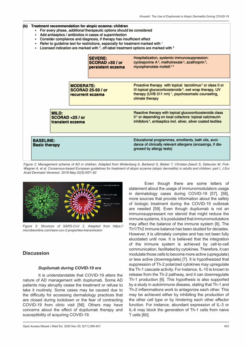
cessation by repairing the skin barrier and reducing the itch. The importance of education about the nature of the ailment, skin hydration, pharmacological regime, identification, and elimination of flare causal factors is often highlighted in virtually all guidelines [10], [14], [27]. Therefore, holistic and multi-faceted approaches are needed to manage this ailment. [8], [10], [28]. The Japanese guideline for AD believes that the management of AD must be based on three fundamental aspects. First is the investigation and countermeasures of the causal and exacerbating elements. Second is the repairment if the skin defect (skincare). Last is pharmacotherapy [28]. Similarly, the European Consensus Guidelines’ treatment option is quite similar to most guidelines and literature, aside from its agreement to divide the management into four phases: Baseline, mild, moderate, and severe [8]. The phases depend on the SCORing AD (SCORAD) (Appendix 1). SCORAD is one of the tools that can be used in assessing the extent and severity of AD. Less than SCORAD 25 is defined as mild, 25–50 as moderate, and more than 50 as severe. It is also suggested that in each phase, adding additional medication and antiseptic or antibiotic may be beneficial in treating superinfection.
A Brief Review of Dupilumab
Dupilumab is a human analogue monoclonal antibody that blocks IL-4 and IL-13 pathways by binding a shared α-subunit of IL-4 and IL-13, both of which are the major cytokines for Th-2 inflammation in AD [4], [29], [30].
Some experimental research demonstrated that early treatment with IL-4 and IL-3 blocking agents will dampen the responses to IFN and IL -17. In brief, when the early lesion of AD is exposed to IL-4 and IL-13, long-lasting persistent effects are doable [31]. Therefore, not only will dupilumab decrease the flare but it may also prevent the course of recalcitrant AD in the future. Dupilumab can be used either as monotherapy or combination therapy. Studies of dupilumab in 4 weeks and 12 weeks as monotherapy and as a combination with topical glucocorticoid in moderate-severe AD show significant improvement [5]. Not only were skin lesions improved but the severity of itch also rapidly decreased, allowing individuals to have a better quality of life. These results were also supported by a separate study, where Eczema Area and Severity Index score and peak pruritus Numerical Rating Score were reported to be significantly improved by the end of week 16th [32]. In terms of adverse effects, both placebo and intervention groups were almost equal [5], [32]. However, in a two phase 3 trial, it is also observed that nasopharyngitis was the second most common adverse effect after infection and infestation in the dupilumab groups [32]. A similar result is also discovered in a study of dupilumab and asthmatic patients [33].

Kosasih. The Use of Dupilumab in Atopic Dermatitis During COVID-19
Open Access Maced J Med Sci. 2020 Nov 05; 8(T1):399-407. 401
COVID-19
What is COVID-19?
History
In December 2019, several pneumonia-like cases with unknown etiology were reported in Wuhan, China. This disease has started with suggestive symptoms of progressive respiratory infection, with some patients developing acute respiratory distress syndrome (ARDS), acute respiratory failure, and other life-threatening complications [34]. A novel beta-CoV was discovered later in January 2020 to be the culprit. International Virus Classification Commission named the virus as Severe Acute Respiratory Syndrome-CoV 2 (SARS-CoV 2), while the World Health Organization (WHO) officially named the disease as COVID-19 in the next month [1], [35]. In March 2020, the WHO asserted that this disease is a global emergency, affecting every aspect of life, and thus, declared COVID-19 as a pandemic [36] (Figure 3).
Incubation and Clinical Characteristic
Data show that symptoms of COVID-19 usually appear after an incubation period of 5.1–12 days [37]. Fever, dry cough, and fatigue are the most common symptoms. However, other symptoms such as headache, hemoptysis, and gastrointestinal symptoms such as diarrhea and vomiting are likely to be exhibited as well. In addition, dyspnea is found to be developed in more than half of the patients [1], [38], [39]. A recent study also discovered that olfactory dysfunction such as anosmia and hyposmia was found prominently in COVID-19 patients [40]. However, COVID-19 can still yield in a person without showing any symptom, which makes this ailment easily transmitted.
Route of Transmission
Human-to-human transmission is feasible due to respiratory fomites or droplets. It is also suggested that direct and non-direct contacts through mucous membrane of eyes, nose, mouth, and skin are another potential routes of transmission [39], [41]. However, a recent study discovered that aerosol transmission is highly plausible through smaller droplets or droplet nuclei. Therefore, proper inter-personal distancing and usage of mask are very essential to control the spread of infection [42], [43]. Due to a recent study that shows COVID-19 cases with enteric symptoms, it is also
suggested that the digestive tract might be another possible route of transmission [44].
An article also suggests that percutaneous transmission is possible due to the high expression of Angiotensin-Converting Enzyme-2 (ACE2) in the skin tissue cells [45]. ACE-2 is known to facilitate the entry of the virus (further explanation will be explained in the next chapter). However, a thorough study is still needed to elucidate this hypothesis.
Pathogenesis
Virus structure
CoVs are enveloped with single-stranded, positive-strand RNA genome (26-32kb in weight) which comes from Coronaviridae family. There are four genera of CoVs; α, β, γ, δ, and COVID-19 belongs to the beta-CoV genus. Within beta genus itself, there are four lineages (A, B, C, and D) [3], [46].
The appearance of the virus is a rough, spherical and has prominent club-shaped elongations which contain its spike protein. This novel CoV has shown 88% similarity to bat-related SARS-like CoVs’ sequence (bat-SL-CoVZC45 and bat-SL-CoVZXC21), and approximately 50% identical to Middle East Respiratory Syndrome CoVs’ sequence. Due to its similar structure, the pathogenesis of SARS-CoV 2 can be postulated. However, the complete pathogenesis of COVID-19 has not been fully elucidated [47].Table 1: Major and minor features. Adapted from Goldsmith et al. and Correale et al. [14], [20]Major features
1. Pruritus2. Recurrent or relapse course of dermatitis3. Typical lesion (Facial and/or extensor rashes in infants and young children, and
flexural lichenification in older children and adults)4. Family history of atopic diatheses (asthma, allergic rhinitis, and atopic dermatitis)
Minor Features1. Xerosis2. Ichthyosis/palmar hyperlinearity, and keratosis pilaris3. Immediate (type I) skin test reaction4. Elevated IgE level5. Early age of onset6. Tendency toward cutaneous infections (especially staph. aureus and herpes
simplex), impaired cell- mediated immunity7. Tendency toward non-specific hand or foot dermatitis 8. Nipple eczema9. Cheilitis10. Recurrent conjunctivitis11. Dennie-Morgan infraorbital fold12. Keratoconus13. Anterior subcapsular cataracts14. Orbital darkening15. Facial pallor, and facial erythema16. Pityriasis alba17. Anterior neck folds18. Itch when sweating19. Intolerance to wool and lipid solvents20. Perifollicular accentuation21. Food intolerance22. Relaps influenced by environmental and emotional factor23. White dermographism, delayed blanch

T1 - Thematic Issue “Coronavirus Disease (COVID-19)” Narrative Review Article
402 https://www.id-press.eu/mjms/index
Figure 1: Management scheme of AD in adults. Adapted from Wollenberg A, Barbarot S, Bieber T, Christen-Zaech S, Deleuran M, Fink-Wagner A, et al. Consensus-based European guidelines for treatment of atopic eczema (atopic dermatitis) in adults and children: part I. J Eur Acad Dermatol Venereol. 2018 May;32(5):657–82
Host Entrance and Immune Response
SARS-CoV 2 can enter the host cells by direct membrane fusion. First, the envelope spike glycoprotein will bind to the host cellular receptor, facilitated by ACE2 [48]. After entering the cell, the virus RNA genome will be released to the cytoplasm and then commenced to the replication phase [49].
Subsequently, after the virus has successfully hijacked the cells, its antigen will be presented to the antigen presentation cells. Antigen presentation will evoke host immune response which is both humoral and cellular immunity. T cells and B cells play a major role as the immune mediators in this event [35].
Since COVID-19 is caused by a virus, similar to any viral infection, the innate immune pathway is the first line of defense. However, a further aberrant and disarrayed immune response might damage the immune systems, leading to fatality [3], [50]. This event is often known as a cytokine storm. Several studies report that ARDS is the main cause of mortality of COVID-19 patients, and ARDS is one of the results of the cytokine storm [3], [51], [52]. This dramatic cytokines response makes COVID-19 difficult to manage and threaten lives.
T-helper type 1 (Th-1) cascade plays an essential role in COVID-19 infection. It is observed that cytokines that generate Th-1 pathway such as IL-1B, IFN-γ, IP-10, and monocyte chemoattractant protein 1 rise [51]. This hypothesis is also supported by several studies and reports discovering highly expressed Th-1 related cytokines in many COVID-19 patients. For instance, a study by Huang et al. revealed that levels of IL-7, IL-8, IL-9, IL-10, fibroblast growth factor, granulocyte-colony stimulating factor, granulocyte-macrophage colony-stimulating factor, MIP-1A,MIP1-B, platelet-derived growth factor, tumor necrosis factor (TNF)α, and VEGF surge in both ICU and non-ICU required COVID-19 patients compared to healthy individuals [51].
An analyzing case study in China also saw a high expression of IL-10, IL-6, and TNF-α in severe cases compared to moderate cases [53]. An identical result is also found in a study of assessment of laboratory data reporting that IL-6 level significantly rises in severe cases compared to mild cases [54]. A multicenter study also supports previous data, in which it is observed that IL-6 was found higher in mortality cases than successful cases [55].
Kosasih. The Use of Dupilumab in Atopic Dermatitis During COVID-19
Open Access Maced J Med Sci. 2020 Nov 05; 8(T1):399-407. 403
Figure 3: Structure of SARS-CoV 2. Adapted from https://microbeonline.com/sars-cov-2-properties-transmission
Discussion
Dupilumab during COVID-19 era
It is understandable that COVID-19 alters the nature of AD management with dupilumab. Some AD patients may abruptly cease the treatment or refuse to take it routinely. Some cases may be caused due to the difficulty for accessing dermatology practices that are closed during lockdown or the fear of contracting COVID-19 from clinic visit [56]. Others may have concerns about the effect of dupilumab therapy and susceptibility of acquiring COVID-19.
Even though there are some letters of statement about the usage of immunomodulators usage in dermatology cases during COVID-19 [57], [58], more sources that provide information about the safety of biologic treatment during the COVID-19 outbreak are needed [59]. Even though dupilumab is not an immunosuppressant nor steroid that might reduce the immune systems, it is postulated that immunomodulators may affect the balance of the immune system [6]. The Th1/Th2 immune balance has been studied for decades. However, it is ultimately complex and has not been fully elucidated until now. It is believed that the integration of the immune system is achieved by cell-to-cell communication, facilitated by cytokines. Therefore, it can modulate those cells to become more active (upregulate) or less active (downregulate) [7]. It is hypothesized that suppression of Th-2 polarized cytokines may upregulate the Th-1 cascade activity. For instance, IL-10 is known to release from the Th-2 pathway, and it can downregulate Th-1 production [6]. This hypothesis is also supported by a study in autoimmune disease, stating that Th-1 and Th-2 inflammations work to antagonize each other. This may be achieved either by inhibiting the production of the other cell type or by hindering each other effector function. For instance, abundant expression of IL-3 or IL-6 may block the generation of Th-1 cells from naive T cells [60].
Figure 2: Management scheme of AD in children. Adapted from Wollenberg A, Barbarot S, Bieber T, Christen-Zaech S, Deleuran M, Fink-Wagner A, et al. Consensus-based European guidelines for treatment of atopic eczema (atopic dermatitis) in adults and children: part I. J Eur Acad Dermatol Venereol. 2018 May;32(5):657–82

T1 - Thematic Issue “Coronavirus Disease (COVID-19)” Narrative Review Article
404 https://www.id-press.eu/mjms/index
In other words, it is plausible that the production of Th-1 polarized cytokines is upregulated due to the decreased Th-2 activity, resulting from the lower expression of IL-4 and IL-13, blocked by dupilumab. This may worsen or increase the risk of aberrant cytokine storm in COVID-19 patients. However, a study disputes that dupilumab affects Th-1 activity. It is shown that no elevation of Th-1/IFN-g-related gene expression was observed in AD patients with dupilumab [61]. Moreover, a hypothesis is also proposed that dupilumab might give AD patients more protection from COVID-19 infection. It is known that expression of IL-6, one of the infamous cytokines that play role in cytokine storm, is depended on endogenous production of IL-4, which obviously decreases in patient on dupilumab. This mechanism gives the possibility of protective effect of dupilumab in the nature of COVID-19 [62].
Many have proposed that the concept of Th-1/Th-2 immune balance is very complex and not only influenced by the cytokines profile but also by antigen presentation, immunogenic and non-immunogenic cells, genetic, hormones, oxidative stress, and environment [6], [60], [63], [64]. Thus, further research is needed to elucidate this matter.
Limitations and Recommendation
One of the limitations of this article is the sparse data of COVID-19 due to its novelty. Another limitation includes the strength of the data of dupilumab, since the most common available sources are randomized-controlled trials. In addition, most of the sources of the immunology cascades are theoretical data, and there is still little clinical evidence that can support the theory. It is recommended that physicians strictly follow-up patients with dupilumab and record their development during this pandemic.
The use of biologic modality in dermatological conditions during the pandemic may be challenging. Any decision either discontinuation or continuation of the modality may be obtained based on the evaluation of patient’s profile and the risk of contracting COVID-19, particularly in high caseload zone. The continuation of biologic treatment is highly suggested with careful monitoring of any undesirable or uncommon side effects. Finally, further studies are required urgently to elucidate this matter.
References
1. Rothan HA, Byrareddy SN. The epidemiology and pathogenesis of coronavirus disease (COVID-19) outbreak. J Autoimmun. 2020;109:102433. https://doi.org/10.1016/j.jaut.2020.102433
PMid:321137042. Kakodkar P, Kaka N, Baig M. A comprehensive literature
review on the clinical presentation, and management of the pandemic coronavirus disease 2019 (COVID-19). Cureus. 2020;12(4):e7560. https://doi.org/10.7759/cureus.7560
PMid:322698933. Ye Q, Wang B, Mao J. The pathogenesis and treatment of the
“Cytokine Storm’’ in COVID-19. J Infect. 2020;80(6):607-13. PMid:322831524. Wang FP, Tang XJ, Wei CQ, Xu LR, Mao H, Luo FM. Dupilumab
treatment in moderate-to-severe atopic dermatitis: A systematic review and meta-analysis. J Dermatol Sci. 2018;90(2):190-8. https://doi.org/10.1016/j.jdermsci.2018.01.016
PMid:294721195. Beck LA, Thaçi D, Hamilton JD, Graham NM, Bieber T,
Rocklin R, et al. Dupilumab treatment in adults with moderate-to-severe atopic dermatitis. N Engl J Med. 2014;371(2):130-9. https://doi.org/10.1056/nejmoa1314768
6. Kidd P. Th1/Th2 balance: The hypothesis, its limitations, and implications for health and disease. Altern Med Rev. 2003;8(3):223-46.
PMid:129462377. Balkwill FR. The Cytokine Network. Oxford: Oxford University
Press; 2000. Available from: https://www.books.google.com.vc/books?id=8tdCmwEACAAJ. [Last accessed on 2020 Aug 07].
8. Wollenberg A, Barbarot S, Bieber T, Christen-Zaech S, Deleuran M, Fink-Wagner A, et al. Consensus-based European guidelines for treatment of atopic eczema (atopic dermatitis) in adults and children: Part I. J Eur Acad Dermatol Venereol. 2018;32(5):657-82. https://doi.org/10.1111/jdv.14891
PMid:296765349. Asher MI, Montefort S, Björkstén B, Lai CK, Strachan DP,
Weiland SK, et al. Worldwide time trends in the prevalence of symptoms of asthma, allergic rhinoconjunctivitis, and eczema in childhood: ISAAC Phases One and Three repeat multicountry cross-sectional surveys. Lancet (London, England). 2006;368(9537):733-43. https://doi.org/10.1016/s0140-6736(06)69283-0
PMid:1693568410. Kapur S, Watson W, Carr S. Atopic dermatitis. Allergy
Asthma Clin Immunol. 2018;14(2):52. https://doi.org/10.1186/s13223-018-0281-6
PMid:3027584411. Koszorú K, Borza J, Gulácsi L, Sárdy M. Quality of life in patients
with atopic dermatitis. Cutis. 2019;104(3):174-7. PMid:3167539312. Price KN, Frew JW, Hsiao JL, Shi VY. COVID-19 and
immunomodulator/immunosuppressant use in dermatology. J Am Acad Dermatol. 2020;82(5):e173-5.
PMid:3222427713. Krakowski AC, Eichenfield LF, Dohil MA. Management of
atopic dermatitis in the pediatric population. Pediatrics. 2008;122(4):812-24. https://doi.org/10.1542/peds.2007-2232
PMid:1882980614. Goldsmith LA, Katz SI, Gilchrest BA, Paller AS, Leffell DJ,
Wolff K. Fitzpatrick’s Dermatology in General Medicine. 8th ed. New York: McGraw-Hill; 2012. p. 165-7. https://doi.org/10.1111/j.1524-4725.2008.34211.x
15. Boguniewicz M, Fonacier L, Leung DY. Atopic and Contact Dermatitis: Clinical Immunology. 5th ed. Netherland: Elsevier Ltd.; 2006. p. 611-24.
16. Tada J. Diagnostic standard for atopic dermatitis. JMAJ. 2002;45(4511):460-5l.
17. Hanifin JM, Rajla G. Diagnostic features of AD. Acta Dermatovener (Stockholm). 1980;92:44-7.

Kosasih. The Use of Dupilumab in Atopic Dermatitis During COVID-19
Open Access Maced J Med Sci. 2020 Nov 05; 8(T1):399-407. 405
18. McKenna SP, Doward LC. Quality of life of children with atopic dermatitis and their families. Curr Opin Allergy Clin Immunol. 2008;8(3):228-31.
PMid:1856029719. Lifschitz C. The impact of atopic dermatitis on quality of life. Ann
Nutr Metab. 2015;66(Suppl 1):34-40. PMid:2592533920. Correale CE, Walker C, Murphy L, Craig TJ. Atopic dermatitis:
A review of diagnosis and treatment. Am Fam Physician. 1999;60(4):1191-8, 1209-10.
PMid:1050774821. Manousaki D, Paternoster L, Standl M, Moffatt MF, Farrall M,
Bouzigon E, et al. Vitamin D levels and susceptibility to asthma, elevated immunoglobulin E levels, and atopic dermatitis: A mendelian randomization study. PLoS Med. 2017;14(5):e1002294. https://doi.org/10.1371/journal.pmed.1002294
PMid:2848647422. Martin MJ, Estravís M, García-Sánchez A, Dávila I, Isidoro-
García M, Sanz C. Genetics and epigenetics of atopic dermatitis: An updated systematic review. Genes (Basel). 2020;11(4):442. https://doi.org/10.3390/genes11040442
PMid:3232563023. Candi E, Schmidt R, Melino G. The cornified envelope: A model
of cell death in the skin. Nat Rev Mol Cell Biol. 2005;6(4):328-40. https://doi.org/10.1038/nrm1619
PMid:1580313924. Maliyar K, Sibbald C, Pope E, Gary Sibbald R. Diagnosis
and management of atopic dermatitis: A review. Adv Skin Wound Care. 2018;31(12):538-50. https://doi.org/10.1097/01.asw.0000547414.38888.8d
PMid:3047528325. Fujii M. Current understanding of pathophysiological
mechanisms of atopic dermatitis: Interactions among skin barrier dysfunction, immune abnormalities and pruritus. Biol Pharm Bull. 2020;43(1):12-9. https://doi.org/10.1248/bpb.b19-00088
PMid:3190291726. Seltmann J, Roesner LM, von Hesler FW, Wittmann M, Werfel T.
IL-33 impacts on the skin barrier by downregulating the expression of filaggrin. J Allergy Clin Immunol. 2015;135(6):1659-61.e4. https://doi.org/10.1016/j.jaci.2015.01.048
PMid:2586397727. Mayba JN, Gooderham MJ. Review of atopic dermatitis and
topical therapies. J Cutan Med Surg. 2017;21(3):227-36. https://doi.org/10.1177/1203475416685077
PMid:2830044028. Katayama I, Aihara M, Ohya Y, Saeki H, Shimojo N, Shoji S,
et al. Japanese guidelines for atopic dermatitis 2017. Allergol Int. 2017;66(2):230-47. https://doi.org/10.1016/j.alit.2016.12.003
PMid:2820932529. Mollanazar NK, Smith PK, Yosipovitch G. Mediators of chronic
pruritus in atopic dermatitis: Getting the itch out? Clin Rev Allergy Immunol. 2016;51(3):263-92. https://doi.org/10.1007/s12016-015-8488-5
30. Leung D, Boguniewicz M. Atopic dermatitis and allergic contact dermatitis. In: Middleton’s Allergy Essentials. amsterdam: Elsevier Inc.; 2017. p. 265-300. https://doi.org/10.1016/b978-0-323-37579-5.00011-8
31. Rich RR, Fleisher TA, Shearer WT, Schroeder HW Jr., Frew AJ. Clinical Immunology: Principles and Practice. 5th ed. Amsterdam, Netherlands: Elsevier; 2019.
32. Thaçi D, Simpson E, Deleuran M, Kataoka Y, Chen Z, Gadkari A, et al. Efficacy and safety of dupilumab monotherapy in adults with moderate-to-severe atopic dermatitis: A pooled analysis
of two phase 3 randomized trials (LIBERTY AD SOLO 1 and LIBERTY AD SOLO 2). J Dermatol Sci. 2019;94(2):266-75. https://doi.org/10.1016/j.jdermsci.2019.02.002
PMid:3110965233. Wenzel S, Ford L, Pearlman D, Spector S, Sher L, Skobieranda
F, et al. Dupilumab in persistent asthma with elevated eosinophil levels. N Engl J Med. 2013;368(26):2455-66. https://doi.org/10.1056/nejmoa1304048
PMid:2368832334. Chen N, Zhou M, Dong X, Qu J, Gong F, Han Y, et al.
Epidemiological and clinical characteristics of 99 cases of 2019 novel coronavirus pneumonia in Wuhan, China: A descriptive study. Lancet. 2020;395(10223):507-13. https://doi.org/10.1016/s0140-6736(20)30211-7
PMid:3200714335. Li X, Geng M, Peng Y, Meng L, Lu S. Molecular immune
pathogenesis and diagnosis of COVID-19. J Pharm Anal. 2020;10(2):102-8.
PMid:3228286336. World Health Organization. Timeline of WHO’s response to
COVID-19. World Health Organ. 2020;1:1-27.37. Lauer SA, Grantz KH, Bi Q, Jones FK, Zheng Q, Meredith HR,
et al. The incubation period of coronavirus disease 2019 (CoVID-19) from publicly reported confirmed cases: Estimation and application. Ann Intern Med. 2020;172(9):577-82. https://doi.org/10.7326/m20-0504
PMid:3215074838. Wang D, Hu B, Hu C, Zhu F, Liu X, Zhang J, et al. Clinical
characteristics of 138 hospitalized patients with 2019 novel coronavirus infected pneumonia in Wuhan, China. JAMA. 2020;323(11):1061-9. https://doi.org/10.1001/jama.2020.1585
PMid:3203157039. Guo YR, Cao QD, Hong ZS, Tan YY, Chen SD, Jin HJ, et al. The
origin, transmission and clinical therapies on coronavirus disease 2019 (COVID-19) outbreak an update on the status. Mil Med Res. 2020;7(1):11. https://doi.org/10.1186/s40779-020-00240-0
40. Meng X, Deng Y, Dai Z, Meng Z. COVID-19 and anosmia: A review based on up-to-date knowledge. Am J Otolaryngol. 2020;41(5):102581. https://doi.org/10.1016/j.amjoto.2020.102581
PMid:3256301941. World Health Organization. Report of the WHO-China Joint
Mission on Coronavirus Disease 2019 (COVID-19). WHO-China Jt Mission Coronavirus Disease. Geneva: World Health Organization; 2019. Available from: https://www.who.int/docs/default-source/coronaviruse/who-china-joint-mission-on-covid-19-final-report.pdf. [Last accessed on 2020 Aug 03]. https://doi.org/10.3410/f.737509210.793572110
42. Morawska L, Tang JW, Bahnfleth W, Bluyssen PM, Boerstra A, Buonanno G, et al. How can airborne transmission of COVID-19 indoors be minimised? Environ Int. 2020;142:105832. https://doi.org/10.1016/j.envint.2020.105832
PMid:3252134543. Setti L, Passarini F, De Gennaro G, Barbieri P, Perrone MG,
Borelli M, et al. Airborne transmission route of covid-19: Why 2 meters/6 feet of inter-personal distance could not be enough. Int J Environ Res Public Health. 2020;17(8):2932. https://doi.org/10.3390/ijerph17082932
PMid:3234034744. Zhang H, Kang Z, Gong H, Xu D, Wang J, Li Z, et al. The
Digestive System is a Potential Route of 2019-nCov Infection: A Bioinformatics Analysis Based on Single-Cell Transcriptomes. bioRxiv; 2020. Available from: http://www.biorxiv.org/content/early/2020/01/31/2020.01.30.927806.abstract. [Last accessed on 2020 Aug 03]. https://doi.org/10.1101/2020.01.30.927806
45. Xue X, Mi Z, Wang Z, Pang Z, Liu H, Zhang F. High expression of ACE2 on keratinocytes reveals skin as a potential target

T1 - Thematic Issue “Coronavirus Disease (COVID-19)” Narrative Review Article
406 https://www.id-press.eu/mjms/index
for SARS-CoV-2. J Invest Dermatol. 2020;1:1-5. https://doi.org/10.1016/j.jid.2020.05.087
46. Su S, Wong G, Shi W, Liu J, Lai AC, Zhou J, et al. Epidemiology, genetic recombination, and pathogenesis of coronaviruses. Trends Microbiol. 2016;24(6):490-502.
PMid:2701251247. Lu R, Zhao X, Li J, Niu P, Yang B, Wu H, et al. Genomic
characterisation and epidemiology of 2019 novel coronavirus: Implications for virus origins and receptor binding. Lancet. 2020;395(10224):565-74.
PMid:3200714548. Tanonaka K, Marunouchi T. Angiotensin-converting enzyme
2. Folia Pharmacol Jpn. 2016;147(2):120-1. https://doi.org/10.1254/fpj.147.120
49. Perlman S, Netland J. Coronaviruses post-SARS: Update on replication and pathogenesis. Nat Rev Microbiol. 2009;7(6):439-50. https://doi.org/10.1038/nrmicro2147
PMid:1943049050. Channappanavar R, Fehr AR, Vijay R, Mack M, Zhao J,
Meyerholz DK, et al. Dysregulated Type I interferon and inflammatory monocyte-macrophage responses cause lethal pneumonia in SARS-CoV-infected mice. Cell Host Microbe. 2016;19(2):181-93. https://doi.org/10.1016/j.chom.2016.01.007
PMid:2686717751. Huang C, Wang Y, Li X, Ren L, Zhao J, Hu Y, et al. Clinical
features of patients infected with 2019 novel coronavirus in Wuhan, China. Lancet. 2020;395(10223):497-506. https://doi.org/10.1016/s0140-6736(20)30183-5
52. Li H, Liu SM, Yu XH, Tang SL, Tang CK. Coronavirus disease 2019 (COVID-19): Current status and future perspectives. Int J Antimicrob Agents. 2020;55(5):105951. https://doi.org/10.1016/j.ijantimicag.2020.105951
PMid:3223446653. Chen G, Wu D, Guo W, Cao Y, Huang D, Wang H, et al. Clinical
and immunological features of severe and moderate coronavirus disease 2019. J Clin Invest. 2020;130(5):2620-9.
PMid:3221783554. Gao Y, Li T, Han M, Li X, Wu D, Xu Y, et al. Diagnostic utility
of clinical laboratory data determinations for patients with the severe COVID-19. J Med Virol. 2020;92(7):791-6. https://doi.org/10.1002/jmv.25770
PMid:3218191155. Ruan Q, Yang K, Wang W, Jiang L, Song J. Clinical predictors of
mortality due to COVID-19 based on an analysis of data of 150
patients from Wuhan, China. Intensive Care Med. 2020;46:846-8. https://doi.org/10.1007/s00134-020-05991-x
PMid:3212545256. Patruno C, Nisticò SP, Fabbrocini G, Napolitano M.
COVID-19, quarantine, and atopic dermatitis. Med Hypotheses. 2020;143(1):109852. https://doi.org/10.1016/j.mehy.2020.109852
PMid:3244709957. Wollenberg A, Flohr C, Simon D, Cork MJ, Thyssen JP, Bieber T,
et al. European Task Force on Atopic Dermatitis statement on severe acute respiratory syndrome coronavirus 2 (SARS-Cov-2) infection and atopic dermatitis. J Eur Acad Dermatology Venereol. 2020;34(6):e241-2. https://doi.org/10.1111/jdv.16411
PMid:3222300358. Napolitano M, Patruno C, Ruggiero A, Nocerino M, Fabbrocini G.
Safety of dupilumab in atopic patients during COVID-19 outbreak. J Dermatolog Treat. 2020;1:1-2. https://doi.org/10.1080/09546634.2020.1771257
59. Georgakopoulos JR, Yeung J. Patient-driven discontinuation of dupilumab during the COVID-19 pandemic in two academic hospital clinics at the University of Toronto. J Cutan Med Surg. 2020;1:1-2. https://doi.org/10.1177/1203475420930223
60. Lafaille JJ. The role of helper T cell subsets in autoimmune diseases. Cytokine Growth Factor Rev. 1998;9(2):139-51. https://doi.org/10.1016/s1359-6101(98)00009-4
PMid:975470861. Hamilton JD, Suárez-Fariñas M, Dhingra N, Cardinale I, Li X,
Kostic A, et al. Dupilumab improves the molecular signature in skin of patients with moderate-to-severe atopic dermatitis. J Allergy Clin Immunol. 2014;134(6):1293-300. https://doi.org/10.1016/j.jaci.2014.10.013
PMid:2548287162. Patruno C, Stingeni L, Fabbrocini G, Hansel K, Napolitano M.
Dupilumab and COVID-19: What should we expect? Dermatol Ther. 2020;33(4):13502. https://doi.org/10.1111/dth.13502
63. Nicolson KS, Streeter HB, Verhagen J, Sabatos-peyton CA, Morgan DJ, Wraith DC. Negative feedback control of the autoimmune response through antigen-induced differentiation of IL-10-secreting Th1 cells. J Exp Med. 2009;206(8):1755-67. https://doi.org/10.1084/jem.20082118
PMid:1963586264. Fowler DH. Th1/Th2 and Tc1/Tc2 cells: Experimental
models and clinical translation. J Allergy Clin Immunol. 2001;107(2):337-44.

Kosasih. The Use of Dupilumab in Atopic Dermatitis During COVID-19
Open Access Maced J Med Sci. 2020 Nov 05; 8(T1):399-407. 407
Appendix
Appendix 1: SCORAD. Adapted from. Honari G. (2017) Clinical Scoring of Atopic Dermatitis. In: Humbert P., Fanian F., Maibach H., Agache P. (eds) Agache’s Measuring the Skin. Springer, Cham. https://doi.org/10.1007/978-3-319-32383-1_94


![ATOPIC DERMATITIS – US DRUG FORECAST AND MARKET ANALYSIS … · US DRUG FORECAST AND MARKET ANALYSIS TO ... [US] key opinion leader, September 2013 [dupilumab ... Atopic Dermatitis](https://static.fdocuments.us/doc/165x107/5adc60997f8b9aa5088b7580/atopic-dermatitis-us-drug-forecast-and-market-analysis-drug-forecast-and.jpg)