The use of bovine fibrin-streptokinase films for the determination of recombinant human plasminogen
6
Biologicals (1992) 20, 197-202 The Use of Bovine Fibrin-Streptokinase Films for the Determination of Recombinant Human Plasminogen lan Dodd,* Diane L. Mitchell, Conrad G. Chapman and Richard A. G. Smith Department of Biotechnology, SmithKIineBeecham Pharmaceuticals, Yew Tree Bottom Road, Epsom, Surrey KT18 5XQ, U.K. Abstract. Plasminogen is a key component of the haemostatic system in man and the plasma-derived protein molecule has been actively investigated. Within the last few years cDNA and the gene encod- ing plasminogen have been cloned and the protein has been expressed in a number of eukaryotic or prokaryotic systems. Yields of expressed plasminogen are frequently low. Currently available assays for plasminogen generally rely on the determination of antigen or utilize tripeptide substrates for mea- suring functional activity, and they have certain limitations. Assays employing relevant protein sub- strates offer an alternative way to measure function and overcome the drawbacks associated with the other tests. The use of fibrin films for the assay of low levels of recombinant plasminogen has not been described fully before. The two fibrin film-based assays described in this paper are significant addi- tions to the array of assays available for plasminogen molecules. Introduction Plasminogen is a serine protease zymogen that plays a major role in many physiological events. The most widely known, and possibly most important, function of plasmin (EC 3.4.21.7), the active form of plas- minogen, is its ability to degrade fibrin,' allowing recanalization of blocked blood vessels. It has also been implicated in neoplasia, 2 activation of prohor- mones 3 and tissue degradation? Plasminogen recep- tors have been identified on a number of peripheral blood cells including platelets 5 and lymphocytes, 6 and also on surface adherent cells such as fibroblasts. 7 Because of these diverse known and unknown func- tions the plasminogen protein has been extensively researched and its structure and properties are now well described (for review see ref. 8). Briefly, plasminogen is a 791 amino acid protein composed essentially of five kringle domains and a serine protease moiety. The molecule is modified post- translationally in a number of ways. The most notable modifications are the removal of the preactivation peptide to generate a new N-terminus, at Lys78, the presence (Type I) or absence (Type II) of carbohydrate at Asn 346 and proteolytic cleavage between residues Arg 561 and Val 562 to generate the active enzyme. More recently there has been an interest in the molecular cloning of cDNAs encoding plasmino- * Corresponding author. 1045-1056/92/030197+ 06 $08.00/0 © gen; ~-'° in addition, plasminogen cDNA has been expressed in a variety of systems including insect u''2 and mammalian '3-~5 cells. During the course of stud- ies in our laboratory we recognized that the majority of activity assays available for plasminogen were modifications of ones developed for the determina- tion of plasma plasminogen and, therefore, generally showed poor sensitivity. In addition, most utilized tripeptide substrates rather than the natural sub- strate, fibrin or (in the case of the clinically relevant Streptokinase (SK)-plasmin) plasminogen itself. In the present paper we describe two fibrin film- based assays that allow the quantitation of recombi- nant plasminogen at low levels and also utilize protein substrates. Materials and methods Materials The plasmid pTR800, encoding IleGs~ -~ Val plas- minogen (plasminogen I682V) has been described.'5 The products of pTR312 (empty vector) and pTR315 (tissue-type plasminogen activator, EC 3.4.21.) from HeLa cells have been described before. ~ Human Lys78-plasminogen was from Immuno (Vienna, Aus- tria). Human Glul-plasminogen, Streptokinase (SK) and H-D-val-leu-lys-p.nitroanilide.2HC1 (S2251) were from Flow Laboratories (Rickmansworth, U.K.). Agarose grade ME and Gel Bond plastic sheets were 1992 The International Association of BiologicalStandardization
Transcript of The use of bovine fibrin-streptokinase films for the determination of recombinant human plasminogen

Biologicals (1992) 20, 197-202
The Use of Bovine Fibrin-Streptokinase Films for the Determination of Recombinant Human Plasminogen
lan Dodd,* Diane L. Mitchell, Conrad G. Chapman and Richard A. G. Smith Department of Biotechnology, SmithKIine Beecham Pharmaceuticals, Yew Tree Bottom Road,
Epsom, Surrey KT18 5XQ, U.K.
Abstract. Plasminogen is a key component of the haemostatic system in man and the plasma-derived protein molecule has been actively investigated. Within the last few years cDNA and the gene encod- ing plasminogen have been cloned and the protein has been expressed in a number of eukaryotic or prokaryotic systems. Yields of expressed plasminogen are frequently low. Currently available assays for plasminogen generally rely on the determination of antigen or utilize tripeptide substrates for mea- suring functional activity, and they have certain limitations. Assays employing relevant protein sub- strates offer an alternative way to measure function and overcome the drawbacks associated with the other tests. The use of fibrin films for the assay of low levels of recombinant plasminogen has not been described fully before. The two fibrin film-based assays described in this paper are significant addi- tions to the array of assays available for plasminogen molecules.
Introduction
Plasminogen is a serine protease zymogen that plays a major role in many physiological events. The most widely known, and possibly most important, function of plasmin (EC 3.4.21.7), the active form of plas- minogen, is its ability to degrade fibrin,' allowing recanalization of blocked blood vessels. It has also been implicated in neoplasia, 2 activation of prohor- mones 3 and tissue degradation? Plasminogen recep- tors have been identified on a number of peripheral blood cells including platelets 5 and lymphocytes, 6 and also on surface adherent cells such as fibroblasts. 7 Because of these diverse known and unknown func- tions the plasminogen protein has been extensively researched and its structure and properties are now well described (for review see ref. 8).
Briefly, plasminogen is a 791 amino acid protein composed essentially of five kringle domains and a serine protease moiety. The molecule is modified post- translationally in a number of ways. The most notable modifications are the removal of the preactivation peptide to generate a new N-terminus, at Lys78, the presence (Type I) or absence (Type II) of carbohydrate at Asn 346 and proteolytic cleavage between residues Arg 561 and Val 562 to generate the active enzyme.
More recently there has been an interest in the molecular cloning of cDNAs encoding plasmino-
* Corresponding author.
1045-1056/92/030197 + 06 $08.00/0 ©
gen; ~-'° in addition, plasminogen cDNA has been expressed in a variety of systems including insect u''2 and mammalian '3-~5 cells. During the course of stud- ies in our laboratory we recognized that the majority of activity assays available for plasminogen were modifications of ones developed for the determina- tion of plasma plasminogen and, therefore, generally showed poor sensitivity. In addition, most utilized tripeptide substrates rather than the natural sub- strate, fibrin or (in the case of the clinically relevant Streptokinase (SK)-plasmin) plasminogen itself.
In the present paper we describe two fibrin film- based assays that allow the quantitation of recombi- nant plasminogen at low levels and also utilize protein substrates.
Materials and methods
Materials The plasmid pTR800, encoding IleGs~ -~ Val plas-
minogen (plasminogen I682V) has been described.'5 The products of pTR312 (empty vector) and pTR315 (tissue-type plasminogen activator, EC 3.4.21.) from HeLa cells have been described before. ~ Human Lys78-plasminogen was from Immuno (Vienna, Aus- tria). Human Glul-plasminogen, Streptokinase (SK) and H-D-val-leu-lys-p.nitroanilide.2HC1 (S2251) were from Flow Laboratories (Rickmansworth, U.K.). Agarose grade ME and Gel Bond plastic sheets were
1992 The International Association of Biological Standardization

198 I. Dodd et al.
purchased from Miles Laboratories Ltd. (Stoke Poges, U.K.), Immobilon PVDF membrane was from Milli- pore (UK) Ltd. (Harrow, U.K.). Bovine fibrinogen (Type IV, 95% clottable), bovine serum albumin (BSA) type V and Soybean Trypsin Inhibitor (SBTI) were purchased from Sigma (Poole, U.K.). Janssen biotiny- lated secondary antibody and Immunogold Silver Stain kit were purchased from ICN Biochemicals (High Wycombe, U.K.). American Diagnostica murine monoclonal antibody #3642 directed against plasminogen was purchased from Ortho Diagnostics (High Wycombe, U.K.).
Methods Expression of recombinant plasminogen. Re-
combinant plasminogen I682V was produced in a HeLa cell t ransient expression system. DNA was transfected using calcium co-precipitation followed by glycerol shock and overnight t reatment in growth medium containing 5 rm~ sodium butyrate. ~7 Plas- minogen secreted by transfected cells was harvested in RPMI 1640 media containing 4c~ (w/v) Soybean peptone for 24 h. Media samples were frozen at - 40°C until assay.
Streptokinase/S2251 assay. Plasminogen was mea- sured in a chromogenic substrate assay, calibrated with Lys78-plasminogen (0-3 to 30 nM) in control, unused harvest medium. 25 pl standard or test medium samples were mixed with 275 pl 1 mM $2251 in 0.1 M Trien pH 8.0 containing 0-2 mg/ml SBTI with or without 3.0 pM SK. Solutions were incubated at 37°C for 6 h. A4o.~ values were determined using a Dynatech Minireader II microtitre plate reader.
Fibrin plate assay. Plasminogen was also measured by fibrin plate assay, using Glul-plasminogen or Lys78-plasminogen (0-1-30 nM) as standards. Plates contained bovine fibrinogen (4 mg/ml), bovine throm- bin (4 NIHu/ml) and SK (3, 10, 30 or 100 nM). Ten microlitre samples of medium or standard were applied in duplicate to fibrin plates and incubated at 37°C for 16 h. Essentially linear calibration lines could be obtained by plotting log plasminogen con- centration against lysis zone diameter.
Sodium dodecyl sulphate polyacrylamide gel elec- trophoresis (SDS PAGE). SDS PAGE was carried out using 10~ separating gels and 4% stacking gels, but otherwise as described. ~s Harvest medium samples (100 pl) were treated with 10% (w/v) SDS (10 pl) and incubated at 37°C for 30 min prior to application of the samples to the gel. Electrophoresis was carried
out at 20°C, constant current (40 mA) until the phe- nol red dye front reached the bottom of the gel.
Fibrin zymography. SDS PAGE gels were screened for plasminogen using the fibrin zymography tech- nique, essentially as described. 19 SK (30 or 100 nM) was substituted for the added plasminogen. Zymo- grams were prepared on Gel Bond plastic sheets and allowed to develop at 37°C until clear lysis zones were apparent; zymograms were then processed as described previously. 2°
/mmunob/otting. Following SDS PAGE the poly- acrylamide gel was equilibrated for 20 min in trans- fer buffer--48 mM Wris, 39 mM Glycine, 1.3 mM SDS pH 9.2, containing 20~. (v/v) methanol. Elec- trophoretic transfer onto Immobilon was carried out using Sartoblot II semi-dry electroblotting system (Sartorius) according to the manufacturer 's instruc- tions. The Immobilon was blocked by incubation with 5% (w/v) BSA in 0.02 M NaH._,PO4, 0-3 M NaCl, 0-5~, Tween 80 pH 7.4 for 1 h and then probed with the pri- mary antibody, #3642, at a final concentration of 3 pg/ml, for 4 h. The proteins were visualized by treat- ment with biotinylated secondary antibody followed by Immunogold Silver Stain according to the manu- facturers' instructions. All steps were carried out at room temperature, with constant agitation.
R e s u l t s
Plasmid vectors encoding plasminogen I682V or con- trols were transfected into HeLa cells in culture. The activity of conditioned media from the cultures was checked using established assays.
The first test employed the chromogenic substrate H-D val-leu-lys pNA.2HC1 ($2251) to detect the pres- ence ofplasmin, the active form ofplasminogen. Vari- ant plasminogen was present at 1.3 nM in the conditioned medium. Medium from cells transfected with the empty vector (pTR 312) or one expressing an irrelevant protein (t-PA; pTR 315 ) were below the sen- sitivity of the assay (<0-2 nM). Controls lacking SK were used at all times to check that the turnover of $2251 was SK-dependent.
The second test investigated the immunoreactiv- ity of the expressed protein. Samples were separated by SDS PAGE, transferred to a PVDF membrane and the blot was probed with antibody #3642, which is directed against an epitope in the K1 to K3 region of plasminogen. Plasminogen I682V and plasma- derived plasminogen controls gave positive signals on the immunoblot (Fig. 1). Medium from pTR312 or

Approximate opporont Mr
I00 000 - -
000 i
I Z 3 4 II II 1' II
Use of fibrin films in determining human plasminogen 199
Figure 1. Immunoblot of plasminogen species. Samples of plasminogen or controls were separated by SDS PAGE, transferred to Immobilon and then probed with the monoclonal antibody directed against a region within kringles 1 to 3 of plasminogen. Full details are given in Materials and Methods. The silver-stained blot is shown. The lanes are: 1, 2, Glul plasminogen (30 and 10 ng); 3, 4, Lys78 plasminogen (30 and 10 ng); 5, conditioned medium (CM) h'om pTR312; 6, CM from pTR 315 (t-PA); 7, 8, CM from pTR800.
pTR315 t rans fec ted cul tures gave no positive response. The ant ibody #3642 did not recognise samples to which 2-mercaptoe thanol had been added.
The functional act ivi ty of p lasminogen I682V was examined in two tes t sys tems uti l izing protein sub- s t ra tes . The first tes t used bovine fibrin plates in which SK had been incorpora ted into the fibrin layer. In the assay, human plasminogen reacts with SK to produce SK-plasmin, which ac t ivates the bovine plas- minogen presen t in the bovine fibrinogen prepara- tions; the bovine plasmin in tu rn degrades the fibrin. In p re l iminary s tudies SK was tes ted at 3, 10, 30 or 100 nM. A submaximal response was shown by both 3 and 10 nM SK whereas 30 nM gave a response simi- lar to tha t of 100 aM. The 30 nM concentra t ion- response lines are shown fi)r both G lu l and Lys78 forms of p lasminogen (Fig. 2). Condi t ioned medium from the cells t ransfcc ted with the vector encoding plasminogen I682V exhibi ted 1.3 nM plasminogen content (1.3 ng actual ly detected) the same as tha t de te rmined in the $2251 assay using Lys78 plas- minogen as s tandard .
The second fibrin film assay uti l ized SDS PAGE followed by fibrin zymography and incorpora ted the knowledge gained from the fibrin plate assays car-
2 0
E
o o
"5
o c~
15
IO
f,, J
O- I 0 -5 I '0 5"0 10 30
Plosmmogen (riM)
Figure 2. Fibrin plate assay ol'plasminogen species. Sam- pies of human plasminogen were spotted onto streptoki- nase-containing bovine fibrin plates and the plates allowed to incubate at 37"C for li4 h. The concentration-response lines represent Glu 1 plasminogen (O) or Lys78 plasmim)- gen (()) on 30 nM SK-containing plates.
ried out previously. Thus , G l u l and Lys78 plasmino- gen and condit ioned medium from a cell cul ture express ing plasminogen I682V were e lect rophoresed and the gel subsequent ly overlaid on a bovine fibrin zymogram incorporat ing bovine plasminogen and

200 I. Dodd et aL
Approximate apparent M,
90 000
30 000
I 2 3 4. ,5 6 7 8 9 I0 I I 12 13
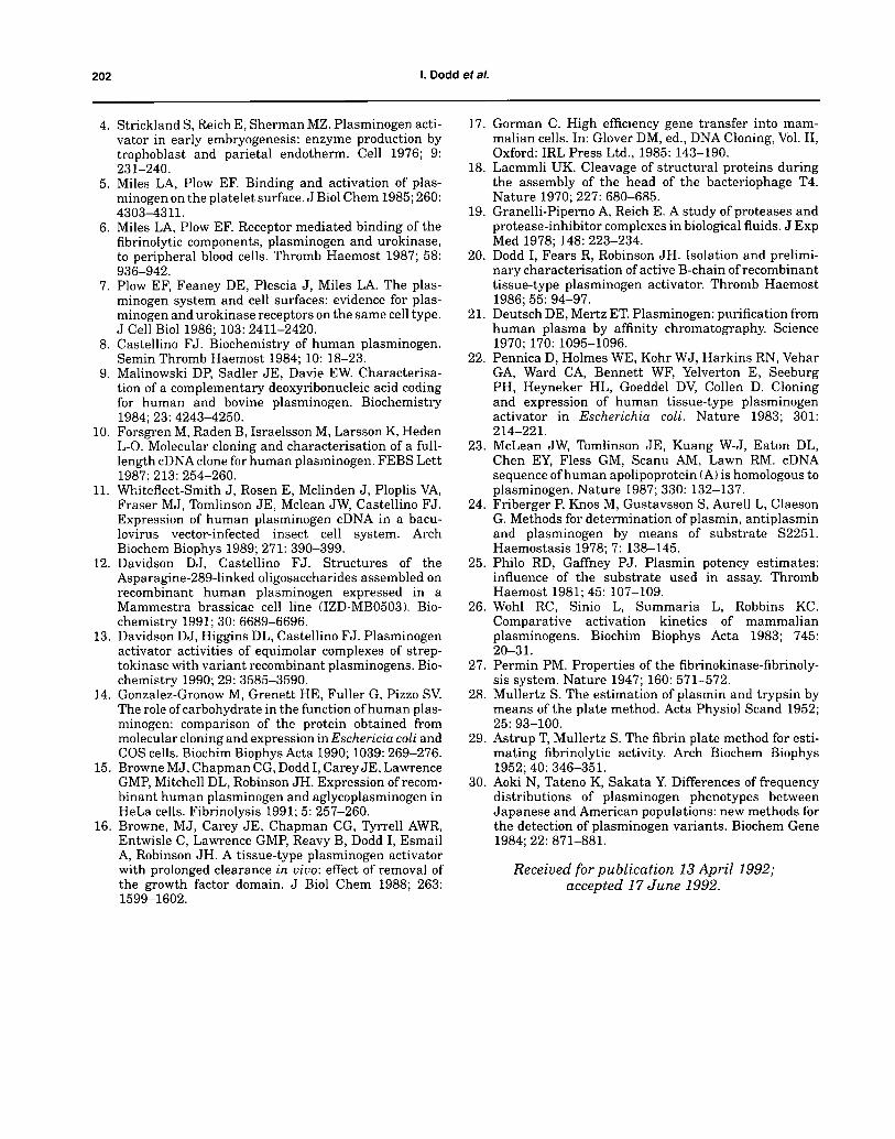
Figure 3. SDS PAGE followed by bovine fibrin zymography of human plas- minogen species. Samples of plasminogen were separated by SDS PAGE and the gel then overlaid with an SK-containing bovine fibrin zymogram. Full details are given in Materials and Methods. The stained zymogram is shown. The lanes are: 1-6, Glul plasminogen (0.3, 1, 3, 10, 30 and 100 ng); 7-12, Lys78 plasminogen (same concentrations); 13, conditioned media from pTR800.
30 nM SK. The demonstrated detection limit of both Glul and Lys78 plasminogen was 0.3 ng/applied sam- ple (20 pl) and functional activity at Mr approximately 90 000 was clearly demonstrated in the harvest medium sample containing the recombinant plas- minogen (Fig. 3).
Conditioned medium from cells transfected with the empty vector pTR312 was inactive (not shown).
Interestingly, an additional species at Mr approxi- mately 30 000 was detected in the Lys78 plasmino- gen preparation. The species may be Val 443 plasminogen; the peptide bond at Val 442/Val 443 is known to be susceptible to proteolytic breakdown.
Discussion
Plasminogen used hitherto for research purposes has been obtained mainly from plasma where the normal concentration is approximately 2 pM. The discovery that it could be easily purified on lysine Sepharose 2~ meant that large amounts were readily available, and as a consequence it was not necessary to develop sen- sitive assays. The recent interest in the cloning and expression ofcDNAs encoding plasminogen has neces-
sitated the development of assays capable of detect- ing the small amounts of material (0.1-10 nM) initially obtained from cell culture expression systems.
Commonly-used assays for plasminogen are of two types: those that identify protein related immuno- logically to plasminogen; and those tha t identify func- tion. Assays based on assessing the immunoreactivity of the target protein suffer from numerous draw- backs. The most important of these are: first, tha t any cross-reacting protein may not be plasminogen--e.g. several other proteins such as tissue-type plasmino- gen activator 22 and apolipoprotein A 23 possess simi- lar tert iary structures that may cross-react with the selected antibody; and second, the tests yield no infor- mation on functional activity, and it is possible tha t even though the cDNA encoding the protein is trans- lated it is not expressed functionally. A relevant example of this is the ability of Escherichia coli to produce immunologically-related but apparently functionally inactive recombinant plasminogen. ~4 Nevertheless, tests based on immunological cross- reaction can show whether the target protein is trans- lated and provide information on chain nature or degradation of impure protein.

Use of fibrin films in determining human plasminogen 201
Functional assays for plasminogen have tended to rely on the use of chromogenic 24 or fluorogenic '4 tripeptides rather than protein substrates. However, it has been shown previously with another serine pro- tease (t-PA) tha t cleavage of the natural substrate can be decreased considerably while cleavage of a tripeptide chromogenic substrate is unaltered 2° and a similar point has also been noted for plasmin itself. 25 It is, therefore, important to assess the activity of recombinant plasminogen molecules in assays resembling more closely the physiological setting. The natural substrate for activated plasminogen is fibrin. In addition, plasminogen itself can act as a sub- strate under certain circumstances; in the clinical management of thrombosis with SK, SK forms a com- plex with circulating plasminogen and the complex acts as a plasminogen activator.
The results presented in this paper demonstrate the use of plasminogen-containing fibrin films for the determination of recombinant human plasminogen. However, we wished to show first that the plasmino- gen variant demonstrated activity in more widely used detection systems.
Plasminogen I682V was used as the model plas- minogen protein. The variant was identified during the cloning ofplasminogen from a A g t l l human liver cDNA library and it is not known whether the con- servative substitution is a polymorphic variant or a cloning artefact. A plasmid vector encoding the vari- ant molecule was expressed in HeLa cells in culture. Conditioned medium from the cells was shown to be active in an $2251 assay (see Results) and contained immunoreactive material at the correct molecular weight for native plasminogen (Fig. 1). These results indicated the vector was expressing the encoded pro- tein.
The two fibrin film-based assays described here uti- lize the ability of SK to complex with human but not bovine plasminogen. 26 Because the SK is incorporated into the fibrin film rather than the test sample the assays are both simple and reproducible.
The first of these assays was a modification of the fibrin plate assay of Astrup and colleagues, which, in its original form, was widely evaluated for the quantitation of plasma-derived plasmin. '-'v-29 The optimal concentration of SK in the fibrin film was shown to be 30 or 100 nM and the lower concentra- tion was used routinely. The response of Glul and Lys78-plasminogen in the assay was similar. (The sensitivity limit demonstrated was 0.3 ng/10 pl). In contrast the response in the $2251 assay was differ- en t - -Glu l plasminogen gave a two-fold greater response.
The second fibrin film based test utilized SDS PAGE as we considered it important to demonstrate tha t the functional activity could be correlated with a known physico-chemical property of plasminogen. Zymography techniques have been widely used for the study of plasminogen polymorphism, for example,3°but sensitivity of the assays is poor. The method described in this paper follows the principles of the fibrin plate method described earlier and allows the simple and sensitive determination of plasmino- gen after electrophoresis in an SDS polyacrylamide gel. The method was shown to be capable of detect- ing 0.3 ng of(plasma-derived) plasminogen and could discriminate the two glycoforms (Type I and Type II) and the two N-terminal forms (Glul and Lys78). The recombinant variant plasminogen migrates at a position corresponding to the Glul form, and the Type II glycoform is predominant. The Type II > Type I ratio was also observed for recombinant (con- sensus) plasminogen in the same expression system. ~ A significant advantage of the zymography test is its ability to relate functional activity to a physico-chemical parameter of the molecule in the same test.
In summary, the two fibrin film-based assays described in this paper are significant additions to the portfolio of assays available for plasminogen molecules. The interest in plasminogen polymor- phism and the genetic basis for it indicates substan- tial research activity in the plasminogen cloning area. The assays described in this paper should aid these studies considerably.
Acknowledgements
We would like to thank Drs M. J. Browne, J. H. Robinson and J. M. Dewdney for their interest, encouragement and discussion.
R e f e r e n c e s
1. Astrup T. Cell-induced fibrinolysis: a fundamental pro- cess. In: Reich E, Rifkin DB, Shaw E, eds. Proteases and Biological Control. Cold Spring Harbor, N.Y.: Cold Spring Harbor Laboratory, 1975: 345.
2. Unkeless JC, Gordon S, Reich E. Secretion of plas- minogen activator by stimulated macrophages. J Exp Med 1974; 139: 834-850.
3. Virgi MAD, Vassalli JD, Estensen RD, Reich E. Plasminogen activator of islets of Langerhans: modulation by glucose and correlation with insulin production. Proc Natl Acad Sci U.S.A. 1980; 77: 875-879.
2 0 2 I. D o d d et aL
4. Strickland S, Reich E, Sherman MZ. Plasminogen acti- vator in early embryogenesis: enzyme production by trophoblast and parietal endotherm. Cell 1976; 9: 231-240.
5. Miles LA, Plow EF. Binding and activation of plas- minogen on the platelet surface. J Biol Chem 1985; 260: 4303-4311.
6. Miles LA, Plow EF. Receptor mediated binding of the fibrinolytic components, plasminogen and urokinase, to peripheral blood cells. Thromb Haemost 1987; 58: 936-942.
7. Plow EF, Feaney DE, Plescia J, Miles LA. The plas- minogen system and cell surfaces: evidence for plas- minogen and urokinase receptors on the same cell type. J Cell Biol 1986; 103: 2411-2420.
8. Castellino FJ. Biochemistry of human plasminogen. Semin Thromb Haemost 1984; 10: 18-23.
9. Malinowski DP, Sadler JE, Davie EW. Characterisa- tion of a complementary deoxyribonucleic acid coding for human and bovine plasminogen. Biochemistry 1984; 23: 4243-4250.
10. Forsgren M, Raden B, Israelsson M, Larsson K, Heden L-O. Molecular cloning and characterisation of a full- length cDNA clone for human plasminogen. FEBS Lett 1987; 213: 254-260.
11. Whitefleet-Smith J, Rosen E, Mclinden J, Ploplis VA, Fraser MJ, Tomlinson JE, Mclean JW, Castellino FJ. Expression of human plasminogen cDNA in a bacu- lovirus vector-infected insect cell system. Arch Biochem Biophys 1989; 271: 390-399.
12. Davidson DJ, Castellino FJ. Structures of the Asparagine-289-1inked oligosaccharides assembled on recombinant human plasminogen expressed in a Mammestra brassicae cell line (IZD-MB0503). Bio- chemistry 1991; 30: 6689-6696.
13. Davidson DJ, Higgins DL, Castellino FJ. Plasminogen activator activities of equimolar complexes of strep- tokinase with variant recombinant plasminogens. Bio- chemistry 1990; 29: 3585-3590.
14. Gonzalez-Gronow M, Grenett HE, Fuller G, Pizzo SV. The role of carbohydrate in the function of human plas- minogen: comparison of the protein obtained from molecular cloning and expression in Eschericia coli and COS cells. Biochim Biophys Acta 1990; 1039: 269-276.
15. Browne MJ, Chapman CG, Dodd I, Carey JE, Lawrence GMP, Mitchell DL, Robinson JH. Expression of recom- binant human plasminogen and aglycoplasminogen in HeLa cells. Fibrinolysis 1991; 5: 257-260.
16. Browne, MJ, Carey JE, Chapman CG, Tyrrell AWR, Entwisle C, Lawrence GMP, Reavy B, Dodd I, Esmail A, Robinson JH. A tissue-type plasminogen activator with prolonged clearance in vivo: effect of removal of the growth factor domain. J Biol Chem 1988; 263: 1599-1602.
17. Gorman C. High efficiency gene transfer into mam- malian cells. In: Glover DM, ed., DNA Cloning, Vol. II, Oxford: IRL Press Ltd., 1985: 143-190.
18. Laemmli UK. Cleavage of structural proteins during the assembly of the head of the bacteriophage T4. Nature 1970; 227: 680-685.
19. Granelli-Piperno A, Reich E. A study of proteases and protease-inhibitor complexes in biological fluids. J Exp Med 1978; 148: 223-234.
20. Dodd I, Fears R, Robinson JH. Isolation and prelimi- nary characterisation of active B-chain of recombinant tissue-type plasminogen activator. Thromb Haemost 1986; 55: 94-97.
21. Deutsch DE, Mertz ET. Plasminogen: purification from human plasma by affinity chromatography. Science 1970; 170: 1095-1096.
22. Pennica D, Holmes WE, Kohr WJ, Harkins RN, Vehar GA, Ward CA, Bennett WF, Yelverton E, Seeburg PH, Heyneker HL, Goeddel DV, Collen D. Cloning and expression of human tissue-type plasminogen activator in Escherichia coli. Nature 1983; 301: 214-221.
23. McLean JW, Tomlinson JE, Kuang W-J, Eaton DL, Chen EY, Fless GM, Scanu AM, Lawn RM. cDNA sequence of human apolipoprotein (A) is homologous to plasminogen. Nature 1987; 330: 132-137.
24. Friberger P, Knos M, Gustavsson S, Aurell L, Claeson G. Methods for determination of plasmin, antiplasmin and plasminogen by means of substrate $2251. Haemostasis 1978; 7: 138-145.
25. Philo RD, Gaffney PJ. Plasmin potency estimates: influence of the substrate used in assay. Thromb Haemost 1981; 45: 107-109.
26. Wohl RC, Sinio L, Summaria L, Robbins KC. Comparative activation kinetics of mammalian plasminogens. Biochim Biophys Acta 1983; 745: 20-31.
27. Permin PM. Properties of the fibrinokinase-fibrinoly- sis system. Nature 1947; 160: 571-572.
28. Mullertz S. The estimation of plasmin and trypsin by means of the plate method. Acta Physiol Scand 1952; 25: 93-100.
29. Astrup T, Mullertz S. The fibrin plate method for esti- mating fibrinolytic activity. Arch Biochem Biophys 1952; 40: 346-351.
30. Aoki N, Tateno K, Sakata Y. Differences of frequency distributions of plasminogen phenotypes between Japanese and American populations: new methods for the detection of plasminogen variants. Biochem Gene 1984; 22: 871-881.
Received for publication 13 April 1992; accepted 17 June 1992.