THE TOP TEN THINGS YOU SHOULD KNOW ABOUT THE OCULOMOTOR SYSTEM.
75
THE TOP TEN THINGS YOU SHOULD KNOW ABOUT THE OCULOMOTOR SYSTEM
-
Upload
carmel-bennett -
Category
Documents
-
view
217 -
download
1
Transcript of THE TOP TEN THINGS YOU SHOULD KNOW ABOUT THE OCULOMOTOR SYSTEM.

THE TOP TEN THINGS YOU SHOULD KNOW ABOUT
THE OCULOMOTOR SYSTEM

10. Movements of the eyes are produced by six extra-ocular muscles. If they, or the neural pathways controlling them,
are not functioning normally, eye movements are abnormal.
• Additionally, accommodation and pupillary responses are produced by intraocular muscles
Video OF Duane’s
Video of Opsoclonus

Meet the musclesMuscle Primary action Example
Medial rectus Adduction Towards themidline/nose
Lateral rectus Abduction Away from themidline/nose
Superior rectus Elevation
Inferior rectus Depression
Superior oblique Intorsion
Inferior oblique Extorsion

Meet the muscles (Cont.)
Muscle Primary action
Ciliary muscle Positive accommodation:acts against suspensoryligaments
Sphincter pupillaeiris muscle
Pupilloconstriction
Dilator pupillaeiris muscle
Pupillodilation

9. The stretch reflex is absent. Gently press on your eye and you’ll see the
world move.
• Proprioceptive feedback from the extra-ocular muscles is not used to keep track of eye position.
• The brain keeps track of eye position by keeping track of the signals sent to the motoneurons that innervate the extra-ocular muscles. This is known as efference copy or corollary discharge.
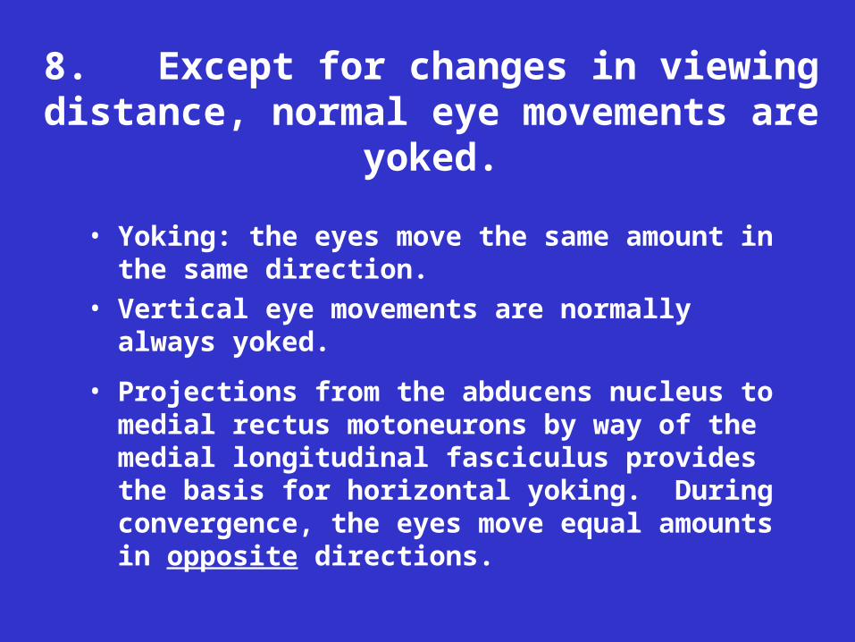
8. Except for changes in viewing distance, normal eye movements are yoked.
• Yoking: the eyes move the same amount in the same direction.
• Vertical eye movements are normally always yoked.
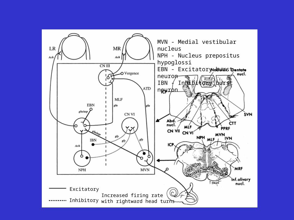
• Projections from the abducens nucleus to medial rectus motoneurons by way of the medial longitudinal fasciculus provides the basis for horizontal yoking. During convergence, the eyes move equal amounts in opposite directions.

Excitatory
Inhibitory
MVN - Medial vestibular nucleusNPH - Nucleus prepositus hypoglossiEBN - Excitatory burst neuronIBN - Inhibitory burst neuron
VIDEO SHOWING INTERNUCLEAR OPHTHALMOPLEGIA

7. Eye movements are controlled by distinct neurological subsystems.
• Eye movements stabilize the image of the external world on the retina
• Eye movements bring images of objects of interest onto the fovea

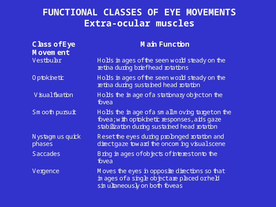
FUNCTIONAL CLASSES OF EYE MOVEMENTSExtra-ocular muscles
Class of EyeMovement
Main Function
Vestibular Holds images of the seen world steady on theretina during brief head rotations
Optokinetic Holds images of the seen world steady on theretina during sustained head rotation
Visual fixation Holds the image of a stationary object on thefovea
Smooth pursuit Holds the image of a small moving target on thefovea; with optokinetic responses, aids gazestabilization during sustained head rotation
Nystagmus quickphases
Reset the eyes during prolonged rotation anddirect gaze toward the oncoming visual scene
Saccades Bring images of objects of interest onto thefovea
Vergence Moves the eyes in opposite directions so thatimages of a single object are placed or heldsimultaneously on both foveas
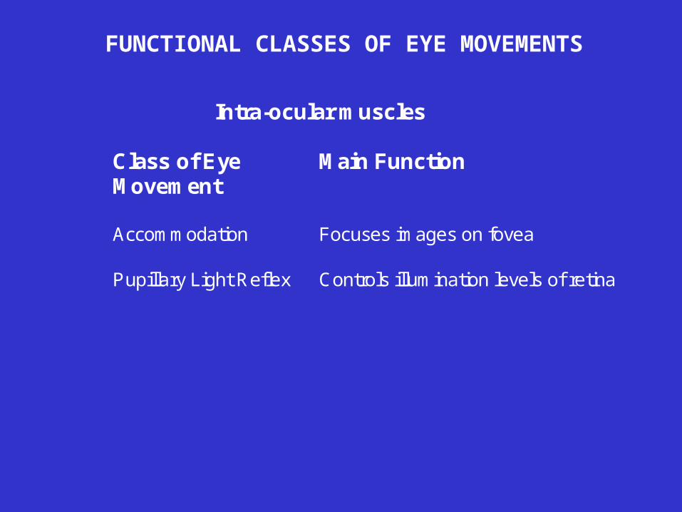
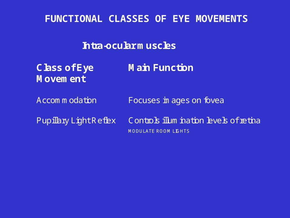
FUNCTIONAL CLASSES OF EYE MOVEMENTS
Intra-ocular muscles
Class of EyeMovement
Main Function
Accommodation Focuses images on fovea
Pupillary Light Reflex Controls illumination levels of retina

6. Vestibular responses. You can’t read without them.
Your head turns in one direction with acertain velocity, and because of thevestibular ocular reflex (VOR), your eyesturn with an equal velocity (if the VORgain is 1.0) in the opposite direction. Thisreflex has a latency of less than 10millseconds.
Once the transient head rotation ceases,your eyes have turned to a new position.
They need to remain at that position andnot drift back to primary position.
To achieve this, a tonic signalproportional to the integral of the eyevelocity signal is generated and sent tothe extraocular motoneurons to maintainthe new eye position.


VOR gain is low at low frequencies

Vestibulo-ocular reflex
Increased firing rate with rightward head turns
Excitatory
Inhibitory
MVN - Medial vestibular nucleusNPH - Nucleus prepositus hypoglossiEBN - Excitatory burst neuronIBN - Inhibitory burst neuron


FUNCTIONAL CLASSES OF EYE MOVEMENTSExtra-ocular muscles
Class of EyeMovement
Main Function
Vestibular Holds images of the seen world steady on theretina during brief head rotations
Optokinetic Holds images of the seen world steady on theretina during sustained head rotation
Visual fixation Holds the image of a stationary object on thefovea
Smooth pursuit Holds the image of a small moving target on thefovea; with optokinetic responses, aids gazestabilization during sustained head rotation
Nystagmus quickphases
Reset the eyes during prolonged rotation anddirect gaze toward the oncoming visual scene
Saccades Bring images of objects of interest onto thefovea
Vergence Moves the eyes in opposite directions so thatimages of a single object are placed or heldsimultaneously on both foveas
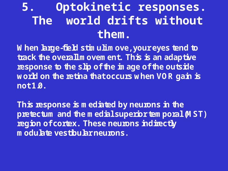
5. Optokinetic responses. The world drifts without them.
When large-field stimuli move, your eyes tend totrack the overall movement. This is an adaptiveresponse to the slip of the image of the outsideworld on the retina that occurs when VOR gain isnot 1.0.
This response is mediated by neurons in thepretectum and the medial superior temporal (MST)region of cortex. These neurons indirectlymodulate vestibular neurons.


VESTIBULAR NUCLEUS NEURON
A. ROTATION IN DARKNESS (Vestibular but no Optokinetic)
B. ROTATION IN LIGHT (Vestibular and Optokinetic)
C. NO ROTATION. OPTIC FLOW. (Optokinetic but no Vestibular)

Vestibular-optokinetic interactions
When rotation stops, nystagmus starts in the opposite direction (postrotatory nystagmus, PRN). In the middle panel, an optokinetic stimulus (drum rotation to the right) causes a sustained optokinetic nystagmus (OKN), with slow phases to the right during the entire period of stimulation. When the lights are turned off during stimulation, eye movements do not stop immediately but persist as optokinetic after-nystagmus (OKAN). In the lower panel, the subject is rotated in the light (natural situation of self-rotation). This gives a combined vestibular and optokinetic stimulus. The response is a sustained nystagmus. When the chair stops rotating, eye movements stop nearly completely: postrotatory nystagmus is suppressed by the opposite-directed optokinetic after-nystagmus and by visual fixation of the stationary world.
Schematic summary of vestibular-optokinetic interaction occurring in response to velocity-step rotations. Graphs on the left show characteristics of the stimulus (head velocity during rotation or drum velocity during optokinetic stimulation); graphs on the right show the responses (slow-phase eye velocity, quick phases having been removed). R, right; L, left; t, time. In the top panel, constant-velocity rotation to the left in the dark produces slow-phase movements to the right (per-rotatory nystagmus, RN) with initial eye velocities equal to head velocity (VOR gain = 1.0).

FUNCTIONAL CLASSES OF EYE MOVEMENTSExtra-ocular muscles
Class of EyeMovement
Main Function
Vestibular Holds images of the seen world steadyon the retina during brief headrotations
Optokinetic Holds images of the seen world steadyon the retina during sustained headrotation
Visual fixation Holds the image of a stationary objecton the fovea
Smooth pursuit Holds the image of a small movingtarget on the fovea; with optokineticresponses, aids gaze stabilizationduring sustained head rotation
VOLUNTARY
Saccades Bring images of objects of interestonto the fovea
VOLUNTARY
Vergence Moves the eyes in opposite directionsso that images of a single object areplaced or held simultaneously on bothfoveas
VOLUNTARY


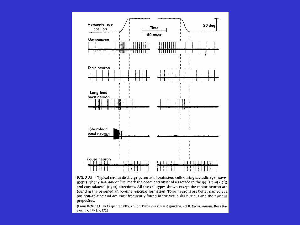
4. Saccadic eye movements. You can’t look at anything interesting without them.
• FAST - 40-90 MS IN TOTAL DURATION
•BALLISTIC
B A
AB
BA


Pulse of firing rate is requiredto produce the transient forceneeded to move the eyerapidly despite viscous drag
Sustained firing rate isrequired to hold the eye in anew postion despite theelastic forces that try to returnit to primary position

Right Medial Rectus Motoneuron - Saccades

Horizontal saccades are generated in the paramedian pontine reticular formation (PPRF)
VERTICAL SACCADES ARE GENERATED HERE
Vertical saccades are generated in the rostral interstitial nucleus of the medial longitudinal fasciculus (riMLF)

Increased firing rate with rightward head turns
Excitatory
Inhibitory
MVN - Medial vestibular nucleusNPH - Nucleus prepositus hypoglossiEBN - Excitatory burst neuronIBN - Inhibitory burst neuron

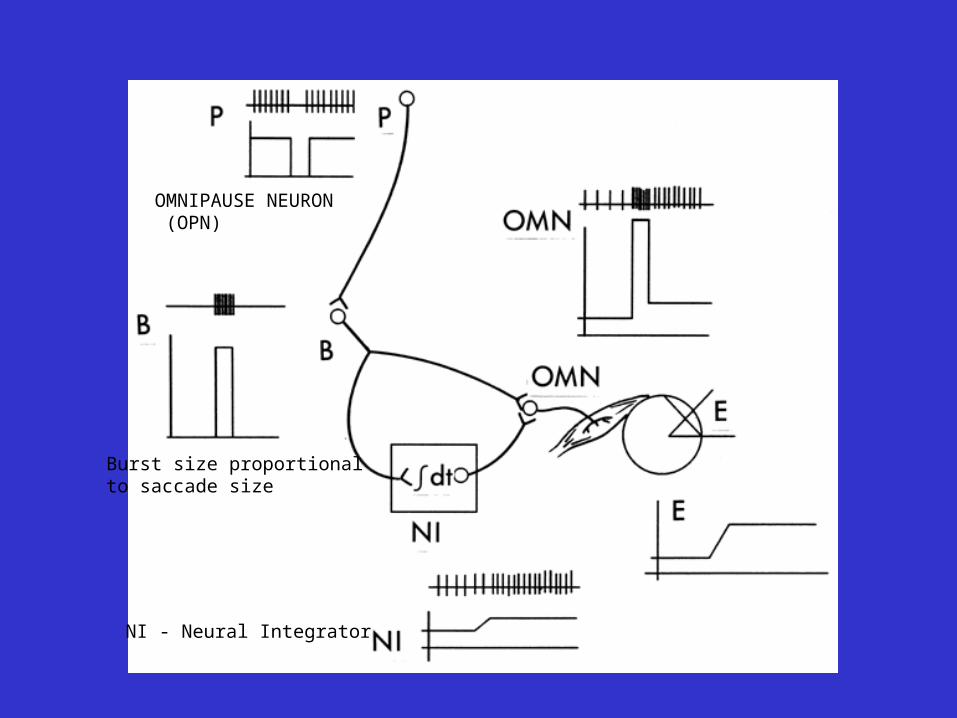
EBN
Burst size proportional to saccade size
OMNIPAUSE NEURON (OPN)
NI - Neural Integrator

Excitatory burst neuron- small saccade

Excitatory burst neuron- medium saccade

Excitatory burst neuron - large saccade


Omnipause neuron - various saccades
EBN
Burst size proportional to saccade size
OMNIPAUSE NEURON (OPN)
NI - Neural Integrator

LOCAL FEEDBACK MODEL

THE SUPERIOR COLLICULUS PROJECTS TO THE PPRF
2-D map of contralateral saccades
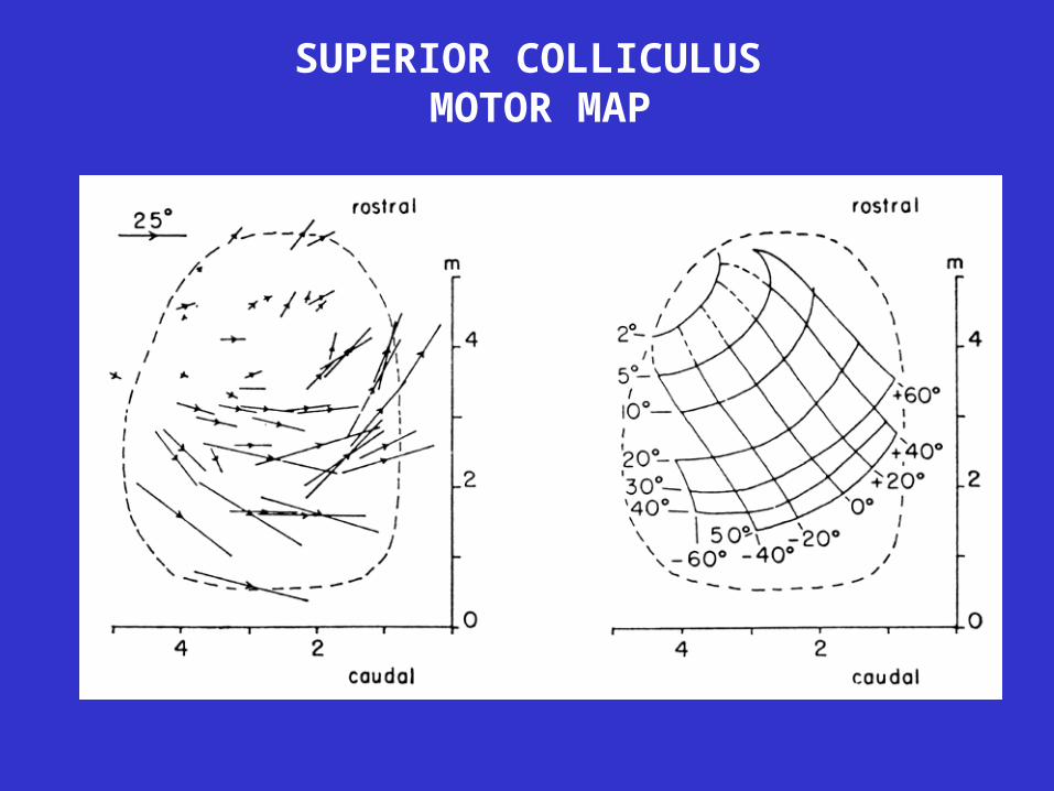
SUPERIOR COLLICULUS MOTOR MAP

A block diagram of the major structures that project to the brain stem saccade generator (premotor burst neurons in PPRF and riMLF). Also shown are projections from cortical eye fields to superior colliculus. FEF, frontal eye fields; SEF, supplementary eye fields; DLPC, dorsolateral prefrontal cortex; IML, intramedullary lamina of thalamus; PEF, parietal eye fields (LIP); PPC, posterior parietal cortex; SNpr, substantia nigra, pars reticulata. Not shown are the pulvinar, which has connections with the superior colliculus and both the frontal and parietal lobes, and certain projections, such as that from the superior colliculus to nucleus reticularis tegmenti pontis (NRTP).

Disorders of the saccadic pulse and step. Innervation patterns are shown on the left, eye movements on the right. Dashed lines indicate the normal response. (A) Normal saccade. (B) Hypometric saccade: pulse amplitude (width ´ height) is too small but pulse and step are matched appropriately. (C) Slow saccade: decreased pulse height with normal pulse amplitude and normal pulse-step match. (D) Gaze-evoked nystagmus: normal pulse, poorly sustained step. (E) Pulse-step mismatch (glissade): step is relatively smaller than pulse. (F) Pulse-step mismatch due to internuclear ophthalmoplegia (INO): the step is larger than the pulse, and so the eye drifts onward after the initial rapid movement.
Experimental cerebellectomy completely abolishes the adaptive capability-for both the pulse size and the pulse-step match.296 Monkeys with lesions restricted to the dorsal cerebellar vermis cannot adapt the size of the saccadic pulse; they have pulse-size dysmetria .416,416a On the other hand, monkeys with floccular lesions cannot match the saccadic step to the pulse to eliminate pulse-step mismatch dysmetria.298 This evidence suggests that the repair of conjugate saccadic dysmetria is mediated by two different cerebellar structures: the dorsal cerebellar vermis and the fastigial nuclei control pulse size, and the flocculus and paraflocculus control the pulse-step match.
FUNCTIONAL CLASSES OF EYE MOVEMENTSExtra-ocular muscles
Class of EyeMovement
Main Function
Vestibular Holds images of the seen world steady on theretina during brief head rotations
Optokinetic Holds images of the seen world steady on theretina during sustained head rotation
Visual fixation Holds the image of a stationary object on thefovea
Smooth pursuit Holds the image of a small moving target on thefovea; with optokinetic responses, aids gazestabilization during sustained head rotation
Nystagmus quickphases
Reset the eyes during prolonged rotation anddirect gaze toward the oncoming visual scene
Saccades Bring images of objects of interest onto thefovea
Vergence Moves the eyes in opposite directions so thatimages of a single object are placed or heldsimultaneously on both foveas

• Smooth pursuit: Tracking eye movements - conjugate. Velocity of visual target
• Visual cue: retinal slip velocity of visual target.
AB
BB AA

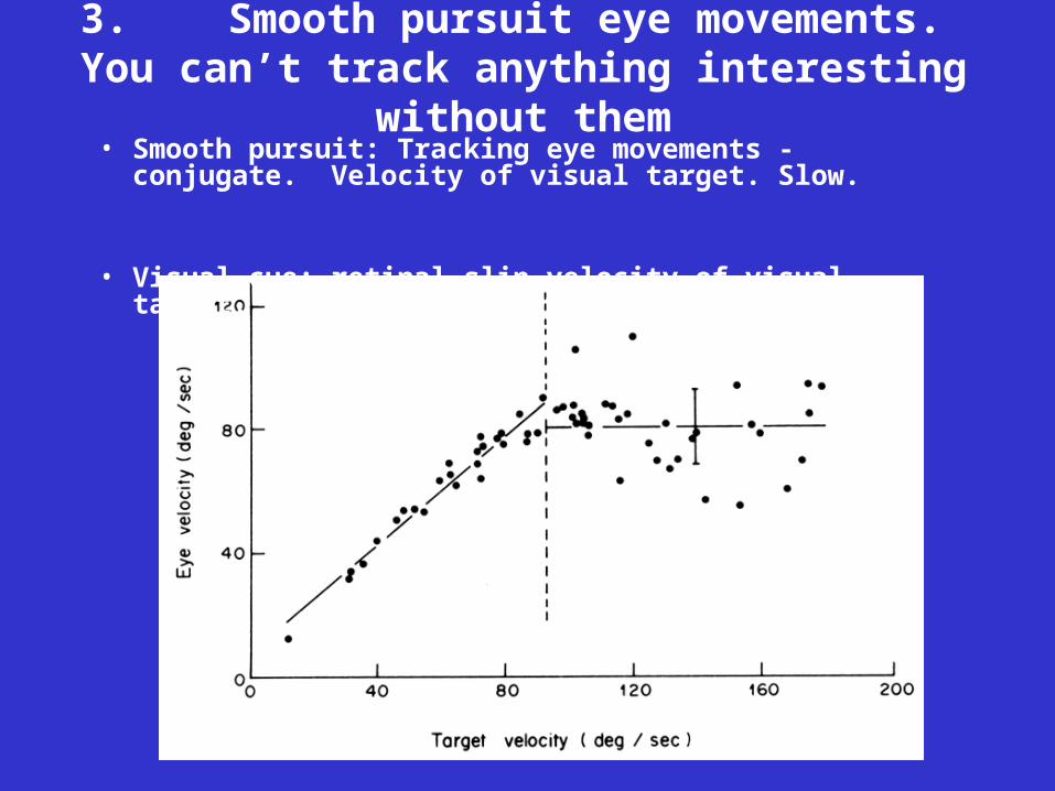
3. Smooth pursuit eye movements. You can’t track anything interesting without them
• Smooth pursuit: Tracking eye movements - conjugate. Velocity of visual target. Slow.
• Visual cue: retinal slip velocity of visual target.

Right Medial Rectus Motoneuron - Smooth pursuit

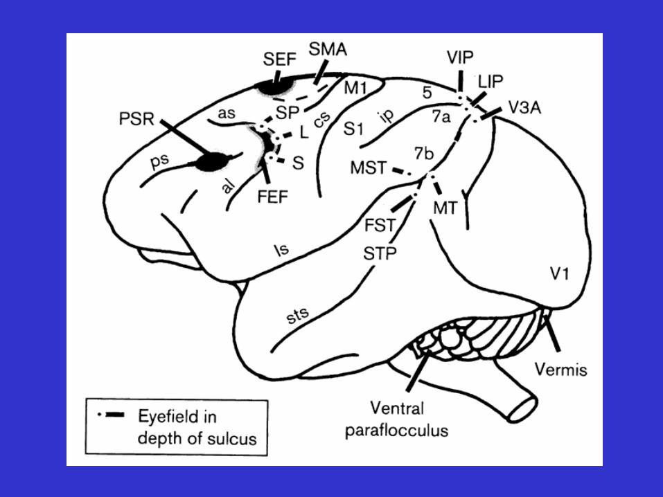
SMOOTH PURSUIT PATHWAYS

FUNCTIONAL CLASSES OF EYE MOVEMENTSExtra-ocular muscles
Class of EyeMovement
Main Function
Vestibular Holds images of the seen world steady on theretina during brief head rotations
Optokinetic Holds images of the seen world steady on theretina during sustained head rotation
Visual fixation Holds the image of a stationary object on thefovea
Smooth pursuit Holds the image of a small moving target on thefovea; with optokinetic responses, aids gazestabilization during sustained head rotation
Nystagmus quickphases
Reset the eyes during prolonged rotation anddirect gaze toward the oncoming visual scene
Saccades Bring images of objects of interest onto thefovea
Vergence Moves the eyes in opposite directions so thatimages of a single object are placed or heldsimultaneously on both foveas

• Vergence: Eye movements in depth. Disconjugate - left and right eyes move in opposite directions.
B
BAA
A
B
2. Vergence. Without it, you can’t get a closer look.
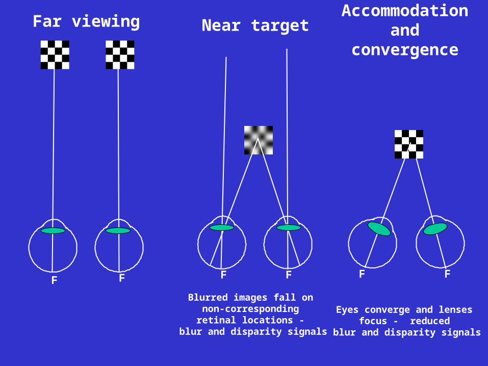
Far viewing Near target Accommodation and convergence
Blurred images fall on non-corresponding retinal locations -
blur and disparity signals
F F F F F F
Eyes converge and lenses focus - reduced
blur and disparity signals
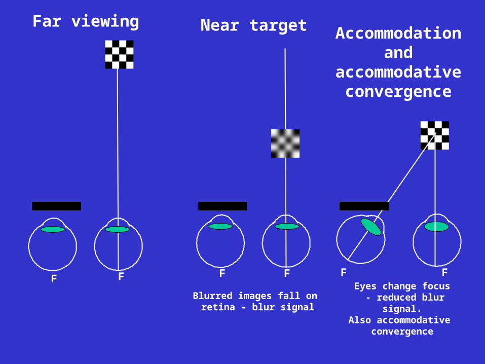
Far viewing Near target Accommodation and
accommodative convergence
Blurred images fall on retina - blur signal
F F F F FF
Eyes change focus - reduced blur signal.Also accommodative
convergence
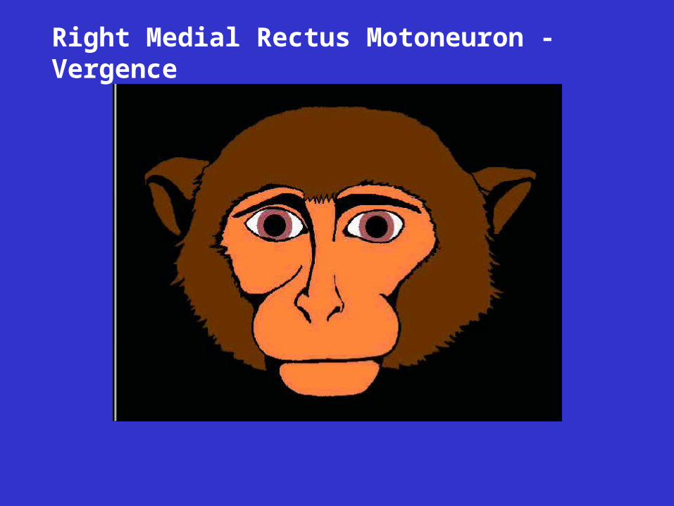
Right Medial Rectus Motoneuron - Vergence
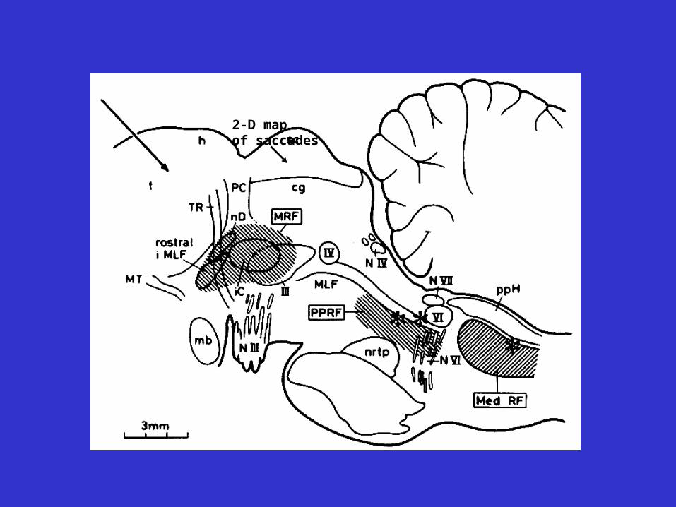
2-D map of saccades
Increased firing rate with rightward head turns
Excitatory
Inhibitory
MVN - Medial vestibular nucleusNPH - Nucleus prepositus hypoglossiEBN - Excitatory burst neuronIBN - Inhibitory burst neuron

NEAR RESPONSE NEURON - VERGENCE

NEAR RESPONSE NEURON - SACCADES

Internuclear ophthalmoplegia - adduction during convergence is not reduced
FUNCTIONAL CLASSES OF EYE MOVEMENTS
Intra-ocular muscles
Class of EyeMovement
Main Function
Accommodation Focuses images on fovea
Pupillary Light Reflex Controls illumination levels of retinaMODULATE ROOM LIGHTS

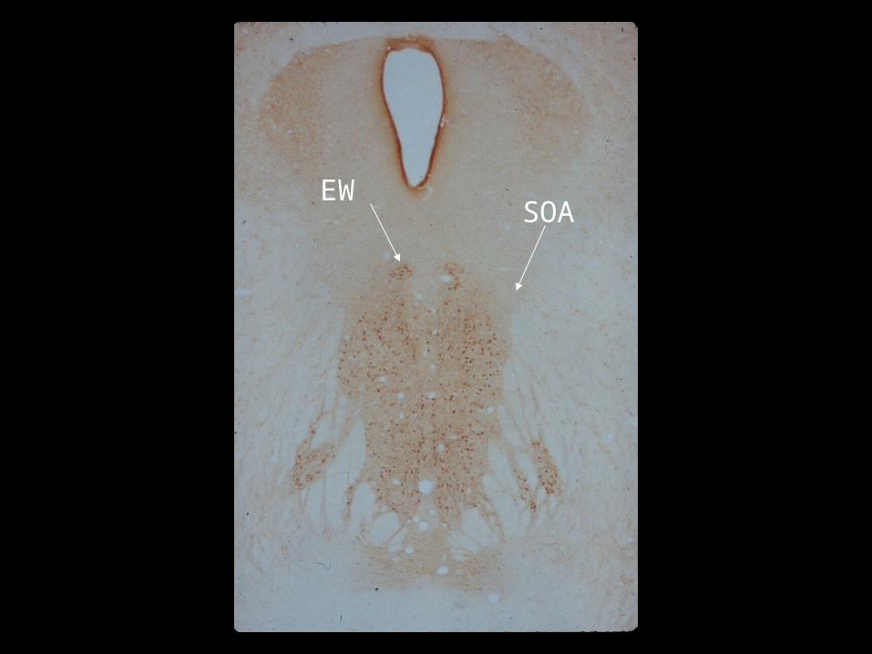
SOA
EW

Edinger-Westphal neuron


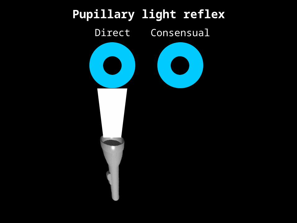
Pupillary light reflex
Direct Consensual

Direct Consensual
Pupillary light reflex

Direct Consensual
Pupillary light reflex
Direct Consensual
Pupillary light reflex

1. Pupillary light reflex. If it’s absent,
there’s a problem. AFFERENT DEFECTS:PUPILS APPROX. EQUAL IN SIZE. BUT RESPONSE TO LIGHT IN ONE EYE IS LESS THAN THE RESPONSE TO LIGHT IN THE OTHER EYE.
EFFERENT DEFECTS:PUPILS MAY BE OF DIFFERENT SIZES (ANISOCORIA). PUPIL OF ONE EYE REACTS MORE TO LIGHT IN EITHER EYE THAN THE PUPIL OF THE OTHER EYE TO LIGHT IN EITHER EYE.

Pupillary light reflex: Afferent deficit

Pupillary light reflex: Afferent deficit
Neutral Density Filter (0.5 log unit)
0.5 log unit Relative AfferentPupillary Deficit (RAPD)
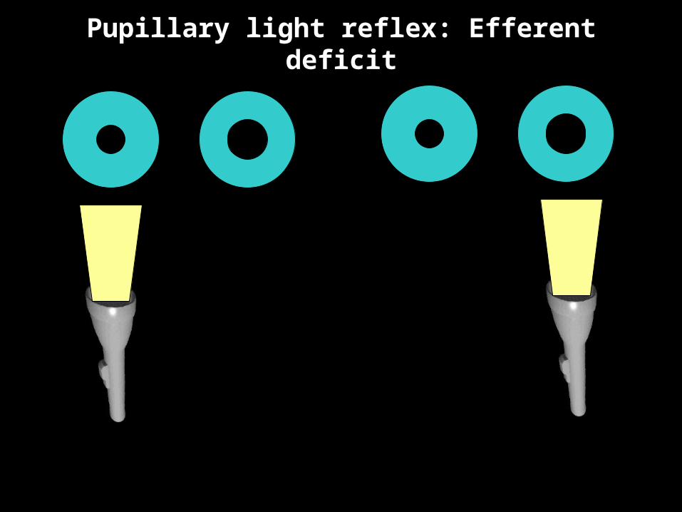
Pupillary light reflex: Efferent deficit

1. Pupillary light reflex. If it’s absent, there’s a problem.2. Vergence. Without it, you can’t get a closer look.3. Smooth pursuit eye movements. You can’t track anything
interesting without them4. Saccadic eye movements. You can’t look at anything interesting
without them.5. Optokinetic responses. The world drifts without them.6. Vestibular responses. You can’t read without them.7. Eye movements are controlled by distinct neurological
subsystems.8. Except for changes in viewing distance, normal eye movements
are yoked. 9. The stretch reflex is absent. Gently press on your eye and you’ll
see the world move.10. Movements of the eyes are produced by six extra-ocular
muscles. If they, or the neural pathways controlling them, are not functioning normally, eye movements are abnormal.
TOP TEN LIST