The tissue, cellular, and molecular regulation of ... · cytokines, growth factors, enzymes, ......
20
European Journal of Orthodontics 28 (2006) 221–240 doi:10.1093/ejo/cjl001 © The Author 2006. Published by Oxford University Press on behalf of the European Orthodontics Society. All rights reserved. For permissions, please email: [email protected]. The tissue, cellular, and molecular regulation of orthodontic tooth movement: 100 years after Carl Sandstedt Murray C. Meikle Department of Oral Sciences, Faculty of Dentistry, University of Otago, Dunedin, New Zealand SUMMARY The first experimental investigation of orthodontic tooth movement was published by Sandstedt in 1904–1905. After 100 years, there is a good understanding of the sequence of events at both tissue and cellular levels and now the current focus of research is at the molecular level. The techniques of reverse transcription–polymerase chain reaction and in situ hybridization to detect mRNAs of interest have revolutionized tooth movement studies and an expanding list of antibodies and enzyme-linked immunosorbent assays directed against human and animal proteins will facilitate their identification in tissue sections and/or culture supernatants. Nevertheless, although this technology has greatly simplified research for the clinical and laboratory investigator, message is not always translated into protein, and the presence of a protein does not necessarily mean it is biologically active. In vivo and in vitro methods have been widely used in tooth movement studies. However, data from in vitro models, in which the mechanical stimulus can be carefully controlled (tension versus compression; intermittent versus continuous), should be correlated with in vivo data from animal models. The current evidence suggests that downstream from the initial mechanotransduction event at focal adhesions which link the extracellular matrix to the cytoskeleton, mechanically induced remodelling is mediated by a complex feedback mechanism involving the synthesis of cytokines such as interleukin-1 (IL-1), IL-6, and receptor activator of nuclear factor B ligand by cells of the osteoblast and/or fibroblast lineages. These in turn act in an autocrine/paracrine fashion to regulate the expression of transcription factors, cytokines, growth factors, enzymes, and structural molecules involved in the differentiation, proliferation, and function of mesenchymal and other cell types. Contrary to the impression gained from the literature, tooth movement is not confined to events within the periodontal ligament. Orthodontic tooth movement involves two interrelated processes: (1) deflection or bending of the alveolar bone and (2) remodelling of the periodontal tissues. Introduction It is now 100 years since a three-part article on the theory of tooth movement was published (Sandstedt, 1904, 1905). It seems appropriate therefore to review the progress in our understanding of the tissue, cellular, and molecular mechanisms involved in orthodontic tooth movement since that time. The literature is now extensive (for previous reviews, see Reitan, 1951; Davidovitch, 1991; Sandy et al., 1993), which creates difficulties when attempting to present a coherent narrative. To add some structure to the subject, it will be addressed largely chronologically and from the biological viewpoint, with a focus on how knowledge has evolved with the discovery of new molecules and the introduction of new experimental techniques. This approach shows that, in common with the rest of the biological sciences, the methodology of tooth movement research has become progressively more reductionist, making the subject less accessible to the clinician. The significance of mechanical stimuli in the maintenance and structure of skeletal tissues has been recognized since the middle of the 19th century (von Meyer, 1867; Wölff, 1892), and is the traditional starting point for any discussion of bone remodelling and tooth movement. The skeleton is continuously remodelled during life by osteoclasts resorbing old bone and osteoblasts subsequently forming new bone. Bone resorption and bone formation are therefore said to be coupled, a process of renewing the skeleton while maintaining its structural integrity (Frost, 1963). Remodelling is orchestrated by cells of the osteoblast lineage and involves a complex network of cell–cell and cell–matrix interactions involving systemic hormones, locally produced cytokines, growth factors, many of which are sequestrated within the bone matrix, as well as the mechanical environment of the cells. Orthodontic and orthopaedic theory and practice have much in common. Both involve the remodelling of bone and both require a thorough understanding of bone biology, particularly the relationship between mechanical stress and the various cell types in bone. However, tooth movement is a more complicated process requiring changes in the periodontal ligament (PDL) as well as the supporting alveolar bone, tissues with different cell populations and remodelling characteristics. During locomotion and other forms of physical activity, the skeleton is loaded intermittently. by guest on May 31, 2012 http://ejo.oxfordjournals.org/ Downloaded from
Transcript of The tissue, cellular, and molecular regulation of ... · cytokines, growth factors, enzymes, ......

European Journal of Orthodontics 28 (2006) 221–240doi:10.1093/ejo/cjl001
© The Author 2006. Published by Oxford University Press on behalf of the European Orthodontics Society.All rights reserved. For permissions, please email: [email protected].
The tissue, cellular, and molecular regulation of orthodontic
tooth movement: 100 years after Carl Sandstedt
Murray C. Meikle Department of Oral Sciences, Faculty of Dentistry, University of Otago, Dunedin, New Zealand
SUMMARY The fi rst experimental investigation of orthodontic tooth movement was published by Sandstedt in 1904 – 1905. After 100 years, there is a good understanding of the sequence of events at both tissue and cellular levels and now the current focus of research is at the molecular level. The techniques of reverse transcription – polymerase chain reaction and in situ hybridization to detect mRNAs of interest have revolutionized tooth movement studies and an expanding list of antibodies and enzyme-linked immunosorbent assays directed against human and animal proteins will facilitate their identifi cation in tissue sections and/or culture supernatants. Nevertheless, although this technology has greatly simplifi ed research for the clinical and laboratory investigator, message is not always translated into protein, and the presence of a protein does not necessarily mean it is biologically active. In vivo and in vitro methods have been widely used in tooth movement studies. However, data from in vitro models, in which the mechanical stimulus can be carefully controlled (tension versus compression; intermittent versus continuous), should be correlated with in vivo data from animal models. The current evidence suggests that downstream from the initial mechanotransduction event at focal adhesions which link the extracellular matrix to the cytoskeleton, mechanically induced remodelling is mediated by a complex feedback mechanism involving the synthesis of cytokines such as interleukin-1 (IL-1), IL-6, and receptor activator of nuclear factor � B ligand by cells of the osteoblast and/or fi broblast lineages. These in turn act in an autocrine/paracrine fashion to regulate the expression of transcription factors, cytokines, growth factors, enzymes, and structural molecules involved in the differentiation, proliferation, and function of mesenchymal and other cell types. Contrary to the impression gained from the literature, tooth movement is not confi ned to events within the periodontal ligament. Orthodontic tooth movement involves two interrelated processes: (1) defl ection or bending of the alveolar bone and (2) remodelling of the periodontal tissues.
Introduction
It is now 100 years since a three-part article on the theory of tooth movement was published ( Sandstedt, 1904 , 1905 ). It seems appropriate therefore to review the progress in our understanding of the tissue, cellular, and molecular mechanisms involved in orthodontic tooth movement since that time. The literature is now extensive (for previous reviews, see Reitan, 1951 ; Davidovitch, 1991 ; Sandy et al. , 1993 ), which creates diffi culties when attempting to present a coherent narrative. To add some structure to the subject, it will be addressed largely chronologically and from the biological viewpoint, with a focus on how knowledge has evolved with the discovery of new molecules and the introduction of new experimental techniques. This approach shows that, in common with the rest of the biological sciences, the methodology of tooth movement research has become progressively more reductionist, making the subject less accessible to the clinician.
The signifi cance of mechanical stimuli in the maintenance and structure of skeletal tissues has been recognized since the middle of the 19th century ( von Meyer, 1867 ; Wölff, 1892 ), and is the traditional starting point for any discussion
of bone remodelling and tooth movement. The skeleton is continuously remodelled during life by osteoclasts resorbing old bone and osteoblasts subsequently forming new bone. Bone resorption and bone formation are therefore said to be coupled, a process of renewing the skeleton while maintaining its structural integrity ( Frost, 1963 ). Remodelling is orchestrated by cells of the osteoblast lineage and involves a complex network of cell – cell and cell – matrix interactions involving systemic hormones, locally produced cytokines, growth factors, many of which are sequestrated within the bone matrix, as well as the mechanical environment of the cells.
Orthodontic and orthopaedic theory and practice have much in common. Both involve the remodelling of bone and both require a thorough understanding of bone biology, particularly the relationship between mechanical stress and the various cell types in bone. However, tooth movement is a more complicated process requiring changes in the periodontal ligament (PDL) as well as the supporting alveolar bone, tissues with different cell populations and remodelling characteristics. During locomotion and other forms of physical activity, the skeleton is loaded intermittently.
by guest on May 31, 2012
http://ejo.oxfordjournals.org/D
ownloaded from

M. C. MEIKLE222
Alveolar bone is also loaded intermittently during mastication, but is subject to a continuous deformation or strain during orthodontic tooth movement, and if the applied force is of suffi cient magnitude, bending of the alveolus will occur. In the skeleton, the relationship between mechanical stress and bone resorption is poorly understood and the application of mechanically induced strain appears to be primarily an osteogenic stimulus ( H ě rt et al. , 1971 ; Lanyon and Baggott, 1976 ). Furthermore, the osteogenic response of bone to external loading appears to be due to a reactivation of quiescent bone-lining cells ( Pead et al. , 1988 ; Chambers et al. , 1993 ), and not dependent on a preliminary phase of bone resorption ( Chow et al. , 1998 ). Alveolar bone in contrast undergoes both resorption and deposition during orthodontic tooth movement, the extent of which is dependent upon the magnitude, direction, and duration of the applied force.
Tissue reaction to orthodontic tooth movement
The idea that orthodontic tooth movement is dependent on the resorption and deposition of the bone of the socket dates back at least to 1839 ( Harris, 1839 ). It was not until the turn of the 20th century, however, that the original histological investigation that forms the foundation of our present knowledge was carried out by Sandstedt (1904 , 1905 ) on dogs and published shortly after his death (Persson, 2005).
Laying the foundations: Sandstedt and Oppenheim
In Sandstedt’s experimental model, a labial arch was bent to engage the six maxillary incisors of a dog and inserted into horizontal tubes attached to bands on the canines. The appliance was activated over a 3-week period by screws distal to the buccal tubes and during that time the crowns of the incisors were moved lingually by 3 mm. Sandstedt found that bone was deposited on the alveolar wall on the tension side of the tooth with both heavy and light forces, and that the newly formed bone spicules followed the orientation of the periodontal fi bre bundles. On the pressure side, with light forces, alveolar bone was resorbed directly by numerous multinucleate osteoclasts in Howship’s lacunae ( Figure 1 ). With heavy forces, the periodontal tissues were compressed, leading to capillary thrombosis, cell death, and the production of localized cell-free areas of what he called hyalinization (owing to its glasslike appearance resembling hyaline cartilage in histological sections). At these sites, osteoclastic resorption of the adjacent alveolar wall did not take place directly, but was initiated by a process referred to by Sandstedt as ‘ undermining resorption ’ from the neighbouring marrow spaces.
Oppenheim (1911 , 1930 ) published the results of experimental work carried out on the primary teeth of monkeys. His observations proved to be substantially different from those of Sandstedt as far as the response of the bone to compression was concerned. He found that
Figure 1 Sandstedt’s histological fi ndings following the application of a light force to the upper incisors of a dog; tooth cut in cross-section. (Left) Pressure side. Z, root surface; P, compressed periodontal ligament; R, resorptive bone surface with numerous osteoclasts in Howship’s lacunae; K, old alveolar bone with Haversian systems with no evidence of bone transformation as described by Oppenheim. (Right) Tension side. Z, root surface showing dentine and cementum; P, periodontal ligament; T, new bony trabeculae orientated along the principal fi bres of the ligament; G, junction between new bone and old compact bones, K. (From Schwarz, 1932 , International Journal of Orthodontia, Oral Surgery and Radiography.)
by guest on May 31, 2012
http://ejo.oxfordjournals.org/D
ownloaded from

223THE REGULATION OF TOOTH MOVEMENT
where a tooth had been tipped labially, the original bone disappeared completely from the labial surface and was replaced by new bone. He concluded that
… bone tissue, be it compact or cancellated, reacts to pressure by a transformation of its entire architecture; this takes place by resorption of the bone present and deposition of new bone tissue; both processes occur simultaneously. Deposition fi nally preponderates over resorption.
Schwarz (1932) attempted to explain the difference between the fi ndings of Sandstedt and Oppenheim by the fact that Oppenheim had killed his animals several days after the appliance had been last activated and did not therefore see the acute effect of the applied force, only a stage of regeneration after the force had been exhausted. It seems more likely that what Oppenheim was describing was the response of the periosteal and endosteal bone surfaces to the bending of the labial alveolar plate. The labial alveolar bone is particularly thin in his illustrations and would have been easily deformed in a monkey in the primary dentition ( Figure 2 ). As discussed later, when bones are subjected to continuous mechanical deformation, concave surfaces are characterized by osteogenesis, and convex surfaces by bone resorption.
Whatever the explanation Oppenheim had lectured at the Angle School of Orthodontics and his research reported in the American Orthodontist, a journal published by the Angle Alumni Society ( Oppenheim 1911, 1912 ). His theory of bone transformation also supported Angle’s (1907) non-extraction philosophy and the widespread belief at the time that orthodontic appliances could ‘ grow bone ’ . Oppenheim’s previous research on monkeys was reported again in 1930 together with the original photomicrographs and the fi ndings of a new series of experiments carried out on dogs ( Oppenheim, 1930 ). He also published articles on root resorption and repair in extracted human premolars following experimental tooth movement ( Oppenheim, 1935 , 1936 ). As a result, it was Oppenheim’s work that appeared in mainstream English language orthodontic textbooks up until the 1950s and beyond. Sandstedt’s early death and the lack of an English translation of his work did not help. He consequently became the forgotten man of tooth movement research until partially rescued from obscurity in the paper by Schwarz (1932) .
Reitan and the post-war period
Following the work of Sandstedt and Oppenheim, reports by other investigators appeared in the literature ( Gottlieb and Orban, 1931 ; Schwarz, 1932 ; Breitner, 1940 ), but it was not until the 1950s that tooth movement studies attracted wider attention. The leading histomorphological investigator of the period was the Norwegian orthodontist Kaare Reitan whose classic memoir was published in 1951 ( Reitan, 1951 ).
Reitan (1957 , 1964 ) made extensive use of human material, particularly premolars that were destined for orthodontic extractions. His work highlighted the complexity of the tissue response to orthodontic treatment depending upon (1) the type (continuous versus intermittent) and magnitude of the force applied, (2) the mechanics involved (tipping versus bodily movement), and (3) the variation in tissue reaction between individual patients. He observed that during the initial stages of a tipping movement, cell-free or hyalinized areas were frequently created with a continuous force of 30 g. The time taken to remove such tissue by undermining resorption varied from 2 to 4 weeks and occasionally longer depending on the length of the root.
Figure 2 In 1930, Oppenheim republished his earlier research on tooth movement (which had been illustrated by drawings and criticized as such) together with the original photomicrographs. This is Figure 1 from the paper showing (A) the original drawing and (B) the photomicrograph of the bone changes following labial movement of a lower incisor for 40 days; tooth sectioned vertically. The text states … ‘ It hardly leaves any doubt that while osteoclasts ( ok ) are absorbing bone on the inner wall, simultaneous formation of new bone is under way on the opposite side of the process. ’ At ob , near the crest of the alveolus, compact bone has disappeared and has been replaced by cancellous bone; the trabeculae of the latter have been arranged perpendicular to the long axis of the tooth; k 1 , new bone trabeculae lined with osteoblasts ( ob ). (From Oppenheim, 1930 , International Journal of Orthodontia, Oral Surgery and Radiography.)
by guest on May 31, 2012
http://ejo.oxfordjournals.org/D
ownloaded from

M. C. MEIKLE224
Figure 3 (A) The application of a torquing force of 200 g to the second incisor of a dog resulted in tip and hyalinization (H) of the periodontal ligament (PDL) at sites of excessive compression. R, direct bone resorption; F, bone formation along the principal fi bre bundles of the PDL. A torquing force of 200 g (B) was not accompanied by tip. R, resorption of bone along the surface of the alveolus and cementum and dentine of the opposing root surface (1, 2). Arrows indicate the direction of tooth movement. (Redrawn from Reitan, 1964 .)
Intermittent forces of 70 – 100 g were also found to produce hyalinization. Cell-free areas were more common in tipping than in bodily movements presumably because, in the latter, the force was more evenly distributed along the root – bone interface ( Figure 3 ). These experiments showed that even with an applied force as low as 30 g, some degree of hyalinization and root resorption appeared inevitable.
In a study of rotated teeth in young dogs, Reitan (1959) found that while the principal fi bres of the PDL were rearranged or remodelled within 28 days, even after a retention period of 232 days, some of the free gingival fi bres remained displaced and stretched. He concluded that rotational relapse was caused primarily by a contraction of the supra-alveolar gingival fi bres (which contain elastic or oxytalin fi bres) and advised the over-rotation and/or transection (pericision) of these fi bres during retention to ensure tooth stability.
The concept of differential force
Storey (1955a , b , c ) who, in addition to analysing the histological changes produced by torsion springs applied to rodent and largomorph incisors, also carried out a series of experiments with canine retraction springs to determine what force levels should be used in clinical practice ( Storey and Smith, 1952 ; Smith and Storey, 1952 ). In an investigation involving nine patients, they found that movement of the canine teeth into premolar extraction sites occurred rapidly when the value of the applied force was in the range of 150 – 250 g (5 – 9 ounces); however, below 150 g, the canines did
not move signifi cantly. When the springs were activated to apply forces in the range of 400 – 600 g (14 – 21 ounces), the anchor teeth (molars and second premolars) moved forward, with the canines remaining relatively stationary.
These experiments gave rise to the differential force concept and the idea that there is an optimum range of force values that will produce the maximum rate of tooth movement. Subsequent research, however, found that the rate of canine retraction using the forces recommended by Storey and Smith (1952) and Smith and Storey (1952) was highly variable between individual patients ( Hixon et al. , 1970 ; Boester and Johnston, 1974 ). This does not necessarily invalidate the concept as implied at the time, but indicates that the optimal force will be different for each patient. In any event, the magnitude of the applied force is just one of the many variables affecting the rate of tooth movement.
Autoradiographic investigations
One of the key technical developments during the 1960s as far as histomorphometry is concerned was the introduction of autoradiography. Tritium-labelled molecules, in particular, enabled, for the fi rst time, changes in cell proliferation and metabolic activity to be measured with reasonable accuracy. Using a rat molar tooth movement model, Baumrind (1969) and Baumrind and Buck (1970) reported that cell proliferation (measured by 3 H-thymidine incorporation) and metabolic activity ( 3 H-uridine incorporation) were increased, and protein synthesis ( 3 H-proline incorporation) was decreased, on both the ‘ tension ’ and ‘ pressure ’ sides of the PDL. This led them to question whether signifi cant differences between the two sides actually existed.
Roberts and Jee (1974) , Roberts et al. (1974) , and Smith and Roberts (1980) used the same model to study the cellular kinetics of 3 H-thymidine incorporation into PDL cells at tension sites. Smith and Roberts (1980) reported that over a time course of 20 hours, a continuous force produced a three-stage proliferative response. An interesting fi nding was a burst of mitotic activity within 2 hours, suggesting that the initial effect of mechanical strain was to allow G 2 -blocked cells to enter the cell cycle and undergo mitosis, as well as G 1 -blocked cells to synthesize DNA. The application of mechanical stress to the rat model also suggested that under strained conditions the cells of the PDL were primarily osteogenic ( Roberts and Chase, 1981 ). This observation was supported by subsequent in vitro studies. PDL fi broblasts are functionally heterogeneous and contain a subpopulation of cells able to produce the osteoblast-related matrix proteins osteopontin, alkaline phosphatase (ALP), and bone sialoprotein (BSP; Lekic et al. , 2001 ; Murakami et al. , 2003 ).
Hyalinization and root resorption
The Scandinavian tradition of movement research was continued by Kvam (1972) and Rygh (1972a , b ), with
by guest on May 31, 2012
http://ejo.oxfordjournals.org/D
ownloaded from
225THE REGULATION OF TOOTH MOVEMENT
particular emphasis on the cellular and tissue changes on the compression side. It had been realized by Sandstedt that hyalinization was related to changes in vasculature, and Gianelly (1969) also showed that resorption was dependent on the maintenance of patent vascular channels. Rygh (1972b) studied the ultrastructural changes in blood vessels in both human and rat material and found (1) packing of erythrocytes in dilated blood vessels within 30 minutes, (2) fragmentation of erythrocytes after 2 – 3 hours, and (3) disintegration of blood vessel walls and extravasation of their contents after 1 – 7 days. He also observed necrotic changes in PDL fi broblasts such as dilatation of the endoplasmic reticulum and mitochondrial swelling within 30 minutes, followed by rupture of the cell membrane and nuclear fragmentation after 2 hours; cellular and nuclear fragments remained within hyalinized zones for several days ( Rygh, 1972a ). There was no difference in the response of rat and human tissues apart from timing; changes observed after 2 days in humans were seen after 2 hours in rats.
Kvam (1972) and Rygh (1972a , b ) showed that root resorption is a side-effect of the cellular activity associated with the removal of the necrotic hyalinized tissue. Scanning electron microscopy of premolar root surfaces following the application of a 50 g force to the crown in a lateral direction revealed resorption cavities extending into the dentine, but where the hyalinized tissue remained intact, the root surface was unaffected ( Figure 4 ; Kvam, 1972 ). Most recently, tartrate-resistant acid phosphatase (TRAP) staining in a rat tooth movement model has highlighted the involvement of TRAP-positive macrophages and multinucleate giant cells in the removal of hyalinized tissue ( Brudvik and Rygh, 1994 ), and clearly demonstrated that on reaching the adjacent root surface, TRAP-positive cells continue to remove the cementum and subjacent dentine-producing resorption lacunae ( Figure 5 ).
The pressure – tension hypothesis
The idea that pressure and tension sites are generated within the PDL is fi rmly embedded in the orthodontic subconscious and it continues to play a key role in organizing our ideas, as well as advancing our understanding of a complex biological process. However, there are two major conceptual problems associated with the hypothesis. First, does stretching of the principal fi bre bundles generate tension and second, can differential pressures be developed within the tissues of the periodontium?
Does stretching of the principal fi bres of the PDL generate tension?
A persistent dogma of the orthodontic literature is that the collagen fi bres of the PDL are stretched during tooth movement. Tension is thereby generated in the fi bres responsible for the cellular response, particularly the stimulation of osteogenesis at the cortical bone surface into which the fi bres are inserted. This belief seems to have been partly due to the traditional textbook representation of a tooth suspended in its socket by the PDL, which overemphasized the role of collagen in tooth support, and also due to a lack of knowledge of the structure and function of the ligamental proteoglycans and other non-collagenous structural molecules that are not easily visualized by conventional histological processing.
Direct measurements of the experimental intrusion of teeth ( Parffi tt, 1960 ; Bien and Ayres, 1965 ; Picton, 1965 ) suggested that, when mechanically loaded, the periodontal tissues behave as a viscoelastic gel which fl ows when subjected to a steady force but ‘ bounces ’ when a load is briefl y applied and then removed. The damping of the mechanical forces acting on a tooth was originally attributed to three distinct but interacting fl uid systems: (1) the vascular system, (2) cells and periodontal fi bres, and (3) the interstitial fl uid continuum ( Bien, 1966 ). While these fl uid systems undoubtedly play a
Figure 4 Scanning electron micrographs of premolar root surfaces following experimental tooth movement in which a force of 50 g was applied in a lateral direction. Teeth were from patients aged 10 – 12 years who required extractions for orthodontic treatment. (A) Resorption cavity extending into the dentine after 76 days. Original magnifi cation: ×58. (B) Resorption defects after 35 days. The area covered by hyalinized tissue (c) was unaffected. ×23. (Reprinted from Kvam, 1972 , with permission from Blackwell Publishing.)
by guest on May 31, 2012
http://ejo.oxfordjournals.org/D
ownloaded from

M. C. MEIKLE226
part, the shock-absorbing function of the ligament is more likely to result from the ability of the hydrophilic proteoglycan molecules to form a strongly hydrated space-fi lling gel, whose displacement is limited by the collagen fi bre network and lamina dura of the alveolar bone. Momentary pressures created by the sudden forces of mastication are therefore of different physiological signifi cance to the prolonged pressures of orthodontic appliances, since biting forces in the neighbourhood of 1500 g/cm 2 do not crush the periodontal membrane and impact the tooth through bone ( Bien, 1966 ).
To test the hypothesis that tension generated in the collagen fi bres of the PDL provides the stimulus for osteogenesis, Heller and Nanda (1979) disrupted collagen metabolism and function in rats by the systemic administration of the lathyritic agent β -aminoproprionitrile; this inhibits the intermolecular cross-linking of the polypeptide chains of the collagen molecule. They found that in lathyritic rats the histological response of the bone to orthodontic tooth movement appeared to be normal. This suggests that when a tooth is continuously loaded during orthodontic treatment, it is unlikely that the principal fi bres of the PDL undergo signifi cant tensile strain, or transfer forces directly to the alveolar bone via Sharpey’s fi bres.
Can differential pressures be generated within the PDL?
Baumrind (1969) proposed that since the PDL appears to behave as a continuous hydrostatic system, any force
delivered to it will, in accordance with Pascal’s law, be transmitted equally to all regions of the ligament. But is the analogy appropriate? This depends on how one defi nes a fl uid. To what extent can the supporting structures of a tooth (cells, collagen, proteoglycans, blood vessels, tissue fl uids) be regarded as a fl uid? And can the lamina dura of the tooth socket with its numerous vascular perforations be regarded as a closed vessel? Apart from minor adjustments following normal occlusal loading (and perhaps even then), the evidence from tooth movement experiments (localized vascular stasis, hyalinization, direct versus undermining bone resorption) would seem to overwhelmingly support the hypothesis that differential pressures can be generated within the periodontium.
The role of bone bending in orthodontic tooth movement
Baumrind (1969) also observed that the crown of the fi rst molar was displaced, on average, 10 times more than the average reduction in PDL width on the pressure side, suggesting that bone deforms more readily than the PDL. Chapter 6 in Angle’s Seventh Edition (Angle, 1907, p. 132) begins with the following paragraph:
When a force is exerted upon the teeth to be moved two principal changes take place in the alveolar process. First, a bending of the process; second, absorption of the
Figure 5 Neighbouring sections from the compressed area of the mesio-lingual root of a rat maxillary fi rst molar after tooth movement for 7 days. The hyalinized zone (H) between the alveolar bone (B) and root (T) reveals a fi brillar structure. Resorption of alveolar bone occurs from the marrow spaces (arrows). Note the resorption lacuna in the dentine at the periphery of the hyalinized zone (arrowhead). (A) Haematoxylin and eosin stain. (B) tartrate-resistant acid phosphatase (TRAP) stain highlighting TRAP-positive cells in the adjacent narrow spaces and at the margin of hyalinized tissue. (C) Compressed area after 10 days. Hyalinized tissue almost removed with resorption lacunae on both the bone and dentine surfaces. The multinucleate cells within the necrotic tissue (arrows) and lining the surface of the dentine (arrowheads) were shown in adjacent sections to be TRAP positive. Haematoxylin and eosin stain. Bars measure 50 μ m. (Reprinted from Brudvik and Rygh, 1994 , with permission from Oxford University Press.)
by guest on May 31, 2012
http://ejo.oxfordjournals.org/D
ownloaded from

227THE REGULATION OF TOOTH MOVEMENT
process in advance of the moving teeth and deposition of bone behind it. These changes vary greatly: according to the age of the patient, in different patients of the same age, in the direction of movement and also in the rapidity of movement.
The role of bone bending in orthodontic tooth movement was then ignored for the next 50 years, apparently even by Angle.
Bone defl ection studies
Laboratory investigations that demonstrated unequivocally that bone defl ection accompanied tooth displacement ( Mühlemann, 1954 ; Mühlemann and Zander, 1954 ; Picton, 1965 ) were not undertaken with orthodontics in mind, but were physiological studies of tooth support. Experiments of Mühlemann and Zander (1954) with Macaca mulatta (rhesus) monkeys found that forces of 50 – 100 g were required to initiate labial or lingual displacement of a tooth from its physiological rest position and deformation of the alveolar bone started when forces greater than 100 g were applied to the teeth. Picton (1965) measured the distortion of alveolar bone with horizontal and axial forces in adult monkeys and noted that bone displacement started in response to forces less than 100 g and occurred in a linear manner up to 1 kg. Horizontal forces of more than 50 g caused the labial and lingual plates to be displaced in the same direction as the applied force; however, intrusive forces of the same magnitude caused dilatation of the socket, making it diffi cult to explain tooth support in terms of a tensional mechanism.
Although most bone defl ection studies have been carried out on laboratory animals, experiments in humans have shown that the interseptal bone can also be bent. Measurements of interseptal bone deformation adjacent to tooth extraction sites were undertaken in two 12-year-old patients by Grimm (1972) . Apart from variations in the rate and magnitude of tooth movement, bending of the alveolar crest was detectable with forces of less than 50 g and reached 35 μ m with a load of 1.5 kg. Somewhat surprisingly, he recorded a 5 to 30 μ m initial defl ection of the crestal bone in the opposite direction to the tooth movement; this effect was transitory, however, and reversed to a slow positive trend after 1 minute.
The cellular and molecular response to bone defl ection
Osteocytes are sensitive to mechanical deformation, and the idea that the cytoplasmic syncytium may detect changes in mechanical loading and therefore act as mechanoreceptors in bone has a long history ( Pritchard, 1956 ). Experimental studies using both in vitro and in vivo models indicate that osteocytes are sensitive to stress applied to intact bone and may indeed function as mechanoreceptors. Load-related increases in glucose-6-phosphate dehydrogenase,
3 H-uridine, c-fos, and insulin-like growth factor-I expression have been detected in osteocytes within 6 hours ( Skerry et al. , 1989 ; El Haj et al. , 1990 ; Lean et al. , 1996 ), demonstrating that intermittent loading at physiological strain magnitude produces rapid changes in metabolic activity.
Recently, Pavlin et al. (2001) , Pavlin and Gluhak-Heinrich (2001) , and Gluhak-Heinrich et al. (2003) focussed on the temporal expression of osteoblast- and osteocyte-associated genes following the loading of alveolar bone in a mouse molar tooth movement model. They found the response of osteoblast-associated genes to deformation in cells at the PDL – bone interface to be 10- to 20-fold greater than the increase in their number, suggesting that differentiation and increased cell function were the initial responses to loading the bone. ALP and BSP gene expression were detected after 24 hours, followed by the concomitant stimulation of osteocalcin and collagen type I expression from 24 to 48 hours ( Pavlin et al. , 2001 ). Most interesting, however, was the response of the osteocytes; the expression of dentine matrix protein-1 mRNA in the osteocytes of alveolar bone was increased twofold as early as 6 hours after loading, on both the formative and resorptive sides of the tooth ( Gluhak-Heinrich et al. , 2003 ).
Stress-generated electrical effects in bone
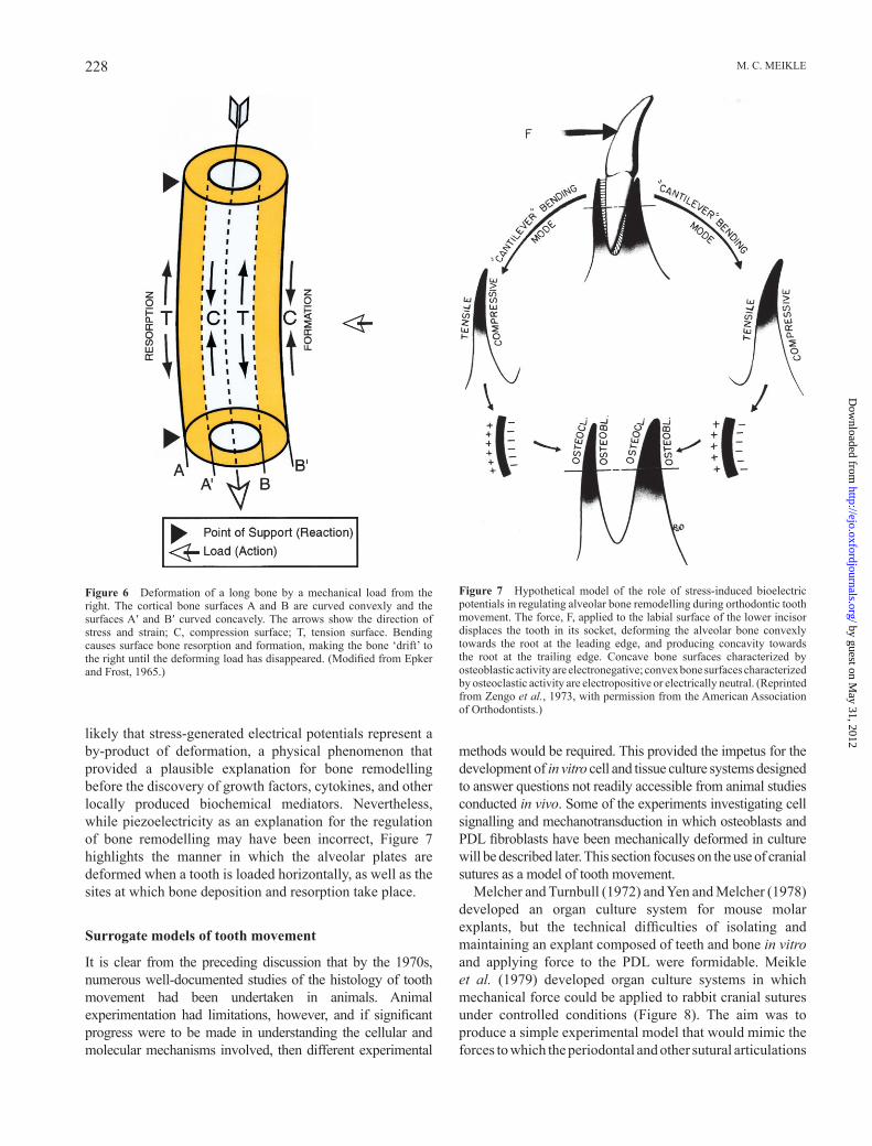
When an external load is applied to a long bone, deformation occurs, thus producing changes in the surface curvature ( Figure 6 ). The external surface of the left cortex elongates in tension, while the internal surface shortens in compression; opposite effects are produced on the internal and external surfaces of the right cortex ( Epker and Frost, 1965 ). Increasing concavity has consistently been shown to be associated with electronegativity and bone formation. In contrast, increasing convexity is associated with electropositivity and bone resorption ( Bassett and Becker, 1962 ).
Stress-generated electrical potentials have been recorded with surface electrodes in a dog mandible following the application of a mechanical force to the teeth ( Gillooly et al. , 1968 ), and Zengo et al. (1973) proposed that electrical potentials are responsible for regulating osteogenesis and bone resorption in the course of orthodontic tooth movement ( Figure 7 ). During the 1960s and 1970s, the regulation of bone remodelling by piezoelectrical effects enjoyed considerable support within the orthopaedic and orthodontic communities. However, the problems from a biological point of view are whether electrical phenomena are suffi ciently discriminatory to be able to regulate the metabolic activity of cell types as diverse as osteoblasts and osteoclasts, which function in close proximity, and also the fact that piezoelectricity does not require the presence of living cells; dead bone displays the same effects, which appear to be generated by shearing forces acting on the collagen fi bres of the bone matrix. The more that is learnt about cell – cell and cell – matrix interactions, the more it is
by guest on May 31, 2012
http://ejo.oxfordjournals.org/D
ownloaded from
M. C. MEIKLE228
likely that stress-generated electrical potentials represent a by-product of deformation, a physical phenomenon that provided a plausible explanation for bone remodelling before the discovery of growth factors, cytokines, and other locally produced biochemical mediators. Nevertheless, while piezoelectricity as an explanation for the regulation of bone remodelling may have been incorrect, Figure 7 highlights the manner in which the alveolar plates are deformed when a tooth is loaded horizontally, as well as the sites at which bone deposition and resorption take place.
Surrogate models of tooth movement
It is clear from the preceding discussion that by the 1970s, numerous well-documented studies of the histology of tooth movement had been undertaken in animals. Animal experimentation had limitations, however, and if signifi cant progress were to be made in understanding the cellular and molecular mechanisms involved, then different experimental
methods would be required. This provided the impetus for the development of in vitro cell and tissue culture systems designed to answer questions not readily accessible from animal studies conducted in vivo . Some of the experiments investigating cell signalling and mechanotransduction in which osteoblasts and PDL fi broblasts have been mechanically deformed in culture will be described later. This section focuses on the use of cranial sutures as a model of tooth movement.
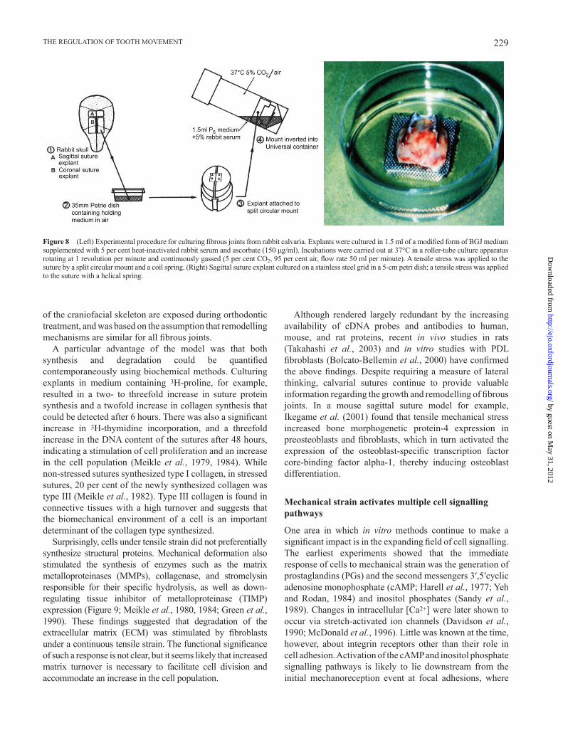
Melcher and Turnbull (1972) and Yen and Melcher (1978) developed an organ culture system for mouse molar explants, but the technical diffi culties of isolating and maintaining an explant composed of teeth and bone in vitro and applying force to the PDL were formidable. Meikle et al. (1979) developed organ culture systems in which mechanical force could be applied to rabbit cranial sutures under controlled conditions ( Figure 8 ). The aim was to produce a simple experimental model that would mimic the forces to which the periodontal and other sutural articulations
Figure 6 Deformation of a long bone by a mechanical load from the right. The cortical bone surfaces A and B are curved convexly and the surfaces A ′ and B ′ curved concavely. The arrows show the direction of stress and strain; C, compression surface; T, tension surface. Bending causes surface bone resorption and formation, making the bone ‘ drift ’ to the right until the deforming load has disappeared. (Modifi ed from Epker and Frost, 1965 .)
Figure 7 Hypothetical model of the role of stress-induced bioelectric potentials in regulating alveolar bone remodelling during orthodontic tooth movement. The force, F, applied to the labial surface of the lower incisor displaces the tooth in its socket, deforming the alveolar bone convexly towards the root at the leading edge, and producing concavity towards the root at the trailing edge. Concave bone surfaces characterized by osteoblastic activity are electronegative; convex bone surfaces characterized by osteoclastic activity are electropositive or electrically neutral. (Reprinted from Zengo et al. , 1973 , with permission from the American Association of Orthodontists.)
by guest on May 31, 2012
http://ejo.oxfordjournals.org/D
ownloaded from
229THE REGULATION OF TOOTH MOVEMENT
of the craniofacial skeleton are exposed during orthodontic treatment, and was based on the assumption that remodelling mechanisms are similar for all fi brous joints.
A particular advantage of the model was that both synthesis and degradation could be quantifi ed contemporaneously using biochemical methods. Culturing explants in medium containing 3 H-proline, for example, resulted in a two- to threefold increase in suture protein synthesis and a twofold increase in collagen synthesis that could be detected after 6 hours. There was also a signifi cant increase in 3 H-thymidine incorporation, and a threefold increase in the DNA content of the sutures after 48 hours, indicating a stimulation of cell proliferation and an increase in the cell population ( Meikle et al. , 1979 , 1984 ). While non-stressed sutures synthesized type I collagen, in stressed sutures, 20 per cent of the newly synthesized collagen was type III ( Meikle et al. , 1982 ). Type III collagen is found in connective tissues with a high turnover and suggests that the biomechanical environment of a cell is an important determinant of the collagen type synthesized.
Surprisingly, cells under tensile strain did not preferentially synthesize structural proteins. Mechanical deformation also stimulated the synthesis of enzymes such as the matrix metalloproteinases (MMPs), collagenase, and stromelysin responsible for their specifi c hydrolysis, as well as down-regulating tissue inhibitor of metalloproteinase (TIMP) expression ( Figure 9 ; Meikle et al. , 1980 , 1984 ; Green et al. , 1990 ). These fi ndings suggested that degradation of the extracellular matrix (ECM) was stimulated by fi broblasts under a continuous tensile strain. The functional signifi cance of such a response is not clear, but it seems likely that increased matrix turnover is necessary to facilitate cell division and accommodate an increase in the cell population.
Although rendered largely redundant by the increasing availability of cDNA probes and antibodies to human, mouse, and rat proteins, recent in vivo studies in rats ( Takahashi et al. , 2003 ) and in vitro studies with PDL fi broblasts ( Bolcato-Bellemin et al. , 2000 ) have confi rmed the above fi ndings. Despite requiring a measure of lateral thinking, calvarial sutures continue to provide valuable information regarding the growth and remodelling of fi brous joints. In a mouse sagittal suture model for example, Ikegame et al. (2001) found that tensile mechanical stress increased bone morphogenetic protein-4 expression in preosteoblasts and fi broblasts, which in turn activated the expression of the osteoblast-specifi c transcription factor core-binding factor alpha-1, thereby inducing osteoblast differentiation.
Mechanical strain activates multiple cell signalling pathways
One area in which in vitro methods continue to make a signifi cant impact is in the expanding fi eld of cell signalling. The earliest experiments showed that the immediate response of cells to mechanical strain was the generation of prostaglandins (PGs) and the second messengers 3 ′ ,5 ′ cyclic adenosine monophosphate (cAMP; Harell et al. , 1977 ; Yeh and Rodan, 1984 ) and inositol phosphates ( Sandy et al. , 1989 ). Changes in intracellular [Ca 2+ ] were later shown to occur via stretch-activated ion channels ( Davidson et al. , 1990 ; McDonald et al. , 1996 ). Little was known at the time, however, about integrin receptors other than their role in cell adhesion. Activation of the cAMP and inositol phosphate signalling pathways is likely to lie downstream from the initial mechanoreception event at focal adhesions, where
Figure 8 (Left) Experimental procedure for culturing fi brous joints from rabbit calvaria. Explants were cultured in 1.5 ml of a modifi ed form of BGJ medium supplemented with 5 per cent heat-inactivated rabbit serum and ascorbate (150 μ g/ml). Incubations were carried out at 37°C in a roller-tube culture apparatus rotating at 1 revolution per minute and continuously gassed (5 per cent CO 2 , 95 per cent air, fl ow rate 50 ml per minute). A tensile stress was applied to the suture by a split circular mount and a coil spring. (Right) Sagittal suture explant cultured on a stainless steel grid in a 5-cm petri dish; a tensile stress was applied to the suture with a helical spring.
by guest on May 31, 2012
http://ejo.oxfordjournals.org/D
ownloaded from

M. C. MEIKLE230
integrin receptors link the ECM to the cytoskeleton ( Wang et al. , 1993 ; DeMali et al. , 2003 ).
The role of PGs
The fi rst evidence that PGs might be involved in mechanical force transduction came from the cell culture studies of Hong et al. (1976) , who observed that perturbing cell membranes by detaching cells mechanically from culture dishes caused an increase in PG synthesis. They suggested that mechanical distortion acts by changing the conformation of the cell membrane, thereby exposing membrane phospholipids to the action of phospholipases to release free arachidonic acid. These fi ndings, plus research showing that changes in intracellular cyclic nucleotides followed mechanical deformation ( Rodan et al. , 1975 ; Davidovitch and Shanfi eld, 1975 ), provided the impetus for an in vitro study by Harell et al. (1977) . They deformed petri dishes with orthodontic screws cemented to the base on which osteoblast-like cells had been cultured and suggested the following sequence of events: (1) activation of phospholipase A 2 with the subsequent release of arachidonic acid resulted in a rapid (threefold) synthesis of PGE 2 at 5 minutes, (2) this in turn activated adenylate cyclase and a transient increase in intracellular cAMP which peaked at 15 minutes, and (3) an increase in intracellular [Ca 2+ ] and a stimulation of DNA synthesis. The ability to mimic or inhibit these changes by exogenous PGE 2 or indomethacin [a cyclooxygenase-1 (COX-1) inhibitor] provided further evidence that the transduction of mechanical deformation into a biological response was mediated by PG production ( Harell et al. , 1977 ; Somjen et al. , 1980 ). Other in vitro models have implicated PGs as mediators of mechanical stress in a number of cell types, including mechanically deformed osteoblasts ( Yeh and Rodan, 1984 ), gingival fi broblasts ( Ngan et al. , 1988 ), and PDL fi broblasts ( Ngan et al. , 1990 ).
In an interesting practical application of the fi ndings of Harell et al. (1977) , Yamasaki et al. (1980) investigated the role of PGs in orthodontic tooth movement initially with the rat molar model. They found that indomethacin produced a dose-related inhibition in the appearance of osteoclasts and bone resorption that was limited to the fi rst 12 hours of tooth movement. This explains why tooth movement is slower in patients who consume large quantities of non-steroidal anti-infl ammatory drugs (NSAIDs). Injections of PGE 1 and PGE 2 into the gingival tissues near the upper fi rst molar also stimulated osteoclast formation and bone resorption. This led to further work that showed local PG injections could increase the rate of tooth movement ( Yamasaki et al. , 1982 , 1984 ).
Leukotrienes
A rabbit model showed that the COX inhibitor fl urbiprofen signifi cantly reduced osteoclast numbers, but interestingly not the degree of tooth movement ( Sandy and Harris, 1984 ). This paradoxical observation suggested that PGs alone did not account for the bone remodelling associated with tooth movement. Leukotrienes (LTs) are also metabolites of arachidonic acid (but produced via the lipoxygenase pathway) and could have accounted for this discrepancy since they are potent stimulators of bone resorption ( Meghji et al. , 1988 ). It was subsequently shown that AA861, a selective inhibitor of the LT pathway, caused a signifi cant inhibition of LTB 4 and a reduction in tooth movement in rats ( Mohammed et al. , 1989 ).
The cAMP pathway
The second messenger system classically associated with mechanical force transduction is cAMP. The fi rst evidence for the involvement of the cAMP pathway in mechanical signal transduction was provided independently by Rodan
Figure 9 (Left) The effect of tensile mechanical stress on collagenase and tissue inhibitor of metalloproteinase (TIMP) production. Four pairs of coronal suture explants were mechanically deformed and culture supernatants collected daily over 4 days. Culture media were assayed for collagenase and TIMP activity using the standard fi bril assay and show that as collagenase increases, there is a concomitant decrease in TIMP. Values are expressed as mean ± standard error of the mean. ( ■ ) Denotes active enzyme. * P < 0.05, ** P < 0.01. (Right) Immunolocalization of collagenase in a stressed coronal suture showing positive cells with diffuse fl uorescence of the surrounding extracellular matrix indicative of the presence of active enzyme.
by guest on May 31, 2012
http://ejo.oxfordjournals.org/D
ownloaded from
231THE REGULATION OF TOOTH MOVEMENT
et al. (1975) and Davidovitch and Shanfi eld (1975) . Rodan et al. (1975) showed that a compressive force of 60 g/cm 2 applied to 16-day-old chick tibia in vitro inhibited the accumulation of cAMP in the epiphyses, as well as in cells isolated from the proliferative zone of the growth plate. The effect was mediated by an enhanced uptake of Ca 2+ which inhibited membrane-associated adenyl cyclase activity ( Bourret and Rodan, 1976 ).
Davidovitch and Shanfi eld (1975) sampled alveolar bone from compression and tension sites surrounding orthodontically tipped canines in cats. They found that cAMP levels initially decreased, followed by an increase after 1 – 2 days that remained elevated to the end of the experimental period of 28 days. They suggested that the initial decrease at the compression sites was due to necrosis of PDL cells, and at the tension sites to a rapid increase in the cell population; the elevation in cAMP observed 2 weeks after the initiation of treatment was probably a refl ection of increased bone remodelling activity. Subsequently, Davidovitch et al. (1976) , in a study on the cellular localization of cAMP in the same model, found an increase in the number of cAMP-positive cells in areas of the PDL where bone resorption or deposition subsequently occurred. Osteocytes in the adjacent alveolar bone, however, appeared to be relatively unaffected by the mechanical force.
The phosphoinositide pathway
The phosphoinositide (PI) pathway had been investigated in the 1950s, but the importance of membrane phospholipids as cellular second messengers was not fully appreciated until the 1980s, when Streb et al. (1983) showed that products of inositol lipid breakdown caused a release of intracellular Ca 2+ . The PI pathway accounts for many of the changes seen in mechanically deformed tissues, including the elevation of intracellular [Ca 2+ ] from the endoplasmic reticulum and increased DNA synthesis.
PGE 2 and parathyroid hormone (PTH) elevate inositol phosphates as well as cAMP in mouse calvarial osteoblast cultures; however, the effect of PTH on inositol phosphate mobilization was not direct, being mediated indirectly by PGs in an autocrine/paracrine fashion ( Farndale et al. , 1988 ). Evidence that mechanical stress was also PG-mediated made it unlikely that only one pathway would be selectively mobilized, and subsequently, Sandy et al. (1989) found that both cAMP and inositol phosphates were elevated in mouse osteoblasts following intermittent mechanical strain. Activation of cAMP and inositol phosphates by PGE 2 has been shown to induce the expression of transcription factors such as Erg-1 (early growth response gene-1) and the proto-oncogenes c-jun and c-fos (referred to as immediate early genes). These activate or repress the transcription of late-acting genes required for bringing about changes in phenotype. Erg-1 is involved in cell
proliferation and has been shown to be elevated in rat osteoblasts by mechanical stretch within 15 minutes ( Dolce et al. , 1996 ).
Signal transduction by integrins and focal adhesions
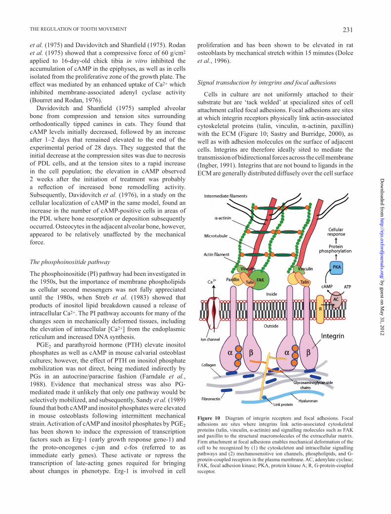
Cells in culture are not uniformly attached to their substrate but are ‘ tack welded ’ at specialized sites of cell attachment called focal adhesions. Focal adhesions are sites at which integrin receptors physically link actin-associated cytoskeletal proteins (talin, vinculin, α -actinin, paxillin) with the ECM ( Figure 10 ; Sastry and Burridge, 2000 ), as well as with adhesion molecules on the surface of adjacent cells. Integrins are therefore ideally sited to mediate the transmission of bidirectional forces across the cell membrane ( Ingber, 1991 ). Integrins that are not bound to ligands in the ECM are generally distributed diffusely over the cell surface
Figure 10 Diagram of integrin receptors and focal adhesions. Focal adhesions are sites where integrins link actin-associated cytoskeletal proteins (talin, vinculin, α -actinin) and signalling molecules such as FAK and paxillin to the structural macromolecules of the extracellular matrix. Firm attachment at focal adhesions enables mechanical deformation of the cell to be recognized by (1) the cytoskeleton and intracellular signalling pathways and (2) mechanosensitive ion channels, phospholipids, and G-protein-coupled receptors in the plasma membrane. AC, adenylate cyclase; FAK, focal adhesion kinase; PKA, protein kinase A; R, G-protein-coupled receptor.
by guest on May 31, 2012
http://ejo.oxfordjournals.org/D
ownloaded from
M. C. MEIKLE232
and appear not to be linked to the actin cytoskeleton; association with the actin cytoskeleton is induced by binding to the ECM ( Wang et al. , 1993 ). Integrins are composed of structurally distinct α and β subunits that combine to generate heterodimeric receptors with unique binding properties; for example, α 2 β 1 integrin binds to collagen and laminin and α v β 3 to vitronectin. Integrins bind to adhesive and structural proteins in the ECM via the peptide sequence RGD (arginine – glycine – aspartate). Integrin-mediated adhesive interactions play key roles in cell migration, proliferation, and differentiation, and also regulate intracellular signal transduction pathways that control adhesion-induced (outside – in) changes in cell physiology ( Wang et al. , 1993 ; Clarke and Brugge, 1995 ). Integrins thus function both as cell adhesion molecules and intracellular signalling receptors.
The numerous signalling pathways activated by integrins are beyond the scope of this discussion, but the low-molecular weight guanosine triphosphatases, Rab and Rho, as well as mitogen-activated protein (MAP) kinase subtypes that are components of integrin-mediated signalling have been shown to be altered in mechanically stretched PDL fi broblasts ( Basdra et al. , 1995 ) and osteoblasts ( Peverali et al. , 2001 ). MAP kinase phosphorylates and activates phospholipase A 2 , leading to the hydrolysis of glycerophospholipids to yield arachidonic acid. Furthermore, integrin-dependent increases in intracellular [Ca 2+ ] appear to involve either inositol triphosphate-induced Ca 2+ mobilization from the endoplasmic reticulum or the transport of extracellular Ca 2+ through mechanosensitive ion channels ( Clarke and Brugge, 1995 ).
Evidence is accumulating making it increasingly likely that the previously reported changes in cell signalling in response to mechanical deformation are downstream of events mediated by integrins at focal adhesions. In a study designed to determine whether cells sense mechanical signals through generalized membrane distortion or through integrins, Meyer et al. (2000) found that mechanical stress altered the cAMP signalling cascade and downstream gene transcription through signals generated by activated integrin receptors, and in a G-protein-dependent manner. In an earlier report ( Wilson et al. , 1995 ), the proliferative response of cultured vascular smooth muscle cells to cyclic mechanical strain was found to be dependent on interactions between integrins and specifi c matrix proteins (collagen, fi bronectin, vitronectin) since the response to strain was neutralized by antibodies to β 3 and α v β 5 integrins. Carvalho et al. (1996) have further shown that mechanical stimulation of bone cells results in a marked increase in β 1 mRNA expression after 30 minutes ( α v expression did not change). Firm attachment to the ECM at focal adhesions is evidently necessary to enable mechanical distortion to be recognized by (1) the cytoskeleton and intracellular signalling pathways and (2) mechanosensitive ion channels, phospholipids, and G-protein-coupled receptors in the cell membrane.
Cytokines
One of the most signifi cant advances in connective tissue biology during the 1980s was the demonstration that many cytokines, in addition to mediating the host immunological response to exogenous antigens, were also produced by connective tissue cells such as fi broblasts and osteoblasts and involved in normal physiological turnover and bone remodelling. Cytokines are low-molecular weight proteins (mw < 25 kDa) produced by cells that regulate or modify the action of other cells in an autocrine (acting on the cell of origin) or paracrine (acting on adjacent cells) manner. The defi nition includes the interleukins (ILs), tumour necrosis factors (TNFs), interferons, growth factors, and colony-stimulating factors. A major diffi culty in understanding cytokine biology is the sheer number and complexity of these factors. Another problem is that several factors such as IL-1 and TNF exhibit overlapping biological activities (redundancy) and many have multiple biological effects (pleiotropy).
The role of cytokines in cell – cell signalling in bone
Cells of the osteoblast lineage play a pivotal role in bone remodelling, a process that involves interactions between osteoblasts and osteoclasts, systemic hormones, cytokines, and growth factors. Systemic hormones and mechanical stimuli infl uence the process via their ability to control the synthesis and/or action of cytokines. Since bone remodelling occurs at discrete sites throughout the skeleton, osteoblast-derived cytokines are ideally placed to regulate or modify the action of other cell types in bone. The fi rst cytokine shown to play a role in bone turnover was IL-1, which in addition to activating lymphocytes also proved to be a potent bone resorptive agent ( Gowen et al. , 1983 ; Heath et al. , 1985 ). Not long afterwards, the TNFs were shown to stimulate bone resorption and inhibit bone formation in vitro ( Bertolini et al. , 1986 ), and subsequent studies have implicated numerous others.
Experiments in which bone marrow had been cultured in the presence of bone resorptive agents such as PTH, 1,25(OH) 2 vitamin D 3 , or IL-1 had shown that osteoclast formation and function was dependent upon the presence of osteoblasts/stromal cells. This suggested that cells of the osteoblast lineage produced soluble factors that were involved in osteoblast – osteoclast signalling. After many attempts to purify, characterize, and sequence such an osteoclast-activating factor using biochemical methods, a cell-surface protein that was found to be identical to receptor activator of nuclear factor κ B ligand (RANKL, a member of the membrane-associated TNF ligand family) was simultaneously discovered by two research teams ( Lacey et al. , 1998 ; Yasuda et al. , 1998 ) using molecular techniques. RANKL and its receptor RANK expressed on osteoclasts and their precursor cells turned out to be the
by guest on May 31, 2012
http://ejo.oxfordjournals.org/D
ownloaded from
233THE REGULATION OF TOOTH MOVEMENT
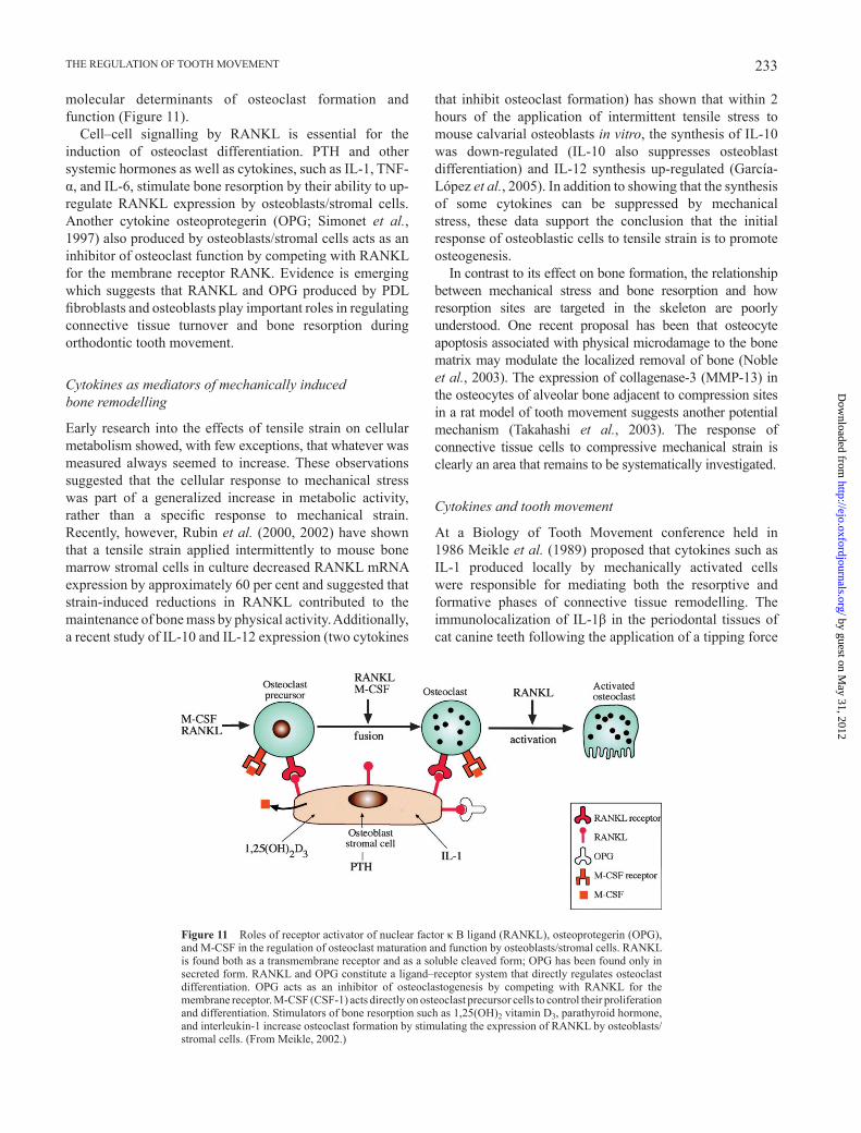
molecular determinants of osteoclast formation and function ( Figure 11 ).
Cell – cell signalling by RANKL is essential for the induction of osteoclast differentiation. PTH and other systemic hormones as well as cytokines, such as IL-1, TNF- α , and IL-6, stimulate bone resorption by their ability to up-regulate RANKL expression by osteoblasts/stromal cells. Another cytokine osteoprotegerin (OPG; Simonet et al. , 1997 ) also produced by osteoblasts/stromal cells acts as an inhibitor of osteoclast function by competing with RANKL for the membrane receptor RANK. Evidence is emerging which suggests that RANKL and OPG produced by PDL fi broblasts and osteoblasts play important roles in regulating connective tissue turnover and bone resorption during orthodontic tooth movement.
Cytokines as mediators of mechanically induced bone remodelling
Early research into the effects of tensile strain on cellular metabolism showed, with few exceptions, that whatever was measured always seemed to increase. These observations suggested that the cellular response to mechanical stress was part of a generalized increase in metabolic activity, rather than a specifi c response to mechanical strain. Recently, however, Rubin et al. (2000 , 2002 ) have shown that a tensile strain applied intermittently to mouse bone marrow stromal cells in culture decreased RANKL mRNA expression by approximately 60 per cent and suggested that strain-induced reductions in RANKL contributed to the maintenance of bone mass by physical activity. Additionally, a recent study of IL-10 and IL-12 expression (two cytokines
that inhibit osteoclast formation) has shown that within 2 hours of the application of intermittent tensile stress to mouse calvarial osteoblasts in vitro , the synthesis of IL-10 was down-regulated (IL-10 also suppresses osteoblast differentiation) and IL-12 synthesis up-regulated ( García-López et al. , 2005 ). In addition to showing that the synthesis of some cytokines can be suppressed by mechanical stress, these data support the conclusion that the initial response of osteoblastic cells to tensile strain is to promote osteogenesis.
In contrast to its effect on bone formation, the relationship between mechanical stress and bone resorption and how resorption sites are targeted in the skeleton are poorly understood. One recent proposal has been that osteocyte apoptosis associated with physical microdamage to the bone matrix may modulate the localized removal of bone ( Noble et al. , 2003 ). The expression of collagenase-3 (MMP-13) in the osteocytes of alveolar bone adjacent to compression sites in a rat model of tooth movement suggests another potential mechanism ( Takahashi et al. , 2003 ). The response of connective tissue cells to compressive mechanical strain is clearly an area that remains to be systematically investigated.
Cytokines and tooth movement
At a Biology of Tooth Movement conference held in 1986 Meikle et al. (1989) proposed that cytokines such as IL-1 produced locally by mechanically activated cells were responsible for mediating both the resorptive and formative phases of connective tissue remodelling. The immunolocalization of IL-1 β in the periodontal tissues of cat canine teeth following the application of a tipping force
Figure 11 Roles of receptor activator of nuclear factor κ B ligand (RANKL), osteoprotegerin (OPG), and M-CSF in the regulation of osteoclast maturation and function by osteoblasts/stromal cells. RANKL is found both as a transmembrane receptor and as a soluble cleaved form; OPG has been found only in secreted form. RANKL and OPG constitute a ligand – receptor system that directly regulates osteoclast differentiation. OPG acts as an inhibitor of osteoclastogenesis by competing with RANKL for the membrane receptor. M-CSF (CSF-1) acts directly on osteoclast precursor cells to control their proliferation and differentiation. Stimulators of bone resorption such as 1,25(OH) 2 vitamin D 3 , parathyroid hormone, and interleukin-1 increase osteoclast formation by stimulating the expression of RANKL by osteoblasts/stromal cells. (From Meikle, 2002 .)
by guest on May 31, 2012
http://ejo.oxfordjournals.org/D
ownloaded from

M. C. MEIKLE234
( Davidovitch et al. , 1988 ) provided the fi rst experimental evidence in support of this hypothesis. Clinical studies have since shown that IL-1 β , TNF- α , IL-6, and epidermal growth factor are all elevated in gingival crevicular fl uid collected from patients during the early phases of orthodontic tooth movement ( Grieve et al. , 1994 ; Lowney et al. , 1995 ; Uematsu et al. , 1996 ). These fi ndings have since been confi rmed by numerous other investigations.
The response of PDL cells to mechanical strain in vitro
Cultured human PDL cells have been widely used to study the effects of tensile mechanical strain on gene expression at both message and protein levels, particularly for investigating intermediate- and late-acting genes. The results can sometimes be ambiguous, however, depending on the culture conditions, stress regimen (intermittent or cyclic versus continuous strain), and length of culture. For example, cyclic tension has been reported to both stimulate ( Saito et al. , 1991 ; Shimizu et al. , 1994 ) and inhibit ( Long et al. , 2001 ) IL-1 β activity by PDL cells. While a continuous tensile strain applied to PDL cells stimulated MMP-1 (collagenase-1), MMP-2 (gelatinase-A), TIMP-1, and TIMP-2 expression ( Bolcato-Bellemin et al. , 2000 ), centrifugation of PDL cells in culture fl asks up-regulated mRNA levels for MMP-1, but had little or no effect on type
I collagen or TIMP expression ( Redlich et al. , 2004 ). Cyclic stretching (15 per cent elongation) decreased ALP activity in PDL cells ( Chiba and Mitani, 2004 ), but the synthesis of other matrix proteins such as type I collagen and fi bronectin by PDL cells exposed to 5 per cent deformation was increased ( Howard et al. , 1998 ). Also one cannot assume that data derived from one differentiated cell type is transferable to another; the synthesis of IL-10 was up-regulated by cyclic tension in human PDL cells ( Long et al. , 2001 ) and down-regulated in murine osteoblasts ( García-López et al. , 2005 ). From all the evidence available to date, however, it is possible to construct the hypothetical model in Figure 12 , showing the response of the cells on the tension side of the PDL to orthodontic tooth movement.
To date, few researchers have utilized in vitro methods to investigate the response of either osteoblasts or PDL cells to compression forces. Kanzaki et al. (2002) found that culture media from compressed PDL fi broblasts stimulated osteoclastogenesis in peripheral blood mononuclear cell cultures and that RANKL mRNA expression was up-regulated in the cells. OPG expression in contrast remained unchanged. These data suggest that the PDL plays a pivotal role in osteoclast differentiation and function during orthodontic tooth movement. In another study, He et al. (2004) showed that PDL cells subjected to tensional and compressive forces were able to perceive two different
Figure 12 Remodelling of the periodontium: tension side. In this hypothetical model periodontal ligament (PDL) fi broblasts under tensile strain synthesize cytokines such as interleukin-1 (IL-1) and IL-6 (1); IL-1 and IL-6 in turn stimulate matrix metalloproteinase (MMP) and inhibit tissue inhibitor of metalloproteinase (TIMP) synthesis by PDL cells via autocrine and paracrine mechanisms (2); vascular endothelial growth factor (VEGF) produced by mechanically activated fi broblasts promotes angiogenesis (3). Degradation of the extracellular matrix by MMPs facilitates cell proliferation and capillary growth; PDL cells (4), osteoblasts, and bone-lining cells (5) enter a biosynthetic phase with the synthesis of structural and other matrix molecules.
by guest on May 31, 2012
http://ejo.oxfordjournals.org/D
ownloaded from

235THE REGULATION OF TOOTH MOVEMENT
forms of mechanical stimuli and responded in a differential manner as regards ECM synthesis (type I collagen, fi bronectin) and degradation (MMP-2, TIMP-2).
Molecular studies of tooth movement in the rat
Connective tissue degradation and bone resorption involves the interaction of several differentiated cell types. In vivo methods are therefore essential if the cascade of molecular events responsible for the recruitment and activation of osteoclasts, as well as multinucleated giant cells, is to be fully understood.
The application of in situ hybridization techniques to tooth movement studies in the rat has proven to be particularly useful in this respect. The expression of IL-1 β and IL-6 mRNAs (but not TNF- α ) has been shown to be up-regulated in both PDL cells and osteoblasts on the compression side ( Alhashimi et al. , 2001 ), as is MMP-8 (collagenase-2) and MMP-13 (collagenase-3; Takahashi et al. , 2003 ). Both RANKL and OPG mRNAs are widely expressed in osteoblasts and PDL cells throughout the periodontal tissues ( Ogasawara et al. , 2004 ), and following tooth movement using the Waldo method ( Waldo and Rothblatt, 1954 ), positive signals for RANKL and RANK were detected in multinucleate osteoclasts at sites of active bone resorption. IL-1 β and TNF- α expression was also identifi ed in some osteoclasts. Figure 13 represents a
hypothetical model of the events involved in remodelling the periodontium on the ‘ compression ’ side during orthodontic tooth movement.
Is orthodontic tooth movement an infl ammatory process?
The proposal that tooth movement involved the development of infl ammation seems to have originated with a review paper by Storey (1973) . In the 1950s, he published a series of papers on the histological changes produced by helical torsion springs applied to rodent (rat, guinea pig) and largomorph (rabbit) incisors ( Storey, 1955a , b , c ). The magnitude of the force applied to the teeth (25, 50, 250 g) was clearly excessive in this experimental model judging from the photomicrographs, and although a good deal of tissue destruction was evident, at no time was any reference made to the involvement of an infl ammatory response. The demonstration that cytokines (frequently referred to in the literature as infl ammatory mediators or pro-infl ammatory cytokines), as well as neurotransmitters such as calcitonin gene-related peptide and substance P, are involved in remodelling the PDL has revived the concept that tooth movement is an infl ammatory process ( Davidovitch et al. , 1988 ; Davidovitch, 1995 ). This raises two questions: fi rst, are cytokines unique to infl ammation and second, to what
Figure 13 Remodelling of the periodontium: compression side. In this hypothetical model periodontal ligament (PDL) cells under compressive strain synthesize interleukin-1 (IL-1) and IL-6 (1); IL-1 and IL-6 act in an autocrine and paracrine manner to up-regulate receptor activator of nuclear factor κ B ligand (RANKL) (2) and matrix metalloproteinase (MMP) (3) expression by PDL cells and osteoblasts. Osteoblast-derived MMPs degrade the non-mineralized surface osteoid layer of bone, while MMPs produced by PDL cells degrade their extracellular matrix; (4) RANKL stimulates the formation and function of osteoclasts from mononuclear precursor cells which access the bone surface and degrade the mineralized matrix; (5) deformation of the alveolar bone up-regulates MMPs expression by osteocytes adjacent to the bone surface.
by guest on May 31, 2012
http://ejo.oxfordjournals.org/D
ownloaded from

M. C. MEIKLE236
extent can tooth movement be regarded as an infl ammatory event?
The answer to the fi rst question is no. The naming of cytokines has customarily been based on the biological activity by which they were originally identifi ed. For example, before being sequenced, IL-1 was known by several names (lymphocyte-activating factor, endogenous pyrogen, catabolin, mononuclear cell factor) depending on the bioassay in which it was active and the research interests of the group that had identifi ed it. The fi rst cytokines were discovered in lymphocytes and monocytes through their role in mediating various aspects of the infl ammatory and immune response and called lymphokines and monokines, respectively, by immunologists. Many, including IL-1 and TNF- α (cachectin), were eventually shown to be produced by numerous cell types including connective tissue cells and to be involved in normal physiological turnover and bone remodelling. Furthermore, both IL-6 (B cell growth factor-II, B cell stimulatory factor, interferon- β 2, plasmacytoma growth factor) and IL-11 (adipogenesis inhibitory factor) have been shown to stimulate bone resorption and IL-10 (cytokine synthesis inhibitory factor) to inhibit bone resorption, a property not immediately obvious from their original names. Does that make bone remodelling an infl ammatory process? It seems unlikely this is the case. Cytokines and PGs are not unique to infl ammation; they are ubiquitous cell signalling molecules involved in numerous pathophysiological processes throughout the body. The statement of Tuncay (2004) that tooth movement is the end-product of an infl ammatory response because it is diminished by NSAIDs merely shows that arachidonic acid metabolites are involved in PDL remodelling and their activity can be blocked by COX inhibitors.
The answer to the second question is more complex and depends on the defi nition of infl ammation. Infl ammation is commonly understood to be the localized host response to microbial infection or tissue damage. Under normal circumstances, tooth movement is a sterile process and the extent of any damage to the tissues will depend upon the magnitude of the applied force. Furthermore, tooth movement does not meet the four classical criteria for infl ammation (redness, swelling, heat, and pain), perhaps apart from pain. In the article by Storey (1973) , the question of infl ammation in tooth movement is raised only in connection with the application of heavy forces. For example,
Those forces which exceed the bioplastic limit result in biodisruptive deformation with interruption of nutrition, ischaemia, and cell death as well as infl ammation and rupture of connective tissues. Biodisruptive deformation is usually followed by the classic process of tissue repair (p 293). Or (p 304) … heavy forces rapidly disrupt the
periodontal tissues and initiate an infl ammatory process but, when the tooth stops moving, repair occurs; mitotic fi gures in fi broblasts are numerous, and within the fi rst day or two a fi ne fi brin network fi lls the gap and cells and fi bres bridge discontinuities in the tissue while macrophages remove cell debris. During this repair phase, vessels are no longer abnormally permeable … .
This description sounds remarkably like wound healing, a process composed of a continuous sequence of infl ammation and repair designed to restore normal tissue continuity and function. Describing orthodontic tooth movement as an infl ammatory process creates the impression that it is a pathological event. For the most part it is not, unless the clinician or animal investigator happens to be applying excessive forces to the teeth. If one wishes to employ a label to describe the tissue response to tooth movement, it is best regarded as an exaggerated form of normal physiological turnover combined with foci of tissue repair, particularly at compression sites where hyalinized tissue and adjacent bone and cementum are being remodelled.
Conclusions
After 100 years, we have reasonably good understanding of the sequence of events involved in orthodontic tooth movement at the tissue and cellular levels on both the tensile and compression sides of the periodontium. At the molecular level, however, although signifi cant progress has been made over the last 20 years, our knowledge remains far from complete. Both in vivo and in vitro methods have been widely used to investigate the response of cells to mechanical deformation and it is important to stress that the two approaches are complementary; data from in vitro model systems in which the mechanical stimulus applied to the cells can be carefully regulated (tension versus compression; intermittent versus continuous) should be correlated with in vivo data obtained from animal models.
Cell cultures have been widely used to investigate signal transduction mechanisms particularly in osteoblasts and, more recently, to study the expression of intermediate- and late-acting genes in both osteoblast and PDL cultures. Nevertheless, the focus to date has been on the effects of tensile strain. The development of instruments based on the vacuum operated apparatus described by Banes et al. (1985) designed to apply static or cyclic variable compression to cells in vitro should resolve this defi ciency. Future investigations of gene expression involving PDL fi broblasts should also consider culturing the cells in a type I collagen gel matrix to create an artifi cial three-dimensional (3D) PDL. Unlike cells on two-dimensional surfaces which have a spread morphology, fi broblasts in 3D matrices develop an elongated or stellate form more closely resembling their shape in vivo which may infl uence signalling pathways and gene expression ( DeMali et al. , 2003 ).
by guest on May 31, 2012
http://ejo.oxfordjournals.org/D
ownloaded from
237THE REGULATION OF TOOTH MOVEMENT
The molecular techniques of reverse transcription - polymerase chain reaction and in situ hybridization to detect gene expression in cell and tissue sections have revolutionized tooth movement studies, particularly in the rat, which despite some anatomical and physiological shortcomings ( Ren et al. , 2004 ) remains the experimental animal of choice. In addition to detecting message levels, an expanding list of antibodies and enzyme-linked immunosorbent assays directed against human and animal proteins will facilitate their identifi cation in tissue sections and/or culture supernatants. Although this technology has greatly simplifi ed research for the clinical and laboratory investigator, one should not forget that the message is not always translated into protein, and the presence of a protein does not necessarily mean that it is biologically active. An MMP such as collagenase may be in a latent or active form or even complexed with TIMP.
The current evidence from these and other model systems suggests that downstream from the initial mechanotransduction event at focal adhesions which link the ECM to the cytoskeleton, connective tissue remodelling is mediated by a complex feedback mechanism involving the synthesis of osteotropic cytokines such as IL-1, IL-6, and RANKL by cells of the osteoblast and/or PDL fi broblast lineages. These in turn act in an autocrine/paracrine manner to regulate the expression of transcription factors, cytokines, growth regulatory molecules, and enzymes involved in the differentiation and function of mesenchymal and other cell types.
It would appear that Sandstedt and Oppenheim arrived at different conclusions regarding the tissue response to orthodontic tooth movement on the seemingly trivial difference of the plane of section — in Sandstedt’s fi gures the tissue blocks were sectioned horizontally and in Oppenheim’s vertically. Oppenheim (1930) was resistant to the idea that bone could be physically deformed but the pattern of the cellular response in his illustrations strongly suggests he was describing precisely that. It is perhaps ironic therefore that while Oppenheim is identifi ed in many people’s minds as the key fi gure in tooth movement research, Sandstedt’s concepts dominate orthodontic theory today. The message for the contemporary investigator is that (1) a 3D perspective is essential for a complete understanding of events and (2) tooth movement involves two interrelated processes: the bending of alveolar bone and remodelling of the periodontal tissues.
Address for correspondence
Professor Murray C. MeikleFaculty of DentistryUniversity of OtagoP.O. Box 647DunedinNew ZealandE-mail: [email protected]
Acknowledgement
The author wishes to thank Dr Dirk Bister, King’s College London Dental Institute, for translating the Sandstedt articles from their original German.
References Alhashimi N, Frithiof L, Brudvik P, Bakhiet M 2001 Orthodontic tooth
movement and de novo synthesis of proinfl ammatory cytokines. American Journal of Orthodontics and Dentofacial Orthopedics 119 : 307 – 312
Angle E H 1907 Treatment of malocclusion of the teeth. Angle’s system, 7th edn. S S White Dental Manufacturing Company, Philadelphia
Banes A J, Gilbert J, Taylor D, Monbureau O 1985 A new vacuum-operated stress-providing instrument that applies static or variable cyclic tension or compression to cells in vitro. Journal of Cell Science 75 : 35 – 42
Basdra E K, Papavassiliou A G, Huber L A 1995 Rab and rho GTPases are involved in specifi c response of periodontal ligament fi broblasts to mechanical stretching. Biochimica et Biophysica Acta 1268 : 209 – 213
Bassett C A L, Becker R O 1962 Generation of electrical potentials in bone in response to mechanical stress. Science 137 : 1063 – 1064
Baumrind S 1969 A reconsideration of the propriety of the “ pressure-tension ” hypothesis. American Journal of Orthodontics 55 : 12 – 22
Baumrind S, Buck D 1970 Rate changes in cell replication and protein synthesis in the periodontal ligament incident to tooth movement. American Journal of Orthodontics 57 : 109 – 131
Bertolini D R, Nedwin G E, Bringham T S, Smith D D, Mundy G R 1986 Stimulation of bone resorption and inhibition of bone formation in vitro by human tumour necrosis factors. Nature 319 : 516 – 518
Bien S M 1966 Hydrodynamic damping of tooth movement. Journal of Dental Research 45 : 907 – 914
Bien S M, Ayres H D 1965 Responses of rat maxillary incisors to loads. Journal of Dental Research 44 : 517 – 520
Boester C H, Johnston L E 1974 A clinical investigation of the concepts of differential and optimal force in canine retraction. Angle Orthodontist 44 : 113 – 119
Bolcato-Bellemin A L, Elkaim R, Abehsera A, Fausser J L, Haikel Y, Tenenbaum H 2000 Expression of mRNAs encoding for α and β integrin subunits, MMPs and TIMPs in stretched human periodontal ligament and gingival fi broblasts. Journal of Dental Research 79 : 1712 – 1716
Bourret L A, Rodan G A 1976 The role of calcium in the inhibition of cAMP accumulation in epiphyseal cartilage cells exposed to physiological pressure. Journal of Cell Physiology 88 : 353 – 362
Breitner C 1940 Bone changes resulting from experimental orthodontic treatment. American Journal of Orthodontics and Oral Surgery 26 : 521 – 547
Brudvik P, Rygh P 1994 Root resorption beneath the main hyalinized zone. European Journal of Orthodontics 16 : 249 – 263
Carvalho R S, Bumann A, Schwarzer C, Scott E, Yen E H K 1996 A molecular mechanism of integrin regulation from bone cells stimulated by mechanical forces. European Journal of Orthodontics 18 : 227 – 235
Chambers T J, Evans M, Gardner T N, Turner-Smith A, Chow J W M 1993 Induction of bone formation in rat tail vertebrae by mechanical loading. Bone and Mineral 20 : 167 – 178
Chiba M, Mitani H 2004 Cytoskeletal changes and the system of regulation of alkaline phosphatase in human periodontal ligament cells induced by mechanical stress. Cellular Biochemistry and Function 22 : 249 – 256
Chow J W M, Wilson A J, Chambers T J, Fox S W 1998 Mechanical loading stimulates bone formation by reactivation of bone lining cells
by guest on May 31, 2012
http://ejo.oxfordjournals.org/D
ownloaded from

M. C. MEIKLE238
in 13-week-old rats. Journal of Bone and Mineral Research 13 : 1760 – 1767
Clarke E A, Brugge J S 1995 Integrins and signal transduction pathways: the road taken. Science 268 : 233 – 239
Davidovitch Z 1991 Tooth movement. Critical Reviews in Oral Biology and Medicine 2 : 411 – 450
Davidovitch Z 1995 Cell biology associated with tooth movement. In: Berkovitz B, Moxham B, Newman H N (eds) The periodontal ligament in health and disease, 2nd edn. Mosby-Wolfe, Philadelphia, pp. 259 – 278
Davidovitch Z, Shanfi eld J L 1975 Cyclic AMP levels in alveolar bone of orthodontically-treated cats. Archives of Oral Biology 20 : 567 – 574
Davidovitch Z, Montgomery P C, Eckerdal O, Gustafson G T 1976 Cellular localization of cyclic AMP in periodontal tissue during experimental tooth movement in cats. Calcifi ed Tissue Research 18 : 316 – 329
Davidovitch Z, Nicolay O F, Ngan P W, Shanfi eld J L 1988 Neurotransmitters, cytokines and the control of alveolar bone remodeling in orthodontics. Dental Clinics of North America 32 : 411 – 435
Davidson R M, Tatakis D W, Auerbach A L 1990 Multiple forms of mechanosensitive channels in osteoblast-like cells. Pfl ugers Archiv 416 : 646 – 651
DeMali K A, Wennerberg K, Burridge K 2003 Integrin signaling to the actin cytoskeleton. Current Opinion in Cell Biology 15 : 72 – 582
Dolce C, Kinniburgh A J, Dziak R 1996 Immediate early-gene induction in rat osteoblastic cells after mechanical deformation. Archives of Oral Biology 41 : 1101 – 1108
El Haj A J, Minter S L, Rawlinson S C, Suswillo R, Lanyon L E 1990 Cellular responses to mechanical loading in vitro. Journal of Bone and Mineral Research 5 : 923 – 932
Epker B N, Frost H M 1965 Correlation of bone resorption and formation with the physical behavior of loaded bone. Journal of Dental Research 44 : 33 – 41
Farndale R W, Sandy J R, Atkinson S J, Pennington S R, Meghji S, Meikle M C 1988 Parathyroid hormone and prostaglandin E 2 stimulate both inositol phosphates and cyclic AMP accumulation in mouse osteoblast cultures. Biochemical Journal 252 : 263 – 268
Frost H M 1963 Bone remodelling dynamics. Charles C Thomas, Springfi eld
García-López S et al. 2005 Mechanical deformation inhibits IL-10 and stimulates IL-12 production by mouse calvarial osteoblasts in vitro. Archives of Oral Biology 50 : 449 – 452
Gianelly A A 1969 Force induced changes in the vascularity of the periodontal ligament. American Journal of Orthodontics 55 : 5 – 11
Gillooly C J, Hosley R T, Mathews J R, Jewett D L 1968 Electrical potentials recorded from mandibular alveolar bone as a result of forces applied to the tooth. American Journal of Orthodontics 54 : 649 – 654
Gluhak-Heinrich J et al. 2003 Mechanical loading stimulates dentin matrix protein 1 (DMP1) expression in osteocytes in vivo. Journal of Bone and Mineral Research 18 : 807 – 817
Gottlieb B, Orban B 1931 Die Veränderungen der Gewebe Bei Übermässiger Beanspruchung der Zähne. Verlag von Georg Thieme, Leipzig
Gowen M, Meikle M C, Reynolds J J 1983 Stimulation of bone resorption in vitro by a non-prostanoid factor released by human monocytes in culture. Biochimica et Biophysica Acta 762 : 471 – 474
Green D D, Hembry R M, Atkinson S J, Reynolds J J, Meikle M C 1990 Immunolocalization of collagenase and tissue inhibitor of metalloproteinases (TIMP) in mechanically deformed fi brous joints. American Journal of Orthodontics and Dentofacial Orthopedics 97 : 281 – 288
Grieve W G, Johnson G K, Moore R N, Reinhardt R A, DuBois L M 1994 Prostaglandin E (PGE) and interleukin-1 β (IL-1 β ) levels in gingival crevicular fl uid during human orthodontic tooth movement. American Journal of Orthodontics and Dentofacial Orthopedics 105 : 369 – 374
Grimm F M 1972 Bone bending, a feature of orthodontic tooth movement. American Journal of Orthodontics 62 : 384 – 393
Harell A, Dekel S, Binderman I 1977 Biochemical effect of mechanical stress on cultured bone cells. Calcifi ed Tissue Research 22 : 202 – 207
Harris C A 1839 The dental art, practical treatise on dental surgery. Armstrong and Berry, Baltimore
He Y, Macarak E J, Korostoff J M, Howard P S 2004 Compression and tension: differential effects on matrix accumulation by periodontal ligament fi broblasts in vitro. Connective Tissue Research 45 : 28 – 39
Heath J K, Saklatvala J, Meikle M C, Atkinson S J, Reynolds J J 1985 Pig interleukin 1 (catabolin) is a potent stimulator of bone resorption in vitro. Calcifi ed Tissue International 37 : 95 – 97
Heller I J, Nanda R 1979 Effect of metabolic alteration of periodontal fi bers on orthodontic tooth movement. An experimental study. American Journal of Orthodontics 75 : 239 – 258
H ě rt J, Lisková M, Landa J 1971 Reaction of bone to mechanical stimuli. Part 1. Continuous and intermittent loading of tibia in rabbit. Folia Morphologica (Praha) 19 : 290 – 300
Hixon E H et al. 1970 On force and tooth movement. American Journal of Orthodontics 57 : 476 – 489
Hong S-C, Polsky-Cynkin R, Levine L 1976 Stimulation of prostaglandin biosynthesis by vasoactive substances in methylcholanthrene-transformed BALB/3T3 cells. Journal of Biological Chemistry 251 : 776 – 780
Howard P S, Kucich U, Taliwal R, Korostoff J M 1998 Mechanical forces alter extracellular matrix synthesis by human periodontal ligament fi broblasts. Journal of Periodontal Research 33 : 500 – 508
Ikegame M et al. 2001 Tensile stress induces bone morphogenetic protein 4 in preosteoblastic and fi broblastic cells, which later differentiate into osteoblasts leading to osteogenesis in the mouse calvariae in organ culture. Journal of Bone and Mineral Research 16 : 24 – 32
Ingber D E 1991 Integrins as mechanochemical transducers. Current Opinion in Cell Biology 3 : 841 – 848
Kanzaki H, Chiba M, Shimizu Y Mitani H 2002 Periodontal ligament cells under mechanical stress induce osteoclastogenesis by receptor activator of nuclear factor κ B ligand up-regulation via prostaglandin E 2 synthesis. Journal of Bone and Mineral Research 17 : 210 – 220
Kvam E 1972 Scanning electron microscopy of tissue changes on the pressure surface of human premolars following tooth movement. Scandinavian Journal of Dental Research 80 : 357 – 368
Lacey D L et al. 1998 Osteoprotegerin ligand is a cytokine that regulates osteoclast differentiation and activation. Cell 93 : 165 – 176
Lanyon L E, Baggott D G 1976 Mechanical function as an infl uence on the structure and form of bone. Journal of Bone and Joint Surgery 58B : 436 – 443
Lean J M, Mackay A G, Chow J W, Chambers T J 1996 Osteocytic expression of mRNA for c-fos and IGF-I: an immediate early gene response to an osteogenic stimulus. American Journal of Physiology 270 : E937 – E945
Lekic P, Rojas J, Birek C, Tenenbaum H, McCulloch C A G 2001 Phenotypic comparison of periodontal cells in vivo and in vitro. Journal of Periodontal Research 36 : 71 – 79
Long P, Hu J, Piesco N, Buckley M, Agarwal S 2001 Low magnitude of tensile strain inhibits IL-1 β -dependent induction of pro-infl ammatory cytokines and induces synthesis of IL-10 in human periodontal ligament cells in vitro. Journal of Dental Research 80 : 1416 – 1420
Lowney J J, Norton L A, Shafer D M, Rossomondo E F 1995 Orthodontic forces increase tumor necrosis factor alpha in the human gingival sulcus. American Journal of Orthodontics and Dentofacial Orthopedics 108 : 519 – 524
McDonald F, Somasundaram B, McCann T J, Mason W T, Meikle M C 1996 Calcium waves in fl uid fl ow stimulated osteoblasts are G protein mediated. Archives of Biochemistry and Biophysics 326 : 31 – 38
Meghji S, Sandy J R, Scutt A M, Harvey W, Harris M 1988 Stimulation of bone resorption by lipoxygenase metabolites of arachidonic acid. Prostaglandins 36 : 139 – 149
by guest on May 31, 2012
http://ejo.oxfordjournals.org/D
ownloaded from
239THE REGULATION OF TOOTH MOVEMENT
Meikle M C 2002 Craniofacial development, growth and evolution. Bateson Publishing, Bressingham, Norfolk, UK
Meikle M C, Reynolds J J, Sellers A, Dingle J T 1979 Rabbit cranial sutures in vitro : a new experimental model for studying the response of fi brous joints to mechanical stress. Calcifi ed Tissue International 28 : 137 – 144
Meikle M C, Sellers A, Reynolds J J 1980 Effect of tensile mechanical stress on the synthesis of metalloproteinases by rabbit coronal sutures in vitro. Calcifi ed Tissue International 30 : 77 – 82
Meikle M C, Heath J K, Hembry R M, Reynolds J J 1982 Rabbit cranial suture fi broblasts under tension express a different collagen phenotype. Archives of Oral Biology 27 : 609 – 613
Meikle M C, Heath J K, Reynolds J J 1984 The use of in vitro models for investigating the response of fi brous joints to tensile mechanical stress. American Journal of Orthodontics 85 : 141 – 153
Meikle M C, Heath J K, Atkinson S J, Hembry R M, Reynolds J J 1989 Molecular biology of stressed connective tissues at sutures and hard tissues in vitro. In: Norton L A, Burstone C J (eds) The biology of tooth movement. CRC Press, Boca Raton, pp. 71 – 86
Melcher A H, Turnbull R S 1972 Organ culture in studies of the periodontium. In: Balls M, Monnickendam M (eds) Organ culture in biomedical research. Cambridge University Press, Cambridge, pp. 149 – 163
Meyer C J, Alenghat F J, Rim P, Fong J H, Fabry B, Ingber D E 2000 Mechanical control of cyclic AMP signaling and gene transcription through integrins. Nature Cell Biology 2 : 666 – 668
Mohammed A H, Tatakis D N, Dziak R 1989 Leukotrienes in orthodontic tooth movement. American Journal of Orthodontics and Dentofacial Orthopedics 95 : 231 – 237
Mühlemann H R 1954 Tooth mobility. The measuring method. Initial and secondary tooth mobility. Journal of Periodontology 25 : 22 – 29
Mühlemann H R, Zander H 1954 The mechanism of tooth mobility. Journal of Periodontology 25 : 128 – 132
Murakami Y, Kojima T, Nasagawa T, Kobayashi H, Ishikawa I 2003 Novel isolation of alkaline phosphatase-positive subpopulation from periodontal ligament fi broblasts. Journal of Periodontology 74 : 780 – 786
Ngan P W, Crock B, Vargese J, Lanese R, Shanfi eld J, Davidovitch Z 1988 Immunohistochemical assessment of the effect of chemical and mechanical stimuli on cAMP and prostaglandin E levels in human gingival fi broblasts in vitro. Archives of Oral Biology 33 : 163 – 174
Ngan P W, Saito S, Saito M, Shanfi eld J, Davidovitch Z 1990 The interactive effects of mechanical stress and interleukin 1 β on prostaglandin E and cyclic AMP production in human gingival fi broblasts in vitro : comparison with cloned osteoblastic cells of mouse (MC3T3-E1). Archives of Oral Biology 35 : 717 – 725
Noble B S et al. 2003 Mechanical loading: biphasic osteocyte survival and targeting of osteoclasts for bone destruction in rat cortical bone. American Journal of Physiology. Cell Physiology 284 : C934 – C943
Oppenheim A 1911 , 1912 Tissue changes, particularly of the bone, incident to tooth movement. American Orthodontist 3 : 56 – 67, 113 – 132
Oppenheim A 1930 Bone changes during tooth movement. International Journal of Orthodontia, Oral Surgery and Radiography 16 : 535 – 551
Oppenheim A 1935 Biologic orthodontic theory and reality. A theoretical and practical treatise. Angle Orthodontist 5 : 159 – 211
Oppenheim A 1936 Biologic orthodontic theory and practice. Angle Orthodontist 6 : 5 – 38, 69 – 116, 153 – 183
Ogasawara T et al. 2004 In situ expression of RANKL, RANK, osteoprotegerin and cytokines in osteoclasts of rat periodontal tissue. Journal of Periodontal Research 39 : 42 – 49
Parffi tt G J 1960 Measurement of the physiological mobility of individual teeth in an axial direction. Journal of Dental Research 39 : 608 – 618
Pavlin D, Gluhak-Heinrich J 2001 Effect of mechanical loading on periodontal cells. Critical Reviews in Oral Biology and Medicine 12 : 414 – 424
Pavlin D, Zadro R, Gluhak-Heinrich J 2001 Temporal pattern of osteoblast-associated genes during mechanically-induced osteogenesis in vivo : early responses of osteocalcin and type I collagen. Connective Tissue Research 42 : 135 – 148
Pead M J, Skerry T M, Lanyon L E 1988 Direct transformation from quiescence to bone formation in the adult periosteum following a single brief period of bone loading. Journal of Bone and Mineral Research 3 : 647 – 656
Persson M 2005 A 100th anniversary: Sandstedt’s experiments on tissue changes during tooth movement. Journal of Orthodontics 32 : 27 – 28
Peverali F A, Basdra E K, Papavassiliou A G 2001 Stretch-mediated activation of selective MAPK subtypes and potentiation of AP-1 binding in human osteoblastic cells. Molecular Medicine 7 : 68 – 78
Picton D C A 1965 On the part played by the socket in tooth support. Archives of Oral Biology 10 : 945 – 955.
Pritchard J J 1956 General anatomy and histology of bone. In: Bourne G H (ed.) The biochemistry and physiology of bone. Academic Press, New York, pp. 1 – 25
Redlich M et al. 2004 The effect of centrifugal force on mRNA levels of collagenase, collagen type-I, tissue inhibitors of metalloproteinases and β -actin in cultured human periodontal ligament fi broblasts. Journal of Periodontal Research 39 : 27 – 32
Reitan K 1951 The initial tissue reaction incident to orthodontic tooth movement as related to the infl uence of function. An experimental histological study on animal and human material. Acta Odontologica Scandinavica 9 (Supplement 6)
Reitan K 1957 Some factors determining the evaluation of forces in orthodontics. American Journal of Orthodontics 43 : 32 – 45
Reitan K 1959 Tissue arrangement during retention of orthodontically rotated teeth. Angle Orthodontist 29 : 105 – 113
Reitan K 1964 Effects of force magnitude and direction of tooth movement on different alveolar types. Angle Orthodontist 34 : 244 – 255
Ren Y, Maltha J C, Kuipers-Jagtman A M 2004 The rat as a model for orthodontic tooth movement — a critical review and a proposed solution. European Journal of Orthodontics 26 : 483 – 490
Roberts W E, Chase D C 1981 Kinetics of cell proliferation and migration associated with orthodontically-induced osteogenesis. Journal of Dental Research 60 : 174 – 181
Roberts W E, Jee W S S 1974 Cell kinetics of orthodontically-stimulated and non-stimulated periodontal ligament in the rat. Archives of Oral Biology 19 : 17 – 20
Roberts W E, Chase D C, Jee W S S 1974 Counts of labelled mitoses in the orthodontically-stimulated periodontal ligament in the rat. Archives of Oral Biology 19 : 665 – 670
Rodan G A, Bouret L A, Harvey A, Mensi T 1975 Cyclic AMP and cyclic GMP: mediators of the mechanical effects of bone remodeling. Science 189 : 467 – 471
Rubin J, Murphy T, Nanes M S, Fan X 2000 Mechanical strain inhibits expression of osteoclast differentiation factor by murine stromal cells. American Journal of Physiology. Cell Physiology 278 : C1126 – C1132
Rubin J, Murphy Y C, Fan X, Goldschmidt M, Taylor W R 2002 Activation of extracellular signal-regulated kinase is involved in mechanical strain inhibition of RANKL expression in bone stromal cells. Journal of Bone and Mineral Research 17 : 1452 – 1460
Rygh P 1972 a Ultrastructural cellular reactions in pressure zones of rat molar periodontium incident to orthodontic tooth movement. Acta Odontologica Scandinavica 30 : 575 – 593
Rygh P 1972 b Ultrastructural vascular changes in pressure zones of rat molar periodontium incident to orthodontic movement. Scandinavian Journal of Dental Research 80 : 307 – 321
Saito M, Saito S, Ngan P W, Shanfi eld J, Davidovitch Z 1991 Interleukin 1 beta and prostaglandin E are involved in the response of periodontal cells to mechanical stress in vivo and in vitro. American Journal of Orthodontics and Dentofacial Orthopedics 99 : 226 – 240
Sandstedt C 1904 Einige Beiträge zur Theorie der Zahnregulierung. Nordisk Tandläkare Tidskrift 5 : 236 – 256
by guest on May 31, 2012
http://ejo.oxfordjournals.org/D
ownloaded from
M. C. MEIKLE240
Sandstedt C 1905 Einige Beiträge zur Theorie der Zahnregulierung. Nordisk Tandläkare Tidskrift 6 : 1 – 25, 141 – 168
Sandy J R, Harris M 1984 Prostaglandins and tooth movement. European Journal of Orthodontics 6 : 175 – 182
Sandy J R, Meghji S, Farndale R W, Meikle M C 1989 Dual elevation of cyclic AMP and inositol phosphates in response to mechanical deformation of murine osteoblasts. Biochimica et Biophysica Acta 1010 : 265 – 169
Sandy J R, Farndale R W, Meikle M C 1993 Recent advances in understanding mechanically induced bone remodeling and their relevance to orthodontic theory and practice. American Journal of Orthodontics and Dentofacial Orthopedics 103 : 212 – 223
Sastry S K, Burridge K 2000 Focal adhesions: a nexus for intracellular signaling and cytoskeletal dynamics. Experimental Cell Research 261 : 25 – 36
Schwarz A M 1932 Tissue changes incidental to orthodontic tooth movement. International Journal of Orthodontia, Oral Surgery and Radiography 18 : 331 – 352
Shimizu N et al. 1994 Cyclic-tension force stimulates interleukin-1 α production by human periodontal ligament cells. Journal of Periodontal Research 29 : 328 – 333
Simonet W S et al. 1997 Osteoprotegerin: a novel secreted protein involved in the regulation of bone density. Cell 89 : 309 – 319
Skerry T M, Bitensky L, Chayen J, Lanyon L E 1989 Early strain-related changes in enzyme activity in osteocytes following bone loading in vivo. Journal of Bone and Mineral Research 4 : 783 – 788
Smith R, Storey E 1952 The importance of force in orthodontics. The design of cuspid retraction springs. Australian Journal of Dentistry 56 : 291 – 304
Smith R K, Roberts W E 1980 Cell kinetics of the initial response to orthodontically induced osteogenesis in rat molar periodontal ligament. Calcifi ed Tissue International 30 : 51 – 56
Somjen D, Binderman I, Berger E, Harell A 1980 Bone remodelling induced by physical stress is prostaglandin E 2 mediated. Biochimica et Biophysica Acta 627 : 91 – 100
Storey E 1955 a Bone changes associated with tooth movement. A histological study of the effect of force in the rabbit, guinea pig and rat. Australian Journal of Dentistry 59 : 147 – 161
Storey E 1955 b Bone changes associated with tooth movement. A histological study of the effect of force for varying durations in the rabbit, guinea pig and rat. Australian Journal of Dentistry 59 : 209 – 219
Storey E 1955 c Bone changes associated with tooth movement. A histological study of the effect of age and sex in the rabbit and guinea pig. Australian Journal of Dentistry 59 : 220 – 224
Storey E 1973 The nature of tooth movement. American Journal of Orthodontics 63 : 292 – 314
Storey E, Smith R 1952 Force in orthodontics and its relation to tooth movement. Australian Journal of Dentistry 56 : 11 – 18
Streb H, Irvine R F, Berridge M J, Schulz I 1983 Release of Ca 2+ from a nonmitochondrial intracellular store in pancreatic acinar cells by inositol-1,4,5-triphosphate. Nature 306 : 67 – 69
Takahashi I et al. 2003 Expression of MMP-8 and MMP-13 genes in the periodontal ligament during tooth movement in rats. Journal of Dental Research 82 : 646 – 651
Tuncay O C 2004 Taking stock: Hippocratic and Platonic thoughts on orthodontic tooth movement. Orthodontics and Craniofacial Research 7 : 162 – 164
Uematsu S, Mogi M, Deguchi T 1996 Interleukin (IL)-1 β , IL-6, tumor necrosis factor- α , epidermal growth factor, and B 2 -microglobulin levels are elevated in gingival crevicular fl uid during human orthodontic tooth movement. Journal of Dental Research 75 : 562 – 567
von Meyer H 1867 Die Architektur der Spongiosa. Archiv für Anatomie und Physiologie 47 : 615 – 628
Waldo C M, Rothblatt J M 1954 Histologic response to tooth movement in the laboratory rat. Journal of Dental Research 33 : 481 – 486
Wang N, Butler J P, Ingber D E 1993 Mechanotransduction across the cell surface and through the cytoskeleton. Science 269 : 1124 – 1127
Wilson E, Sudhir K, Ives H E 1995 Mechanical strain of rat vascular smooth muscle cells is sensed by specifi c extracellular matrix/integrin interactions. Journal of Clinical Investigation 96 : 2364 – 2372
Wölff J D 1892 Das Gesetz der Transformation der Knochen. Verlag von August Hirschwald, Berlin
Yamasaki K, Miura F, Suda T 1980 Prostaglandin as a mediator of bone resorption induced by experimental tooth movement in rats. Journal of Dental Research 59 : 1635 – 1642
Yamasaki K, Shibata Y, Fukuhara T 1982 The effect of prostaglandins on experimental tooth movement in monkeys ( Macaca fuscata ). Journal of Dental Research 61 : 1444 – 1446
Yamasaki K, Shibata Y, Imani S, Tani Y, Shibasaki Y, Fukuhara T 1984 Clinical application of prostaglandin E 1 (PGE 1 ) upon orthodontic tooth movement. American Journal of Orthodontics 85 : 508 – 518
Yasuda H et al. 1998 Osteoclast differentiation factor is a ligand for osteoprotegerin/osteoclastogenesis-inhibitory factor and is identical to TRANCE/RANKL. Proceedings of the National Academy of Sciences of the United States of America 95 : 3597 – 3600
Yeh C-K, Rodan G A 1984 Tensile forces enhance prostaglandin E 2 synthesis in osteoblastic cells grown on collagen ribbons. Calcifi ed Tissue International 36 : S67 – S71
Yen E H K, Melcher A H 1978 A continuous fl ow culture system for organ culture of large explants of adult tissue. Effect of oxygen tension on mouse molar periodontium. In Vitro 14 : 811 – 818
Zengo A N, Pawluk R J, Bassett C A L 1973 Stress-induced bioelectric potentials in the dentoalveolar complex. American Journal of Orthodontics 64 : 17 – 27
by guest on May 31, 2012
http://ejo.oxfordjournals.org/D
ownloaded from