Tissues Definition: Tissues Definition: Collections of cells.
Biophysical Journal Volume 69 October 1995 1259-1271
The Thermodynamic Response of Soft Biological Tissues to PulsedUltraviolet Laser Irradiation
V. Venugopalan,** N. S. Nishioka,* and B. B. Mikik9*Wellman Laboratories of Photomedicine, Harvard Medical School, Massachusetts General Hospital, Boston, Massachusetts 02114; and*Department of Mechanical Engineering, Massachusetts Institute of Technology, Cambridge, Massachusetts 02139 USA
ABSTRACT The physical mechanisms that enable short pulses of high-intensity ultraviolet laser radiation to remove tissue,in a process known as laser ablation, remain obscure. The thermodynamic response of biological tissue to pulsed laserirradiation was investigated by measuring and subsequently analyzing the stress transients generated by pulsed argonfluorine (ArF, A = 193 nm) and krypton fluorine (KrF, A = 248 nm) excimer laser irradiation of porcine dermis using thin-filmpiezoelectric transducers. For radiant exposures that do not cause material removal, the stress transients are consistent withrapid thermal expansion of the tissue. At the threshold radiant exposure for ablation, the peak stress amplitude generated by248 nm irradiation is more than an order of magnitude larger than that produced by 193 nm irradiation. For radiant exposureswhere material removal is achieved, the temporal structure of the stress transient indicates that the onset of material removaloccurs during irradiation. In this regime, the variation of the peak compressive stress with radiant exposure is consistent withlaser-induced rapid surface vaporization. For 193 nm irradiation, ionization of the ablated material occurs at even greaterradiant exposures and is accompanied by a change in the variation of peak stress with radiant exposure consistent with aplasma-mediated ablation process. These results suggest that absorption of ultraviolet laser radiation by the extracellularmatrix of tissue leads to decomposition of tissue on the time scale of the laser pulse. The difference in volumetric energydensity at ablation threshold between the two wavelengths indicates that the larger stresses generated by 248 nm irradiationmay facilitate the onset of material removal. However, once material removal is achieved, the stress measurementsdemonstrate that energy not directly responsible for target decomposition contributes to increasing the specific energy of theplume (and plasma, when present), which drives the gas dynamic expansion of ablated material. This provides direct evidencethat ultraviolet laser ablation of soft biological tissues is a surface-mediated process and not explosive in nature.
GLOSSARY
mass number, (-)speed of light in vacuum = 2.99792 X 10⁸ m S-1propagation speed of a longitudinal acoustic wave (m s-1)propagation speed of a longitudinal acoustic wave in region i,i being an integer (m s-1)specific heat at constant pressure (J kg-' K-1)specific heat at constant volume (J kg-' K-1)capacitance, (F)proton charge = 1.60219 X 10⁻¹⁹ Cpiezoelectric stress constant of the PVDF film (CN-1)photon energy with wavelength A (eV)exponential of xFourier number of laser irradiation relative to the opticalpenetration depth (= agtp), (-)
Fourier number of laser irradiation relative to the size of a
tissue matrix element (= atp/82) (-)
Gaunt factor (-)specific enthalpy (J kg-') or Planck's constant = 6.62618 X
10-34 J s
h/21-r = 1.05459 X 10⁻³⁴ J s
Boltzmann constant = 1.38066 X 10⁻²³ J K-1natural logarithm of xelectron mass = 9.10953 X 10⁻³¹ kgproton mass = 1.67265 X 10⁻²⁷ kg
Received for publication 6 February 1995 and in finalform 5 July 1995.Address reprint requests to Vasan Venugopalan, Massachusetts GeneralHospital, M/S: BHX 630, 50 Blossom St., Boston, MA 02114. Tel.:617-726-1589; Fax: 617-726-4103; E-mail:[email protected] 1995 by the Biophysical Society0006-3495/95/10/1259/13 $2.00
m"ne
ninp
C(x)Pi/'
qttRSt
tpTT.T.Uj
upus
UVUj
wXAzz
mass flux (kg m-2 s-1)Mach number of flow in region i, (= u,Ici), (-)electron density (m-3)ion density (m-3)proton density (m-3)on the order of xpressure in region i, i being an integer (Pa)laser irradiance (W m-2)incident laser irradiance, (W m-2)universal gas constant = 8.314 J mol-1 K-1optical thickness of a plasma layer, (-)time, (s)laser pulse duration, (s)temperature, (K)electron temperature, (K)ambient temperature, (°C)velocity in region i, i being an integer, (m s-1)plasma velocity (m s-')shock velocity (m s-1)vapor velocity (m s-1)specific volume in region i, i being an integer (m3 kg-1)voltage (V)work done per unit area (J m-2)mass fraction of component A (-)vapor thickness (m)atomic number (-)
Greek Symbolsa thermal diffusivity (m2 s-1)
characteristic size of the tissue chromophore (m)5etch thickness of tissue etched away by the ablation process (m)AH change in enthalpy (J mol-F)AS change in entropy (J mol-F K-1)
NomenclatureAc
Ca
Ci
cp
cv
ee33EA
exp(x)Fo
Fo8
gh
kln(x)me
mp
1 259
Biophysical Journal
Eo permittivity of free space = 8.85419 X 10⁻¹² c2 j-1 m-1E specific internal energy (J kg-1)
elf incident radiant exposure (J m-2)Elf threshold radiant exposure at which bulk material removal is
achieved (J m-2)E" volumetric energy density (J m-3)E't threshold volumetric energy density at which bulk material
removal is achieved (= Utk ) (J m 3)(Di Mi (M2 + 3)/2 (-)'yi ratio of specific heats (= cplcv) in region i (-)A laser wavelength (m)A multiplicative constant used in scaling law for rapid surface
vaporization (kg"rm s 2/3)Amax largest allowable value for A, defined by Eq. 4 (kg13m-'s-2/3)Ap multiplicative constant used in scaling law for plasma-
mediated ablation (kg1/4m- s- 1/2)PAa optical absorption coefficient of incident radiation within target
(m-l)p plasma optical absorption coefficient (m-1)Q1 Arrhenius damage integral defined by Eq. 7Ir 3.14159...H compression ratio across the shock front produced by rapid
surface vaporization, defined by Eq. 16p material density (kg m-3)
up peak compressive stress (Pa)0th peak compressive stress generated by irradiation at the
threshold radiant exposure Elf (Pa)Tm dimensionless mechanical equilibration time of the layer
heated by laser irradiation (= uaCatp), ()defined by Eq. 25, (i6kg- 2 S-3)
INTRODUCTION
Pulsed lasers are widely used in surgery because of theirability to rapidly and precisely coagulate, incise, and excisetissue. They are currently employed in numerous applica-tions in a range of surgical subspecialties such as gynecol-ogy, ophthalmology, orthopedics, otolaryngology, and neu-rosurgery (Dixon, 1982). Despite the widespread use oflasers, the physical mechanisms that control laser-tissueinteractions are poorly understood. A deeper understandingof these processes would facilitate the development of newlaser applications, avoid potential deleterious effects of la-ser radiation, and advance basic research dealing with bio-logical effects that stem from these interactions. This hasmotivated many investigators to examine the mechanismsby which pulsed laser radiation removes biological tissue.Over the last decade research has shown that tissue removal,also termed laser ablation, can be mediated by photothermal(e.g., vaporization, pyrolysis), photomechanical (e.g., ther-mally induced fracture), and perhaps photochemical (e.g.,ablative photodecomposition) mechanisms (Albagli et al.,1994a; Srinivasan et al., 1987; Walsh et al., 1988; Zweig,1991). The relative contribution of these mechanisms to theonset of material removal can be modulated through theappropriate choice of laser wavelength, optical penetrationdepth, laser spot size and pulse duration. Nevertheless, it isstill not possible to make an a priori prediction of themechanism that leads to the onset of material removal whenthe laser and tissue parameters are specified. Such a capa-bility would be tremendously powerful, since it would lead
of tissue to a given laser-tissue interaction. In addition, thiscapability would limit the number of experiments necessaryto assess the suitability of a laser and optimize its parame-ters for a given clinical application.
Such a conceptual understanding of these processes, andthe conditions under which they are operative, is more likelyto be achieved when a time-resolved measurement of thedynamic response of tissue to irradiation is combined withtheoretical analysis to relate the measurements with charac-teristics of known phenomena. As ablation processes areconfined to small spatial and temporal scales, both tasks aredifficult because of the limitations of existing experimentalapproaches and inadequate information about the dynamicoptical and physical properties of tissues. In this study wemeasure the stress transients generated by pulsed ultraviolet(UV) laser irradiation and ablation of tissue and analyze thisinformation to determine the mechanism that mediates theinteraction.
In this report, time-resolved stress measurements arecombined with modeling and theoretical analysis to charac-terize the dynamics of pulsed UV laser irradiation andablation of soft biological tissues. The measurement oflaser-induced stress transients was chosen because thesestresses arise through transient heating of the target as wellas by recoil of ablated material. This permits an examinationof the mechanisms responsible for the onset of materialremoval at very low radiant exposures as well as anychanges in the ablation mechanism that may occur at largerradiant exposures once material removal has been achieved.In this respect, the measurement of stress transients hasbroader applicability than other diagnostic techniques suchas flash photography, pump-probe techniques to measureplume dynamics, and interferometric techniques that mea-sure front surface deformation of the target (Albagli et al.,1994a; Domankevitz and Nishioka, 1990; Walsh andDeutsch, 1991). In addition, because the recoil stressesgenerated by laser ablation of tissues have been shown tocause cellular injury in the region surrounding the ablationsite (Doukas et al., 1995; Lustmann et al., 1992; Yashima etal., 1991), the magnitude and temporal structure of thestresses generated by ablative recoil are of great interest.This may facilitate a correlation between the characteristicsof the stress transients and the extent of the resulting cellularinjury.
Material removal requires the breaking of bonds, whichcan be achieved by several processes. For example, irradi-ation causes the rapid thermal expansion of the tissue whoseassociated mechanical stresses may cause material fracture(Dingus and Scammon, 1991). Alternatively, the absorptionof laser energy may produce a phase change in the cellularor extracellular tissue elements or the tissue water. Also,possible modification of the tissue's mechanical integritythrough the absorption of radiant energy must be considered.In this study we examine the following two hypotheses.
First, we postulate that the chromophore and its role inpreserving the mechanical structure of tissue are importantfactors that affect the mechanism and dynamics of the
Volume 69 October 19951 260
to a better prediction of the physical and biological response
UV Laser Irradiation of Soft Bio-Tissues
ablation process when the following two conditions aresatisfied. 1) The laser pulse duration tp is small relative tothe characteristic thermal diffusion time across the opticalpenetration depth, i/agu2. This condition is satisfied whenthe Fourier number is less than or on the order of 1. That is,when Fo = agt2tp ' C(1), where a and t'1 are thermaldiffusivity of the tissue and the characteristic optical pene-tration depth of the laser radiation (scattering is assumednegligible), respectively. 2) The laser pulse duration tp issmall relative to the characteristic thermal diffusion timeacross the characteristic length scale of the chromophore82/a. That is, when Fo6 = at1/82 < C(1), where 8 is thecharacteristic size of the tissue chromophore.
Under these conditions, energy transport away from theheated volume via diffusion is negligible during irradiation,and the energy absorbed by the chromophore is not trans-ported to neighboring structures. Thus, deposited energy isconfined to the tissue chromophore and selective photother-molysis (Anderson and Parrish, 1983) of the chromophoreshould be considered as a potential mechanism for laserablation. If the chromophore is the tissue extracellular ma-trix (ECM), the mechanical integrity of the tissue is directlytargeted and may result in the breaking of bonds to allowmaterial removal. Conversely, if the chromophore does notplay an active role in preserving the mechanical integrity oftissue, the dynamics of material removal should be quitedifferent. For example, if water is the dominant chro-mophore (e.g., infrared (IR) laser ablation), the mechanicalintegrity of the ECM is not targeted directly. In this case, toachieve material removal the heated water must expand,thereby straining and finally fracturing the ECM compo-nents. This would likely lead to a slower, albeit moreexplosive, dynamic process. As such, one may expect afundamentally different ablation mechanism by simplychanging the tissue chromophore without changing param-eters which would alter the thermal and mechanical tran-sients that are generated on the macroscopic scale. There iscircumstantial evidence to support this hypothesis. A recentstudy by Edwards et al. (1994) presents results (gross his-tology and mass removal data) that indicate that ablation oftissue using 6.45 ,um radiation absorbed primarily by theamide II band of the collagen molecule may cause materialremoval of soft tissues through modification of the tissuecollagen. Such a process would be unlike other IR ablationprocesses where water is the dominant chromophore (Ed-wards et al., 1994). However, data that would directly pointto a different dynamic process in this case are still lacking.
Second, we postulate that the contribution of photome-chanical mechanisms to material removal are likely to besignificant only when the magnitude of laser-inducedstresses is sufficient to cause mechanical fracture or inducecavitation. As suggested by Albagli et al. (1994b), a me-chanical mechanism for material removal should be ener-getically more efficient than vaporization processes becauseevery bond in the ablated mass need not be broken formaterial fracture. The contribution of photomechanical
when the stresses generated at radiant exposures below theonset of material removal approach the ultimate tensilestrength of the target. This is likely to occur when the laserpulse duration tp becomes shorter or comparable to themechanical equilibration time of the heated volume. That is,when the dimensionless mechanical equilibration time ofthe laser-heated layer, Tm = 1laCatp C C(1) where Ca is thespeed of longitudinal wave propagation in the medium.
In this study the stress transients generated in reticularporcine dermis by nanosecond pulses of 193 and 248 nm
radiation were measured in vitro. By using ultraviolet wave-lengths, the dynamics of ablation in which the tissue ECMis directly targeted by the radiation can be examined. Also,because mechanical equilibration of the heated volume isallowed during 193 nm irradiation and disallowed for 248nm irradiation, the contribution of photomechanical mech-anisms to the material removal process when the tissueECM is directly targeted is also examined.At both 193 and 248 nm, collagen is the dominant chro-
mophore of the laser radiation in reticular dermis (Venugo-palan, 1994). Collagen type I is the primary ECM protein inthe dermis, comprising roughly 30% of the dermis byweight and responsible for maintaining the structural integ-rity of the dermis (Parry and Craig, 1984). Specifically, 193nm radiation is likely absorbed by the peptide bonds(-CONH-) along the backbone of the collagen molecule.For 248 nm radiation the aromatic amino acids tryptophan,tyrosine and phenylalanine, present as residues in the pri-
mary protein sequence of collagen, are the dominant chro-mophores. The contribution of nucleic acids, which absorblight about 10 times more strongly as do the aromatic aminoacids at 248 nm, can be ignored since the reticular dermis isalmost acellular and nearly devoid of nucleic acids (Lynchet al., 1987). Thus, for irradiation at these wavelengths themechanism for the onset of tissue removal should beardirect connection to the absorption of laser radiation by thecollagen within the reticular dermis or by the stress tran-sients induced by the absorption of laser energy.
MATERIALS AND METHODS
Thin sections of porcine reticular dermis ('400 ,um thick) were preparedusing a pneumatic vibrating dermatome (Zimmer, Concord, MA) fromspecimens acquired immediately postmortem. Samples were kept refriger-ated and hydrated in saline (Baxter Healthcare Corp., Deerfield, IL) untiluse. Sections were irradiated within 18 hours of acquisition with pulsesgenerated by an excimer laser (Lambda Physik EMG 103/MSC, Acton,MA) using either argon fluorine or krypton fluorine gas fills. The specificlaser and tissue parameters and the characteristic time scales involved inthe laser-tissue interaction are shown in Table 1.
The active element of the stress transducer was a 9 ,um thick film ofpiezoelectric polyvinylidene fluoride film with 60 nm thick aluminumelectrodes vapor deposited on both sides (AMP Inc., Harrisburg, PA). Thesections of dermis were acoustically coupled to the film surface with a thinlayer of saline. The film was attached to a plexiglas stub using rubbercement. This stub has an acoustical impedance nearly identical to the tissueand serves as a medium through which stress waves can pass from thetissue. The electrical output of the transducer was taken via a 320 mm longcoaxial cable and 50 fl matching resistor in series to a 1-Mfl impedance
mechanisms to material removal should become significant
Venugopalan et al. 1 261
preamplifier (Tektronix 7A26, Beaverton, OR) and digitizer (Tektronix
Volume 69 October 1995
TABLE I Relevant laser parameters and dimensionless time scales for ArF and KrF-excimer laser irradiation of porcine dermis
Laser A (nm) E. (eV) tp (ns) pla (gtm) Tm (= PUaCatp) Fo (- ap.2tp) Fo8* ( atpAS2)
KrF-excimer 248 5.0 24 30t 1.2 3.5 X 10⁻⁶ 0.3-3.0 X 10⁻²ArF-excimer 193 6.4 22 0.35§ 94 3.2 X 10⁻² 0.3-3.0 X 10⁻²
*A range for this parameter exists because the diameter of collagen fibrils vary between 1 and 3 ,um in reticular dermis (Smith et al., 1982).1Value reported by Dyer and Al-Dhahir (1990).§Value derived by applying a Beer's law model for material blow-off from etch depth versus radiant exposure data as presented by (Ediger et al., 1993).This value represents an upper bound for A-1 because material removal commences during the laser pulse.
AD 7912, 750-MHz bandwidth). In this arrangement, the time-varyingvoltage V(t) generated between the film electrodes by a uniaxial stresstransient whose stress component perpendicular to the film surface o(t) isgiven by Dyer and Srinivasan (1986) and Schoeffmann et al. (1988):
V(t) =C + C A0(t) (1)CD + L
where e33 is the piezoelectric constant of the PVDF film in pure compres-sion; CD and CL are the capacitances of the transducer and the load,respectively; and A is the surface area of the region affected by the stresstransient. The temporal resolution of the transducer is '5 ns (Venugo-palan, 1994) and provides a reproducible response for stresses up to ar =1010 Pa (Lee et al., 1990). The signal recorded by the digitizer wastransferred to a microcomputer (Macintosh LC, Apple Computer, Cuper-tino, CA) via an RS-232 port.
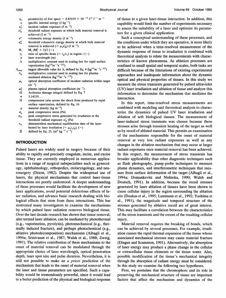
The optical components were arranged as shown in Fig. 1. An 8-mmdiameter aperture was used to select a uniform potion of the laser output.The laser beam then propagated through a set of attenuators, a beamsplitter, a 160-mm focal length lens, and a prism that deflected the beamonto the target surface. The optics were composed of UV-grade quartz. Thelaser pulse energy incident on the target was determined by measuring theenergy of the split-off portion of the beam with a pyroelectric detector(Molectron Detector, Inc., Portland, OR, Model J3-09) and correcting forthe reflection losses at the optical interfaces and the absorption losses in thequartz optics. The pulse energy was varied by changing the number and/orthickness of quartz flats used as attenuators. The average radiant exposure,EO incident on the target surface was calculated by dividing the incidentlaser energy by the area of the irradiated spot. The spot area was deter-mined by the pattern formed by laser irradiation on laser burn paper(Kentek, Pitsfield, NH) which was found to correspond to the lie2 spot sizewithin ±20%.
The measurements were made as follows. The tissue sample was placedon the transducer surface with a thin layer of saline in between to provide
To Oscillosco
acoustic contact. The sample was sufficiently thick to absorb all thedelivered radiation. The irradiation resulted in the generation of stresseswithin the heated region as well as recoil stresses imparted to the tissuesurface when material removal was achieved. These stresses traversed thetissue thickness and stressed the piezoelectric film, thereby producingthe measured voltages. To ensure that the stress waves remained planarduring their transit across the tissue thickness, the laser spots used werealways >1 mm in diameter. This eliminated geometric attenuation of thestress wave and minimized effects of acoustic absorption and dispersion onthe stress transient (Sigrist, 1986; Venugopalan, 1994). The large spot sizealso ensured that the expansion of ablated material away from the targetsurface took place in a one-dimensional geometry.
RESULTS
KrF-excimer laser experiments: (A = 248 nm)
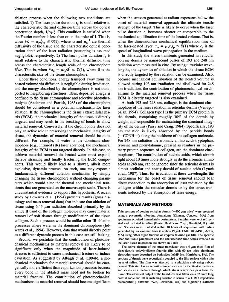
Typical signals from the transducer generated by KrF-exci-mer laser irradiation (A = 248 nm) of porcine dermis areshown in Fig. 2. A positive stress denotes compression andeach trace is normalized relative to its peak compressivestress. The signals produced by radiant exposures rangingfrom a subablative energy dose to a strongly ablative doseare represented by traces 2 a-d. Trace 2 a is a bipolar stresstransient, (i.e., it has both compressive and tensile compo-nents) and is characteristic of stress transients producedby the rapid thermal expansion of the tissue (Dyer andAl-Dhahir, 1990), sometimes referred to as the thermoelas-
1.(
)pe cc 0.5a)
a~~~~; ()0.0
-1.0
Transient Digitizer
FIGURE 1 Experiment setup for measurement of laser-induced stresstransients in tissue.
-50 0 50 100 150 200 250 300tinme (ns)
FIGURE 2 Transducer signals resulting from KrF-excimer laser irradi-ation (A = 248 nm) of porcine dermis in air. The radiant exposure and peakcompressive stress for each trace are: (a) e' = 420 J m-2, ap = 5.2 X 10⁶Pa; (b) E' = 2060 J m-2,o-p = 2.4 X10⁶ Pa; (c)E' = 2200 J m2,-2.6 X 10⁶ Pa; (d) e" = 1.1 X 10⁴ J m-2, op = 2.0 X 10⁷ Pa.
1262 Biophysical Journal
UV Laser Irradiation of Soft Bio-Tissues
tic effect. The stresses return to baseline in approximately220 ns. As the radiant exposure is increased to the thresholdradiant exposure of ablation (as determined in the Anal-ysis section), the stress transient (trace 2 b) remainsessentially unchanged with the exception that the tensileportion of the thermoelastic stress recovers to baselinemore quickly. Once the radiant exposure exceeds thisthreshold, the magnitude of the tensile portion of thestress wave is reduced relative to the compressive com-ponent as shown in trace 2 c. Finally, at large radiantexposures where material removal is observed visually,the tensile stresses produced by the thermoelastic mech-anism are completely obscured by the compressive recoilstress imparted to the tissue surface by the ablationproducts. In this case, only a unipolar compressive stresstransient is measured. This stress transient reaches itsmaximum value on the same time scale as the ther-moelastic stresses and returns to baseline after a fewhundred nanoseconds.The peak compressive stress op generated by KrF-
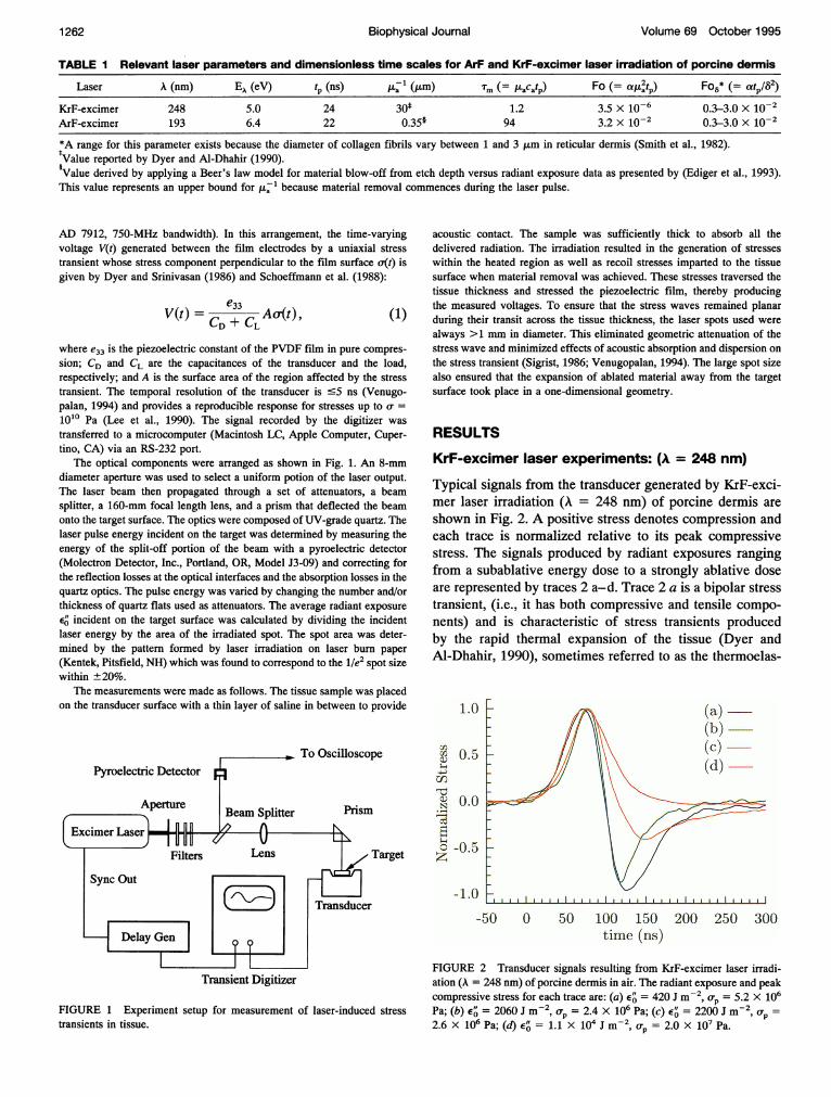
excimer laser irradiation of porcine dermis is plottedversus incident radiant exposure in Fig. 3. For radiantexposures below ablation threshold, the peak stress in-creases linearly with radiant exposure as expected fromthermoelastic stress generation. However, at a radiantexposure E' 2000 J m-2 the peak compressive stressproduced by ablative recoil dominate those produced bythe thermoelastic response and the variation of the peakcompressive stress versus incident radiant exposure de-viates from this linear behavior.
ArF-excimer laser experiments: (A = 193 nm)
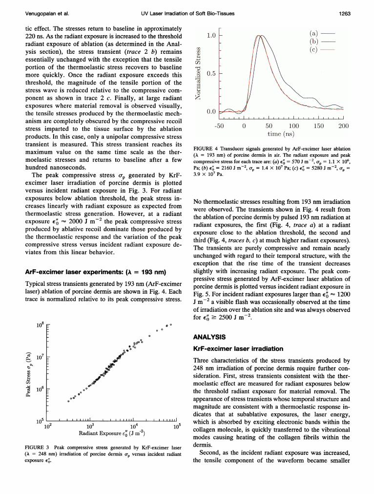
Typical stress transients generated by 193 nm (ArF-excimerlaser) ablation of porcine dermis are shown in Fig. 4. Eachtrace is normalized relative to its peak compressive stress.
108
Ca 7
;, 10
bl'
cn
1 106
104
10,
0
8000o
Ped_'
450
o
0 0
00oO0
0
I I I I II I, I II
103 104Radiant Exposure Eg (J m-2)
FIGURE 3 Peak compressive stress generated by KrF-excimer laser(A = 248 nm) irradiation of porcine dermis o-p versus incident radiantexposure EO.
1.0
Crl
u:n
.)
z
r s0.5
.0 1 . .1 f1 i. I
-50 0 50 100timle (rus)
150 200
FIGURE 4 Transducer signals generated by ArF-excimer laser ablation(A = 193 nm) of porcine dermis in air. The radiant exposure and peakcompressive stress for each trace are: (a) E' = 570 J m-2, o(p = 1.1 X 10⁶,Pa; (b) E' = 2160 J m-2, Up = 1.4 X 10⁷ Pa; (c) E' = 5280 J m-2, a-p =3.9 x 10⁷ Pa.
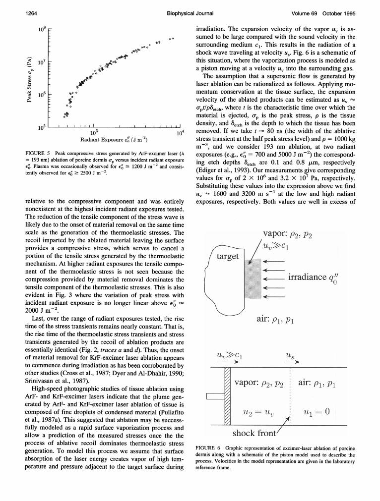
No thermoelastic stresses resulting from 193 nm irradiationwere observed. The transients shown in Fig. 4 result fromthe ablation of porcine dermis by pulsed 193 nm radiation atradiant exposures, the first (Fig. 4, trace a) at a radiantexposure close to the ablation threshold, the second andthird (Fig. 4, traces b, c) at much higher radiant exposures).The transients are purely compressive and remain nearlyunchanged with regard to their temporal structure, with theexception that the rise time of the transient decreasesslightly with increasing radiant exposure. The peak com-pressive stress generated by ArF-excimer laser ablation ofporcine dermis is plotted versus incident radiant exposure inFig. 5. For incident radiant exposures larger than E'- 1200J m-2 a visible flash was occasionally observed at the timeof irradiation over the ablation site and was always observedfor E'0 2500 J m-2.
ANALYSIS
KrF-excimer laser irradiation
Three characteristics of the stress transients produced by248 nm irradiation of porcine dermis require further con-sideration. First, stress transients consistent with the ther-moelastic effect are measured for radiant exposures belowthe threshold radiant exposure for material removal. Theappearance of stress transients whose temporal structure andmagnitude are consistent with a thermoelastic response in-dicates that at subablative exposures, the laser energy,which is absorbed by exciting electronic bands within thecollagen molecule, is quickly transferred to the vibrationalmodes causing heating of the collagen fibrils within thedermis.
Second, as the incident radiant exposure was increased,the tensile component of the waveform became smaller
Venugopalan et al. 1263
.1
9
Volume 69 October 1995
108
Caaq1-1
bQ.
rncn
24-.'.)cn
I(1)
P-4
107
106
105
Gs000
0080
sof~
c90
_- 00
0o
10
1 i IEIsI m 2)
Radiant Exposure e" (J m-2)
W i
104
FIGURE 5 Peak compressive stress generated by ArF-excimer laser (A= 193 nm) ablation of porcine dermis op versus incident radiant exposureE". Plasma was occasionally observed for E' 2 1200 J m-2 and consis-tently observed for ej' ' 2500 J m
relative to the compressive component and was entirelynonexistent at the highest incident radiant exposures tested.The reduction of the tensile component of the stress wave islikely due to the onset of material removal on the same timescale as the generation of the thermoelastic stresses. Therecoil imparted by the ablated material leaving the surfaceprovides a compressive stress, which serves to cancel aportion of the tensile stress generated by the thermoelasticmechanism. At higher radiant exposures the tensile compo-nent of the thermoelastic stress is not seen because thecompression provided by material removal dominates thetensile component of the thermoelastic stresses. This is alsoevident in Fig. 3 where the variation of peak stress withincident radiant exposure is no longer linear above E"2000 J m-2.
Last, over the range of radiant exposures tested, the risetime of the stress transients remains nearly constant. That is,the rise time of the thermoelastic stress transients and stresstransients generated by the recoil of ablation products areessentially identical (Fig. 2, traces a and d). Thus, the onsetof material removal for KrF-excimer laser ablation appearsto commence during irradiation as has been corroborated byother studies (Cross et al., 1987; Dyer and Al-Dhahir, 1990;Srinivasan et al., 1987).
High-speed photographic studies of tissue ablation usingArF- and KrF-excimer lasers indicate that the plume gen-erated by ArF- and KrF-excimer laser ablation of tissue iscomposed of fine droplets of condensed material (Puliafitoet al., 1987a). This suggested that ablation may be success-fully modeled as a rapid surface vaporization process andallow a prediction of the measured stresses once the theprocess of ablative recoil dominates thermoelastic stressgeneration. To model this process we assume that surfaceabsorption of the laser energy creates vapor of high tem-perature and pressure adjacent to the target surface during
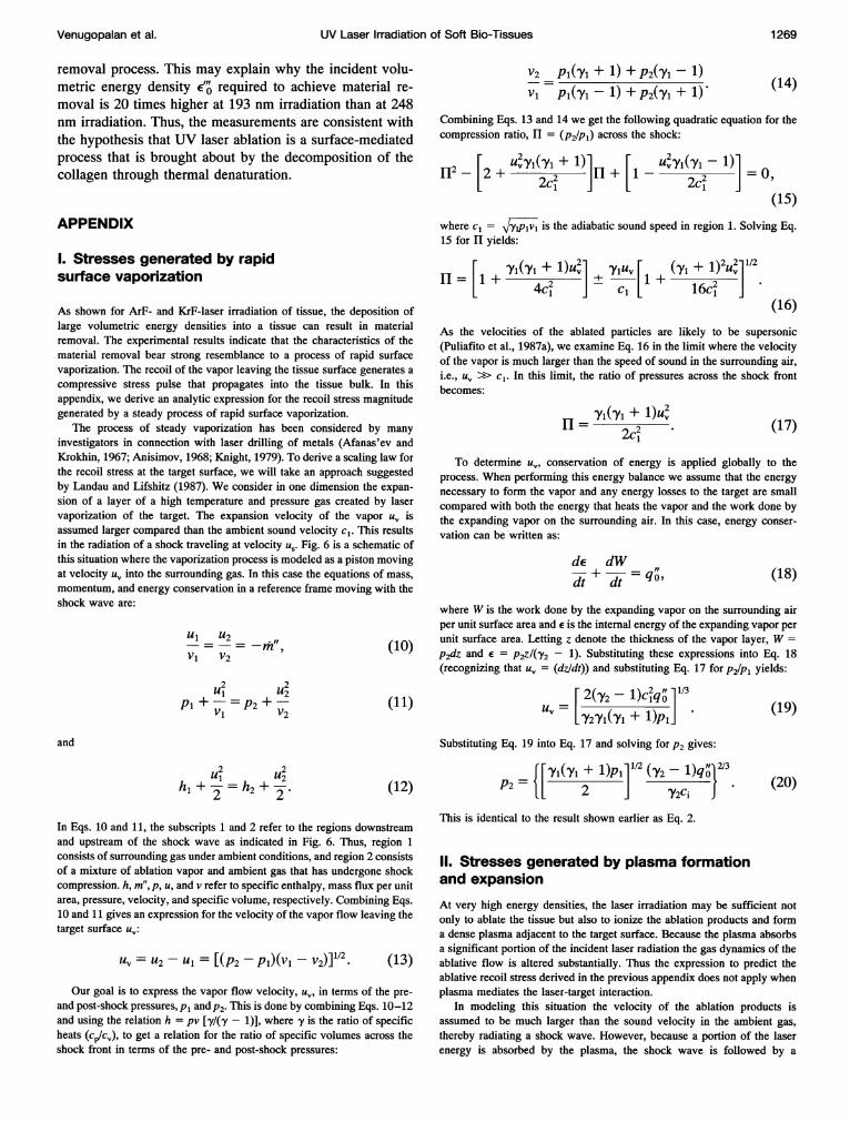
irradiation. The expansion velocity of the vapor u, is as-sumed to be large compared with the sound velocity in thesurrounding medium c1. This results in the radiation of ashock wave traveling at velocity us. Fig. 6 is a schematic ofthis situation, where the vaporization process is modeled asa piston moving at a velocity uv into the surrounding gas.The assumption that a supersonic flow is generated by
laser ablation can be rationalized as follows. Applying mo-mentum conservation at the tissue surface, the expansionvelocity of the ablated products can be estimated as uvO'pt/P6etch, where t is the characteristic time over which thematerial is ejected, o(p is the peak stress, p is the tissuedensity, and Setch is the depth to which the tissue has beenremoved. If we take t 80 ns (the width of the ablativestress transient at the half peak stress level) and p = 1000 kgm-3, and we consider 193 nm ablation, at two radiantexposures (e.g., E" = 700 and 5000 J m-2) the correspond-ing etch depths Setch are 0.1 and 0.8 ,um, respectively(Ediger et al., 1993). Our measurements give correspondingvalues for op of 2 X 10⁶ and 3.2 X 10⁷ Pa, respectively.Substituting these values into the expression above we findUV --1600 and 3200 m s-1 at the low and high radiantexposures, respectively. Both values are well in excess of
ut.>C1
vapor: P2, P2
- irradiance q'
air: Pl, Pi
Us
FIGURE 6 Graphic representation of excimer-laser ablation of porcinedermis along with a schematic of the piston model used to describe theprocess. Velocities in the model representation are given in the laboratoryreference frame.
1 264 Biophysical Journal
UV Laser Irradiation of Soft Bio-Tissues
cl = 331 m s-1 under STP conditions, making supersonicflow likely.
Analysis proceeds by solving the mass, momentum, andenergy conservation equations to determine the pressure inthe vapor P2, which is equal to the recoil stress perpendic-ular to the target surface. Details of the analysis are pre-sented in Appendix I, and only the result is shown anddiscussed here. Assuming the velocity of the vapor to bemuch larger than the speed of sound in the surrounding airc1, the pressure of the vapor adjacent to the target P2 is givenby:
P2=[~1~1 +1)p,]112 (,y2 (2)2/P2=
2 Z (2)
where Pi is the ambient pressure and Yl and y2 are the ratioof specific heats, cp/cv, in regions 1 and 2, respectively.
Eq. 2 may not be directly applicable to the data becausethe laser irradiance delivered to the target was not constantin time. Also, because the laser pulses are of such shortduration, a steady-state vaporization process may not beestablished. However, because the dynamics of the ablationprocess show little temporal delay with respect to the laserpulse (Dyer and Srinivasan, 1986; Srinivasan et al., 1987),changes in the laser irradiance result in a nearly instanta-neous change in the ablation dynamics and recoil stress.Thus, we can modify Eq. 2 in the following manner. As thetemporal shape of the laser pulse is invariant for changes inE", the incident irradiance qO can be replaced by incidentradiant exposure divided by the pulse duration, Ea/tp. Inaddition, because the threshold radiant exposure E4 doesnot contribute directly to heating the plume of ablatedmaterial it should be subtracted from the incident radiantexposure. This leads to the following scaling law for thepeak recoil stress:
= A(Eo'- E)2/3. (3)
where A is a parameter that can be adjusted subject to therestriction that it is less than the value given by the steady-state solution, as this would violate the conservation equa-tions. Thus A must satisfy the condition:
A -< Amax = {[iYi+1)(42 y-1}1{[ 2 ] Y2CItp}
The value for Amax is 5.9 X 10⁴ kg1/3 m-1 S-2/3 forKrF-excimer laser ablation and 6.2 X 10⁴ kg1/3 m-1 S-2/3for ArF-excimer laser ablation. A derived value of A sig-nificantly lower than Amax would indicate that a steady-statevaporization process is not achieved early in the laser pulseand that a significant amount of laser energy contributes tothe acceleration of the ablated mass to the steady-statevelocity.
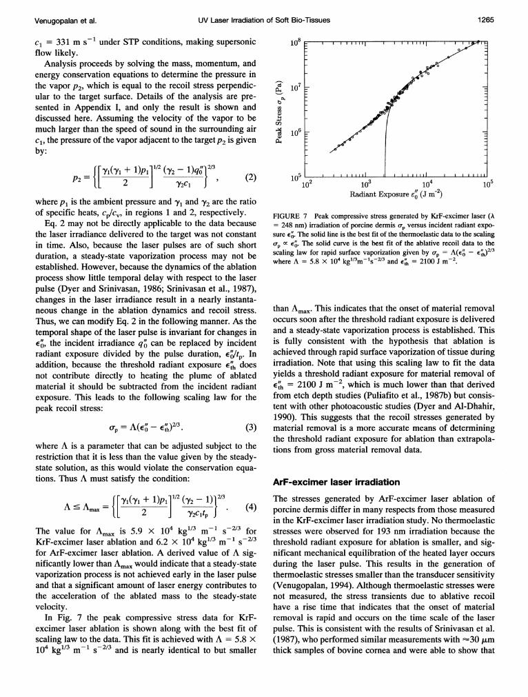
In Fig. 7 the peak compressive stress data for KrF-excimer laser ablation is shown along with the best fit ofscaling law to the data. This fit is achieved with A = 5.8 X10⁴ kg"13 m-1 S-2/3 and is nearly identical to but smaller
108
U,U,
a)
a)
107
106
102 103 104Radiant Exposure e0 (J m-2)
105
FIGURE 7 Peak compressive stress generated by KrF-excimer laser (A= 248 nm) irradiation of porcine dermis o(p versus incident radiant expo-sure E4. The solid line is the best fit of the thermoelastic data to the scalingop X E'. The solid curve is the best fit of the ablative recoil data to thescaling law for rapid surface vaporization given by op = A(E' - E )2/3where A = 5.8 X 10⁴ kg"3m- S-2/3 and E' = 2100 J m-2.
than Amax. This indicates that the onset of material removaloccurs soon after the threshold radiant exposure is deliveredand a steady-state vaporization process is established. Thisis fully consistent with the hypothesis that ablation isachieved through rapid surface vaporization of tissue duringirradiation. Note that using this scaling law to fit the datayields a threshold radiant exposure for material removal of
th = 2100 J m-2, which is much lower than that derivedfrom etch depth studies (Puliafito et al., 1987b) but consis-tent with other photoacoustic studies (Dyer and Al-Dhahir,1990). This suggests that the recoil stresses generated bymaterial removal is a more accurate means of determiningthe threshold radiant exposure for ablation than extrapola-tions from gross material removal data.
ArF-excimer laser irradiation
The stresses generated by ArF-excimer laser ablation ofporcine dermis differ in many respects from those measuredin the KrF-excimer laser irradiation study. No thermoelasticstresses were observed for 193 nm irradiation because thethreshold radiant exposure for ablation is smaller, and sig-nificant mechanical equilibration of the heated layer occursduring the laser pulse. This results in the generation ofthermoelastic stresses smaller than the transducer sensitivity(Venugopalan, 1994). Although thermoelastic stresses werenot measured, the stress transients due to ablative recoilhave a rise time that indicates that the onset of materialremoval is rapid and occurs on the time scale of the laserpulse. This is consistent with the results of Srinivasan et al.(1987), who performed similar measurements with --30 ,tmthick samples of bovine cornea and were able to show that
Venugopalan et al. 1 265
105
Volume 69 October 1995
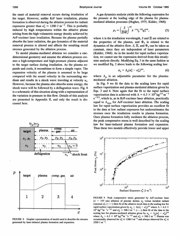
the onset of material removal occurs during irradiation ofthe target. However, unlike KrF laser irradiation, plasmaformation is observed during the ablation process for radiantexposures greater than E'- 1200 J m-2. This is probablyinduced by high temperatures within the ablative plumearising from the high volumetric energy density achieved byArF-excimer laser irradiation. Because the plasma partiallyabsorbs the laser radiation, the gas dynamics of the materialremoval process is altered and affects the resulting recoilstresses generated by the ablation process.To model plasma-mediated ablation we consider a one-
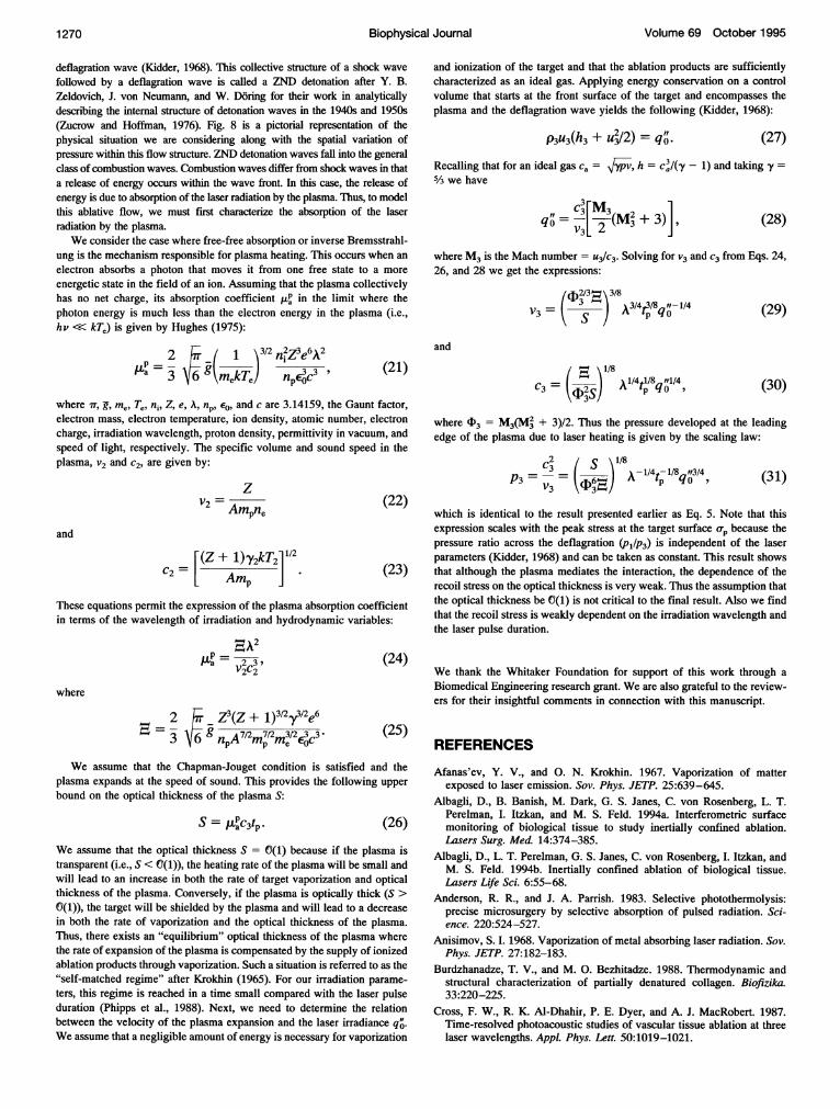
dimensional geometry and assume the ablation process cre-ates a high-temperature and high-pressure plasma adjacentto the target surface during irradiation. As the plasma ex-pands and cools, it recombines to form a simple vapor. Theexpansion velocity of the plasma is assumed to be largecompared with the sound velocity in the surrounding me-dium and results in a shock wave traveling at velocity us.However, because the plasma also absorbs some energy, theshock wave will be followed by a deflagration wave. Fig. 8is a schematic of this situation along with a representation ofthe variation in pressure in this flow. Details of this analysisare presented in Appendix II, and only the result is dis-cussed here.
pl-asnma air
P2, P2 ' P3, P3 ' P4, P4
U2 4u3 =0- -\ L*
\ shock front
airtarget plasma ,comp. gas,
'~~ ~
1 22
3
A gas dynamics analysis yields the following expression forthe pressure at the leading edge of the plasma for plasma-mediated ablation processes (Hughes, 1975; Kidder, 1968):
S)1/8P3= ,,,s,6)~x1-/4tp-1/8q 13/4 (5)
where A is the irradiation wavelength, S and . are related tothe properties of the plasma, and (D3 is related to thedynamics of the ablative flow. S, , and cI3 can be taken asconstant, since they are independent of laser parameters(Kidder, 1968). As in the model for rapid surface vaporiza-tion, we cannot use the expression derived from this steady-state analysis directly. Modifying Eq. S in the same fashion aswe modified Eq. 2 above leads to the following scaling law:
rj, = AP(Efof -E)3/4 (6)
where AP is an adjustable parameter for the plasma-mediated ablation.
In Fig. 9 we fit the data to the scaling laws for rapidsurface vaporization and plasma-mediated ablation given byEqs. 3 and 6. Note again that the fit to the rapid surfacevaporization data is achieved with A = 6.3 X 10⁴ kg"3 m-'s-2/3, which is, as in KrF-excimer laser ablation, practicallyequal to Amax for ArF-excimer laser ablation. The scalinglaw for rapid surface vaporization provides an excellent fitto the data at low radiant exposures but underestimates thestresses once the irradiation results in plasma formation.Once plasma formation fully mediates the ablation process,the peak compressive stress is well described by the scalinglaw for laser-induced plasma formation and expansion.Thus these two models effectively provide lower and upper
108
_,
a)U)S.,
cn
107
106
105104103
Radiant Exposure -0' (J m-2)
4
x
FIGURE 8 Graphic representation of model used to describe the stressesgenerated by laser-induced plasma formation and expansion.
FIGURE 9 Peak compressive stress generated by ArF-excimer laser(A = 193 nm) ablation of porcine dermis op versus incident radiantexposure 4'. ( ) Best fit of the ablative recoil data to the scaling law forrapid surface vaporization given by op = A(Eo- E"j)213 where A = 6.3 X10⁴ kgl3m- 's-2/3 and E' = 500 J m-2. (----) Best fit of the data to thescaling law for plasma-mediated ablation given by 0p = Ap(e' - E )3/4where Ap = 6.3 x 10⁴ kg"4m-ls-1/2 and E4 = 500 J m-2 Plasma wasoccasionally observed for e' 2 1200 J m-2 and always observed for E' 22500 J 2
Biophysical Journal1 266
u

UV Laser Irradiation of Soft Bio-Tissues
bounds for the compressive ablative recoil stress generatedby 193 nm laser ablation.
DISCUSSION
For both KrF- and ArF-excimer laser ablation of porcinedermis, we have established that the characteristics of theprocess are consistent with the view that UV irradiationproduces dissolution of the tissue during the laser pulsethrough the deposition of the threshold incident radiantexposure for ablation 4. Further, the energy that does notcontribute directly to material removal increases the specificenergy of the ablation plume, thereby increasing its temper-ature, which sometimes results in plasma formation andpressure, which drives a gas-dynamic expansion. However,the mechanism by which the vaporization or dissolution oftissue is achieved by the irradiation has not been addressed.While the results of the experiments cannot directly resolvethis issue, we can use physical reasoning to infer what thesemechanisms might be.
Photomechanical processes are unlikely to be the solemeans for material removal because the recoil stresses areconsistent with a process of rapid surface vaporization, andthe stresses generated at E4 are much smaller than theultimate tensile strength (UTS) of porcine dermis, sUTs8-10 MPa (Yamada, 1970). Accordingly, we search for amechanism that allows for decomposition or modificationof the mechanical integrity of tissue during the laser pulse.This mechanism must provide a direct means for materialremoval or, in the case of KrF irradiation, allow the sub-stantial thermoelastic stresses generated by the laser irradi-ation to facilitate material removal. It is possible that tem-perature rise produced by the absorption of laser energyby the ECM leads to denaturation of tissue proteins on the
130
125
v120
B 115
S 110Q0
105
100 _-lo-lo
time scale of the laser pulse (Edwards et al., 1994; Venugo-palan, 1994). Because denaturation of collagen is accompa-nied by the rupture of intermolecular hydrogen bonds(Burdzhanadze and Bezhitadze, 1988), which are neces-sary for collagen fibers to bear physical stresses (Nimni,1983), denaturation is a viable mechanism for tissuedecomposition.To establish the time scale of denaturation, the denatur-
ation process will be treated via the theory of absolutereaction rates. Within this context, denaturation can bethought of as a transformation from a native state A to adenatured state B through a process that passes through anactivated state A*, which lies at a Gibbs free energy greaterthan both states A and B. Since the equilibrium concentra-tions of A and A* are temperature dependent, the transfor-mation A -> B is temperature dependent. Thus, to determinethe amount of complex A that is converted to B, the tem-perature history must be known. Using the theory of abso-lute reaction rates one can derive the time-varying concen-tration of complex A xA(t) remaining at any time during thetemperature transient T(t) (Glasstone et al., 1941):
XA(t°) ]fl(t) = -In[JA(t) 0]
JtkT(t') ex-[H- T(t')AS]h
I RT(t')
(7)
where k, h, AH, AS, and R are the Boltzmann constant,Planck constant, activation enthalpy, activation entropy, anduniversal gas constant, respectively.
Eq. 7 shows that both enthalpic and entropic contribu-tions are important when considering the kinetics of suchrate processes. For protein denaturation both the enthalpyand entropy of activation are quite large (Glasstone et al.,1941). In this limiting case, AH - TAS becomes the dif-ference between two large numbers and the exponentialterm in Eq. 7 becomes such a strong function of temperaturethat the preexponential term can be taken as approximatelyconstant. This permits the definition of a critical tempera-ture, Tc, such that the region of denaturation (defined asQ = 1) can be taken as all points where this temperature isreached. This simplification allows Tc to be approximatedby the implicit relation (Hu and Barnes, 1970):
AHC R ln(kTctJh) + AS
I, IIII,I,,,,, I,,I,,,, I,, ,
10-9 10-8Exposure time te (s)
1o-7
FIGURE 10 Plot of critical temperature, Tc versus exposure time t, forthermal denaturation of dermis. Denaturation is considered complete whenQ = 1.
(8)
where te is the time the complex is exposed to Tc.For collagen denaturation, the activation enthalpy and
entropy have been determined in mouse dermis to be AH =
4.247 X 10⁵J molF1 and AS = 995.3 J molF1 K'1, respec-tively (Jacques and Prahl, 1987). This permits a calculationof the critical temperature Tc versus the exposure time, te.The result of this calculation is shown in Fig. 10, whichindicates that a temperature rise of -95-100°C is necessaryto achieve protein denaturation on a time scale of 1-10 ns
1267Venugopalan et al.
Volume 69 October 1995
for an initial temperature of 22°C. To determine whetherdenaturation can be expected to occur over the time scale ofthe laser pulse, we must find the temperature rise achievedwithin the ECM of the tissue. If no significant energy lossesoccur during the irradiation time, the temperature rise in theECM at the tissue surface is given by:
PECMCV,ECMXECM'
where PECM, CV,ECM, and XECM are the density, specific heat,and mass fraction of the ECM, respectively. Assuming thatthe properties of the ECM are dominated by the propertiesof native collagen at a hydration level of 0.35 g H20/gtissue, PECM, CV,ECM, and XECM take values of approximately780 kg m-3, 2300 J kg-1 K-1, and 0.3, respectively (Longet al., 1993; Mrevlishvili, 1977; Nomura et al., 1977; Parryand Craig, 1984). Thus, KrF-excimer laser irradiation at thethreshold radiant exposure of E4 = 2100 J m-2 produces atemperature change at the surface of 130°C that is sufficientto achieve denaturation of tissue during the irradiation. Weshould also mention that because these experiments weredone on a multiple shot basis, the collagen lining the mar-gins of the ablation crater are already denatured. Thus, atlow radiant exposures, the laser pulses may be simplyremoving tissue already denatured by previous pulses.However, at larger radiant exposures, the etch depth islarger than the zone of residual damage, and tissue removalincludes material not denatured by previous laser pulses.The mechanism for material removal in ArF-excimer
laser ablation is likely similar to that of KrF-excimer laserablation. This is manifest in the fact that the scaling law forrapid surface vaporization provides an adequate fit to theablative recoil stress data before the onset of plasma forma-tion. However, the volumetric energy density necessary formaterial removal E'h is about 20 times larger in the case ofArF-excimer laser ablation. While this discrepancy can beattributed in part to the uncertainty regarding the opticalpenetration depth of 193 nm laser radiation in tissue (Edigeret al., 1993; Puliafito et al., 1985) it is also likely that thethermoelastic stresses generated by KrF-excimer laser irra-diation reduce the energy needed to "detach" the ablatedmaterial from the bulk tissue. Because the magnitude ofthermoelastic stresses generated at the ablation threshold(orth) by 193 nm laser radiation is at least 10 times smaller(Orth = 0.2 MPa, whereas 0th = 3 MPa for 248 nm irradi-ation), they may not facilitate the material removal processwhich results in the higher energy density necessary toachieve ablation.
This mechanism of material removal achieved throughtargeted destruction of the tissue chromophore implies thatessentially all collagen that is denatured is removed. Thismay be the key reason why the morphology of in vitro ArF-and KrF- excimer laser etching of tissue is free of grossmechanical injury and exhibits minimal thermal injury(Lane et al., 1985; Puliafito et al., 1985, 1987b). In contrast,
and TEA CO2 lasers, which are absorbed by water, grossmechanical tearing and extensive thermal injury occur(Cummings and Walsh, 1993; Walsh et al., 1988, 1989)despite comparable optical absorption depths. At thesewavelengths the material removal process is presumablygoverned by explosive vaporization of tissue water, whichmay lead to significant amounts of the thermal energy at theablation site and induce considerable tissue tearing. How-ever, detailed measurement of the dynamics of the IR laserablation process will be needed to validate this hypothesis.
CONCLUSIONS
The stress transients generated by pulsed excimer laserirradiation and ablation of porcine dermis at A = 193 and248 nm have been measured. Modeling and theoreticalanalysis permits the correlation of the temporal structureand amplitude of the measured transients with the natureand dynamics of the tissue response to UV laser irradiation.Specifically, we have shown that laser exposures that do notachieve material removal produce stress transients that areconsistent with dynamic thermal expansion of the tissueknown as thermoelastic stress generation. When ablation isachieved the recoil stresses produced at both wavelengthsare fully consistent with the view that the onset of materialremoval occurs during irradiation and proceeds via a pro-cess of laser-induced rapid surface vaporization. This per-mits the formulation of a scaling law that relates the mea-sured recoil stress with the incident and threshold radiantexposures. For 193 nm irradiation, ignition of the ablationplume occurs at high radiant exposures leading to a plasma-mediated ablation process. Again, a gas dynamic analysispermits the formulation of a scaling law that describes thefunctional variation of the recoil stress with incident radiantexposure in this regime. The success of the rapid surfacevaporization and plasma models in predicting the ablativerecoil stresses suggests that the ablation process is surface-mediated because energy in excess of that necessary toremove material contributes to the specific energy of theplume and drives the gas dynamic expansion of the ablateddebris.
Using the classical theory of absolute reaction rates, wedetermined that the temperature rise produced by absorptionof the threshold radiant exposure by collagen within thetissue ECM can result in its denaturation during irradiation.Since denatured collagen is unable to bear physical stresses,this is a viable mechanism for material removal. The com-parison of 193 and 248 nm ablation provides insight into therole of laser-induced stresses with regard to the onset ofmaterial removal. At the threshold radiant exposure forablation, the peak stress amplitude generated by 248 nmirradiation is more than an order of magnitude greater thanthat produced by 193 nm irradiation. These measurementsindicate that the stresses generated by KrF-excimer laserirradiation at the threshold for material removal, while un-able to fracture the tissue alone, may facilitate the material
Biophysical Journal1 268
with IR ablation using sources such as Q-switched Er:YAG
UV Laser Irradiation of Soft Bio-Tissues
removal process. This may explain why the incident volu-metric energy density E"' required to achieve material re-moval is 20 times higher at 193 nm irradiation than at 248nm irradiation. Thus, the measurements are consistent withthe hypothesis that UV laser ablation is a surface-mediatedprocess that is brought about by the decomposition of thecollagen through thermal denaturation.
APPENDIX
1. Stresses generated by rapidsurface vaporization
As shown for ArF- and KrF-laser irradiation of tissue, the deposition oflarge volumetric energy densities into a tissue can result in materialremoval. The experimental results indicate that the characteristics of thematerial removal bear strong resemblance to a process of rapid surfacevaporization. The recoil of the vapor leaving the tissue surface generates acompressive stress pulse that propagates into the tissue bulk. In thisappendix, we derive an analytic expression for the recoil stress magnitudegenerated by a steady process of rapid surface vaporization.
The process of steady vaporization has been considered by manyinvestigators in connection with laser drilling of metals (Afanas'ev andKrokhin, 1967; Anisimov, 1968; Knight, 1979). To derive a scaling law forthe recoil stress at the target surface, we will take an approach suggestedby Landau and Lifshitz (1987). We consider in one dimension the expan-sion of a layer of a high temperature and pressure gas created by laservaporization of the target. The expansion velocity of the vapor u, isassumed larger compared than the ambient sound velocity cl. This resultsin the radiation of a shock traveling at velocity us. Fig. 6 is a schematic ofthis situation where the vaporization process is modeled as a piston movingat velocity u, into the surrounding gas. In this case the equations of mass,momentum, and energy conservation in a reference frame moving with theshock wave are:
U_ U2it=-=-m, (10)
Vl V2
2 2Ul U2P1 + P2+ (11)V1 V2
and
hi + # = h2 + 2" (12)
V2 P(PYl + 1) + P2(Y1-1)Vl Pl('Yl-1) + P2(Y1 + 1) (14)
Combining Eqs. 13 and 14 we get the following quadratic equation for thecompression ratio, HI = (P2/Pi) across the shock:
F - U2y(1 )n2 2 + 22y1(y+ 1-]+[ 24 1=0
(15)
where cl = V7ipivi is the adiabatic sound speed in region 1. Solving Eq.15 for Hl yields:
Y+(,Y + 1)U2] + 1u, [1 + 1)2U 122/ /U (Yi 161 1/H + + 1 +2
(16)
As the velocities of the ablated particles are likely to be supersonic(Puliafito et al., 1987a), we examine Eq. 16 in the limit where the velocityof the vapor is much larger than the speed of sound in the surrounding air,i.e., uv >> cl. In this limit, the ratio of pressures across the shock frontbecomes:
Yi (Yl + 1)uV1 242 (17)
To determine us, conservation of energy is applied globally to theprocess. When performing this energy balance we assume that the energynecessary to form the vapor and any energy losses to the target are smallcompared with both the energy that heats the vapor and the work done bythe expanding vapor on the surrounding air. In this case, energy conser-vation can be written as:
dE dWdt dt 0° (18)
where W is the work done by the expanding vapor on the surrounding airper unit surface area and e is the internal energy of the expanding vapor perunit surface area. Letting z denote the thickness of the vapor layer, W =p2dz and E = p2z/(y2 - 1). Substituting these expressions into Eq. 18(recognizing that uv = (dz/dt)) and substituting Eq. 17 for P2/P1 yields:
2(y2 - 1)C2qO' 11/3U Y=L 2Y1(Y1 + 1)PlJ
Substituting Eq. 19 into Eq. 17 and solving for P2 gives:
P2 e (e + 1)p,1/2 (y2 1)qit2/3
(19)
(20)
In Eqs. 10 and 11, the subscripts 1 and 2 refer to the regions downstreamand upstream of the shock wave as indicated in Fig. 6. Thus, region 1consists of surrounding gas under ambient conditions, and region 2 consistsof a mixture of ablation vapor and ambient gas that has undergone shockcompression. h, m", p, u, and v refer to specific enthalpy, mass flux per unitarea, pressure, velocity, and specific volume, respectively. Combining Eqs.10 and 11 gives an expression for the velocity of the vapor flow leaving thetarget surface u,:
UV= U2- Ul = [(P2 -p (v1 -v2)]112. (13)
Our goal is to express the vapor flow velocity, uv, in terms of the pre-
and post-shock pressures, Pi and P2. This is done by combining Eqs. 10-12and using the relation h = pv [y/(y - 1)], where -y is the ratio of specificheats (cplcv), to get a relation for the ratio of specific volumes across theshock front in terms of the pre- and post-shock pressures:
This is identical to the result shown earlier as Eq. 2.
II Stresses generated by plasma formationand expansionAt very high energy densities, the laser irradiation may be sufficient notonly to ablate the tissue but also to ionize the ablation products and forma dense plasma adjacent to the target surface. Because the plasma absorbsa significant portion of the incident laser radiation the gas dynamics of theablative flow is altered substantially. Thus the expression to predict theablative recoil stress derived in the previous appendix does not apply whenplasma mediates the laser-target interaction.
In modeling this situation the velocity of the ablation products isassumed to be much larger than the sound velocity in the ambient gas,thereby radiating a shock wave. However, because a portion of the laserenergy is absorbed by the plasma, the shock wave is followed by a
Venugopalan et al. 1 269
1270 Biophysical Journal Volume 69 October 1995
deflagration wave (Kidder, 1968). This collective structure of a shock wavefollowed by a deflagration wave is called a ZND detonation after Y. B.Zeldovich, J. von Neumann, and W. D6ring for their work in analyticallydescribing the internal structure of detonation waves in the 1940s and 1950s(Zucrow and Hoffman, 1976). Fig. 8 is a pictorial representation of thephysical situation we are considering along with the spatial variation ofpressure within this flow structure. ZND detonation waves fall into the generalclass of combustion waves. Combustion waves differ from shock waves in thata release of energy occurs within the wave front. In this case, the release ofenergy is due to absorption of the laser radiation by the plasma. Thus, to modelthis ablative flow, we must first characterize the absorption of the laserradiation by the plasma.We consider the case where free-free absorption or inverse Bremsstrahl-
ung is the mechanism responsible for plasma heating. This occurs when anelectron absorbs a photon that moves it from one free state to a moreenergetic state in the field of an ion. Assuming that the plasma collectivelyhas no net charge, its absorption coefficient ,P in the limit where thephoton energy is much less than the electron energy in the plasma (i.e.,hv << kTe) is given by Hughes (1975):
2 J /1\3/2 n 2Z3e6A2= 3 \gkmekTe n (21)
where T, g,me, Te, ni, Z, e, A, np, E0, and c are 3.14159, the Gaunt factor,electron mass, electron temperature, ion density, atomic number, electroncharge, irradiation wavelength, proton density, permittivity in vacuum, andspeed of light, respectively. The specific volume and sound speed in theplasma, v2 and c2, are given by:
z=2 (22)
and
E (Z + 1)-y2kT2l"12C2 = A J(23)
[ Amp ] 23These equations permit the expression of the plasma absorption coefficientin terms of the wavelength of irradiation and hydrodynamic variables:
0- A2a (24)
V2 2
where
2 - Z3(Z + 1)312y3a2e63 9 7/12M712M3/2e (25)
We assume that the Chapman-Jouget condition is satisfied and theplasma expands at the speed of sound. This provides the following upperbound on the optical thickness of the plasma S:
S = lp c3t. (26)We assume that the optical thickness S = f(1) because if the plasma istransparent (i.e., S < C(1)), the heating rate of the plasma will be small andwill lead to an increase in both the rate of target vaporization and opticalthickness of the plasma. Conversely, if the plasma is optically thick (S >Y(1)), the target will be shielded by the plasma and will lead to a decrease
in both the rate of vaporization and the optical thickness of the plasma.Thus, there exists an "equilibrium" optical thickness of the plasma wherethe rate of expansion of the plasma is compensated by the supply of ionizedablation products through vaporization. Such a situation is referred to as the"self-matched regime" after Krokhin (1965). For our irradiation parame-ters, this regime is reached in a time small compared with the laser pulseduration (Phipps et al., 1988). Next, we need to determine the relationbetween the velocity of the plasma expansion and the laser irradiance q'.We assume that a negligible amount of energy is necessary for vaporization
and ionization of the target and that the ablation products are sufficientlycharacterized as an ideal gas. Applying energy conservation on a controlvolume that starts at the front surface of the target and encompasses theplasma and the deflagration wave yields the following (Kidder, 1968):
p3u3(h3 + u2/2) = q" (27)
Recalling that for an ideal gas ca = ypv, h = C3(y - 1) and taking y =5/3 we have
q= V3 2L' 3 + 3)] (28)
where M3 is the Mach number = U3C3. Solving for V3 and C3 from Eqs. 24,26, and 28 we get the expressions:
()3 3/4/8 1/4 (29)
and
C3= (D2S tvqo (30)
where F3 = M3(M2 + 3)/2. Thus the pressure developed at the leadingedge of the plasma due to laser heating is given by the scaling law:
2 ' 1/8C3 I S14p-18 /P3= A-D (31)
which is identical to the result presented earlier as Eq. 5. Note that thisexpression scales with the peak stress at the target surface crp because thepressure ratio across the deflagration (P1/P3) is independent of the laserparameters (Kidder, 1968) and can be taken as constant. This result showsthat although the plasma mediates the interaction, the dependence of therecoil stress on the optical thickness is very weak. Thus the assumption thatthe optical thickness be Y(1) is not critical to the final result. Also we findthat the recoil stress is weakly dependent on the irradiation wavelength andthe laser pulse duration.
We thank the Whitaker Foundation for support of this work through aBiomedical Engineering research grant. We are also grateful to the review-ers for their insightful comments in connection with this manuscript.
REFERENCES
Afanas'ev, Y. V., and 0. N. Krokhin. 1967. Vaporization of matterexposed to laser emission. Sov. Phys. JETP. 25:639-645.
Albagli, D., B. Banish, M. Dark, G. S. Janes, C. von Rosenberg, L. T.Perelman, I. Itzkan, and M. S. Feld. 1994a. Interferometric surfacemonitoring of biological tissue to study inertially confined ablation.Lasers Surg. Med. 14:374-385.
Albagli, D., L. T. Perelman, G. S. Janes, C. von Rosenberg, I. Itzkan, andM. S. Feld. 1994b. Inertially confined ablation of biological tissue.Lasers Life Sci. 6:55-68.
Anderson, R. R., and J. A. Parrish. 1983. Selective photothermolysis:precise microsurgery by selective absorption of pulsed radiation. Sci-ence. 220:524-527.
Anisimov, S. I. 1968. Vaporization of metal absorbing laser radiation. Sov.Phys. JETP. 27:182-183.
Burdzhanadze, T. V., and M. 0. Bezhitadze. 1988. Thermodynamic andstructural characterization of partially denatured collagen. Biofizika.33:220-225.
Cross, F. W., R. K. Al-Dhahir, P. E. Dyer, and A. J. MacRobert. 1987.Time-resolved photoacoustic studies of vascular tissue ablation at threelaser wavelengths. Appi. Phys. Lett. 50:1019-1021.
Venugopalan et al. UV Laser Irradiation of Soft Bio-Tissues 1271
Cummings, J. P., and J. T. Walsh Jr. 1993. Tissue tearing caused by pulsedlaser-induced ablation pressure. Appl. Opt. 32:494-503.
Dingus, R. S., and R. J. Scammon. 1991. Griineisen-stress induced ablationof biological tissue. Proc. SPIE. 1427:45-54.
Dixon, J. A. 1982. Surgical applications of lasers. Proc. IEEE. 70:579-588.
Domankevitz, Y., and N. S. Nishioka. 1990. Measurement of laser ablationthreshold with a high-speed framing camera. IEEE J. Quantum Elec.26:2276-2278.
Doukas, A. G., D. J. McAuliffe, S. Lee, V. Venugopalan, and T. J. Flotte.1995. Physical factors involved in stress-wave-induced cell injury: theeffect of stress gradient. Ultrasound Med. Bio. In press.
Dyer, P. E., and R. K. Al-Dhahir. 1990. Transient photoacoustic studies oflaser tissue ablation. Proc. SPIE. 1202:46-60.
Dyer, P. E., and R. Srinivasan. 1986. Nanosecond photoacoustic studies onultraviolet laser ablation of organic polymers. Appl. Phys. Lett. 48:445-447.
Ediger, M. N., G. H. Pettit, R. P. Weiblinger, and C. H. Chen. 1993.Transmission of corneal collagen during ArF excimer laser ablation.Lasers Surg. Med. 13:204-210.
Edwards, G., R. Logan, M. Copeland, L. Reinisch, J. Davidson, B. John-son, R. Maciunas, M. Mendenhall, R. Ossoff, J. Tribble, J. Werkhaven,and D. O'Day. 1994. Tissue ablation by a free-electron laser tuned to theamide II band. Nature. 371:416-419.
Glasstone, S., K. J. Laidler, and H. Eyring. 1941. The Theory of RateProcesses. McGraw Hill Book Co. Inc., New York.
Hu, C-L., and F. S. Barnes. 1970. The thermal-chemical damage inbiological material under laser irradiation. IEEE Trans. Biomed. Eng.BME-17:220-229.
Hughes, T. P. 1975. Plasmas and Laser Light. Adam Hilger Ltd, London.Jacques, S. L., and S. A. Prahl. 1987. Modeling optical and thermal
distributions in tissue during laser irradiation. Lasers Surg. Med.6:494-503.
Kidder, R. E. 1968. Application of lasers to the production of high-temperature and high-pressure plasma. Nucl. Fusion. 8:3-12.
Knight, C. J. 1979. Theoretical modeling of rapid surface vaporization withback pressure. AIAA Journal. 17:519-523.
Krokhin, 0. N. 1965. "Matched" plasma heating mode using laser radia-tion. Sov. Phys. Tech. Phys. 9:1024-1026.
Landau, L. D., and E. M. Lifshitz. 1987. Fluid Mechanics, 2nd ed.Pergamon Press, Oxford.
Lane, R. J., R. Linsker, J. J. Wynne, A. Torres, and R. G. Geronemus.1985. Ultraviolet-laser ablation of skin. Arch. Dermatol. 121:609-617.
Lee, L. M., D. A. Hyndman, R. P. Reed, and F. Bauer. 1990. PVDFapplications in shock measurements. In Shock Waves in CondensedMatter-1989. S. C. Schmidt, J. N. Johnson, L. W. Davison, editors.Elsevier Science Publishers B. V., Amsterdam. 821-824.
Long, C. G., E. Braswell, D. Zhu, J. Apigo, J. Baum, and B. Brodsky. 1993.Characterization of collagen-like peptides containing interruptions in therepeating Gly-X-Y sequence. Biochemistry. 32:11688-11695.
Lustmann, J., M. Ulmansky, A. Fuxbrunner, and A. Lewis. 1992. Photo-acoustic injury and bone healing following 193nm excimer laser abla-tion. Lasers Surg. Med. 12:390-396.
Lynch, S. E., J. C. Nixon, R. B. Colvin, and H. N. Antoniades. 1987. Roleof platelet-derived growth factor in would healing: synergistic effectswith other growth factors. Proc. Natl. Acad. Sci. USA. 84:7696-7700.
Mrevlishvili, G. M. 1977. Thermodynamic properties of biopolymers in thehelical and coiled state in the temperature interval 4-400°K. Biofizika.22:180-191.
Nimni, M. E. 1983. Collagen: Structure, function, and metabolism innormal and fibrotic tissues. Semin. Arthritis Rheum. 13:1-86.
Nomura, S., A. Hiltner, J. B. Lando, and E. Baer. 1977. Interaction of waterwith native collagen. Biopolymers. 16:231-246.
Parry, D. A. D., and A. S. Craig. 1984. Growth and development ofcollagen fibrils in connective tissue. In Ultrastructure of the ConnectiveTissue Matrix. A. Ruggeri and P. M. Motta, editors. Martinus NijhoffPublishers, Boston, MA. 34-64.
Phipps, Jr., C. R., T. P. Turner, R. F. Harrison, G. W. York, W. Z. Osborne,G. K. Anderson, X. F. Corlis, L. C. Haynes, H. S. Steele, K. C. Spicochi,and T. R. King. 1988. Impulse coupling to targets in vacuum by KrF,HF, and CO2 single-pulse lasers. J. Appl. Phys. 64:1083-1096.
Puliafito, C. A., R. F. Steinert, T. F. Deutsch, F. Hillenkamp, E. J. Dehm,and C. M. Adler. 1985. Excimer laser ablation of the cornea and lens.Ophthalmology. 92:741-748.
Puliafito, C. A., D. Stern, R. R. Kreuger, and E. R. Mandel. 1987a.High-speed photography of excimer laser ablation of the cornea. Arch.Ophthalmol. 105:1255-1259.
Puliafito, C. A., K. Wong, and R. F. Steinert. 1987b. Quantitative andultrastructural studies of excimer laser ablation of the cornea at 193 and248 nanometers. Lasers Surg. Med. 7:155-159.
Schoeffmann, H., H. Schmidt-Kloiber, and E. Reichel. 1988. Time-resolved investigations of laser-induced shock waves in water by use ofpolyvinylidenefluoride hydrophones. J. Appl. Phys. 63:46-51.
Sigrist, M. W. 1986. Laser generation of acoustic waves in liquids andgases. J. Appl. Phys. 60:R83-R121.
Smith, L. T., K. A. Holbrook, and P. H. Byers. 1982. Structure of thedermal matrix during development and in the adult. J. Invest. Dermatol.79:93s-104s.
Srinivasan, R., P. E. Dyer, and B. Braren. 1987. Far-ultraviolet laserablation of the cornea: photoacoustic studies. Lasers Surg. Med.6:514-519.
Venugopalan, V. 1994. The thermodynamic response of polymers andbiological tissues to pulsed laser irradiation. Sc.D. thesis, MassachusettsInstitute of Technology, Cambridge, MA.
Walsh, Jr., J. T., and T. F. Deutsch. 1991. Measurement of Er:YAG laserablation plume dynamics. Appl. Phys. B. 52:217-224.
Walsh, J. T. Jr., T. J. Flotte, R. R. Anderson, and T. F. Deutsch. 1988.Pulsed CO2 laser tissue ablation: effect of tissue type and pulse durationon thermal damage. Lasers Surg. Med. 8:108-118.
Walsh, J. T. Jr., T. J. Flotte, and T. F. Deutsch. 1989. Er:YAG laserablation of tissue: effect of pulse duration and tissue type on thermaldamage. Lasers Surg. Med. 9:314-326.
Yamada, H. 1970. Strength of Biological Materials. Williams & Wilkins,Baltimore, MD.
Yashima, Y., D. J. McAuliffe, S. L. Jacques, and T. J. Flotte. 1991.Laser-induced photoacoustic injury of skin: effect of inertial confine-ment. Lasers Surg. Med. 11:62-68.
Zucrow, M. J., and J. D. Hoffman. 1976. Gas Dynamics. John Wiley andSons, Inc., New York.
Zweig, A. D. 1991. A thermo-mechanical model for laser ablation. J. Appl.Phys. 70:1684-1691.