The Tentorium Cerebelli: A Comprehensive Review Including ...
14
Providence St. Joseph Health Providence St. Joseph Health Digital Commons Journal Articles and Abstracts 7-31-2018 e Tentorium Cerebelli: A Comprehensive Review Including Its Anatomy, Embryology, and Surgical Techniques. Rabjot Rai Joe Iwanaga Gaffar Shokouhi Rod J Oskouian Neurosurgery, Swedish Neuroscience Institute, Seale, USA. R Shane Tubbs Follow this and additional works at: hps://digitalcommons.psjhealth.org/publications Part of the Neurology Commons , Pathology Commons , and the Surgery Commons is Article is brought to you for free and open access by Providence St. Joseph Health Digital Commons. It has been accepted for inclusion in Journal Articles and Abstracts by an authorized administrator of Providence St. Joseph Health Digital Commons. For more information, please contact [email protected]. Recommended Citation Rai, Rabjot; Iwanaga, Joe; Shokouhi, Gaffar; Oskouian, Rod J; and Tubbs, R Shane, "e Tentorium Cerebelli: A Comprehensive Review Including Its Anatomy, Embryology, and Surgical Techniques." (2018). Journal Articles and Abstracts. 804. hps://digitalcommons.psjhealth.org/publications/804
Transcript of The Tentorium Cerebelli: A Comprehensive Review Including ...

Providence St. Joseph HealthProvidence St. Joseph Health Digital Commons
Journal Articles and Abstracts
7-31-2018
The Tentorium Cerebelli: A ComprehensiveReview Including Its Anatomy, Embryology, andSurgical Techniques.Rabjot Rai
Joe Iwanaga
Gaffar Shokouhi
Rod J OskouianNeurosurgery, Swedish Neuroscience Institute, Seattle, USA.
R Shane Tubbs
Follow this and additional works at: https://digitalcommons.psjhealth.org/publications
Part of the Neurology Commons, Pathology Commons, and the Surgery Commons
This Article is brought to you for free and open access by Providence St. Joseph Health Digital Commons. It has been accepted for inclusion in JournalArticles and Abstracts by an authorized administrator of Providence St. Joseph Health Digital Commons. For more information, please [email protected].
Recommended CitationRai, Rabjot; Iwanaga, Joe; Shokouhi, Gaffar; Oskouian, Rod J; and Tubbs, R Shane, "The Tentorium Cerebelli: A ComprehensiveReview Including Its Anatomy, Embryology, and Surgical Techniques." (2018). Journal Articles and Abstracts. 804.https://digitalcommons.psjhealth.org/publications/804

Received 07/20/2018 Review began 07/21/2018 Review ended 07/23/2018 Published 07/31/2018
© Copyright 2018Rai et al. This is an open accessarticle distributed under the terms ofthe Creative Commons AttributionLicense CC-BY 3.0., which permitsunrestricted use, distribution, andreproduction in any medium,provided the original author andsource are credited.
The Tentorium Cerebelli: A ComprehensiveReview Including Its Anatomy, Embryology,and Surgical TechniquesRabjot Rai , Joe Iwanaga , Gaffar Shokouhi , Rod J. Oskouian , R. Shane Tubbs
1. Department of Anatomy, St. George's University School of Medicine, St. George's, GRD 2. SeattleScience Foundation, Seattle, USA 3. Neurosciences Research Center, Tabriz, IRN 4. Neurosurgery,Swedish Neuroscience Institute, Seattle, USA 5. Neurosurgery, Seattle Science Foundation, Seattle, USA
Corresponding author: Joe Iwanaga, [email protected] Disclosures can be found in Additional Information at the end of the article
AbstractThe tentorium cerebelli functions as a partition, dispelling the burden of weight fromsupratentorial structures upon inferior brain matter. Clinicians and neurosurgeons, whenassessing pathological findings, should have knowledge regarding the tentorium cerebellianatomy. This work of literature is a comprehensive review of the tentorium cerebelli,including its anatomy, embryology, and clinical and surgical implications. The evolutionarypattern demonstrates sequential stages to higher mammalian lineage. An understanding of thecomplexity of the neurovascular structures and the anatomy of the tentorium cerebelli iscrucial for surgical procedures by neurosurgeons.
Categories: Pathology, Miscellaneous, NeurosurgeryKeywords: tentorium cerebelli, tentorial notch, incisura, dural sinus, embryology
Introduction And BackgroundThree layers make up the meninges: the dura, arachnoid, and pia mater. The dura is also knownas the thick meninx, or pachymeninx, while the arachnoid and pia mater are known as the thinmeninx or leptomeninges [1]. An extension of the dura includes the dural reflections, whichconsist of four distinguishable dura folds, including the falx cerebri, tentorium cerebelli, falxcerebelli, and diaphragma sellae [2]. This review paper will look comprehensively at the tent-shaped structure forming the roof of the posterior cranial fossa, the tentorium cerebelli (Figures1-3) [3]. The tent shape of the tentorium cerebelli helps maintain the anatomy of the brain byproviding protection against the pressure caused by the heavier upper part of the brain [4-5]. Ifthe tentorium cerebellum or falx cerebri were severed, sagging of the brain would take place [4].However, the presence of the tentorium cerebelli can cause trouble during times of cranialswelling or displacement by space-occupying lesions [5].
1 2 3 4 5
Open Access ReviewArticle DOI: 10.7759/cureus.3079
How to cite this articleRai R, Iwanaga J, Shokouhi G, et al. (July 31, 2018) The Tentorium Cerebelli: A Comprehensive ReviewIncluding Its Anatomy, Embryology, and Surgical Techniques. Cureus 10(7): e3079. DOI10.7759/cureus.3079

FIGURE 1: Right lateral view of the cranium afterhemicraniectomy and removal of right cerebral hemisphere ina cadaver.Note the tentorium cerebelli and its relationship to the medial brain structures. Also, note theolfactory tract (right arrow) and optic nerve (left arrow).
2018 Rai et al. Cureus 10(7): e3079. DOI 10.7759/cureus.3079 2 of 13

FIGURE 2: Left: the tentorial incisura (white arrows), straightsinus (blue arrows) and transverse sinus (yellow arrow); Right,schematic drawing of left hemicranium noting the numerousneurovascular structures just medial to the tentorial incisurae.g., basal vein of Rosenthal (blue).The anterior half of the tentorium cerebelli is cut away at the blue arrow. Also note the straightsinus (SS).
FIGURE 3: Superior view of a cadaveric tentorium cerebelliwith brain superior to the brainstem removed.Note the midbrain (MB) and the optic chiasm (OC). White arrows point to the right tentorialincisura.
ReviewAnatomyThe tentorium cerebelli, the second-largest dural reflection, is a crescent-shaped dura fold thatextends over the posterior cranial fossa, separating the occipital and temporal cerebralhemisphere from the cerebellum and infratentorial brainstem [1,6]. The tentorial cerebelli is adistinguishing landmark and divides the cranial cavity into the supratentorial andinfratentorial spaces [1]. This dural reflection has a free and fixed margin. The fixed margins ofthe tentorium cerebelli are attached to the superior borders of the petrous part of the temporalbone, known as the posterior clinoid process via the anterior and posterior petroclinoid folds
2018 Rai et al. Cureus 10(7): e3079. DOI 10.7759/cureus.3079 3 of 13

(Figure 4) and along the transverse sinuses grooves on the occipital bone posteriorly. The freemargin is located at the anterior edge and forms a U-shape termed the tentorial notch or theincisura tentoria (Figures 5-7). The tentorial notch allows for the presence of a gap, whichlodges the midbrain [1,6]. The midbrain inhabits the anterior portion of the incisura and theposterior half is occupied by the superior vermis or the splenium of the corpus collosum. Theremainder of the area is composed of the cerebrospinal fluid, blood vessels, and nerves withinthe perimesencephalic and superior vermis cisterns [3]. The free margin runs anteriorly, crossesthe attached borders, and adheres to the anterior clinoid process bilaterally; this forms thelateral part of the cavernous sinus. At the location of the border crossing, cranial nerves III andIV pass through toward the lateral wall of the cavernous sinus [6]. The trigeminal nerve andtrigeminal ganglion emerge between the recess formed by the apex of the petrous bone and thebottom layer of the tentorium pouched anteriorly beneath the superior petrosal sinus [1,6].Above the free margin sits the hippocampal gyrus and the posterior cerebral artery; thisanatomical finding is clinically important in cases involving transtentorial (uncal) herniations[7]. Lastly, the falx cerebri and falx cerebelli, which are also dural reflections, are attached tothe tentorium cerebelli superiorly and inferiorly, respectively (Figures 1-3) [6].
FIGURE 4: Schematic drawing of the anterior extensions of thetentorium cerebelli and the anterior petroclinoid and posteriorpetroclinoid folds, which make up the lateral and posteriorborders of the oculomotor trigone.
2018 Rai et al. Cureus 10(7): e3079. DOI 10.7759/cureus.3079 4 of 13

FIGURE 5: Right subtemporal view of the tentorium cerebelliand structures along its medial border such as the trochlearnerve.Anteriorly, note the internal carotid artery (transected) (ICA) for reference.
FIGURE 6: Superior view of the left tentorium cerebelli and itsincisura.
2018 Rai et al. Cureus 10(7): e3079. DOI 10.7759/cureus.3079 5 of 13

Note, the left cerebral hemisphere has been removed.
FIGURE 7: Inferior view of the tentorial incisura (white arrows)following a skull base approach.Note the posterior cerebral (PCA), superior cereberllar (SCA), and posterior communicatingarteries (PComm).
Comparative anatomyKlintworth’s (1968) study of the comparative anatomy found the tentorium cerebelli absent inamphibians, fish, and reptiles but present in birds and mammals [4]. The morphology ofstructure varied among different species, for instance, the tentorium was found as a separatedbilateral delicate partition, unjoined at the midline, and dividing only the lateral portion of thecerebrum and cerebellum in bats, pigs, gerbils, hamsters, opossums, rats, and mice. While inother mammals (18 species mentioned), including cats, dogs, humans, rhesus monkeys, minks,and goats), the bilateral folds fused together at the midline give rise to a crescentic partitionposterior to the brain stem, separating the posteroinferior portions of the cerebral hemispherefrom the cerebellum. In these species, the posterior falx cerebri and, occasionally, a falxcerebelli adhered with the tentorium cerebelli. In these animals only, where the tentoriumcerebelli and falx cerebri united in the median plane posteriorly, a straight sinus existed. Itslength in relation to the tentorial notch varied among different species [4,8]. Klintworth [4] alsofound differences in the degree of tentorial ossification amongst various mammals. In hisstudy, he found that in adult cats, the entire tentorium cerebelli was completely ossified. But,in dogs, dolphins, minks, porpoises, and wallabies, the ossification was limited to theposteromedial portion of the tentorium. According to Klintworth [4], the property of tentorialossification provides an evolutionary pattern for mammalian lines. It reflects the retention ofthe osteogenic potentialities, which occur within the ectomeninx during the development ofthe neurocranium.
2018 Rai et al. Cureus 10(7): e3079. DOI 10.7759/cureus.3079 6 of 13
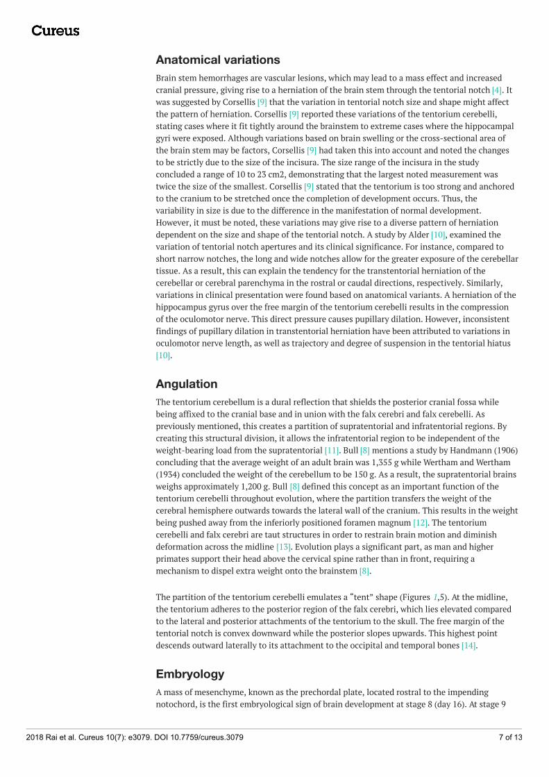
Anatomical variationsBrain stem hemorrhages are vascular lesions, which may lead to a mass effect and increasedcranial pressure, giving rise to a herniation of the brain stem through the tentorial notch [4]. Itwas suggested by Corsellis [9] that the variation in tentorial notch size and shape might affectthe pattern of herniation. Corsellis [9] reported these variations of the tentorium cerebelli,stating cases where it fit tightly around the brainstem to extreme cases where the hippocampalgyri were exposed. Although variations based on brain swelling or the cross-sectional area ofthe brain stem may be factors, Corsellis [9] had taken this into account and noted the changesto be strictly due to the size of the incisura. The size range of the incisura in the studyconcluded a range of 10 to 23 cm2, demonstrating that the largest noted measurement wastwice the size of the smallest. Corsellis [9] stated that the tentorium is too strong and anchoredto the cranium to be stretched once the completion of development occurs. Thus, thevariability in size is due to the difference in the manifestation of normal development.However, it must be noted, these variations may give rise to a diverse pattern of herniationdependent on the size and shape of the tentorial notch. A study by Alder [10], examined thevariation of tentorial notch apertures and its clinical significance. For instance, compared toshort narrow notches, the long and wide notches allow for the greater exposure of the cerebellartissue. As a result, this can explain the tendency for the transtentorial herniation of thecerebellar or cerebral parenchyma in the rostral or caudal directions, respectively. Similarly,variations in clinical presentation were found based on anatomical variants. A herniation of thehippocampus gyrus over the free margin of the tentorium cerebelli results in the compressionof the oculomotor nerve. This direct pressure causes pupillary dilation. However, inconsistentfindings of pupillary dilation in transtentorial herniation have been attributed to variations inoculomotor nerve length, as well as trajectory and degree of suspension in the tentorial hiatus[10].
AngulationThe tentorium cerebellum is a dural reflection that shields the posterior cranial fossa whilebeing affixed to the cranial base and in union with the falx cerebri and falx cerebelli. Aspreviously mentioned, this creates a partition of supratentorial and infratentorial regions. Bycreating this structural division, it allows the infratentorial region to be independent of theweight-bearing load from the supratentorial [11]. Bull [8] mentions a study by Handmann (1906)concluding that the average weight of an adult brain was 1,355 g while Wertham and Wertham(1934) concluded the weight of the cerebellum to be 150 g. As a result, the supratentorial brainsweighs approximately 1,200 g. Bull [8] defined this concept as an important function of thetentorium cerebelli throughout evolution, where the partition transfers the weight of thecerebral hemisphere outwards towards the lateral wall of the cranium. This results in the weightbeing pushed away from the inferiorly positioned foramen magnum [12]. The tentoriumcerebelli and falx cerebri are taut structures in order to restrain brain motion and diminishdeformation across the midline [13]. Evolution plays a significant part, as man and higherprimates support their head above the cervical spine rather than in front, requiring amechanism to dispel extra weight onto the brainstem [8].
The partition of the tentorium cerebelli emulates a “tent” shape (Figures 1,5). At the midline,the tentorium adheres to the posterior region of the falx cerebri, which lies elevated comparedto the lateral and posterior attachments of the tentorium to the skull. The free margin of thetentorial notch is convex downward while the posterior slopes upwards. This highest pointdescends outward laterally to its attachment to the occipital and temporal bones [14].
EmbryologyA mass of mesenchyme, known as the prechordal plate, located rostral to the impendingnotochord, is the first embryological sign of brain development at stage 8 (day 16). At stage 9
2018 Rai et al. Cureus 10(7): e3079. DOI 10.7759/cureus.3079 7 of 13
(day 20), the mesoderm of the head is developed from the lateral migration of cells from theprechordal plate. The prechordal plate continues to generate mesenchyme. At stage 11 (day 24),the pia mater is identified at the caudal aspect of the medulla oblongata in reference tooccipital somites derived from the neural crest. By stage 12 (day 26), the pia mater advances tothe level of the mesencephalic. At this point, there is no distinction between neural crests, asthey become incorporated with cranial-nerve ganglia and head mesenchyme. At stage 13 (day28), the mesenchyme from the prechordal plate arranges bilaterally, causing the formation ofpremandibular condensation. At this time, the cellular sheath of the notochord is apparentwithin the occipital area and develops caudally. Between stages 14 and 17, the development ofthe medial aspect of tentorium cerebelli begins. At stage 14 (day 32), the extension of thenotochordal cellular sheath is seen into the mesencephalic flexure. The premandibularcondensation becomes continuous alongside the notochordal cellular sheath, reaching the tipof the notochord and resuming into the medial aspect of the future tentorium cerebelli. Thedevelopment of the medial part of the tentorium cerebelli is predominantly leptomeningeal asthe beginning of the formation is largely due to the involvement of the notochordal cellularsheath. In stage 17 (day 41), the dural limiting layer, where the pori durales for cranial nervesIII, IV, V, and XII appear, begins formation within the basal areas. This is also wheremesenchymal condensation for the formation of the future chondrocranium is forming. Thedural limiting layer, lateral to the diencephalon, forms the rostrolateral portion of thetentorium cerebelli. During stage 19 (day 48), the leptomeningeal layer seen internal to thelimiting layer is looser while the pachymeninx, where the future dural sinus will be located, isdenser. The medial aspect of the tentorium cerebelli is condensed to fibrous tissue, and bystage 21 (day 52), it extends from the sella turcica to the mammillary body, however, thismedial part starts to become thinner. By stage 23 (day 57), the lateral portions of the tentoriumextend close to the ridge of the mesencephalon. The rostrolateral portion meets thecaudolateral at the medial part, which begins to disintegrate. The line of union of the twotentorial leaves at the site of the future tentorial notch. The medial part disappears, and therostrolateral and caudolateral portions form the definitive tentorial cerebelli [15].
HistologyThe tentorium cerebelli is visible under the microscope in a crown-rump length of 20 mm. Byeight weeks, the embryos tentorium crown-rump length is approximately 30 mm and can begrossly viewed as delicate bilateral folds. By three months, with a crown-rump of 55 mm, theunion of the bilateral folds posteriorly give rise to the development of the straight sinus. As thefetus grows, the tentorium and tentorial notch increase in size progressively. Microscopically,at the stage of the six to eight weeks embryo, the tentorium examination comprised of looselypacked areolar connective tissue of spindle or stellate cells is bordered by a layer of flattenedmesothelial cells superiorly and inferiorly. As the tentorium continues ontogenesis, furtherhistogenesis identifies bundles of collagen at the central core amassing, which evolve into adense fibrous membrane [16].
Arterial vasculature supplyThe various anastomotic networks between adjacent arterial regions further complicate thecomplexity of the vascular supply of the tentorium cerebelli [2]. The meningeal branches of theexternal carotid, internal carotid, posterior cerebral, and vertebral arteries are all involved inthe blood supply to the dura of the posterior fossa, including the tentorium cerebelli. A smallmeningohypophyseal trunk arising from the intracavernous portion of the internal carotidartery branches into two arteries, the tentorial and dorsal meningeal (Figure 8). The tentorialartery, also known as the artery of Bernasconi and Cassinari, exits from the cavernous sinus andtravels between the dural foldings of the tentorium. The tentorial artery branchesposterolaterally and becomes parallel to the free margin, one branching off and supplying thetentorium as it heads towards the straight sinus [17-18]. The other branching
2018 Rai et al. Cureus 10(7): e3079. DOI 10.7759/cureus.3079 8 of 13

laterally provides vasculature to the lateral portion of the tentorium [18]. The posterior cerebralartery gives rise to small infratentorial branches, known as the artery of Davidoff andSchechter, which travel around the brain stem and under the free margin toward the tentoriumapex, where they supply the medial aspect of the tentorium cerebelli. The internal carotid arteryand branches of the external carotid artery provide vascular supply to the petrosal insertion ofthe tentorium [19]. The vertebral artery branches into the posterior meningeal artery, where itssupratentorial branches supply the inferior falx cerebri, adjacent dura, and adjacent tentoriumcerebelli [20].
FIGURE 8: Small vessels of the skull base noting the tentorialbranch to the free edge of the tentorium from themeningohypophyseal trunk of the cavernous internal carotidartery.
Dural venous sinuses associated with the tentorium cerebelliStraight Sinus
The straight sinus is formed by the merger of the inferior sagittal sinus with the great cerebralvein of Galen. The straight sinus is at the junction between the tentorium cerebelli and the falxcerebri. The sinus drains into the confluence of the sinuses or transverse sinus [21]. The straightsinus is angled downward in a rostrocaudal direction when viewed vertically [22].
Transverse Sinus
The transverse sinus emerges laterally from the confluence of the sinus while adhering to thetentorium cerebelli posteriorly. The right transverse sinus is usually larger compared to the left,which is primarily caused by drainage from the superior sagittal sinus directing to the right. Asa result, the right transverse sinus, right sigmoid sinus, and right internal jugular vein drainvenous blood from the superficial areas of the brain, while the left transverse sinus, left sigmoidsinus, and left internal jugular vein drain blood from deeper regions via the internal cerebralvessels and the basal and great veins [21].
Superior Petrosal Sinus
2018 Rai et al. Cureus 10(7): e3079. DOI 10.7759/cureus.3079 9 of 13
The superior petrosal sinus travels in the attached part of the tentorium cerebelli [21-22]. Thesuperior petrosal sinus drains the cavernous sinus and extends toward the transverse sinus.
Tentorial Sinuses
Tentorial sinuses collect various supra and infratentorial bridging veins and are bilateral butmore or less asymmetrical venous channels. The medial group is formed by the coalescence ofthe veins from the superior surface of the cerebellum, and the lateral group is commonly formedby the convergence of veins arising from the basal and lateral surface of the temporal andoccipital lobes. The medial group arises about 1.5 to 2.5 cm from the midline and about 1.0 to2.5 cm from the transverse sinus. The lateral sinuses are often found 3.0 to 4.0 cm from themidline, 2-10 mm from the transverse sinus, and 1.0 cm from the petrous ridge.
InnervationThe recurrent meningeal branches of V1 (ophthalmic division of the trigeminal nerve), alsoknown as the nervus tentorii of Arnold, innervates the tentorium cerebelli. The distribution ofthe nervus tentorii investigated by Lee et al. [23] identified four morphological divisions. Thepattern of type 1 had the highest prevalence in the study, which concluded that the fibersextend to the straight and transverse sinus. Type 2 exhibited the fibers extending only to thetransverse sinus and lateral convexity. Type 3 projected the fibers medially towards only thestraight sinus and posterior portion of the falx cerebri. Lastly, type 4 exhibited the nerve fibersterminating within the tentorium cerebelli. The study conducted by Lee et al. [23] found type 1to have the highest prevalence (the incidence rate of type 1-4 were 71.2%, 21.2%, 3.8%, and3.8%, respectively) from 52 samples of the tentorium cerebelli from 29 cadavers (both sidesfrom 23 cadavers, and one side from six cadavers). The distribution patterns of the nervustentorii were asymmetrical when comparing the left and right sides of the tentorium cerebelli.
The posterior projections of these tentorial nerves form a plexus within the tentorium cerebelliand are predominate at the superior wall of the transverse sinus and the posterior half of thestraight sinus. Lee et al. [23] found innervation to be the most scarce at the tentorial notch andthe anterior half of the straight sinus. This provided clinical significance so as to avoidretraction at the posterior aspect of the falx cerebri and tentorium cerebelli. Manipulation inthis area was shown to cause hemodynamic fluctuations, including hypotension, bradycardia,arrhythmia, asystole, or apnea (known as the trigeminocardiac reflex) by eliciting neural signalsstimulated by the sensory endings of the trigeminal nerve through the trigeminal ganglion tothe sensory nucleus of the trigeminal nerve in the brainstem [23-24].
ImagingThe use of contrast-enhanced computed tomography (CT) is able to visualize the tentorium in99% of cases but can be difficult to recognize on non-contrasted images. The inclination inwhich the image was taken contributes to the varied configuration for which the tentorium isrecognized. It can be viewed as four types: (1) Gothic arch — a plane of the incisura showing thefull contour of the tentorium from the anterior clinoid process to the apex of the tentorialnotch, (2) V configuration — a plane of the torcular showing with the tentorium travelingmedially, (3) Diverging bands — a plane below the torcular showing where the tentoriumdeparts posterolaterally towards its lateral attachments, and (4) M configuration — a planethrough the torcular.
With pathological findings, the tentorial bands may be more prominent on contrast CT scans,including in arteriovenous malformation, venous sinus thrombosis, and causes of tentorialhypervascularity [3]. A majority of the intracranial space-occupying lesions can be identifiedthrough cranial CT alone without the use of neuroradiological procedures. The CT scans are
2018 Rai et al. Cureus 10(7): e3079. DOI 10.7759/cureus.3079 10 of 13

able to localize the mass lesions, however, secondary effects are less visible [25]. As mentioned,visualization of the tentorium on non-contrast images is difficult, however, when pathologicalstates occur, the visualization of the tentorium becomes evident. Pathological findings, such astentorial calcification, subarachnoid hemorrhages, tentorial hypervascularity, andjuxtatentorial brain atrophy are able to demarcate the tentorium on non-contrast CT images[3].
The thin nature of the tentorium cerebelli causes its contrast imaging to be low compared tosurrounding tissues. With gadolinium-enhanced T1-weighted magnetic resonance imaging(MRI), the visibility of the tentorium is improved, however, a limiting factor is accessibility tothe contrast [13]. The use of contrast-enhanced cranial CT and MRI are the best methods for theimaging of the tentorium cerebelli.
Clinical and surgical nuancesBrain Herniation
Brain herniation is a term to describe the displacement of brain matter through a rigid openingof the skull or dura mater, such as through the dural reflections of the tentorium cerebelli andfalx cerebri. The pathological states of space-occupying lesions, such as brain tumors,intracranial hemorrhage, or cerebral edema, can cause the brain to herniate when sufficientcompensation is not achievable. The most common brain herniation includes uncal, central,subfalcine, upward, and tonsillar. From these, the uncal, upward, and central are associatedwith the tentorium cerebellum, being the rigid opening for the protrusion of brain tissue.Herniations are a life-threatening syndrome requiring immediate attention. Although surgicalintervention is crucial, stabilizing measures can be taken place including hyperventilation,osmotic therapy, and elevating the head of the bed in order to acutely decrease intracranialpressure and improve cerebral perfusion pressure. It is important to note that the mainstay oftreatment is surgical intervention. It is essential in order to decrease intracranial pressure viathe removal of the mass effect, the removal of cerebral structures (lobectomy), ordecompressive craniotomy in order to create another opening for the brain to displace, thusreducing the downward and lateral compression [7]. Herniation might be caused as a result ofintracranial hemorrhages from the subdural or epidural hematoma; the standard of careincludes burr-hole exploration and emergency craniotomy [26].
Tentorial Neoplasms
Due to the vascular supply and neural structures surrounding the tentorium cerebelli, surgicalintervention to debulk tumors within the tentorium can be difficult [27]. Bassiouni et al. [27]investigated the microsurgical approach to tentorial meningioma and found a prevalence of3%-6% amongst all intracranial meningioma. The best approach to prevent reoccurrenceinvolved tumor removal with a resection of dural involvement and diseased bone if cancer cellsinvaded. Surgical manipulation in the tentorium cerebelli raises concerns about the possibleresection of the dural venous sinus. Due to the high vascularity of the region, Bassiouni et al.[27] used a more conservative approach when tumors invade a patent sinus wall, coagulateresidual tumor on the sinus wall, or resect the outer dural layer of the infiltrated sinus wall.This option leaves the remnants of the tumor behind. On the other hand, there is an overallconsensus that a completely occluded and non-patent sinus wall can be removed safely.
Surgical Approaches
The surgical technique for approaching a tumor varies based on the location of the lesion inrelation to the tentorium cerebelli. Ideally, a lateral tentorial lesion is approached both above
2018 Rai et al. Cureus 10(7): e3079. DOI 10.7759/cureus.3079 11 of 13

and below the tentorium in order to expose margins beyond the tumor. The central core of thelesion is removed, and then the surrounding tumor along the occipital and cerebellum aredissected. Tumors located medially to the tentorium at the tentorial notch are best approachedvia an occipital transtentorial, compared to a supracerebellar infratentorial approach, allowingfor the tumor to be devascularized prior to resection via sectioning around the tentorium [15].The tentorium can be split to offer a larger surgical corridor. The main complication in thisapproach is an injury to the striate cortex affecting vision [27]. The most common location fortumors is at the petrosal apex, and these are best approached either via the subtemporal orretromastoid approaches depending on whether the tumor is supratentorially orinfratentorially located, respectively. The subtemporal approach allows for a bettervisualization of the anterior and lateral midbrain, pons, posterior cerebral, posteriorcommunicating, and superior cerebellar arteries and oculomotor and trochlear nerves [15]. Thisroute may require temporal lobe resection, thus, a venogram to assess the anatomy should beperformed prior to lobectomy [27]. The retromastoid approach, on the other hand, allows forthe safer exposure of the lower cranial nerves [15]. Approaches to the tentorial notch, especiallywhen this structure is transected, must appreciate the trochlear nerve and avoid it as anteriorly,it will pierce the tentorium to enter the cavernous sinus and this point can be variable. Alongthe attached margin of the tentorium to the petrous ridge, one must take care not to injure thesuperior petrosal sinus, especially as it crosses the trigeminal nerve at the opening of Meckel’scave.
ConclusionsAs the higher primates evolved from quadrupedalism to bipedalism with erect posture, thetentorium cerebellum displays successive stages of evolution through mammalian lines. As theweight shifts from the front end to above the cervical spine, the tentorium cerebelli addsstrength to the midline to aid in supporting the extra weight. The difference in the shape andsize of the tentorium throughout development may have a clinical effect during herniation,resulting in different brain tissues herniating through the rigid tentorial incisura depending onanatomical variant. The understanding of the complexity of the neurovascular structuresaround the tentorium cerebelli is crucial for neurosurgeons when operating. The higherneuronal structures in the posterior tentorium cerebelli should be avoided during surgicalprocedures. Prior to operating, a venogram to depict the venous and dural structure is indicatedfor individual variation. Knowledge of the anatomy will support clinical diagnosis and guidemanagement for the neurosurgeon.
Additional InformationDisclosuresConflicts of interest: In compliance with the ICMJE uniform disclosure form, all authorsdeclare the following: Payment/services info: All authors have declared that no financialsupport was received from any organization for the submitted work. Financial relationships:All authors have declared that they have no financial relationships at present or within theprevious three years with any organizations that might have an interest in the submitted work.Other relationships: All authors have declared that there are no other relationships oractivities that could appear to have influenced the submitted work.
References1. Adeeb N, Mortazavi MM, Tubbs RS, Cohen-Gadol AA: The cranial dura mater: a review of its
history, embryology, and anatomy. Child Nerv Syst. 2012, 28:827-837. 10.1007/s00381-012-1744-6
2. Shukla V, Hayman LA, Ly C, Fuller G, Taber KH: Adult cranial dura I: intrinsic vessels. J CompuAssist Tomo. 2002, 26:1069-1074. 10.1097/00004728-200211000-00038
2018 Rai et al. Cureus 10(7): e3079. DOI 10.7759/cureus.3079 12 of 13

3. Naidich TP, Leeds NE, Kricheff II, Pudlowski RM, Naidich JB, Zimmerman RD: The tentoriumin axial section. I. normal CT appearance and non-neoplastic pathology. Radiology. 1977,123:631-638. 10.1148/123.3.631
4. Klintworth GK: The comparative anatomy and phylogeny of the tentorium cerebelli . Anat Rec.1968, 160:635-641. 10.1002/ar.1091600312
5. Meyer A: Herniation of the brain . Arch Neuro Psychiatr. 1920, 4:387-400.10.1001/archneurpsyc.1920.02180220036003
6. Snell RS: Clinical Neuro Anat. Lippincott Williams & Wilkins, Baltimore, US; 2010. 7:428-431.7. Aminoff MJ, & Daroff RB: Encyclopedia of the Neurological Sciences . Academic Print, London;
2014. 2:484-485, 554-556, 1036.8. Bull JW: Tentorium cerebelli. Proc R Soc Med. 1969, 62:1301-1310.9. Corsellis JA: Individual variation in the size of the tentorial opening . J Neurol Neurosurgery
Ps. 1958, 21:279-283. 10.1136/jnnp.21.4.27910. Adler DE, Milhorat TH: The tentorial notch: anatomical variation, morphometric analysis, and
classification in 100 human autopsy cases. J Neurosurg. 2002, 96:1103-1112.10.3171/jns.2002.96.6.1103
11. Jeffery N: Differential regional brain growth and rotation of the prenatal human tentoriumcerebelli. J Anat. 2002, 200:135-144. 10.1046/j.0021-8782.2001.00017.x
12. Grine FE: Evolutionary History of the "Robust" Australopithecines . Aldine Transaction,Brunswick, NJ; 2009. 108.
13. Glaister J, Carass A, Pham DL, Butman JA, Prince JL: Automatic falx cerebri and tentoriumcerebelli segmentation from magnetic resonance images. Proc SPIE Int Soc Opt Eng. 2017,10137. 10.1117/12.2255640
14. Wilson M: The Anatomical Foundation of Neuroradiology of the Brain . Little Brown & Co,Boston; 1963. 17-19.
15. OʼRahilly R, Möller F: The meninges in human development. J Neuropath and Exp Neur. 1986,45:588-608. 10.1097/00005072-198609000-00008
16. Klintworth GK: The ontogeny and growth of the human tentorium cerebelli . Anat Rec. 1967,158:433-441. 10.1002/ar.1091580407
17. Banerjee A, Ezer H, Nanda A: The artery of Bernasconi and Cassinari: a morphometric studyfor superselective catheterization. Am J Neuroradiol. 2011, 32:1751-1755. 10.3174/ajnr.a2552
18. Newton TH: The anterior and posterior meningeal branches of the vertebral artery . Radiology.1968, 91:271-279. 10.1148/91.2.271
19. Umeoka K, Takusakawa Y, Kominami S, Kobayashi S, Morita A: The meningeal branches ofthe superior cerebellar artery: a surgical observation study. J Neurosurg. 2016, 124:244-247.10.3171/2014.12.jns141190
20. Newton TH, Weidner W, Greitz T: Dural arteriovenous malformation in the posterior fossa .Radiology. 1968, 90:27-35. 10.1148/90.1.27
21. Kiliç T, Akakın A: Anatomy of Cerebral Veins and Sinuses . Front Neurol Neurosci. Karger AG,Basel; 2008. 23:4-15. 10.1159/000111256
22. Kaplan HA, Browder J, Krieger AJ: Venous channels within the intracranial dural partitions .Radiology. 1975, 115:641-645. 10.1148/15.3.641
23. Lee S, Shin K, Koh K, Song W: Visualization of the tentorial innervation of human dura mater .J Anat. 2017, 231:683-689. 10.1111/joa.12659
24. Kemp WJ, Tubbs RS, Cohen-Gadol AA: The innervation of the cranial dura mater:neurosurgical case correlates and a review of the literature. World Neurosurg. 2012, 78:505-510. 10.1016/j.wneu.2011.10.045
25. Osborn A, Heaston D, Wing S: Diagnosis of ascending transtentorial herniation by cranialcomputed tomography. Am J of Roentgenol. 1978, 130:755-760. 10.2214/ajr.130.4.755
26. Nussbaum EA, Aizik BA, Wolf D, Sebring L, Mirvis S: Complete temporal lobectomy forsurgical resuscitation of patients with transtentorial herniation secondary to unilateralhemispheric swelling. Neurosurgery. 1991, 29:62-66. 10.1227/00006123-199107000-00010
27. Bassiouni H, Hunold A, Asgari S, Stolke D: Tentorial meningiomas: clinical results in 81patients treated microsurgically. Neurosurgery. 2004, 55:108-118.10.1227/01.neu.0000126886.48372.49
2018 Rai et al. Cureus 10(7): e3079. DOI 10.7759/cureus.3079 13 of 13