The T6SSs of Pseudomonas aeruginosa Strain PAO1 and Their ...
9
HAL Id: hal-01458173 https://hal.archives-ouvertes.fr/hal-01458173 Submitted on 30 Mar 2020 HAL is a multi-disciplinary open access archive for the deposit and dissemination of sci- entific research documents, whether they are pub- lished or not. The documents may come from teaching and research institutions in France or abroad, or from public or private research centers. L’archive ouverte pluridisciplinaire HAL, est destinée au dépôt et à la diffusion de documents scientifiques de niveau recherche, publiés ou non, émanant des établissements d’enseignement et de recherche français ou étrangers, des laboratoires publics ou privés. Distributed under a Creative Commons Attribution| 4.0 International License The T6SSs of Pseudomonas aeruginosa Strain PAO1 and Their Effectors: Beyond Bacterial-Cell Targeting. Thibault G Sana, Benjamin Berni, Sophie Bleves To cite this version: Thibault G Sana, Benjamin Berni, Sophie Bleves. The T6SSs of Pseudomonas aeruginosa Strain PAO1 and Their Effectors: Beyond Bacterial-Cell Targeting.. Frontiers in Cellular and Infection Microbiology, Frontiers, 2016, 6, pp.61. 10.3389/fcimb.2016.00061. hal-01458173
Transcript of The T6SSs of Pseudomonas aeruginosa Strain PAO1 and Their ...

HAL Id: hal-01458173https://hal.archives-ouvertes.fr/hal-01458173
Submitted on 30 Mar 2020
HAL is a multi-disciplinary open accessarchive for the deposit and dissemination of sci-entific research documents, whether they are pub-lished or not. The documents may come fromteaching and research institutions in France orabroad, or from public or private research centers.
L’archive ouverte pluridisciplinaire HAL, estdestinée au dépôt et à la diffusion de documentsscientifiques de niveau recherche, publiés ou non,émanant des établissements d’enseignement et derecherche français ou étrangers, des laboratoirespublics ou privés.
Distributed under a Creative Commons Attribution| 4.0 International License
The T6SSs of Pseudomonas aeruginosa Strain PAO1 andTheir Effectors: Beyond Bacterial-Cell Targeting.
Thibault G Sana, Benjamin Berni, Sophie Bleves
To cite this version:Thibault G Sana, Benjamin Berni, Sophie Bleves. The T6SSs of Pseudomonas aeruginosa StrainPAO1 and Their Effectors: Beyond Bacterial-Cell Targeting.. Frontiers in Cellular and InfectionMicrobiology, Frontiers, 2016, 6, pp.61. �10.3389/fcimb.2016.00061�. �hal-01458173�
MINI REVIEWpublished: 09 June 2016
doi: 10.3389/fcimb.2016.00061
Frontiers in Cellular and Infection Microbiology | www.frontiersin.org 1 June 2016 | Volume 6 | Article 61
Edited by:
Damien F. Meyer,
CIRAD, France
Reviewed by:
Suzanne Mariane Janete Fleiszig,
University of California, Berkeley, USA
Susu M. Zughaier,
Emory University, USA
*Correspondence:
Thibault G. Sana
Sophie Bleves
†Present Address:
Thibault G. Sana
Department of Microbiology and
Immunology, Stanford School of
Medicine, Stanford University,
Stanford, CA, USA
Received: 08 March 2016
Accepted: 23 May 2016
Published: 09 June 2016
Citation:
Sana TG, Berni B and Bleves S (2016)
The T6SSs of Pseudomonas
aeruginosa Strain PAO1 and Their
Effectors: Beyond Bacterial-Cell
Targeting.
Front. Cell. Infect. Microbiol. 6:61.
doi: 10.3389/fcimb.2016.00061
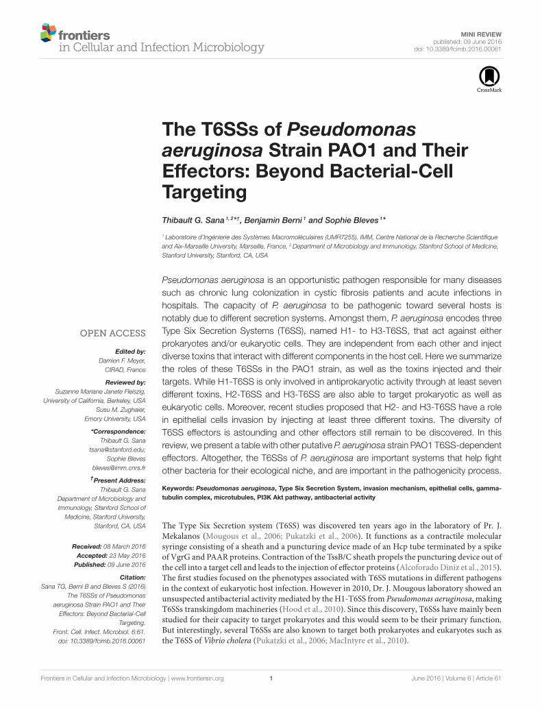
The T6SSs of Pseudomonasaeruginosa Strain PAO1 and TheirEffectors: Beyond Bacterial-CellTargeting
Thibault G. Sana 1, 2*†, Benjamin Berni 1 and Sophie Bleves 1*
1 Laboratoire d’Ingénierie des Systèmes Macromoléculaires (UMR7255), IMM, Centre National de la Recherche Scientifique
and Aix-Marseille University, Marseille, France, 2Department of Microbiology and Immunology, Stanford School of Medicine,
Stanford University, Stanford, CA, USA
Pseudomonas aeruginosa is an opportunistic pathogen responsible for many diseases
such as chronic lung colonization in cystic fibrosis patients and acute infections in
hospitals. The capacity of P. aeruginosa to be pathogenic toward several hosts is
notably due to different secretion systems. Amongst them, P. aeruginosa encodes three
Type Six Secretion Systems (T6SS), named H1- to H3-T6SS, that act against either
prokaryotes and/or eukaryotic cells. They are independent from each other and inject
diverse toxins that interact with different components in the host cell. Here we summarize
the roles of these T6SSs in the PAO1 strain, as well as the toxins injected and their
targets. While H1-T6SS is only involved in antiprokaryotic activity through at least seven
different toxins, H2-T6SS and H3-T6SS are also able to target prokaryotic as well as
eukaryotic cells. Moreover, recent studies proposed that H2- and H3-T6SS have a role
in epithelial cells invasion by injecting at least three different toxins. The diversity of
T6SS effectors is astounding and other effectors still remain to be discovered. In this
review, we present a table with other putative P. aeruginosa strain PAO1 T6SS-dependent
effectors. Altogether, the T6SSs of P. aeruginosa are important systems that help fight
other bacteria for their ecological niche, and are important in the pathogenicity process.
Keywords: Pseudomonas aeruginosa, Type Six Secretion System, invasion mechanism, epithelial cells, gamma-
tubulin complex, microtubules, PI3K Akt pathway, antibacterial activity
The Type Six Secretion system (T6SS) was discovered ten years ago in the laboratory of Pr. J.Mekalanos (Mougous et al., 2006; Pukatzki et al., 2006). It functions as a contractile molecularsyringe consisting of a sheath and a puncturing device made of an Hcp tube terminated by a spikeof VgrG and PAAR proteins. Contraction of the TssB/C sheath propels the puncturing device out ofthe cell into a target cell and leads to the injection of effector proteins (AlcoforadoDiniz et al., 2015).The first studies focused on the phenotypes associated with T6SS mutations in different pathogensin the context of eukaryotic host infection. However in 2010, Dr. J. Mougous laboratory showed anunsuspected antibacterial activity mediated by the H1-T6SS from Pseudomonas aeruginosa, makingT6SSs transkingdommachineries (Hood et al., 2010). Since this discovery, T6SSs have mainly beenstudied for their capacity to target prokaryotes and this would seem to be their primary function.But interestingly, several T6SSs are also known to target both prokaryotes and eukaryotes such asthe T6SS of Vibrio cholera (Pukatzki et al., 2006; MacIntyre et al., 2010).
Sana et al. P. aeruginosa T6SS Effectors
P. aeruginosa is one of the most virulent opportunistichuman pathogens and is responsible for many diseases such asbroncho-alveolar colonization in cystic fibrosis patients or acuteinfections of lungs and burned skin that can lead to septicemia.Its genome encodes many virulence factors including severalsecretion systems that help P. aeruginosa control its environmentand the activity of host cells (Bleves et al., 2010). Among theses,P. aeruginosa harbors three independent Type Six SecretionSystems (T6SS). This review will aim to describe the roles,effectors, and targets of these three T6SSs.
P. AERUGINOSA USES T6SSS ASANTIPROKARYOTIC WEAPONS
T6SSs are present in more than 200 Gram-negative bacteriaincluding P. aeruginosa, whose genome encodes three differentT6SS loci named H1-, H2-, and H3-T6SS (Table 1). Historically,H1-T6SS is the first T6SS machinery that was shown to displayan antibacterial activity (Hood et al., 2010). H1-T6SS serves asa counter-attack weapon to outcompete other T6SS+ bacteriathat coexist in a same ecological niche, and confers a growthadvantage upon P. aeruginosa (Basler et al., 2013). Morespecifically, P. aeruginosa targets other bacteria through H1-T6SS dependent injection of effector Tse2 and also produces ananti-toxin Tsi2, protecting itself against the intrinsic effect ofthe toxin and from attack by sister-cells (Hood et al., 2010). Itwas recently shown that Tse2 induces quiescence in bacterialtarget cells, and that Tsi2 directly interacts with Tse2 in thecytoplasm to inactivate its lethal activity (Li et al., 2012). Recently,Tse2 toxicity was shown to be NAD-dependent and may involvean ADP-ribosyltransferase activity (Robb et al., 2016). BesidesTse2, which acts in the cytoplasm of prey cells, Tse1 andTse3 are injected into the periplasm of target bacterial cellsthrough H1-T6SS (Russell et al., 2011). Tse1 and Tse3 hydrolysepeptidoglycan, providing a fitness advantage for P. aeruginosain competition with other bacteria. To protect from killingby sister-cells, P. aeruginosa uses the periplasmic immunityproteins Tsi1 and Tsi3 which counteract Tse1 and Tse3 toxicity(Russell et al., 2011). Later, X-ray studies revealed that Tse1cleaves the γ-D-glutamyl-l-meso-diaminopimelic acid amidebond of crosslinked peptidoglycan (Benz et al., 2012; Chou et al.,2012). Moreover, the crystal structure of Tse1 in interactionwith Tsi1 demonstrates that the immunity protein occludes theactive site of Tse1 abolishing its enzyme activity (Benz et al.,2012). Tse3 functions as a muramidase, cleaving the β-1,4-linkage between N-acetylmuramic acid and N-acetylglucosaminein peptidoglycan (Lu et al., 2013). These three effectors werediscovered in 2010 thanks to their coregulation with the H1-T6SSmachinery (Table 1), and other H1-T6SS toxins were describedlater. Tse4 was identified as a H1-T6SS effector using quantitativecellular proteomics in interaction with Hcp (Whitney et al.,2014). Tse5, Tse6, and Tse7 were identified by their geneticassociation with VgrGs and the H1-T6SS (Hachani et al., 2014;Whitney et al., 2014). Those four effectors display antibacterialactivity and are associated with cognate immunities (Table 1).Recently Tse6 was shown to degrade the essential dinucleotides
NAD(+) and NADP(+) leading to bacteriostasis in the targetbacterium (Whitney et al., 2015). Intriguingly Tse6 delivery intothe host cytoplasm requires translation elongation factor Tu (EF-Tu). The interaction of a toxin with a house keeping proteinmay suggest that it can target phylogenetically diverse bacteria(Whitney et al., 2015). EF-Tu may facilitate Tse6 translocationinto recipient cells or by driving the H1-T6SS needle at thecell surface of the preys either by favoring the passage ofthe toxin once delivered into the periplasm to the cytoplasm.Indeed EF-Tu is known as a moonlighting protein or anchorlessmultifunctional protein that is capable, when localized to the cellsurface, of interfering with bacterial adherence (for a review seeHenderson and Martin, 2011). Importantly work done on H1-T6SS toxins has revealed different conserved mechanisms fortargeting T6SS effectors to the T6SSmachinery (Table 1): (i) Hcp-dependent recruitment in the case of Tse1-4, (ii) direct VgrG-targeting for Tse5, (iii) VgrG-targeting for Tse7 through a PAARmotif and for Tse6 through an adaptator/chaperonne proteincalled EagT6. Altogether, H1-T6SS is a formidable antibacterialweapon, injecting many different effectors to compete bacterialcells, and allowing P. aeruginosa to overwhelm them duringcompetition for the same ecological niche.
H2-T6SS and H3-T6SS also display antibacterial activity byinjecting the phospholipase D enzymes PldA and PldB thatbelong to the Tle5 (type VI lipase effector) family into otherbacterial cells (Russell et al., 2013; Jiang et al., 2014). ThePldA toxin functions by degrading the major constituent ofbacterial membranes, phosphatidylethanolamine (Russell et al.,2013). However, despite strong evidence that P. aeruginosaT6SSs participate widely in bacterial competition, several recentreports have focused on the ability of H2 and H3-T6SS to targetepithelial cells.
P. AERUGINOSA T6SSS ALSO TARGETEUKARYOTIC CELLS
So far, H1-T6SS has never been shown to be directly involvedin anti-eukaryotic activity. However, Hcp1 has been foundin pulmonary secretions of cystic fibrosis patients as well asHcp1-specific antibodies in their sera (Mougous et al., 2006),suggesting that the antiprokaryotic activity of H1-T6SS couldbe necessary for host colonization in a complex microbialcommunity. Interestingly, several members of the gut microbiotaactually encode T6SSs that could lead to the contact-dependentkilling of other bacteria, including Bacteroidetes fragilis (Russellet al., 2014). This opens a new field of research at the interfacebetween the pathogen, host and microbiota, giving a protectiverole for the commensal microbiota through T6SS-dependentkilling of the pathogen. In support of this hypothesis, V. choleraeneeds some of its antitoxins to establish in the host gut, stronglysuggesting that it is subject to T6SS attacks from the microbiota(Fu et al., 2013). Moreover, it was shown that about half ofthe Bacteroidales genomes, the most prevalent Gram-negativebacterial order of the human gut, encode at least one T6SS (Coyneet al., 2016). Finally, a recent report shows that the antibacterialactivity of Bacteroidetes fragilis is active in the mice gut and that
Frontiers in Cellular and Infection Microbiology | www.frontiersin.org 2 June 2016 | Volume 6 | Article 61

Sana et al. P. aeruginosa T6SS Effectors
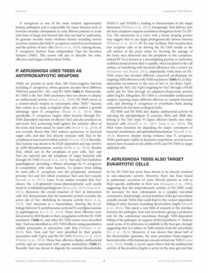
TABLE1|Im
munityproteins,enzymaticactivities,targets,localizations,andrecruitmentoftheT6SSeffectors
oftheP.aeruginosastrain
PAO1.
PAO1Effector
PAnumber
Immunity
Target-cell
Host-cell
localization
Regulation
Activity
Activity/Function
Recruitmentto
theT6SS
machinery
References
H1-T6SS
RetS
repression
QSrepression
Mougousetal.,
2006;Lesic
etal.,
2009
Tse1(Typ
esix
exp
orted)
PA1844
Tsi1(Typ
esix
immunity)(PA1845)
Bacteria
Perip
lasm
RetS
repression
Amidase
Peptid
oglycan
degradatio
n
Hcp1(PA0085)-dependent
Hoodetal.,
2010;Russell
etal.,
2011;Benzetal.,
2012;
Chouetal.,
2012;Silverm
an
etal.,
2013
Tse2
PA2702
Tsi2(PA2703)
Bacteria
Cytoplasm
RetS
repression
NADdependent
toxicity
Bacterio
static
Hcp1(PA0085)-dependent
Hoodetal.,
2010;Lietal.,
2012;Robbetal.,
2016;
Silverm
anetal.,
2013
Tse3
PA3484
Tsi3(PA3485)
Bacteria
Perip
lasm
RetS
repression
Muramidase
Peptid
oglycan
degradatio
n
Hcp1(PA0085)-dependent
Hoodetal.,
2010;Russell
etal.,
2011;Luetal.,
2013;
Silverm
anetal.,
2013
Tse4
PA2774
Tsi4
(PA2775)
Bacteria
Perip
lasm
?4transm
embrane
segments
Hcp1(PA0085)-dependent
Whitn
eyetal.,
2015
Tse5(RhsP
1)
PA2684
Tsi5
(PA2684.1)
Bacteria
Perip
lasm
/
membrane
RetS
repression
?RHS/YDrepeat,
toxicity
VgrG
1c(PA2685)-dependent
Hachanietal.,
2014;Whitn
ey
etal.,
2014
Tse6
PA0093
Tsi6
(PA0092)
Bacteria
Cytoplasm
NAD(P)+
glycohyd
rolase
Bacterio
static,
NAD(P)+
depletio
n,
PAARmotif
VgrG
1a(PA0091)-dependent
trhoughaPAARmotif
&the
EagT6(effector-associatedgene
with
Tse)(P
A0094)chaperone
AlcoforadoDinizetal.,
2015;
Whitn
eyetal.,
2015
Tse7
PA0099
?Bacteria
Cytoplasm
?Endonuclease
?TOX-G
HH2signature
VgrG
1b(PA0095)-dependent
Hachanietal.,
2014
H2-T6SS
QSactivatio
nFur
repressionPsrA
repression
Exp
onentialp
hase
RpoNrepression
Kangetal.,
2008;Siehnel
etal.,
2010;Sanaetal.,
2012,
2013
PldA(Tle5a)(Typ
esix
lipase
effector)
PA3487
Tli5a(PA3488)(Typ
e
sixlipase
immunity)
Bacteria
euka
ryote
Perip
lasm
cytoso
l
Phosp
holipase
DCellwallintegrity
internalizatio
nthrough
Akt
binding
VgrG
4b(PA3486)-dependent?
Wilderm
anetal.,
2001;Russell
etal.,
2013;Jiangetal.,
2014;
SpencerandBrown,2015
VgrG
2b
PA0262
Euka
ryotes
Cytoso
lExp
onentialp
hase
Protease
?γ-TurC
and
microtubule-dependent
internalizatio
n
Evo
lvedVgrG
Sanaetal.,
2015
H2-T
6SSPutative
Tle1
PA3290
Tli1
(PA3291)
Bacteria
Perip
lasm
Phosp
holipase
A2To
xicity
VgrG
4a(PA3294)-dependent
trhoughachaperonne(PA3293)
with
aDUF4123?
Barretetal.,
2011;Russell
etal.,
2013;Huetal.,
2014;
AlcoforadoDinizetal.,
2015
(Continued)
Frontiers in Cellular and Infection Microbiology | www.frontiersin.org 3 June 2016 | Volume 6 | Article 61
Sana et al. P. aeruginosa T6SS Effectors
TABLE1|Continued
PAO1Effector
PAnumber
Immunity
Target-cell
Host-cell
localization
Regulation
Activity
Activity/Function
Recruitmentto
theT6SS
machinery
References
Tle3
PA0260
Tli3(PA0259)
Bacteria
Perip
lasm
Lipolytic
Toxicity
VgrG
2b(PA0262)-dependent?
Barretetal.,
2011;Russell
etal.,
2013;Sanaetal.,
2015
Tle4
PA1510
Tli4
(PA1509)
Bacteria
Perip
lasm
Lipolytic
Toxicity
VgrG
2a(PA1511)-dependent?
Barretetal.,
2011;Luetal.,
2013;Russelletal.,
2013;
Sanaetal.,
2015
PA1508
PA1508
??
??
PAARmotif
VgrG
2a(PA1511)-dependent
trhoughaPAARmotif?
Thisreview
H3-T6SS
QSactivatio
n
RpoNStatio
nary
phase
Lesicetal.,
2009;Sanaetal.,
2013
PldB(Tle5b)
PA5089
Tli5b1(PA5086)
Tli5b2(PA5087)
Tli5b3(PA5088)
Bacteria
euka
ryote
Perip
lasm
cytoso
l
Statio
nary
phase
Phosp
holipase
DCellwallintegrity
internalizatio
nthrough
Akt
binding
VgrG
5(PA5090)-dependent?
Russelletal.,
2013;Jiang
etal.,
2014
Putative
effectorsarehighlightedinlightblue.GreenisH1-T6SS,darkblueisH2-T6SS,andorangeisH3-T6SS.Relatedeffectorsarelistedbelowuntilnewcolorandnewsecretionisdescribed.Lightbluearetheputative
effectorsof
H2-T6SS.
it kills several members of the microbiota in vitro, suggesting arole in the gut colonization (Chatzidaki-Livanisa et al., 2016).Altogether, the T6SS antibacterial activity clearly has a role in theeukaryotic host as well, and should be studied into more details.
Despite being considered an extracellular pathogen, severalreports demonstrate that P. aeruginosa actively invades non-phagocytic cells, such as the epithelial cells that line the mucosalbarrier and the endothelial cells that form the vascular lumen(Chi et al., 1991; Engel and Eran, 2011). The entry step requiresthe actin network, most probably to allow membrane protrusion(Fleiszig et al., 1995). This is thought to help bacteria avoidingthe immune system or to invade deeper tissues during theinfection process. Although, bacteria are present in the lumenand therefore at the apical side of the epithelium, P. aeruginosacan only internalize through membrane that displays basolateralcharacteristics (Figure 1). To circumvent this, P. aeruginosa isable to transform apical membrane into basolateral membrane,creating a local microenvironment that facilitates colonizationand entry into the epithelium (Kierbel et al., 2007). Interestingly,P. aeruginosa is also able to transmigrate through an epithelialbarrier, taking advantage of cell division sites and senescent cellextrusion (Golovkine et al., 2016). Altogether, these convergentmechanisms for entering or crossing the epithelial barrier suggestthat this ability is essential for successful colonization of the hostby P. aeruginosa.
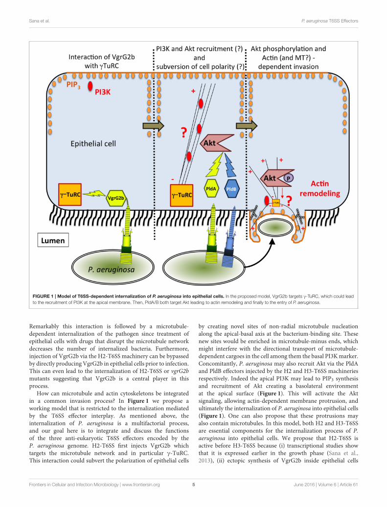
Themechanism by which P. aeruginosa recruits host factors tointernalize within non-phagocytic cells is still poorly understood.Among the various factors required for this process (for areview see Engel and Eran, 2011), we demonstrated thatthe H2-T6SS machinery (Figure 1) promotes the uptake ofP. aeruginosa into pulmonary epithelial cells but, at the time,the identity of the cognate effector(s) involved remained tobe discovered (Sana et al., 2012). Two recent reports haveenabled key new insights into the T6SS-mediated invasionmechanism of P. aeruginosa. Host-cell invasion requires twophospholipase D enzymes, PldA and PldB, which are injectedvia the H2-T6SS or H3-T6SS machineries, respectively (Jianget al., 2014; Table 1). The H3-T6SS machinery is thus requiredfor P. aeruginosa internalization. PldA and PldB both targetthe host PI3K (phosphoinositide 3-kinase)/Akt pathway, whichis hijacked during the internalization process (Kierbel et al.,2005; Engel and Eran, 2011). After injection into epithelialcells, the two T6SS effectors were shown to directly bind Akt,which may lead to activation of the PI3K-Akt signaling pathway.Indeed, Akt phosphorylation is thought to promote a profoundremodeling of the apical membrane in which protrusionsenriched in PIP3 (phosphatidylinositol-3,4,5-triphosphate) andactin form, facilitating further entry of P. aeruginosa (Bleveset al., 2014; Jiang et al., 2014). Interestingly, PldA and PldBare also known to target bacterial cells, making them trans-kingdom effectors (Bleves et al., 2014). Host-cell invasionalso requires the evolved VgrG2b effector (Sana et al., 2015).VgrG2b is injected via the H2-T6SS into epithelial cells whereit targets the microtubule network and more interestingly thegamma-tubulin ring complex components (γ-TuRC) of themicrotubule nucleating-center (Kollman et al., 2011; Table 1).
Frontiers in Cellular and Infection Microbiology | www.frontiersin.org 4 June 2016 | Volume 6 | Article 61
Sana et al. P. aeruginosa T6SS Effectors
FIGURE 1 | Model of T6SS-dependent internalization of P. aeruginosa into epithelial cells. In the proposed model, VgrG2b targets γ-TuRC, which could lead
to the recruitment of PI3K at the apical membrane. Then, PldA/B both target Akt leading to actin remodeling and finally to the entry of P. aeruginosa.
Remarkably this interaction is followed by a microtubule-dependent internalization of the pathogen since treatment ofepithelial cells with drugs that disrupt the microtubule networkdecreases the number of internalized bacteria. Furthermore,injection of VgrG2b via the H2-T6SS machinery can be bypassedby directly producing VgrG2b in epithelial cells prior to infection.This can even lead to the internalization of H2-T6SS or vgrG2bmutants suggesting that VgrG2b is a central player in thisprocess.
How can microtubule and actin cytoskeletons be integratedin a common invasion process? In Figure 1 we propose aworking model that is restricted to the internalization mediatedby the T6SS effector interplay. As mentioned above, theinternalization of P. aeruginosa is a multifactorial process,and our goal here is to integrate and discuss the functionsof the three anti-eukaryotic T6SS effectors encoded by theP. aeruginosa genome. H2-T6SS first injects VgrG2b whichtargets the microtubule network and in particular γ-TuRC.This interaction could subvert the polarization of epithelial cells
by creating novel sites of non-radial microtubule nucleationalong the apical-basal axis at the bacterium-binding site. Thesenew sites would be enriched in microtubule-minus ends, whichmight interfere with the directional transport of microtubule-dependent cargoes in the cell among them the basal PI3Kmarker.Concomitantly, P. aeruginosa may also recruit Akt via the PldAand PldB effectors injected by the H2 and H3-T6SS machineriesrespectively. Indeed the apical PI3K may lead to PIP3 synthesisand recruitment of Akt creating a basolateral environmentat the apical surface (Figure 1). This will activate the Aktsignaling, allowing actin-dependent membrane protrusion, andultimately the internalization of P. aeruginosa into epithelial cells(Figure 1). One can also propose that these protrusions mayalso contain microtubules. In this model, both H2 and H3-T6SSare essential components for the internalization process of P.aeruginosa into epithelial cells. We propose that H2-T6SS isactive before H3-T6SS because (i) transcriptional studies showthat it is expressed earlier in the growth phase (Sana et al.,2013), (ii) ectopic synthesis of VgrG2b inside epithelial cells
Frontiers in Cellular and Infection Microbiology | www.frontiersin.org 5 June 2016 | Volume 6 | Article 61
Sana et al. P. aeruginosa T6SS Effectors
trigger internalization of T6SS mutants (Sana et al., 2015), and(iii) PldA and PldB can compensate for each other duringinfection with stationary-phase grown bacteria (Jiang et al.,2014). However, the exact molecular mechanism by whichVgrG2b acts on the γ-TuRC and the microtubule network hasyet to be deciphered. Also, what is the nature of the effectordomain of the evolved VgrG2b? How does the interaction of Aktwith these two phospholipases D trigger its activation? Finally,the intracellular lifestyle of P. aeruginosa has to be studied ingreater detail, particularly in light of very interesting reportswhich propose that P. aeruginosa creates its own bleb-niche inepithelial cells where it can replicate (Angus et al., 2008; Jollyet al., 2015).
More efforts have to be made to decipher this entiremechanism because it could lead to important biomedicalapplications. Indeed, P. aeruginosa is known to induce acuteinfection in patients with burned skin. Rationally, in thisscenario, the first barrier P. aeruginosa will have to cross willbe the skin, which is basically composed of epithelial cells.We also know that H2-T6SS and H3-T6SS are important forfull virulence in worm models as well as in mouse models(Lesic et al., 2009; Sana et al., 2013). Therefore, it begs thequestion as to whether H2 or H3-T6SS are responsible forpathogen entry through the burned skin barrier. It will thereforebe very interesting over the next years to study this invasionmechanism more deeply using for example a three dimensionalmodel of burned skin (Shepherd et al., 2009). Thus, H2- andH3-T6SS of P. aeruginosa are potentially good candidates fornew therapeutic targets. And finally, although most invasivebacteria manipulate host actin for entry (Cossart and Sansonetti,2004) this T6SS-mediated entry mechanism could be common inother pathogens such as Campylobacter jejuni, and Citrobacterfreundii, Neisseria gonorrhoeae, or Burkholderia cepacia thatalso appear to modulate the microtubule network to invadeepithelial cells (Donnenberg et al., 1990; Oelschlaeger et al., 1993;Grassme et al., 1996; Yoshida and Sasakawa, 2003; Taylor et al.,2010).
CONCLUSIONS
The T6SS machineries of P. aeruginosa must be considered asversatile weapons that are able to target both prokaryotic andeukaryotic cells. In the future, studies should aim at determiningthe role of their antiprokaryotic activity in vivo because H1-T6SS is clearly active in cystic fibrosis patients. One could alsoask whether this T6SS-driven antibacterial activity is a commonweapon used by pathogens in vivo to outcompete either thecommensal microbiota or other pathogens. As shown in Table 1
the repertoire of T6SS effectors in P. aeruginosa may not becomplete and at least 4 H2-T6SS putative effectors can beproposed according to their genetic linkage with known effectorgenes (Table 1). Also, the exact mechanism of T6SS-dependentinternalization within epithelial cells should be studied in moredetail and its role in colonization and pathogenicity should bebetter understood.
AUTHOR CONTRIBUTIONS
TS and SB wrote the review and created Figure 1. BB and SBcreated Table 1.
FUNDING
TS was financed by a Ph.D. fellowship from the FrenchResearch Ministry and with a “Teaching and Research”fellowship from AMU. This work is supported by a grant(N◦RF20150501346/1/69) from “Association GregoryLemarchal” and “Vaincre la Mucoviscidose” and by AMUand CNRS.
ACKNOWLEDGMENTS
We thank Chantal Soscia and Romé Voulhoux’s team forconstant support, and Theodore Hu and Ben Field for carefulreading of the manuscript.
REFERENCES
Alcoforado Diniz, J., Liu, Y. C., and Coulthurst, S. J. (2015). Molecular weaponry:
diverse effectors delivered by the Type VI secretion system. Cell. Microbiol. 17,
1742–1751. doi: 10.1111/cmi.12532
Angus, A. A., Lee, A. A., Augustin, D. K., Lee, E. J., Evans, D. J., and Fleiszig, S.
M. (2008). Pseudomonas aeruginosa induces membrane blebs in epithelial cells,
which are utilized as a niche for intracellular replication and motility. Infect.
Immun. 76, 1992–2001. doi: 10.1128/IAI.01221-07
Barret, M., Egan, F., Fargier, E., Morrissey, J. P., and O’Gara, F. (2011).
Genomic analysis of the type VI secretion systems in Pseudomonas spp.: novel
clusters and putative effectors uncovered. Microbiology 157, 1726–1739. doi:
10.1099/mic.0.048645-0
Basler, M., Ho, B. T., and Mekalanos, J. J. (2013). Tit-for-tat: type VI secretion
system counterattack during bacterial cell-cell interactions. Cell 152, 884–894.
doi: 10.1016/j.cell.2013.01.042
Benz, J., Sendlmeier, C., Barends, T. R., and Meinhart, A. (2012).
Structural insights into the effector-immunity system Tse1/Tsi1 from
Pseudomonas aeruginosa. PLoS ONE 7:e40453. doi: 10.1371/journal.pone.
0040453
Bleves, S., Sana, T. G., and Voulhoux, R. (2014). The target cell genus does not
matter. Trends Microbiol. 22, 304–306. doi: 10.1016/j.tim.2014.04.011
Bleves, S., Viarre, V., Salacha, R., Michel, G. P., Filloux, A., and Voulhoux,
R. (2010). Protein secretion systems in Pseudomonas aeruginosa: a
wealth of pathogenic weapons. Int. J. Med. Microbiol. 300, 534–543. doi:
10.1016/j.ijmm.2010.08.005
Chatzidaki-Livanisa, M., Geva-Zatorsky, N., and Comstocka, L. E. (2016).
Bacteroides fragilis type VI secretion systems use novel effector and immunity
proteins to antagonize human gut Bacteroidales species. Proc. Natl. Acad. Sci.
U.S.A. 113, 3627–3632. doi: 10.1073/pnas.1522510113
Chi, E., Mehl, T., Nunn, D., and Lory, S. (1991). Interaction of Pseudomonas
aeruginosa with A549 pneumocyte cells. Infect. Immun. 59, 822–828.
Chou, S., Bui, N. K., Russell, A. B., Lexa, K. W., Gardiner, T. E., LeRoux, M.,
et al. (2012). Structure of a peptidoglycan amidase effector targeted to Gram-
negative bacteria by the type VI secretion system. Cell Rep. 1, 656–664. doi:
10.1016/j.celrep.2012.05.016
Cossart, P., and Sansonetti, P. J. (2004). Bacterial invasion: the paradigms of
enteroinvasive pathogens. Science 304, 242–248. doi: 10.1126/science.1090124
Coyne, M. J., Roelofs, K. G., and Comstock, L. E. (2016). Type VI secretion systems
of human gut Bacteroidales segregate into three genetic architectures, two of
Frontiers in Cellular and Infection Microbiology | www.frontiersin.org 6 June 2016 | Volume 6 | Article 61
Sana et al. P. aeruginosa T6SS Effectors
which are contained on mobile genetic elements. BMC Genomics 17, 58. doi:
10.1186/s12864-016-2377-z
Donnenberg, M. S., Donohue-Rolfe, A., and Keusch, G. T. (1990). A comparison
of HEp-2 cell invasion by enteropathogenic and enteroinvasive Escherichia coli.
FEMS Microbiol. Lett. 57, 83–86. doi: 10.1111/j.1574-6968.1990.tb04179.x
Engel, J., and Eran, Y. (2011). Subversion of mucosal barrier
polarity by Pseudomonas aeruginosa. Front. Microbiol. 2:114. doi:
10.3389/fmicb.2011.00114
Fleiszig, S. M., Zaidi, T. S., and Pier, G. B. (1995). Pseudomonas aeruginosa invasion
of and multiplication within corneal epithelial cells in vitro. Infect. Immun. 63,
4072–4077.
Fu, Y., Waldor, M. K., and Mekalanos, J. J. (2013). Tn-Seq analysis of
Vibrio cholerae intestinal colonization reveals a role for T6SS-mediated
antibacterial activity in the host. Cell Host Microbe. 14, 652–663. doi:
10.1016/j.chom.2013.11.001
Golovkine, G., Faudry, E., Bouillot, S., Elsen, S., Attree, I., and Huber, P.
(2016). Pseudomonas aeruginosa transmigrates at epithelial cell-cell junctions,
exploiting sites of cell division and senescent cell extrusion. PLoS Pathog.
12:e1005377. doi: 10.1371/journal.ppat.1005377
Grassme, H. U., Ireland, R. M., and van Putten, J. P. (1996). Gonococcal opacity
protein promotes bacterial entry-associated rearrangements of the epithelial
cell actin cytoskeleton. Infect. Immun. 64, 1621–1630.
Hachani, A., Allsopp, L. P., Oduko, Y., and Filloux, A. (2014). The VgrG proteins
are “a la carte” delivery systems for bacterial type VI effectors. J. Biol. Chem.
289, 17872–17884. doi: 10.1074/jbc.M114.563429
Henderson, B., and Martin, A. (2011). Bacterial virulence in the moonlight:
multitasking bacterial moonlighting proteins are virulence determinants in
infectious disease. Infect. Immun. 79, 3476–3491. doi: 10.1128/IAI.00179-11
Hood, R. D., Singh, P., Hsu, F., Guvener, T., Carl, M. A., Trinidad, R. R., et al.
(2010). A type VI secretion system of Pseudomonas aeruginosa targets a toxin
to bacteria. Cell Host Microbe. 7, 25–37. doi: 10.1016/j.chom.2009.12.007
Hu, H., Zhang, H., Gao, Z., Wang, D., Liu, G., Xu, J., et al. (2014). Structure
of the type VI secretion phospholipase effector Tle1 provides insight into its
hydrolysis and membrane targeting. Acta Crystallogr. D Biol. Crystallog. 70,
2175–2185. doi: 10.1107/S1399004714012899
Jiang, F., Waterfield, N. R., Yang, J., Yang, G., and Jin, Q. (2014). A
Pseudomonas aeruginosa type VI secretion phospholipase D effector targets
both prokaryotic and eukaryotic cells. Cell Host Microbe. 15, 600–610. doi:
10.1016/j.chom.2014.04.010
Jolly, A. L., Takawira, D., Oke, O. O., Whiteside, S. A., Chang, S. W., Wen, E.
R., et al. (2015). Pseudomonas aeruginosa-induced bleb-niche formation in
epithelial cells is independent of actinomyosin contraction and enhanced by
loss of cystic fibrosis transmembrane-conductance regulator osmoregulatory
function.MBio 6, e02533. doi: 10.1128/mBio.02533-14
Kang, Y., Nguyen, D. T., Son, M. S., and Hoang, T. T. (2008). The
Pseudomonas aeruginosa PsrA responds to long-chain fatty acid signals to
regulate the fadBA5 beta-oxidation operon. Microbiology 154, 1584–1598. doi:
10.1099/mic.0.2008/018135-0
Kierbel, A., Gassama-Diagne, A., Mostov, K., and Engel, J. N. (2005).
The phosphoinositol-3-kinase-protein kinase B/Akt pathway is critical for
Pseudomonas aeruginosa strain PAK internalization. Mol. Biol. Cell. 16,
2577–2585. doi: 10.1091/mbc.E04-08-0717
Kierbel, A., Gassama-Diagne, A., Rocha, C., Radoshevich, L., Olson, J., Mostov,
K., et al. (2007). Pseudomonas aeruginosa exploits a PIP3-dependent pathway
to transform apical into basolateral membrane. J. Cell Biol. 177, 21–27. doi:
10.1083/jcb.200605142
Kollman, J. M., Merdes, A., Mourey, L., and Agard, D. A. (2011). Microtubule
nucleation by gamma-tubulin complexes. Nat. Rev. Mol. Cell Biol. 12, 709–721.
doi: 10.1038/nrm3209
Lesic, B., Starkey, M., He, J., Hazan, R., and Rahme, L. G. (2009). Quorum sensing
differentially regulates Pseudomonas aeruginosa type VI secretion locus I and
homologous loci II and III, which are required for pathogenesis. Microbiology
155, 2845–2855. doi: 10.1099/mic.0.029082-0
Li, M., Le Trong, I., Carl, M. A., Larson, E. T., Chou, S., De Leon, J. A., et al.
(2012). Structural basis for type VI secretion effector recognition by a cognate
immunity protein. PLoS Pathog. 8:e1002613. doi: 10.1371/journal.ppat.1002613
Lu, D., Shang, G., Yu, Q., Zhang, H., Zhao, Y., Cang, H., et al. (2013). Expression,
purification and preliminary crystallographic analysis of the T6SS effector
protein Tse3 from Pseudomonas aeruginosa. Acta Crystallogr. Sect. F Struct.
Biol. Cryst. Commun. 69, 524–527. doi: 10.1107/S1744309113007148
MacIntyre, D. L., Miyata, S. T., Kitaoka, M., and Pukatzki, S. (2010). The
Vibrio cholerae type VI secretion system displays antimicrobial properties.
Proc. Natl. Acad. Sci. U.S.A. 107, 19520–19524. doi: 10.1073/pnas.1012
931107
Mougous, J. D., Cuff, M. E., Raunser, S., Shen, A., Zhou, M., Gifford, C.
A., et al. (2006). A virulence locus of Pseudomonas aeruginosa encodes a
protein secretion apparatus. Science 312, 1526–1530. doi: 10.1126/science.11
28393
Oelschlaeger, T. A., Guerry, P., and Kopecko, D. J. (1993). Unusual microtubule-
dependent endocytosis mechanisms triggered by Campylobacter jejuni and
Citrobacter freundii. Proc. Natl. Acad. Sci. U.S.A. 90, 6884–6888. doi:
10.1073/pnas.90.14.6884
Pukatzki, S., Ma, A. T., Sturtevant, D., Krastins, B., Sarracino, D., Nelson, W. C.,
et al. (2006). Identification of a conserved bacterial protein secretion system in
Vibrio cholerae using the Dictyostelium host model system. Proc. Natl. Acad.
Sci. U.S.A. 103, 1528–1533. doi: 10.1073/pnas.0510322103
Robb, C. S., Robb, M., Nano, F. E., and Boraston, A. B. (2016). The structure of
the Toxin and Type Six Secretion System Substrate Tse2 in complex with its
immunity protein. Structure 24, 277–284. doi: 10.1016/j.str.2015.11.012
Russell, A. B., Hood, R. D., Bui, N. K., LeRoux,M., Vollmer,W., andMougous, J. D.
(2011). Type VI secretion delivers bacteriolytic effectors to target cells. Nature
475, 343–347. doi: 10.1038/nature10244
Russell, A. B., LeRoux, M., Hathazi, K., Agnello, D. M., Ishikawa, T., Wiggins,
P. A., et al. (2013). Diverse type VI secretion phospholipases are functionally
plastic antibacterial effectors. Nature 496, 508–512. doi: 10.1038/nature
12074
Russell, A. B., Wexler, A. G., Harding, B. N., Whitney, J. C., Bohn, A. J., Goo,
Y. A., et al. (2014). A type VI secretion-related pathway in Bacteroidetes
mediates interbacterial antagonism. Cell Host Microbe. 16, 227–236. doi:
10.1016/j.chom.2014.07.007
Sana, T. G., Baumann, C., Merdes, A., Soscia, C., Rattei, T., Hachani, A., et al.
(2015). Internalization of Pseudomonas aeruginosa strain PAO1 into epithelial
cells is promoted by interaction of a T6SS effector with the microtubule
network.MBio 6:e00712. doi: 10.1128/mBio.00712-15
Sana, T. G., Hachani, A., Bucior, I., Soscia, C., Garvis, S., Termine, E., et al.
(2012). The second type VI secretion system of Pseudomonas aeruginosa strain
PAO1 is regulated by quorum sensing and Fur andmodulates internalization in
epithelial cells. J. Biol. Chem. 287, 27095–27105. doi: 10.1074/jbc.M112.376368
Sana, T. G., Soscia, C., Tonglet, C. M., Garvis, S., and Bleves, S. (2013). Divergent
control of two type VI secretion systems by RpoN in Pseudomonas aeruginosa.
PLoS ONE 8:e76030. doi: 10.1371/journal.pone.0076030
Shepherd, J., Douglas, I., Rimmer, S., Swanson, L., and MacNeil, S. (2009).
Development of three-dimensional tissue-engineered models of bacterial
infected human skin wounds. Tissue Eng. Part C Methods 15, 475–484. doi:
10.1089/ten.tec.2008.0614
Siehnel, R., Traxler, B., An, D. D., Parsek, M. R., Schaefer, A. L., and Singh, P.
K. (2010). A unique regulator controls the activation threshold of quorum-
regulated genes in Pseudomonas aeruginosa. Proc. Natl. Acad. Sci. U.S.A. 107,
7916–7921. doi: 10.1073/pnas.0908511107
Silverman, J. M., Agnello, D. M., Zheng, H., Andrews, B. T., Li, M., Catalano,
C. E., et al. (2013). Haemolysin coregulated protein is an exported receptor
and chaperone of type VI secretion substrates. Mol. Cell. 51, 584–593. doi:
10.1016/j.molcel.2013.07.025
Spencer, C., and Brown, H. A. (2015). Biochemical characterization of a
Pseudomonas aeruginosa phospholipase D. Biochemistry 54, 1208–1218. doi:
10.1021/bi501291t
Taylor, J. B., Hogue, L. A., LiPuma, J. J., Walter, M. J., Brody, S. L., and Cannon,
C. L. (2010). Entry of Burkholderia organisms into respiratory epithelium:
CFTR, microfilament and microtubule dependence. J. Cyst. Fibros. 9, 36–43.
doi: 10.1016/j.jcf.2009.10.002
Whitney, J. C., Beck, C. M., Goo, Y. A., Russell, A. B., Harding, B. N., De Leon,
J. A., et al. (2014). Genetically distinct pathways guide effector export through
the type VI secretion system. Mol. Microbiol. 92, 529–542. doi: 10.1111/mmi.
12571
Whitney, J. C., Quentin, D., Sawai, S., LeRoux, M., Harding, B. N., Ledvina, H.
E., et al. (2015). An interbacterial NAD(P)(+) glycohydrolase toxin requires
Frontiers in Cellular and Infection Microbiology | www.frontiersin.org 7 June 2016 | Volume 6 | Article 61
Sana et al. P. aeruginosa T6SS Effectors
elongation factor Tu for delivery to target cells. Cell 163, 607–619. doi:
10.1016/j.cell.2015.09.027
Wilderman, P. J., Vasil, A. I., Johnson, Z., and Vasil, M. L. (2001). Genetic and
biochemical analyses of a eukaryotic-like phospholipase D of Pseudomonas
aeruginosa suggest horizontal acquisition and a role for persistence in a chronic
pulmonary infection model. Mol. Microbiol. 39, 291–303. doi: 10.1046/j.1365-
2958.2001.02282.x
Yoshida, S., and Sasakawa, C. (2003). Exploiting host microtubule dynamics: a new
aspect of bacterial invasion. Trends Microbiol. 11, 139–143. doi: 10.1016/S0966-
842X(03)00023-4
Conflict of Interest Statement: The authors declare that the research was
conducted in the absence of any commercial or financial relationships that could
be construed as a potential conflict of interest.
Copyright © 2016 Sana, Berni and Bleves. This is an open-access article distributed
under the terms of the Creative Commons Attribution License (CC BY). The use,
distribution or reproduction in other forums is permitted, provided the original
author(s) or licensor are credited and that the original publication in this journal
is cited, in accordance with accepted academic practice. No use, distribution or
reproduction is permitted which does not comply with these terms.
Frontiers in Cellular and Infection Microbiology | www.frontiersin.org 8 June 2016 | Volume 6 | Article 61