The skin is the largest organ of the body. It consists of several different types of tissues working...
36
-
Upload
dominic-fisher -
Category
Documents
-
view
212 -
download
0
Transcript of The skin is the largest organ of the body. It consists of several different types of tissues working...

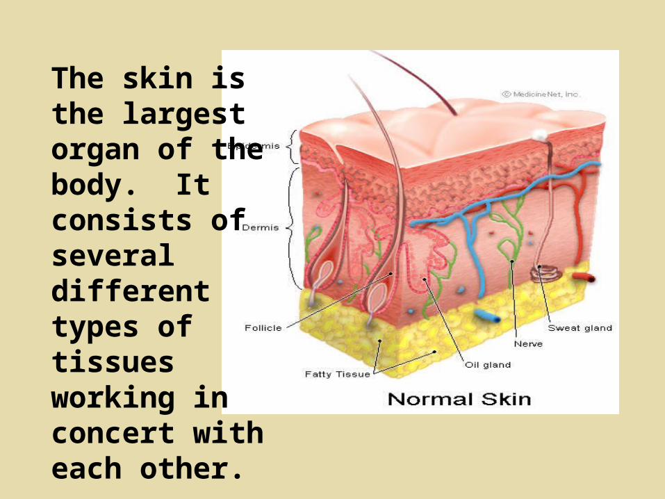
The skin is the largest organ of the body. It consists of several different types of tissues working in concert with each other.

SKIN:
Millions of sensors
Extensive vascular system
Protective and pliable covering
Continually replaced
Includes hair, nails, glands

Friction Ridges
Found on digits, palms, and soles
Assist in grasping ability
2700 ridge units per square inch of skin
Form on fetus before birth and do not change except for injury, disease, death

"The various configurations are not determined by self-limited mechanism within the skin. The skin possesses the capacity to form ridges, but the alignments of these ridges are as responsive to stresses in growth as are the alignments of sand to sweeping by wind or wave.”
Harold Cummins Ph.D.
Volar Pads Emerge At 10 Weeks

Babler has examined the developmental relationships between epidermal ridges and the developing bone skeleton of the hand.
He has shown a significant prenatal relationship between epidermal ridge dimension and bone dimension of the hand.
Whorl patterns tend to be associated with shorter distal phalanges. Whorl patterns also tend to be associated with less ossification, suggesting either early ridge development relative to bone maturity or delayed bone development relative to ridge formation.
In other words, friction ridge patterns are not just the result of genetic factors but also random physical stresses and tensions within the womb.

Friction ridge

Five Layers of the Epidermis

Stratum corneum: 25-30 layers of dead squamous epithelial cells, that are constantly shed.
Stratum lucidum: present only in thick skin: palms & soles
Stratum granulosum: 3-4 layers of cells in the process of dying
Stratum spinosum: living layers of epidermis
Stratum basale:single layer-mitotically active-the generating layer

Four types of cells are present in the stratum basale layer:
Keratinocytes (90%) - responsible for waterproofing and toughening the skin
Melanocytes (8%) - synthesize the pigment melanin which absorbs and disperses ultraviolet radiation
Tactile cells - very sparse and function in touch reception
Nonpigmented granular dendrocytes - cells that ingest bacteria and foreign debris.

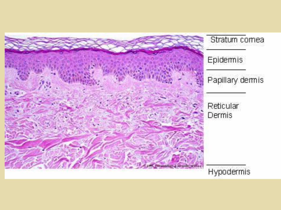
Identify
the
layers.

Dermis: thicker than epidermis.
Consists of two layers.


The primary function of the dermis is to sustain and support the epidermis.
The papillary layer (DPL) is made up of connective tissue with fine elastic fibres.
The surface area of this layer is increased by the dermal papillae (DP).
These fingerlike formations greatly increase the surface area for the exchange of oxygen, nutrients and waste products between the dermis and the epidermis.

Layers of Skin


Sweat Glands/Eccrine Glands
Concentrated in palms and soles of feet
Simple, coiled tubular glands
Sweat contains around 99% water and 1% solids ( mostly NaCl)
Development of Friction Ridges (as described by William J. Babler, PHD in his article " Embryologic Development of Epidermal Ridges and Their Configurations"- Birth Defects: Original Article Series 27(2): 95-112, 1991)

Minutiae = the small characteristics of ridges
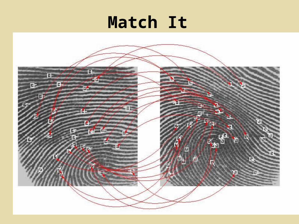
Match It
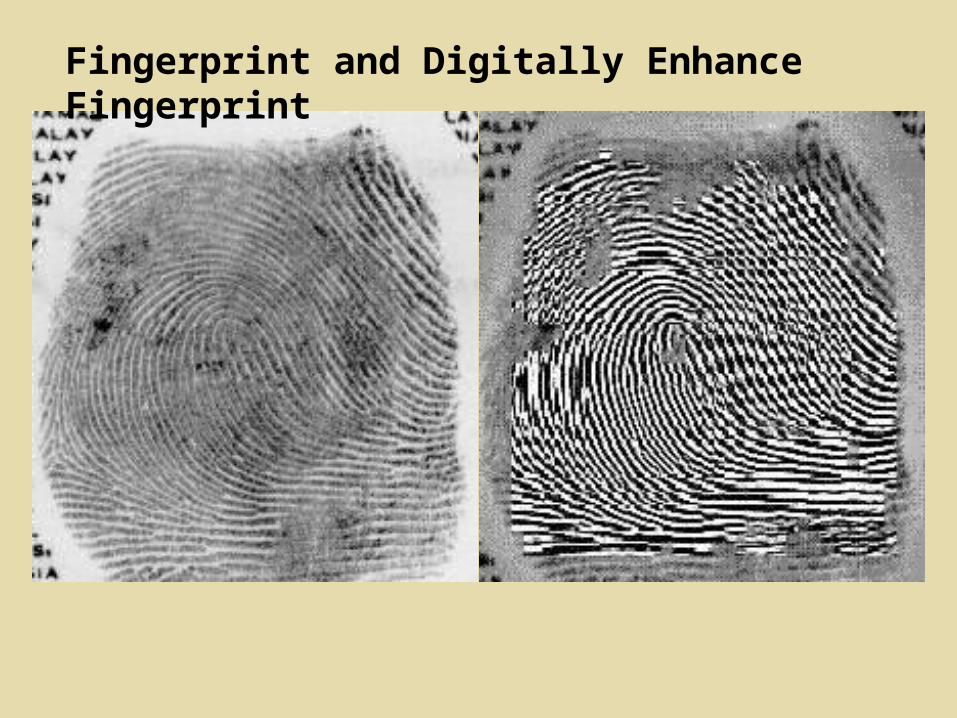
Fingerprint and Digitally Enhance Fingerprint

Problems With Fingerprints

The Arch

The Tented Arch

The Loop
Radial Ulnar

The Whorl

The Twinned Loop or Double Loop
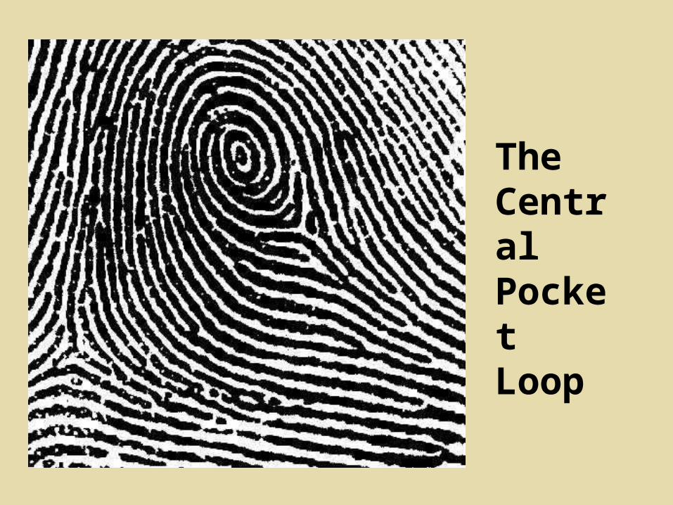
The Central Pocket Loop
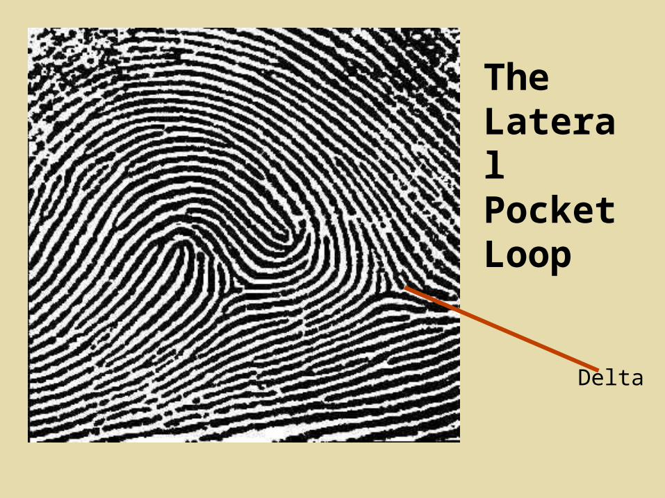
The Lateral Pocket Loop
Delta

The Composite

The Accidental


Lift fingerprints using proper methodology from following objects: 1. Plastic water bottle
2. White coffee cup
3. Pop can
4. Black card/Green Metal Powder Tech.
Latent Lifting and Minutiae Identification Lab

a. Lift best single print from each object and tape onto white card.
b. Label backside of card with date, object, and location and your name(s) as lab technician(s).
c. Examine, identify and label 6 different minutiae characteristics of each print.


![Adagio e Cantabile. Transcribed for concert Organ solo ...€¦ · Adagio e Cantabile. Transcribed for concert Organ solo. [from Concerto "Il Piacere" RV 180] Transcribed for concert](https://static.fdocuments.us/doc/165x107/5eacc3c1cad0900a403344f1/adagio-e-cantabile-transcribed-for-concert-organ-solo-adagio-e-cantabile-transcribed.jpg)
![[Cycle of Organ] Concert for the inauguration of the Organ ...](https://static.fdocuments.us/doc/165x107/61823aa573f1a621b922d884/cycle-of-organ-concert-for-the-inauguration-of-the-organ-.jpg)