The Skin Also known as the Integumentary System= covers all body surfaces.
71
The Skin Also known as the Integumentary System= covers all body surfaces
-
Upload
helen-jenkins -
Category
Documents
-
view
217 -
download
0
Transcript of The Skin Also known as the Integumentary System= covers all body surfaces.

The Skin
Also known as the Integumentary System= covers all body surfaces

Skin Video

Contains these tissues:
• 1. Epithelial Tissue– a. Found in outer layer of skin (covers body)
• 2. Connective Tissue– a. Consists of tough, flexible fibers that holds
the body together.
• 3. Muscle Tissue– a. Push hairs up in skin (goose bumps)
• 4. Nervous Tissue– a. Helps detect pain, pressure, heat, cold

Consists of three major regions
– Epidermis – outermost superficial region
– Dermis – middle region
– Hypodermis (superficial fascia) – deepest region

Functions
• Covers all body surface:
• inside and out• Ex. Lung linings• capillaries
• anchored by basement membrane
• reproduce rapidly
• tightly packed
• attached by desmosomes


The Two Layers of the Skin(Outer and Inner)
• 1. Epidermis– a. Outermost layer of skin
• 1. Exterior – Consists of 25 to 30 layers of dead cells (continually
being shed)– Contains keratininized stratified squamous
epithelium - protective protein– Contains keratinocytes, melanocytes, Merkel cells,
and Langerhans’ cells

• b. Characteristics– 1. No blood vessels– 2. Newer cells below push up to surface– 3. Older cells layer up and die– 4. Hardening = keratinizing– 5. calluses form– 6. shields against moisture loss– 7. Prevents invasions of organisms– 8. Ability to tan

Over view….
• 2. Outer portion of the skin is exposed to the external environment and functions in protection
3. InteriorContains living cells (continually forming new cells)Melanin (pigment) found in this layer

• 3. Epidermal ridges– a. Known as fingerprints

Identifying Markers

Types of Fingerprints:
• 1. Arch

• 2. Whorl

• 3. Loops

• 4. Double Loops
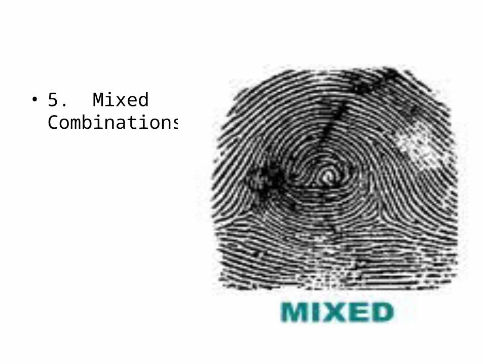
• 5. Mixed Combinations

Layers of Epidermis
• From outer to inner:• 1. Stratum Lucidum = (Clear Layer)• 2. Stratum Granulosum = (Granular Layer)• a. Granular in appearance
3. Stratum Spinosum = (Prickly Layer)– a. Thick part
• 4. Stratum Basale = (Basal Layer)ba. Deepest layer
5. Stratum Corneum = (Horny Layer)
a. Layer of hard cells
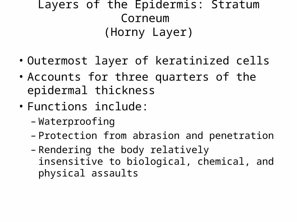
Layers of the Epidermis: Stratum Corneum (Horny Layer)
• Outermost layer of keratinized cells
• Accounts for three quarters of the epidermal thickness
• Functions include:– Waterproofing– Protection from abrasion and penetration– Rendering the body relatively insensitive to
biological, chemical, and physical assaults

Layers of the Epidermis: Stratum Lucidum (Clear Layer)
• Thin, transparent band superficial to the stratum granulosum
• Consists of a few rows of flat, dead keratinocytes
• Present only in thick skin

Layers of the Epidermis: Stratum Granulosum (Granular Layer)
• Thin; three to five cell layers in which drastic changes in keratinocyte appearance occurs


Layers of the Epidermis: Stratum Spinosum (Prickly Layer)
• Cells contain a weblike system of intermediate filaments attached to desmosomes
• Melanin granules and Langerhans’ cells are abundant in this layer

Langerhans Cells


Langerhans Cells…..
• On infection of an area of skin, the local Langerhans' cells will take up and process microbial antigens.
• They are similar in shape and function to macrophages.

Merkel Cells
• Merkel cells are present in small numbers in the stratum basale, or the deepest layer, of the epidermis.
• They are located near areas of well-vascularised, richly innervated connective tissue. Each Merkel cell is intimately associated with a nerve terminal, forming a structure known as a Merkel cell-neuron complex, or a Merkel disc.
• Merkel cells possess desmosomes and keratin filaments, which suggests that they may have an epithelial origin.

Layers of the Epidermis: Stratum Basale (Basal Layer)
• Deepest epidermal layer firmly attached to the dermis
• Consists of a single row of the youngest keratinocytes
• Cells undergo rapid division, hence its alternate name, stratum germinativum

• The basal cell layer contains cells called melanocytes. • Melanocytes produce the skin coloring or pigment known
as melanin, which gives skin its tan or brown color and helps protect the deeper layers of the skin from the harmful effects of the sun.
• Sun exposure causes melanocytes to increase production of melanin in order to protect the skin from damaging ultraviolet rays, producing a suntan.
• Patches of melanin in the skin cause birthmarks, freckles and age spots.
• Melanoma develops when melanocytes undergo malignant transformation.

Skin Color
• Three pigments contribute to skin color– Melanin – yellow to reddish-brown to black
pigment, responsible for dark skin colors• Freckles and pigmented moles – result from local
accumulations of melanin
– Carotene – yellow to orange pigment, most obvious in the palms and soles of the feet
– Hemoglobin – reddish pigment responsible for the pinkish hue of the skin

Layers of the Epidermis: Stratum Basale (Basal Layer)

• 3. Dermis– a. Known as the inner, thicker portion of skin– b. Contains:
• Blood vessels• Nerves• Nerve endings• Hair follicles• Sweat glands• Oil glands

– c. Attached to an underlying layer of subcutaneous (fat) layer.
• This fat layer helps absorb impacts, retain heat, store food.

Dermis
• Second major skin region containing strong, flexible connective tissue
• Cell types include fibroblasts, macrophages, and occasionally mast cells and white blood cells
• Composed of two layers – papillary (fingerlike projections) and reticular

Layers of the Dermis: Papillary Layer
• Papillary layer– Areolar connective tissue with collagen and
elastic fibers– Its superior surface contains peglike
projections called dermal papillae– Dermal papillae contain capillary loops,
Meissner’s corpuscles, and free nerve endings
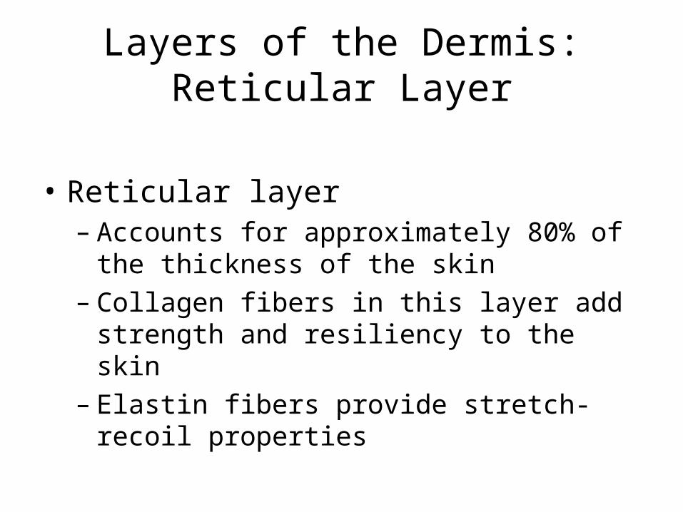
Layers of the Dermis: Reticular Layer
• Reticular layer– Accounts for approximately 80% of the
thickness of the skin– Collagen fibers in this layer add strength and
resiliency to the skin– Elastin fibers provide stretch-recoil properties

Dermis Characteristics
• Uneven surface• Contains:
– Fingerlike papillae– Fibrous and connective tissue– Muscle fibers– Hair follicles– Blood vessels– Nerve fibers– Pacinian Corpuscles = heavy pressure– Meissner’s Corpuscles = light pressure-- Sevbaceous glands--Sweat glands

Sweat Glands
• Different types prevent overheating of the body; secrete cerumen and milk– Eccrine sweat glands – found in palms, soles of the
feet, and forehead– Apocrine sweat glands – found in axillary and
anogenital areas– Ceruminous glands – modified apocrine glands in
external ear canal that secrete cerumen– Mammary glands – specialized sweat glands that
secrete milk

Sebaceous Glands
• Simple alveolar glands found all over the body
• Soften skin when stimulated by hormones
• Secrete an oily secretion called sebum

Structure of a Nail
• Scalelike modification of the epidermis on the distal, dorsal surface of fingers and toes

Hair
• Filamentous strands of dead keratinized cells produced by hair follicles
• Contains hard keratin which is tougher and more durable than soft keratin of the skin
• Made up of the shaft projecting from the skin, and the root embedded in the skin
• Consists of a core called the medulla, a cortex, and an outermost cuticle
• Pigmented by melanocytes at the base of the hair

Hair Function and Distribution
• Functions of hair include:– Helping to maintain warmth– Alerting the body to presence of insects on the skin – Guarding the scalp against physical trauma, heat
loss, and sunlight
• Hair is distributed over the entire skin surface except– Palms, soles, and lips– Nipples and portions of the external genitalia
Hair Follicle
• Root sheath extending from the epidermal surface into the dermis
• Deep end is expanded forming a hair bulb
• A knot of sensory nerve endings (a root hair plexus) wraps around each hair bulb
• Bending a hair stimulates these endings, hence our hairs act as sensitive touch receptors
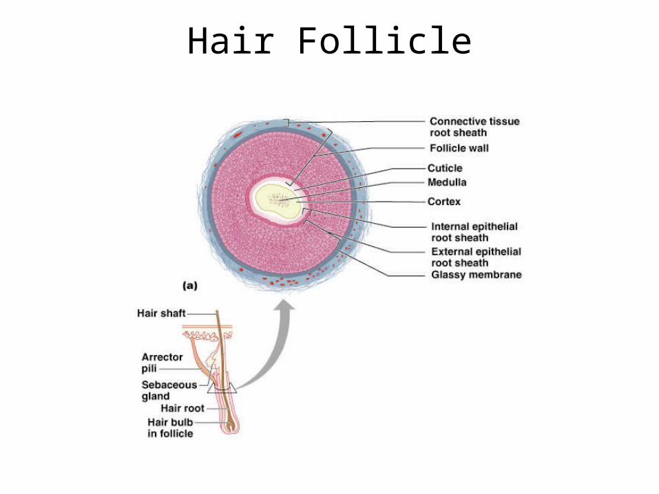
Hair Follicle

Hair Follicle
Types of Hair
• Vellus – pale, fine body hair found in children and the adult female
• Terminal – coarse, long hair of eyebrows, scalp, axillary, and pubic regions

Hair Thinning and Baldness
• Hair Thinning and Baldness
• Alopecia – hair thinning in both sexes
• True, or frank, baldness – Genetically determined and sex-influenced
condition – Male pattern baldness – caused by follicular
response to DHT

Functions of the Integumentary System
• Protection – chemical, physical, and mechanical barrier
• Body temperature regulation is accomplished by:– Dilation (cooling) and constriction (warming) of dermal
vessels– Increasing sweat gland secretions to cool the body
• Cutaneous sensation – exoreceptors sense touch and pain

Functions of the Integumentary System
• Metabolic functions – synthesis of vitamin D in dermal blood vessels
• Blood reservoir – skin blood vessels store up to 5% of the body’s blood volume
• Excretion – limited amounts of nitrogenous wastes are eliminated from the body in sweat

Skin Cancer
• Most skin tumors are benign and do not metastasize
• A crucial risk factor for nonmelanoma skin cancers is the disabling of the p53 gene
• Newly developed skin lotions can fix damaged DNA

Skin Cancer
• The three major types of skin cancer are:– Basal cell carcinoma– Squamous cell carcinoma– Melanoma

Basal Cell Carcinoma
• Least malignant and most common skin cancer• Stratum basale cells proliferate and invade the
dermis and hypodermis• Slow growing and do not often metastasize• Can be cured by surgical excision in 99% of the
cases• Hard, dry scaly growths with reddish base• Fair skinned people most at risk• Over forty and out in sun at risk
• Sun causes DNA damage

Squamous Cell Carcinoma
• Arises from keratinocytes of stratum spinosum
• Arise most often on scalp, ears, and lower lip
• Grows rapidly and metastasizes if not removed
• Prognosis is good if treated by radiation therapy or removed surgically

Melanoma
• Cancer of melanocytes is the most dangerous type of skin cancer because it is:– Highly metastatic– Resistant to chemotherapy
• Known as melanocarcinomas• Caused by sunburn

Melanoma
• Melanomas have the following characteristics (ABCD rule)– A: Asymmetry; the two sides of the
pigmented area do not match – B: Border is irregular and exhibits
indentations– C: Color (pigmented area) is black, brown,
tan, and sometimes red or blue– D: Diameter is larger than 6 mm (size of a
pencil eraser)

Melanoma
• Treated by wide surgical excision accompanied by immunotherapy
• Chance of survival is poor if the lesion is over 4 mm thick
Burns
• First-degree – only the epidermis is damaged– Symptoms include localized redness, swelling, and pain– Heals in 2 weeks, no scars
• Second-degree – epidermis and upper regions of dermis are damaged– Symptoms mimic first degree burns, but blisters also appear– Damage is to dermis
• Third-degree – entire thickness of the skin is damaged– Burned area appears gray-white, cherry red, or black; there is no
initial edema or pain (since nerve endings are destroyed)– Dermis destroyed

Rules of Nine
• Body is divided into 11 areas, each accounting for 9% of the total body surface area (water loss)

Rule of Nines
• Estimates the severity of burns
• Burns considered critical if:– Over 25% of the body has second-degree
burns– Over 10% of the body has third-degree burns– There are third-degree burns on face, hands,
or feet
Developmental Aspects of the Integument: Fetal
• Epidermis develops from ectoderm• Dermis and hypodermis develop from
mesoderm• Lanugo – downy coat of delicate hairs
covering the fetus• Vernix caseosa – substance produced by
sebaceous glands that protects the skin of the fetus in the amnion

Developmental Aspects of the Integument: Adolescent to Adult
• Skin and hair become oilier and acne may appear
• Skin shows the effects of cumulative environmental assaults around age 30
• Scaling and dermatitis become more common

Developmental Aspects of the Integument: Old Age
• Epidermal replacement of cells slows and skin becomes thinner
• Skin becomes dry and itchy• Subcutaneous fat layer diminishes, leading to
intolerance of cold• Decreased elasticity and loss of subcutaneous
tissue leads to wrinkles• Decreased numbers of melanocytes and
Langerhans’ cells increase the risk of skin cancer

Hypodermis
• Subcutaneous layer deep to the skin
• Composed of adipose and areolar connective tissue

4. Maintains homeostasis
a. Skin sweats to cool off body when too hot. Blood vessels also dilate to radiate heat away from body.
b. Goose bumps form to trap a layer of air next to the skin to keep warmer.
c. Information sent to brain from skin about sensory changes.
d. Vitamin D is produced when sun activates changes in the skin.
e. Skin acts as a barrier.

Skin is a Barrier

Steps to healing an injury
• 1. Cut
• 2. Epidermal cells divide
• 3. Blood clots at injury site
• 4. A scab forms
• 5. White blood cells gather (pus)
• 6. Gap fills pushing scab loose.

Shallow Cut
• New epithelial cells will fill in.

Deep Cut
• Broken blood vessels allow clots to form– Fibrin + blood cells + platelets– Fluids dry to form scab– Fibroblasts are attracted by chemicals at wound site– New collagenous fibers form– Phagocysts eat dead cells– Scab falls off– Connective tissue forms scars– Deep wounds look granulated

Burns
• 1. First degree burns– a. Involve damage to epidermal cells– B. Usually heal in one week

• 2. Second degree burn– a. Damage to both epidermis and dermis– B. Blistering and scarring

• 3. Third degree burns– a. Destroys both the epidermis and dermis– b. Skin function lost– c. Skin grafts needed