Simple Machines Levers Quiz school/story.php?title=levers-pulleys- 2512.
Upload
lenard-gibsonCategory
view
216download
2

• The skeleton cannot stand alone.
• The bony levers are held in position, as well as moved, by the muscles.
• In addition, many functions which support life involve muscular movement.

• There are more than 400 muscles in the body.
• Approximately 40% of body mass is made up of muscle tissue; the purpose of much of it is to move bones.
• They are extensively supplied with blood vessels and nerves and their action is controlled by the nervous system
• When they contract, they cause movement and, as a result, perform mechanical work.

The muscular system has 3 main functions:1. Locomotion / Movement
muscles supply force to propel the body in movement patterns such as walking, running, throwing and swimming
2. Internal movement internal muscles are actively involved in life support
functions, such as the circulation of the blood, the passage of food through the digestive tract and the movement of the chest, diaphragm and abdomen during breathing
3. Posture the skeleton is assisted in resisting gravity’s effects
by muscles and ligaments to maintain the alignment of the body in a variety of positions such as sitting, standing and walking

Types of Muscle
• There are 3 types of muscle tissue which differ in structure and function.
• They are:– smooth or involuntary– cardiac (heart)– skeletal (striated or striped)
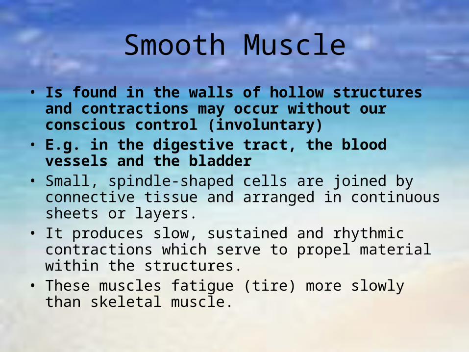
Smooth Muscle
• Is found in the walls of hollow structures and contractions may occur without our conscious control (involuntary)
• E.g. in the digestive tract, the blood vessels and the bladder
• Small, spindle-shaped cells are joined by connective tissue and arranged in continuous sheets or layers.
• It produces slow, sustained and rhythmic contractions which serve to propel material within the structures.
• These muscles fatigue (tire) more slowly than skeletal muscle.


Cardiac Muscle
• It is found only in the heart.• Cardiac muscle fibres and cells are intertwined in a
network and the muscle contracts and relaxes rhythmically without nervous stimulation to individual muscle cells.
• The rhythm is controlled normally by an initial nerve impulse arriving at the heart, which is relayed from cell to cell through the muscle network.
• The heart is not under conscious control.• The contractions are involuntary.• It has the unique ability to contract and relax in a rapid
succession and is difficult to fatigue.

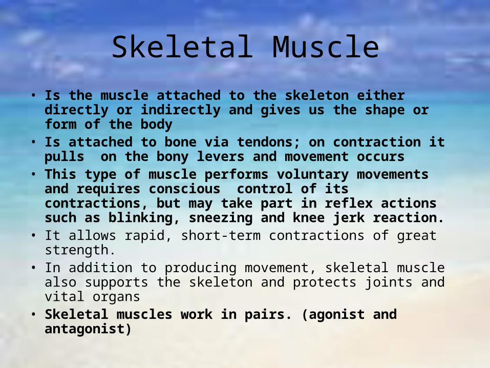
Skeletal Muscle
• Is the muscle attached to the skeleton either directly or indirectly and gives us the shape or form of the body
• Is attached to bone via tendons; on contraction it pulls on the bony levers and movement occurs
• This type of muscle performs voluntary movements and requires conscious control of its contractions, but may take part in reflex actions such as blinking, sneezing and knee jerk reaction.
• It allows rapid, short-term contractions of great strength.• In addition to producing movement, skeletal muscle also
supports the skeleton and protects joints and vital organs• Skeletal muscles work in pairs. (agonist and
antagonist)

Structure of Skeletal Muscle
• Each skeletal muscle consists of a number of bundles containing hundreds of individual muscle cells held together by connective tissues.
• The fibrous connective tissue between and surrounding the muscle bundles is continuous with the tendon which anchors the muscle to the bone and transmit the force of muscle contraction.
• Muscle cells are long, narrow and threadlike, and are referred to as muscle fibres.– A muscle fibre is the basic structural unit of the
muscle.
• Each fibre is composed of many microscopic threads called myofibrils.– Myofibrils are the smaller building blocks of the
muscle fibre and are responsible for muscular contraction
• They are bound together by connective tissue and contained a fluid called sarcoplasm– The sarcoplasm also contains other cellular material
such as fats, glycogen, mitochondria (powerhouses of the cell which receive oxygen) and some myoglobin (a protein in muscle cells which carries oxygen)


Muscle Attachment• Skeletal muscles must have a rigid
attachment to a bone at one end and be attached across a joint to another more easily movable bone at the other end in order to effect movement.
• The muscle’s point of attachment to the more stationary bone is called the origin– usually attaches either directly or indirectly to the
bone via a tendon and tends to be closer to the body’s main mass (proximal)
• The muscle’s movable point of attachment is called the insertion– This end of the muscle usually attaches to the bone
by means of a strong, non-elastic tendon and tends to be away from the body’s main mass (distal)

Major Skeletal Muscles
• Muscles are usually named from their various characteristics or locations.
• E.g. the pectoralis major is named for its size and position.– it is the large (major) of the chest
• The deltoid muscle forms the triangular shape of the Greek letter “delta” on the shoulder.
• The biceps in the upper arm has 2 points of the origin, as the name implies (“two heads”).

UPPER LIMB
• DELTOID – highly visible joint
covering the shoulder joint

• BICEPS– the anterior muscle of
the upper arm
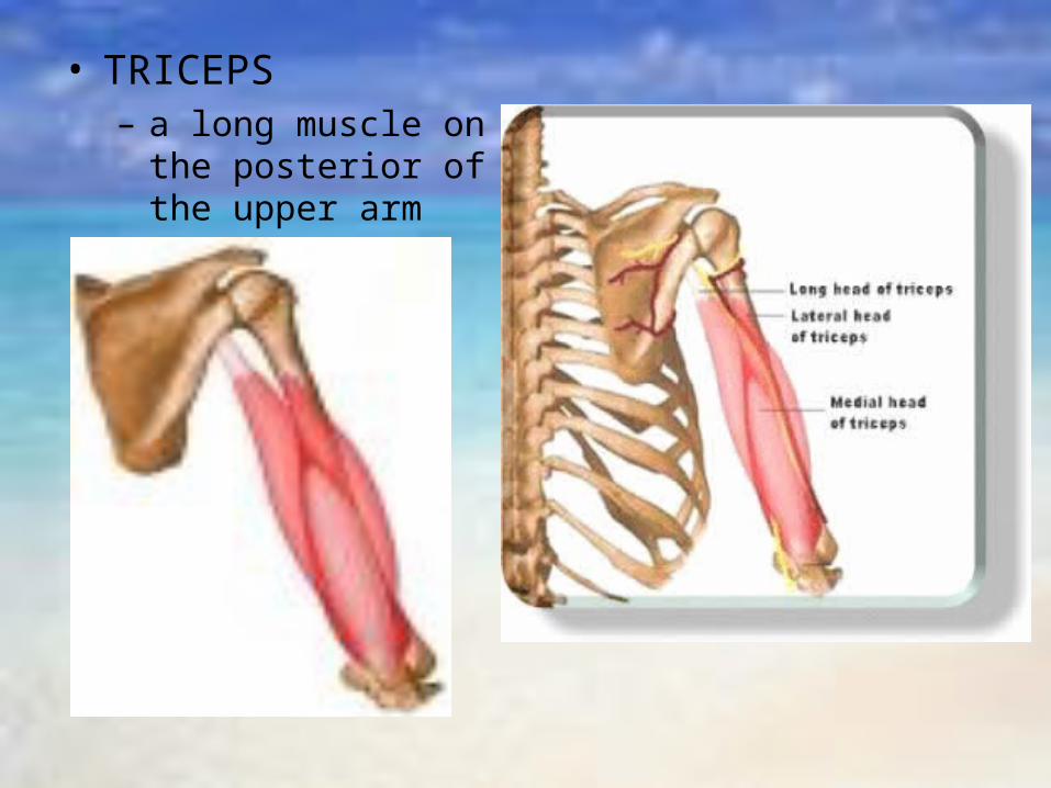
• TRICEPS– a long muscle on the
posterior of the upper arm

• HAND & FINGER FLEXORS– involved in flexion– a group of muscles on the anterior of the forearm
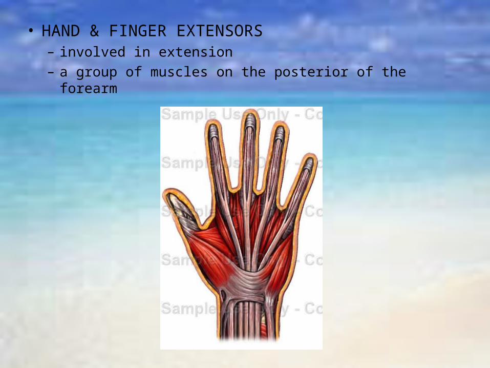
• HAND & FINGER EXTENSORS– involved in extension– a group of muscles on the posterior of the forearm
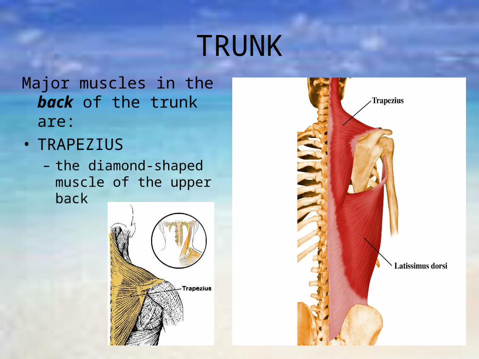
TRUNKMajor muscles in the
back of the trunk are:• TRAPEZIUS
– the diamond-shaped muscle of the upper back
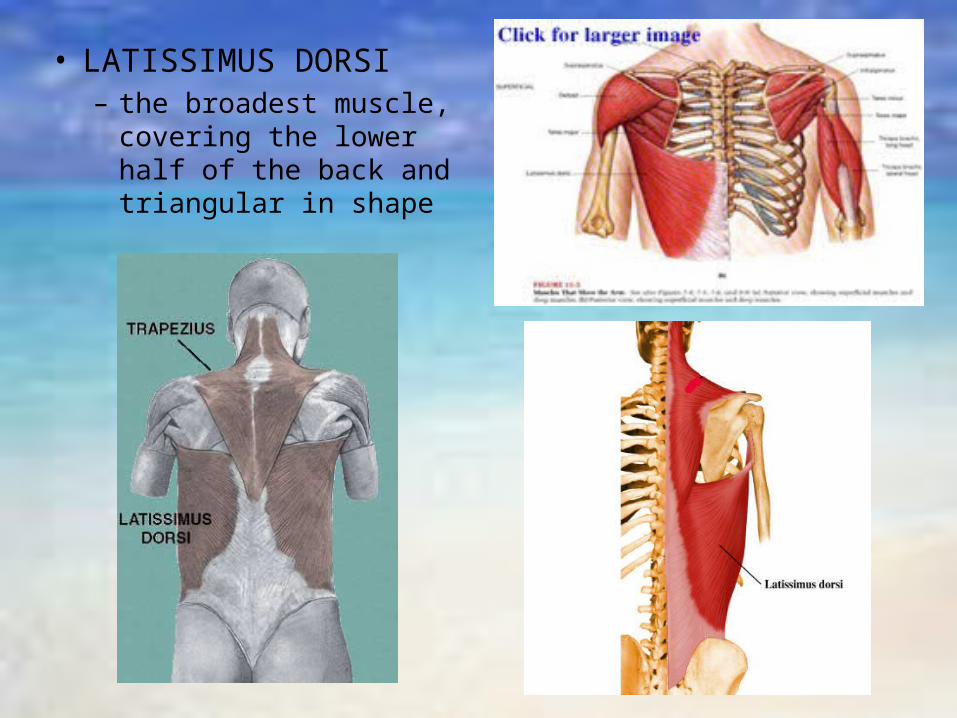
• LATISSIMUS DORSI– the broadest muscle,
covering the lower half of the back and triangular in shape
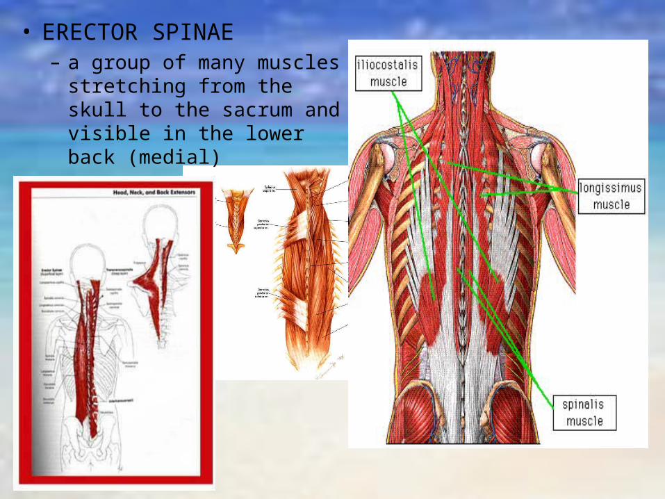
• ERECTOR SPINAE– a group of many muscles
stretching from the skull to the sacrum and visible in the lower back (medial)
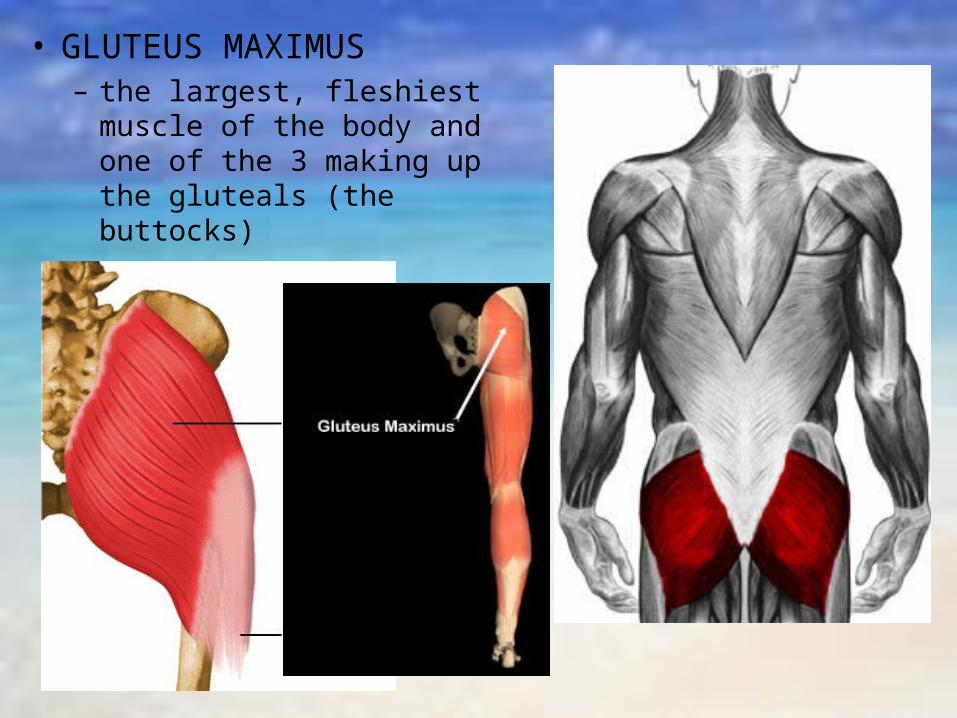
• GLUTEUS MAXIMUS– the largest, fleshiest
muscle of the body and one of the 3 making up the gluteals (the buttocks)
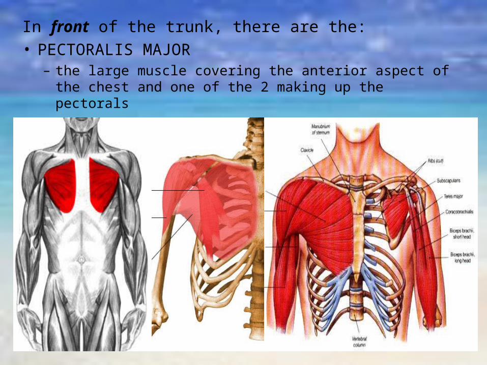
In front of the trunk, there are the:• PECTORALIS MAJOR
– the large muscle covering the anterior aspect of the chest and one of the 2 making up the pectorals

• RECTUS ABDOMINUS– a vertical muscle mass extending the length of the
abdomen and one of the abdominals

• EXTERNAL OBLIQUES– visible laterally at the ribs and
are flat, wide sheets of muscles which run at angles to the abdominal wall (obliques)
Internal Obliques
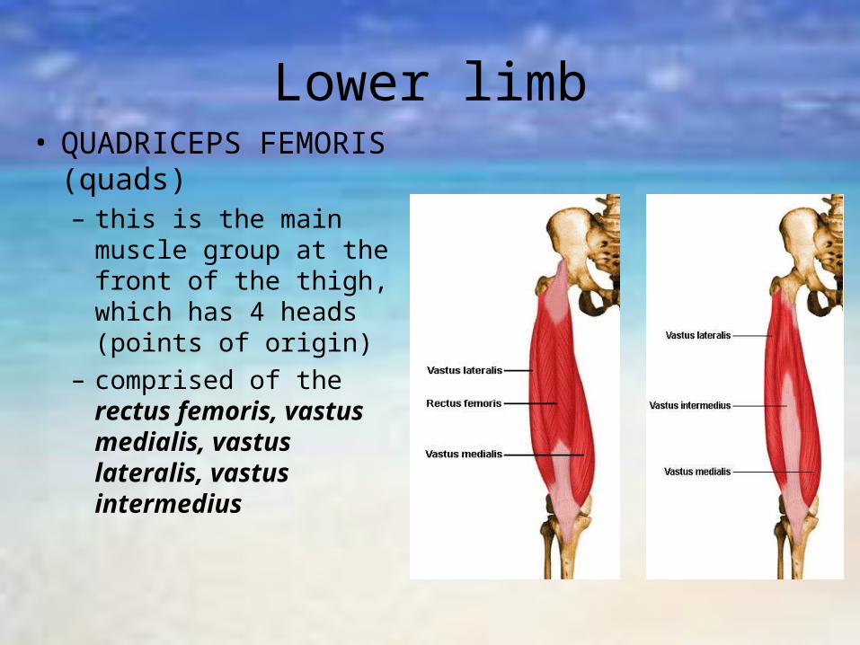
Lower limb• QUADRICEPS
FEMORIS (quads)– this is the main muscle
group at the front of the thigh, which has 4 heads (points of origin)
– comprised of the rectus femoris, vastus medialis, vastus lateralis, vastus intermedius

Rectus Femoris
Vastus Intermedius
Vastus Medialis
Vastus Lateralis

• SARTORIUS– the longest muscle in the body, crossing
the front of the thigh obliquely

• HAMSTRINGS– this group consists of
3 separate muscles which lie on the posterior of the thigh, with cordlike tendons behind the knee
– the muscles ate the biceps femoris, semi-membranosus and semi-tendinosus

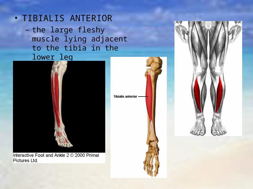
• TIBIALIS ANTERIOR– the large fleshy muscle
lying adjacent to the tibia in the lower leg

• GASTROCNEMIUS– a bulging mass of the calf
which inserts into the heel via the achilles tendon
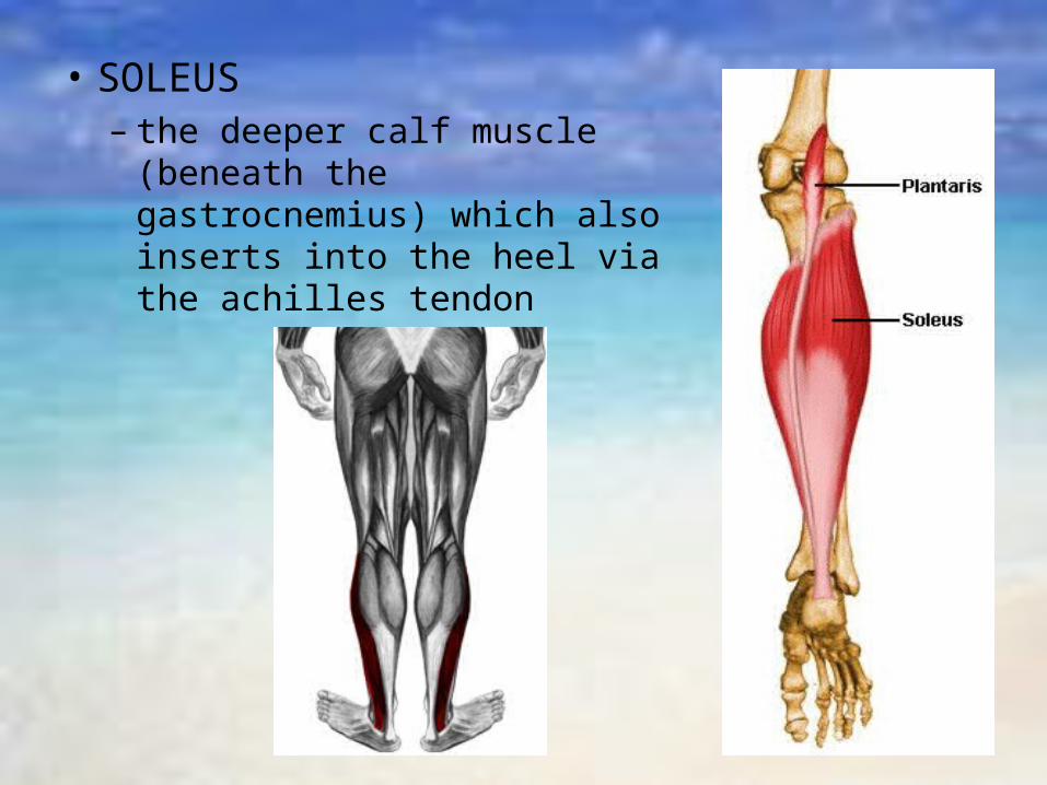
• SOLEUS– the deeper calf muscle
(beneath the gastrocnemius) which also inserts into the heel via the achilles tendon

Muscle action
• Most skeletal muscles are attached to bones by tendons at their origin (fixed end) and insertion (movable end) and act over a joint
• Some muscle act over 2 joints, e.g., the triceps, and are involved in the movement of more than one joint
• To perform any movement of the body, we ultimately depend on the contraction (shortening) and the relaxation (lengthening) of muscle tissue
• Muscles contract or relax when stimulated (triggered) by the central nervous system (CNS)

• During contraction, muscles usually shorten and thicken, pulling the insertion and the origin towards each other.
• During relaxation, the muscle returns to its original length.
• The pull on the bone then produces a resulting movement known as muscle action.
• This occurs in a specific direction• The location of the origin and insertion
determine the direction in which the muscle pulls a particular bone.
• Muscles can only produce movement at the joints over which they act; i.e. between their respective origin and insertion.
• Muscles can only pull, they cannot push.
• Hence, they produce movement by acting in pairs or groups.
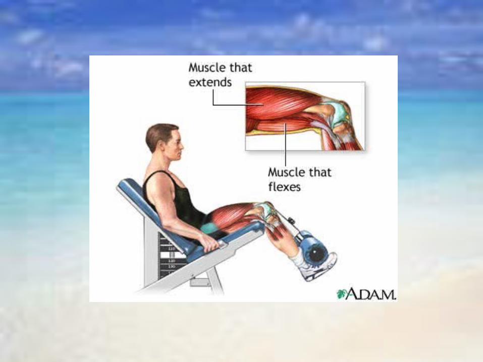
• E.g. in the thigh, as the quadriceps contracts and shortens, the hamstrings lengthens to the same extent
• The muscle that flexes the joint in one direction, is opposed by one that extends the joint in the other direction
• In producing a particular movement, a muscle performs one of 4 roles:– Agonist (prime mover)
• this is the muscle causing the major action• there are agonists for all movable joints and usually more than one
is involved in a particular joint movement– Antagonist
• this muscle must relax and lengthen to allow the agonist to contract, thus helping to control the action
• agonist works as a pair with the antagonist muscle• the 2 roles are interchangeable depending on the direction of the
movement– Synergist (or assistance)
• this muscle assists the agonist to produce a particular joint action by contracting or relaxing, thus any excess or unnecessary movement
– Stabiliser (or fixator)• these muscles act at a joint to stabilise it, giving the muscles a
fixed base• the muscle shortens very little during its contraction, causing
minimal movement• this permits the action to be carried out correctly and allows other
joints to work more effectively
Major Muscles & their main Action
• The following table summarises the action of those muscles or muscle groups which are most relevant to the body movement
• The origin and insertion have been included only for the purpose of helping you understand how a particular muscle produces a specific action
• That is, which bone or bones a muscle pulls on, over which joint it acts and the direction of the pull
Musculoskeletal system and exercise
• During exercise, components of the musculoskeletal system may be placed under stress or strain due to forces acting upon muscles, tendons, ligaments and joints.
• In order to avoid injury, there a number of exercise considerations which relate specifically to those structural and functional aspects of the system.
1. Since muscles usually work in pairs, any training of muscle groups should be given to both the agonist and the antagonist
• If one muscle group is worked excessively without working the opposing muscle group, creating muscle imbalance, the area may be predisposed to injury
• E.g. shin splints (overworking the strong calf muscles at the expense of the weaker shin muscles)
2. As postural muscles are stronger than phasic muscles, they must be relaxed before phasic muscles can be worked.
• E.g. in performing sit-ups, the strong hip flexor needs to be restricted in its involvement in the movement so that weaker abdominals have an increased involvement if abdominal strengthening is to occur.
• Thus, sit-ups should be carried out with bent knees

3. A relaxed muscle can be stretched further than a tense one
– the more relaxed the antagonist, the faster and more powerfully we can contract the agonist
– antagonist muscles are frequently torn because the contracting agonist controls a particular muscle action
4. Increasing the temperature (warming-up) of a muscle or muscle group allows it to be stretched further
• this places it at less risk from sudden strain and also allows a faster and more forceful contraction

5. Weak muscles makes joint unstable and increase the risk of injury
• the strength of muscles acting over any major joint is a vital factor in maintaining joint stability
6. Excessive extension or flexion• an extreme movement which goes
beyond a safe range of motion of a joint is potentially dangerous due to the stress and strain placed on joint structures
• The injuries associated with activity are due to either internal or external forces, or from overuse or unaccustomed use
• Some of the more common injuries are:– Muscle strain– Muscle cramp– Tendonitis– Joint sprain– Fracture
Musculoskeletal injuries
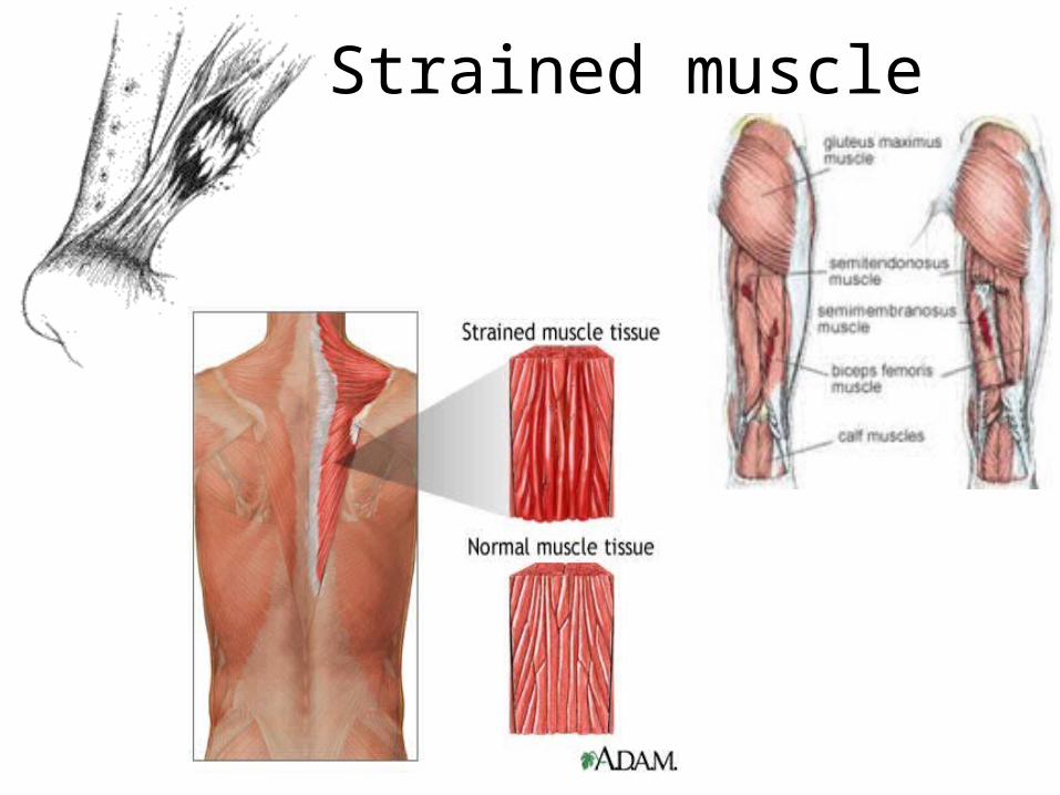
Strained muscle
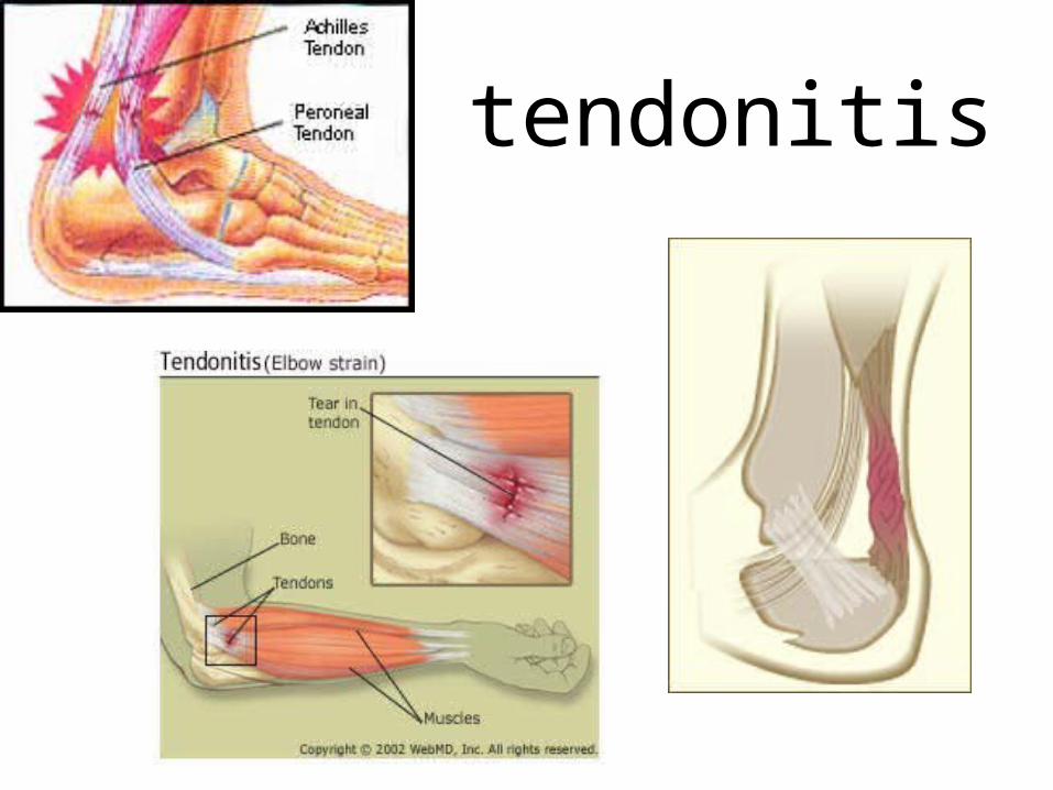
tendonitis

sprains

http://www.nucleusinc.com/medical-animations.php
Total Knee Replacement