The Skeletal System Chapter 2. Imaging Consideration Diagnostic images include soft tissue and bony...
80
The Skeletal System Chapter 2
-
Upload
edith-rodgers -
Category
Documents
-
view
216 -
download
1
Transcript of The Skeletal System Chapter 2. Imaging Consideration Diagnostic images include soft tissue and bony...

The Skeletal System
Chapter 2

Imaging Consideration
Diagnostic images include soft tissue and bony structure of interest.
Soft tissue areas often hold clues to the diagnosis. Any signs of muscle wasting, soft tissue swelling, calcifications, opaque foreign bodies, or the presence of gas may indicate disease.

MRIMagnetic Resonance Imaging
Provides soft tissue detail because of its superior contrast resolution.
Exams: staging of soft tissue tumors of the extremities, joints, bone marrow imaging… just to name a few

CTComputed Tomography
Better contrast resolution than radiography.
Exams: trauma, extend of fractures, dislocation, joint abnormalities, excellent ability to display bony margins and trabecular patterns.

NMNuclear Medicine
Demonstrates metabolic function
Exam: bone scanAllows the metabolic function of
the entire skeletal system to be evaluated at one time. Demonstrates the metabolic processes of the bone caused by disease processes.

DEXABone Mineral Densitometry
Double-energy x-ray absorbtiometry
Evaluate the bone density by evaluating the bone mass of the distal radius, femoral neck, and lumbar spine

Congenital and Hereditary Diseases

Osteogenesis Imperfecta

Osteogenesis Imperfecta

Achondroplasia
• Most common inherited disorder affecting the skeletal system
• Results in deformity and dwarfism
• Cartilage in the epiphyses does not convert to bone normally
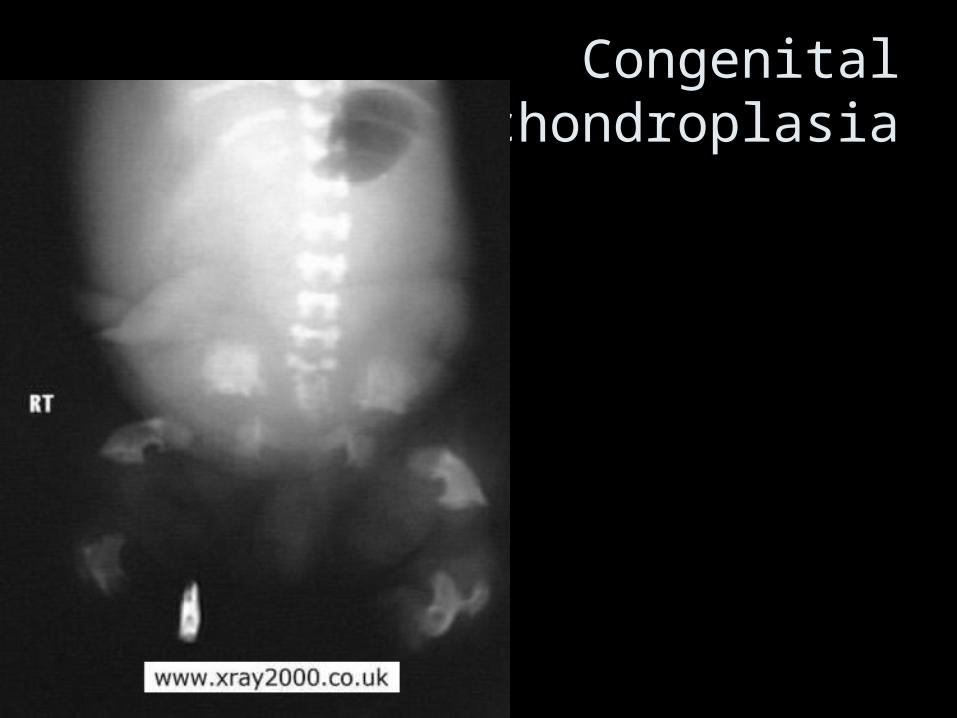
Congenital Achondroplasia

Achondroplasia

Osteopetrosis
• Increased exposure factors are required
• Some cases, adequate penetration
may never be achieved

Infant Osteopetrosis

Malformations

Syndactyly

Polydactyly

Clubfoot (talipes)

Congenital hip dislocation

Congenital scoliosis

Scoliosis & Rotoscoliosis

bilateral lumbar ribs

spina bifida occulta


fetus with anencephaly

Inflammatory Disease

Chronic osteomyelitis

rheumatoid arthritis“swan sign”

rheumatoid arthritis

R.A.

Arthritis –joint inflammation
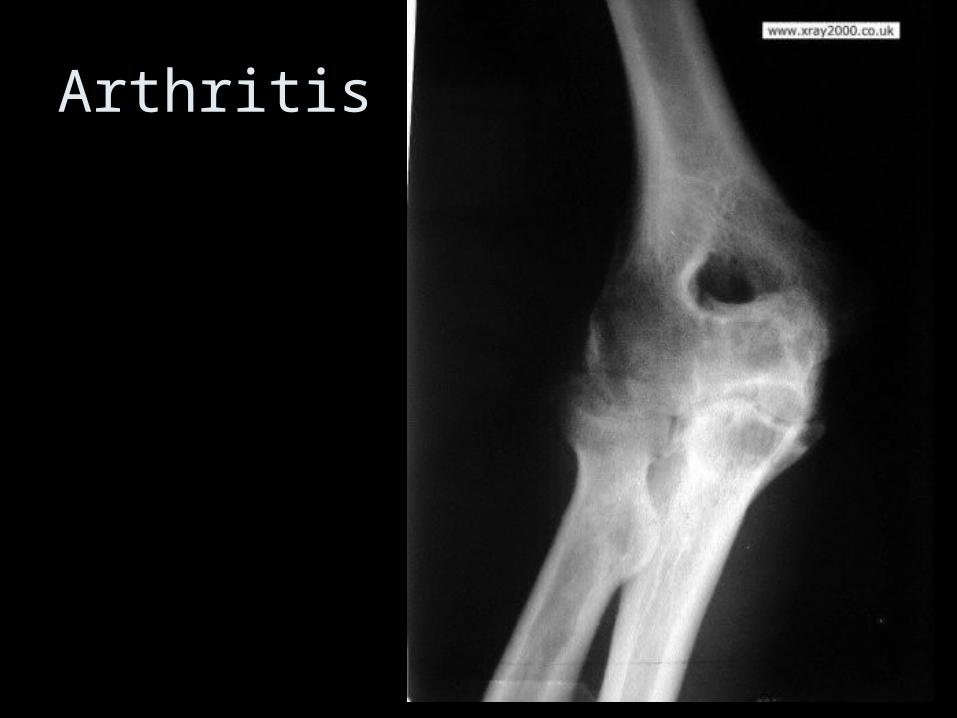
Arthritis

Cystic Arthritis

Arthritic Dislocation - Patella
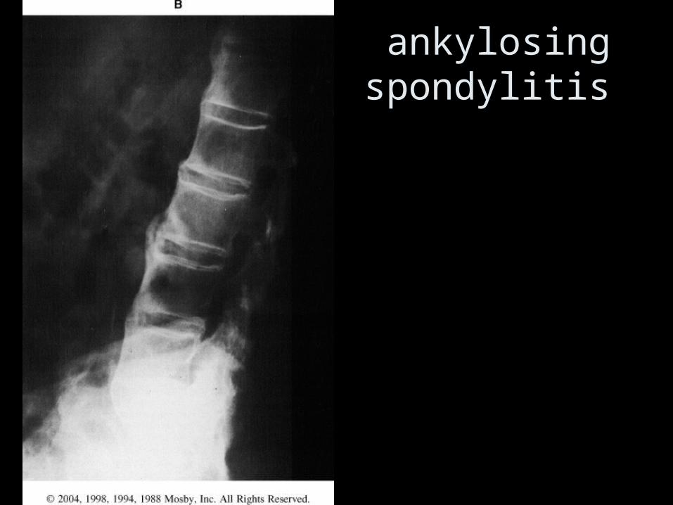
ankylosing spondylitis

Ankylosing Spondylitis
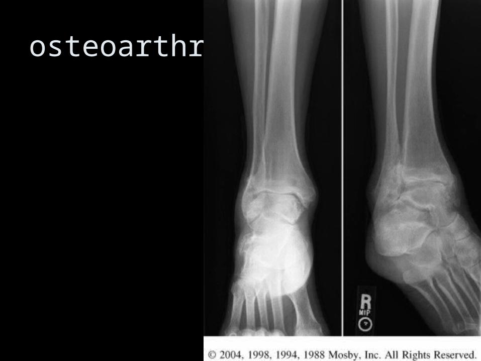
osteoarthritis
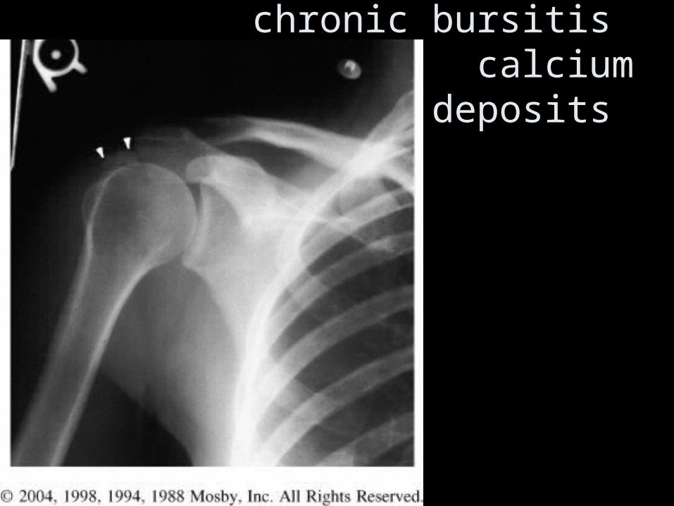
chronic bursitis calcium
deposits

gout

gout

Metabolic Disease

osteopenia steroid use

osteopenia
• A radiographically visible decrease in bone density
• Bone loss must be at least 30% to radiographically demonstrate

Ostopenia

Rickets or Osteomalacia

Paget’s disease
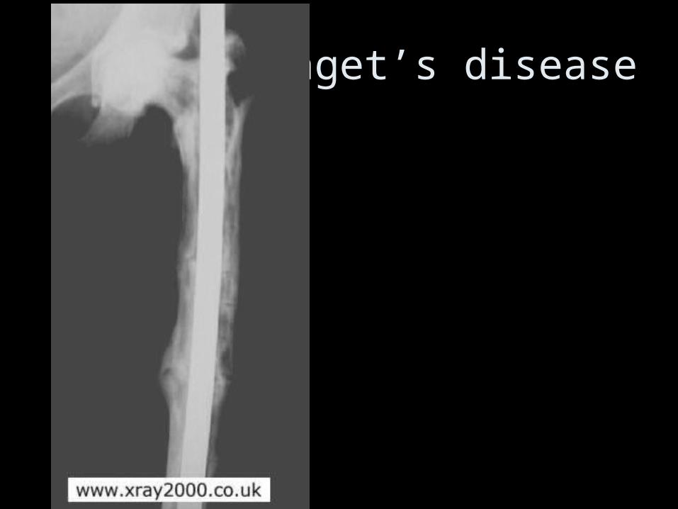
Paget’s disease

Paget’s disease

Arachnodactilia from Acromegaly

Marfans Syndrome

Bone Spur

spondylolisthesis

spondylolisthesis

spondylolisthesis

Neoplastic Disease

Bone Cyst

Bone Cyst

simple bone cyst
FX ?

Ostocondroma

Ostocondroma
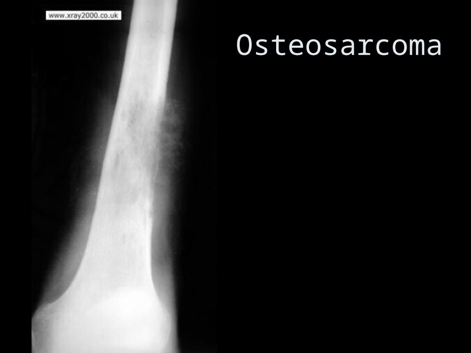
Osteosarcoma

metastatic disease

Dislocation

Dislocation

Dislocation of the Patella

osteosarcoma

osteosarcoma
Trauma & other stuff

Dislocation

Dislocation

Dislocation

Fat Pad Sign

Fracture?

Diabetic Changes

Avascular Necrosis

Avascular Necrosis

Free air from puncture wound

Calcified Bakers Cyst
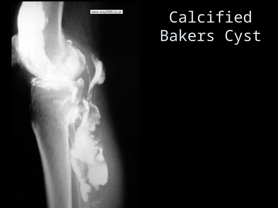
Calcified Bakers Cyst

Syphilis