The Skeletal System: Appendicular Division 218/07... · The appendicular skeleton is involved in...
53
C h a p t e r 7 The Skeletal System: Appendicular Division PowerPoint ® Lecture Slides prepared by Jason LaPres North Harris College Houston, Texas Copyright © 2009 Pearson Education, Inc., publishing as Pearson Benjamin Cummings
-
Upload
nguyenxuyen -
Category
Documents
-
view
240 -
download
0
Transcript of The Skeletal System: Appendicular Division 218/07... · The appendicular skeleton is involved in...

C h a p t e r
7
The Skeletal System:
Appendicular Division
PowerPoint® Lecture Slides
prepared by Jason LaPres
North Harris College
Houston, Texas
Copyright © 2009 Pearson Education, Inc.,
publishing as Pearson Benjamin Cummings

Introduction
The appendicular skeleton is involved in
changing your position in the external
environment.
Standing
Walking
Sitting
Dressing
Driving a car
Copyright © 2009 Pearson Education, Inc., publishing as Pearson Benjamin Cummings

Introduction
Figure 7.1 The Appendicular SkeletonCopyright © 2009 Pearson Education, Inc., publishing as Pearson Benjamin Cummings

M
Copyright © 2009 Pearson Education, Inc., publishing as Pearson Benjamin Cummings
The Pectoral Girdle and Upper Limb
The Pectoral Girdle
The Pectoral Girdle
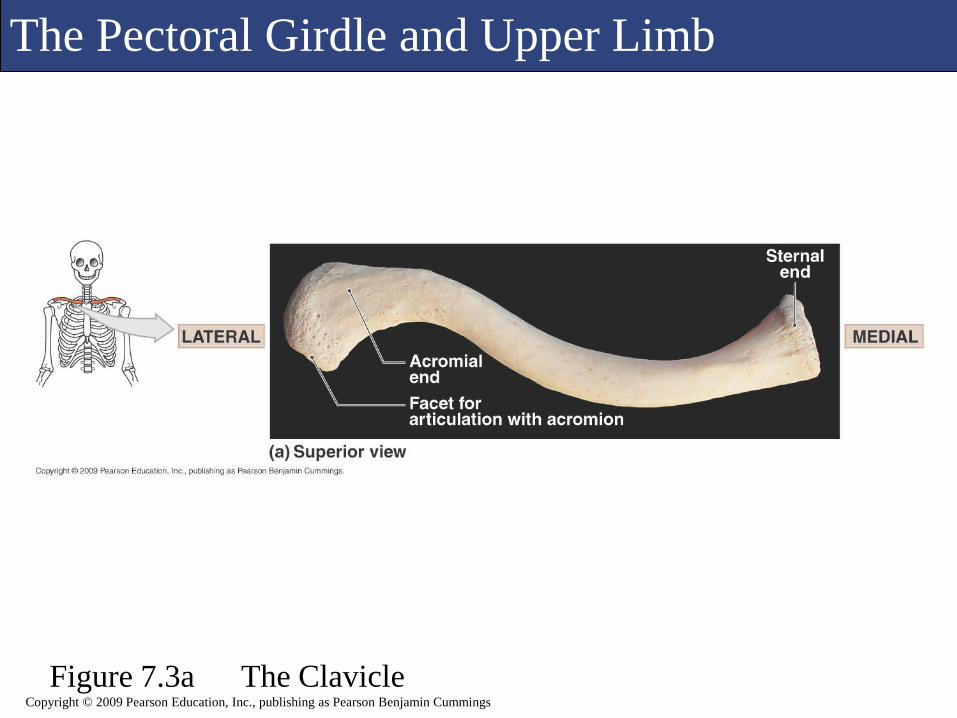
Includes the S-shaped clavicle (collarbone) and the
flattened scapula (shoulder blade)
The clavicle articulates with the manubrium of
the sternum and is the only direct connection
between the axial skeleton and the pectoral girdle.
The scapula is attached to the clavicle anteriorly
but has no connection to the actual axial skeleton;
instead skeletal muscles and ligaments support it.
The Pectoral Girdle and Upper Limb
Figure 7.3a The ClavicleCopyright © 2009 Pearson Education, Inc., publishing as Pearson Benjamin Cummings

The Pectoral Girdle and Upper Limb
Figure 7.3b The Clavicle
Copyright © 2009 Pearson Education, Inc., publishing as Pearson Benjamin Cummings

The Pectoral Girdle and Upper Limb
Figure 7.4 Mobility of the Pectoral GirdleCopyright © 2009 Pearson Education, Inc., publishing as Pearson Benjamin Cummings
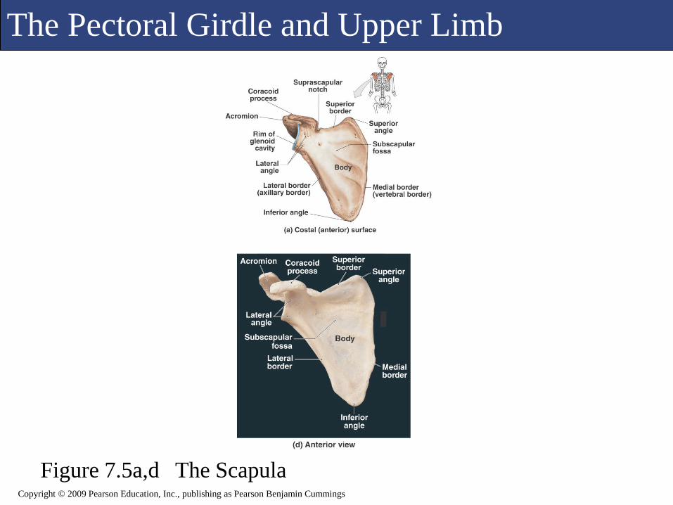
The Pectoral Girdle and Upper Limb
Figure 7.5a,d The ScapulaCopyright © 2009 Pearson Education, Inc., publishing as Pearson Benjamin Cummings

The Pectoral Girdle and Upper Limb
Figure 7.5b,e The ScapulaCopyright © 2009 Pearson Education, Inc., publishing as Pearson Benjamin Cummings

The Pectoral Girdle and Upper Limb
Figure 7.5c,f The ScapulaCopyright © 2009 Pearson Education, Inc., publishing as Pearson Benjamin Cummings

M
Copyright © 2009 Pearson Education, Inc., publishing as Pearson Benjamin Cummings
The Pectoral Girdle and Upper Limb
The Upper Limb
The upper limb consists of the
Brachium (humerus)
Antebrachium (ulna and radius)
Wrist (carpals)
Hand (metacarpals and phalanges)

The Pectoral Girdle and Upper Limb
Figure 7.6a The Humerus
Copyright © 2009 Pearson Education, Inc., publishing as Pearson Benjamin Cummings

The Pectoral Girdle and Upper Limb
Figure 7.6b,c The Humerus
Copyright © 2009 Pearson Education, Inc., publishing as Pearson Benjamin Cummings
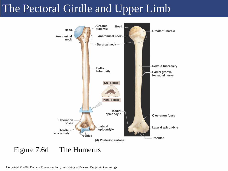
The Pectoral Girdle and Upper Limb
Figure 7.6d The Humerus
Copyright © 2009 Pearson Education, Inc., publishing as Pearson Benjamin Cummings

The Pectoral Girdle and Upper Limb
Figure 7.7a The Radius and Ulna
Copyright © 2009 Pearson Education, Inc., publishing as Pearson Benjamin Cummings

The Pectoral Girdle and Upper Limb
Figure 7.7b,c The Radius and Ulna
Copyright © 2009 Pearson Education, Inc., publishing as Pearson Benjamin Cummings

The Pectoral Girdle and Upper Limb
Figure 7.7d The Radius and Ulna
Copyright © 2009 Pearson Education, Inc., publishing as Pearson Benjamin Cummings

The Pectoral Girdle and Upper Limb
Figure 7.7e,f The Radius and Ulna
Copyright © 2009 Pearson Education, Inc., publishing as Pearson Benjamin Cummings

M
Copyright © 2009 Pearson Education, Inc., publishing as Pearson Benjamin Cummings
The Pectoral Girdle and Upper Limb
The Wrist and Hand
The Wrist and Hand
The carpal bones are the 8 bones of the wrist.
The metacarpal bones (5) articulate with the distal carpal
bones and make up the palm of the hand.
The 14 phalanges of the hand make up the finger bones.

The Pectoral Girdle and Upper Limb
Figure 7.8a The Bones of the Wrist and HandCopyright © 2009 Pearson Education, Inc., publishing as Pearson Benjamin Cummings

The Pectoral Girdle and Upper Limb
Figure 7.8b The Bones of the Wrist and HandCopyright © 2009 Pearson Education, Inc., publishing as Pearson Benjamin Cummings
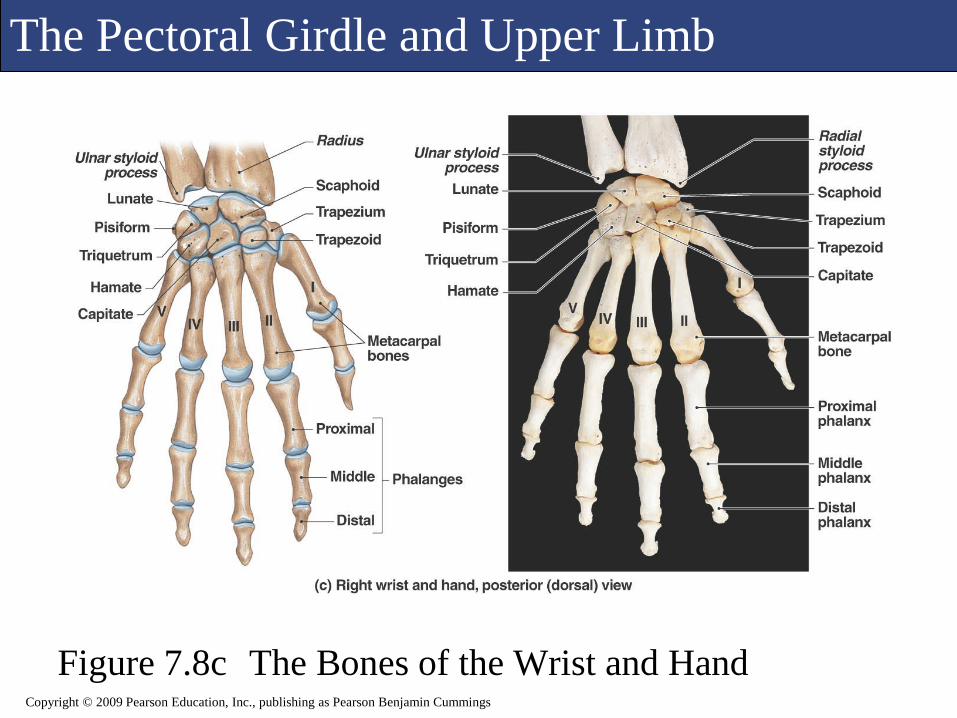
The Pectoral Girdle and Upper Limb
Figure 7.8c The Bones of the Wrist and HandCopyright © 2009 Pearson Education, Inc., publishing as Pearson Benjamin Cummings

M
Copyright © 2009 Pearson Education, Inc., publishing as Pearson Benjamin Cummings
The Pelvic Girdle and Lower Limb
The Pelvic Girdle and Lower Limb
The Pelvic Girdle
Supports and protects the lower viscera and
developing fetus in females
The pelvic girdle consists of two ossa coxae
bones.
The bones of the pelvic girdle and lower limb are
much more massive than their homologues of the
upper limb.

The Pelvic Girdle and Lower Limb
Figure 7.9 The Pelvic Girdle and Lower LimbCopyright © 2009 Pearson Education, Inc., publishing as Pearson Benjamin Cummings
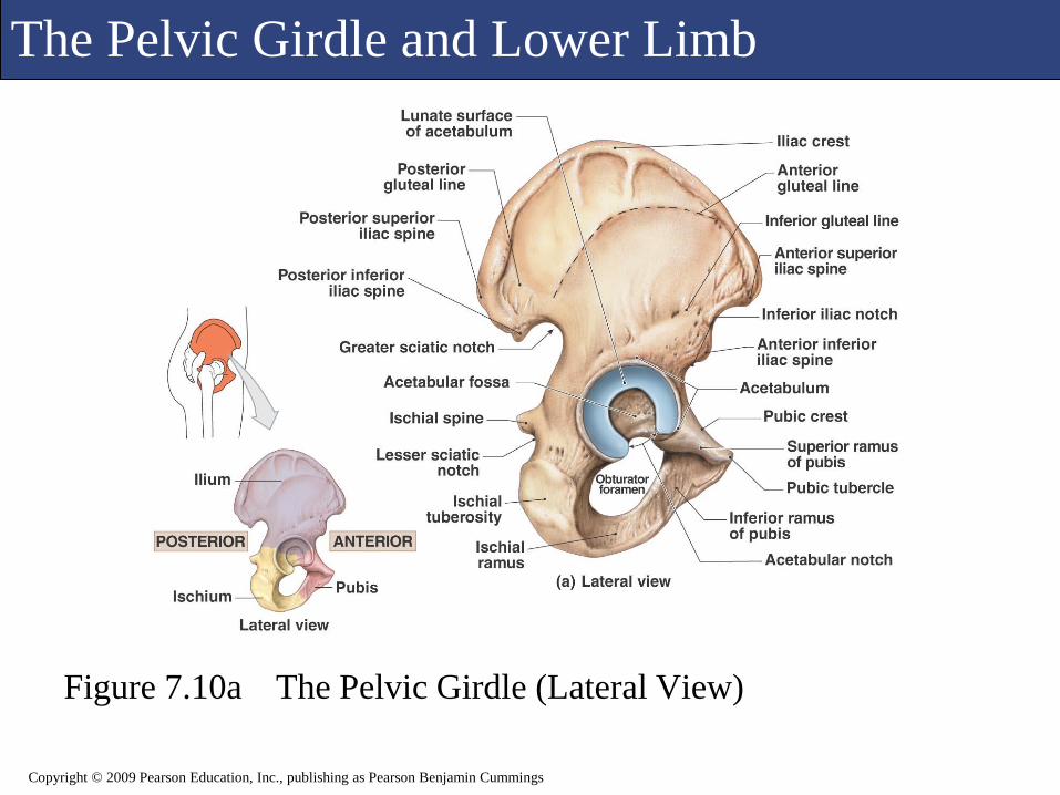
The Pelvic Girdle and Lower Limb
Figure 7.10a The Pelvic Girdle (Lateral View)
Copyright © 2009 Pearson Education, Inc., publishing as Pearson Benjamin Cummings

The Pelvic Girdle and Lower Limb
Figure 7.10a The Pelvic Girdle (Lateral View)
Copyright © 2009 Pearson Education, Inc., publishing as Pearson Benjamin Cummings

The Pelvic Girdle and Lower Limb
Figure 7.10b The Pelvic Girdle (Medial View)
Copyright © 2009 Pearson Education, Inc., publishing as Pearson Benjamin Cummings

The Pelvic Girdle and Lower Limb
Figure 7.10b The Pelvic Girdle (Medial View)
Copyright © 2009 Pearson Education, Inc., publishing as Pearson Benjamin Cummings

The Pelvic Girdle and Lower Limb
Figure 7.11a The Pelvis (Anterior View)
Copyright © 2009 Pearson Education, Inc., publishing as Pearson Benjamin Cummings

The Pelvic Girdle and Lower Limb
Figure 7.11a The Pelvis (Anterior View)
Copyright © 2009 Pearson Education, Inc., publishing as Pearson Benjamin Cummings
The Pelvic Girdle and Lower Limb
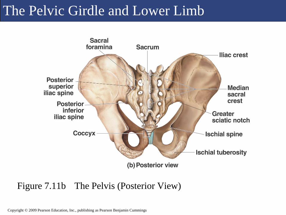
Figure 7.11b The Pelvis (Posterior View)
Copyright © 2009 Pearson Education, Inc., publishing as Pearson Benjamin Cummings

The Pelvic Girdle and Lower Limb
Figure 7.11b The Pelvis (Posterior View)
Copyright © 2009 Pearson Education, Inc., publishing as Pearson Benjamin Cummings
The Pelvic Girdle and Lower Limb
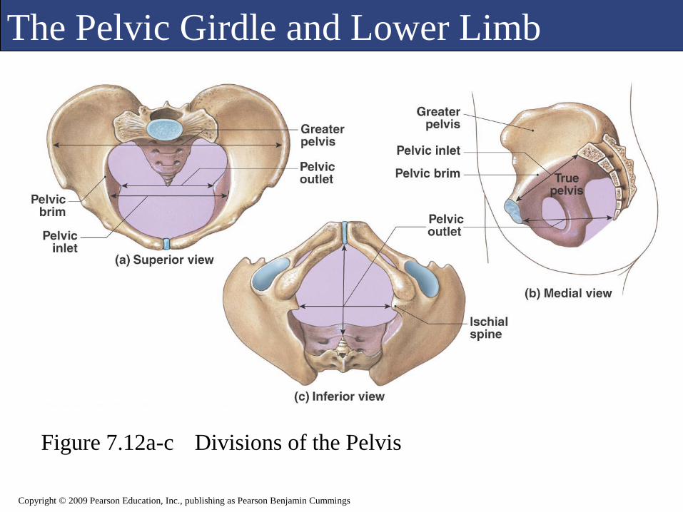
Figure 7.12a-c Divisions of the Pelvis
Copyright © 2009 Pearson Education, Inc., publishing as Pearson Benjamin Cummings

The Pelvic Girdle and Lower Limb
Figure 7.12d Divisions of the Pelvis
Copyright © 2009 Pearson Education, Inc., publishing as Pearson Benjamin Cummings

M
Copyright © 2009 Pearson Education, Inc., publishing as Pearson Benjamin Cummings
The Pelvic Girdle and Lower Limb
The Pelvis
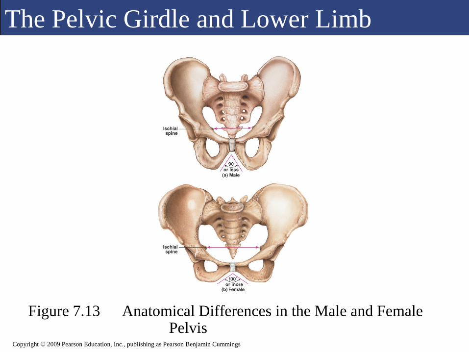
The male and female pelvis contains numerous differences.
Generally the male pelvis is heavier with more prominent markings due to the larger muscles attached to it.
Differences are noted as how the female compares to the male
Enlarged pelvic outlet, due to wider ischial spines
Less curvature of the sacrum and coccyx
Wider, more circular pelvic inlet
Broader, lower pelvis
Widely fanning ilia
Pubic angle greater than 100 degrees
The Pelvic Girdle and Lower Limb
Figure 7.13 Anatomical Differences in the Male and Female Pelvis
Copyright © 2009 Pearson Education, Inc., publishing as Pearson Benjamin Cummings

M
Copyright © 2009 Pearson Education, Inc., publishing as Pearson Benjamin Cummings
The Pelvic Girdle and Lower Limb
The Lower Limb
Responsible for transferring the body weight to the ground
Consists of the following structures:
The femur (thigh)
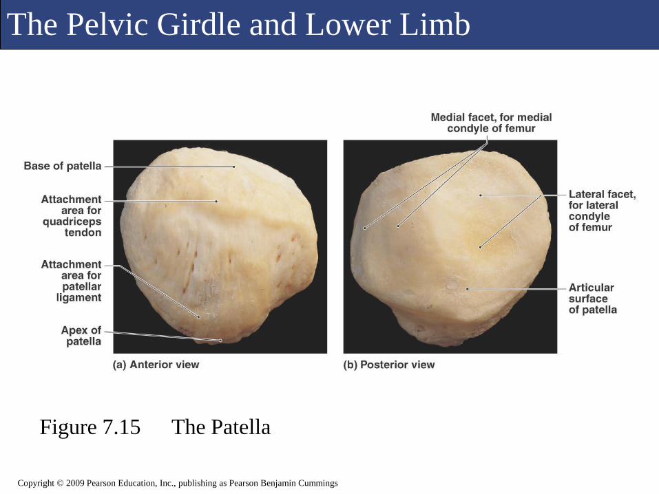
The patella (kneecap)
The tibia (leg)
The fibula (leg)
Tarsal bones of the ankle
Metatarsal bones and phalanges of the foot
The Pelvic Girdle and Lower Limb
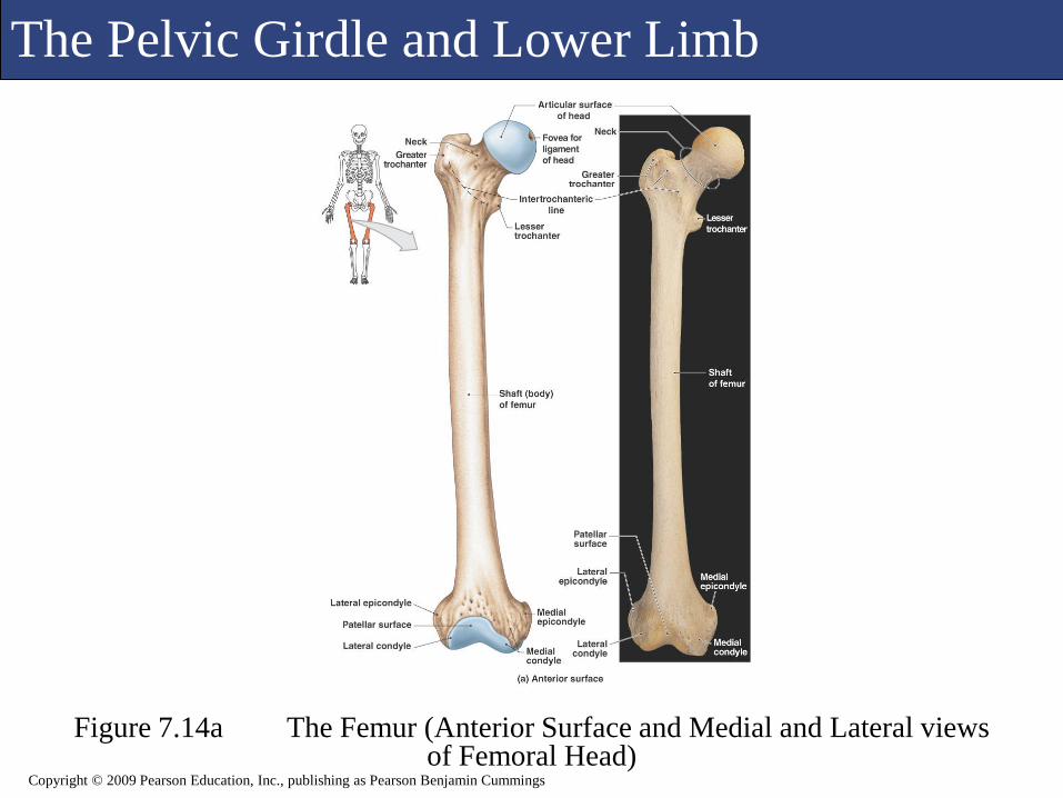
Figure 7.14a The Femur (Anterior Surface and Medial and Lateral views of Femoral Head)
Copyright © 2009 Pearson Education, Inc., publishing as Pearson Benjamin Cummings
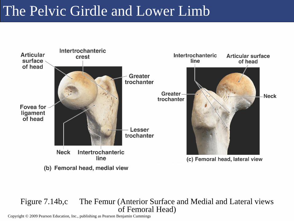
The Pelvic Girdle and Lower Limb
Figure 7.14b,c The Femur (Anterior Surface and Medial and Lateral views of Femoral Head)
Copyright © 2009 Pearson Education, Inc., publishing as Pearson Benjamin Cummings

The Pelvic Girdle and Lower Limb
Figure 7.14d The Femur Copyright © 2009 Pearson Education, Inc., publishing as Pearson Benjamin Cummings

The Pelvic Girdle and Lower Limb
Figure 7.14e,f The Femur Copyright © 2009 Pearson Education, Inc., publishing as Pearson Benjamin Cummings
The Pelvic Girdle and Lower Limb
Figure 7.15 The Patella
Copyright © 2009 Pearson Education, Inc., publishing as Pearson Benjamin Cummings

The Pelvic Girdle and Lower Limb
Figure 7.16a The Tibia and FibulaCopyright © 2009 Pearson Education, Inc., publishing as Pearson Benjamin Cummings

The Pelvic Girdle and Lower Limb
Figure 7.16b,c The Tibia and Fibula
Copyright © 2009 Pearson Education, Inc., publishing as Pearson Benjamin Cummings

The Pelvic Girdle and Lower Limb
Figure 7.16d,e The Tibia and FibulaCopyright © 2009 Pearson Education, Inc., publishing as Pearson Benjamin Cummings

M
Copyright © 2009 Pearson Education, Inc., publishing as Pearson Benjamin Cummings
The Pelvic Girdle and Lower Limb
The Ankle and Foot
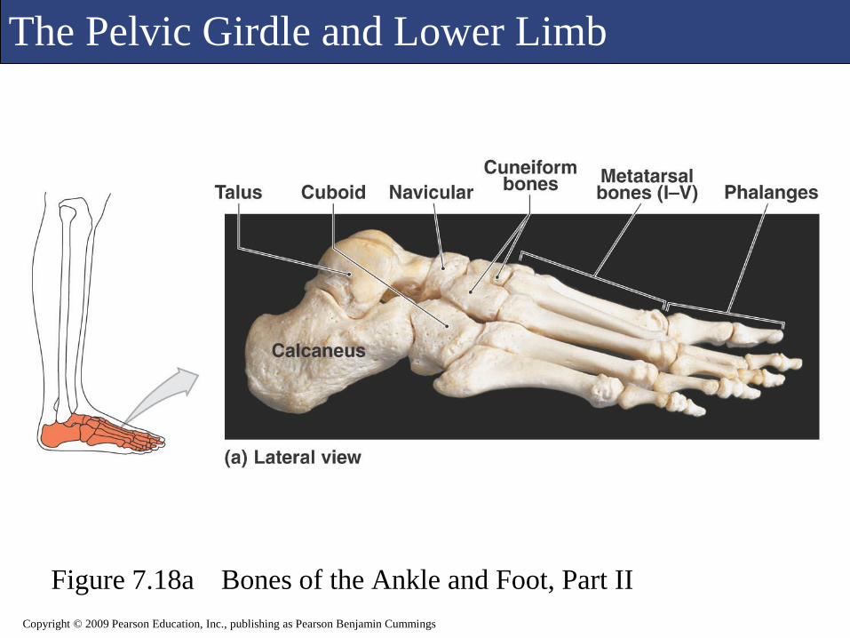
The Ankle and Foot
There are 7 tarsal bones that make up the ankle.
The metatarsal bones (5) articulate with the distal
tarsal bones and make up the arches of the foot.
The 14 phalanges of the foot make up the toe
bones.

The Pelvic Girdle and Lower Limb
Figure 7.17a Bones of the Ankle and Foot, Part I
Copyright © 2009 Pearson Education, Inc., publishing as Pearson Benjamin Cummings

The Pelvic Girdle and Lower Limb
Figure 7.17b Bones of the Ankle and Foot, Part I
Copyright © 2009 Pearson Education, Inc., publishing as Pearson Benjamin Cummings
The Pelvic Girdle and Lower Limb
Figure 7.18a Bones of the Ankle and Foot, Part II
Copyright © 2009 Pearson Education, Inc., publishing as Pearson Benjamin Cummings

The Pelvic Girdle and Lower Limb
Figure 7.18b Bones of the Ankle and Foot, Part II
Copyright © 2009 Pearson Education, Inc., publishing as Pearson Benjamin Cummings

M
Copyright © 2009 Pearson Education, Inc., publishing as Pearson Benjamin Cummings
Individual Variation in the Skeletal System
Your skeleton can reveal information about
you, such as your race, medical history,
gender, body size, muscle mass, and age.
As you age, a number of changes and events
take place in the skeletal system

Individual Variation in the Skeletal System
Copyright © 2009 Pearson Education, Inc., publishing as Pearson Benjamin Cummings

Individual Variation in the Skeletal System
Copyright © 2009 Pearson Education, Inc., publishing as Pearson Benjamin Cummings