
THE SIGNIFICANCE OF LEFT AXIS DEVIATION IN HEART DISEASE ...
10
THE SIGNIFICANCE OF LEFT AXIS DEVIATION IN HEART DISEASE OF THE AFRICAN BY L. SCHAMROTH AND D. BLUMSOHN From the Baragwaniath Hospital and University of the Witwatersrand, Johannesburg Received December 5, 1960 The pattern of heart disease in the African (Negro) differs from that in the White (Caucasian) in two notable respects-the virtual absence of myocardial infarction (0.400, Schwartz et al., 1958; 1.4%, Laurie et al., 1960) and the presence of a high proportion of cases of obscure etiology (37.50, Schwartz et al., 1958; 31.5%, Laurie et al., 1960). The latter have been referred to by various authors as idiopathic heart disease, nutritional heart disease (Gillanders, 1951), cardio- vascular collagenosis with parietal endocardial thrombosis (Becker et al., 1953) and cryptogenic heart disease (Schwartz et al., 1958). Cryptogenic cardiomyopathy would appear to be a better term for this group of cases. They present with congestive cardiac failure, moderate to gross cedema, generalized cardiomegaly, gallop rhythm, occasional systolic murmurs, a hypokinetic circulation, and an inert heart on fluoroscopy-a presentation not unlike myxcedematous heart disease or pericardial effusion. There is no evidence of ischmmic, hypertensive, rheumatic, or pul- monary heart disease. Pathological examination reveals a basic pattern of dilatation of all cham- bers of the heart, moderate myocardial hypertrophy, and a patchy interstitial or endocardial fibrosis which is seldom marked. Antemortem intramural thrombi are commonly found in all chambers. Widespread endomyocardial fibrosis found in other parts of Africa (Bedford and Konstam, 1946; Ball et al., 1954; Davies and Ball, 1955) is extremely rare in South Africa. Some of these cases have been observed in this hospital (Seftel and Susser, 1960) during late pregnancy, the puerperium, and the lactational period-post-partum heart disease. This pattern is similar to that described as post-partal heart disease (Malvin, 1947), post-partum myocardosis (Vilter and McKee, 1953), and idiopathic myocardial degeneration of the puerperium (Gouley et al., 1937), by observers in America. Recent work (Grant, 1956; Davies and Evans, 1960) has indicated that significant left axis deviation is not due to left ventricular hypertrophy per se, but to a conduction defect involving the anterior superior division of the left bundle branch. This conduction defect is most frequently the result of myocardial infarction or diseases associated with fibrosis of the left ventricle. The left bundle branch divides into a number of small branches almost immediately after leaving the bundle of His. These branches divide into two major sweeps or radiations that innervate the anterior superior aspect-the anterior superior division-and the posterior inferior aspect of the left ventricle -the posterior inferior division (Fig. 1). Excitation and conduction normally progress simul- taneously through the fibres of both divisions. If, however, interruption of conduction occurs in the fibres of the anterior superior division, activation of the ventricular walls will occur chiefly through the posterior inferior division without necessarily causing prolongation of the QRS complex. As the principal excitation via the posterior inferior division is terminally upwards and to the left (arrow 2, Fig. 1), the dominant vector will be upward and to the left resulting in left axis deviation. The possibility that the patchy fibrosis found in the cases of cryptogenic cardiomyopathy of the 405 on December 14, 2021 by guest. Protected by copyright. http://heart.bmj.com/ Br Heart J: first published as 10.1136/hrt.23.4.405 on 1 July 1961. Downloaded from
Transcript of THE SIGNIFICANCE OF LEFT AXIS DEVIATION IN HEART DISEASE ...

THE SIGNIFICANCE OF LEFT AXIS DEVIATION IN HEARTDISEASE OF THE AFRICAN
BY
L. SCHAMROTH AND D. BLUMSOHN
From the Baragwaniath Hospital and University of the Witwatersrand, Johannesburg
Received December 5, 1960
The pattern of heart disease in the African (Negro) differs from that in the White (Caucasian)in two notable respects-the virtual absence of myocardial infarction (0.400, Schwartz et al., 1958;1.4%, Laurie et al., 1960) and the presence of a high proportion of cases of obscure etiology(37.50, Schwartz et al., 1958; 31.5%, Laurie et al., 1960). The latter have been referred to byvarious authors as idiopathic heart disease, nutritional heart disease (Gillanders, 1951), cardio-vascular collagenosis with parietal endocardial thrombosis (Becker et al., 1953) and cryptogenicheart disease (Schwartz et al., 1958). Cryptogenic cardiomyopathy would appear to be a betterterm for this group of cases. They present with congestive cardiac failure, moderate to grosscedema, generalized cardiomegaly, gallop rhythm, occasional systolic murmurs, a hypokineticcirculation, and an inert heart on fluoroscopy-a presentation not unlike myxcedematous heartdisease or pericardial effusion. There is no evidence of ischmmic, hypertensive, rheumatic, or pul-monary heart disease. Pathological examination reveals a basic pattern of dilatation of all cham-bers of the heart, moderate myocardial hypertrophy, and a patchy interstitial or endocardial fibrosiswhich is seldom marked. Antemortem intramural thrombi are commonly found in all chambers.Widespread endomyocardial fibrosis found in other parts of Africa (Bedford and Konstam, 1946;Ball et al., 1954; Davies and Ball, 1955) is extremely rare in South Africa. Some of these cases havebeen observed in this hospital (Seftel and Susser, 1960) during late pregnancy, the puerperium, andthe lactational period-post-partum heart disease. This pattern is similar to that described aspost-partal heart disease (Malvin, 1947), post-partum myocardosis (Vilter and McKee, 1953), andidiopathic myocardial degeneration of the puerperium (Gouley et al., 1937), by observers inAmerica.
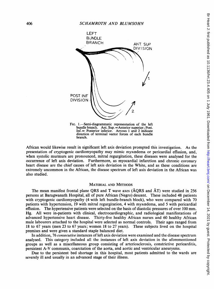
Recent work (Grant, 1956; Davies and Evans, 1960) has indicated that significant left axisdeviation is not due to left ventricular hypertrophy per se, but to a conduction defect involving theanterior superior division of the left bundle branch. This conduction defect is most frequently theresult of myocardial infarction or diseases associated with fibrosis of the left ventricle. The leftbundle branch divides into a number of small branches almost immediately after leaving the bundleof His. These branches divide into two major sweeps or radiations that innervate the anteriorsuperior aspect-the anterior superior division-and the posterior inferior aspect of the left ventricle-the posterior inferior division (Fig. 1). Excitation and conduction normally progress simul-taneously through the fibres of both divisions. If, however, interruption of conduction occurs in thefibres of the anterior superior division, activation of the ventricular walls will occur chiefly throughthe posterior inferior division without necessarily causing prolongation of the QRS complex. Asthe principal excitation via the posterior inferior division is terminally upwards and to the left(arrow 2, Fig. 1), the dominant vector will be upward and to the left resulting in left axis deviation.
The possibility that the patchy fibrosis found in the cases of cryptogenic cardiomyopathy of the405
on Decem
ber 14, 2021 by guest. Protected by copyright.
http://heart.bmj.com
/B
r Heart J: first published as 10.1136/hrt.23.4.405 on 1 July 1961. D
ownloaded from
SCHAMROTH AND BLUMSOHN
LEFTBUNDLEBRANCH ANT SUP
DIVISION \\ / D
FIG. 1.-Semi-diagrammatic representation of the leftbundle branch. Ant. Sup. =Anterior superior; Post.Inf.= Posterior inferior. Arrows 1 and 2 indicatedirection of terminal vector forces of each bundlebranch.
African would likewise result in significant left axis deviation prompted this investigation. As thepresentation of cryptogenic cardiomyopathy may mimic myxcedema or pericardial effusion, and,when systolic murmurs are pronounced, mitral regurgitation, these diseases were analysed for theoccurrence of left axis deviation. Furthermore, as myocardial infarction and chronic coronaryheart disease are the chief causes of left axis deviation in the White, and as these conditions areextremely uncommon in the African, the disease spectrum of left axis deviation in the African wasalso studied.
MATERIAL AND METHODSThe mean manifest frontal plane QRS and T wave axes (AQRS and AT) were studied in 256
persons at Baragwanath Hospital, all of pure African (Negro) descent. These included 48 patientswith cryptogenic cardiomyopathy (4 with left bundle-branch block), who were compared with 70patients with hypertension, 19 with mitral regurgitation, 4 with myxcedema, and 5 with pericardialeffusion. The hypertensive patients were selected on the basis of diastolic pressures of over 100 mm.Hg. All were in-patients with clinical, electrocardiographic, and radiological manifestations ofadvanced hypertensive heart disease. Thirty-five healthy African nurses and 60 healthy Africanmale labourers attached to the hospital were selected as normal controls. Their ages ranged from18 to 67 years (men 23 to 67 years; women 18 to 27 years). These subjects lived on the hospitalpremises and were given a standard staple balanced diet.
In addition, 76 consecutive instances of left axis deviation were examined and the disease spectrumanalysed. This category included all the instances of left axis deviation in the aforementionedgroups as well as a miscellaneous group consisting of arteriosclerosis, constrictive pericarditis,persistent A-V communis, coarctation of the aorta, and aortic and ventricular aneurysms.
Due to the persistent bed shortage in this hospital, most patients admitted to the wards areseverely ill and usually in an advanced stage of their illness.
406
on Decem
ber 14, 2021 by guest. Protected by copyright.
http://heart.bmj.com
/B
r Heart J: first published as 10.1136/hrt.23.4.405 on 1 July 1961. D
ownloaded from

LEFT AXIS DEVIATION 407
All electrocardiograms were recorded by a direct writing electrocardiograph with the subjects inthe supine position.
Axes were plotted on Bayley's triaxial reference system with the addition of the unipolar limblead axes forming a hexaxial reference system (Fig. 2 and 3).
F-90
L - .t15.I0.
FIG. 2.- Hexaxial reference system, showing normal FIG. 3.-Hexaxial reference system, showing range ofrange of AQRS. slight left axis deviation (light shading) and significant
left axis deviation (dark shading).
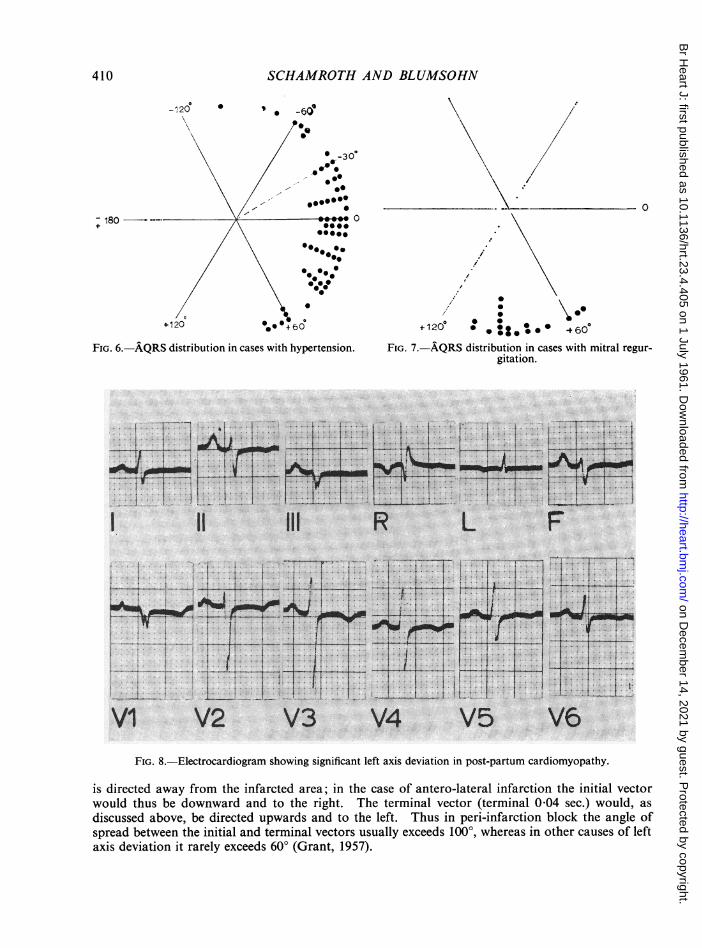
The normal range of AQRS is usually between 0 and +90°. Axes between 0 and-~29° mayoccasionally be found in the normal subject, especially during pregnancy or in those withmuch obesity or a stocky build. It may also be associated with severe ascites or abdominaldistension. However, axes in this zone should be viewed with suspicion as they are frequently dueto organic heart disease. Grant (1956) considers an AQRS more leftward than-1l5° as significant.The criteria for significant left axis deviation in this paper are stringently defined as a leftward shiftof -30° or greater. This implies dominant S waves in leads II and III and usually dominant Rwaves in standard lead I (Fig. 8). Axes in the zone of 0 to -29° are referred to as slight left axisdeviation (Fig. 3). An AQRS-T angle spread greater than 600 is regarded as abnormal.
RESULTS
Normal Subjects. Only one of the 95 normal controls had left axis deviation. This was a managed 42 in whom AQRS was-~30°. The T wave axis in this subject was normal and there were noother electrocardiographic abnormalities. A&QRS in the remaining subjects ranged from 0 to+90° with a mean of +470 (Fig. 4). Age and sex were without influence on the distribution ofAQRS. (It should be noted that the youngest subject was 18 years old.) Four men had a QRS-Tspread of 600 and the subject with left axis deviation referred to above had a QRS-T spread of 75°.The remainder had QRS-T angles of less than 600. The electrocardiogram in one woman showedthe Wenckebach phenomenon. There were no other cardiographic abnormalities in our cases.
These results are similar to those reported in the White (Caucasian) ethnic group. Thus Daviesand Evans (1960) found that 3 out of 200 normal subjects had left axis deviation of AQRS, all threecases being over 40 years of age. Of 558 cases studied by Zao et at. (1958) AQRS was locatedbetween 0 and +900 in all except one in whom it was-~15°. The QRS-T angles ranged from 0 to 45°.
on Decem
ber 14, 2021 by guest. Protected by copyright.
http://heart.bmj.com
/B
r Heart J: first published as 10.1136/hrt.23.4.405 on 1 July 1961. D
ownloaded from

SCHAMROTH AND BLUMSOHN
Cryptogenic Cardiomyopathy. (a) Cases without QRS Prolongation. Of the 44 patients, significant leftaxis deviation was found in 20 (46%). Slight left axis deviation was present in 10 (23%). Normal axeswere found in 11 (25%), and the remaining 3, all of whom had gross functional tricuspid regurgitation,showed right axis deviation (Fig. 5).
The electrocardiograms also showed other abnormalities. In 31 cases the T waves were inverted overV5 and V6. In 26 cases the QRS-T angle exceeded 600. In addition, there were instances of uni- andmultifocal ventricular extrasystoles, first degree A-V block, and atrial fibrillation. There were no instancesof U wave inversion, sharp-angled ST-T junctions, or plane depression of the S-T segments. The T waveswere not pointed or symmetrical.
(b) Cases with QRS Prolongation. There were 4 cases with left bundle-branch block. Two of these hadterminal vectors showing significant left axis deviation (-750 and - 600) (Fig. 9). Moreover, the terminalvectors were more leftward than the initial vectors indicating that left axis deviation was present before thedevelopment of left bundle-branch block (Grant, 1957). In both these cases the angle between initial andterminal vectors did not exceed 600. In the third case both initial and terminal vectors were normallydirected.
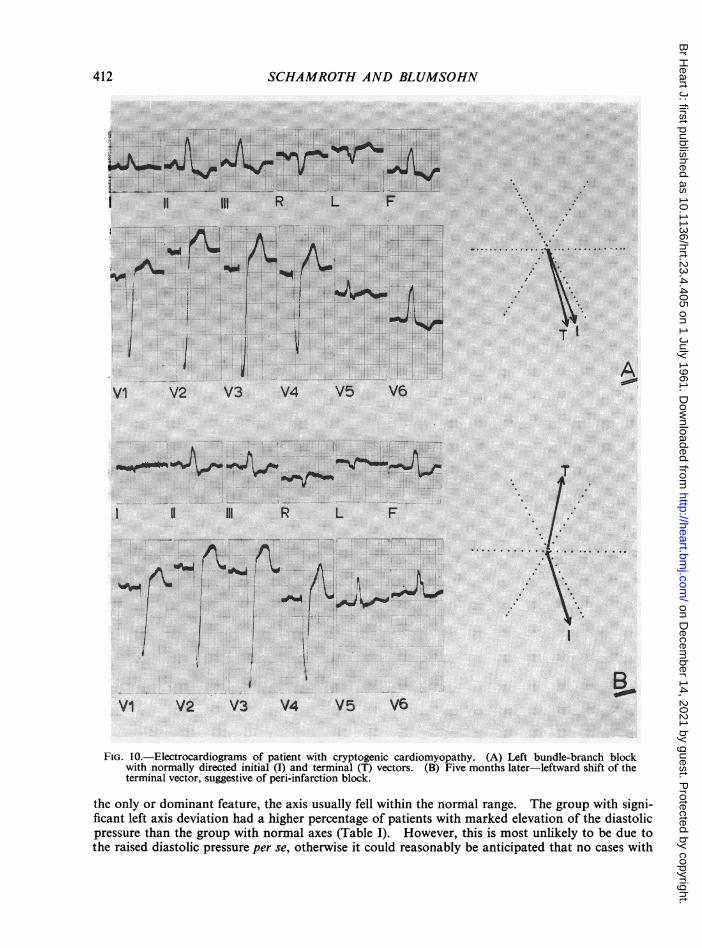
The fourth case also showed normally directed initial and terminal vectors (Fig. IOA). However, 5months later the terminal vector had shifted to the left, showing significant left axis deviation (-80°): Fig.lOB); the initial vector remained normal (+700). This spread of 1500 indicates a pattern suggestive ofperi-infarction block (vide infra). This patient denied any chest pain and there was no evidence of myocardialinfarction in the electrocardiogram.
-120 *0*
%.06~~~~~~~~~~~~~~~~
+120~~~~~~~~~~
FIG. 4.-AQRS distribution in normal African subjects. FIcG. 5.-AQRS distribution in cases with cryptogeniccardiomyopathy.
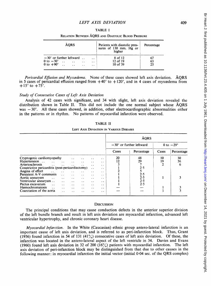
Hypertension. Significant left axis deviation was found in 12 of 70 patients(17%)0Nearly allwere examples of long-standing hypertensive disease with complicating factors such as cardiacfailure, diabetes mellitus, or renal or cerebral vascular disease. Slight left axis deviation was foundin 19 (27%) Normal axes were present in 39 (56%) (Fig. 6).
The average age for those with left axis deviation was 58 years and for those with normal axes45 years. The categories with slight or significant left axis deviation had a higher percentage ofpatientswith marked elevation of the diastolic pressures than the group with normal axes (Table I).
Mitral Regurgitation. Of these 19 patients none showed significant or even slight left axisdeviation. The tendency was for a deviation of AQRS to the right or frank right axis deviation(greater than +900)(Fig. 7). The QRS-T angles ranged from 0 to 50°. T waves were tall andupright in leads V5 and V6 in all cases.
408
on Decem
ber 14, 2021 by guest. Protected by copyright.
http://heart.bmj.com
/B
r Heart J: first published as 10.1136/hrt.23.4.405 on 1 July 1961. D
ownloaded from
LEFT AXIS DEVIATION
TABLE I
RELATION BETWEEN AQRS AND DIASTOLIC BLOOD PRESSURE
AQRS Patients with diastolic pres- Percentagesures of 130 mm. Hg or
higher
- 30° or further leftward .. .. 8 of 12 67Oto-30° .. .. .. .. 12of19 63Oto +900 .. .. .. .. 10of39 25
409
Pericardial Effusion and Myxa?dema. None of these cases showed left axis deviation. AQRSin 5 cases of pericardial effusion ranged from +40° to + 120°, and in 4 cases of myxcedema from+ 150 to +750.
Study of Consecutive Cases ofLeft Axis DeviationAnalysis of 42 cases with significant, and 34 with slight, left axis deviation revealed the
distribution shown in Table II. This did not include the one normal subject whose AQRSwas -30°. All these cases showed, in addition, other electrocardiographic abnormalities eitherin the patterns or in rhythm. No patterns of myocardial infarction were observed.
TABLE II
LEF Axis DEVIATION IN VARIOUS DISEASES
AQRS-30° or further leftward 0 to -29°
Cases Percentage Cases Percentage
Cryptogenic cardiomyopathy .. .. .. .. 20 48 10 30Hypertension .. .. .. .. .. 12 29 19 56Arteriosclerosis .. .. .. .. .. .. 3 7 2 6Constrictive pericarditis (post-pericardiectomy) .. .. 1 2 5Angina of effort .. .. .. .. .. .. 2 5Persistent A-V communis .. .. .. .. .. 1 2 5Aortic aneurysm .. .. .. .. .. .. 1 2-5 1 3Ventricular aneurysm .. .. .. .. .. .. 1 2 5Pectus excavatum .. .. .. .. .. .. 1 2 5Hemochromatosis .. .. .. .. .. .. 1 3Coarctation of the aorta ... .. .. ..3- - 1
DISCUSSIONThe principal conditions that may cause conduction defects in the anterior superior division
of the left bundle branch and result in left axis deviation are myocardial infarction, advanced leftventricular hypertrophy, and chronic coronary heart disease.
Myocardial Infarction. In the White (Caucasian) ethnic group antero-lateral infarction is animportant cause of left axis deviation, and is referred to as peri-infarction block. Thus, Grant(1956) found infarction in 54 of 131 (41%) consecutive cases of left axis deviation. Of these, theinfarction was located in the antero-lateral aspect of the left ventricle in 34. Davies and Evans(1960) found left axis deviation in 32 of 200 (16%) patients with myocardial infarction. The leftaxis deviation of peri-infarction block may be distinguished from that due to other causes in thefollowing manner: in myocardial infarction the initial vector (initial 0-04 sec. of the QRS complex)
on Decem
ber 14, 2021 by guest. Protected by copyright.
http://heart.bmj.com
/B
r Heart J: first published as 10.1136/hrt.23.4.405 on 1 July 1961. D
ownloaded from
410 SCHAMROTH AND BLUMSOHN
-120 * '0 -60'
030
0~~~~~~~~~~~~~~~
0
~~~*3Q/
+120~~~~ *0 /e60
\ /,' -.0....: 0
+7180--- 0.0.0°
00~~~~~~~
/0\/\ *f .;\~~~~~/
V120 000+60 +120° *.::. * 460FIG. 6. AQRS distribution in cases with hypertension. FIG. 7. AQRS distribution in cases with mitral regur-
gitation.
1 1i 111 R L F
-~--1- -i-,-X--1b1 1 I l I 1X
VI V2 V34 V5 V6FIG. 8.-Electrocardiogram showing significant left axis deviation in post-partum cardiomyopathy.
is directed away from the infarcted area; in the case of antero-lateral infarction the initial vectorwould thus be downward and to the right. The terminal vector (terminal 0 04 sec.) would, asdiscussed above, be directed upwards and to the left. Thus in peni-infarction block the angle ofspread between the initial and terminal vectors usually exceeds 1000, whereas in other causes of leftaxis deviation it rarely exceeds 60° (Grant, 1957).
on Decem
ber 14, 2021 by guest. Protected by copyright.
http://heart.bmj.com
/B
r Heart J: first published as 10.1136/hrt.23.4.405 on 1 July 1961. D
ownloaded from
LEFT AXIS DEVIATION 411
i T
I II 2it R L Fl
.. .... ........... _. ........J _......
~ ~ ~ ~ ~ /..................~~~~~~~~~~~.......". .....; ....
-.............................
VI V2 V3 V4 V5 '6
FIG. 9.-Electrocardiogram in a case of cryptogenic cardiomyopathy, showing left bundle-branch block, and leftaxis deviation of both initial (I) and terminal (T) vectors. The terminal vector is further leftward than theinitial vector.
By virtue of the fact that myocardial infarction is extremely rare or virtually absent in theAfrican population, peri-infarction block does not constitute any significant cause of left axisdeviation. Indeed, we did not encounter one case of myocardial infarction in the 256 electro-cardiograms examined. Furthermore, we found only one example of peri-infarction block in allour cases of left axis deviation: this was in a patient with cryptogenic cardiomyopathy. Thus, asindicated by Davies and Evans (1960) peri-infarction block does not necessarily connote infarctionin its literal meaning, as it may occur with fibrosis alone. The term should therefore be retained asan electrocardiographic conception alone.
Advanced Left Ventricular Hypertrophy. This is sometimes associated with left axis deviation.However, as Grant (1957) has indicated "it is not the hypertrophy itself that produces the left axisdeviation, because the majority of cases of even severe left ventricular hypertrophy have a normallydirected mean QRS axis. Probably the myocardial fibrosis which so often accompanies markedleft ventricular hypertrophy is responsible, by involving certain peripheral parts of the left ventricularconducting system." This condition is analogous to what has previously been called "parietalblock" (Segers et al., 1947; Alzamora-Castro et al., 1953).
Davies and Evans (1960) found left axis deviation in only 8 of 50 patients with severe hyper-tension, 7 of whom either had chest pain or electrocardiographic evidence of myocardial infarction.Likewise, Grant (1956), after the exclusion of all cases of myocardial infarction, noted that left axisdeviation did not necessarily correlate with the heart weight or body build. Nevertheless, theincidence of left axis deviation was higher among cases with hypertrophied left ventricles thanamong those with normal heart weights, and he attributed this to the myocardial fibrosis thataccompanies severe left ventricular hypertrophy.
In the present series 44 per cent of patients with hypertension showed left axis deviation. How-ever, it should be stressed that these were all advanced or long-standing cases of hypertension withcardiac failure, marked retinitis, and complicated in the majority of instances by renal disease,diabetes mellitus, or cerebral vascular accidents. Indeed, in those cases where hypertension was
on Decem
ber 14, 2021 by guest. Protected by copyright.
http://heart.bmj.com
/B
r Heart J: first published as 10.1136/hrt.23.4.405 on 1 July 1961. D
ownloaded from
412 SCHAMROTH AND BLUMSOHAr
...
ItI11|1 R L F -
'''i'-*'t@-9lw,,,,,..^l01 V1 d'', t ..""%:.
A............~~~~~~~~~~~~~~~~~~~~~...VI V2 V3 V4 V5 V6
I II I R L F /
I~~~~~~~~~~~~~~~I
V2 V3 V4 V5 V6
FIG. 10.-Electrocardiograms of patient with cryptogenic cardiomyopathy. (A) Left bundle-branch blockwith normally directed initial (I) and terminal (T) vectors. (B) Five months later-leftward shift of theterminal vector, suggestive of peri-infarction block.
the only or dominant feature, the axis usually fell within the normal range. The group with signi-ficant left axis deviation had a higher percentage of patients with marked elevation of the diastolicpressure than the group with normal axes (Table I). However, this is most unlikely to be due tothe raised diastolic pressure per se, otherwise it could reasonably be anticipated that no cases with
on Decem
ber 14, 2021 by guest. Protected by copyright.
http://heart.bmj.com
/B
r Heart J: first published as 10.1136/hrt.23.4.405 on 1 July 1961. D
ownloaded from
LEFT AXIS DEVIATION
much rise of diastolic pressure would have normally directed axes. Rather, it is the structural changeaccompanying the raised diastolic pressure that is more likely to be the cause.
Chronic Coronary Heart Disease. This also is a cause of left axis deviation by virtue ofthe fibrosisit produces. This is probably the cause of left axis deviation in the elderly patient where myocardialinfarction and long-standing hypertension can be excluded. As with myocardial infarction, chroniccoronary heart disease is rare among the African population. Thus we found 3 patients (aged 103,65, and 49 years) where significant left axis deviation could possibly be attributed to chronic coronaryheart disease. Significant left axis deviation was also found in 2 patients (aged 65 and 50 years)who presented with angina of effort. However, in our normal controls there were 12 men over 50years of age who did not show left axis deviation. Of the 200 normal subjects examined by Daviesand Evans (1960), the three with left axis deviation were all over 40 years of age. We had onesubject among our normal controls, a man aged 42, with AQRS of -30°; and although chroniccoronary heart disease may have been a factor, we doubt this because, apart from the rarity ofchronic coronary heart disease in this population, he showed none of its stigmata. It may bepossible, as suggested by Manning (quoted by Grant, 1958) that this may be an example of a rarecongenital malformation of the distal radicle of the left bundle branch. Subsequent to theirinvestigations, Davies and Evans (1960) found left axis deviation in four young subjects with noevidence of heart disease in whom this mechanism of congenital agenesis was postulated.
Cardiomyopathy. Davies and Evans (1960) found left axis deviation in 16 (64%) of 25 cases withobscure cardiomyopathy, They also found it in a number of cases of familial cardiomegaly,Friedreich's disease, cardiac amyloidosis, myotonia atrophica, and hemochromatosis.
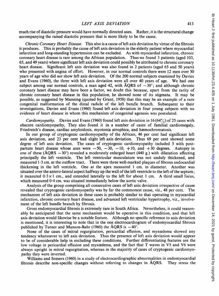
In our group of cryptogenic cardiomyopathy of the African, 46 per cent had significant leftaxis deviation, and 23 per cent had slight left axis deviation. Thus 69 per cent showed somedegree of left axis deviation. The cases of cryptogenic cardiomyopathy included 5 with post-partum heart disease whose axes were -50, -30, -10, +10, and +30 degrees. Autopsy inone of these (AQRS -30') revealed a moderately enlarged heart (440 g.) with dilatation affectingprincipally the left ventricle. The left ventricular musculature was not unduly thickened, andmeasured 1-3 cm. at the outflow tract. There were three well-marked plaques of fibrous endocardialthickening in the left ventricle. One at the apex measured 1 cm. in diameter. Another wassituated over the antero-lateral aspect halfway up the wall of the left ventricle to the left of the septum;it measured 0-3 x 1 cm., and extended laterally to the left for about 1 cm. A third small focus,which measured 0 4 cm. was situated immediately below the aortic valve.
Analysis of the group comprising all consecutive cases of left axis deviation irrespective of causerevealed that cryptogenic cardiomyopathy was by far the commonest cause, viz., 48 per cent. Themechanism of left axis deviation in these cases is probably similar to that operating in myocardialinfarction, chronic coronary heart disease, and advanced left ventricular hypertrophy, viz., involve-ment of the left bundle branch by fibrosis.
Gross endomyocardial fibrosis is extremely rare in South Africa. Nevertheless, it could reason-ably be anticipated that the same mechanism would be operative in this condition, and that leftaxis deviation would likewise be a notable feature. Although no specific reference to axis deviationis mentioned, it is interesting to note that in the one electrocardiogram of endomyocardial fibrosispublished by Turner and Manson-Bahr (1960) the AQRS is -40°.
None of the cases of mitral regurgitation, pericardial effusion, and myxcedema showed anytendency whatsoever to left axis deviation. Thus the presence of left axis deviation would appearto be of considerable help in excluding these conditions. Further differentiating features are thelow voltage in pericardial effusion and myxcedema, and the fact that T waves in V5 and V6 werealways upright in mitral regurgitation, whereas in the majority of cases of cryptogenic cardiomyo-pathy they were inverted.
Williams and Somers (1960) in a study of electrocardiographic abnormalities in endomyocardialfibrosis describe non-specific changes without referring to changes in AQRS. They stress the
413
on Decem
ber 14, 2021 by guest. Protected by copyright.
http://heart.bmj.com
/B
r Heart J: first published as 10.1136/hrt.23.4.405 on 1 July 1961. D
ownloaded from

SCHAMROTH AND BLUMSOHN
difficulty of distinguishing endomyocardial fibrosis from chronic rheumatic mitral disease andpericarditis, a difficulty previously noted by Ball et al. (1954). However, as with cryptogeniccardiomyopathy the presence of left axis deviation may well be a distinguishing feature and warrantsfurther investigation.
It is apparent that in the local African cryptogenic cardiomyopathy is the commonest causeof left axis deviation. This is in contrast to the Caucasian where myocardial infarction andcoronary heart disease are the chief causes of left axis deviation. Thus in an African patientpresenting with cardiomegaly in the absence of advanced hypertension, the presence of left axisdeviation is suggestive of cryptogenic cardiomyopathy.
It should be stressed that left axis deviation is not specific to any of the disease in which it occurs,but merely represents a conduction defect most commonly due to non-specific fibrosis. In theabsence of myocardial infarction, coronary heart disease, and advanced hypertension, the presenceof left axis deviation, irrespective of race and particularly in the younger subject is a pointer to acardiomyopathy.
SUMMARYThe mean manifest frontal plane QRS and T wave axes (AQRS and AT) were studied in cases
of cryptogenic cardiomyopathy in the African (Negro) and compared with cases of hypertension,mitral regurgitation, pericardial effusion, and myxcedema.
Electrocardiograms of healthy African male orderlies and nurses attached to the hospital wereused as normal controls. With one exception, AQRS in the normal controls was located between0 and + 900. Cases with cryptogenic cardiomyopathy showed a significantly high incidence ofleft axisdeviation and formed the major wtiological group of all causes of left axis deviation in the African.
Significant left axis deviation (-30° or further leftward) was found only in those hypertensivepatients who had advanced disease with complicating factors.
The presence of left axis deviation may help to distinguish cryptogenic cardiomyopathy frompericardial effusion, myxcedema, or mitral regurgitation. In the absence of coronary heart diseaseand advanced hypertension the presence of left axis deviation is a pointer to a cardiomyopathy.
We wish to express our thanks to Dr. I. Frack, Superintendent of Baragwanath Hospital for permission to publishthese cases, Dr. E. Kahn for his helpful criticism of the manuscript, Dr. C. Isaacson for the pathological report, andMr. A. Shevitz of the Photographic Dept. of the University of the Witwatersrand for the photographic reproductions.
REFERENCESAlzamora-Castro, V., Abugattas, R., Rubio, C., Bouroncle, J., Zapata, C., Santa-Maria, E., Battilana, G., Binder, T.,
Subiria, R., and Paredes, D. (1953). Circulation, 7, 108.Ball, J. D., Davies, J. N. P., and Williams, A. W. (1954). Lancet, 1, 1049.Becker, B. J. P., Chatgidakis, C. B., and van Lingen, B. (1953). Circulation, 7, 345.Bedford, D. E., and Konstam, G. L. S. (1946). Brit. Heart J., 8, 236Davies, H., and Evans, W. (1960). Brit. Heart J., 22, 551.Davies, J. N. P., and Ball, J. D. (1955). Brit. Heart J., 17, 337.Gillanders, A. D. (1951). Brit. Heart J., 13, 177.Gouley, B. A., McMillan, T. M., and Bellet, S. (1937). Amer. J. med. Sci., 194, 185.Grant, R. P. (1956). Circulation, 14, 233.
(1957). Clinical Electrocardiography. McGraw-Hill Book Co. Inc. (New York).(1958). Third World Congress of Cardiology. Brussels.
Laurie, W., Woods, J. D., and Roach, G. (1960). Amer. J. Cardiol., 5, 48.Melvin, J. P. (1947). Ann. intern. Med., 27, 596.Schwartz, M. B., Schamroth, L., and Seftel, H. C. (1958). Med. Proc., 4, 275.Seftel, H. C., and Susser, M. (1961). Brit. Heart J. 23, 43.Segers, M., Vastesaeger, M., and Denolin, H. (1947). Acta. Cardiol., 2, 166.Turner, P. P., and Manson-Bahr, P. E. C. (1960). Brit. Heart J., 22, 305.Vilter, R. W., and McKee, E. E. (1943). Ohio State med. J., 39, 142.Williams, A. W., and Somers, K. (1960). Brit. Heart J., 22, 311.Zao, Z. Z., Hermann, G. R., and Hejtmancik, M. R. (1958). Amer. Heart J., 56, 65.
414
on Decem
ber 14, 2021 by guest. Protected by copyright.
http://heart.bmj.com
/B
r Heart J: first published as 10.1136/hrt.23.4.405 on 1 July 1961. D
ownloaded from


![Maxillofacial Fractures due to Falls: does Fall …FISS, and fracture location. Data were expressed as mean and standard deviation (M [SD]). Statistical significance was determined](https://static.fdocuments.us/doc/165x107/5f2b65d86e4fe637f359b8da/maxillofacial-fractures-due-to-falls-does-fall-fiss-and-fracture-location-data.jpg)