The Seminal Contributions of Gregorio Weber to Modern ...
26
1 The Seminal Contributions of Gregorio Weber to Modern Fluorescence Spectroscopy David M. Jameson Department of Genetics and Molecular Biology University of Hawaii 1960 East-West road Honolulu, Hawaii 96822 USA [email protected] Abstract Gregorio Weber is acknowledged to be the person responsible for many of the more important theoretical and experimental developments in modern fluorescence spectroscopy. In particular, Weber pioneered the application of fluorescence spectroscopy to the biological sciences. His list of achievements includes: the synthesis and use of dansyl chloride as a probe of protein hydrodynamics; the extension of Perrin’s theory of fluorescence polarization to fluorophores associated with random orientations with ellipsoids of revolution and to mixtures of fluorophores; the first spectral resolution of the fluorescence of the aromatic amino acids and of intrinsic fluorescence of proteins; the first demonstration that both FAD and NADH make internal complexes; the first report on aromatic secondary amines, which are strongly fluorescent in apolar solvents, but hardly in water, the most spectacular case being the anilino-naphthalene sulfonates (ANS); the first description of the use of the fluorescence of small molecules as probes for the viscosity of micelles, with implications for membrane systems; a general formulation of depolarization by energy transfer; the discovery of the “red-edge” effect in homo-energy transfer; the development of modern cross-correlation phase fluorometry; the development of the excitation-emission matrix method for resolving contributions from multiple fluorophores; the synthesis of several novel fluorophores, including pyrenebutyric acid, IAEDANS, bis-ANS, PRODAN and LAURDAN, designed to probe dynamic aspects of biomolecules. In addition to these seminal contributions, Gregorio Weber also trained and inspired generations of spectroscopists and biophysicists who went on to make important contributions to their fields, including both basic research as well as the commercialization of fluorescence methodologies and their extension into the clinical and biomedical disciplines.
Transcript of The Seminal Contributions of Gregorio Weber to Modern ...
1
The Seminal Contributions of Gregorio Weber to Modern Fluorescence Spectroscopy
David M. Jameson
Department of Genetics and Molecular Biology University of Hawaii 1960 East-West road Honolulu, Hawaii 96822 USA
Abstract Gregorio Weber is acknowledged to be the person responsible for many of the more important theoretical and experimental developments in modern fluorescence spectroscopy. In particular, Weber pioneered the application of fluorescence spectroscopy to the biological sciences. His list of achievements includes: the synthesis and use of dansyl chloride as a probe of protein hydrodynamics; the extension of Perrin’s theory of fluorescence polarization to fluorophores associated with random orientations with ellipsoids of revolution and to mixtures of fluorophores; the first spectral resolution of the fluorescence of the aromatic amino acids and of intrinsic fluorescence of proteins; the first demonstration that both FAD and NADH make internal complexes; the first report on aromatic secondary amines, which are strongly fluorescent in apolar solvents, but hardly in water, the most spectacular case being the anilino-naphthalene sulfonates (ANS); the first description of the use of the fluorescence of small molecules as probes for the viscosity of micelles, with implications for membrane systems; a general formulation of depolarization by energy transfer; the discovery of the “red-edge” effect in homo-energy transfer; the development of modern cross-correlation phase fluorometry; the development of the excitation-emission matrix method for resolving contributions from multiple fluorophores; the synthesis of several novel fluorophores, including pyrenebutyric acid, IAEDANS, bis-ANS, PRODAN and LAURDAN, designed to probe dynamic aspects of biomolecules. In addition to these seminal contributions, Gregorio Weber also trained and inspired generations of spectroscopists and biophysicists who went on to make important contributions to their fields, including both basic research as well as the commercialization of fluorescence methodologies and their extension into the clinical and biomedical disciplines.
2
Overview
During the last few decades, fluorescence spectroscopy has evolved from a narrow, highly specialized technique into an important discipline widely utilized in the biological, chemical and physical sciences. Fluorescence methodologies have also assumed an increasingly important role in the clinical and medical sciences. There are now world-renown centers for fluorescence spectroscopy, highly successful commercial enterprises specializing in fluorescence instrumentation, fluorescence based clinical instruments in virtually all hospital laboratories, and thousands of practitioners worldwide. As in all scientific disciplines, the development of modern fluorescence spectroscopy has benefited from the contributions of many individuals from many countries. However, one individual, Gregorio Weber, can be singled out for his outstanding and far-reaching contributions to this field.
Early years
Born in Buenos Aires, Argentina, on July 4, 1916, Weber demonstrated an early aptitude for science, mathematics and linguistics. He was greatly impressed by his high school teacher of Geology and Mineralogy - not just by his teaching skills, but also by his broad knowledge of science. Weber told this teacher that he would like to have a career in science and asked which area - chemistry, physics, et cetera - he would recommend. The teacher replied that it was very difficult to find steady employment in Argentina as a pure scientist at that time and he recommended that Weber go to Medical School, since that would afford him the opportunity to study diverse areas of science and, if all else failed, provide him with a profession with which to support himself. Taking this advice, Weber enrolled in the University of Buenos Aires and completed his M.D. degree in 1943. While a medical student, from 1939 to 1943, he worked in the Department of Physiology and Biochemistry as a teaching assistant for Bernardo Alberto Houssay who had already achieved renown as a physiologist for his work on the endocrine system and in particular the pituitary gland (Houssay shared the 1947 Nobel Prize for Physiology and Medicine with Carl and Gerty Cori). Houssay recognized the ability of his young protege and suggested that Weber apply for a prestigious British Council Fellowship to support graduate studies toward a Ph.D. at Cambridge University. Although in 1943 the war was still raging in Europe, and London was being subjected to V-1 attacks, Weber enthusiastically embraced the opportunity to pursue his love of science.
Cambridge
Travel to England during the war years was an adventure and Weber’s voyage took 44 days in a convoy, which languished off the coast of Africa for weeks and endured occasional U-boat attacks. In Cambridge, Weber initially thought to study
3
colloid and surface chemistry and joined the laboratory of Eric Riddeal. Soon, however, he became enamored of proteins and went to talk to Malcolm Dixon, the well-known enzymologist, about applying techniques of Physical Chemistry to the study of proteins. Dixon suggested that Weber consider applying fluorescence techniques to the study of the naturally fluorescent flavin and flavoprotein systems. At that time, Weber knew little about fluorescence but soon learned that there were a number of low molecular weight flavin compounds, such as riboflavin and FAD, that differed greatly in fluorescence intensity. Weber was thus given the task of “sorting out” this area.
When Gregorio Weber began his graduate studies, the fluorescence of substances extracted from organisms had already attracted the attention of biologists and biochemists. In these early studies, however, fluorescence was used as an aid in isolation and purification and also in the quantitative determination of fluorescent substances such as riboflavin, porphryins, and pterines. The relationship between the fluorescence from chlorophylls and biochemical aspects of the photosynthetic process seemed undoubted but was far from clear. Almost all of the work done on fluorescence substances had been descriptive in nature, i.e., concerned mainly with the conditions under which the fluorescence could be most readily observed as well as with the color and intensity of the fluorescent emission. The situation was different in the physical sciences, though. Physicists, such as Enrique Gaviola (a fellow Argentinean), Jean and Francis Perrin, S.I. Vavilov, F. Weigert, P. Pringsheim, and others had already introduced important concepts such as the excited state lifetime, the polarization of the fluorescence and the quantum yield of the emission process, yet these ideas had not penetrated into the Chemical or Biological fields (for a comprehensive, lucid and exceptionally well-documented overview of the history of photoluminescence in the first half of the 20th century, the reader is referred to the series of articles by Bernhard Nickel [1-3]). Weber recognized the need for a quantitative understanding of fluorescence phenomena and to this end he spared himself no pains. He wrote in his Ph.D. thesis [4] “I feel that a knowledge, as deep as possible, of the physical principles concerned is indispensable. Even close collaboration with a physicist cannot spare this task to the biochemist. I am tempted to believe that a biologist having n ideas related to the biological side of the problem and a physicist possessing another n relating to the physical side would result in some 2n useful combinations whereas the same ideas collected in one brain would lead to a number of combinations more like n!”.
Francis Perrin’s influence
In later years, Weber would say that the work that most influenced him and which he liked the best was that of Francis Perrin. Weber=s introduction to polarization started when he read Perrin=s classic paper of 1926 in the Journal de Physique on the depolarization of fluorescence by Brownian rotations [5]. In Weber’s own words [6] “I remember that Malcolm Dixon came to me one day, handed me a little piece of paper, and said that somebody at King’s College – I wish I could

4
remember his name – had said that there was a paper on fluorescence that I should read. The little piece of paper had written on it: F. Perrin, J. de Physique, 1926. So I went to the Cambridge library, which I positively thought of as a temple of learning and looks indeed like one, and I read the famous paper of Perrin on depolarization of the fluorescence by Brownian rotations, not one but many times. Argentine secondary education in the first half of the century included French language and literature so that I could not only understand the scientific content, but also enjoy the literary quality of the writing. It was written in that transparent, terse style of XVIII century France, which I have tried, perhaps unsuccessfully, to imitate from then onwards. The clarity of Perrin's thought and his ability to do the right experiment were really remarkable.” Weber went on to note [6] “It was from reading Perrin's papers that I conceived three ideas on the use of polarization: determination of the change in the fluorescence lifetime as one quenches the fluorescence by addition of an appropriate chemical, determination of the molecular volume of proteins by fluorescent conjugates with known dyes and determination of the viscosity of a medium through the polarization of the emission from a known fluorescent probe.”
Ph.D. thesis
It goes without saying that all fluorescence instrumentation had to be home built at that time. The original apparatus built by Weber is shown in figure 1, which is reproduced from his thesis. The light source (L) was a carbon arc, originally developed for use in searchlights during the war. The exciting light was first filtered through a layer of concentrated NaNO2 (U) to remove UV light (< 420nm) and then polarized by a Nicol prism (N1). Additional glass filters (F1 and F2) were used to further delimit the exciting light and to isolate the emission. The actual measurement of the polarization of the fluorescence was realized using visual compensation techniques involving observation of interference patterns as a “pile-of-plates” polarizer P (the compensator of Arago) was rotated. Using these simple methods and only his eye as the detector, Weber was able to quantify levels of polarized light reaching only 1 or 2%. There was a price to be paid, however, for these visual compensation methods. Like many of the pioneering spectroscopists, Weber suffered acute eye aliments in latter years as a result of excessive exposure to infrared and ultraviolet light, which led to removal of his lenses, detached retinas and eventually cornea transplants1. These difficulties resulted in a marked photophobia, which required Gregorio Weber to wear sunglasses most of the time – the sunglasses became almost a trademark for “The Professor” as he was knownto his students.
1 An insight into the rationality characteristic of Gregorio Weber is his remark, in the author’s presence, to the physician who removed the bandages from the second eye, which had received the new cornea – “I have a homogeneous, clear, binocular visual field” – a statement conveying the maximum of information with the minimum of words.

5
Fig. 1. Original drawing from Gregorio Weber’s Ph.D. thesis showing the optical arrangement of the instrument he constructed for polarization measurements.
A large portion of Weber’s thesis was devoted to measurements on the quenching of fluorescence of riboflavin and on development of a general theory of quenching by complex formation. This lead to his first publication entitled: The quenching of fluorescence in liquids by complex formation. Determination of the mean life of the complex [7]. This paper was the first to demonstrate that fluorescence quenching can take place after formation of molecular complexes of finite duration rather than collisions. His second publication entitled Fluorescence of riboflavin and flavin-adenine dinucleotide [8], was the first demonstration of an internal complex in FAD. Years later he was to follow up this work with the first demonstration that NADH also formed an internal complex [9] and with more complete characterizations of the excited state properties of FAD and NADH [10-13]. Weber completed his doctoral thesis entitled Fluorescence of Riboflavin, Diaphorase and Related Substances in 1947 (figure 2). Interestingly, the final chapter of his thesis was devoted to the application of polarization measurements to the determination of the microscopic viscosity of gels. This work followed on the original, seminal observations of F. Weigert [14] in 1920 that the percentage of polarized light from a dye in solution increased rapidly with the viscosity of the solution, and the more detailed studies of Vavilov and Levshin in 1923 who reported on the polarization of the fluorescence of 26 dyes in water and glycerol

6
and also on dyes in colloidal solutions [15] - observations which were, of course, important for F. Perrin’s experimental and theoretical studies. Weber observed that the polarization of fluorescein in gels such as agar, gelatin and silicic acid did not increase as the gels solidified. These observations were important in deciding between different theories on the nature of colloidal suspensions, namely supporting the theory that the colloidal particles built a continuous, fibrillar structure, which would leave pockets of solvent in between the gel structures. These pockets could then accommodate the fluorescein molecules, which would rotate freely in the embedded solvent. Weber clearly articulated the concept of microviscosity as opposed to macroviscosity or “bulk” viscosity and suggested that fluorescence polarization might be fruitfully applied to the study of cell protoplasm. This prescient observation anticipated the work he would publish 24 years later which first delineated the application of fluorescence probes to study the physical state of lipid systems [16,17]. In fact, Weber never lost his fascination with the rotation of small molecules. In later years he developed the theory of differential phase fluorometry [18] and applied the method to demonstrate the anisotropic rotation of small molecules in isotropic solvents [19,20]. Fig. 2. Gregorio Weber graduating from St. John’s College, Cambridge University, 1947.

7
Postdoctoral
From 1948 to 1952 Weber carried out independent investigations at the Sir William Dunn Institute of Biochemistry at Cambridge, supported by a British Beit Memorial Fellowship. At that time he began to delve more deeply into the theory of fluorescence polarization and also began to develop methods which would allow him to study proteins which did not contain an intrinsic fluorophore such as FAD or NADH (the fluorescence of the aromatic amino acids had not yet been discovered). To this end, he invested considerable time and effort in synthesizing a fluorescent probe which could be covalently attached to proteins and which possessed absorption and emission characteristics appropriate for the instrumentation available in post-war England. The result of two years of effort was the still popular probe dimethylaminonaphthalene sulfonyl chloride or dansyl chloride. With this tool in hand and with new instrumentation he began to investigate several protein systems, publishing his theory and experimental results in two classic papers published in 1952, namely, Polarization of the fluorescence of macromolecules. I. Theory and experimental method [21] and Polarization of the fluorescence of macromolecules. II. Fluorescent conjugates of ovalbumin and bovine serum albumin [22]. The theory paper (which interestingly contains an acknowledgement to F. Perrin for his suggestions) includes an extension of Perrin’s theory of depolarization due to rotation of ellipsoidal molecules. Specifically, Weber showed that Perrin’s complex equations, which required a knowledge of the orientation of the fluorophore’s absorption and emission oscillators with respect to the axis of rotation of the ellipsoid, could be considerably simplified if the fluorophores carrying the oscillators were assumed to be randomly oriented on the macromolecule. This paper also contained a formulation of the law of additivity of polarizations, namely:
⎟⎠
⎞⎜⎝
⎛∑⎟⎠
⎞⎜⎝
⎛31 -
P1 f =
31 -
P1
i
1-
iobs
1-
where Pobs is the actual polarization observed arising from i-components, fi represents the fractional contribution of the ith component to the total emission intensity and Pi is the polarization of the ith component. The motivation for considering this additivity function arose from Weber’s realization that a population of fluorescent molecules differing in their size or excited state lifetime (for example free and protein-bound probe) would contribute separately to the observed polarization. Similarly, a population of nonspherical proteins, such as prolate or oblate ellipsoids, could give rise to a distribution of rotational rates depending upon the orientation of the probe along the respective rotational axes. Hence, a clear understanding of the ways in which the individual contributions sum to the total signal was important. These considerations led immediately (in fact in the paper directly following Weber’s two articles in Biochemical Journal) to the work of Laurence who first described the application of polarization methods to
8
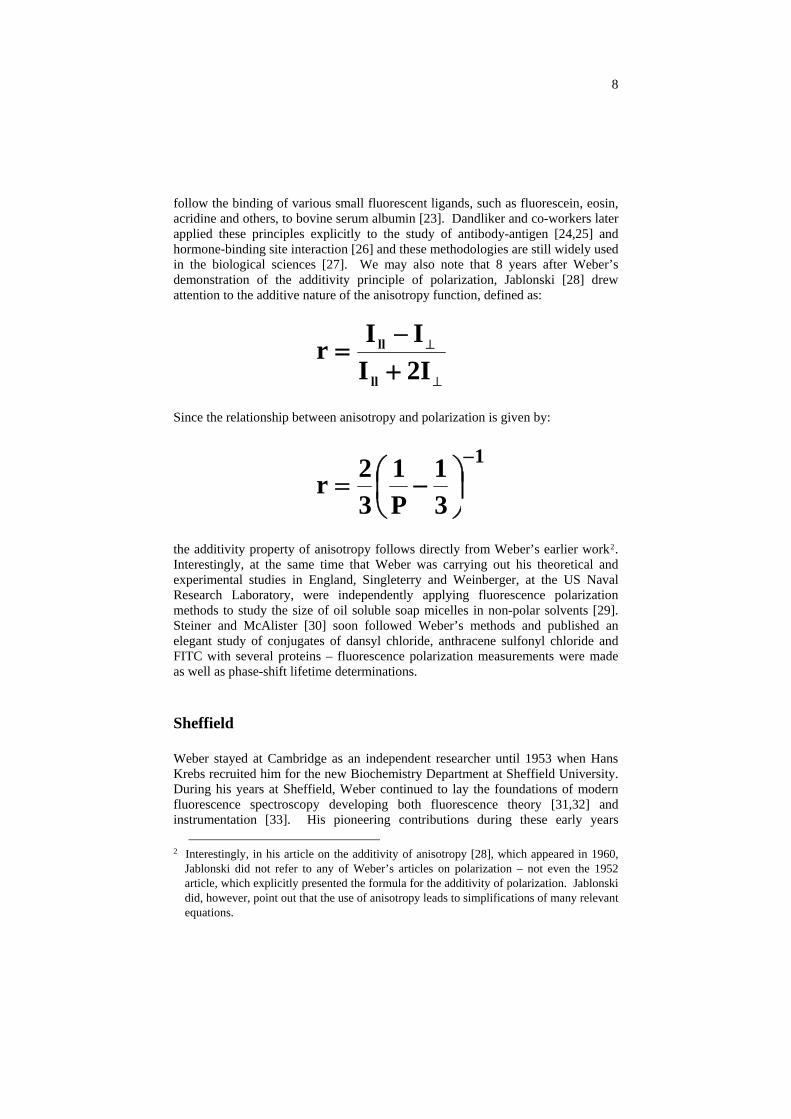
follow the binding of various small fluorescent ligands, such as fluorescein, eosin, acridine and others, to bovine serum albumin [23]. Dandliker and co-workers later applied these principles explicitly to the study of antibody-antigen [24,25] and hormone-binding site interaction [26] and these methodologies are still widely used in the biological sciences [27]. We may also note that 8 years after Weber’s demonstration of the additivity principle of polarization, Jablonski [28] drew attention to the additive nature of the anisotropy function, defined as:
⊥
⊥
+−
=I2I
IIrll
ll
Since the relationship between anisotropy and polarization is given by:
1
31
P1
32r
−
⎟⎠⎞
⎜⎝⎛ −=
the additivity property of anisotropy follows directly from Weber’s earlier work2. Interestingly, at the same time that Weber was carrying out his theoretical and experimental studies in England, Singleterry and Weinberger, at the US Naval Research Laboratory, were independently applying fluorescence polarization methods to study the size of oil soluble soap micelles in non-polar solvents [29]. Steiner and McAlister [30] soon followed Weber’s methods and published an elegant study of conjugates of dansyl chloride, anthracene sulfonyl chloride and FITC with several proteins – fluorescence polarization measurements were made as well as phase-shift lifetime determinations.
Sheffield
Weber stayed at Cambridge as an independent researcher until 1953 when Hans Krebs recruited him for the new Biochemistry Department at Sheffield University. During his years at Sheffield, Weber continued to lay the foundations of modern fluorescence spectroscopy developing both fluorescence theory [31,32] and instrumentation [33]. His pioneering contributions during these early years
2 Interestingly, in his article on the additivity of anisotropy [28], which appeared in 1960,
Jablonski did not refer to any of Weber’s articles on polarization – not even the 1952 article, which explicitly presented the formula for the additivity of polarization. Jablonski did, however, point out that the use of anisotropy leads to simplifications of many relevant equations.
9
included his report with Laurence [34] of aromatic secondary amines, which were strongly fluorescent in apolar solvents but very weakly fluorescent in water, the most spectacular case being the anilino-naphthalene sulfonates (ANS). It is interesting to note that even today, more than 50 years after that first report, ANS is still being used in protein studies, quite often as an indicator of the “molten globule state.”
Intrinsic protein fluorescence
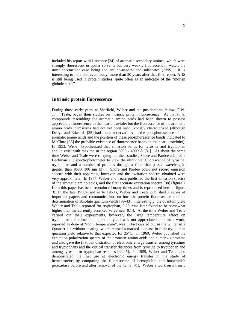
During those early years at Sheffield, Weber and his postdoctoral fellow, F.W. John Teale, began their studies on intrinsic protein fluorescence. At that time, compounds resembling the aromatic amino acids had been shown to possess appreciable fluorescence in the near ultraviolet but the fluorescence of the aromatic amino acids themselves had not yet been unequivocally characterized (although Debye and Edwards [35] had made observations on the phosphorescence of the aromatic amino acids and the position of these phosphorescence bands indicated to McClure [36] the probable existence of fluorescence bands in the near ultraviolet). In 1953, Weber hypothesized that emission bands for tyrosine and tryptophan should exist with maxima in the region 3000 - 4000 Å [31]. At about the same time Weber and Teale were carrying out their studies, Shore and Pardee adapted a Beckman DU spectrophotometer to view the ultraviolet fluorescence of tyrosine, tryptophan and a number of proteins through a filter that passed wavelengths greater than about 300 nm [37]. Shore and Pardee could not record emission spectra with their apparatus, however, and the excitation spectra obtained were very approximate. In 1957, Weber and Teale published the first emission spectra of the aromatic amino acids, and the first accurate excitation spectra [38] (figure 7 from this paper has been reproduced many times and is reproduced here in figure 3). In the late 1950's and early 1960's, Weber and Teale published a series of important papers and communications on intrinsic protein fluorescence and the determination of absolute quantum yields [39-43]. Interestingly, the quantum yield Weber and Teale reported for tryptophan, 0.20, was later found to be somewhat higher than the currently accepted value near 0.14. At the time Weber and Teale carried out their experiments, however, the large temperature effect on tryptophan’s lifetime and quantum yield was not appreciated and their work, reported as done at “room temperature”, was in fact carried out in the winter in a Quonset hut without heating, which caused a marked increase in their tryptophan quantum yield relative to that expected for 25°C. In 1960, Weber published the excitation polarization spectra of the aromatic amino acids and numerous proteins and also gave the first demonstration of electronic energy transfer among tyrosines and tryptophans and the critical transfer distances from tyrosine to tryptophan and among tyrosine or tryptophan residues [44,45]. In 1959, Weber and Teale also demonstrated the first use of electronic energy transfer in the study of hemeproteins by comparing the fluorescence of hemoglobin and horseradish peroxidase before and after removal of the heme [41]. Weber’s work on intrinsic
10
Fig. 3. Figure 7 from [38] giving the first emission spectra of the aromatic amino acids.
tryptophan fluorescence inspired Velick to study the binding of NADH to dehydrogenases by following the quenching of the tryptophan fluorescence due to energy transfer [46]. Soon afterwards, Velick et al., [47] then applied this method to study the binding of aromatic ligands (in particular 2,4 dinitrophenol) to specific antibodies - work which led to the significant finding of affinity maturation (increase in the affinity of the antibodies produced with time after induction of antibody production), which was to have a major impact on the field of immunology. Weber followed up his early interest in heme protein fluorescence years later with the first report of the emission spectrum of hemoglobin [48]. During the last 20 years, numerous papers have appeared from many laboratories reporting studies on the intrinsic fluorescence of hemeproteins such as hemoglobin, myoglobin and horseradish peroxidase. Weber’s interest in the photophysics of tryptophan also resulted in a publication with Bernard Valeur, in 1977, of an important and often quoted paper [49] on the 1La and 1Lb excitation bands of indole and tryptophan. During the four decades since the first description of protein fluorescence, thousands of papers have been written on the fluorescence of tryptophan, tyrosine or phenylalanine or some aspect of intrinsic protein fluorescence. The study of intrinsic protein fluorescence has, in fact, become one of the most important techniques used in protein research and has been of great
11
importance in establishing the dynamic nature of proteins. This potential was certainly not lost on Weber who presented a classic paper at the “Light and Life” conference held in 1960 and, in a true understatement, summarized his presentation by saying “There are many ways in which the properties of the excited state can be utilized to study points of ignorance of the structure and function of proteins” [50]. In fact, in an earlier communication [43] (presented at the annual meeting of the British Biochemical Society on April 3, 1959) Weber estimated that the excited state lifetime of tryptophan in proteins was on the order of 4 ns and commented “These values are too short to permit measurements of fluorescence polarization to be of value in the determination of the rotational relaxation times of proteins in solution, but can give useful information on local conditions about the tryptophan or tyrosine residues.” Now that present day methods of site-directed mutagenesis permit the facile removal and/or addition of tryptophan residues to allow the creation of novel single-tryptophan containing proteins, Weber’s vision of the utility of intrinsic protein fluorescence is being fully realized.
Red-edge effects
In 1924, Gaviola and Pringsheim first observed decreases in the polarization of solutions of some fluorophores in glycerol as the fluorophore concentration increased but the explanation of this phenomenon as being due to dipole-dipole energy transfer over distances larger than the contact distance was due to Jean Perrin [51], the father of Francis Perrin. One of the first quantitative treatments of concentration dependent energy transfer was due to Weber [52]. In his original observations on energy transfer among the aromatic amino acids [44] Weber also pointed out that homotransfer, i.e., indole to indole or tryptophan to tryptophan, became much less effective upon excitation near the red-edge of the absorption. In 1970, Weber and Shinitsky [53] published a more comprehensive study and showed that this “red-edge effect” was a very general phenomenon of aromatic fluorophores. Homotransfer and the failure of such transfer upon red-edge excitation have been used to study dynamic aspects of numerous macromolecular systems (see, for example, [54,55]). Weber’s interest in red-edge effects persisted and in the late 1970’s he published two articles with Bernard Valeur, which described a new red-edge effect producing apparent rotational anomalies of fluorophores [56,57].
EEM
Always mindful of the effect of heterogeneity on observed signals, during his stay in Sheffield, Weber conceived of a method to elucidate the number of fluorescing compounds in mixtures of fluorophores by variation of the excitation and emission wavelength and construction of a matrix of the resulting intensities [58]. Years later, with the advent of computer controlled instrumentation and data analysis,

12
Weber’s matrix approach would become widely utilized in Analytical Chemistry and would be known as the EEM (Excitation-Emission Matrix) technique [59,60].
Brandeis
In 1960, Weber was a Visiting Professor at Brandeis University. While there he gave a series of lectures in fluorescence and inspired a number of students and postdoctoral fellows with the potential of fluorescence methods. Among those were two individuals, Lubert Stryer and Ludwig Brand, who went on to establish themselves as leading researchers and who made many important contributions in the biological applications of fluorescence spectroscopy.
University of Illinois
At around this time, I.C. “Gunny” Gunsalus, then the head of the Biochemistry Division of the Department of Chemistry at the University of Illinois at Urbana-Champaign, recruited Weber. Gunny relates the story that while he was convincing his colleagues that Gregorio Weber was an exceptional scientist, someone commented that Weber didn’t have as many publications as one might expect from a senior professor. Gunny explained that while this was true, Weber’s ratio of outstanding papers to total papers was unity and that this ratio - known thereafter as the Weber ratio - was certainly the more important consideration. The reader should pause at this point to estimate his/her own “Weber ratio!”
Gregorio Weber joined the University of Illinois in 1962 and built a research program that continued actively until his death from leukemia on July 17, 1997. During the early years in Urbana, Weber continued to develop novel fluorescence instrumentation and probes and extended his studies of protein systems. In the mid-1960’s, Philippe Wahl visited Weber’s laboratory. Building on previous studies by Gottlieb and Wahl on dansyl labeled polymer systems [61], Wahl and Weber published one of the first reports delineating the effects of thermally activated local probe mobility on fluorescence depolarization studies of protein systems (specifically, dansyl conjugates of gamma globulins) [62]. One of Weber’s lasting contributions to the biological fluorescence field, in fact, was his approach of deciding what questions he wanted to ask about a biomolecular system, and then designing and synthesizing a fluorophore with the optimum spectroscopic properties to achieve the goal. Among the fluorescence probes Weber developed in Urbana were pyrenebutyric acid [63] (which had a lifetime of 100 - 150 ns and thus extended the polarization method to proteins with molecular weights of 106), bis-ANS [64] (which binds to many proteins with much higher affinity than ANS and which also binds to many nucleotide binding sites), IAEDANS [65] (the first sulfhydryl specific fluorescence probe), and PRODAN
[66] (2-dimethylamino- 6-propionyl-naphthalene; a probe designed by Weber to have an exceptionally large excited state dipole moment and hence to possess an

13
extreme environmental sensitivity). Weber also made derivatives of PRODAN such as LAURDAN, which included a lauric acid tail to render the probe lipid soluble (LAURDAN has been very extensively used in recent years as a probe of membrane dynamics – see, for example [67-70]) and DANCA, which had a cyclohexanoic group attached that increased the affinity of the probe for heme binding sites [71,72]. Soon after PRODAN appeared another group synthesized ACRYLODAN, a sulfhydryl specific derivative of PRODAN [73].
One of Weber’s colleagues at the University of Illinois was Nelson Leonard, a world-renowned organic chemist. Leonard once audited a course on fluorescence given by Weber (I can testify to the fact that Gregorio Weber’s fluorescence courses were usually so packed with auditors that the poor graduate students actually taking the course for credit were hard-pressed to find seats!) and was inspired by these lectures to develop fluorescent analogs of nucleosides and coenzymes, which would clearly be of great value in investigations of coenzyme-enzyme and nucleic acid-protein interactions [74]. Leonard and his research group synthesized a series of analogs, such as εATP, εCTP, cyclic εAMP and others, rendered fluorescent by reaction of the nucleoside with chloroacetaldehyde. Most of the early characterizations of the fluorescent properties of these analogs were made in Weber’s laboratory [75-77] (figure 4).
Fig. 4. Gregorio Weber in his laboratory in the Roger Adams Laboratory building at the University of Illinois – circa 1970.

14
Phase fluorometry
During his days at Sheffield, Weber began to work on designing a fluorescence lifetime instrument. Influenced perhaps by the work of fellow Argentinean Enrique Gaviola [78], who built the first phase fluorometer in 1926, Weber focused on phase fluorometry (one should note though that J.B. Birks in Manchester, England and others were also working on phase fluorometry in the late 1950’s and early 1960’s). It was only in Urbana, however, in the mid-1960's that Weber, together with his graduate student Richard Spencer, succeeded in constructing a highly versatile phase and modulation fluorometer utilizing the principle of cross-correlation [79]. (For an excellent overview of the early history of phase fluorometry the reader is referred to an article by F.W. John Teale [80]). Their cross-correlation method proved to be the key to modern phase fluorometry and is still used universally today. In addition to its use in commercial phase fluorometers, the cross-correlation method is also used in time-resolved fluorescence microscopy [81], and in clinical and biomedical instrumentation which apply frequency domain measurements of photon migration through thick tissues to study problems as diverse as blood oxygenation levels [82] and mammography [83]. Weber also extended the theory of phase fluorometry. For example, he and Spencer described the effect of Brownian rotation and energy transfer on phase lifetime determinations [84]. Weber also solved the daunting problem of deriving the analytical solution to resolving multiple lifetimes from multifrequency phase and modulation data [85]. While working on this problem, Weber developed a mathematical technique he had not seen before. He discussed this technique with his mathematician friends at the University of Illinois and one of them told him that he had seen this approach before and eventually found a reference. I remember going with Gregorio Weber to the Math Library on the University of Illinois campus and finding the article by R. de Prony in Volume 1 of the 1795 issue of J. Ecole Polytech. When Weber wrote his article on this topic, one of the section headings was titled: “Computation of the Component Lifetimes from the Moments by Prony’s Method.” I asked Weber why he referenced de Prony’s article – almost two centuries old - rather than simply state that he had developed the method himself. Weber replied that since de Prony had found the method first he must receive the credit! In the days when only two or three light modulation frequencies were readily available, Weber’s algorithm was useful for resolving heterogeneous lifetimes (see, for example, [86]). However, as continuously variable frequency instrumentation developed (vide infra) it was found that Weber’s algorithm was not generally applicable since the precision required in the phase and modulation lifetime values became impossibly high as the number of frequencies being utilized increased [87]. Hence, the phase and modulation field adopted non-linear least-squares data fitting routines [87,88]. In recent years, however, with the advent of time-resolved microscopy utilizing phase and modulation methodologies and typically one or two light modulation frequencies, Weber’s algorithm is again proving useful since it allows for an extremely rapid analysis of an unknown lifetime component if the second component is known [89].

15
Fig. 5. Gregorio Weber and Enrico Gratton on the University of Illinois campus – circa 1985.
While Enrico Gratton was a postdoctoral fellow in Gregorio Weber’s laboratory from 1975 - 1976, he worked, at the suggestion of Weber, on development of a continuously variable frequency phase and modulation fluorometer. At that time the phase and modulation instrument used a Debye-Sears ultrasonic tank to achieve the light modulation and only two or three frequencies were readily available from each radio crystal utilized - changing crystals and extending the accessible frequency range was a time-consuming enterprise. Enrico returned to Urbana in 1978 as an Assistant Professor in the Physics Department (figure 5). By this time, he had built the first true multifrequency phase and modulation instrument, utilizing a Pockels cell as the light modulator [90], thus completing Weber’s vision. Gregorio Weber still had more contributions to make to the development of phase fluorometry, though, as he helped with the establishment of a multifrequency
16
phase and modulation instrument at the Frascati ADONE Synchrotron Radiation Source which utilized the harmonic content of the light pulses to generate the modulation frequencies [91] – a method which is now widely utilized with pulsed laser sources. Still later Weber was involved with the setup of another phase fluorometer at the Wisconsin Aladdin Synchrotron Radiation Center [92].
Polarization revisited
During his years in Urbana, Weber continued to extend the theory of polarization. In 1971 he published an article describing a “phenomenological” treatment of depolarization due to rotational diffusion [93] and in 1972, with G.G. and R.L. Belford (Professors of Mathematics and Physical Chemistry, respectively, at the University of Illinois) published a re-examination of treatments by several groups (including Weber’s own work which he critically re-examined) of the theory of fluorescence depolarization [94]. This article presents the generally accepted master equation - with five exponential terms - for the time-dependence of fluorescence depolarization owing to rotational diffusion of fluorophores attached to rigid macromolecules (this equation was also derived independently at the same time by Ehrenberg and Rigler [95]). Years later, in 1989, Weber showed that he still thought deeply about polarization and rotational diffusion as he published a paper entitled: Perrin revisited: Parametric theory of the motional depolarization of fluorescence [96]. This article presented a formulation in which depolarization results from exchanges between a fixed number of oscillator orientations in thermodynamic equilibrium – a treatment which Weber considered would be appropriate for many cases of biological systems wherein the fluorophores may occupy only a finite number of positions.
Students, postdocs and visitors
During his career, Gregorio Weber trained a significant fraction of the people who later became leading fluorescence researchers. His students and postdoctoral fellows during his years in England included D.J.R. Laurence, James Longworth, Audrey White and F.W. John Teale. Postdoctoral fellows and students during the early years in Illinois included Terry Pasby, Sonia Anderson, K. Rosenheck, Carl Rosen, John Brewer, Ezra Daniels, Jim Knopp, Allen Rawitch, Earl Hudson, Bill Vaughan, and Ana Jonas.
During the time that I was a graduate student in Weber’s laboratory (1971-1977), I overlapped with graduate students, David Kolb, Jim Stewart, Moraima Winkler, Kathy Gibbons, Joe Lakowicz, Alex Paladini, Jr., J. Fenton Williams, John Wehrly, Bob Hall, Wayne Richards and Tom Li, and with postdoctoral fellows Francisco Barrantes, Roberto Morero, Fumio Tanaka, I. Iweibo, Yueh-hsiu Chien, Louise Slade, Bob Mustacich, Richard Spencer, George Mitchell, Bernard Valeur, Antoine Visser, Bill Mantulin and Enrico Gratton. Other individuals who

17
spent formative periods in Weber’s laboratory include Philippe Wahl, Meir Shinitzky, John Olson, Ken Jacobson, Bob Clegg, Greg Reinhart and George Fortes. In the 1980's and 1990's Weber’s students included, Parkson Chong, Lan King, Cathy Royer, Sue Scarlata, Chris Luddington, Rob Macgregor, Peter Torgerson and Gerard Marriott and postdoctoral fellows included Maite Coppey, Frank Kaufman, Mohamed Rholam, Dave Edmundson, Kancheng Ruan, Andre Kasprzak, Gen-Jun Xu, Larry Morrison, Edith Miles, Don Nealon, Leonardo Erijman, Patricio Rodriguez, Suzana Sanchez, Jerson Silva and Debora Foguel. During my years in Gregorio Weber’s laboratory (as a student and later as a postdoc), visitors who came to carry out experiments included Nicole Cittanova, Bill Cramer, Andy Cossins, Pierre Sebban, Serge Pin, Bernard Alpert, Christian Zentz, Patrick Tauc, Maurice Eftink, Tiziana Parasassi and José Delfino. No doubt I am missing some names and I apologize for my failing memory. I should mention that during most of Gregorio Weber’s years in Urbana, his technician Fay Farris served as his hands and eyes in almost all of the chemical syntheses he undertook to design new fluorescence probes.
Commercialization of fluorescence
In the late 1960's and early 1970's Weber had three people working with him who would go on to make an important contribution to the commercialization of fluorescence. Richard Spencer was first a graduate student and then a postdoctoral fellow; George Mitchell was a postdoctoral fellow and Dave Laker was a machinist in the Chemistry Department at the University of Illinois. Spencer and Mitchell were largely responsible for the development of a new generation of photon-counting instrumentation in Weber’s laboratory [97,98] - at a time when photon counting was still a novelty outside of physics and astronomy. In the early 1970's, Spencer, Laker and Mitchell formed the company SLM. In the beginning this company (originally Spencer and Laker Instruments, Inc.) worked out of a garage until they were able to lease some space, and Weber contributed fatherly support, in terms of advise and finances, to the fledgling enterprise. SLM went on to become one of the most innovative and important developers of research quality fluorescence instrumentation, which helped push the entire field forward.
Weber’s influence in the commercialization of fluorescence extended to other companies as well. In the late 1960's and early 1970's he was a consultant for Hitachi-Perkin Elmer, which at that time was producing the MPF-2 and MPF-3 series of spectrofluorimeters. In 1975, David Kolb received his Ph.D. degree with Weber and immediately joined SPEX Industries where he went on to become Product Manager and helped to design new instrumentation. In the early 1980's Enrico Gratton, consulting with an Italian Industrialist, helped to start Instrumenzione Scientificia Sperimentale - I.S.S. Eventually I.S.S., under the leadership of Beniamino Barbieri, became located in Urbana, Illinois, which due to Gregorio Weber’s presence, had become the Mecca of Fluorescence. In fact, the first commercial, continuously variable frequency, phase and modulation fluorometer, delivered by I.S.S. in 1984, was named the Greg 200 in honor of

18
Gregorio Weber. In the mid-1970's, Abbott Laboratories consulted Weber about the development of a polarization instrument for clinical assays. The result was the Abbott TDx instrument, which has become the one of the leading clinical instruments for analysis of a wide variety of biomolecules – tens of thousands of TDx instruments are currently in use.
National Laboratories
In 1986, the Laboratory for Fluorescence Dynamics was formed at the University of Illinois at Urbana-Champaign. The LFD, a National Research Resource supported by the National Institutes of Health, was started by Enrico Gratton and William Mantulin, both of whom had spent a postdoctoral period with Gregorio Weber in the mid-1970's. In the 1990’s, the Center for Fluorescence Spectroscopy, supported by the National Institutes of Health, was started at the University of Maryland by Joseph Lakowicz, who had been a graduate student with Gregorio Weber in the early 1970's. More recently, the Gregorio Weber Laboratory for Protein Association and Virus Assembly was initiated at the Universidad Federal de Rio de Janeiro by Jerson Silva, who was a postdoctoral fellow with Gregorio Weber in the early 1980's.
Honors
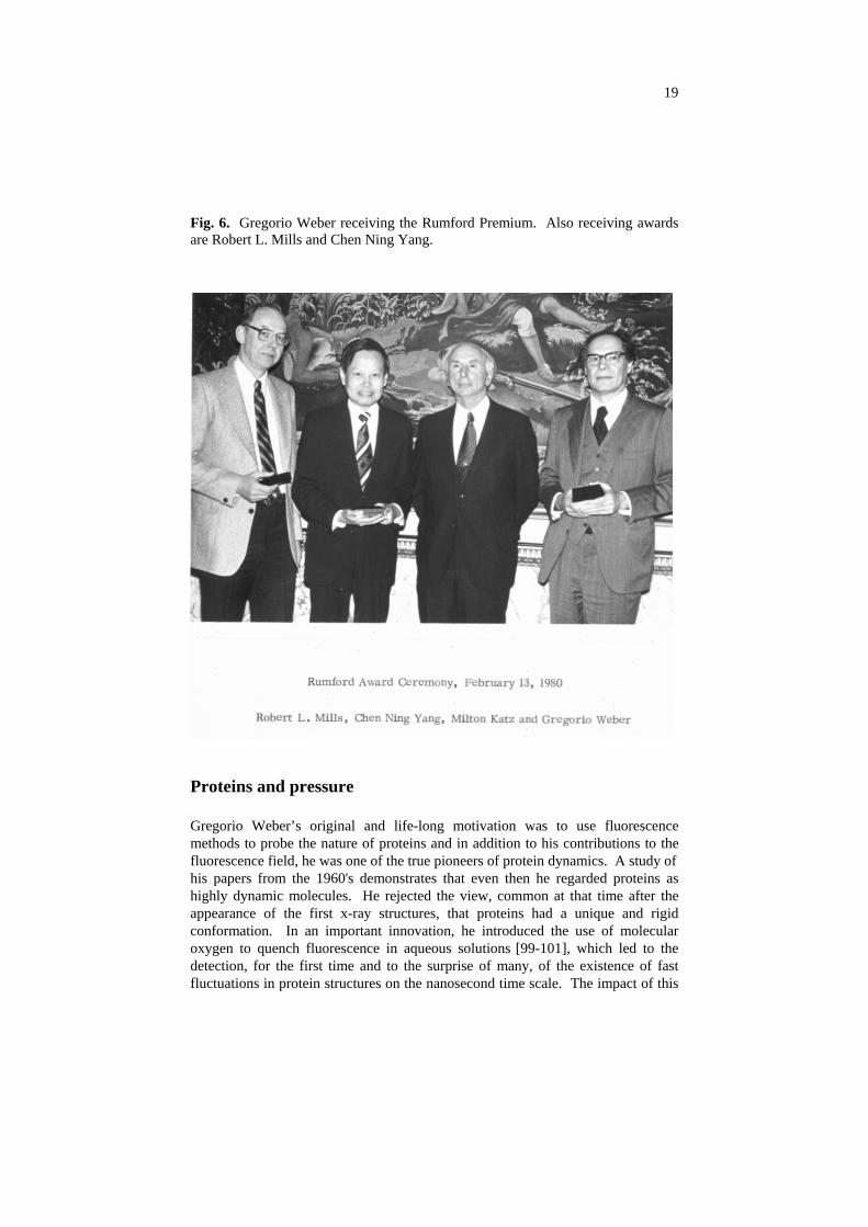
Gregorio Weber’s scientific achievements were recognized by many honors and awards. These include election to the US National Academy of Sciences, election to the American Academy of Arts and Sciences, election as a corresponding member to the National Academy of Exact Sciences of Argentina, the first National Lecturer of the Biophysical Society, the Rumford Premium of the American Academy of Arts and Sciences, the ISCO Award for Excellence in Biochemical Instrumentation, the first Repligen Award for the Chemistry of Biological Processes (awarded by the American Chemical Society) and the first International Jablonski Award for Fluorescence Spectroscopy. It is worth noting that the Rumford Premium is one of the oldest scientific awards given in the United States. It was created by a bequest to the Academy from Benjamin Thompson, Count Rumford, in 1796 - previously awardees include J. Willard Gibbs, A.A. Michelson, Thomas Edison, R.W. Wood, Percy Bridgman, Irving Langmuir, Enrico Fermi, S. Chandrasekhar, Hans Bethe, Lars Onsanger and other highly original thinkers. The Rumford award committee recommended that the 1979 award be given to two physicists, Robert L. Mills and Chen Ning Yang, for their joint work on the theory of gauge invariance of the electromagnetic field, and to Gregorio Weber, “Acknowledged to be the person responsible for modern developments in the theory and application of fluorescent techniques to chemistry and biochemistry” (figure 6).
19
Fig. 6. Gregorio Weber receiving the Rumford Premium. Also receiving awards are Robert L. Mills and Chen Ning Yang.
Proteins and pressure
Gregorio Weber’s original and life-long motivation was to use fluorescence methods to probe the nature of proteins and in addition to his contributions to the fluorescence field, he was one of the true pioneers of protein dynamics. A study of his papers from the 1960's demonstrates that even then he regarded proteins as highly dynamic molecules. He rejected the view, common at that time after the appearance of the first x-ray structures, that proteins had a unique and rigid conformation. In an important innovation, he introduced the use of molecular oxygen to quench fluorescence in aqueous solutions [99-101], which led to the detection, for the first time and to the surprise of many, of the existence of fast fluctuations in protein structures on the nanosecond time scale. The impact of this

20
work was shown by the increasing interest in experimental and theoretical work in protein dynamics, which followed. Weber’s early description of proteins in solution as “kicking and screaming stochastic molecules” [102] has, in recent years, been fully verified both from theoretical and experimental studies. Since this chapter is meant to explore Gregorio Weber’s contribution to fluorescence spectroscopy, I cannot go deeply into his far-reaching contributions to protein research – a topic requiring an additional chapter! These contributions were recognized by the American Chemical Society in 1986, which named Weber as the first recipient of Repligen Award for the Chemistry of Biological Processes. In the 1970's, initially in collaboration with H.G. Drickamer, Weber combined fluorescence and hydrostatic pressure methods to the study of molecular complexes and proteins. It is interesting to note that the initial system he thought to study was the complex formed by isoalloxazine and adenine, one of his original research interests [103]. These observations confirmed the applicability of fluorescence and high-pressure techniques to problems of structure, and particularly dynamics, at the molecular level. Weber and collaborators, in papers published from 1980 to the present, demonstrated that most proteins made up of subunits can be dissociated by the application of hydrostatic pressure, and opened, in this way, a new method to study protein-protein interactions (see [104] for a review of this area). In these studies, quite unexpected properties of protein aggregates were revealed and a new approach to problems in biology and medicine was opened by these observations. For example, Weber and his collaborators demonstrated the possibility of destroying the infectivity of viruses, without affecting their immunogenic capacity, by subjecting them to hydrostatic pressure, and thus opened the possibility of developing viral vaccines that contain, without covalent modification, all the antigens present in the original virus [105,106].
As a result of his investigations employing fluorescence techniques in conjunction with perturbations by pressure and temperature, Weber presented, in the last few years of his life, a novel proposal, which ran contrary to the generally accepted opinion that the properties of the solvent (water) are the determinants of the folding and association of proteins. Instead, he proposed that the very large residual entropy of the protein was responsible for many of the observed properties [107,108]. The correctness of this view, considered iconoclastic and heretical by some, remains to be determined, but Weber’s approach to this topic demonstrates not only his originality of thought, but also his willingness to critically examine scientific data and commonly held opinions, and to draw his own conclusions. Weber’s philosophy in this regard is exemplified by the dedication in his book, Protein Interactions [109]: “Dedicated to Those Who Put Doubt Above Belief.” All scientists would do well to remember his words.
21
Acknowledgements
I wish to thank Bernard Valeur and Jean-Claude Brochon for the invitation to attend the 6th International MAFS conference. I am also indebted to Bernhard Nickel for reprints of his manuscripts on the history of photoluminescence, which helped to serve as an inspiration for this article. I thank Edward Voss, John Croney and Oliver Holub for their suggestions and help with this manuscript. Finally I am grateful to all of the colleagues and friends around the world, in the extended Weber family, with whom I have interacted over the years in Gregorio Weber’s laboratory and afterwards. The support of the National Science Foundation (Grant MCB 9808427) is also gratefully acknowledged.
References
1. Nickel B (1996) Pioneers in photochemistry: From the Perrin diagram to the Jablonski
diagram. EPA Newsletter 58:9-38 2. Nickel B (1997) Pioneers in photophysics: From the Perrin diagram to the Jablonski
diagram. Part 2. EPA Newsletter 61:27-60 3. Nickel B (1998) Pioneers in photophysics: From Wiedemann’s discovery to the
Jablonski diagram. EPA Newsletter 64:19-72 4. Weber G (1947) Fluorescence of riboflavin, diaphorase and related substances. Ph.D.
Dissertation, Cambridge University 5. Perrin F (1926) Polarisation de la lumière de fluorescence. Vie moyenne des molécules
dans l’etat excité. Jour de Phys, VIe Série, 7:390-401 6. Weber G (1989) Final words at Bocca di Magra. In, Jameson DM, Reinhart GD (eds)
Fluorescent biomolecules: Methodologies and applications. Plenum Press, NY pp 343-349
7. Weber G (1948) The quenching of fluorescence in liquids by complex formation. Determination of the mean life of the complex. Trans Faraday Soc 44: 185-189
8. Weber G (1950) Fluorescence of riboflavin and flavin-adenine dinucleotide. Biochem J 47:114-121
9. Weber G (1957) Intramolecular transfer of electronic energy in dihydrodiphospho- pyridine nucleotide. Nature 180:1409
10. Weber G (1958) Transfert d'energie dans la dihydro-diphosphopyridine. J Chim Physique 55:878-886
11. Weber G (1966) Intramolecular complexes of flavins. In, E.C. Slater (ed) Flavins and Flavoproteins, Elsevier Publishing Co., Amsterdam, pp 15-21
12. Scott GT, Spencer RD, Leonard N, Weber G (1970) Emission properties of NADH. Studies of fluorescence lifetimes and quantum efficiencies of NADH, AcPyADH, and simplified synthetic models. J Amer Chem Soc 92:687-695
13. Spencer RD, Weber G (1972) Thermodynamics and kinetics of the intramolecular complex in flavin adenine dinucleotide. In, Akeson A, Ehrenberg A (eds) Structure and function of oxidation reduction enzymes, Pergamon, Oxford-New York, pp. 393-399
14. Weigert F (1920) Über polarisiertes Fluorszenzlicht. Verh d D Phys Ges 1:100-102

22
15. Vavilov SI, Levschin WL (1923) Beiträge zur Frage über polarisiertes Fluoreszenzlicht von Farbstofflösungen. II. Z Physik 16:134-154
16. Shinitzky M, Dianoux AC, Gitler C, Weber G (1971) Microviscosity and order in the hydrocarbon region of micelles and membranes determined with fluorescent probes. I. Synthetic micelles. Biochemistry 10:2106-2113
17. Cogan, U., Shinitzky, M., Weber, G. and Nishida, T. (1973) Microviscosity and order in the hydrocarbon region of phospholipid and phospholipid-cholesterol dispersions determined with fluorescent probes. Biochemistry 12:521-527
18. Weber G (1977) Theory of differential phase fluorometry: Detection of anisotropic molecular rotations. J Chem Phys 66: 4081-4091
19. Weber G, Mitchell GM (1976) Detection of anisotropic rotations by differential phase fluorometry In, Birks, JB (ed) Excited states of biological membranes, Wiley, London, pp. 72-76
20. Mantulin WW, Weber G (1977) Rotational anisotropy and solvent fluorophore bonds: An investigation by differential polarized phase fluorometry. J Chem Phys 66:4092-4099
21. Weber G (1952) Polarization of the fluorescence of macromolecules. I. Theory and experimental method. Biochem J 51:145-155
22. Weber G (1952) Polarization of the fluorescence of macromolecules. II. Fluorescent conjugates of ovalbumin and bovine serum albumin. Biochem J 51:155-167
23. Laurence DJR (1952) A study of the adsorption of dyes on bovine serum albumin by the method of polarization of fluorescence. Biochem J 51:168-177
24. Dandliker WB, Feijen GA (1961) Quantification of the antigen-antibody reaction by the polarization of fluorescence. Biochem Biophys Res Comm 5:299-304
25. Dandliker WB, deSaussure VA (1970) Fluorescence polarization in immunochemistry. Immunochemistry 7:799-828
26. Levinson SA, Dandliker WB, Brawn RJ, Vanderlaan WP (1976) Fluorescence polarization measurement of the hormone-binding site interaction. Endocrinology 99:1129-1143
27. Jameson DM, Sawyer, WH (1995) Fluorescence anisotropy applied to biomolecular interactions. Methods Enzymol. 246:283-300
28. Jablonski A (1960) On the notion of emission anisotropy. Bull. Acad Polon Sci, Série des sci math et phys 8:259-264
29. Singleterry CR, Weinberger LA (1951) The size of soap micelles in benzene from osmotic pressure and from the depolarization of fluorescence. J Am Chem Soc 73:4574-4579
30. Steiner RF, McAlister AJ (1957) Use of the fluorescence technique as an absolute method for obtaining mean relaxation times of globular proteins. J Polymer Sci 24:105-123
31. Weber, G (1953) Rotational Brownian motion and polarization of the fluorescence of solutions. Adv Prot Chem 8, 415-459
32. Weber G (1954) Concentration depolarization of the fluorescence of solutions. Trans Faraday Soc 50:552-557
33. Weber G (1956) Photoelectric method for the measurement of the polarization of the fluorescence of solutions. J Opt Soc Amer 46: 962-970
34. Weber G and Laurence DJR (1954) Fluorescent indicators of adsorption in aqueous solution and on the solid phase. Biochem J 56:xxxi
35. Debye P, Edwards JO (1952) A note on the phosphorescence of proteins. Science 116:143-144
23
36. McClure DS (1949) Triplet-singlet transitions in organic molecules: Lifetime measurements of the triplet state. J Chem Phys 17;905-913
37. Shore VG, Pardee AB (1956) Fluorescence of some proteins, nucleic acids and related compounds. Arch Biochem Biophys 60:100-107
38. Teale FWJ, Weber G (1957) Ultraviolet fluorescence of the aromatic amino acids. Biochem J 53:476-482
39. Weber G, Teale FWJ (1957) Determination of the absolute quantum yield of fluorescent solutions. Trans Faraday Soc 53:646-655
40. Weber G, Teale FWJ (1958) Fluorescence excitation spectrum of organic compounds in solution. Trans Faraday Soc 54:640-648
41. Weber G, Teale FWJ (1959) Electronic energy transfer in heme proteins. Faraday Soc Discussions 27:134-141
42. Teale FWJ, Weber G (1959) Ultraviolet fluorescence of proteins. Biochem J 72:15p 43. Weber G, Tea1e FWJ (1959) Polarization of the ultraviolet fluorescence and electronic
energy transfer in proteins. Biochem. J 72:15p 44. Weber G (1960) Fluorescence polarization spectrum and electronic energy transfer in
tyrosine, tryptophan and related compounds. Biochem J 75: 335-345 45. Weber G (1960) Fluorescence-polarization spectrum and electronic energy transfer in
proteins. Biochem. J. 75, 345-352 46. Velick S (1958) Fluorescence spectra and polarization of glyceraldehyde-3-phosphate
and lactic dehydrogenase complexes. J Biol Chem 237:1455-1467 47. Velick S, Parker CW, Eisen HN (1960) Excitation energy transfer and the quantitative
study of the antibody hapten reaction. Proc Natl Acad Sci USA 46:1470-1482 48. Alpert B, Jameson DM, Weber G (1980) Tryptophan emission From human
hemoglobin and its isolated subunits. Photochem. Photobiol. 31:1-4. 49. Valeur B, Weber G (1977) Resolution of the fluorescence excitation spectrum of indole
into the 1La and 1Lb excitations bands. Photochem. Photobiol. 25:441-444 50. Weber G (1961) Excited states of proteins. In, Light and Life, W.D. McElroy and
Bently Glass (eds.), Johns Hopkins Press, Baltimore, p. 82-106 51. Perrin J (1926) Lumière et réactions chimiques. In, Structure et activité chimiques,
rapports et discussions, Gauthier-Villars, Paris, pp 322-399 52. Weber G (1954) Dependence of the polarization of the fluorescence on the
concentration. Trans Faraday Soc 50:552-555 53. Weber G, Shinitsky M (1970) Failure of energy transfer between identical aromatic
molecules on excitation at the long wave edge of the absorption spectrum. Proc Natl Acad Sci USA 65:823-830
54. Hamman BD, Oleinikov AV, Jokhadze GG, Traut RR, Jameson DM (1996) Dimer/monomer equilibrium and subunit exchange of Escherichia coli ribosomal protein L7/L12. Biochemistry 35:16680-16686
55. Helms MK, Hazlett TL, Mizuguchi H, Hasemann CA, Uyeda K, Jameson DM (1998) Site-directed mutants of rat testis fructose 6-phosphate, 2-kinase:fructose 2,6-bisphosphatase: Localization of conformational alterations induced by ligand binding. Biochemistry 37:14057-14064
56. Valeur B, Weber G (1977) Anisotropic rotations in 1-naphthylamine. Existence of a red-edge transition moment normal to the ring plane. Chem Phys Lett 45:140-144
57. Valeur B, Weber G (1978) A new red-edge effect in aromatic molecules: Anomaly of apparent rotation revealed by fluorescence polarization. J Chem Phys 69:2393-2400
58. Weber G (1960) Enumeration of components in complex systems by fluorescence spectrophotometry. Nature 190:27-29
24
59. Christian GD, Callis JN, Davidson ER (1981) Array detectors and excitation-emission matrices in multicomponent analysis. In, Wenry EL (ed) Modern Fluorescence Spectroscopy, Plenum Press, Chapter 4
60. Soper SA, McGown LB, Warner IM (1994) Molecular fluorescence, phosphorescence, and chemiluminescence spectrometry. Anal Chem 15:428R-444R
61. Gottlieb YY, Wahl P (1963) Étude théorique de la polarisation de fluorescence des macromolécules portant un groupe émetteur mobile autour d’un axe de rotation. J Chim Phys 60:849-856
62. Wahl P, Weber G (1967) Fluorescence depolarization of rabbit gamma globulin conjugates. J Mol Biol 30:371-382
63. Knopp JA, Weber G (1969) Fluorescence polarization of pyrenebutyric bovine serum albumin and pyrenebutyric-human macroglobulin conjugates. J Biol Chem 244:6309-6315
64. Rosen CG, Weber G (1969) Dimer formation from 1-anilino-8-naphthalene sulfonate catalyzed by bovine serum albumin - A new fluorescent molecule with exceptional binding properties. Biochemistry 8:3915-3920
65. Hudson EN, Weber G (1973) Synthesis and characterization of two fluorescent sulfhydryl reagents. Biochemistry 12:4154-4161
66. Weber G, Farris FJ (1979) Synthesis and spectral properties of a hydrophobic fluorescent probe: 2-dimethylamino-6-propionylnaphthalene. Biochemistry 18: 3075-3078
67. Parasassi T, Di Stefano M, Loiero M, Ravagnan G, Gratton E (1994) Influence of cholesterol on phospholipid bilayers phase domains as detected by Laurdan fluorescence. Biophys J 66:120-132
68. Parasassi T, Gratton E, Yu W, Wilson P, Levi M (1997) Two-photon fluorescence microscopy of laurdan gp-domains in model and natural membranes. Biophys J 72: 2413-2429
69. Bagatolli LA, Gratton E, Fidelio GD (1998) Water dynamics in glycosphingolipid aggregates studied by LAURDAN fluorescence. Biophys J 75:331-341
70. Bagatolli LA, Gratton E (1999) Two-photon fluorescence microscopy observation of shape changes at the phase transition in phospholipid giant unilamellar vesicles. Biophys J 77:2090-2101
71. Macgregor RB, Weber G (1981) Fluorophores in polar media: Spectral effects of the Langevin distribution of electrostatic interactions. Annals of the N.Y. Acad. Sci. 366: 140-150
72. Macgregor RB, Weber G (1986) Estimation of the polarity of the protein interior by optical spectroscopy. Nature 319:70-72
73. Prendergast FG, Meyer M, Carlson GL, Iida S, Potter, JD (1983) Synthesis, spectral properties, and use of 6-acryloyl-2-dimethylaminonaphthalene (Acrylodan). A thiol-selective, polarity-sensitive fluorescence probe. J Biol Chem 25:7541-7544
74. Leonard NJ (1997) The ‘chemistry’ of research collaboration. Tetrahedron 53:2323-2355
75. Secrist, J.A., III, Barrio, J.R., Leonard, N.J. and Weber, G. (1972) Fluorescent modifications of adenosine containing coenzymes. Biological activities and spectroscopic properties. Biochemistry 11:3499-3506
76. Barrio JR, Tolman GL, Leonard NJ, Spencer, RD, Weber G (1973) Flavin 1, N6-ethenoadenine dinucleotide: Dynamic and static quenching of fluorescence. Proc Natl Acad Sci USA 70:941-943

25
77. Spencer RD, Weber G, Tolman GL, Barrio JR, Leonard NJ (1974) Species responsible for the fluorescence of 1, N6-ethenoadenosine. Eur J Biochem 45:425-429
78. Gaviola E (1926) Die Abklingzeiten der Fluoeszenz von Farbstofflösungen. Z. Physik 35:748-756
79. Spencer RD, Weber G (1969) Measurement of subnanosecond fluorescence lifetimes with a cross-correlation phase fluorometer. Annals New York Acad Sci 158:361-376
80. Teale FWJ (1983) Phase and modulation fluorometry. In, Cundall RB, Dale RE (eds) Time-resolved fluorescence spectroscopy in biochemistry and biology. NATO ASI Series A:Life Sciences Vol 69, Plenum Press, London, pp 59-80
81. French T, So PTC, Dong CY, Berland KM, Gratton E (1997) Fluorescence lifetime imaging techniques for microscopy. In, Sluder G, Wolf D (eds) Methods in Cell Biology, Video Microscopy Vol. 56, pp. 227-304
82. Franceschini MA, Wallace D, Barbieri B, Fantini S, Mantulin WW, Pratesi S, Donzelli GP, Gratton E (1997) Optical study of the skeletal muscle during exercise with a second generation frequency-domain tissue oximeter. SPIE Proc 2979:807-814
83. Franceschini MA, Moesta KT, Fantini S, Gaida G, Gratton E, Jess H, MantulinWW, Seeber M, Schlag PM, Kaschke M (1997) Frequency-domain instrumentation techniques enhances optical mammography: Initial clinical results. Proc Natl Acad Sci USA, 94:6468-6473
84. Spencer RD, Weber G (1970) Influence of brownian rotations and energy transfer upon the measurements of fluorescence lifetime. J Chem Phys 52:1654-1663
85. Weber G (1981) Resolution of the fluorescence lifetimes in a heterogeneous system by phase and modulation measurements. J Phys Chem 85:949-953
86. Jameson DM, Weber G (1981) Resolution of the pH dependent heterogeneous fluorescence decay of tryptophan by phase and modulation measurements. J Phys Chem 85:953-958
87. Jameson DM, Gratton E (1983) Analysis of Heterogeneous Emissions by Multifrequency Phase and Modulation Fluorometry” In, Eastwood, D (ed) New Directions Molecular Luminescence, ASTM STP 822, American Society for Testing and Materials, Philadelphia, pp. 67-81
88. Jameson DM, Gratton E, Hall RD (1984) The Measurement and Analysis of Hetero-geneous Emissions by Multifrequency Phase and Modulation Fluorometry. App Spectros Rev 20:55-106
89. Gadella TWJ, Clegg RM, Jovin TM (1994) Fluorescence lifetime imaging microscopy: pixel by pixel analysis of phase-modulation data. Bioimaging 2:139-159
90. Gratton E, Limkeman M (1983) A continuously variable frequency cross-correlation phase fluorometer with picosecond resolution. Biophys J 44:315-324
91. Gratton E, Jameson DM, Rosato N, Weber G (1984) A multifrequency cross-correlation phase fluorometer using synchrotron radiation. Rev Sci Instrum 55:486-494
92. Gratton E, Mantulin WW, Weber G, Royer CA, Jameson DM, Reininger R, Hansen RWC (1996) Fluorescence dynamics of biological systems using synchrotron radiation. Rev Sci Instrum 67:1-7
93. Weber G (1971) Theory of fluorescence depolarization by anisotropic Brownian rotations: Discontinuous distribution approach. J Chem Phys 55:2399-2407
94. Belford GC, Belford RL, Weber G (1972) Dynamics of fluorescence polarization macromolecules. Proc Natl Acad Sci USA 69:1392-1393
95. Ehrenberg M, Rigler R (1972) Polarized fluorescence and rotational brownian motion. Chem Phys Lett 14:539-544
26
96. Weber G (1989) Perrin revisited: Parametric theory of the motional depolarization of fluorescence. J Phys Chem 93:6069-6973
97. Jameson DM, Spencer RD, Weber G (1976) Construction and performance of a scanning, photon-counting spectrofluorometer. Rev Sci Instru 47:1034-1038
98. Jameson DM, Weber G, Spencer RD, Mitchell G (1978) Fluorescence polarization: measurements with a photon-counting photometer. Rev Sci Instru 49:510-514
99. Vaughan WM, Weber G (1970) Oxygen quenching pyrenebutyric acid fluorescence in water. A dynamic probe of the microenvironment. Biochemistry 9:464-473
100. Lakowicz JR, Weber G (1973) Quenching of fluorescence by oxygen. A probe for structural fluctuations in macromolecules. Biochemistry 12:4161-4170
101. Lakowicz JR, Weber G (1973) Quenching of protein fluorescence by oxygen. Detection of structural fluctuations in proteins in the nanosecond time scale. Biochemistry 12:4171-4179
102. Weber G (1975) Energetics of ligand binding to proteins. Adv Prot Chem 29:1-83 103. Weber G, Tanaka F, Okamoto BY, Drickamer HG (1974) The effect of pressure on the
molecular complex of isoalloxazine and adenine. Proc Natl Acad Sci USA 71:1264-1266
104. Silva, JL, Weber G (1993) Pressure stability of proteins. Annu Rev Phys Chem 44:89-113
105. Silva JL, Luan P, Glaser M, Voss EW, Weber G (1992) Effects of hydrostatic pressure on a membrane-enveloped virus: High immunogenicity of the pressure-inactivated Virus. J Virology 66:2111-2117
106. Juriekwicz E, Villas-Boas M, Silva JL, Weber G, Hunsmann G, Clegg, RM (1995) Inactivation of simian immunodeficiency virus by hydrostatic pressure. Proc Natl Acad Sci USA 92:6935-6937
107. Weber G (1993) Thermodynamics of the association and the pressure dissociation of oligomeric proteins. J Phys Chem 97:7108-7115
108. Weber G (1998) Thermodynamic concepts in protein condensation. Comm Mol Cell Biophys 9:201-218
109. Weber G (1992) Protein Interactions. Chapman and Hall, New York