The routes of infection spread in central skull-base ...
11
RESEARCH ARTICLE Open Access The routes of infection spread in central skull-base osteomyelitis and the diagnostic role of CT and MRI scans J. Mejzlik 1* , M. Cerny 1 , L. Zeinerova 1 , J. Dedkova 2 , J. Kopriva 2 , K. Zadrobilek 3 , J. Adamkov 3 , V. Chrobok 1 and V. Pellantova 4 Abstract Background: Central skull-base osteomyelitis (CSBO) represents a life-threatening complication of external ear canal infection. Computed tomography (CT) and magnetic resonance imaging (MRI) play key roles in assessment of CSBO progression. Methods: Twelve patients with CSBO were included in a retrospective clinical study. In total, 62 scans (30 CTs and 32 MRIs) were performed to evaluate the extent of inflammatory changes. The scans were read independently by two radiologists specialised in imaging of the head and neck. The regions under the skull base were specified using the online Anatomy Atlas of the skull base. To clarify the timeline, the time period was divided into four parts, and inflammatory changes in the skull-base regions were tracked. Data were statistically analysed. Results: In early stages of the disease, CT scan detects inflammatory changes closely related to the stylomastoid foramen and medially to the posterior belly of the digastric muscle, changes which have been proved to be crucial for the diagnosis of CSBO. Later the infection spreads to the contralateral side causing demineralisation of the bones. Conclusion: Imaging methods play a crucial role not only in establishing the diagnosis, but also in anticipating the direction of infection spread underneath the skull base. Keywords: Central skull-base osteomyelitis, Imaging, Treatment, Cranial nerve palsy, computed tomography Background Malignant otitis externa usually begins as an external audi- tory canal skin infection followed by osteomyelitis of the temporal bone. Spread to the skull base occurs through the tympanomastoid suture to affect the stylomastoid and jugu- lar foramina. Similarly, central skull-base osteomyelitis (CSBO) represents a rare but life-threatening complication, involving the temporal bone pyramid, clivus, sphenoid bone wings, and adjacent soft tissues beneath the central skull base. Typically, the CSBO infection begins in the external ear canal, but spread of infection from paranasal sinuses has also been reported [1]. CSBO usually develops slowly as otitis externa lasting for weeks or months. The infection spreads under the periosteum of the tympanic bone, crosses the petrotympanic and tympanomastoid sutures, and con- tinues towards the temporal bone apex and clivus, and also reaches the bony structures on the contralateral side. The inflammatory changes can be traced not only within the bone but also in the soft tissues below the skull base. Mani- festation of cranial nerve palsy in patients with otitis externa leads to suspicion of CSBO. The group most af- fected are elderly diabetics with recurrent otitis externa [2]. The most common infectious agents include Pseudomonas aeruginosa, Staphylococcus species, or fungi [1, 3]. The presence of symptoms such as severe otalgia, discharge and cranial nerve palsy (VI, VII, VIII, IX, X, XI, XII) are crucial for establishing the diagnosis of CSBO [2], as laboratory pa- rameters are usually inconclusive and the results of both bi- opsy and microbial culture are frequently negative. © The Author(s). 2019 Open Access This article is distributed under the terms of the Creative Commons Attribution 4.0 International License (http://creativecommons.org/licenses/by/4.0/), which permits unrestricted use, distribution, and reproduction in any medium, provided you give appropriate credit to the original author(s) and the source, provide a link to the Creative Commons license, and indicate if changes were made. The Creative Commons Public Domain Dedication waiver (http://creativecommons.org/publicdomain/zero/1.0/) applies to the data made available in this article, unless otherwise stated. * Correspondence: [email protected] 1 Department of Otorhinolaryngology and Head and Neck Surgery, Faculty of Medicine in Hradec Kralove, University Hospital Hradec Kralove, Charles University in Prague, Sokolska 581, Hradec Kralove, 500 05 Hradec Kralove, Czech Republic Full list of author information is available at the end of the article Mejzlik et al. BMC Medical Imaging (2019) 19:60 https://doi.org/10.1186/s12880-019-0331-7
Transcript of The routes of infection spread in central skull-base ...

RESEARCH ARTICLE Open Access
The routes of infection spread in centralskull-base osteomyelitis and the diagnosticrole of CT and MRI scansJ. Mejzlik1* , M. Cerny1, L. Zeinerova1, J. Dedkova2, J. Kopriva2, K. Zadrobilek3, J. Adamkov3, V. Chrobok1 andV. Pellantova4
Abstract
Background: Central skull-base osteomyelitis (CSBO) represents a life-threatening complication of external ear canalinfection. Computed tomography (CT) and magnetic resonance imaging (MRI) play key roles in assessment of CSBOprogression.
Methods: Twelve patients with CSBO were included in a retrospective clinical study. In total, 62 scans (30 CTs and32 MRIs) were performed to evaluate the extent of inflammatory changes. The scans were read independently bytwo radiologists specialised in imaging of the head and neck. The regions under the skull base were specified usingthe online Anatomy Atlas of the skull base. To clarify the timeline, the time period was divided into four parts, andinflammatory changes in the skull-base regions were tracked. Data were statistically analysed.
Results: In early stages of the disease, CT scan detects inflammatory changes closely related to the stylomastoidforamen and medially to the posterior belly of the digastric muscle, changes which have been proved to be crucialfor the diagnosis of CSBO. Later the infection spreads to the contralateral side causing demineralisation of thebones.
Conclusion: Imaging methods play a crucial role not only in establishing the diagnosis, but also in anticipating thedirection of infection spread underneath the skull base.
Keywords: Central skull-base osteomyelitis, Imaging, Treatment, Cranial nerve palsy, computed tomography
BackgroundMalignant otitis externa usually begins as an external audi-tory canal skin infection followed by osteomyelitis of thetemporal bone. Spread to the skull base occurs through thetympanomastoid suture to affect the stylomastoid and jugu-lar foramina. Similarly, central skull-base osteomyelitis(CSBO) represents a rare but life-threatening complication,involving the temporal bone pyramid, clivus, sphenoid bonewings, and adjacent soft tissues beneath the central skullbase. Typically, the CSBO infection begins in the externalear canal, but spread of infection from paranasal sinuseshas also been reported [1]. CSBO usually develops slowly as
otitis externa lasting for weeks or months. The infectionspreads under the periosteum of the tympanic bone, crossesthe petrotympanic and tympanomastoid sutures, and con-tinues towards the temporal bone apex and clivus, and alsoreaches the bony structures on the contralateral side. Theinflammatory changes can be traced not only within thebone but also in the soft tissues below the skull base. Mani-festation of cranial nerve palsy in patients with otitisexterna leads to suspicion of CSBO. The group most af-fected are elderly diabetics with recurrent otitis externa [2].The most common infectious agents include Pseudomonasaeruginosa, Staphylococcus species, or fungi [1, 3]. Thepresence of symptoms such as severe otalgia, discharge andcranial nerve palsy (VI, VII, VIII, IX, X, XI, XII) are crucialfor establishing the diagnosis of CSBO [2], as laboratory pa-rameters are usually inconclusive and the results of both bi-opsy and microbial culture are frequently negative.
© The Author(s). 2019 Open Access This article is distributed under the terms of the Creative Commons Attribution 4.0International License (http://creativecommons.org/licenses/by/4.0/), which permits unrestricted use, distribution, andreproduction in any medium, provided you give appropriate credit to the original author(s) and the source, provide a link tothe Creative Commons license, and indicate if changes were made. The Creative Commons Public Domain Dedication waiver(http://creativecommons.org/publicdomain/zero/1.0/) applies to the data made available in this article, unless otherwise stated.
* Correspondence: [email protected] of Otorhinolaryngology and Head and Neck Surgery, Faculty ofMedicine in Hradec Kralove, University Hospital Hradec Kralove, CharlesUniversity in Prague, Sokolska 581, Hradec Kralove, 500 05 Hradec Kralove,Czech RepublicFull list of author information is available at the end of the article
Mejzlik et al. BMC Medical Imaging (2019) 19:60 https://doi.org/10.1186/s12880-019-0331-7

CT is used abundantly to determine the extent oftemporal bone lesions. For example, in chronic otitismedia with cholesteatoma the preoperative classifica-tion is based on CT examination [4]. Magnetic reson-ance imaging (MRI) is essential for determining softtissue involvement, and in the area of the externalauditory canal, not only the size of the tumour butalso the type of malignancy can be judged on thebasis of diffusion-weighted MRI [5]. In CSBO, com-puted tomography (CT) demonstrates incipient bonedestruction, but demineralization of the bones is evi-dent in CT as a late sign of inflammation. Magneticresonance imaging (MRI) reveals alterations of theclivus on T1-weighted unenhanced images. 67Galliumscintigraphy interestingly reveals higher metabolic ac-tivity in the temporal bone pyramid and the clivus.To monitor the spread of inflammation in the cranialbase, repeat CT or MRI examinations should be per-formed. In the area of the cranial base, near the den-tition, a number of artefacts can degrade both CTand MRI images. Radiation exposure can be reducedby low-dose CT and post-processing data canminimize artefacts [6]. The Curve-Like Structure Ex-traction method may help to track linear structuressuch as fracture lines or vessels [7]. The objective ofthis research is to highlight the role of CT in detect-ing the moment of infection spread from the tem-poral bone to the central skull base. A CT scan, evennon-contrast enhanced, in the early stages of the dis-ease can identify the soft tissue changes before theosteolysis becomes obvious.
MethodsEleven male and one female patients with CSBO wereidentified and assessed retrospectively in the Departmentof Otorhinolaryngology and Head and Neck Surgery, Uni-versity Hospital Hradec Kralove during the period 2012–2017. The data were collected from medical records. Theaverage age of patients was 78.0 years (range 70–85 years),average weight 84.1 kg (range 57-101 kg), and averagebody mass index (BMI) 29.1 (range 21.1–38.1). All the pa-tients had various comorbidities including diabetes melli-tus (DM) 6 (50.0%), ischemic heart disease (IHD) 4(33.3%), renal failure (RF) 4 (33.3%), chronic pulmonarydisease (PD) 7 (58.3%), and others 5 (41.7%).The disease progressed over time in all cases, and the
patients were admitted to the hospital at different stages,with an average hospitalisation time of 20 days (range 4–40 days). The inclusion criteria were: otitis externa notresponsive to conservative outpatient treatment; charac-teristic clinical findings such as severe otalgia, ear dis-charge, one or more cranial nerve palsies (VI, VII, VIII,IX, X, XI, XII); osteolysis of the clivus on CT or alter-ation of the clivus on MRI.
In all cases one or more biopsies were obtained from in-flamed regions: 3 (25%) biopsies were from mastoid cells,9 (75%) from the retropharyngeal space trans-nasally, and1 (8.3%) orbital.Malignancy, mycobacterial infection and granulomato-
sis with polyangiitis were always excluded by laboratoryfindings, imaging methods and biopsy.Microbiological cultures were taken both prior to and
after antibiotic therapy. CT investigation of the temporalbones was performed as a first imaging method,followed by MRI. Both CT and MRI scans were reviewedretrospectively. Altogether 62 scans were performed, ofwhich 30 were CTs and 32 MRIs. The scans were readindependently by two radiologists specialised in imagingof the head and neck. High Resolution Computed Tom-ography (HRCT) was the preferred imaging method toexclude osteomyelitis of the temporal bone. Once thediagnosis of CSBO was confirmed, the disease wasfollowed up by non-contrast enhanced MRI; hence thedifference in the numbers of CTs and MRIs.The regions under the skull base were specified according
to the terminology from the online Anatomy Atlas of theskull base: external_meatus, clivus, longus_capitis_m, rectus_-capitis_m, ICA, petrous_apex, mastoid, foramen_lacerum,Eustachian_tube, medial_pterygoid_m., lateral_pterygoid_m,tensor_velipalatini_m, levator_veli_palatini_m, torus_tubarius,digastric_m, bulbus_jugularis, styloid_process, foramen stylo-mastoideum, condylus_occipitalis [8].The patients were followed up for different periods of
time. To simplify the timeline, the follow-up period wasstructured into four parts: day 0, days 5–75, days 93–177, and days 209–477.Inflammation levels were tracked using erythrocyte
sedimentation rate (ESR), C-reactive protein (CRP), andwhite blood cell count (WBC).The data from all the laboratory results of the patients
were gathered, and missing data (10%) were ignored inthe analysis. Statistical analysis was performed using theanalytical software Statistica version 13.3. Descriptivedata are presented as a number (percent in the file) orthe median (range). Pearson Chi-square statistics wereused to evaluate the statistical significance (p) of DM,IHD, RF, PD, bacterial branches, stylomastoid region in-volvement, and time of hospital stay after diagnosis ofCSBO. Regression analysis was performed to determinethe impact of individual factors on the observed vari-ables. Data are presented as odds ratio (OR), (95% confi-dence interval (CI)), and p.
ResultsMicrobiological cultures revealed one or more bacterialstrains including Pseudomonas aeruginosa in 9 cases(75%), Staphylococcus aureus in 8 cases (66.7%), Can-dida albicans in 2 cases (16.7%), Citrobacter in 1 case
Mejzlik et al. BMC Medical Imaging (2019) 19:60 Page 2 of 11

(8.3%), Candida parapsilosis in 1 case (8.3%), Malasseziafurfur in 1 case (8.3%), Staphylococcus coagulase-nega-tive in 1 case (8.3%), Stenotrophomonas maltophila in 1case (8.3%), and Klebsiella pneumoniae in one case(8.3%). Biopsies showed acute or sub-acute inflammatorygranulation tissue covered by squamous or respiratoryepithelium in all cases.Palsy of one or more cranial nerves developed in all
cases (Table 1).At the time of admission the average levels of inflam-
mation markers were: ESR 60.5 mm/1 h (range 1.3–219.5)/ 80.2 mm/2 h (range 24–149); CRP 33.1 mg/l(range 1.3–219.5); WBC 8.3 × 10⁹/l (range 2.9–16.6).The involvement of selected regions was assessed
(Table 2). In all 12 cases, clinical infection started in theexternal auditory canal. By the time the CSBO diagnosiswas made, changes on imaging were present in only 9patients. In the early stages of the disease, CT scansshowed involvement of the soft tissues surrounding theexternal ear canal without evidence of bony lesions. Theindividual structure involvement is depicted in Figs. 1and 2 provides details of tissue involvement on primaryimaging evaluation. As the disease progressed, CT scansdetected inflammatory changes in fat tissue closely re-lated to the stylomastoid foramen medially to the poster-ior belly of the digastric muscle (Figs. 3 and 4). Oncereviewed, this latter finding was found crucial for thediagnosis of CSBO (Fig. 5). Only in the later stages ofthe disease did non-enhanced T1-weighted images dis-play destruction of the tympanic and temporal bones,osteolytic changes, and alterations of the clivus, as wellas enhancement of the soft tissues beneath the skull base(Figs. 6 and 7).
In all 12 patients, CT scans confirmed the presence offluid in the mastoid cells. However, this is a non-specificfinding, and can be seen even in individuals without ac-tive inflammatory processes. In one patient, the infectionalso involved the temporomandibular joint and theupper eyelid on the affected side.Additionally, MRI investigations in the early stage
of the disease again illustrated fluid in the mastoidcells and oedema of the soft tissues adjacent to theexternal ear canal. Soft tissue enhancement was vis-ible on axial gadolinium-enhanced T1-weighted im-aging, originating from the digastric muscle insertionand continuing to the insertions of the musculuslongus capitis and musculus rectus capitis anterior(Fig. 8). The pharyngobasilar fascia was in close con-tact with the pharyngeal process of the ossis occipita-lis in the middle line, and represented the frontalborder of the infection. Contralateral spread beneaththe clivus in the retropharyngeal space was consist-ently noted. In advanced stages of infection thebutterfly-shaped soft tissue enhancement was locatedunderneath both petrosal bones and the clivus (Figs.6 and 7). Progression of infection from the temporalbone to the other skull base bones was not noticed,except for progression through the fissura sphenooc-cipitalis towards the occipital bone (Fig. 8). When thetreatment proved to be successful, gradual improve-ment of inflammatory changes in the individual re-gions was observed (Figs. 9 and 10).
TherapyAll patients were treated with oral antibiotics as outpa-tients prior to admission to the hospital. As the diagno-sis of CSBO was confirmed, oral or intravenous systemicantibiotic treatment was started according to the specificmicrobial sensitivity and findings (Table 3).Mastoidectomy was indicated for facial nerve paralysis
progression, headaches, persisting ear discharge, or forosteolytic changes of the temporal bone indicated onCT. Histology revealed mild nonspecific inflammatorychanges in all cases. In one case, a thyroplasty type I wasperformed for hoarseness and aspirations caused by cra-nial nerve X palsy. In 10 cases, local control of infectionwas established, cranial nerve palsies disappeared, andheadaches improved, although changes in soft tissuespersisted on MRI. Despite intensive antibiotic therapy, 2patients died in the regional hospitals as a result of thedisease.
Statistical analysisAnalysis was focused on variables that would be early pre-dictors of CSBO development. As this is a retrospectivestudy, the data were not taken consistently in the timeline
Table 1 List of cranial nerves palsies. (n. VI. – n. XII.) which werediagnosed during the course of the disease. Abducens nerve(VI.) palsy is manifested by a disturbance of lateral eye bulbmovement; facial nerve (VII.) palsy by movement disorder of halfof the face; vestibulocochlear nerve (n. VIII.) palsy by sensorineuralhearing loss and disequilibrium; glossopharyngeal nerve (IX.) palsyby disorder of soft palate mobility; vagus nerve (X.) palsy byunilateral vocal cord paresis; accessory nerve (XI.) palsy by inabilityto raise the arm above the horizontal; hypoglossal nerve (XII.)palsy by tongue movement disorders. Most patients had morethan one cranial nerve palsy
Cranial nerve palsy No. % n = 12
n.VIII. 7 58.3
n.VII. 6 50.0
n.IX. 3 25.0
n.X. 3 25.0
n.XII. 3 25.0
n.VI. 1 8.3
n.XI. 1 8.3
Mejzlik et al. BMC Medical Imaging (2019) 19:60 Page 3 of 11

of the disease. Pearson Chi-square test was found to besuitable for testing the statistical significance (p) of vari-ables. From the range of all possible variables that weretested, comorbidities: DM, IHD, RF, PD, others; bacteria:Pseudomonas aeruginosa, Staphylococcus aureus; and stylo-mastoid involvement and duration of hospital stay werefound to be of interest. However, only stylomastoid involve-ment achieved statistical significance p = 0.021 (Table 4).In the knowledge that this was statistically significant,
effort was focused on detection of parameters related tostylomastoid involvement. Among the variables tested,the ESR_mm_1h: OR 1.041, 95%CI 1.010–1.073, p 0.027;and ESR_mm_2h: OR 0.980, 95%CI 0.9511.000, p 0.039were found to be statistically significant, albeit the OR isrelatively low (Table 5).The duration of hospital stay was also tested as a
marker of seriousness of the disease, but was not foundsignificant.
DiscussionInfections of the external auditory canal are not al-ways easy to cure, particularly if the infection spreadsinside the tympanic, mastoid or petrous bones. Themost advanced stage of the infection is CSBO. Thetreatment recommendations are usually based on case
Table 2 Overview of affected regions on the affected and contralateral sides during the time period: day 0 was the day of establishingthe diagnosis, days 5–75 represent early stage, days 73–177 represent advanced stage, and days 209–477 represent follow-up. Resultsfrom the affected and contralateral sides are in separate halves of the Table
Affected Contralateral
Days 0 5–75 93–177 209–477 0 5–75 93–177 209–477
External_meatus 9 12 6 2 0 0 3 3
Clivus 5 10 7 7 2 10 9 7
Longus_capitis_m 3 8 7 6 1 7 8 5
Rectus_capitis_m 4 9 6 6 1 5 8 5
ICA 0 0 0 0 0 0 0 0
Petrous_apex 5 7 6 7 1 4 7 5
Mastoid 7 11 7 4 0 0 9 5
Foramen_lacerum 4 8 5 7 1 4 7 5
Eustachian_tube 4 11 7 6 1 2 8 5
Medial_pterygoid_m. 2 6 3 0 0 2 7 3
Lateral_pterygoid_m 2 4 3 0 0 0 2 3
Tensor_velipalatini_m 4 10 7 6 1 2 8 5
Levator_veli_palatini_m 4 10 7 6 1 3 8 5
Torus_tubarius 4 6 6 6 1 3 8 5
Digastric_m 3 12 3 0 0 0 5 3
Bulbus_jugularis 5 9 7 4 1 4 5 2
Styloid_process 1 6 2 0 0 0 0 0
Foramen stylomastoideum 8 13 6 0 1 3 8 3
Condylus_occipitalis 3 9 5 2 0 0 5 0
Fig. 1 Day 0. Non-contrast enhanced CT in axial plane, sectionthickness 1 mm. The fatty tissues adjacent to the stylomastoidforamen are infiltrated, and cortical bone within the mastoid processis discretely eroded. The mastoid cells are filled with oedematousmucosa and soft tissue
Mejzlik et al. BMC Medical Imaging (2019) 19:60 Page 4 of 11
reports or case series reports [2]. Early diagnosisbased on clinical symptoms and imaging is crucial forsuccessful therapy. Unfortunately, early diagnosis isoften missed as the patients are presented to theENT department after weeks or months of problems[9], and this was the experience in our group. Oncethe greater part of the skull base has been affected,the diagnosis becomes easier to establish but theprognosis worsens. Thus the authors highlight the im-portance of early diagnosis that allows an early startto treatment.
ImagingMedical imaging techniques represent a valuable methodfor diagnostics and follow-up. HRCT is used to excludebone involvement in malignant otitis externa. In ourgroup, HRCT was always performed as the first imagingmethod.Contrast-enhanced CT scans focus on soft tissue
imaging beneath the skull base, gadolinium-enhancedMRI scans are excellent for tracking the involvementof the clivus, and 67Ga scintigraphy can evaluate skullbase metabolic activity [10]. The MRI finding typicallyincludes enhancement of the clivus with hypointensityin the bone marrow space on T1-weighted images,hyperintensity on T2-weighted images, and effacementof the parapharyngeal fat planes and soft tissues atthe skull base [11]. Diffusion-weighted imaging can
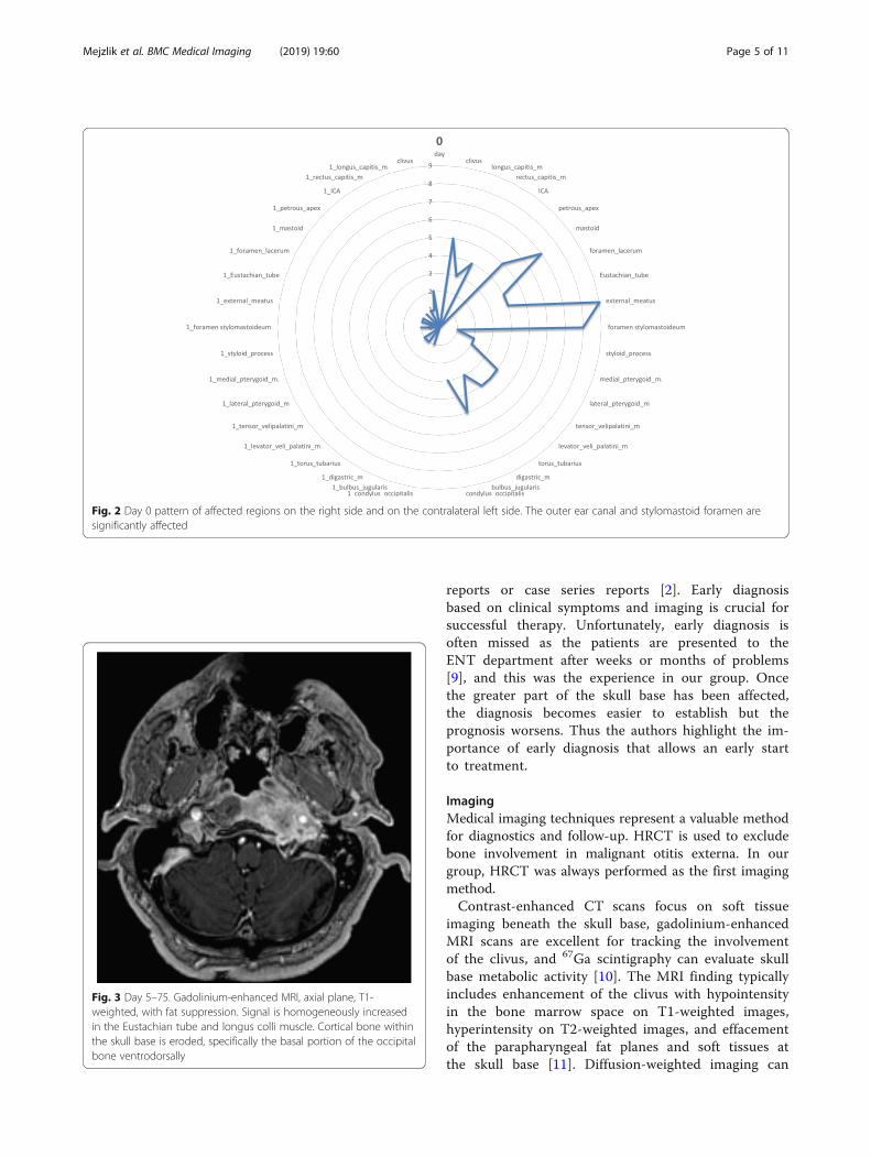
Fig. 3 Day 5–75. Gadolinium-enhanced MRI, axial plane, T1-weighted, with fat suppression. Signal is homogeneously increasedin the Eustachian tube and longus colli muscle. Cortical bone withinthe skull base is eroded, specifically the basal portion of the occipitalbone ventrodorsally
Fig. 2 Day 0 pattern of affected regions on the right side and on the contralateral left side. The outer ear canal and stylomastoid foramen aresignificantly affected
Mejzlik et al. BMC Medical Imaging (2019) 19:60 Page 5 of 11

increase the prominence of the skull lesions and canbe used to distinguish between malignant and benignlesions [12, 13]. To exclude malignancy, one or morebiopsies were usually taken. Without exception, theCT or MRI scans were performed for neurologicalreasons to exclude stroke prior to ENT examination.Once the diagnosis had been established, the MRIwas repeated bi-monthly.Sreepada et al. consider the tympanomastoid suture
crucial for spread of infection [14]. The swelling of soft
Fig. 5 Contrast-enhanced axial CT image of temporal and occipitalbones. Oedematous changes in soft tissues adjacent to the stylomastoidforamen on the left side represent the first sign of osteomyelitis spreadbeyond the temporal bone (arrows)
Fig. 4 Day 5–75: the inflammation spreads to further regions on the affected side
Fig. 6 Day 93–177. Gadolinium-enhanced MRI, axial plane, T1-weighted, with fat suppression. The signal increases homogeneouslyon the left side in retrostyloid and pre-styloid regions, longus collimuscle, and Eustachian tubes on both sides. The signal mildly andhomogeneously increases in the temporomandibular joint, lateralpterygoid, and masseter muscles on the left side. Bone marrowshows signs of infiltration in the basal part of the occipital bone
Mejzlik et al. BMC Medical Imaging (2019) 19:60 Page 6 of 11

tissues adjacent to the tympanomastoid and petrotympa-nic sutures and stylomastoid foramen was visible in allcases on non-contrast CT scans. Therefore the authorsconsider it as a warning sign suggestive of further pro-gression of skull base infection, similarly as reported inmalignant otitis externa [15]. Sreepada also recommendsCT follow-up for CSBO even though the osteolyticchanges of the compact bone on CT represents a ratherlate sign, as 30% of bone must be demineralized to
appear eroded on CT scans (Fig. 11). If soft tissuechanges were visible on non-contrast enhanced CTscans, the suspicion of inflammation was raised. MRI isconsidered essential for CSBO diagnosis [16]. Venouschannels and fascial planes facilitate the spread alongthe dural venous sinuses. Two experienced radiologists
Fig. 8 Gadolinium-enhanced axial T1-weighted spin-echo imagewith fat saturation. In advanced stages of an infection, a butterfly-shaped soft tissue enhancement was described underneath bothpetrosal bones and the clivus (arrow)
Fig. 7 Day 93–177: massive spread of infection to the contralateral side (to the left)
Fig. 9 Day 209–477. Gadolinium-enhanced MRI, axial plane, T1-weighted, with fat suppression. Mild homogeneous signal increasepersists in the pharyngobasilar fascia and longus colli muscles onboth sides. A cyst in the right maxillary sinus represents asecondary finding
Mejzlik et al. BMC Medical Imaging (2019) 19:60 Page 7 of 11

were asked to trace the infection spread on CT and MRIscans. After acquisition of the data, the pathways can betraced even by post-processing imaging methods [7].Hence, most active disease was found in the compactbone along the middle and posterior cranial fossa sur-faces with extension to the petrous apex [14]. In patientNo 2 the infection also involved the temporomandibularjoint and parotid gland and the upper eyelid on theaffected side. This was due to an anatomical variant,persistent foramen of Huschke, which allowed infectionspread from the external auditory canal to the
Fig. 10 Day 209–477: slow infection improvement in all regions on affected and contralateral sides
Table 3 Antibiotics used for the treatment of the CSBO. Theantibiotic choice reflects the two most frequently detectedbacteria, Pseudomonas aeruginosa 9 (75%) and Staphylococcusaureus 8 (66.7%). Both were detected using microbiologicalculture and tested for antibiotic resistance
Antibiotics No. % n = 12
Ciprofloxacin 11 91.7
Ceftazidime 9 75.0
Amoxicillin/clavulanic_acid 9 75.0
Gentamycin 4 33.3
Clindamycin 4 33.3
Meropenem 4 33.3
Clarithromycin 3 25.0
Dalacin 3 25.0
Ofloxacin 3 25.0
Piperacillin 3 25.0
Cefuroxime_axetil 2 16.7
Rifampicin 2 16.7
Oxacylinum 2 16.7
Amikacin 2 16.7
Vancomycin 1 8.3
Linezolid 1 8.3
Co-trimoxazole 1 8.3
Clarithromycin 1 8.3
Table 4 Pearson Chi-square statistics were used to find a factorthat would predict the occurrence of CSBO. The list containsfactors with major statistical importance: (DM) diabetes mellitus,(IHD) ischemic heart disease, (RF) renal failure, (PD) pulmonarydisease. Infection in the stylomastoid region on imagingmethods was found to be the only statistically significant factor
Chi-Square p.
DM 0.000 1.000
IHD 1.33 0.248
RF 1.33 0.248
PD 0.33 0.564
Others 0.33 0.564
Pseudomonas aeruginosa 4.5 0.105
Staphylococcus aureus 1.33 0.248
Stylomastoid involvement 5.33 0.021
Hospital stay duration 0.83 1.000
Mejzlik et al. BMC Medical Imaging (2019) 19:60 Page 8 of 11

temporomandibular joint [17]. In this region, advanceddiffusion imaging MRI can be used [18]. The rudimen-tary foramen was also observed in one case and facili-tated infection spread towards the temporomandibularjoint and surrounding tissues.MRI represents an excellent technique for soft tis-
sue imaging. Fascial spaces, periosteum, muscular in-sertions and bone marrow are visible on post-contrastT1 weighted scans. Sutures between skull bones rep-resent a temporary barrier for osteomyelitis extensionto adjacent bones. The periosteum within the skullbase adheres more tightly to musculus rectus capitisanterior and musculus longus capitis insertions. Add-itionally, the periosteum, muscular tendons and fasciabeneath the skull base are the boundaries for infec-tion spread under the skull base. Those margins arethe reasons for soft tissue enhancement inpost-contrast T1 weighted imaging, which is typicallya butterfly shaped zone of increased signal intensitybetween the spine and pharyngobasilar fascia. An in-teresting finding of infection spread was noticed onrepeated MRI scans. The infection crossed the
midline and spread through the retropharyngeal andretrostyloid parts of the parapharyngeal space into thecontralateral temporal bone (Fig. 12. a, b). Originationof the pharyngobasilar fascia in the skull base repre-sents a relatively steady border for infection spread.However, spaces anteriorly and laterally from thementioned fascia are opened towards the sphenoidalbones and orbits. Crucial advantages in MRI scanningwere reported by Ozgen et al. [16]. MRI scans arerecommended to distinguish between malignant andbenign lesions, and Ginat et al. recommend diffusionweighted imaging (DWI) [12].
Table 5 Regression analysis of the inflammatory markersassociated with: 1) dependent variable: stylomastoidinvolvement; 2) independent variables: ESR after 1 and 2 h werefound to be statistically significant but OR remains low
OR 95% CI p.
CRP 1.010 0.990 1.020 0.281
WBC_x10E9_l 1.020 0.951 1.083 0.569
ESR_mm_1h 1.041 1.010 1.073 0.027
ESR_mm_2h 0.980 0.951 1.000 0.039
Fig. 11 Patient No: 3, five months following mastoidectomy andantibiotic treatment: amoxicillin/clavulanic acid, gentamycin,clarithromycin and dalacin. Non-enhanced CT image (bone window)axial scan cross section of temporal and occipital bones. Osteolysisof the compact bone in the left lateral skull base represents a latesign of osteomyelitis (arrow). Minor changes of spongy bone andmajor changes of compact bone highlight spread of infection underthe periosteum, but represents a late sign of the infection
Fig. 12 a Patient 1, 2 months after the first clinical symptoms: MRI ofthe skull base. Contrast-enhanced axial T1-weighted spin-echoimage with fat saturation. Inflammatory changes of the right sidesoft tissues beneath the skull base, musculus longus capitis andmusculus rectus capitis anterior (arrow). The swelling extends to themidline; the infection originates from the right external auditorycanal. b Patient No. 1, 4 months following the first clinicalsymptoms: MRI of the skull base of the same patient. Contrast-enhanced axial T1-weighted spin-echo image with fat saturation.After antibiotic treatment: ceftazidime, ciprofloxacin, clindamycin,Oxacyllin. Inflammatory changes of the soft tissues below the skullbase progressed to the left side (arrow), musculus longus capitis andmusculus rectus capitis anterior. The swelling affects theretropharyngeal and retrostyloid part of the parapharyngeal space inthe midline and progresses to the left side
Mejzlik et al. BMC Medical Imaging (2019) 19:60 Page 9 of 11

DiagnosticsMalignancy should be excluded in the first instance [2].However, this is not possible without biopsy and histo-logical examination. All cases were confirmed histologically.The presence of acute or subacute inflammatory changeshad no impact on further management, but rather thera-peutic response was guided by inflammation markers. Oursmall number of patients does not permit deeper statisticalanalysis; however, the largest reported meta-analysis [2] in-cluded 42 patients in 68 articles over a period of more than60 years. It was proved that only the ESR correlates withthe disease activity [2, 19]. Thus we conclude that our fileof 12 patient merits consideration. The literature mostlyrecommends the use of MRI as the most valuable imagingmethod to determine the extent of infective changes of softtissues below the skull base [1, 20]. Scintigraphy was notconsidered in our study, but new molecular tracers for as-sessment of bacterial infection have already been tested[21].
TherapyThe bacterial strains found in our study were various,but it is well known that patients with CSBO frequentlyhave been treated with a range of antibiotics prior tohospital admission, and cultures of biopsy material arevery often sterile [10].Both malignant otitis externa and CSBO should be
treated by an ENT specialist [22]. Johnson and Batra sys-tematically reviewed a series of 42 cases and recom-mended conservative management and long-termantibiotic treatment covering Pseudomonas aeruginosa orother infectious agents confirmed by cultivation [2, 19].Pseudomonas aeruginosa infection should be suspectedeven if the microbial cultivation is negative [10], andhence the first choice of antibiotic treatment in our groupof patients on admission was ciprofloxacin. Although inall cases the initial course of antibiotics was administeredintravenously, an experimental intra-arterial application ofantibiotic has also been reported [22]. Despite a numberof papers unambiguously recommending conservativemanagement of CSBO, some authors prefer a surgical ap-proach associated with extensive treatment of affectedbones [1]. We provided mastoidectomy in cases of deteri-oration in facial nerve palsy and the progression of osteo-lytic changes on CT. Two patients died due to renal andheart failure, exacerbated by long-lasting infection andeven the side effects of the prolonged administration ofantibiotics. We therefore conclude, in accordance with theliterature, that comorbidities represent the main prognos-tic factor for survival [23].
ConclusionsFor the establishment of CSBO, CT and MRI findingsproved to be crucial. Both surgical and conservative
treatment of CSBO are dependent on CT and MRI scanfindings. Before the inflammation starts to spread towardsthe clivus, the fat tissues adjacent to the styloid processbecomes stranded on enhanced CT scans and the tissuechanges are obvious even on non-enhanced CT scans. Weconsequently strongly recommend that CT or MRI im-aging of the skull base becomes compulsory in elderly dia-betics with otitis externa lasting more than 2 months.
AbbreviationsBMI: Body mass index; CI: Confidence interval; CRP: C-reactive protein;CSBO: Central skull base osteomyelitis; CT: Computed tomography;DM: Diabetes mellitus; ESR: Erythrocyte sedimentation rate; HRCT: Highresolution computed tomography; IHD: Ischemic heart disease;MRI: Magnetic resonance imaging; OR: Odds ratio; p: Statistical significance;PD: Pulmonary disease; RF: Renal failure; WBC: White blood cell count
AcknowledgementsThe authors are grateful to Ian McColl MD, PhD for assistance with themanuscript.
Ethicals approval and consent to particiapteThe manuscript has been approved by the Ethical Committee of UniversityHospital in Hradec Kralove. All procedures performed in studies involvinghuman participants were in accordance with the ethical standards of theinstitutional and national research committee and with the 1964 Helsinkideclaration and its later amendments.
FundingSupported by the project (Ministry of Health, Czech Republic) for conceptualdevelopment of research organization 00179906.
Authors’ contributionsThe roles of author and co-authors and their responsibilities were as follows:J.M. preparation of the manuscript, first and corresponding author. M.C. prepar-ation of the manuscript. J.D. data collection and preparation of the manuscript.J.K. data collection and preparation of the manuscript. L.Z. data collection andpreparation of the manuscript. K.Z. data collection and preparation of themanuscript. J.A. data collection and preparation of the manuscript. V.C. seniorauthor, preparation and revision of the manuscript. V.P. senior author, prepar-ation and revision of the manuscript. All authors read and approved the finalmanuscript.
Consent for publicationFormal consent is not required for this type of study.Raw data are available in the author’s database.
Competing interestsAll authors certify that they have no affiliations with or involvement in anyorganization or entity with any financial interest (such as honoraria;educational grants; participation in speakers’ bureaus; membership,employment, consultancies, stock ownership, or other equity interest; andexpert testimony or patent-licensing arrangements), or non-financial interest(such as personal or professional relationships, affiliations, knowledge or be-liefs) in the subject matter or materials discussed in this manuscript.
Publisher’s NoteSpringer Nature remains neutral with regard to jurisdictional claims inpublished maps and institutional affiliations.
Author details1Department of Otorhinolaryngology and Head and Neck Surgery, Faculty ofMedicine in Hradec Kralove, University Hospital Hradec Kralove, CharlesUniversity in Prague, Sokolska 581, Hradec Kralove, 500 05 Hradec Kralove,Czech Republic. 2Department of Radiology, Faculty of Medicine in HradecKralove, University Hospital Hradec Kralove, Charles University in Prague,Hradec Kralove, Czech Republic. 3Department of Neurosurgery, Faculty ofMedicine in Hradec Kralove, University Hospital Hradec Kralove, Charles
Mejzlik et al. BMC Medical Imaging (2019) 19:60 Page 10 of 11

University in Prague, Hradec Kralove, Czech Republic. 4Department ofInfectious Diseases, Faculty of Medicine in Hradec Kralove, University HospitalHradec Kralove, Charles University in Prague, Hradec Kralove, Czech Republic.
Received: 10 September 2018 Accepted: 11 April 2019
References1. Ridder GJ, Breunig C, Kaminsky J, Pfeiffer J. Central skull base osteomyelitis:
new insights and implications for diagnosis and treatment. Eur ArchOtorhinolaryngol. 2015;272(5):1269–76.
2. Johnson AK, Batra PS. Central skull base osteomyelitis: an emerging clinicalentity. Laryngoscope. 2014;124(5):1083–7.
3. Gabrielli E, Fothergill AW, Brescini L, Sutton DA, Marchionni E, Orsetti E,Staffolani S, Castelli P, Gesuita R, Barchiesi F. Osteomyelitis caused byAspergillus species: a review of 310 reported cases. Clin Microbiol Infect.2014;20(6):559–65.
4. Razek A, Ghonim MR, Ashraf B. Computed tomography staging of middleear cholesteatoma. Pol J Radiol. 2015;80:328–33.
5. Razek A. Assessment of masses of the external ear with diffusion-weightedMR imaging. Otol Neurotol. 2018;39(2):227–31.
6. Yang W, Zhang HJ, Yang J, Wu JS, Yin XR, Chen Y, Shu HZ, Luo LM,Coatrieux G, Gui ZG, et al. Improving low-dose CT image using residualconvolutional network. Ieee Access. 2017;5:24698–705.
7. Chen Y, Zhang YD, Yang J, Cao Q, Yang GY, Chen J, Shu HZ, Luo LM, CoatrieuxJL, Feng QJ. Curve-like structure extraction using minimal path propagationwith backtracking. IEEE Trans Image Process. 2016;25(2):988–1003.
8. Bony skull base: axial anatomy [http://www.headandneckrad.com/].9. Chawdhary G, Hussain S, Corbridge R. Delayed diagnosis of central skull-
base osteomyelitis with abscess: case report and learning points. Ann R CollSurg Engl. 2017;99(1):E24–E7.
10. Spielmann PM, Yu R, Neeff M. Skull base osteomyelitis: current microbiologyand management. J Laryngol Otol. 2013;127(Suppl 1):8–12.
11. Michalowicz M, Ramanathan M. Clival osteomyelitis presenting as a SkullBase mass. J Neurol Surg Reports. 2017;78(2):E93–E5.
12. Ginat DT, Mangla R, Yeaney G, Ekholm S. Diffusion-weighted imaging ofskull lesions. J Neurol Surg B Skull Base. 2014;75(3):204–13.
13. Lesser FD, Derbyshire SG, Lewis-Jones H. Can computed tomographyand magnetic resonance imaging differentiate between malignantpathology and osteomyelitis in the central skull base? J Laryngol Otol.2015;129(9):852–9.
14. Sreepada GS, Kwartler JA. Skull base osteomyelitis secondary to malignantotitis externa. Curr Opin Otolaryngol Head Neck Surg. 2003;11(5):316–23.
15. Murray ME, Britton J. Osteomyelitis of the skull base: the role of highresolution CT in diagnosis. Clin Radiol. 1994;49(6):408–11.
16. Ozgen B, Oguz KK, Cila A. Diffusion MR imaging features of skull baseosteomyelitis compared with skull base malignancy. Am J Neuroradiol.2011;32(1):179–84.
17. Lacout A, Marsot-Dupuch K, Smoker WR, Lasjaunias P. Foramentympanicum, or foramen of Huschke: pathologic cases and anatomic CTstudy. Am J Neuroradiol. 2005;26(6):1317–23.
18. Razek A. Routine and advanced diffusion imaging modules of the salivaryglands. Neuroimaging Clin N Am. 2018;28(2):245–54.
19. Omran AA, El Garem HF, Al Alem RK. Recurrent malignant otitis externa:management and outcome. Eur Arch Otorhinolaryngol. 2012;269(3):807–11.
20. Muranjan SN, Khadilkar SV, Wagle SC, Jaggi ST. Central Skull Baseosteomyelitis: diagnostic dilemmas and management issues. Indian JOtolaryngol Head Neck Surg. 2016;68(2):149–56.
21. Ordonez AA, Weinstein EA, Bambarger LE, Saini V, Chang YS, DeMarco VP,Klunk MH, Urbanowski ME, Moulton KL, Murawski AM, et al. A systematicapproach for developing Bacteria-specific imaging tracers. J Nucl Med. 2017;58(1):144–50.
22. Yamazaki H, Kikuchi M, Shinohara S, Naito Y, Fujiwara K, Kanazawa Y, Tona R.Intra-arterial administration of antibiotics for refractory skull baseosteomyelitis. Auris Nasus Larynx. 2014;41(4):380–3.
23. Rothholtz VS, Lee AD, Shamloo B, Bazargan M, Pan D, Djalilian HR. Skullbase osteomyelitis: the effect of comorbid disease on hospitalization.Laryngoscope. 2008;118(11):1917–24.
Mejzlik et al. BMC Medical Imaging (2019) 19:60 Page 11 of 11