The role of VEGF and its soluble receptor VEGFR-1 in preterm newborns of preeclamptic mothers with...
6
2013 http://informahealthcare.com/jmf ISSN: 1476-7058 (print), 1476-4954 (electronic) J Matern Fetal Neonatal Med, 2013; 26(10): 978–983 ! 2013 Informa UK Ltd. DOI: 10.3109/14767058.2013.766692 The role of VEGF and its soluble receptor VEGFR-1 in preterm newborns of preeclamptic mothers with RDS Salih Kalay 1 , Burak Cakcak 1 , Osman Oztekin 1 , Gonul Tezel 1 , Ozgur Tosun 2 , Mustafa Akcakus 1 , and Nihal Oygur 1 1 Department of Pediatrics, Akdeniz University Medical School, Antalya, Turkey, and 2 Department of Biostatistics, Akdeniz University Medical School, Antalya, Turkey Abstract Objective: We measured vascular endothelial growth factor (VEGF) and soluble VEGF receptor 1(sVEGFR-1) concentrations in cord blood and tracheal aspirate fluid (TAF) in order to investigate the role of them in lung maturation and the severity of respiratory distress syndrome (RDS) in preterm newborns, born to preeclamptic mothers. Methods: Newborns were divided into two groups as preterms born to preeclamptic mothers and preterms born to healthy mothers. They were also divided into two groups as severe RDS (sRDS) and mild RDS (mRDS) according to the need of surfactant and extent or type of ventilatory support. The concentrations of VEGF and sVEGFR-1 in cord blood and TAF (only in preterms with sRDS) were assayed by standardized enzyme-linked immunosorbent assay. Results: When the patients were evaluated as sRDS and mRDS, cord blood VEGF and VEGF/ sVEGFR-1 concentrations of preterms with sRDS were significantly lower than the concentra- tions of preterms with mRDS. Conversely, cord blood sVEGFR-1 concentrations of preterms with sRDS were significantly higher than the concentrations of preterms with mRDS. VEGF and sVEGFR-1 concentrations in TAF could be compared only between sRDS preterms, born to preeclampsia (þ) and () mothers. No statistical significance was detected between the two groups when sVEGFR-1, VEGF and VEGF/sVEGFR-1 concentrations in TAF were compared. Conclusion: Preeclampsia seems not to have an important effect on VEGF and sVEGFR-1 concentrations of preterm newborns both in cord blood and in TAF. Low VEGF and high sVEGFR-1 concentrations seem to be associated with the severity of RDS irrespective of preeclampsia, suggesting that VEGF may be one of the main components of lung maturation. Keywords Preeclampsia, preterm newborn, respiratory distress syndrome, vascular endothelial growth factor, vascular endothelial growth factor receptor 1 History Received 29 July 2012 Revised 1 January 2013 Accepted 10 January 2013 Published online 13 February 2013 Introduction Vascular endothelial growth factor (VEGF) is accepted as an important growth factor in lung maturation. It plays an essential role not in only embryogenesis, but also in postnatal vasculogenesis, angiogenesis and development of the alveolar capillary bed in the lung. It has also been shown that VEGF stimulates surfactant production by alveolar type II cells [1–3]. VEGF exerts its biological effects via 2 tyrosine kinase receptors, VEGF receptor (VEGFR)-1 and VEGFR-2. These receptors function as endogenous dominant-negative regula- tors of VEGF, by complexing with VEGF with similar affinity as the membrane receptor, thereby preventing downstream signal transduction. Soluble VEGFR-1 is accepted as an important receptor in VEGF regulation during lung develop- ment. Studies investigating the role of VEGF and sVEGFR-1 in respiratory distress syndrome (RDS) or chronic lung disease in newborns have shown that preterms with more severe RDS (sRDS) have lower cord blood VEGF levels or preterms which develop bronchopulmonary dysplasia have low VEFG levels in tracheal aspirate fluid (TAF), concurrent with elevated sVEGFR-1 levels on the first day of life [4–7]. Preeclampsia is a multisystem disorder of unknown cause that is unique to human pregnancy. It is characterized by abnormal vascular response to placentation that is associated with increased systemic vascular resistance, enhanced platelet aggregation, activation of the coagulation system and endo- thelial cell dysfunction [8,9]. VEGF and sVEGFR-1 are also thought to have important roles in the pathogenesis of preeclampsia. Due to the results of many studies it has been accepted that in pregnant women with preeclampsia, the placenta produces elevated levels of sVEGFR-1, which captures free VEGF. As a result, the normal vasculature in the kidney, brain, lungs and other organs of mothers are deprived of essential survival and maintenance signals and become dysfunction [10–13]. However, it is not clear whether VEGF, sVEGFR-1 levels and the lung maturation of newborns, born to preeclamptic mothers are also different from babies, born to healthy mothers. Several studies have shown a reduced risk of RDS or earlier lung maturation in infants who were born to patients with preeclampsia and hypertension [14–16]. In contrast, more recently, a few studies have failed to demonstrate any protective effect of preeclampsia in the development of RDS Address for correspondence: Dr Nihal Oygur, Department of Pediatrics, Division of Neonatology, Akdeniz University Faculty of Medicine, Antalya, Turkey. E-mail: [email protected] J Matern Fetal Neonatal Med Downloaded from informahealthcare.com by University of Sussex Library on 09/26/13 For personal use only.
Transcript of The role of VEGF and its soluble receptor VEGFR-1 in preterm newborns of preeclamptic mothers with...

2013
http://informahealthcare.com/jmfISSN: 1476-7058 (print), 1476-4954 (electronic)
J Matern Fetal Neonatal Med, 2013; 26(10): 978–983! 2013 Informa UK Ltd. DOI: 10.3109/14767058.2013.766692
The role of VEGF and its soluble receptor VEGFR-1 in preterm newbornsof preeclamptic mothers with RDS
Salih Kalay1, Burak Cakcak1, Osman Oztekin1, Gonul Tezel1, Ozgur Tosun2, Mustafa Akcakus1, and Nihal Oygur1
1Department of Pediatrics, Akdeniz University Medical School, Antalya, Turkey, and 2Department of Biostatistics, Akdeniz University Medical School,
Antalya, Turkey
Abstract
Objective: We measured vascular endothelial growth factor (VEGF) and soluble VEGF receptor1(sVEGFR-1) concentrations in cord blood and tracheal aspirate fluid (TAF) in order toinvestigate the role of them in lung maturation and the severity of respiratory distresssyndrome (RDS) in preterm newborns, born to preeclamptic mothers.Methods: Newborns were divided into two groups as preterms born to preeclamptic mothersand preterms born to healthy mothers. They were also divided into two groups as severe RDS(sRDS) and mild RDS (mRDS) according to the need of surfactant and extent or type ofventilatory support. The concentrations of VEGF and sVEGFR-1 in cord blood and TAF (only inpreterms with sRDS) were assayed by standardized enzyme-linked immunosorbent assay.Results: When the patients were evaluated as sRDS and mRDS, cord blood VEGF and VEGF/sVEGFR-1 concentrations of preterms with sRDS were significantly lower than the concentra-tions of preterms with mRDS. Conversely, cord blood sVEGFR-1 concentrations of preterms withsRDS were significantly higher than the concentrations of preterms with mRDS. VEGF andsVEGFR-1 concentrations in TAF could be compared only between sRDS preterms, born topreeclampsia (þ) and (�) mothers. No statistical significance was detected between the twogroups when sVEGFR-1, VEGF and VEGF/sVEGFR-1 concentrations in TAF were compared.Conclusion: Preeclampsia seems not to have an important effect on VEGF and sVEGFR-1concentrations of preterm newborns both in cord blood and in TAF. Low VEGF and highsVEGFR-1 concentrations seem to be associated with the severity of RDS irrespective ofpreeclampsia, suggesting that VEGF may be one of the main components of lung maturation.
Keywords
Preeclampsia, preterm newborn, respiratorydistress syndrome, vascular endothelialgrowth factor, vascular endothelial growthfactor receptor 1
History
Received 29 July 2012Revised 1 January 2013Accepted 10 January 2013Published online 13 February 2013
Introduction
Vascular endothelial growth factor (VEGF) is accepted as an
important growth factor in lung maturation. It plays an
essential role not in only embryogenesis, but also in postnatal
vasculogenesis, angiogenesis and development of the alveolar
capillary bed in the lung. It has also been shown that VEGF
stimulates surfactant production by alveolar type II cells
[1–3]. VEGF exerts its biological effects via 2 tyrosine kinase
receptors, VEGF receptor (VEGFR)-1 and VEGFR-2. These
receptors function as endogenous dominant-negative regula-
tors of VEGF, by complexing with VEGF with similar affinity
as the membrane receptor, thereby preventing downstream
signal transduction. Soluble VEGFR-1 is accepted as an
important receptor in VEGF regulation during lung develop-
ment. Studies investigating the role of VEGF and sVEGFR-1
in respiratory distress syndrome (RDS) or chronic lung
disease in newborns have shown that preterms with more
severe RDS (sRDS) have lower cord blood VEGF levels or
preterms which develop bronchopulmonary dysplasia have
low VEFG levels in tracheal aspirate fluid (TAF), concurrent
with elevated sVEGFR-1 levels on the first day of life [4–7].
Preeclampsia is a multisystem disorder of unknown cause
that is unique to human pregnancy. It is characterized by
abnormal vascular response to placentation that is associated
with increased systemic vascular resistance, enhanced platelet
aggregation, activation of the coagulation system and endo-
thelial cell dysfunction [8,9]. VEGF and sVEGFR-1 are also
thought to have important roles in the pathogenesis of
preeclampsia. Due to the results of many studies it has been
accepted that in pregnant women with preeclampsia, the
placenta produces elevated levels of sVEGFR-1, which
captures free VEGF. As a result, the normal vasculature in
the kidney, brain, lungs and other organs of mothers are
deprived of essential survival and maintenance signals and
become dysfunction [10–13]. However, it is not clear whether
VEGF, sVEGFR-1 levels and the lung maturation of
newborns, born to preeclamptic mothers are also different
from babies, born to healthy mothers.
Several studies have shown a reduced risk of RDS or
earlier lung maturation in infants who were born to patients
with preeclampsia and hypertension [14–16]. In contrast,
more recently, a few studies have failed to demonstrate any
protective effect of preeclampsia in the development of RDS
Address for correspondence: Dr Nihal Oygur, Department of Pediatrics,Division of Neonatology, Akdeniz University Faculty of Medicine,Antalya, Turkey. E-mail: [email protected]
J M
ater
n Fe
tal N
eona
tal M
ed D
ownl
oade
d fr
om in
form
ahea
lthca
re.c
om b
y U
nive
rsity
of
Suss
ex L
ibra
ry o
n 09
/26/
13Fo
r pe
rson
al u
se o
nly.

by acceleration of fetal lung maturation [17,18]. However,
these studies are only based on the incidence of RDS in the
newborns of hypertensive mothers.
Studies, investigating the role of VEGF and its soluble
receptor in lung maturation were only about preterms with
RDS, irrespective of maternal preeclampsia [4–6]. Besides,
some studies compared the cord blood VEGF and sVEGFR-1
concentrations of babies born to preeclamptic and healthy
mothers irrespective of RDS [19–21]. We could not detect any
study in literature investigating the role of VEGF and
sVEGFR-1 in the development of RDS in preterms of
preeclamptic mothers.
This study was conducted to investigate whether maternal
production of VEGF and sVEGFR-1 (a) can change the
concentration of this growth hormone and its soluble receptor
in cord blood and TAF in preterm newborns of preeclamptic
mothers and (b) can affect lung maturation and the severity of
RDS in these babies.
Methods
This study was performed with the approval of the Ethics
Committee of Akdeniz University Faculty of Medicine.
Informed Consent Form was read and signed by parents or
guardians prior to the study.
Study subjects
This study was conducted at Akdeniz University Medical
School Hospital between February 2010 and April 2012. All
infants born at or before 32þ 0 weeks of gestation either to
preeclamptic or to healthy mothers were recorded during this
period. The preeclamptic pregnants with chorioamnionitis,
infection, chronic hypertension, chronic renal or cardiac
disease, active asthma, thyroid disease and epilepsia were
excluded from the study. Preterms with the evidence of
congenital heart disease, various lung diseases other than
RDS, hypoxic or traumatic birth, malformation of the central
nervous system, IUGR, circulatory failure or kidney abnorm-
alities were also excluded.
Newborns included in the study were divided into two
groups as preterms born to preeclamptic mothers and preterms
born to healthy mothers. They were also divided into two
groups as severe RDS (sRDS) and mild RDS (mRDS)
according to the need of surfactant and extent or type of
ventilatory support.
Gestational age (GA) was estimated by last menstrual date,
New Ballard Score or prenatal ultrasound. Data about
antenatal betamethasone administration was obtained from
patient charts.
Preeclampsia was defined as an increased diastolic blood
pressure (490 mmHg) or increased diastolic blood pressure of
15 mmHg over baseline value, with proteinuria (4300 mg/
24 h) on urine analysis [22]. Healthy pregnant was defined as
normotensive, non-proteinuric, pregnant women with no
medical or obstetric complications.
RDS was defined as acute respiratory failure in the first
postnatal hours with characteristic chest radiograph changes
in the absence of sepsis, pneumonia, or other causes of
respiratory distress. Preterms with severe nasal flaring,
grunting, tachypnea, retractions, cyanosis and with severe
pulmonary opacification that obscurated the cardiac and
diaphragmatic margins and with also prominent air brocho-
grams on chest radiography, were accepted as sRDS. Preterms
with mild respiratory symptoms and mild reticulogranular
appearance with also limited air bronchograms on chest
radiography were accepted as mRDS. Patients with sRDS
were immediately put on mechanical ventilator and exogen-
ous surfactant was administered within 2 h after birth. Four
patients needed a second dose of surfactant, 8 h after the first
dose. All preterms with sRDS were extubated as soon as
possible and put on CPAP. Preterms with mRDS were directly
put on CPAP and oral caffeine was started.
Data obtained for all infants included: birth weight (BW),
GA, mode of delivery, sex, antenatal use of corticosteroid,
Apgar score at 5 min of life and use of mechanical ventilation.
VEGF and sVEGFR-1 concentrations of plasma samples,
obtained from cord blood and TAF (only sRDS preterms),
were measured with enzyme-linked immunosorbent assay
(ELISA) and the ratios of VEGF to sVEGFR-1 in TAF were
also calculated.
Collection of blood and VEGF, sVEGFR-1 measurement
Umbilical venous cord blood samples were obtained upon
delivery. All the samples were centrifuged within 15 min of
collection. Plasma was kept at �70 �C until analysis by a
technician who was unaware of the patient’s condition. The
concentrations of VEGF and sVEGFR-1 in cord blood were
assayed by ELISA (R&D Systems, Minneapolis, MN) in
duplicate, according to the protocol recommended by the
manufacturer. The lower limit of detection was510 pg/mL.
Tracheal aspiration fluid sample collection
TAF samples were obtained from infants who required
mechanical ventilation by standardized routine tracheal
lavage as previously described [23,24]. TAF samples were
taken within 2 h after birth and before surfactant administra-
tion. Briefly, 1 ml of sterile isotonic saline was instilled into the
endotracheal tube, the patient was manually ventilated for three
breaths, and the trachea was suctioned twice for 5 s. Lavage
fluid was consistent (2–3 ml) in all subjects each time. Tracheal
aspirates were collected into a trap and transferred into tubes
containing 500 IU of aprotinin (Trasylol; Bayer, Leverkusen,
Germany) and 5 mg of deferoxiamine (Desferal; Ciba, Basel,
Switzerland). The tubes were stored at �20 �C until analysis.
The levels of VEGF and sVEGFR-1 in TAF were assayed by a
standardized ELISA (R&D Systems) in duplicate, according to
the protocol recommended by the manufacturer.
Assay for VEGF and soluble receptors in trachealaspirate samples
All of the assays were conducted in duplicate. To determine
the levels of VEGF and sVEGFR-1 in TAF, samples were
assayed using commercial sandwich immunoassay kits (R&D
Systems). All assays were performed according to the
manufacturer’s protocol. VEGF and soluble receptor concen-
trations in the sample were determined from a linear
standard curve ranging from 0 to 2000 pg/mL for VEGF
and sVEGFR-1. The coefficients of variation from interassay
DOI: 10.3109/14767058.2013.766692 VEGF and its soluble receptor VEGFR-1 in preterm newborns 979
J M
ater
n Fe
tal N
eona
tal M
ed D
ownl
oade
d fr
om in
form
ahea
lthca
re.c
om b
y U
nive
rsity
of
Suss
ex L
ibra
ry o
n 09
/26/
13Fo
r pe
rson
al u
se o
nly.
and intra-assay precision assessments were 10% for all of the
assays. To estimate the in situ TAF concentration of VEGF
and sVEGFR-1, a correction for dilution was calculated using
the ratio of urea-N in the tracheal aspirate sample.
Urea assay
Urea concentration in tracheal lavage fluid was quantitatively
assayed with reagents from Sigma Diagnostics (St. Louis,
MO). The concentration of serum urea was measured by the
hospital chemistry laboratory from a blood sample taken for
routine clinical management within 6 h of lavage. Epithelial
lining fluid (ELF) volume was calculated as follows
ELF ¼ TA urea=Serum ureað Þ � TA volume
Concentrations of VEGF and sVEGFR-1 in TA samples
were all normalized to the ELF to correct for dilution during
the sampling procedure.
Statistical analysis
All statistical analyses were performed with Statistical
Package for Social Sciences (SPSS, Version 16.0, Chicago,
IL) software. Data were summarized as median (range).
Shapiro–Wilk test was used to test the normality condition for
distributions of continuous variables. As for univariate
methods, differences between groups were assessed by
either Mann–Whitney U test or Student’s t-test, depending
on their distributions. Group differences in nominal variables
were assessed with Chi-square test.
In order to understand whether VEGF, sVEGFR-1 con-
centrations in cord blood show statistically significant differ-
ence between RDS groups, multivariate General Linear
Model (GLM) was used. Since GA and BW showed
statistically significant associations with VEGF, sVEGFR-1,
GA and BW were assigned as the covariates of GLM model.
Group differences based on VEGF and sVEGFR-1 concen-
trations were then evaluated using GLM, adjusted for
covariates. Statistical significance was assumed for all tests
when p50.05.
Results
Demographic and clinical characteristics of subjects
A total of 180 pregnants delivered preterms of 32þ 0 weeks
or lower gestation during the study period. Out of 180, 42
were preeclamptic. Four of them were excluded from the
study as one had chronic renal disease, two had chronic
hypertension and one had epilepsy. Therefore 37 preeclamptic
pregnant were found eligible for the study. Out of 138 healthy
pregnants, 37 were randomly enrolled in the study.
Out of 37 preterms born to preeclamptic pregnant, five
were excluded from the study due to the different pathologies
as congenital heart disease, various lung diseases other than
RDS, hypoxic or traumatic birth, malformation of the central
nervous system. Parental consent for umbilical cord blood
sample could not be obtained from two preterms. Out of 37
preterms born to healthy pregnants, parental consent could not
also be obtained from 13, for umbilical cord blood sample.
Therefore 54 preterms (preterm infants of 30 preeclamptic and
24 healthy pregnant) were enrolled in this study.
Out of 30 preterms born to preeclamptic mothers, sRDS
developed in nine and mRDS in 21; out of 24 preterms born to
healthy mothers sRDS developed in 10 and mRDS in14.
Demographic and clinical characteristics of the studied
population according to sRDS, mRDS and preeclampsia (þ)
preeclampsia (�) groups were shown in Tables 1 and 2. GA,
BW and Apgar score at 5 min of postnatal life in preterms
with sRDS were significantly lower than in preterms with
mRDS (p: 0.043 for GA; p: 0.036 for BW and p: 0.01 for
Apgar score). GA and BW of preterms born to preeclamptic
mothers were significantly lower than in preterms born to
healthy mothers (p: 0.025 for GA; p: 0.045 for BW). The rate
of caesarean section in mothers with preeclampsia, was higher
than healthy mothers (p: 0.019).
Laboratory results of subjects
VEGF and sVEGFR-1 concentrations were assayed in 54 cord
blood samples. When the patients were evaluated as sRDS
and mRDS, cord blood VEGF concentrations and VEGF/
sVEGFR-1 ratios of preterms with sRDS were significantly
lower than preterms with mRDS (p: 0.001 for VEGF and
p: 0.001 for VEGF/sVEGFR-1). Conversely, cord blood
sVEGFR-1 concentrations of preterms with sRDS were
significantly higher than the sVEGFR-1 concentrations of
preterms with mRDS (p: 0.002) (Table 1 and Figures 1 and 2).
When the patients were evaluated as preeclampsia (þ) and
preeclampsia (�) preterms, cord blood VEGF, sVEGFR-1
concentrations and VEGF/sVEGFR-1 ratios were insignifi-
cant (Table 2).
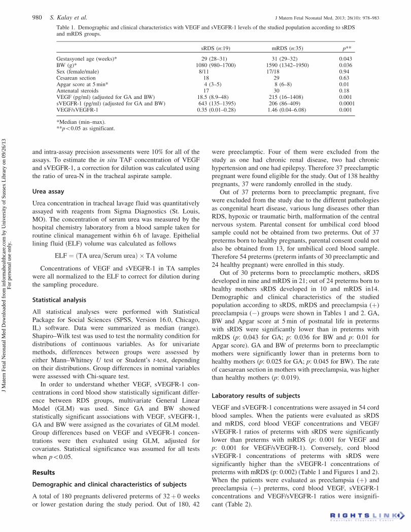
Table 1. Demographic and clinical characteristics with VEGF and sVEGFR-1 levels of the studied population according to sRDSand mRDS groups.
sRDS (n:19) mRDS (n:35) p**
Gestasyonel age (weeks)* 29 (28–31) 31 (29–32) 0.043BW (g)* 1080 (980–1700) 1590 (1342–1950) 0.036Sex (female/male) 8/11 17/18 0.94Cesarean section 18 29 0.63Apgar score at 5 min* 4 (3–5) 8 (6–8) 0.01Antenatal steroids 17 30 0.18VEGF (pg/ml) (adjusted for GA and BW) 18.5 (8.9–48) 215 (16–1408) 0.001sVEGFR-1 (pg/ml) (adjusted for GA and BW) 643 (135–1395) 206 (86–409) 0.0001VEGF/sVEGFR-1 0.35 (0.01–0.28) 1.46 (0.04–6.08) 0.001
*Median (min–max).**p50.05 as significant.
980 S. Kalay et al. J Matern Fetal Neonatal Med, 2013; 26(10): 978–983
J M
ater
n Fe
tal N
eona
tal M
ed D
ownl
oade
d fr
om in
form
ahea
lthca
re.c
om b
y U
nive
rsity
of
Suss
ex L
ibra
ry o
n 09
/26/
13Fo
r pe
rson
al u
se o
nly.

Figure 2. Cord blood sVEGFR-1 levels ofmRDS and sRDS preterms (adjusted for GAand BW).
Figure 1. Cord blood VEGF levels ofmRDS and sRDS preterms (adjusted for GAand BW).
Table 2. Demographic and clinical characteristics with VEGF and sVEGFR-1 levels of the studied population according topreeclampsia (þ) and preeclampsia(�) groups.
Preeclampsia(þ) (n:30) Preeclampsia(�) (n:24) p**
Gestasyonel age (weeks)* 30 (28–31) 31 (29–32) 0.025BW (g)* 1320 (980–1950) 1480 (850–1800) 0.045Sex (female/male) 14/16 11/13 0.86Cesarean section 28 19 0.019Apgar score at 5 min* 6 (3–8) 6 (3–8) 0.96Antenatal steroids 24 16 0.13sRDS 9 10 0.37VEGF levels (pg/ml)* 42 (12–1408) 69 (9–1274) 0.344sVEGFR-1 levels (pg/ml)* 446 (86–1349) 311 (129–857) 0.355VEGF/sVEGFR-1* 0.19 (0.01–6.08) 0.315 (0.01–5.17) 0.284
*Median (min–max).**p50.05 as significant.
DOI: 10.3109/14767058.2013.766692 VEGF and its soluble receptor VEGFR-1 in preterm newborns 981
J M
ater
n Fe
tal N
eona
tal M
ed D
ownl
oade
d fr
om in
form
ahea
lthca
re.c
om b
y U
nive
rsity
of
Suss
ex L
ibra
ry o
n 09
/26/
13Fo
r pe
rson
al u
se o
nly.

Due to the ethical problems, TAF samples could be
obtained only from preterms who have been incubated for
mechanical ventilation, so, VEGF and sVEGFR-1 concentra-
tions in TAF could be compared only between sRDS preterms
born to preeclampsia (þ) mothers and sRDS preterms born to
preeclampsia (�) mothers. A total of 19 TAF samples were
collected after birth. However no statistical significance was
detected between the two groups when sVEGFR-1, VEGF and
VEGF/sVEGFR-1 concentrations in TAF were compared
(p: 0.26 for VEGF, p: 0.96 for sVEGFR-1, p: 0.48 for VEGF/
sVEGFR-1) (Table 3).
After adjustments with GA and BW, the differences in
VEGF and sVEGFR-1 levels between RDS groups were
investigated with GLMs. Both concentrations showed statis-
tically significant difference between RDS groups after the
adjustments (for VEGF: p¼ 0.001 and for sVEGFR-1:
p¼ 0.0001) (Table 1).
Discussion
Soluble VEGFR-1 has been proposed as a possible circulating
endothelial damaging factor in preeclampsia. Soluble
VEGFR-1 mRNA is up-regulated in preeclamptic placentas,
possibly due to placental hypoxia. It acts as a potent VEGF
and placental growth factor (PlGF) antagonist by binding
VEGF and PlGF molecules, thereby reducing free-circulating
concentrations of VEGF and PlGF [8–11,13,25–27]. The
rapid decline of sVEGFR-1 concentrations after delivery
found by Maynard et al. [12] supports a placental origin of
sVEGFR-1 in preeclampsia.
Studies investigating VEGF, sVEGFR-1 levels in pre-
eclamptic pregnant and their newborns had conflicting results.
According to the study done by Staff et al. [25] sVEGFR-1
concentration was elevated in fetal circulation in preeclampsia,
but at a much lower level than in the maternal circulation.
Similarly, Schlembach et al. [9], Catarino et al. [19] and Kwon
et al. [20] reported that umbilical venous serum concentrations
of sVEGFR-1 were significantly elevated and VEGF concen-
trations were significantly decreased in preeclampsia, com-
pared to normal pregnancies. Similarly, Tsao et al. [5] found
sVEGFR-1 levels significantly high in the cord blood of
preeclampsia. In contrast, Galazios [21] detected umbilical
cord serum VEGF levels of newborns born to preeclamptic
women significantly higher than in newborns born to healthy
women. In the light of these suggestions, we hypothesized that
the placental secretion of angiogenic factors in preeclampsia
may affect their concentrations, not only in fetal circulation,
but also in the lung. Therefore, we measured cord blood
concentrations of free VEGF and sVEGFR-1 in preterms of
preeclamptic and healthy mothers. We also measured their
concentrations in TAF only in sRDS preterms with and
without preeclamptic mothers.
According to our blood results, there was no significant
difference between the cord blood VEGF and sVEGFR-1
concentrations of preterms with preeclamptic mothers and
preterms with healthy mothers suggesting that preeclampsia
did not influence or play an important role in VEGF and
sVEGFR-1 concentrations of babies, born to preeclamptic
mothers.
RDS due to surfactant deficiency is a common cause of
morbidity and mortality in premature infants. An increasing
evidence suggests that VEGF may contribute to surfactant
secretion and pulmonary maturation [2,28]. VEGF is a
specific endothelial cell mitogen that regulates endothelial
cell differentiation, angiogenesis, and maintenance of existing
vessels [29–31]. VEGF has been shown to be deposited in the
sub epithelial matrix at the leading edges of branching
airways where it stimulates angiogenesis [32]. Additionally,
VEGF induces airway epithelial-cell proliferation in human
lungs in vitro [33]. In human fetal lung, VEGF is localized in
alveolar epithelial cells and myocytes, suggesting a paracrine
role for VEGF in modulating activities in adjacent vascular
endothelium [30]. Compernolle et al. [2] showed that type II
cells respond to VEGF treatment with increased surfactant
protein (SP)-B and SP-C production. In the study by Lassus
et al. [4], infants with sRDS had less VEGF in their TAF
during the early postnatal period than infants with mRDS and
cord blood VEGF elevation was correlated with absence of
RDS. These data suggested that VEGF might be a marker of
pulmonary maturity.
In our study, cord blood VEGF, VEGF/sVEGFR-1 con-
centrations of preterm with sRDS were found significantly
lower (p: 0.001 for both VEGF and VEGF/sVEGFR-1) and
sVEGFR-1 concentrations were found significantly higher
(p: 0.002) than the concentrations of preterms with mRDS.
When the patients were evaluated as preeclampsia (þ) and
preeclampsia (�) preterms, cord blood VEGF, sVEGFR-1
and VEGF/sVEGFR-1 concentrations were insignificant. Our
results also suggested that VEGF and sVEGFR-1 levels were
related with the severity of RDS or lung maturation but not
with the presence or absence of preeclampsia. This suggestion
was also supported with VEGF, sVEGFR-1 concentrations
and VEGF/sVEGFR-1 ratios of TAF, that all of them were
significantly higher in preterms with sRDS, apart from
preeclampsia.
Studies investigating the relation between preeclampsia
and the occurrence of RDS depend only on the incidence of
RDS in the newborns of hypertensive mothers [14–18,34]. To
our knowledge, our study is the first that investigates the
relationship between the occurrence of RDS and preeclampsia
on the basis of VEGF, sVEGFR-1 concentrations and VEGF/
sVEGFR-1 ratios both in cord blood and TAF. According to
our results, preterm babies of preeclamptic mothers had the
same probability of having sRDS as newborns of normoten-
sive mothers, suggesting that preeclampsia may not protect
against the development of sRDS and fetal lung maturity
seems not to be accelerated in preeclampsia.
In conclusion, preeclampsia seems not to have an import-
ant effect on VEGF and sVEGFR-1 concentrations of
Table 3. VEGF and sVEGFR-1 levels in the TAF of sRDS preterms.
VEGF and VEGF/sVEGFR-1 levelsin TAF
Preeclampsia(þ)sRDS (n:9)
Preeclampsia(�)sRDS (n:10) p**
VEGF (pg/ml)* 25 (10.5–98.3) 43.5 (9.7–1043.8) 0.26sVEGFR-1 (pg/ml)* 234 (50–1936) 222 (54–1010) 0.96VEGF/sVEGFR-1
(pg/ml)*0.10 (0.01–4.08) 0.19 (0.05–5.17) 0.48
*Median (min–max).
982 S. Kalay et al. J Matern Fetal Neonatal Med, 2013; 26(10): 978–983
J M
ater
n Fe
tal N
eona
tal M
ed D
ownl
oade
d fr
om in
form
ahea
lthca
re.c
om b
y U
nive
rsity
of
Suss
ex L
ibra
ry o
n 09
/26/
13Fo
r pe
rson
al u
se o
nly.

newborns both in cord blood and in TAF. Low VEGF and
high sVEGFR-1 concentrations seem to be associated with the
severity of RDS irrespective of preeclampsia, suggesting that
VEGF may be one of the main components of lung
maturation. However, more clinical or experimental studies
are necessary in order to obtain a definitive conclusion about
the effect of VEGF on lung maturation.
Declaration of interest
The authors report no conflicts of interest. The authors alone
are responsible for the content and writing of the paper.
References
1. Ferrara N, Gerber HP, Le Couter J. The biology of VEGF and itsreceptors. Nat Med 2003;9:669–76.
2. Compernolle V, Brusselmans K, Acker T, et al. Loss of HIF-2alphaand inhibition of VEGF impair fetal lung maturation, whereastreatment with VEGF prevents fatal respiratory distress in prema-ture mice. Nat Med 2002;8:702–10.
3. Thebaud B. Angiogenesis in lung development, injury and repair:implications for chronic lung disease of prematurity. Neonatology2007;91:291–7.
4. Lassus P, Ristimaki A, Ylikorkala O, et al. Vascular endothelialgrowth factor in human preterm lung. Am J Respir Crit Care Med1999;159:1429–33.
5. Tsao PN, Wei SC, Chou HC, et al. Vascular endothelial growthfactor in preterm infants with respiratory distress syndrome. PediatrPulmonol 2005;39:461–5.
6. Abdel-Hady S, Abdel-Ghafar E, Abdel-Rehim I, Abdel-Gawad ER.Vascular endothelial growth factor in preterm infants with respira-tory distress syndrome. Egypt J Immunol 2007;14:43–9.
7. Hasan J, Beharry KD, Valencia AM, et al. Soluble vascularendothelial growth factor receptor 1 in tracheal aspirate fluid ofpreterm neonates at birth may be predictive of bronchopulmonarydysplasia/chronic lung disease. Pediatrics 2009;123:1545–7.
8. Sibai B, Dekker G, Kupferminc M. Pre-eclampsia. Lancet 2005;4:785–99.
9. Schlembach D, Beider E. Angiogenic factors in preeclampsia. J SocGyn Invest 2003;10:316A.
10. Levine RJ, Maynard SE, Qian C, et al. Circulating angiogenic fac-tors and the risk of preeclampsia. N Engl J Med 2004;350:672–83.
11. Zhou Y, McMaster M, Woo K, et al. Vascular endothelial growthfactor ligands and receptors that regulate human cytotrophoblastsurvival are dysregulated in severe preeclampsia and hemolysis,elevated liver enzymes, and low platelets syndrome. Am J Pathol2002;160:1405–23.
12. Maynard SE, Min JY, Merchan J, et al. Excess placental solublefmslike tyrosine kinase 1 (sFlt1) may contribute to endothelialdysfunction, hypertension and proteinuria in preeclampsia. J ClinInvest 2003;111:649–58.
13. Sezer SD, Kucuk M, Yenisey C, et al. Comparison of angiogenicand anti-angiogenic factors in maternal and umbilical cord blood inearly- and late-onset pre-eclampsia. Gynecol Endocrinol2012;28:628–32.
14. Bent AE, Gray JH, Luther ER, et al. Assessment of fetal lungmaturity: relationship of gestational age and pregnancy complica-tions to phosphatidylglycerol levels. Am J Obstet Gynecol1982;142:664–9.
15. Ferroni KM, Gross TL, Sokol RJ, Chik L. What affects fetalpulmonary maturation during diabetic pregnancy? Am J ObstetGynecol 1984;150:270–4
16. Shah DM, Shenai JP, Vaughn WK. Neonatal outcome of prematureinfants of mothers with preeclampsia. J Perinatol 1995;15:264–7.
17. Tubman TR, Rollins MD, Patterson C, Halliday HL. Increaseincidence of respiratory distress syndrome in babies of hypertensivemothers. Arch Dis Child 1991;66:52–4.
18. Bowen JR, Leslie GI, Arnold JD, et al. Increased incidence ofrespiratory distress syndrome in infants following pregnanciescomplicated by hypertension. Aust N Z J Obstet Gynaecol1988;28:109–12.
19. Catarino C, Rebelo I, Belo L, et al. Fetal and maternal angiogenic/anti-angiogenic factors in normal and preeclamptic pregnancy.Growth Factors 2009;27:345–51.
20. Kwon JY, Maeng YS, Kwon YG, et al. Decreased endothelialprogenitor cells in umbilical cord blood in severe preeclampsia.Gynecol Obstet Invest 2007;64:103–8.
21. Galazios G, Papazoglou D, Giagloglou K, et al. Umbilical cordserum vascular endothelial growth factor (VEGF) levels in normalpregnancies and in pregnancies complicated by preterm delivery orpre-eclampsia. Int J Gynaecol Obstet 2004;85:6–11.
22. ACOG Committee on Obstetric Practice. ACOG practice bulletin.Diagnosis and management of preeclampsia and eclampsia.American College of Obstetricians and Gynecologists. ObstetGynecol 2002;99:159–67.
23. Darlow BA, Sluis KB, Inder TE, Winterbourn CC. Endotrachealsuctioning of the neonate: comparison of two methods as a sourceof mucus material for research. Pediatr Pulmonol 1997;23:217–21.
24. Varsila, E, Pesonen E, Andersson S. Early protein oxidation in theneonatal lung is related to development of chronic lung disease.Acta Paediatr 1995;84:1296–9.
25. Staff AC, Braekke K, Harsem NK, et al. Circulating concentrationsof sFlt1 (soluble fms-like tyrosine kinase 1) in fetal and maternalserum during pre-eclampsia. Eur J Obstet Gynecol Reprod Biol2005;122:33–9.
26. Luft FC. Pieces of the preeclampsia puzzle. Nephrol DialTransplant 2003;18:2209–10.
27. Roberts JM, Rajakumar A. Preeclampsia and Soluble fms-LikeTyrosine Kinase 1. J Clin Endocrinol Metab 2009;94:2252–4.
28. Chen CM, Wang LF. High-dose vascular endothelial growth factorincreases surfactant protein gene expressions in preterm rat lung.Earl Hum Dev 2007;83:581–4.
29. Peters KG, De Vries C, Williams LT. Vascular endothelial growthfactor receptor expression during embryogenesis and tissue repairsuggests a role in endothelial differentiation and blood vesselgrowth. Proc Natl Acad Sci USA 1993;90:8915–9.
30. Shifren JL, Doldi N, Ferrara N, et al. In the human fetus, vascularendothelial growth factor is expressed in epithelial cells andmyocytes, but not in vascular endothelium: implications for modeof action. J Clin Endocrinol Metabol 1994;79:316–22.
31. Pepper MS, Ferrara N, Orci L, Montesano R. Potent synergismbetween vascular endothelial growth factor and basic fibroblastgrowth factor in the induction of angiogenesis in vitro. BiochemBiophys Res Commun 1992;89:824–31.
32. Healy AM, Morgenthau L, Zhu X, et al. VEGF is deposited in thesubepithelial matrix at the leading edge of branching airways andstimulates neovascularization in the murine embryonic lung. DevDyn 2000;219:341–52.
33. Brown KR, England KM, Goss KL, et al. VEGF induces airwayepithelial cell proliferation in human fetal lung in vitro. Am JPhysiol Lung Cell Mol Physiol 2001;281:1001–10.
34. Chang EY, Menard MK, Vermillion ST, et al. The associationbetween hyaline membrane disease and preeclampsia. Am J ObstetGynecol 2004;191:1414–7.
DOI: 10.3109/14767058.2013.766692 VEGF and its soluble receptor VEGFR-1 in preterm newborns 983
J M
ater
n Fe
tal N
eona
tal M
ed D
ownl
oade
d fr
om in
form
ahea
lthca
re.c
om b
y U
nive
rsity
of
Suss
ex L
ibra
ry o
n 09
/26/
13Fo
r pe
rson
al u
se o
nly.