The role of the right hemisphere in semantic control …epubs.surrey.ac.uk/812744/1/the role of the...
18
The role of the right hemisphere in semantic control: A case-series comparison of right and left hemisphere stroke Hannah E. Thompson n , Lauren Henshall, Elizabeth Jefferies Department of Psychology and York Neuroimaging Centre, University of York, UK article info Article history: Received 16 August 2015 Received in revised form 28 February 2016 Accepted 29 February 2016 Available online 2 March 2016 Keywords: Semantic Right hemisphere Control Summation abstract Semantic control processes guide conceptual retrieval so that we are able to focus on non-dominant associations and features when these are required for the task or context, yet the neural basis of semantic control is not fully understood. Neuroimaging studies have emphasised the role of left inferior frontal gyrus (IFG) in controlled retrieval, while neuropsychological investigations of semantic control deficits have almost exclusively focussed on patients with left-sided damage (e.g., patients with semantic aphasia, SA). Nevertheless, activation in fMRI during demanding semantic tasks typically extends to right IFG. To investigate the role of the right hemisphere (RH) in semantic control, we compared nine RH stroke patients with 21 left-hemisphere SA patients, 11 mild SA cases and 12 healthy, aged-matched controls on semantic and executive tasks, plus experimental tasks that manipulated semantic control in paradigms particularly sensitive to RH damage. RH patients had executive deficits to parallel SA patients but they performed well on standard semantic tests. Nevertheless, multimodal semantic control deficits were found in experimental tasks involving facial emotions and the ‘summation’ of meaning across multiple items. On these tasks, RH patients showed effects similar to those in SA cases – multimodal deficits that were sensitive to distractor strength and cues and miscues, plus increasingly poor perfor- mance in cyclical matching tasks which repeatedly probed the same set of concepts. Thus, despite striking differences in single-item comprehension, evidence presented here suggests semantic control is bilateral, and disruption of this component of semantic cognition can be seen following damage to either hemisphere. & 2016 The Authors. Published by Elsevier Ltd. This is an open access article under the CC BY license (http://creativecommons.org/licenses/by/4.0/). 1. Introduction Semantic cognition allows us to understand the meaning of words and objects that we encounter and to produce appropriate thoughts and behaviour (Lambon Ralph, 2014; Patterson et al., 2007). Successful semantic cognition requires the interaction of several component processes (Jefferies, 2013; Jefferies and Lambon Ralph, 2006; Rogers et al., 2015): (i) stored knowledge about items and their features and associations (e.g., SALT-PEPPER) and (ii) the ability to flexibly control the retrieval of information to suit our current goals and the situation. For example, when we see a sign on the motorway saying: “SALT-SPREADING”, we are able to inhibit strong associations (e.g., dinner table) and retrieve distant associations relevant to this context (Badre et al., 2005; Noonan et al., 2010; Thompson-Schill et al., 1997). These components of semantic cog- nition involve distinct brain networks, and are differentially damaged in different patient populations (for example, semantic dementia vs. aphasia: Jefferies and Lambon Ralph (2006)). They may also show different degrees of lateralisation, since semantic deficits reflecting a loss of conceptual knowledge typically arise from bilateral damage, while those reflecting poor control over re- trieval are largely observed following left-sided damage. Progressive degradation of the store of conceptual knowledge is observed in semantic dementia, SD, following bilateral atrophy in the anterior temporal lobes (ATL; Bozeat et al., 2000; Galton et al., 2001; Mummery et al., 2000; Patterson et al., 2007). Several lines of evidence suggests that both ATLs support the semantic store: (i) Unlike SD patients, patients with unilateral ATL damage – e.g., through resection for temporal lobe epilepsy (Lambon Ralph et al., 2010; Lambon Ralph et al., 2012) – present only subtle semantic impairments. (ii) fMRI studies show bilateral ATL recruitment for tasks involving the comprehension of words, pictures and sounds (Rice et al., 2015; Visser et al., 2010). Nevertheless, both neu- ropsychological and neuroimaging studies of the contribution of ATL to picture and word semantic tasks show some degree of hemispheric specialisation, with more involvement of the left ATL for tasks involving written words and speech production (Lambon Contents lists available at ScienceDirect journal homepage: www.elsevier.com/locate/neuropsychologia Neuropsychologia http://dx.doi.org/10.1016/j.neuropsychologia.2016.02.030 0028-3932/& 2016 The Authors. Published by Elsevier Ltd. This is an open access article under the CC BY license (http://creativecommons.org/licenses/by/4.0/). n Corresponding author. E-mail address: [email protected] (H.E. Thompson). Neuropsychologia 85 (2016) 44–61
Transcript of The role of the right hemisphere in semantic control …epubs.surrey.ac.uk/812744/1/the role of the...
Neuropsychologia 85 (2016) 44–61
Contents lists available at ScienceDirect
Neuropsychologia
http://d0028-39
n CorrE-m
journal homepage: www.elsevier.com/locate/neuropsychologia
The role of the right hemisphere in semantic control: A case-seriescomparison of right and left hemisphere stroke
Hannah E. Thompson n, Lauren Henshall, Elizabeth JefferiesDepartment of Psychology and York Neuroimaging Centre, University of York, UK
a r t i c l e i n f o
Article history:Received 16 August 2015Received in revised form28 February 2016Accepted 29 February 2016Available online 2 March 2016
Keywords:SemanticRight hemisphereControlSummation
x.doi.org/10.1016/j.neuropsychologia.2016.02.032/& 2016 The Authors. Published by Elsevie
esponding author.ail address: [email protected] (H.
a b s t r a c t
Semantic control processes guide conceptual retrieval so that we are able to focus on non-dominantassociations and features when these are required for the task or context, yet the neural basis of semanticcontrol is not fully understood. Neuroimaging studies have emphasised the role of left inferior frontalgyrus (IFG) in controlled retrieval, while neuropsychological investigations of semantic control deficitshave almost exclusively focussed on patients with left-sided damage (e.g., patients with semanticaphasia, SA). Nevertheless, activation in fMRI during demanding semantic tasks typically extends to rightIFG. To investigate the role of the right hemisphere (RH) in semantic control, we compared nine RHstroke patients with 21 left-hemisphere SA patients, 11 mild SA cases and 12 healthy, aged-matchedcontrols on semantic and executive tasks, plus experimental tasks that manipulated semantic control inparadigms particularly sensitive to RH damage. RH patients had executive deficits to parallel SA patientsbut they performed well on standard semantic tests. Nevertheless, multimodal semantic control deficitswere found in experimental tasks involving facial emotions and the ‘summation’ of meaning acrossmultiple items. On these tasks, RH patients showed effects similar to those in SA cases – multimodaldeficits that were sensitive to distractor strength and cues and miscues, plus increasingly poor perfor-mance in cyclical matching tasks which repeatedly probed the same set of concepts. Thus, despitestriking differences in single-item comprehension, evidence presented here suggests semantic control isbilateral, and disruption of this component of semantic cognition can be seen following damage to eitherhemisphere.& 2016 The Authors. Published by Elsevier Ltd. This is an open access article under the CC BY license
(http://creativecommons.org/licenses/by/4.0/).
1. Introduction
Semantic cognition allows us to understand the meaning ofwords and objects that we encounter and to produce appropriatethoughts and behaviour (Lambon Ralph, 2014; Patterson et al.,2007). Successful semantic cognition requires the interaction ofseveral component processes (Jefferies, 2013; Jefferies and LambonRalph, 2006; Rogers et al., 2015): (i) stored knowledge about itemsand their features and associations (e.g., SALT-PEPPER) and (ii) theability to flexibly control the retrieval of information to suit ourcurrent goals and the situation. For example, when we see a sign onthe motorway saying: “SALT-SPREADING”, we are able to inhibit strongassociations (e.g., dinner table) and retrieve distant associationsrelevant to this context (Badre et al., 2005; Noonan et al., 2010;Thompson-Schill et al., 1997). These components of semantic cog-nition involve distinct brain networks, and are differentiallydamaged in different patient populations (for example, semantic
30r Ltd. This is an open access articl
E. Thompson).
dementia vs. aphasia: Jefferies and Lambon Ralph (2006)). Theymay also show different degrees of lateralisation, since semanticdeficits reflecting a loss of conceptual knowledge typically arisefrom bilateral damage, while those reflecting poor control over re-trieval are largely observed following left-sided damage.
Progressive degradation of the store of conceptual knowledge isobserved in semantic dementia, SD, following bilateral atrophy inthe anterior temporal lobes (ATL; Bozeat et al., 2000; Galton et al.,2001; Mummery et al., 2000; Patterson et al., 2007). Several linesof evidence suggests that both ATLs support the semantic store:(i) Unlike SD patients, patients with unilateral ATL damage – e.g.,through resection for temporal lobe epilepsy (Lambon Ralph et al.,2010; Lambon Ralph et al., 2012) – present only subtle semanticimpairments. (ii) fMRI studies show bilateral ATL recruitment fortasks involving the comprehension of words, pictures and sounds(Rice et al., 2015; Visser et al., 2010). Nevertheless, both neu-ropsychological and neuroimaging studies of the contribution ofATL to picture and word semantic tasks show some degree ofhemispheric specialisation, with more involvement of the left ATLfor tasks involving written words and speech production (Lambon
e under the CC BY license (http://creativecommons.org/licenses/by/4.0/).
H.E. Thompson et al. / Neuropsychologia 85 (2016) 44–61 45
Ralph et al., 2001; Mion et al., 2010; Rice et al., 2015; Snowdenet al., 2004).
The semantic deficit in semantic aphasia (SA) following uni-lateral stroke to left inferior frontal gyrus (IFG) and posteriormiddle temporal gyrus (pMTG; Jefferies, and Lambon Ralph, 2006)is qualitatively different to the pattern of impairment in SD. SApatients show deficits of controlled semantic retrieval that affectboth verbal comprehension and non-verbal tasks such as picturematching and object use (Corbett et al., 2009; Corbett et al., 2009,2011; Jefferies, and Lambon Ralph, 2006; Noonan et al., 2010;Thompson et al., 2015). However, unlike SD cases, SA patientsshow inconsistent performance across semantic tasks with dif-ferent retrieval demands. They have high susceptibility to externalconstraints – being aided by cues and misled by miscues that di-rect retrieval towards or away relevant information (Jefferies et al.,2007; Noonan et al., 2010; Soni et al., 2009). SA patients alsoperform more poorly with strong vs. weak distracters (Noonanet al., 2010) and have difficulty selecting amongst competingitems. For example, on cyclical word-picture matching tasks, per-formance starts well, but deteriorates when the same set of se-mantically-related items are repeatedly probed, such that itemsare targets, then become distracters, and then have to be re-se-lected as targets (Gardner et al., 2012; Jefferies et al., 2007; War-rington, and McCarthy, 1983).
Despite semantic control being assessed across multiple neu-ropsychological investigations (Almaghyuli et al., 2012; Corbettet al., 2011; Jefferies and Lambon Ralph, 2006; Noonan et al., 2010;Thompson et al., 2015) and fMRI studies (Badre et al., 2005; Badreand Wagner, 2007; Noonan et al., 2013; Schnur et al., 2009;Thompson-Schill et al., 1997), the focus of the literature to date hasbeen on the role of the left hemisphere (LH) – much less is knownabout the contribution of the right hemisphere (RH) to situationsin which semantic retrieval must be shaped to suit the task orcontext. Neuroimaging studies of diverse manipulations of se-mantic control – including the strength of the probe-target link,the strength/number of distracters and the use of ambiguous vs.non-ambiguous words – elicit a consistent response in both leftand right IFG suggesting that this aspect of cognition may be bi-lateral (Noonan et al., 2013; Rodd et al., 2005; Snyder et al., 2007;Thompson-Schill et al., 1997; Wagner et al., 2001). However, theIFG response is typically stronger and more extensive in the leftthan the right hemisphere (Badre et al., 2005; Noonan et al., 2013;Thompson-Schill et al., 1997). There is also an exclusively left-si-ded response in posterior MTG (Krieger-Redwood et al., 2015;Noonan et al., 2013), indicating that at least some aspects ofcontrolled semantic retrieval may be left-lateralised. However,nearly all of the neuroimaging studies that contributed to theNoonan et al. meta-analysis employed words as stimuli: it istherefore unclear whether this hemispheric asymmetry reflects abias for verbal stimuli in the left hemisphere. A recent study ex-amined the retrieval of strong and weak semantic associationsusing words or pictures and found a left-sided bias for both typesof stimuli (Krieger-Redwood et al., 2015). This result is consistentwith the finding of severe semantic impairment for both verbaland non-verbal semantic tasks in SA patients with unilateral lefthemisphere lesions.
Highly convergent evidence also points to a large-scale multi-demand executive network, which is bilateral (Duncan, 2006,2010; Duncan and Owen, 2000). The semantic control networkoverlaps with the executive control network in several key loca-tions, including dorsolateral prefrontal cortex, intraparietal sulcusand pre-supplementary motor area (Noonan et al., 2013). This islikely to reflect the recruitment of the multi-demand networkduring executively-demanding semantic tasks, as well as in de-manding perceptual judgements, for instance. This might explainwhy SA patients show high correlations between non-semantic
executive tasks and high-control semantic judgements (Jefferiesand Lambon Ralph, 2006). However, at least some parts of thesemantic control network lie outside the multi-demand network –
in particular, left anterior/inferior frontal gyrus and left posteriormiddle temporal gyrus show some of the strongest and most re-liable responses to manipulations of semantic control and yet donot respond to executively-demanding tasks more generally. Thesecomponents are left-lateralised (Noonan et al., 2013). Thus, de-manding semantic judgements may be supported by a partially-separable bilateral executive and left-lateralised semantic controlprocesses. Some degree of hemispheric lateralisation has also beenreported within the multi-demand network: for example, rightPFC has been associated with inhibition, and left PFC with selec-tion (Aron et al., 2014; Erika-Florence et al., 2014; Hampshire et al.,2010; Mostofsky and Simmonds, 2008). Nonetheless, these con-structs are difficult to separate experimentally and the currentstudy does not set out to distinguish different aspects of executivecontrol.
From these observations, we might anticipate that damage toright-hemisphere homologues of regions damaged in SA patientswould result in some degree of semantic control deficit (sincecontrol-demanding semantic tasks activate both left and right IFG,albeit to different degrees). LH patients generally have more severeand pronounced semantic deficits than RH cases particularly fortypically used semantic comprehension tasks such as synonymstests (Robinson et al., 2010, 2012). In addition to quantitative dif-ferences, there may also be qualitative differences in the con-tribution of the two hemispheres to semantic cognition, whichcould reflect differential connectivity to left vs. right ATL or lan-guage regions, or, more controversially, hemispheric differences inthe nature of semantic processing (Dien, 2009; Giora, 1997; Jung-Beeman, 2005). For example, some theories suggest that theanatomy and neuronal structure of the RH is more suited to hol-istic, ‘summative’ processing than the LH (Chechlacz et al., 2015;Hutsler and Galuske, 2003) and that the RH activates a broadersemantic field (‘coarse coding’), which can sustain the meaning ofa wide range of distant associates in a coherent ‘whole’. RH pa-tients have particular problems understanding the nuances ofhigher-order language, including metaphor, irony and sarcasm(Brownell et al., 1990; Foldi et al., 1983; Gagnon et al., 2003; Myers,1983; Rehak et al., 1992; Rinaldi et al., 2004; Winner and Gardner,1977; Zaidel et al., 2002). There is also a well-established linkbetween impaired comprehension of emotional prosody and facialexpressions and RH lesions (e.g., Blonder et al., 1991; Bowers et al.,1991; Kolb and Taylor, 2000; Kucharska-Pietura et al., 2003; Sil-berman and Weingartner, 1986). Some neuroimaging studiessupport this pattern of hemispheric difference – showing a re-versal of the normal pattern of more left than right-sided activityusing highly demanding verbal and picture tasks such as metaphorcomprehension (Anaki et al., 1998; Brownell et al., 1990; Faust andMashal, 2007; Giora et al., 2000; Pobric et al., 2008). Nevertheless,the results are inconsistent, with some studies arguing for anequivalent role between the hemispheres in these tasks, andothers suggesting the LH still makes the most significant con-tribution to these tasks (Chen et al., 2008; Kacinik and Chiarello,2007; Lee and Dapretto, 2006; Rapp et al., 2007; Schmidt et al.,2007; Yang et al., 2009).
1.1. This study
For the first time this study provides a comparison of semanticcontrol deficits in stroke patients with lesions in LH and RH re-gions implicated in semantic control. Neuropsychological studiesof impaired executive-semantic processing have almost ex-clusively focussed on patients with LH lesions who have damage toleft inferior frontal gyrus (IFG) and/or posterior temporoparietal
H.E. Thompson et al. / Neuropsychologia 85 (2016) 44–6146
regions (Jefferies and Lambon Ralph, 2006; Noonan et al., 2010;Schnur et al., 2009; Thompson-Schill et al., 1998; see above). It isnot yet clear whether RH patients who have damage to RIFG andtemporal-parietal cortex (similar to that in SA but in the oppositehemisphere) show a loss of knowledge per se (e.g., of coarse se-mantic relationships), or have problems with the controlled re-trieval of this information. We examined RH stroke cases on tasksthat manipulated semantic control and compared their deficitswith SA patients who have left hemisphere lesions, for the firsttime. It was predicted that: (1) RH patients would be at ceiling onbasic semantic tasks, since activation in simple semantic tasks isstrongly left-lateralised in frontal and temporoparietal regions(Noonan et al., 2013). However, RH patients may show impair-ments on tasks which have been previously associated with theRH. (2) RH patients might show executive control impairmentswhich go beyond the semantic domain, similar to their SA coun-terparts. (3) On tests in which semantic deficits can be detected,RH patients might show multimodal semantic control deficits si-milar to SA cases, resulting in strong effects of cueing and dis-tracter strength. (4) Similarly, on matching tasks designed to besensitive to deficits in RH lesions, we anticipated declining accu-racy when sets of items were presented repeatedly across severalcycles such that the targets became distracters and vice versa.Patients with SA are known to show this pattern for everydayobjects in both word-picture and picture-picture matching tasks(Gardner et al., 2012; Jefferies et al., 2007). Typically, repetitionfacilitates retrieval but this task creates a build-up of competitionbetween semantically similar items and is therefore sensitive todifficulty guiding selection amongst competing alternatives(Gardner et al., 2012; Schnur et al., 2006).
2. Methods
2.1. RH patients
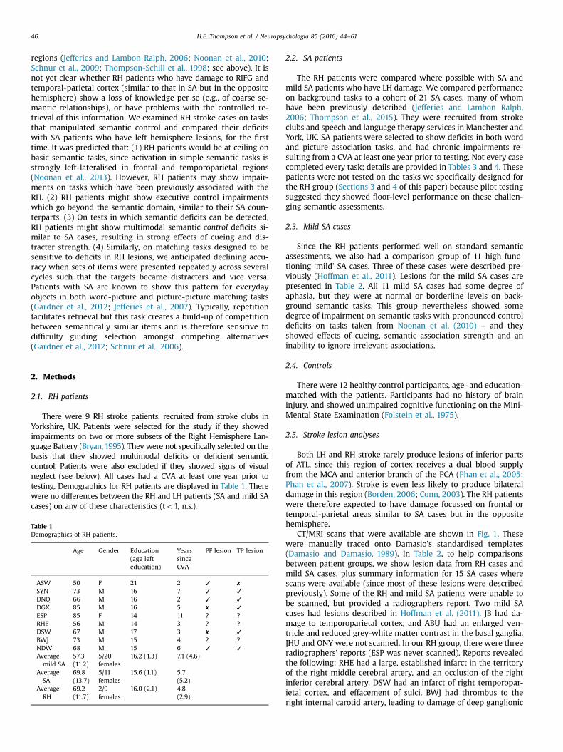
There were 9 RH stroke patients, recruited from stroke clubs inYorkshire, UK. Patients were selected for the study if they showedimpairments on two or more subsets of the Right Hemisphere Lan-guage Battery (Bryan, 1995). They were not specifically selected on thebasis that they showed multimodal deficits or deficient semanticcontrol. Patients were also excluded if they showed signs of visualneglect (see below). All cases had a CVA at least one year prior totesting. Demographics for RH patients are displayed in Table 1. Therewere no differences between the RH and LH patients (SA and mild SAcases) on any of these characteristics (to1, n.s.).
Table 1Demographics of RH patients.
Age Gender Education(age lefteducation)
YearssinceCVA
PF lesion TP lesion
ASW 50 F 21 2 ✓ ✗
SYN 73 M 16 7 ✓ ✓
DNQ 66 M 16 2 ✓ ✓
DGX 85 M 16 5 ✗ ✓
ESP 85 F 14 11 ? ?RHE 56 M 14 3 ? ?DSW 67 M 17 3 ✗ ✓
BWJ 73 M 15 4 ? ?NDW 68 M 15 6 ✓ ✓
Averagemild SA
57.3(11.2)
5/20females
16.2 (1.3) 7.1 (4.6)
AverageSA
69.8(13.7)
5/11females
15.6 (1.1) 5.7(5.2)
AverageRH
69.2(11.7)
2/9females
16.0 (2.1) 4.8(2.9)
2.2. SA patients
The RH patients were compared where possible with SA andmild SA patients who have LH damage. We compared performanceon background tasks to a cohort of 21 SA cases, many of whomhave been previously described (Jefferies and Lambon Ralph,2006; Thompson et al., 2015). They were recruited from strokeclubs and speech and language therapy services in Manchester andYork, UK. SA patients were selected to show deficits in both wordand picture association tasks, and had chronic impairments re-sulting from a CVA at least one year prior to testing. Not every casecompleted every task; details are provided in Tables 3 and 4. Thesepatients were not tested on the tasks we specifically designed forthe RH group (Sections 3 and 4 of this paper) because pilot testingsuggested they showed floor-level performance on these challen-ging semantic assessments.
2.3. Mild SA cases
Since the RH patients performed well on standard semanticassessments, we also had a comparison group of 11 high-func-tioning ‘mild’ SA cases. Three of these cases were described pre-viously (Hoffman et al., 2011). Lesions for the mild SA cases arepresented in Table 2. All 11 mild SA cases had some degree ofaphasia, but they were at normal or borderline levels on back-ground semantic tasks. This group nevertheless showed somedegree of impairment on semantic tasks with pronounced controldeficits on tasks taken from Noonan et al. (2010) – and theyshowed effects of cueing, semantic association strength and aninability to ignore irrelevant associations.
2.4. Controls
There were 12 healthy control participants, age- and education-matched with the patients. Participants had no history of braininjury, and showed unimpaired cognitive functioning on the Mini-Mental State Examination (Folstein et al., 1975).
2.5. Stroke lesion analyses
Both LH and RH stroke rarely produce lesions of inferior partsof ATL, since this region of cortex receives a dual blood supplyfrom the MCA and anterior branch of the PCA (Phan et al., 2005;Phan et al., 2007). Stroke is even less likely to produce bilateraldamage in this region (Borden, 2006; Conn, 2003). The RH patientswere therefore expected to have damage focussed on frontal ortemporal-parietal areas similar to SA cases but in the oppositehemisphere.
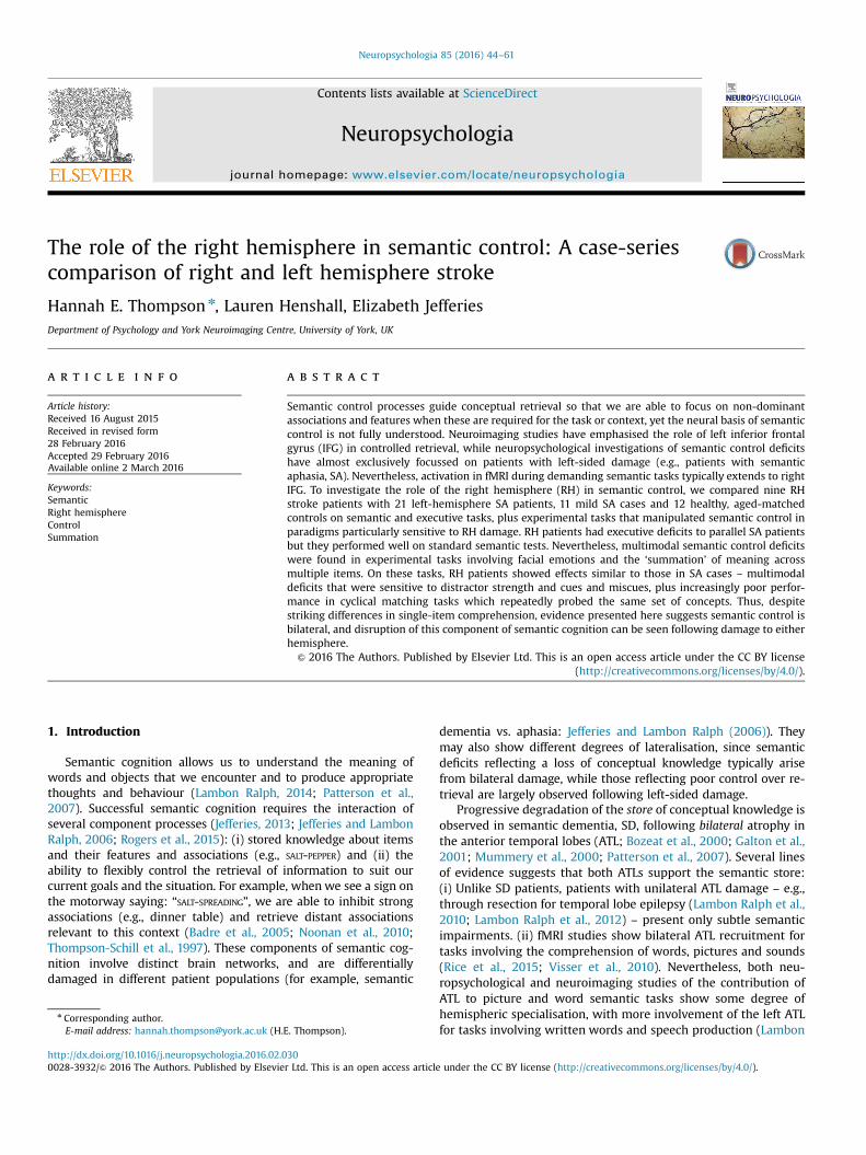
CT/MRI scans that were available are shown in Fig. 1. Thesewere manually traced onto Damasio’s standardised templates(Damasio and Damasio, 1989). In Table 2, to help comparisonsbetween patient groups, we show lesion data from RH cases andmild SA cases, plus summary information for 15 SA cases wherescans were available (since most of these lesions were describedpreviously). Some of the RH and mild SA patients were unable tobe scanned, but provided a radiographers report. Two mild SAcases had lesions described in Hoffman et al. (2011). JB had da-mage to temporoparietal cortex, and ABU had an enlarged ven-tricle and reduced grey-white matter contrast in the basal ganglia.JHU and ONY were not scanned. In our RH group, there were threeradiographers' reports (ESP was never scanned). Reports revealedthe following: RHE had a large, established infarct in the territoryof the right middle cerebral artery, and an occlusion of the rightinferior cerebral artery. DSW had an infarct of right temporopar-ietal cortex, and effacement of sulci. BWJ had thrombus to theright internal carotid artery, leading to damage of deep ganglionic
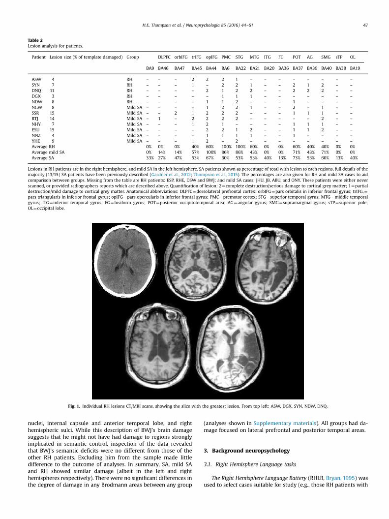
Table 2Lesion analysis for patients.
Patient Lesion size (% of template damaged) Group DLPFC orbIFG trIFG opIFG PMC STG MTG ITG FG POT AG SMG sTP OL
BA9 BA46 BA47 BA45 BA44 BA6 BA22 BA21 BA20 BA36 BA37 BA39 BA40 BA38 BA19
ASW 4 RH – – – 2 2 2 1 – – – – – – – –
SYN 7 RH – – – 1 – 2 2 1 – – 2 1 2 – –
DNQ 11 RH – – – – 2 1 2 2 – – 2 2 2 – –
DGX 3 RH – – – – – 1 1 1 – – – – – – –
NDW 8 RH – – – – 1 1 2 – – – 1 – – – –
NGW 8 Mild SA – – – – 1 2 2 1 – – 2 – 1 – –
SSR 15 Mild SA – – 2 1 2 2 2 – – – 1 1 1 – –
RTJ 14 Mild SA – 1 – 2 2 2 2 – – – – – 2 – –
NHY 7 Mild SA – – – 1 2 1 – – – – 1 1 1 – –
ESU 15 Mild SA – – – – 2 2 1 2 – – 1 1 2 – –
NNZ 4 Mild SA – – – – 1 1 1 1 – – 1 – – – –
YHE 9 Mild SA – – – 1 2 – 2 – – – – – – – –
Average RH 0% 0% 0% 40% 60% 100% 100% 60% 0% 0% 60% 40% 40% 0% 0%Average mild SA 0% 14% 14% 57% 100% 86% 86% 43% 0% 0% 71% 43% 71% 0% 0%Average SA 33% 27% 47% 53% 67% 60% 53% 53% 40% 13% 73% 53% 60% 13% 40%
Lesions in RH patients are in the right hemisphere, and mild SA in the left hemisphere. SA patients shown as percentage of total with lesion to each regions, full details of themajority (13/15) SA patients have been previously described (Gardner et al., 2012; Thompson et al., 2015). The percentages are also given for RH and mild SA cases to aidcomparison between groups. Missing from the table are RH patients: ESP, RHE, DSW and BWJ; and mild SA cases: JHU, JB, ABU, and ONY. These patients were either neverscanned, or provided radiographers reports which are described above. Quantification of lesion: 2¼complete destruction/serious damage to cortical grey matter; 1¼partialdestruction/mild damage to cortical grey matter. Anatomical abbreviations: DLPFC¼dorsolateral prefrontal cortex; orbIFG¼pars orbitalis in inferior frontal gyrus; trIFG,¼pars triangularis in inferior frontal gyrus; opIFG¼pars opercularis in inferior frontal gyrus; PMC¼premotor cortex; STG¼superior temporal gyrus; MTG¼middle temporalgyrus; ITG¼ inferior temporal gyrus; FG¼fusiform gyrus; POT¼posterior occipitotemporal area; AG¼angular gyrus; SMG¼supramarginal gyrus; sTP¼superior pole;OL¼occipital lobe.
Fig. 1. Individual RH lesions CT/MRI scans, showing the slice with the greatest lesion. From top left: ASW, DGX, SYN, NDW, DNQ.
H.E. Thompson et al. / Neuropsychologia 85 (2016) 44–61 47
nuclei, internal capsule and anterior temporal lobe, and righthemispheric sulci. While this description of BWJ's brain damagesuggests that he might not have had damage to regions stronglyimplicated in semantic control, inspection of the data revealedthat BWJ's semantic deficits were no different from those of theother RH patients. Excluding him from the sample made littledifference to the outcome of analyses. In summary, SA, mild SAand RH showed similar damage (albeit in the left and righthemispheres respectively). There were no significant differences inthe degree of damage in any Brodmann areas between any group
(analyses shown in Supplementary materials). All groups had da-mage focused on lateral prefrontal and posterior temporal areas.
3. Background neuropsychology
3.1. Right Hemisphere Language tasks
The Right Hemisphere Language Battery (RHLB, Bryan, 1995) wasused to select cases suitable for study (e.g., those RH patients with
(
(
(
H.E. Thompson et al. / Neuropsychologia 85 (2016) 44–6148
a deficit in two or more subtests). There were six subtests, de-scribed below. In all cases where reading was involved, the ex-perimenter read aloud the written sentences.
(i) Word and picture metaphor tasks. The word task involvedmatching a probe sentence with one of three spoken sen-tences for its correct interpretation. For example: “Probe: Hedidn’t take the changes lying down. Options: 1. He didn’t want tolie down. 2. He protested against the changes. 3. Change madehim tired”. In the picture condition, there was a spoken sen-tence to be matched with one of four pictures, one distractorbeing a literal interpretation of the sentence. There were tenitems in each test.
(ii) An inference test: answering questions about a short storywhich required inference from the text. This used threeparagraphs (and a practice paragraph) with four questionsabout each paragraph.
iii) 20-item word-picture matching, presented with five distractoritems (some of which were semantically, phonologically orvisually related to the target). RH cases were not expected tobe impaired on this simple task.
(iv) A 10-item humour test: selecting which of four possible sen-tences was the best punch line for the joke. For example: “Ajudge had just finished telling the prisoner that he was free to go,as the jury found him not guilty of fraud. The prisoner thenasked…(A) When can I leave sir? (B) What about my friends?(C) Does that mean I can keep the money? (D) What time is itplease?”
(v) A 10-item test of emphatic stress, where the researcher read asentence which described a picture and the participant thendescribed a similar picture using the same prosody.
The Familiar and Novel Language Comprehension test (FANL-C,Kempler and Van Lancker, 1985), used 20 spoken sentences in eachcondition, with a four-choice picture test of (i) novel literal phrasesand (ii) familiar metaphoric phrases. A sentence was presentedverbally and participants were asked to pick which picture out offour options reflects the sentence (e.g., metaphoric – ‘he’s got hishead in the clouds’; literal – ‘he’s chasing after a white duck’).
3.2. Semantic tasks
We ran a standard battery of semantic tasks sensitive to deficitsin SA cases.
(i) The Camel and Cactus task, picture version (CCTp; Bozeat et al.,2000) used 64 items, and involved matching a probe picturewith an associated picture, presented alongside three distrac-ter pictures (e.g., does CAMEL go with CACTUS, TREE, SUNFLOWER, orROSE?).
(ii) Category and Letter fluency: patients were given a minute toname as many ‘animals’ as they could (category fluency), anditems beginning with ‘S’ (letter fluency).
iii) A synonym judgement task involved matching a probe wordwith a target word presented alongside two unrelated dis-tractors. This had 96 items in two frequency bands (high andlow) and three imageability bands (high, medium and low),producing sixteen trials in each of the six frequency-by-im-ageability conditions (see Jefferies et al. (2009)). All of thewords in each trial were in the same frequency/imageabilitybands. For example, a low imageability, low frequency iteminvolved matching SUFFIX with INFLECTION, PERPETRATOR or TEMERITY,while a high imageability, high frequency item involvedmatching MONEY with CASH, CAR or CHURCH. Responses wereuntimed.
3.3. Visual tasks
Visual neglect and visual processing were assessed in RH pa-tients using the following tests.
(i) The Visual Object and Space Processing battery, VOSP (War-rington, and James, 1991): All eight subtests and the screeningtest of these perceptual tasks were presented. Four measurevisual object perception (Incomplete Letters, Silhouettes, Ob-ject Decision, and Progressive Silhouettes) while the other fourmeasure visual space perception (Dot Counting, Position Dis-crimination, Number Location, and Cube Analysis).
(ii) The Bells Cancellation test was used to assess visual neglect.Patients attempted to find 35 images of a bell amongst dis-tractors distributed across a sheet of paper (Gauthier et al.,1989). Patients were not limited in time to complete this test(although RT was recorded). This was scored by counting howmany bells were marked on each side of the page.
3.4. Executive tasks
The same executive tasks were presented to RH and SApatients.
(i) Forward and backward digit span (Wechsler, 1987), assessedauditory working memory.
(ii) Elevator Counting involved counting tones in two conditions. Inthe ‘no distraction’ condition, all of the tones were targets tobe counted. In the ‘with distraction’ condition, patients wereasked to count low pitch tones and ignore high pitch tones.This test was taken from the Test of Everyday Attention (TEA;Robertson et al., 1994)
iii) The Hayling Test involved completing spoken sentences withsingle words (Burgess, and Shallice, 1997). Participants wereasked to complete the sentence with either a sensible word oran unconnected word, in two conditions, e.g., “She posted theletter without a…” “stamp/tomato”.
(iv) Ravens Coloured Progressive Matrices test (RCPM: Raven, 1962),assessed non-verbal reasoning using pattern and rule recogni-tion with shapes and colours in sets A, AB, and B.
(v) The Brixton Spatial Rule Attainment task (BSRA: Burgess, andShallice, 1997), involved making predictions about the move-ment of a dot, based on patterns that it showed across trials,and then adapting these predictions when the patternchanged.
(vi) The Trails Test required participants to draw a line betweenletters and numbers in order, in an easy condition (part A, e.g.,1-2-3…) and difficult condition (part B, e.g., 1-A-2-B-3-C…,Reitan, 1958).
4. Results
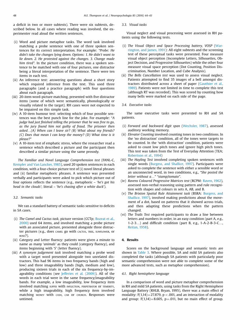
Scores on the background language and semantic tests areshown in Table 3. Where possible, SA and mild SA patients alsocompleted the tasks (although SA patients with particularly poorsemantic comprehension were not able to complete some of themore advanced tests, such as metaphor comprehension).
4.1. Right hemisphere language
In a comparison of word and picture metaphor comprehensionin RH and mild SA patients, using tasks from the Right HemisphereLanguage Battery (RHLB, Bryan, 1995), there was a main effect ofmodality: F(1,14)¼27.879, po .001, and an interaction of modalityand group: F(1,14)¼8.605, p¼ .011, but no main effect of group.
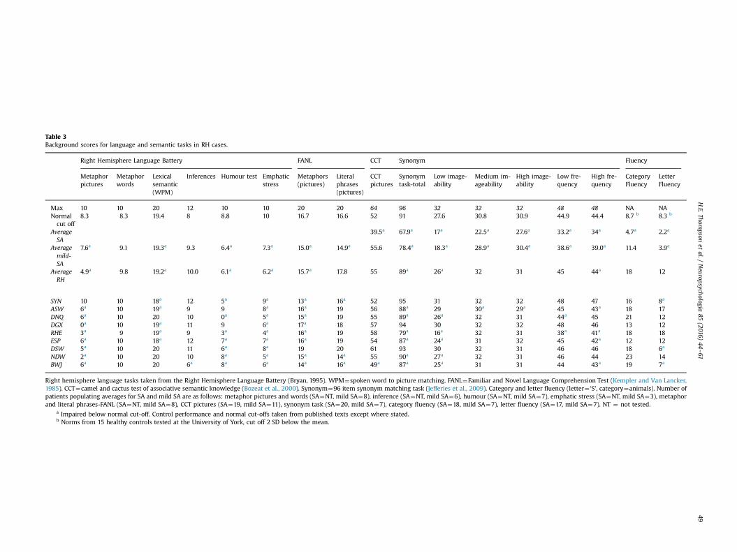
Table 3Background scores for language and semantic tasks in RH cases.
Right Hemisphere Language Battery FANL CCT Synonym Fluency
Metaphorpictures
Metaphorwords
Lexicalsemantic(WPM)
Inferences Humour test Emphaticstress
Metaphors(pictures)
Literalphrases(pictures)
CCTpictures
Synonymtask-total
Low image-ability
Medium im-ageability
High image-ability
Low fre-quency
High fre-quency
CategoryFluency
LetterFluency
Max 10 10 20 12 10 10 20 20 64 96 32 32 32 48 48 NA NANormalcut off
8.3 8.3 19.4 8 8.8 10 16.7 16.6 52 91 27.6 30.8 30.9 44.9 44.4 8.7 b 8.3 b
AverageSA
39.5a 67.9a 17a 22.5a 27.6a 33.2a 34a 4.7a 2.2a
Averagemild-SA
7.6a 9.1 19.3a 9.3 6.4a 7.3a 15.0a 14.9a 55.6 78.4a 18.3a 28.9a 30.4a 38.6a 39.0a 11.4 3.9a
AverageRH
4.9a 9.8 19.2a 10.0 6.1a 6.2a 15.7a 17.8 55 89a 26a 32 31 45 44a 18 12
SYN 10 10 18a 12 5a 9a 13a 16a 52 95 31 32 32 48 47 16 8a
ASW 6a 10 19a 9 9 8a 16a 19 56 88a 29 30a 29a 45 43a 18 17DNQ 6a 10 20 10 0a 5a 15a 19 55 89a 26a 32 31 44a 45 21 12DGX 0a 10 19a 11 9 6a 17a 18 57 94 30 32 32 48 46 13 12RHE 3a 9 19a 9 3a 4a 16a 19 58 79a 16a 32 31 38a 41a 18 18ESP 6a 10 18a 12 7a 7a 16a 19 54 87a 24a 31 32 45 42a 12 12DSW 5a 10 20 11 6a 8a 19 20 61 93 30 32 31 46 46 18 6a
NDW 2a 10 20 10 8a 5a 15a 14a 55 90a 27a 32 31 46 44 23 14BWJ 6a 10 20 6a 8a 6a 14a 16a 49a 87a 25a 31 31 44 43a 19 7a
Right hemisphere language tasks taken from the Right Hemisphere Language Battery (Bryan, 1995). WPM¼spoken word to picture matching. FANL¼Familiar and Novel Language Comprehension Test (Kempler and Van Lancker,1985). CCT¼camel and cactus test of associative semantic knowledge (Bozeat et al., 2000). Synonym¼96 item synonym matching task (Jefferies et al., 2009). Category and letter fluency (letter¼ ‘S’, category¼animals). Number ofpatients populating averages for SA and mild SA are as follows: metaphor pictures and words (SA¼NT, mild SA¼8), inference (SA¼NT, mild SA¼6), humour (SA¼NT, mild SA¼7), emphatic stress (SA¼NT, mild SA¼3), metaphorand literal phrases-FANL (SA¼NT, mild SA¼8), CCT pictures (SA¼19, mild SA¼11), synonym task (SA¼20, mild SA¼7), category fluency (SA¼18, mild SA¼7), letter fluency (SA¼17, mild SA¼7). NT ¼ not tested.
a Impaired below normal cut-off. Control performance and normal cut-offs taken from published texts except where stated.b Norms from 15 healthy controls tested at the University of York, cut off 2 SD below the mean.
H.E.Thom
psonet
al./Neuropsychologia
85(2016)
44–61
49
H.E. Thompson et al. / Neuropsychologia 85 (2016) 44–6150
Mild SA patients performed similarly on word and picture meta-phor tasks, while RH patients were disproportionally impaired atthe picture metaphor task, in line with previously findings (Win-ner and Gardner, 1977). In all other subtests of the RHLB, therewere no noticeable differences between groups (both RH and mildSA patients showed some impairment).
In a similar comparison of literal and metaphorical picturematching (the FANL task), there was a main effect of sentencetype: F(1,13)¼10.024, p¼ .007, an interaction of sentence andgroup: F(1,13)¼7.459, p¼ .017, and no group difference: F(1,13)¼2.321, p¼ .152. Mild SA patients performed similarly in metapho-rical and literal sentences, while RH patients were poorer at me-taphor sentence comprehension.
4.2. Semantic tasks
RH patients performed at a normal level on basic verbal andpictorial semantic tasks, and on assessments requiring both com-prehension and speech production. In a comparison of SA and RHpatients, performance was significantly higher in RH patients in allsemantic tasks: CCTp: t(26)¼4.088, po .001, synonym judgement:t(27)¼4.618, po .001, and category and letter fluency: t(25)Z6.770, po .001. Even mild SA patients were significantly worsethan RH patients on the synonym judgment task: t(14)¼3.510,p¼ .003, and category and letter fluency: t(14)Z2.962, pr .010. Incontrast, there were no significant differences between RH pa-tients and healthy controls on any semantic task.
4.3. Visual tasks
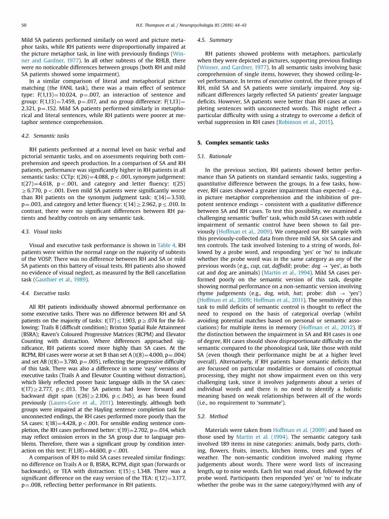
Visual and executive task performance is shown in Table 4. RHpatients were within the normal range on the majority of subtestsof the VOSP. There was no difference between RH and SA or mildSA patients on this battery of visual tests. RH patients also showedno evidence of visual neglect, as measured by the Bell cancellationtask (Gauthier et al., 1989).
4.4. Executive tasks
All RH patients individually showed abnormal performance onsome executive tasks. There was no difference between RH and SApatients on the majority of tasks: t(17)r1.903, pZ .074 for the fol-lowing: Trails B (difficult condition); Brixton Spatial Rule Attainment(BSRA); Raven’s Coloured Progressive Matrices (RCPM) and ElevatorCounting with distraction. Where differences approached sig-nificance, RH patients scored more highly than SA cases. At theRCPM, RH cases were worse at set B than set A (t(8)¼4.000, p¼ .004)and set AB (t(8)¼3.780, p¼ .005), reflecting the progressive difficultyof this task. There was also a difference in some ‘easy’ versions ofexecutive tasks (Trails A and Elevator Counting without distraction),which likely reflected poorer basic language skills in the SA cases:t(17)Z2.777, pr .013. The SA patients had lower forward andbackward digit span (t(26)Z2.106, pr .045), as has been foundpreviously (Laures-Gore et al., 2011). Interestingly, although bothgroups were impaired at the Hayling sentence completion task forunconnected endings, the RH cases performed more poorly than theSA cases: t(18)¼4.428, po .001. For sensible ending sentence com-pletion, the RH cases performed better: t(19)¼2.702, p¼ .014, whichmay reflect omission errors in the SA group due to language pro-blems. Therefore, there was a significant group by condition inter-action on this test: F(1,18)¼44.600, po .001.
A comparison of RH to mild SA cases revealed similar findings:no difference on Trails A or B, BSRA, RCPM, digit span (forwards orbackwards), or TEA with distraction: t(15)r1.348. There was asignificant difference on the easy version of the TEA: t(12)¼3.177,p¼ .008, reflecting better performance in RH patients.
4.5. Summary
RH patients showed problems with metaphors, particularlywhen they were depicted as pictures, supporting previous findings(Winner, and Gardner, 1977). In all semantic tasks involving basiccomprehension of single items, however, they showed ceiling-le-vel performance. In terms of executive control, the three groups ofRH, mild SA and SA patients were similarly impaired. Any sig-nificant differences largely reflected SA patients' greater languagedeficits. However, SA patients were better than RH cases at com-pleting sentences with unconnected words. This might reflect aparticular difficulty with using a strategy to overcome a deficit ofverbal suppression in RH cases (Robinson et al., 2015).
5. Complex semantic tasks
5.1. Rationale
In the previous section, RH patients showed better perfor-mance than SA patients on standard semantic tasks, suggesting aquantitative difference between the groups. In a few tasks, how-ever, RH cases showed a greater impairment than expected – e.g.,in picture metaphor comprehension and the inhibition of pre-potent sentence endings – consistent with a qualitative differencebetween SA and RH cases. To test this possibility, we examined achallenging semantic ‘buffer’ task, which mild SA cases with subtleimpairment of semantic control have been shown to fail pre-viously (Hoffman et al., 2009). We compared our RH sample withthis previously-collected data from three mild SA, six SA cases andten controls. The task involved listening to a string of words, fol-lowed by a probe word, and responding ‘yes’ or ‘no’ to indicatewhether the probe word was in the same category as any of theprevious words (e.g., cup, cat, daffodil; probe: dog - ‘yes’, as bothcat and dog are animals) (Martin et al., 1994). Mild SA cases per-formed poorly on the semantic version of this task, despiteshowing normal performance on a non-semantic version involvingrhyme judgements (e.g., dog, wish, hat; probe: dish - ‘yes’)(Hoffman et al., 2009; Hoffman et al., 2011). The sensitivity of thistask to mild deficits of semantic control is thought to reflect theneed to respond on the basis of categorical overlap (whilstavoiding potential matches based on personal or semantic asso-ciations) for multiple items in memory (Hoffman et al., 2012). Ifthe distinction between the impairment in SA and RH cases is oneof degree, RH cases should show disproportionate difficulty on thesemantic compared to the phonological task, like those with mildSA (even though their performance might be at a higher leveloverall). Alternatively, if RH patients have semantic deficits thatare focussed on particular modalities or domains of conceptualprocessing, they might not show impairment even on this verychallenging task, since it involves judgements about a series ofindividual words and there is no need to identify a holisticmeaning based on weak relationships between all of the words(i.e., no requirement to ‘summate’).
5.2. Method
Materials were taken from Hoffman et al. (2009) and based onthose used by Martin et al. (1994). The semantic category taskinvolved 189 items in nine categories: animals, body parts, cloth-ing, flowers, fruits, insects, kitchen items, trees and types ofweather. The non-semantic condition involved making rhymejudgements about words. There were word lists of increasinglength, up to nine words. Each list was read aloud, followed by theprobe word. Participants then responded ‘yes’ or ‘no’ to indicatewhether the probe was in the same category/rhymed with any of
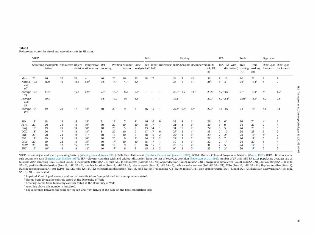
Table 4Background scores for visual and executive tasks in RH cases.
VOSP Bells Hayling TEA Trails Digit span
Screening Incompleteletters
Silhouettes Objectdecision
Progressivesilhouettes
Dotcounting
Position Numberlocation
Cubeanalysis
Lefthalf
Righthalf
Differencee BSRA Sensible Unconnected RCPM(A, AB,B)
TEA TEA (withdistraction)
Trailmaking(A)
Trailmaking(B)
Digit Spanforwards
Digit Spanbackwards
Max 20 20 30 20 – 10 20 10 10 18 17 54 15 15 36 7 10 24 23 8 7Normalcutoff
19.3 16.9 10 10.5 6.0d 9.5 17.1 4.7 5.4 28 11 11 28b 6 3 24c 17.4c 5 2
AverageSA
19.5 11.4a 15.8 6.9a 7.5a 16.2a 8.2 5.3a – – – 20.9a 11.5 9.8a 23.5a 4.7a 3.6 21a 10.1a 4a 1.7a
AveragemildSA
19.3 9.5 19.2 9.1 8.6 – – – 32.1 – – 27.9a 5.2a 2.4a 23.9a 15.8a 5.2 2.8
AverageRH
19a 19 20 17 12a 10 20 9 7 16 15 1 27.2a 10.8a 1.5a 27.2a 6.6 4.6 24 17a 5.8 3.1
SYN 18a 18 13 16 15a 9a 19 7 4a 16 16 0 38 14 1a 26a 6 0a 24 7a 4a 3ASW 20 20 24 18 10a 10 20 10 10 18 17 1 21a 14 6a 30 6 9 24 22 7 4DNQ 19a 19 21 18 10a 10 20 5 8 13 14 1 31 15 7a 24a 6 5 24 23 6 3DGX 18a 20 17 14 13a 8a 20 10 9 17 17 0 27a 12 1a 35 7 10 24 23 5 2RHE 20 20 23 19 11a 10 19 10 7 18 16 2 27a 13 1a 23a 7 2a 24 17a 4a 2ESP 17a 19 15 15 10a 10 20 9 3a 14 12 2 24a 13 5a 21a 6 7 24 17a 5 2DSW 20 19 22 18 15a 10 17a 10 10 18 17 1 39 14 1a 32 7 1a 24 20 6 4NDW 20 18 17 15 12a 10 18 9 9 16 15 1 29 15 4a 31 7 5 24 17a 8 6BWJ 19a 16a 19 14 12a 10 17a 6 6 15 13 2 9a 13 0a 23a 7 2a 24 17a 7 2
VOSP¼visual object and space processing battery (Warrington and James, 1991); Bells Cancellation test (Gauthier, Dehaut and Joanette, 1989); RCPM¼Raven's Coloured Progressive Matrices (Raven, 1962); BSRA¼Brixton spatialrule attainment task (Burgess and Shallice, 1997); TEA¼elevator counting with and without distraction from the test of everyday attention (Robertson et al., 1994). number of SA and mild SA cases populating averages are asfollows: VOSP screening (SA¼10, mild SA¼NT), incomplete letters (SA¼8, mild SA¼3), silhouettes (SA/mild SA¼NT), object decision (SA¼9, mild SA¼NT), progressive silhouettes (SA¼8, mild SA¼NT), dot counting (SA¼18, mildSA¼6), position discrimination (SA¼18, mild SA¼6), number location (SA¼18, mild SA¼9, cube analysis (SA¼18, mild SA¼9), bells cancellation test (SA/mild SA¼NT), BSRA (SA¼19, mild SA¼11), Hayling sensible (SA¼11),Hayling unconnected (SA¼10), RCPM (SA¼20, mild SA¼8), TEA with/without distraction (SA¼18, mild SA¼5), Trail making A/B (SA¼9, mild SA¼8), digit span forwards (SA¼18, mild SA¼10), digit span backwards (SA¼18, mildSA¼9). NT ¼ not tested.
a Impaired. Control performance and normal cut-offs taken from published texts except where stated.b Norms from 20 healthy controls tested at the University of York.c Accuracy norms from 14 healthy controls tested at the University of York.d Anything above this number is impaired.e The difference between the score for the left and right halves of the page on the Bells cancellation task.
H.E.Thom
psonet
al./Neuropsychologia
85(2016)
44–61
51
H.E. Thompson et al. / Neuropsychologia 85 (2016) 44–6152
the preceding words. List lengths of 1, 2 and 5 had 20 trials whilethe remainder had 24 trials. Patients had to be at least 75% accu-rate on each list length in order to proceed to the next list length.Span was defined as the maximum length at which the accuracywas 75% or higher, and if this came between two lengths, span wascalculated with linear interpolation. Prior to beginning the se-mantic buffer task, participants were given a list of the categoriesand were asked to assign each item to one of the categories. If theymade an error, the experimenter told them the correct category.The categorisation accuracy of the RH patients was 98%; mild SAscored 94%.
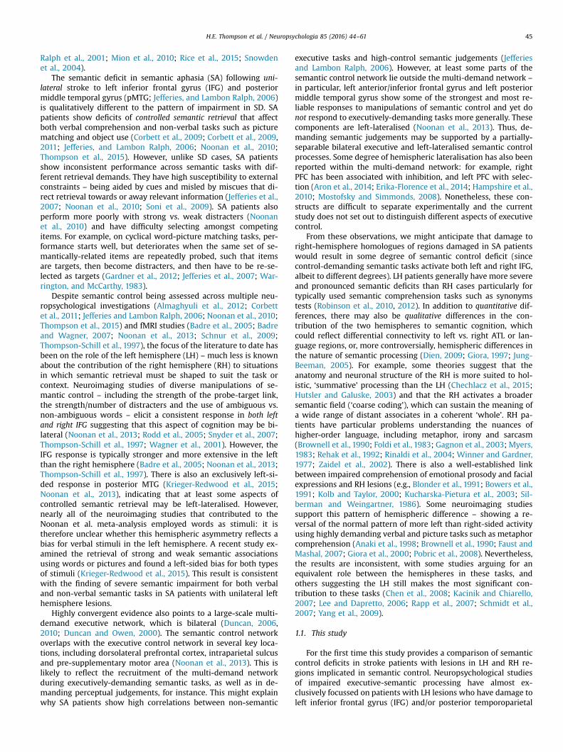
5.3. Results
Performance on these tasks is given in Fig. 2. Data from healthycontrols, SA and mild SA patients is reproduced from Hoffmanet al. (2011). In a comparison of RH and mild SA cases, there wasno main effect of group (Fo1). There was a main effect of task(semantic vs. rhyme): F(1,10)¼21.598, p¼ .001, and an interaction:F(1,10)¼40.401, po .001. Mild SA patients were strikingly better atthe rhyme version of the task, and poor at the semantic version,while RH patients showed similar performance in both tasks. In acomparison of RH and SA patients, there was a main effect ofgroup: F(1,13)¼21.221, po .001: SA patients showed floor effects.There was no main effect of task (F¼1.1), but again there was aninteraction: F(1,13)¼10.141, p¼ .007. SA patients resembled mildSA cases, albeit with a lower overall level of performance.
Additionally, we also looked at performance in each RH patientusing the revised standardised difference test (RSDT; Crawfordet al., 2010), which examines the difference between two tasks(rhyme vs. semantic) for a patient, in relation to controls (using amodified t-statistic). None of the RH patients showed a differencebetween the semantic and rhyme conditions that varied fromcontrols (p4 .05). This was in contrast to the mild SA cases, whoall showed larger difference between the semantic and rhymeconditions than controls (po .01). Despite floor effects, all but oneSA patient (NY) also showed this difference (po .05).
5.4. Summary
Even on a demanding semantic task, which required retrievalof categories that were not explicitly revealed for multiple itemsheld in memory, RH patients showed performance in the normalrange. This supports the suggestion that there may be some dis-tinction in the contribution of the two hemispheres to semanticprocessing. Our RH patients showed no impairment of single wordcomprehension (see also Klepousniotou and Baum (2005)) and the
Fig. 2. Performance on the semantic and rhyme buffer tasks. Error bars showstandard error of mean.
semantic buffer task similarly required words to be categorisedindividually, even though several items were presented in eachtrial (i.e., the meaning of one word did not influence the categor-isation of the next item).
In the remainder of the empirical work, we focus on conceptualdomains that have revealed greater impairment in RH cases toestablish whether there are any parallels with the deficit of se-mantic control in SA. Patients with SA retain conceptual knowl-edge that they nevertheless fail to retrieve in tasks with highcontrol demands, when there are few external cues to constrainretrieval. They show strong cueing and miscueing effects (Jefferieset al., 2008; Noonan et al., 2010; Soni et al., 2009; Soni et al., 2011).Additionally, they show poorer retrieval with strong distractorswhich compete with the target (Noonan et al., 2010). In the nextexperiment, we examined the effect of cueing, miscueing anddistractor strength on performance in RH cases using paradigmsdesigned to be sensitive to deficits in this group. Since previousstudies have suggested that RH cases have difficulty ‘summating’meaning across multiple inputs, and with interpreting emotionalfaces (Adolphs, 2002; Nakamura et al., 1999; Witteman et al.,2011), we manipulated control demands in these tasks.
6. Summation and task-irrelevant information
Beeman et al. (1994) and Jung-Beeman (2005) has suggestedthat the RH shows ‘coarse semantic coding’ grouping togetherdisparate words into an overarching meaning (e.g., EYES – CLOSED –
NIGHT - SLEEP; or FOOT – CRY – GLASS - CUT). Visual field experimentshave revealed faster processing of distantly related words in the‘RH’ (or left visual field) comparison to the ‘LH’ (Anaki et al., 1998;Faust and Mashal, 2007; Mashal and Faust, 2009), and patientstudies have revealed difficulties with semantic comprehension ofdistant relationships after RH damage (Blake et al., 2015; Tompkinset al., 2011, 2008). We varied the semantic control demands withina 4 alternative forced choice (4AFC) ‘summation’ task by(i) providing cues and miscues that either led retrieval towards oraway from relevant aspects of knowledge, and (ii) by manipulatingthe identity of the distracters. If RH cases show strong sensitivityto distractor type and cueing like SA cases, this would suggest thatthey retain semantic knowledge that they cannot always retrieveappropriately. We also compared comprehension within this taskusing words and pictures to investigate if RH cases have a multi-modal deficit of semantic control, like those with SA.
6.1. Rationale and procedure: cues and miscues
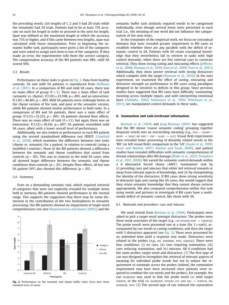
We used stimuli from Beeman et al. (1994). Participants wereasked to pick a target word amongst distractors. The probes werethree weak associates of the target (e.g., CAT-ATTACKS-PAW - SCRATCH).The probe words were presented one at a time (for 1 s each), ac-companied by cue words in cueing conditions, and then the targetwith 3 distractors appeared (see Fig. 3). These were presented foran unlimited time until a response was made. Distractors wererelated to the probes (e.g., HIT, WHISKERS, FOOT, SCRATCH). There werefour conditions: (i) no cues, (ii) cues requiring summation, (iii)cues reducing summation, and (iv) miscues. All 4 conditions hadthe same probes, target word and distractors. (1) The first type ofcue was designed to strengthen the retrieval of relevant aspects ofmeaning for individual probe words but not to reduce the re-quirement to summate across the probes (indeed, the summationrequirement may have been increased since patients were re-quired to combine the cue words and the probes). For example, thecue AGGRESSIVE was used to link the probe word CAT with targetSCRATCH, in the trial CAT AGGRESSIVE, ATTACKS CUT, PAW PAD - SCRATCH, HIT,WHISKERS, FOOT. (2) The second type of cue reduced the summation
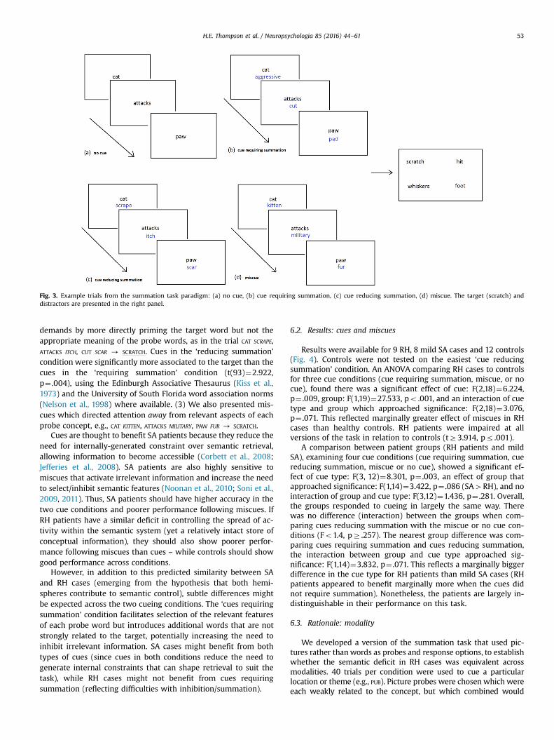
Fig. 3. Example trials from the summation task paradigm: (a) no cue, (b) cue requiring summation, (c) cue reducing summation, (d) miscue. The target (scratch) anddistractors are presented in the right panel.
H.E. Thompson et al. / Neuropsychologia 85 (2016) 44–61 53
demands by more directly priming the target word but not theappropriate meaning of the probe words, as in the trial CAT SCRAPE,ATTACKS ITCH, CUT SCAR - SCRATCH. Cues in the ‘reducing summation’condition were significantly more associated to the target than thecues in the ‘requiring summation’ condition (t(93)¼2.922,p¼ .004), using the Edinburgh Associative Thesaurus (Kiss et al.,1973) and the University of South Florida word association norms(Nelson et al., 1998) where available. (3) We also presented mis-cues which directed attention away from relevant aspects of eachprobe concept, e.g., CAT KITTEN, ATTACKS MILITARY, PAW FUR - SCRATCH.
Cues are thought to benefit SA patients because they reduce theneed for internally-generated constraint over semantic retrieval,allowing information to become accessible (Corbett et al., 2008;Jefferies et al., 2008). SA patients are also highly sensitive tomiscues that activate irrelevant information and increase the needto select/inhibit semantic features (Noonan et al., 2010; Soni et al.,2009, 2011). Thus, SA patients should have higher accuracy in thetwo cue conditions and poorer performance following miscues. IfRH patients have a similar deficit in controlling the spread of ac-tivity within the semantic system (yet a relatively intact store ofconceptual information), they should also show poorer perfor-mance following miscues than cues – while controls should showgood performance across conditions.
However, in addition to this predicted similarity between SAand RH cases (emerging from the hypothesis that both hemi-spheres contribute to semantic control), subtle differences mightbe expected across the two cueing conditions. The ‘cues requiringsummation’ condition facilitates selection of the relevant featuresof each probe word but introduces additional words that are notstrongly related to the target, potentially increasing the need toinhibit irrelevant information. SA cases might benefit from bothtypes of cues (since cues in both conditions reduce the need togenerate internal constraints that can shape retrieval to suit thetask), while RH cases might not benefit from cues requiringsummation (reflecting difficulties with inhibition/summation).
6.2. Results: cues and miscues
Results were available for 9 RH, 8 mild SA cases and 12 controls(Fig. 4). Controls were not tested on the easiest ‘cue reducingsummation’ condition. An ANOVA comparing RH cases to controlsfor three cue conditions (cue requiring summation, miscue, or nocue), found there was a significant effect of cue: F(2,18)¼6.224,p¼ .009, group: F(1,19)¼27.533, po .001, and an interaction of cuetype and group which approached significance: F(2,18)¼3.076,p¼ .071. This reflected marginally greater effect of miscues in RHcases than healthy controls. RH patients were impaired at allversions of the task in relation to controls (tZ3.914, pr .001).
A comparison between patient groups (RH patients and mildSA), examining four cue conditions (cue requiring summation, cuereducing summation, miscue or no cue), showed a significant ef-fect of cue type: F(3, 12)¼8.301, p¼ .003, an effect of group thatapproached significance: F(1,14)¼3.422, p¼ .086 (SA4RH), and nointeraction of group and cue type: F(3,12)¼1.436, p¼ .281. Overall,the groups responded to cueing in largely the same way. Therewas no difference (interaction) between the groups when com-paring cues reducing summation with the miscue or no cue con-ditions (Fo1.4, pZ .257). The nearest group difference was com-paring cues requiring summation and cues reducing summation,the interaction between group and cue type approached sig-nificance: F(1,14)¼3.832, p¼ .071. This reflects a marginally biggerdifference in the cue type for RH patients than mild SA cases (RHpatients appeared to benefit marginally more when the cues didnot require summation). Nonetheless, the patients are largely in-distinguishable in their performance on this task.
6.3. Rationale: modality
We developed a version of the summation task that used pic-tures rather thanwords as probes and response options, to establishwhether the semantic deficit in RH cases was equivalent acrossmodalities. 40 trials per condition were used to cue a particularlocation or theme (e.g., PUB). Picture probes were chosenwhich wereeach weakly related to the concept, but which combined would
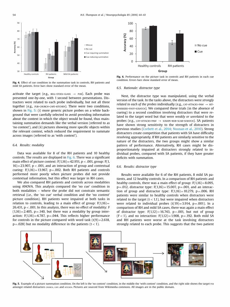
Fig. 4. Effect of cue condition in the summation task in controls, RH patients andmild SA patients. Error bars show standard error of the mean.
Fig. 6. Performance on the picture task in controls and RH patients in each cuecondition. Errors bars show standard error of mean.
H.E. Thompson et al. / Neuropsychologia 85 (2016) 44–6154
activate the target (e.g., BELL-STOOL-GLASS - PUB). Each probe waspresented one-by-one, with 1 second between presentations. Dis-tractors were related to each probe individually, but not all threetogether (e.g., PUB-CHURCH-CAFE-KITCHEN). There were two conditions,shown in Fig. 5: (i) more generic picture probes on a white back-ground that were carefully selected to avoid providing informationabout the context in which the object would be found, thus main-taining summation demands like the verbal version (referred to as‘no context’), and (ii) pictures showing more specific objects withinthe relevant context, which reduced the requirement to summateacross images (referred to as ‘with context’).
6.4. Results: modality
Data was available for 8 of the RH patients and 10 healthycontrols. The results are displayed in Fig. 6. There was a significantmain effect of picture context: F(1,16)¼42.951, po .001, group: F(1,16)¼23.907, po .001, and an interaction of group and contextualcueing: F(1,16)¼13.967, p¼ .002. Both RH patients and controlsperformed more poorly when picture probes did not providecontextual information, but this effect was larger in RH cases.
We also compared RH patients and controls across modalitiesusing ANOVA. This analysis compared the ‘no cue’ condition inboth modalities – where the probe did not constrain semanticretrieval (i.e., the ‘no cue’ verbal condition and the ‘no context’picture condition). RH patients were impaired at both tasks inrelation to controls, leading to a main effect of group: F(1,16)¼26.431, po .001. In this analysis, there was no effect of modality: F(1,16)¼2.405, p¼ .140, but there was a modality by group inter-action: F(1,16)¼4.787, p¼ .044. This reflects higher performancefor controls in the picture compared with word task (t(9)¼2.618,p¼ .028) but no modality difference in the patients (to1).
Fig. 5. Example of a picture summation condition. On the left is the ‘no context’ conditioamongst related distractors CHURCH, CAFE and KITCHEN. Pictures are sourced from Wikimedi
6.5. Rationale: distractor type
Next, the distractor type was manipulated, using the verbalversion of the task. In the tasks above, the distractors were stronglyrelated to each of the probes individually (e.g., CAT-ATTACKS-PAW - HIT-WHISKERS-FOOT-SCRATCH). We compared these trials (in the absence ofcueing) to a second condition involving distractors that were re-lated to the target word but that were weakly or unrelated to theprobes (e.g., CAT-ATTACKS-PAW - SCRAPE-RUB-SCAR-SCRATCH). SA patientshave shown strong sensitivity to the strength of distracters inprevious studies (Corbett et al., 2014; Noonan et al., 2010). Strongdistracters create competition that patients with SA have difficultyresolving appropriately. If RH patients are similarly sensitive to thenature of the distracters, the two groups might show a similarpattern of performance. Alternatively, RH cases might be dis-proportionately impaired at distracters strongly related to in-dividual probes, compared with SA patients, if they have greaterdeficits with summation.
6.6. Results: distractor type
Results were available for 6 of the RH patients, 8 mild SA pa-tients, and 12 healthy controls. In a comparison of RH patients andhealthy controls, there was a main effect of group: F(1,16)¼8.095,p¼ .012, distractor type: F(1,16)¼15.007, p¼ .001, and an interac-tion of group and distractor type: F(1,16)¼10.279, p¼ .006. RHpatients were similar to healthy controls when distractors wererelated to the target (to1.1), but were impaired when distractorswere related to individual probes (t(19)¼3.914, p¼ .001). In acomparison of RH and mild SA cases, there was again a main effectof distractor type: F(1,12)¼16.743, p¼ .001, but not of group(Fo1), and no interaction: F(1,12)¼1.908, p¼ .192. Both mild SAand RH patients were worse at the task involving distractorsstrongly related to each probe. This suggests that the two patient
n, in the middle the ‘with context’ condition, and the right side shows the target PUB
a commons. All images are in the public domain.
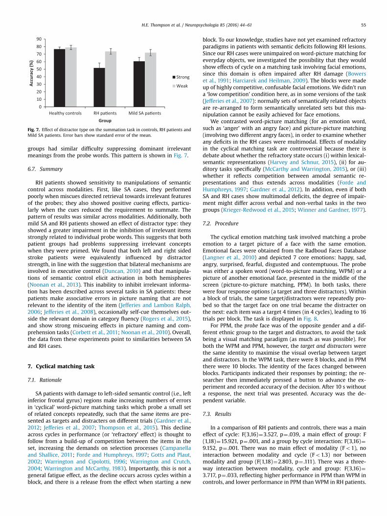
Fig. 7. Effect of distractor type on the summation task in controls, RH patients andMild SA patients. Error bars show standard error of the mean.
H.E. Thompson et al. / Neuropsychologia 85 (2016) 44–61 55
groups had similar difficulty suppressing dominant irrelevantmeanings from the probe words. This pattern is shown in Fig. 7.
6.7. Summary
RH patients showed sensitivity to manipulations of semanticcontrol across modalities. First, like SA cases, they performedpoorly when miscues directed retrieval towards irrelevant featuresof the probes; they also showed positive cueing effects, particu-larly when the cues reduced the requirement to summate. Thepattern of results was similar across modalities. Additionally, bothmild SA and RH patients showed an effect of distractor type: theyshowed a greater impairment in the inhibition of irrelevant itemsstrongly related to individual probe words. This suggests that bothpatient groups had problems suppressing irrelevant conceptswhen they were primed. We found that both left and right sidedstroke patients were equivalently influenced by distractorstrength, in line with the suggestion that bilateral mechanisms areinvolved in executive control (Duncan, 2010) and that manipula-tions of semantic control elicit activation in both hemispheres(Noonan et al., 2013). This inability to inhibit irrelevant informa-tion has been described across several tasks in SA patients: thesepatients make associative errors in picture naming that are notrelevant to the identity of the item (Jefferies and Lambon Ralph,2006; Jefferies et al., 2008), occasionally self-cue themselves out-side the relevant domain in category fluency (Rogers et al., 2015),and show strong miscueing effects in picture naming and com-prehension tasks (Corbett et al., 2011; Noonan et al., 2010). Overall,the data from these experiments point to similarities between SAand RH cases.
7. Cyclical matching task
7.1. Rationale
SA patients with damage to left-sided semantic control (i.e., leftinferior frontal gyrus) regions make increasing numbers of errorsin ‘cyclical’ word-picture matching tasks which probe a small setof related concepts repeatedly, such that the same items are pre-sented as targets and distracters on different trials (Gardner et al.,2012; Jefferies et al., 2007; Thompson et al., 2015). This declineacross cycles in performance (or ‘refractory’ effect) is thought tofollow from a build-up of competition between the items in theset, increasing the demands on selection processes (Campanellaand Shallice, 2011; Forde and Humphreys, 1997; Gotts and Plaut,2002; Warrington and Cipolotti, 1996; Warrington and Crutch,2004; Warrington and McCarthy, 1983). Importantly, this is not ageneral fatigue effect, as the decline occurs across cycles within ablock, and there is a release from the effect when starting a new
block. To our knowledge, studies have not yet examined refractoryparadigms in patients with semantic deficits following RH lesions.Since our RH cases were unimpaired on word-picture matching foreveryday objects, we investigated the possibility that they wouldshow effects of cycle on a matching task involving facial emotions,since this domain is often impaired after RH damage (Bowerset al., 1991; Harciarek and Heilman, 2009). The blocks were madeup of highly competitive, confusable facial emotions. We didn’t runa ‘low competition’ condition here, as in some versions of the task(Jefferies et al., 2007): normally sets of semantically related objectsare re-arranged to form semantically unrelated sets but this ma-nipulation cannot be easily achieved for face emotions.
We contrasted word-picture matching (for an emotion word,such as ‘anger’ with an angry face) and picture-picture matching(involving two different angry faces), in order to examine whetherany deficits in the RH cases were multimodal. Effects of modalityin the cyclical matching task are controversial because there isdebate about whether the refractory state occurs (i) within lexical-semantic representations (Harvey and Schnur, 2015), (ii) for au-ditory tasks specifically (McCarthy and Warrington, 2015), or (iii)whether it reflects competition between amodal semantic re-presentations and thus extends across modalities (Forde andHumphreys, 1997; Gardner et al., 2012). In addition, even if bothSA and RH cases show multimodal deficits, the degree of impair-ment might differ across verbal and non-verbal tasks in the twogroups (Krieger-Redwood et al., 2015; Winner and Gardner, 1977).
7.2. Procedure
The cyclical emotion matching task involved matching a probeemotion to a target picture of a face with the same emotion.Emotional faces were obtained from the Radboud Faces Database(Langner et al., 2010) and depicted 7 core emotions: happy, sad,angry, surprised, fearful, disgusted and contemptuous. The probewas either a spoken word (word-to-picture matching, WPM) or apicture of another emotional face, presented in the middle of thescreen (picture-to-picture matching, PPM). In both tasks, therewere four response options (a target and three distractors). Withina block of trials, the same target/distractors were repeatedly pro-bed so that the target face on one trial became the distracter onthe next: each itemwas a target 4 times (in 4 cycles), leading to 16trials per block. The task is displayed in Fig. 8.
For PPM, the probe face was of the opposite gender and a dif-ferent ethnic group to the target and distractors, to avoid the taskbeing a visual matching paradigm (as much as was possible). Forboth the WPM and PPM, however, the target and distractors werethe same identity to maximise the visual overlap between targetand distractors. In the WPM task, there were 8 blocks, and in PPMthere were 10 blocks. The identity of the faces changed betweenblocks. Participants indicated their responses by pointing; the re-searcher then immediately pressed a button to advance the ex-periment and recorded accuracy of the decision. After 10 s withouta response, the next trial was presented. Accuracy was the de-pendent variable.
7.3. Results
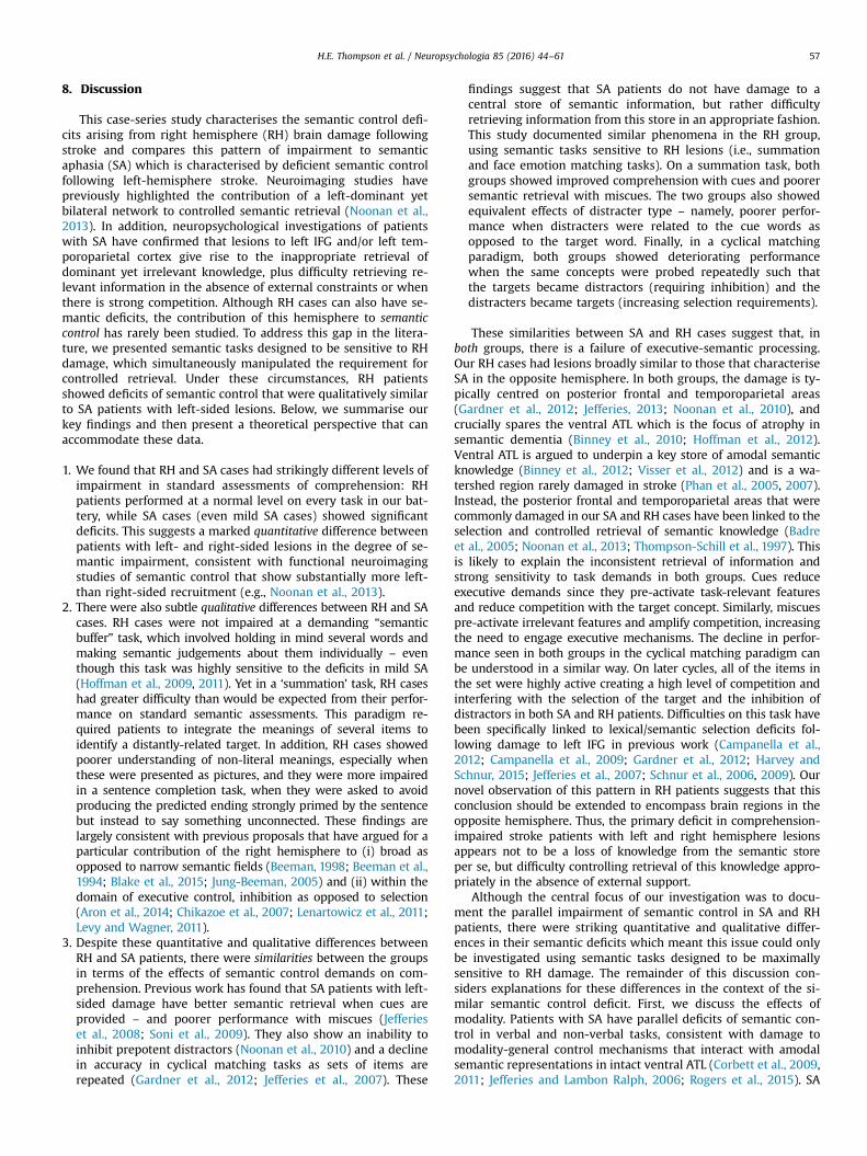
In a comparison of RH patients and controls, there was a maineffect of cycle: F(3,16)¼3.527, p¼ .039, a main effect of group: F(1,18)¼15.921, p¼ .001, and a group by cycle interaction: F(3,16)¼9.152, p¼ .001. There was no main effect of modality (Fo1), nointeraction between modality and cycle (Fo1.3) nor betweenmodality and group (F(1,18)¼2.803, p¼ .111). There was a three-way interaction between modality, cycle and group: F(3,16)¼3.717, p¼ .033, reflecting higher performance in PPM than WPM incontrols, and lower performance in PPM than WPM in RH patients.
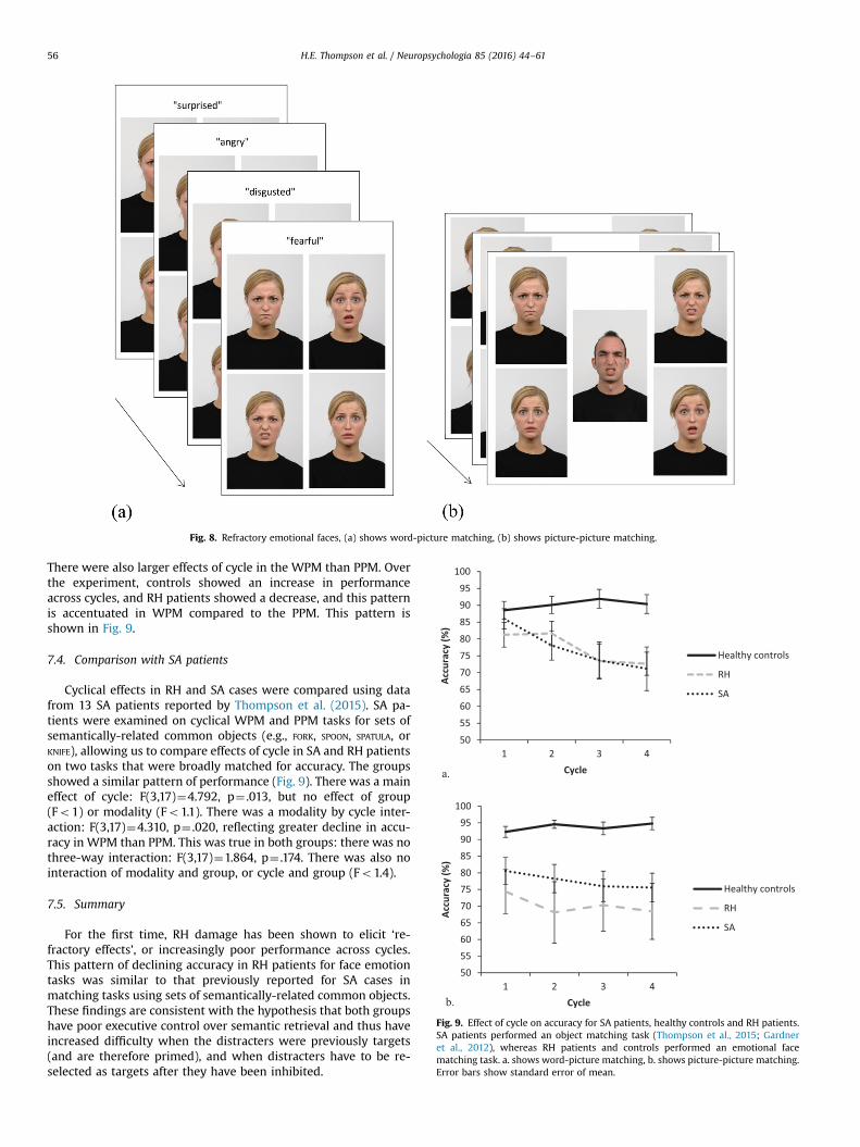
Fig. 8. Refractory emotional faces, (a) shows word-picture matching, (b) shows picture-picture matching.
Fig. 9. Effect of cycle on accuracy for SA patients, healthy controls and RH patients.SA patients performed an object matching task (Thompson et al., 2015; Gardneret al., 2012), whereas RH patients and controls performed an emotional facematching task. a. shows word-picture matching, b. shows picture-picture matching.Error bars show standard error of mean.
H.E. Thompson et al. / Neuropsychologia 85 (2016) 44–6156
There were also larger effects of cycle in the WPM than PPM. Overthe experiment, controls showed an increase in performanceacross cycles, and RH patients showed a decrease, and this patternis accentuated in WPM compared to the PPM. This pattern isshown in Fig. 9.
7.4. Comparison with SA patients
Cyclical effects in RH and SA cases were compared using datafrom 13 SA patients reported by Thompson et al. (2015). SA pa-tients were examined on cyclical WPM and PPM tasks for sets ofsemantically-related common objects (e.g., FORK, SPOON, SPATULA, orKNIFE), allowing us to compare effects of cycle in SA and RH patientson two tasks that were broadly matched for accuracy. The groupsshowed a similar pattern of performance (Fig. 9). There was a maineffect of cycle: F(3,17)¼4.792, p¼ .013, but no effect of group(Fo1) or modality (Fo1.1). There was a modality by cycle inter-action: F(3,17)¼4.310, p¼ .020, reflecting greater decline in accu-racy in WPM than PPM. This was true in both groups: there was nothree-way interaction: F(3,17)¼1.864, p¼ .174. There was also nointeraction of modality and group, or cycle and group (Fo1.4).
7.5. Summary
For the first time, RH damage has been shown to elicit ‘re-fractory effects’, or increasingly poor performance across cycles.This pattern of declining accuracy in RH patients for face emotiontasks was similar to that previously reported for SA cases inmatching tasks using sets of semantically-related common objects.These findings are consistent with the hypothesis that both groupshave poor executive control over semantic retrieval and thus haveincreased difficulty when the distracters were previously targets(and are therefore primed), and when distracters have to be re-selected as targets after they have been inhibited.
H.E. Thompson et al. / Neuropsychologia 85 (2016) 44–61 57
8. Discussion
This case-series study characterises the semantic control defi-cits arising from right hemisphere (RH) brain damage followingstroke and compares this pattern of impairment to semanticaphasia (SA) which is characterised by deficient semantic controlfollowing left-hemisphere stroke. Neuroimaging studies havepreviously highlighted the contribution of a left-dominant yetbilateral network to controlled semantic retrieval (Noonan et al.,2013). In addition, neuropsychological investigations of patientswith SA have confirmed that lesions to left IFG and/or left tem-poroparietal cortex give rise to the inappropriate retrieval ofdominant yet irrelevant knowledge, plus difficulty retrieving re-levant information in the absence of external constraints or whenthere is strong competition. Although RH cases can also have se-mantic deficits, the contribution of this hemisphere to semanticcontrol has rarely been studied. To address this gap in the litera-ture, we presented semantic tasks designed to be sensitive to RHdamage, which simultaneously manipulated the requirement forcontrolled retrieval. Under these circumstances, RH patientsshowed deficits of semantic control that were qualitatively similarto SA patients with left-sided lesions. Below, we summarise ourkey findings and then present a theoretical perspective that canaccommodate these data.
1. We found that RH and SA cases had strikingly different levels ofimpairment in standard assessments of comprehension: RHpatients performed at a normal level on every task in our bat-tery, while SA cases (even mild SA cases) showed significantdeficits. This suggests a marked quantitative difference betweenpatients with left- and right-sided lesions in the degree of se-mantic impairment, consistent with functional neuroimagingstudies of semantic control that show substantially more left-than right-sided recruitment (e.g., Noonan et al., 2013).
2. There were also subtle qualitative differences between RH and SAcases. RH cases were not impaired at a demanding “semanticbuffer” task, which involved holding in mind several words andmaking semantic judgements about them individually – eventhough this task was highly sensitive to the deficits in mild SA(Hoffman et al., 2009, 2011). Yet in a ‘summation’ task, RH caseshad greater difficulty than would be expected from their perfor-mance on standard semantic assessments. This paradigm re-quired patients to integrate the meanings of several items toidentify a distantly-related target. In addition, RH cases showedpoorer understanding of non-literal meanings, especially whenthese were presented as pictures, and they were more impairedin a sentence completion task, when they were asked to avoidproducing the predicted ending strongly primed by the sentencebut instead to say something unconnected. These findings arelargely consistent with previous proposals that have argued for aparticular contribution of the right hemisphere to (i) broad asopposed to narrow semantic fields (Beeman, 1998; Beeman et al.,1994; Blake et al., 2015; Jung-Beeman, 2005) and (ii) within thedomain of executive control, inhibition as opposed to selection(Aron et al., 2014; Chikazoe et al., 2007; Lenartowicz et al., 2011;Levy and Wagner, 2011).
3. Despite these quantitative and qualitative differences betweenRH and SA patients, there were similarities between the groupsin terms of the effects of semantic control demands on com-prehension. Previous work has found that SA patients with left-sided damage have better semantic retrieval when cues areprovided – and poorer performance with miscues (Jefferieset al., 2008; Soni et al., 2009). They also show an inability toinhibit prepotent distractors (Noonan et al., 2010) and a declinein accuracy in cyclical matching tasks as sets of items arerepeated (Gardner et al., 2012; Jefferies et al., 2007). These
findings suggest that SA patients do not have damage to acentral store of semantic information, but rather difficultyretrieving information from this store in an appropriate fashion.This study documented similar phenomena in the RH group,using semantic tasks sensitive to RH lesions (i.e., summationand face emotion matching tasks). On a summation task, bothgroups showed improved comprehension with cues and poorersemantic retrieval with miscues. The two groups also showedequivalent effects of distracter type – namely, poorer perfor-mance when distracters were related to the cue words asopposed to the target word. Finally, in a cyclical matchingparadigm, both groups showed deteriorating performancewhen the same concepts were probed repeatedly such thatthe targets became distractors (requiring inhibition) and thedistracters became targets (increasing selection requirements).
These similarities between SA and RH cases suggest that, inboth groups, there is a failure of executive-semantic processing.Our RH cases had lesions broadly similar to those that characteriseSA in the opposite hemisphere. In both groups, the damage is ty-pically centred on posterior frontal and temporoparietal areas(Gardner et al., 2012; Jefferies, 2013; Noonan et al., 2010), andcrucially spares the ventral ATL which is the focus of atrophy insemantic dementia (Binney et al., 2010; Hoffman et al., 2012).Ventral ATL is argued to underpin a key store of amodal semanticknowledge (Binney et al., 2012; Visser et al., 2012) and is a wa-tershed region rarely damaged in stroke (Phan et al., 2005, 2007).Instead, the posterior frontal and temporoparietal areas that werecommonly damaged in our SA and RH cases have been linked to theselection and controlled retrieval of semantic knowledge (Badreet al., 2005; Noonan et al., 2013; Thompson-Schill et al., 1997). Thisis likely to explain the inconsistent retrieval of information andstrong sensitivity to task demands in both groups. Cues reduceexecutive demands since they pre-activate task-relevant featuresand reduce competition with the target concept. Similarly, miscuespre-activate irrelevant features and amplify competition, increasingthe need to engage executive mechanisms. The decline in perfor-mance seen in both groups in the cyclical matching paradigm canbe understood in a similar way. On later cycles, all of the items inthe set were highly active creating a high level of competition andinterfering with the selection of the target and the inhibition ofdistractors in both SA and RH patients. Difficulties on this task havebeen specifically linked to lexical/semantic selection deficits fol-lowing damage to left IFG in previous work (Campanella et al.,2012; Campanella et al., 2009; Gardner et al., 2012; Harvey andSchnur, 2015; Jefferies et al., 2007; Schnur et al., 2006, 2009). Ournovel observation of this pattern in RH patients suggests that thisconclusion should be extended to encompass brain regions in theopposite hemisphere. Thus, the primary deficit in comprehension-impaired stroke patients with left and right hemisphere lesionsappears not to be a loss of knowledge from the semantic storeper se, but difficulty controlling retrieval of this knowledge appro-priately in the absence of external support.
Although the central focus of our investigation was to docu-ment the parallel impairment of semantic control in SA and RHpatients, there were striking quantitative and qualitative differ-ences in their semantic deficits which meant this issue could onlybe investigated using semantic tasks designed to be maximallysensitive to RH damage. The remainder of this discussion con-siders explanations for these differences in the context of the si-milar semantic control deficit. First, we discuss the effects ofmodality. Patients with SA have parallel deficits of semantic con-trol in verbal and non-verbal tasks, consistent with damage tomodality-general control mechanisms that interact with amodalsemantic representations in intact ventral ATL (Corbett et al., 2009,2011; Jefferies and Lambon Ralph, 2006; Rogers et al., 2015). SA
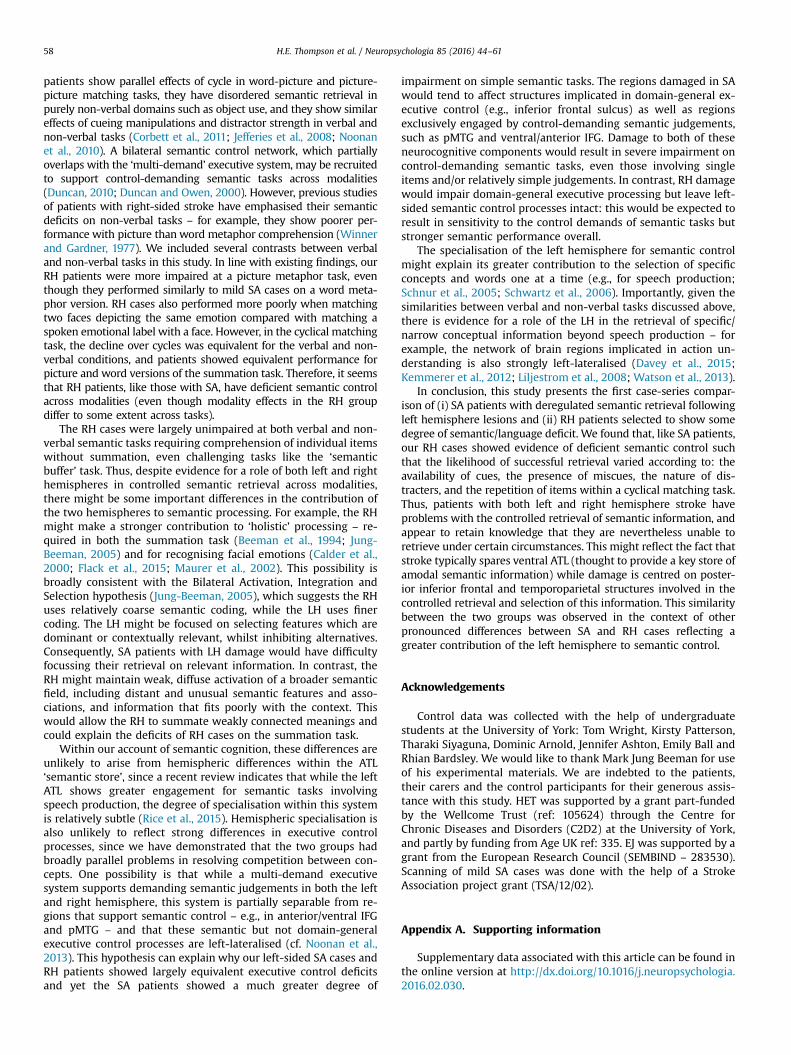
H.E. Thompson et al. / Neuropsychologia 85 (2016) 44–6158
patients show parallel effects of cycle in word-picture and picture-picture matching tasks, they have disordered semantic retrieval inpurely non-verbal domains such as object use, and they show similareffects of cueing manipulations and distractor strength in verbal andnon-verbal tasks (Corbett et al., 2011; Jefferies et al., 2008; Noonanet al., 2010). A bilateral semantic control network, which partiallyoverlaps with the ‘multi-demand’ executive system, may be recruitedto support control-demanding semantic tasks across modalities(Duncan, 2010; Duncan and Owen, 2000). However, previous studiesof patients with right-sided stroke have emphasised their semanticdeficits on non-verbal tasks – for example, they show poorer per-formance with picture than word metaphor comprehension (Winnerand Gardner, 1977). We included several contrasts between verbaland non-verbal tasks in this study. In line with existing findings, ourRH patients were more impaired at a picture metaphor task, eventhough they performed similarly to mild SA cases on a word meta-phor version. RH cases also performed more poorly when matchingtwo faces depicting the same emotion compared with matching aspoken emotional label with a face. However, in the cyclical matchingtask, the decline over cycles was equivalent for the verbal and non-verbal conditions, and patients showed equivalent performance forpicture and word versions of the summation task. Therefore, it seemsthat RH patients, like those with SA, have deficient semantic controlacross modalities (even though modality effects in the RH groupdiffer to some extent across tasks).
The RH cases were largely unimpaired at both verbal and non-verbal semantic tasks requiring comprehension of individual itemswithout summation, even challenging tasks like the ‘semanticbuffer’ task. Thus, despite evidence for a role of both left and righthemispheres in controlled semantic retrieval across modalities,there might be some important differences in the contribution ofthe two hemispheres to semantic processing. For example, the RHmight make a stronger contribution to ‘holistic’ processing – re-quired in both the summation task (Beeman et al., 1994; Jung-Beeman, 2005) and for recognising facial emotions (Calder et al.,2000; Flack et al., 2015; Maurer et al., 2002). This possibility isbroadly consistent with the Bilateral Activation, Integration andSelection hypothesis (Jung-Beeman, 2005), which suggests the RHuses relatively coarse semantic coding, while the LH uses finercoding. The LH might be focused on selecting features which aredominant or contextually relevant, whilst inhibiting alternatives.Consequently, SA patients with LH damage would have difficultyfocussing their retrieval on relevant information. In contrast, theRH might maintain weak, diffuse activation of a broader semanticfield, including distant and unusual semantic features and asso-ciations, and information that fits poorly with the context. Thiswould allow the RH to summate weakly connected meanings andcould explain the deficits of RH cases on the summation task.
Within our account of semantic cognition, these differences areunlikely to arise from hemispheric differences within the ATL‘semantic store’, since a recent review indicates that while the leftATL shows greater engagement for semantic tasks involvingspeech production, the degree of specialisation within this systemis relatively subtle (Rice et al., 2015). Hemispheric specialisation isalso unlikely to reflect strong differences in executive controlprocesses, since we have demonstrated that the two groups hadbroadly parallel problems in resolving competition between con-cepts. One possibility is that while a multi-demand executivesystem supports demanding semantic judgements in both the leftand right hemisphere, this system is partially separable from re-gions that support semantic control – e.g., in anterior/ventral IFGand pMTG – and that these semantic but not domain-generalexecutive control processes are left-lateralised (cf. Noonan et al.,2013). This hypothesis can explain why our left-sided SA cases andRH patients showed largely equivalent executive control deficitsand yet the SA patients showed a much greater degree of
impairment on simple semantic tasks. The regions damaged in SAwould tend to affect structures implicated in domain-general ex-ecutive control (e.g., inferior frontal sulcus) as well as regionsexclusively engaged by control-demanding semantic judgements,such as pMTG and ventral/anterior IFG. Damage to both of theseneurocognitive components would result in severe impairment oncontrol-demanding semantic tasks, even those involving singleitems and/or relatively simple judgements. In contrast, RH damagewould impair domain-general executive processing but leave left-sided semantic control processes intact: this would be expected toresult in sensitivity to the control demands of semantic tasks butstronger semantic performance overall.
The specialisation of the left hemisphere for semantic controlmight explain its greater contribution to the selection of specificconcepts and words one at a time (e.g., for speech production;Schnur et al., 2005; Schwartz et al., 2006). Importantly, given thesimilarities between verbal and non-verbal tasks discussed above,there is evidence for a role of the LH in the retrieval of specific/narrow conceptual information beyond speech production – forexample, the network of brain regions implicated in action un-derstanding is also strongly left-lateralised (Davey et al., 2015;Kemmerer et al., 2012; Liljestrom et al., 2008; Watson et al., 2013).
In conclusion, this study presents the first case-series compar-ison of (i) SA patients with deregulated semantic retrieval followingleft hemisphere lesions and (ii) RH patients selected to show somedegree of semantic/language deficit. We found that, like SA patients,our RH cases showed evidence of deficient semantic control suchthat the likelihood of successful retrieval varied according to: theavailability of cues, the presence of miscues, the nature of dis-tracters, and the repetition of items within a cyclical matching task.Thus, patients with both left and right hemisphere stroke haveproblems with the controlled retrieval of semantic information, andappear to retain knowledge that they are nevertheless unable toretrieve under certain circumstances. This might reflect the fact thatstroke typically spares ventral ATL (thought to provide a key store ofamodal semantic information) while damage is centred on poster-ior inferior frontal and temporoparietal structures involved in thecontrolled retrieval and selection of this information. This similaritybetween the two groups was observed in the context of otherpronounced differences between SA and RH cases reflecting agreater contribution of the left hemisphere to semantic control.
Acknowledgements
Control data was collected with the help of undergraduatestudents at the University of York: Tom Wright, Kirsty Patterson,Tharaki Siyaguna, Dominic Arnold, Jennifer Ashton, Emily Ball andRhian Bardsley. We would like to thank Mark Jung Beeman for useof his experimental materials. We are indebted to the patients,their carers and the control participants for their generous assis-tance with this study. HET was supported by a grant part-fundedby the Wellcome Trust (ref: 105624) through the Centre forChronic Diseases and Disorders (C2D2) at the University of York,and partly by funding from Age UK ref: 335. EJ was supported by agrant from the European Research Council (SEMBIND – 283530).Scanning of mild SA cases was done with the help of a StrokeAssociation project grant (TSA/12/02).
Appendix A. Supporting information
Supplementary data associated with this article can be found inthe online version at http://dx.doi.org/10.1016/j.neuropsychologia.2016.02.030.
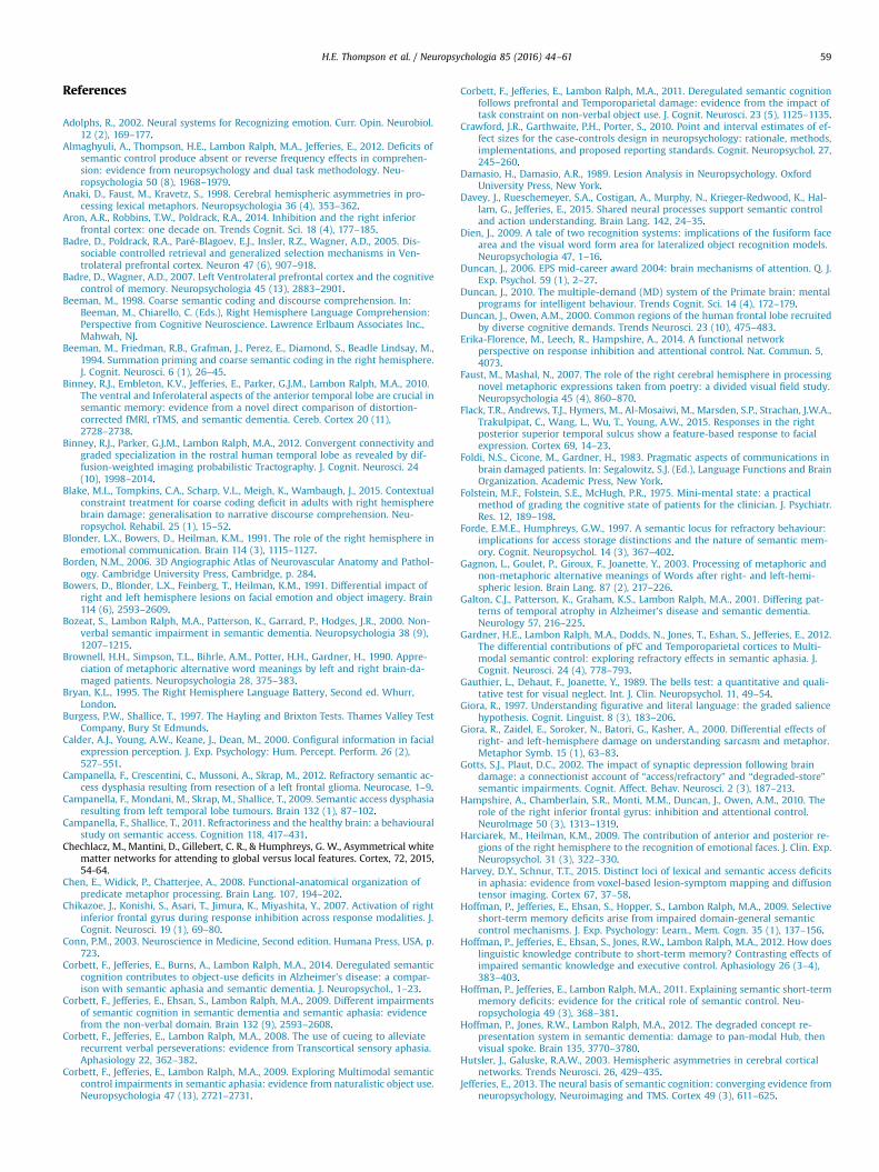
H.E. Thompson et al. / Neuropsychologia 85 (2016) 44–61 59
References
Adolphs, R., 2002. Neural systems for Recognizing emotion. Curr. Opin. Neurobiol.12 (2), 169–177.
Almaghyuli, A., Thompson, H.E., Lambon Ralph, M.A., Jefferies, E., 2012. Deficits ofsemantic control produce absent or reverse frequency effects in comprehen-sion: evidence from neuropsychology and dual task methodology. Neu-ropsychologia 50 (8), 1968–1979.
Anaki, D., Faust, M., Kravetz, S., 1998. Cerebral hemispheric asymmetries in pro-cessing lexical metaphors. Neuropsychologia 36 (4), 353–362.
Aron, A.R., Robbins, T.W., Poldrack, R.A., 2014. Inhibition and the right inferiorfrontal cortex: one decade on. Trends Cognit. Sci. 18 (4), 177–185.
Badre, D., Poldrack, R.A., Paré-Blagoev, E.J., Insler, R.Z., Wagner, A.D., 2005. Dis-sociable controlled retrieval and generalized selection mechanisms in Ven-trolateral prefrontal cortex. Neuron 47 (6), 907–918.
Badre, D., Wagner, A.D., 2007. Left Ventrolateral prefrontal cortex and the cognitivecontrol of memory. Neuropsychologia 45 (13), 2883–2901.
Beeman, M., 1998. Coarse semantic coding and discourse comprehension. In:Beeman, M., Chiarello, C. (Eds.), Right Hemisphere Language Comprehension:Perspective from Cognitive Neuroscience. Lawrence Erlbaum Associates Inc.,Mahwah, NJ.
Beeman, M., Friedman, R.B., Grafman, J., Perez, E., Diamond, S., Beadle Lindsay, M.,1994. Summation priming and coarse semantic coding in the right hemisphere.J. Cognit. Neurosci. 6 (1), 26–45.
Binney, R.J., Embleton, K.V., Jefferies, E., Parker, G.J.M., Lambon Ralph, M.A., 2010.The ventral and Inferolateral aspects of the anterior temporal lobe are crucial insemantic memory: evidence from a novel direct comparison of distortion-corrected fMRI, rTMS, and semantic dementia. Cereb. Cortex 20 (11),2728–2738.
Binney, R.J., Parker, G.J.M., Lambon Ralph, M.A., 2012. Convergent connectivity andgraded specialization in the rostral human temporal lobe as revealed by dif-fusion-weighted imaging probabilistic Tractography. J. Cognit. Neurosci. 24(10), 1998–2014.
Blake, M.L., Tompkins, C.A., Scharp, V.L., Meigh, K., Wambaugh, J., 2015. Contextualconstraint treatment for coarse coding deficit in adults with right hemispherebrain damage: generalisation to narrative discourse comprehension. Neu-ropsychol. Rehabil. 25 (1), 15–52.
Blonder, L.X., Bowers, D., Heilman, K.M., 1991. The role of the right hemisphere inemotional communication. Brain 114 (3), 1115–1127.
Borden, N.M., 2006. 3D Angiographic Atlas of Neurovascular Anatomy and Pathol-ogy. Cambridge University Press, Cambridge, p. 284.
Bowers, D., Blonder, L.X., Feinberg, T., Heilman, K.M., 1991. Differential impact ofright and left hemisphere lesions on facial emotion and object imagery. Brain114 (6), 2593–2609.
Bozeat, S., Lambon Ralph, M.A., Patterson, K., Garrard, P., Hodges, J.R., 2000. Non-verbal semantic impairment in semantic dementia. Neuropsychologia 38 (9),1207–1215.
Brownell, H.H., Simpson, T.L., Bihrle, A.M., Potter, H.H., Gardner, H., 1990. Appre-ciation of metaphoric alternative word meanings by left and right brain-da-maged patients. Neuropsychologia 28, 375–383.
Bryan, K.L., 1995. The Right Hemisphere Language Battery, Second ed. Whurr,London.
Burgess, P.W., Shallice, T., 1997. The Hayling and Brixton Tests. Thames Valley TestCompany, Bury St Edmunds.
Calder, A.J., Young, A.W., Keane, J., Dean, M., 2000. Configural information in facialexpression perception. J. Exp. Psychology: Hum. Percept. Perform. 26 (2),527–551.
Campanella, F., Crescentini, C., Mussoni, A., Skrap, M., 2012. Refractory semantic ac-cess dysphasia resulting from resection of a left frontal glioma. Neurocase, 1–9.
Campanella, F., Mondani, M., Skrap, M., Shallice, T., 2009. Semantic access dysphasiaresulting from left temporal lobe tumours. Brain 132 (1), 87–102.
Campanella, F., Shallice, T., 2011. Refractoriness and the healthy brain: a behaviouralstudy on semantic access. Cognition 118, 417–431.
Chechlacz, M., Mantini, D., Gillebert, C. R., & Humphreys, G. W., Asymmetrical whitematter networks for attending to global versus local features. Cortex, 72, 2015,54-64.
Chen, E., Widick, P., Chatterjee, A., 2008. Functional-anatomical organization ofpredicate metaphor processing. Brain Lang. 107, 194–202.
Chikazoe, J., Konishi, S., Asari, T., Jimura, K., Miyashita, Y., 2007. Activation of rightinferior frontal gyrus during response inhibition across response modalities. J.Cognit. Neurosci. 19 (1), 69–80.
Conn, P.M., 2003. Neuroscience in Medicine, Second edition. Humana Press, USA, p.723.
Corbett, F., Jefferies, E., Burns, A., Lambon Ralph, M.A., 2014. Deregulated semanticcognition contributes to object-use deficits in Alzheimer’s disease: a compar-ison with semantic aphasia and semantic dementia. J. Neuropsychol., 1–23.
Corbett, F., Jefferies, E., Ehsan, S., Lambon Ralph, M.A., 2009. Different impairmentsof semantic cognition in semantic dementia and semantic aphasia: evidencefrom the non-verbal domain. Brain 132 (9), 2593–2608.
Corbett, F., Jefferies, E., Lambon Ralph, M.A., 2008. The use of cueing to alleviaterecurrent verbal perseverations: evidence from Transcortical sensory aphasia.Aphasiology 22, 362–382.
Corbett, F., Jefferies, E., Lambon Ralph, M.A., 2009. Exploring Multimodal semanticcontrol impairments in semantic aphasia: evidence from naturalistic object use.Neuropsychologia 47 (13), 2721–2731.
Corbett, F., Jefferies, E., Lambon Ralph, M.A., 2011. Deregulated semantic cognitionfollows prefrontal and Temporoparietal damage: evidence from the impact oftask constraint on non-verbal object use. J. Cognit. Neurosci. 23 (5), 1125–1135.
Crawford, J.R., Garthwaite, P.H., Porter, S., 2010. Point and interval estimates of ef-fect sizes for the case-controls design in neuropsychology: rationale, methods,implementations, and proposed reporting standards. Cognit. Neuropsychol. 27,245–260.
Damasio, H., Damasio, A.R., 1989. Lesion Analysis in Neuropsychology. OxfordUniversity Press, New York.
Davey, J., Rueschemeyer, S.A., Costigan, A., Murphy, N., Krieger-Redwood, K., Hal-lam, G., Jefferies, E., 2015. Shared neural processes support semantic controland action understanding. Brain Lang. 142, 24–35.
Dien, J., 2009. A tale of two recognition systems: implications of the fusiform facearea and the visual word form area for lateralized object recognition models.Neuropsychologia 47, 1–16.
Duncan, J., 2006. EPS mid-career award 2004: brain mechanisms of attention. Q. J.Exp. Psychol. 59 (1), 2–27.
Duncan, J., 2010. The multiple-demand (MD) system of the Primate brain: mentalprograms for intelligent behaviour. Trends Cognit. Sci. 14 (4), 172–179.
Duncan, J., Owen, A.M., 2000. Common regions of the human frontal lobe recruitedby diverse cognitive demands. Trends Neurosci. 23 (10), 475–483.
Erika-Florence, M., Leech, R., Hampshire, A., 2014. A functional networkperspective on response inhibition and attentional control. Nat. Commun. 5,4073.
Faust, M., Mashal, N., 2007. The role of the right cerebral hemisphere in processingnovel metaphoric expressions taken from poetry: a divided visual field study.Neuropsychologia 45 (4), 860–870.
Flack, T.R., Andrews, T.J., Hymers, M., Al-Mosaiwi, M., Marsden, S.P., Strachan, J.W.A.,Trakulpipat, C., Wang, L., Wu, T., Young, A.W., 2015. Responses in the rightposterior superior temporal sulcus show a feature-based response to facialexpression. Cortex 69, 14–23.
Foldi, N.S., Cicone, M., Gardner, H., 1983. Pragmatic aspects of communications inbrain damaged patients. In: Segalowitz, S.J. (Ed.), Language Functions and BrainOrganization. Academic Press, New York.
Folstein, M.F., Folstein, S.E., McHugh, P.R., 1975. Mini-mental state: a practicalmethod of grading the cognitive state of patients for the clinician. J. Psychiatr.Res. 12, 189–198.
Forde, E.M.E., Humphreys, G.W., 1997. A semantic locus for refractory behaviour:implications for access storage distinctions and the nature of semantic mem-ory. Cognit. Neuropsychol. 14 (3), 367–402.
Gagnon, L., Goulet, P., Giroux, F., Joanette, Y., 2003. Processing of metaphoric andnon-metaphoric alternative meanings of Words after right- and left-hemi-spheric lesion. Brain Lang. 87 (2), 217–226.
Galton, C.J., Patterson, K., Graham, K.S., Lambon Ralph, M.A., 2001. Differing pat-terns of temporal atrophy in Alzheimer’s disease and semantic dementia.Neurology 57, 216–225.
Gardner, H.E., Lambon Ralph, M.A., Dodds, N., Jones, T., Eshan, S., Jefferies, E., 2012.The differential contributions of pFC and Temporoparietal cortices to Multi-modal semantic control: exploring refractory effects in semantic aphasia. J.Cognit. Neurosci. 24 (4), 778–793.
Gauthier, L., Dehaut, F., Joanette, Y., 1989. The bells test: a quantitative and quali-tative test for visual neglect. Int. J. Clin. Neuropsychol. 11, 49–54.
Giora, R., 1997. Understanding figurative and literal language: the graded saliencehypothesis. Cognit. Linguist. 8 (3), 183–206.
Giora, R., Zaidel, E., Soroker, N., Batori, G., Kasher, A., 2000. Differential effects ofright- and left-hemisphere damage on understanding sarcasm and metaphor.Metaphor Symb. 15 (1), 63–83.
Gotts, S.J., Plaut, D.C., 2002. The impact of synaptic depression following braindamage: a connectionist account of “access/refractory” and “degraded-store”semantic impairments. Cognit. Affect. Behav. Neurosci. 2 (3), 187–213.
Hampshire, A., Chamberlain, S.R., Monti, M.M., Duncan, J., Owen, A.M., 2010. Therole of the right inferior frontal gyrus: inhibition and attentional control.NeuroImage 50 (3), 1313–1319.
Harciarek, M., Heilman, K.M., 2009. The contribution of anterior and posterior re-gions of the right hemisphere to the recognition of emotional faces. J. Clin. Exp.Neuropsychol. 31 (3), 322–330.
Harvey, D.Y., Schnur, T.T., 2015. Distinct loci of lexical and semantic access deficitsin aphasia: evidence from voxel-based lesion-symptom mapping and diffusiontensor imaging. Cortex 67, 37–58.
Hoffman, P., Jefferies, E., Ehsan, S., Hopper, S., Lambon Ralph, M.A., 2009. Selectiveshort-term memory deficits arise from impaired domain-general semanticcontrol mechanisms. J. Exp. Psychology: Learn., Mem. Cogn. 35 (1), 137–156.
Hoffman, P., Jefferies, E., Ehsan, S., Jones, R.W., Lambon Ralph, M.A., 2012. How doeslinguistic knowledge contribute to short-term memory? Contrasting effects ofimpaired semantic knowledge and executive control. Aphasiology 26 (3–4),383–403.
Hoffman, P., Jefferies, E., Lambon Ralph, M.A., 2011. Explaining semantic short-termmemory deficits: evidence for the critical role of semantic control. Neu-ropsychologia 49 (3), 368–381.
Hoffman, P., Jones, R.W., Lambon Ralph, M.A., 2012. The degraded concept re-presentation system in semantic dementia: damage to pan-modal Hub, thenvisual spoke. Brain 135, 3770–3780.
Hutsler, J., Galuske, R.A.W., 2003. Hemispheric asymmetries in cerebral corticalnetworks. Trends Neurosci. 26, 429–435.
Jefferies, E., 2013. The neural basis of semantic cognition: converging evidence fromneuropsychology, Neuroimaging and TMS. Cortex 49 (3), 611–625.
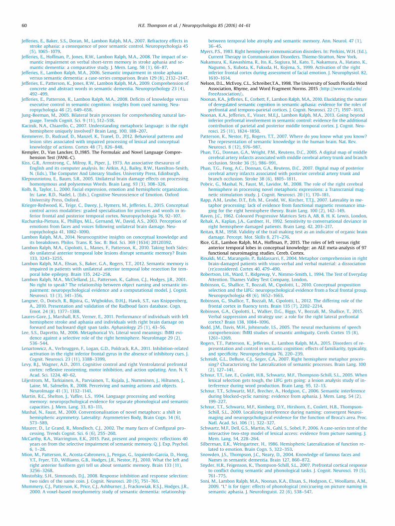
H.E. Thompson et al. / Neuropsychologia 85 (2016) 44–6160
Jefferies, E., Baker, S.S., Doran, M., Lambon Ralph, M.A., 2007. Refractory effects instroke aphasia: a consequence of poor semantic control. Neuropsychologia 45(5), 1065–1079.
Jefferies, E., Hoffman, P., Jones, R.W., Lambon Ralph, M.A., 2008. The impact of se-mantic impairment on verbal short-term memory in stroke aphasia and se-mantic dementia: a comparative study. J. Mem. Lang. 58 (1), 66–87.
Jefferies, E., Lambon Ralph, M.A., 2006. Semantic impairment in stroke aphasiaversus semantic dementia: a case-series comparison. Brain 129 (8), 2132–2147.
Jefferies, E., Patterson, K., Jones, R.W., Lambon Ralph, M.A., 2009. Comprehension ofconcrete and abstract words in semantic dementia. Neuropsychology 23 (4),492–499.
Jefferies, E., Patterson, K., Lambon Ralph, M.A., 2008. Deficits of knowledge versusexecutive control in semantic cognition: insights from cued naming. Neu-ropsychologia 46 (2), 649–658.
Jung-Beeman, M., 2005. Bilateral brain processes for comprehending natural lan-guage. Trends Cognit. Sci. 9 (11), 512–518.
Kacinik, N.A., Chiarello, C., 2007. Understanding metaphoric language: is the righthemisphere uniquely involved? Brain Lang. 100, 188–207.
Kemmerer, D., Rudrauf, D., Manzel, K., Tranel, D., 2012. Behavioral patterns andlesion sites associated with impaired processing of lexical and conceptualknowledge of actions. Cortex 48 (7), 826–848.
Kempler, D., Van Lancker, D.,1985. The Formulaic and Novel Language Compre-hension Test (FANL-C).
Kiss, G.R., Armstrong, C., Milroy, R., Piper, J., 1973. An associative thesaurus ofEnglish and its computer analysis. In: Aitkin, A.J., Bailey, R.W., Hamilton-Smith,N. (Eds.), The Computer And Literary Studies. University Press, Edinburgh.
Klepousniotou, E., Baum, S.R., 2005. Unilateral brain damage effects on processinghomonymous and polysemous Words. Brain Lang. 93 (3), 308–326.
Kolb, B., Taylor, L., 2000. Facial expression, emotion and hemispheric organization.In: Lane, R.D., Nadel, L. (Eds.), Cognitive Neuroscience of Emotion. OxfordUniversity Press, Oxford.
Krieger-Redwood, K., Teige, C., Davey, J., Hymers, M., Jefferies, E., 2015. Conceptualcontrol across modalities: graded specialisation for pictures and words in in-ferior frontal and posterior temporal cortex. Neuropsychologia 76, 92–107.
Kucharska-Pietura, K., Phillips, M.L., Gernand, W., David, A.S., 2003. Perception ofemotions from faces and voices following unilateral brain damage. Neu-ropsychologia 41, 1082–1090.
Lambon Ralph, M.A., 2014. Neurocognitive insights on conceptual knowledge andits breakdown. Philos. Trans. R. Soc. B: Biol. Sci. 369 (1634) 20120392.
Lambon Ralph, M.A., Cipolotti, L., Manes, F., Patterson, K., 2010. Taking both Sides:do unilateral anterior temporal lobe lesions disrupt semantic memory? Brain133, 3243–3255.
Lambon Ralph, M.A., Ehsan, S., Baker, G.A., Rogers, T.T., 2012. Semantic memory isimpaired in patients with unilateral anterior temporal lobe resection for tem-poral lobe epilepsy. Brain 135, 242–258.
Lambon Ralph, M.A., McClelland, J.L., Patterson, K., Galton, C.J., Hodges, J.R., 2001.No right to speak? The relationship between object naming and semantic im-pairment: neuropsychological evidence and a computational model. J. Cognit.Neurosci. 13 (3), 341–356.
Langner, O., Dotsch, R., Bijstra, G., Wigboldus, D.H.J., Hawk, S.T., van Knippenberg,A., 2010. Presentation and validation of the Radboud faces database. Cogn.Emot. 24 (8), 1377–1388.
Laures-Gore, J., Marshall, R.S., Verner, E., 2011. Performance of individuals with lefthemisphere stroke and aphasia and individuals with right brain damage onforward and backward digit span tasks. Aphasiology 25 (1), 43–56.
Lee, S.S., Dapretto, M., 2006. Metaphorical Vs. Literal word meanings: fMRI evi-dence against a selective role of the right hemisphere. NeuroImage 29 (2),536–544.
Lenartowicz, A., Verbruggen, F., Logan, G.D., Poldrack, R.A., 2011. Inhibition-relatedactivation in the right inferior frontal gyrus in the absence of inhibitory cues. J.Cognit. Neurosci. 23 (11), 3388–3399.
Levy, B.J., Wagner, A.D., 2011. Cognitive control and right Ventrolateral prefrontalcortex: reflexive reorienting, motor inhibition, and action updating. Ann. N. Y.Acad. Sci. 1224, 40–62.
Liljestrom, M., Tarkiainen, A., Parviainen, T., Kujala, J., Numminen, J., Hiltunen, J.,Laine, M., Salmelin, R., 2008. Perceiving and naming actions and objects.NeuroImage 41 (3), 1132–1141.
Martin, R.C., Shelton, J., Yaffee, L.S., 1994. Language processing and workingmemory: neuropsychological evidence for separate phonological and semanticcapacities. J. Mem. Lang. 33, 83–111.
Mashal, N., Faust, M., 2009. Conventionalisation of novel metaphors: a shift inhemispheric asymmetry. Laterality: Asymmetries Body, Brain Cogn. 14 (6),573–589.
Maurer, D., Le Grand, R., Mondloch, C.J., 2002. The many faces of Configural pro-cessing. Trends Cognit. Sci. 6 (6), 255–260.
McCarthy, R.A., Warrington, E.K., 2015. Past, present and prospects: reflections 40years on from the selective impairment of semantic memory. Q. J. Exp. Psychol.6, 1–28.
Mion, M., Patterson, K., Acosta-Cabronero, J., Pengas, G., Izquierdo-Garcia, D., Hong,Y.T., Fryer, T.D., Williams, G.B., Hodges, J.R., Nestor, P.J., 2010. What the left andright anterior fusiform gyri tell us about semantic memory. Brain 133 (11),3256–3268.
Mostofsky, S.H., Simmonds, D.J., 2008. Response inhibition and response selection:two sides of the same coin. J. Cognit. Neurosci. 20 (5), 751–761.
Mummery, C.J., Patterson, K., Price, C.J., Ashburner, J., Frackowiak, R.S.J., Hodges, J.R.,2000. A voxel-based morphometry study of semantic dementia: relationship
between temporal lobe atrophy and semantic memory. Ann. Neurol. 47 (1),36–45.
Myers, P.S., 1983. Right hemisphere communication disorders. In: Perkins, W.H. (Ed.),Current Therapy in Communication Disorders. Thieme-Stratton, New York.
Nakamura, K., Kawashima, R., Ito, K., Sugiura, M., Kato, T., Nakamura, A., Hatano, K.,Nagumo, S., Kubota, K., Fukuda, H., Kojima, S., 1999. Activation of the rightinferior frontal cortex during assessment of facial emotion. J. Neurophysiol. 82,1610–1614.
Nelson, D.L., McEvoy, C.L., Schreiber,T.A., 1998. The University of South Florida WordAssociation, Rhyme, and Word Fragment Norms. 2015 ⟨http://www.usf.edu/FreeAssociation/⟩.
Noonan, K.A., Jefferies, E., Corbett, F., Lambon Ralph, M.A., 2010. Elucidating the natureof deregulated semantic cognition in semantic aphasia: evidence for the roles ofprefrontal and temporo-parietal cortices. J. Cognit. Neurosci. 22 (7), 1597–1613.
Noonan, K.A., Jefferies, E., Visser, M.E.J., Lambon Ralph, M.A., 2013. Going beyondinferior prefrontal involvement in semantic control: evidence for the additionalcontribution of parietal and posterior middle temporal cortex. J. Cognit. Neu-rosci. 25 (11), 1824–1850.
Patterson, K., Nestor, P.J., Rogers, T.T., 2007. Where do you know what you know?The representation of semantic knowledge in the human brain. Nat. Rev.Neurosci. 8 (12), 976–987.
Phan, T.G., Donnan, G.A., Wright, P.M., Reutens, D.C., 2005. A digital map of middlecerebral artery infarcts associated with middle cerebral artery trunk and branchocclusion. Stroke 36 (5), 986–991.
Phan, T.G., Fong, A.C., Donnan, G.A., Reutens, D.C., 2007. Digital map of posteriorcerebral artery infarcts associated with posterior cerebral artery trunk andbranch occlusion. Stroke 38 (6), 1805–1811.
Pobric, G., Mashal, N., Faust, M., Lavidor, M., 2008. The role of the right cerebralhemisphere in processing novel metaphoric expressions: a Transcranial mag-netic stimulation study. J. Cognit. Neurosci. 20 (1), 170–181.
Rapp, A.M., Leube, D.T., Erb, M., Grodd, W., Kircher, T.T.J., 2007. Laterality in me-taphor processing: lack of evidence from functional magnetic resonance ima-ging for the right hemisphere theory. Brain Lang. 100 (2), 142–149.
Raven, J.C., 1962. Coloured Progressive Matrices Sets A, AB, B. H. K. Lewis, London.Rehak, A., Kaplan, J.A., Gardner, H., 1992. Sensitivity to conversational deviance in
right hemisphere damaged patients. Brain Lang. 42, 203–217.Reitan, R.M., 1958. Validity of the trail making test as an indicator of organic brain
damage. Percept. Mot. Skills 8, 271–276.Rice, G.E., Lambon Ralph, M.A., Hoffman, P., 2015. The roles of left versus right
anterior temporal lobes in conceptual knowledge: an ALE meta-analysis of 97functional neuroimaging studies. Cereb. Cortex.
Rinaldi, M.C., Marangolo, P., Baldassarri, F., 2004. Metaphor comprehension in rightbrain-damaged patients with visuo-verbal and verbal material: a dissociation(re)considered. Cortex 40, 479–490.
Robertson, I.H., Ward, T., Ridgeway, V., Nimmo-Smith, I., 1994. The Test of EverydayAttention. Thames Valley Test Company, London.
Robinson, G., Shallice, T., Bozzali, M., Cipolotti, L., 2010. Conceptual propositionselection and the LIFG: neuropsychological evidence from a focal frontal group.Neuropsychologia 48 (6), 1652–1663.
Robinson, G., Shallice, T., Bozzali, M., Cipolotti, L., 2012. The differing role of thefrontal cortex in fluency tests. Brain 135 (7), 2202–2214.
Robinson, G.A., Cipolotti, L., Walker, D.G., Biggs, V., Bozzali, M., Shallice, T., 2015.Verbal suppression and strategy use: a role for the right lateral prefrontalcortex? Brain 138, 1084–1096.
Rodd, J.M., Davis, M.H., Johnsrude, I.S., 2005. The neural mechanisms of speechcomprehension: fMRI studies of semantic ambiguity. Cereb. Cortex 15 (8),1261–1269.
Rogers, T.T., Patterson, K., Jefferies, E., Lambon Ralph, M.A., 2015. Disorders of re-presentation and control in semantic cognition: effects of familiarity, typicality,and specificity. Neuropsychologia 76, 220–239.
Schmidt, G.L., DeBuse, C.J., Seger, C.A., 2007. Right hemisphere metaphor proces-sing? Characterizing the Lateralization of semantic processes. Brain Lang. 100(2), 127–141.
Schnur, T.T., Lee, E., Coslett, H.B., Schwartz, M.F., Thompson-Schill, S.L., 2005. Whenlexical selection gets tough, the LIFG gets going: a lesion analysis study of in-terference during word production. Brain Lang. 95, 12–13.
Schnur, T.T., Schwartz, M.F., Brecher, A., Hodgson, C., 2006. Semantic interferenceduring blocked-cyclic naming: evidence from aphasia. J. Mem. Lang. 54 (2),199–227.
Schnur, T.T., Schwartz, M.F., Kimberg, D.Y., Hirshorn, E., Coslett, H.B., Thompson-Schill, S.L., 2009. Localizing interference during naming: convergent Neuroi-maging and neuropsychological evidence for the function of Broca's area. Proc.Natl. Acad. Sci. 106 (1), 322–327.
Schwartz, M.F., Dell, G.S., Martin, N., Gahl, S., Sobel, P., 2006. A case-series test of theinteractive two-step model of lexical access: evidence from picture naming. J.Mem. Lang. 54, 228–264.
Silberman, E.K., Weingartner, H., 1986. Hemispheric Lateralization of function re-lated to emotion. Brain Cogn. 5, 322–353.
Snowden, J.S., Thompson, J.C., Neary, D., 2004. Knowledge of famous faces andNames in semantic dementia. Brain 127, 860–872.
Snyder, H.R., Feigenson, K., Thompson-Schill, S.L., 2007. Prefrontal cortical responseto conflict during semantic and phonological tasks. J. Cognit. Neurosci. 19 (5),761–775.
Soni, M., Lambon Ralph, M.A., Noonan, K.A., Ehsan, S., Hodgson, C., Woollams, A.M.,2009. “L” is for tiger: effects of phonological (mis)cueing on picture naming insemantic aphasia. J. Neurolinguist. 22 (6), 538–547.
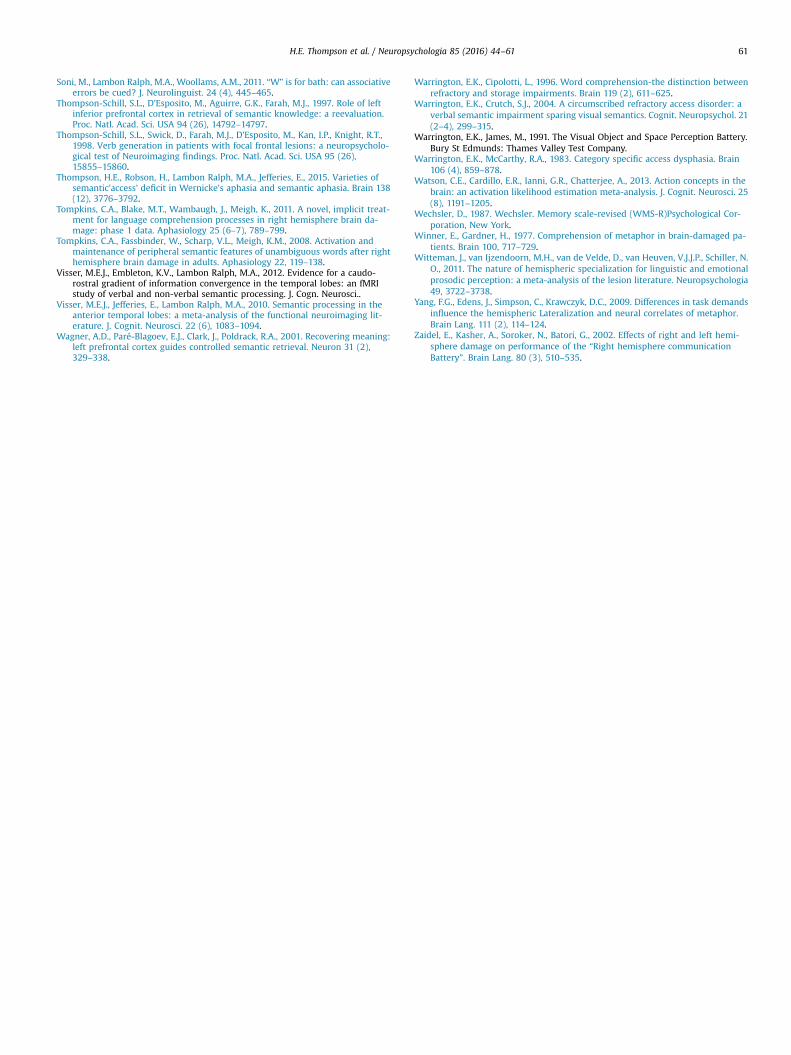
H.E. Thompson et al. / Neuropsychologia 85 (2016) 44–61 61
Soni, M., Lambon Ralph, M.A., Woollams, A.M., 2011. “W” is for bath: can associativeerrors be cued? J. Neurolinguist. 24 (4), 445–465.
Thompson-Schill, S.L., D’Esposito, M., Aguirre, G.K., Farah, M.J., 1997. Role of leftinferior prefrontal cortex in retrieval of semantic knowledge: a reevaluation.Proc. Natl. Acad. Sci. USA 94 (26), 14792–14797.
Thompson-Schill, S.L., Swick, D., Farah, M.J., D’Esposito, M., Kan, I.P., Knight, R.T.,1998. Verb generation in patients with focal frontal lesions: a neuropsycholo-gical test of Neuroimaging findings. Proc. Natl. Acad. Sci. USA 95 (26),15855–15860.
Thompson, H.E., Robson, H., Lambon Ralph, M.A., Jefferies, E., 2015. Varieties ofsemantic'access’ deficit in Wernicke’s aphasia and semantic aphasia. Brain 138(12), 3776–3792.
Tompkins, C.A., Blake, M.T., Wambaugh, J., Meigh, K., 2011. A novel, implicit treat-ment for language comprehension processes in right hemisphere brain da-mage: phase 1 data. Aphasiology 25 (6–7), 789–799.
Tompkins, C.A., Fassbinder, W., Scharp, V.L., Meigh, K.M., 2008. Activation andmaintenance of peripheral semantic features of unambiguous words after righthemisphere brain damage in adults. Aphasiology 22, 119–138.
Visser, M.E.J., Embleton, K.V., Lambon Ralph, M.A., 2012. Evidence for a caudo-rostral gradient of information convergence in the temporal lobes: an fMRIstudy of verbal and non-verbal semantic processing. J. Cogn. Neurosci..
Visser, M.E.J., Jefferies, E., Lambon Ralph, M.A., 2010. Semantic processing in theanterior temporal lobes: a meta-analysis of the functional neuroimaging lit-erature. J. Cognit. Neurosci. 22 (6), 1083–1094.
Wagner, A.D., Paré-Blagoev, E.J., Clark, J., Poldrack, R.A., 2001. Recovering meaning:left prefrontal cortex guides controlled semantic retrieval. Neuron 31 (2),329–338.
Warrington, E.K., Cipolotti, L., 1996. Word comprehension-the distinction betweenrefractory and storage impairments. Brain 119 (2), 611–625.
Warrington, E.K., Crutch, S.J., 2004. A circumscribed refractory access disorder: averbal semantic impairment sparing visual semantics. Cognit. Neuropsychol. 21(2–4), 299–315.
Warrington, E.K., James, M., 1991. The Visual Object and Space Perception Battery.Bury St Edmunds: Thames Valley Test Company.
Warrington, E.K., McCarthy, R.A., 1983. Category specific access dysphasia. Brain106 (4), 859–878.
Watson, C.E., Cardillo, E.R., Ianni, G.R., Chatterjee, A., 2013. Action concepts in thebrain: an activation likelihood estimation meta-analysis. J. Cognit. Neurosci. 25(8), 1191–1205.
Wechsler, D., 1987. Wechsler. Memory scale-revised (WMS-R)Psychological Cor-poration, New York.
Winner, E., Gardner, H., 1977. Comprehension of metaphor in brain-damaged pa-tients. Brain 100, 717–729.
Witteman, J., van Ijzendoorn, M.H., van de Velde, D., van Heuven, V.J.J.P., Schiller, N.O., 2011. The nature of hemispheric specialization for linguistic and emotionalprosodic perception: a meta-analysis of the lesion literature. Neuropsychologia49, 3722–3738.
Yang, F.G., Edens, J., Simpson, C., Krawczyk, D.C., 2009. Differences in task demandsinfluence the hemispheric Lateralization and neural correlates of metaphor.Brain Lang. 111 (2), 114–124.
Zaidel, E., Kasher, A., Soroker, N., Batori, G., 2002. Effects of right and left hemi-sphere damage on performance of the “Right hemisphere communicationBattery”. Brain Lang. 80 (3), 510–535.